Associations Between Delayed Cerebral Ischemia in Spontaneous Subarachnoid Hemorrhage and Dysfunction of Autonomic Cardiovascular Modulation Compared to Transcranial Doppler Ultrasound Findings

 , , and
Abstract
1. Introduction
2. Materials and Methods
2.1. Patient Selection
2.2. Clinical Parameters
2.3. Transcranial Doppler Ultrasound
2.4. Autonomic Cardiovascular Modulation
2.5. Statistical Analysis
3. Results
3.1. Baseline Characteristics
3.2. Comparison of Parameters Between Patients with and Without Delayed Cerebral Ischemia
3.2.1. Transcranial Doppler Ultrasound
3.2.2. Biosignals
3.2.3. Parameters of Autonomic Cardiovascular Modulation
3.2.4. Sympathetic Modulation
3.2.5. Parasympathetic Modulation
3.2.6. Total Autonomic Modulation and Sympathovagal Balance
3.3. Potential Influence of the Therapeutic Regimen on Parameters of Autonomic Cardiovascular Modulation
3.4. Associations Between Parameters of Cardiovascular Autonomic Testing and Transcranial Doppler Ultrasound with Development of DCI
3.5. Associations Between Parameters of Cardiovascular Autonomic Testing and Transcranial Doppler Ultrasound with Functional Outcome
4. Discussion
4.1. Summary of Key Findings
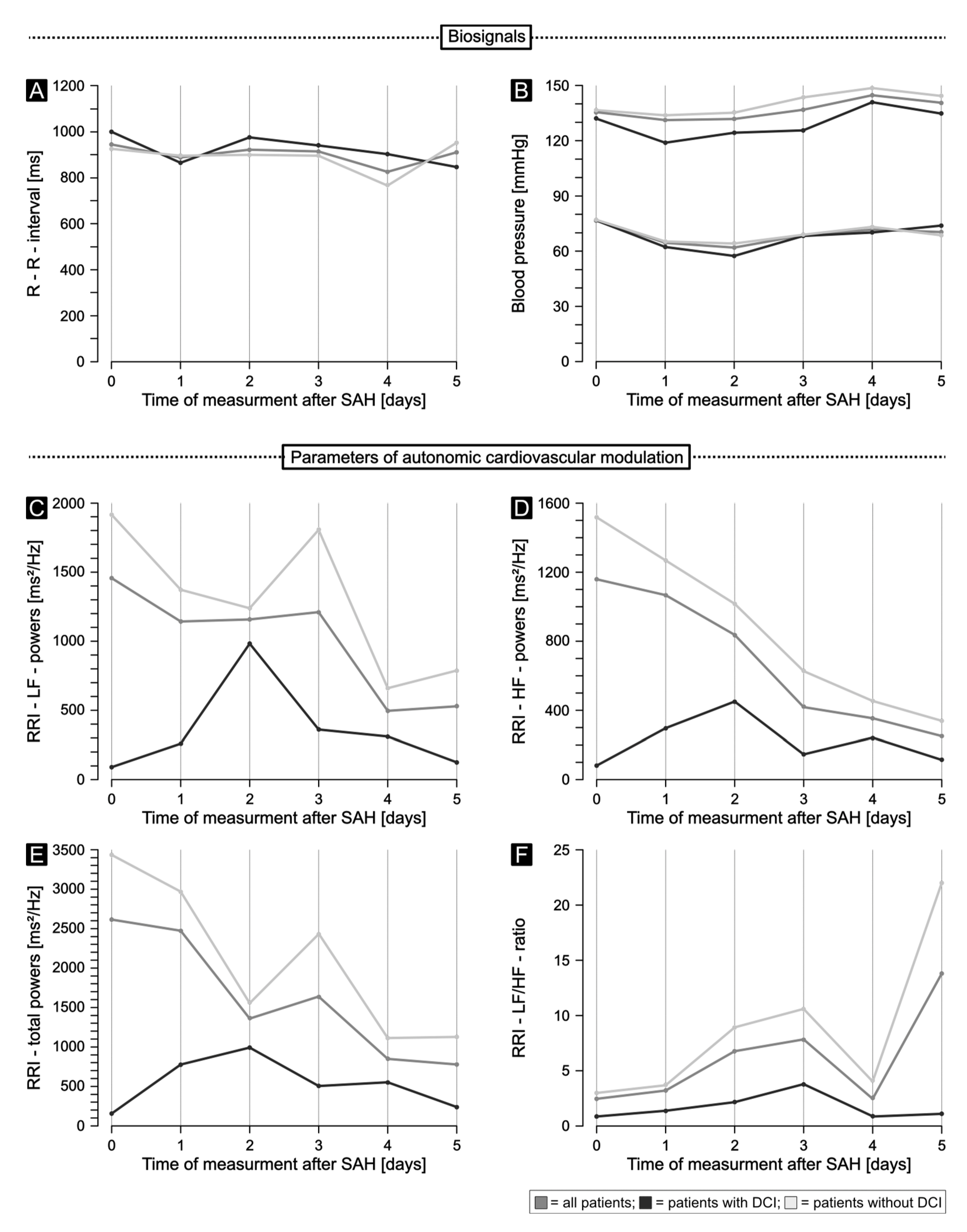
- Not only during the acute phase, i.e., within 24 h [9], but also during the first days after disease onset, spontaneous SAH induces a decrease in sympathetic, parasympathetic and total autonomic cardiovascular modulation.
- Significantly lower values of sympathetic modulation (RRI-LF powers, normalized RRI-LF powers, SBP-LF powers) on days 5 and 9 and significantly lower values of total autonomic modulation (RRI-SD, RRI-CV, RRI-total powers), as well as insignificantly lower values of parasympathetic modulation (RMSSDs, RRI-HF powers) on day 5 after SAH in those patients who develop DCI compared to the No DCI cohort, suggest a clear attribution of autonomic mechanisms on cerebral macro and microvasospasms and finally secondary brain injury.
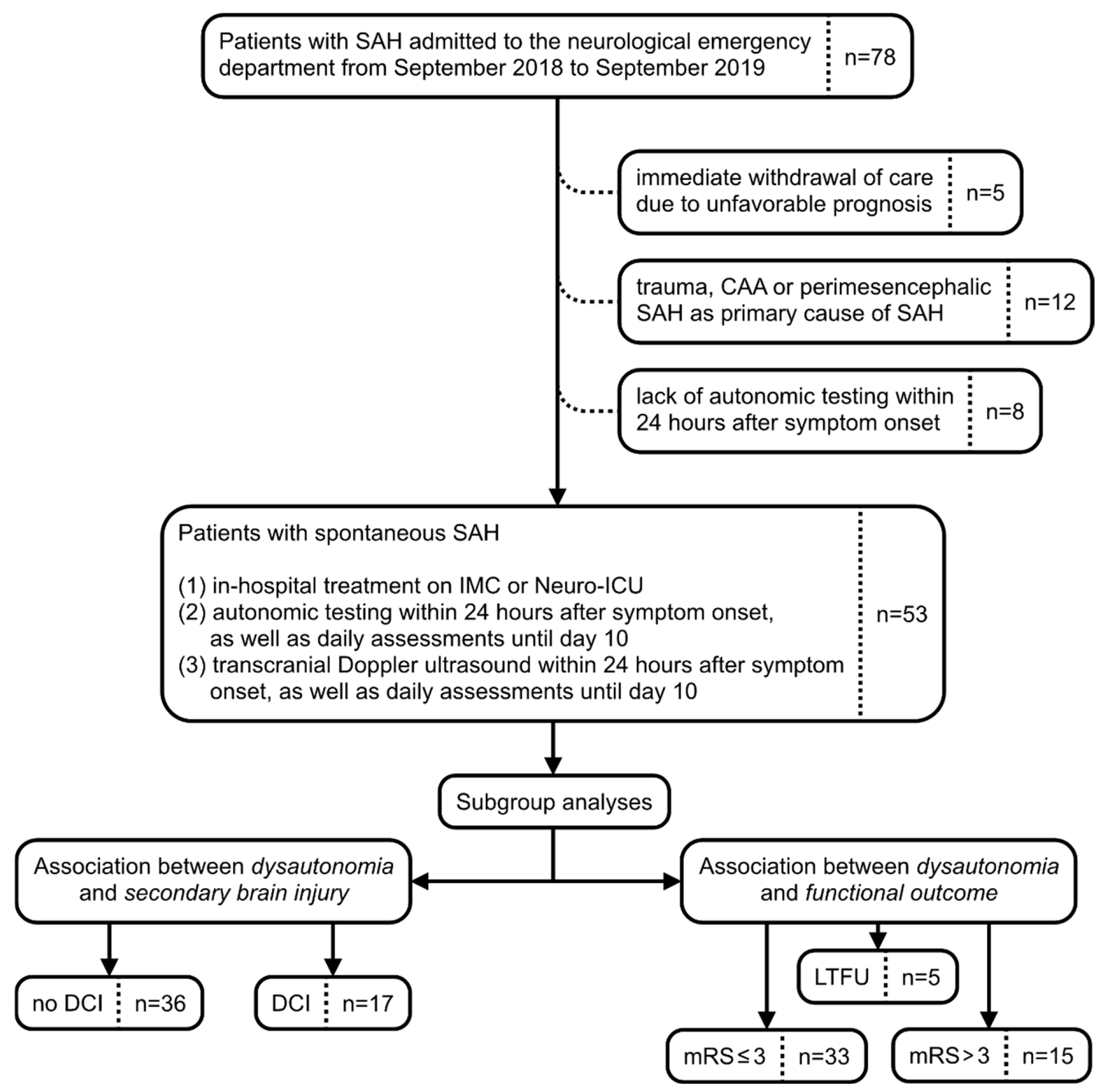
- Regardless of DCI development, significantly different normalized RRI-LF powers and normalized RRI-HF powers, as well as LF/HF ratios on day 5 in patients with mRS > 3 compared to the mRS ≤ 3 cohort three to six months after SAH, revealed potential associations of autonomic dysfunction on clinical outcome. Thus, bedsided assessment of heart rate, and blood-pressure variability may predict augmented risk of cardiovascular complications.
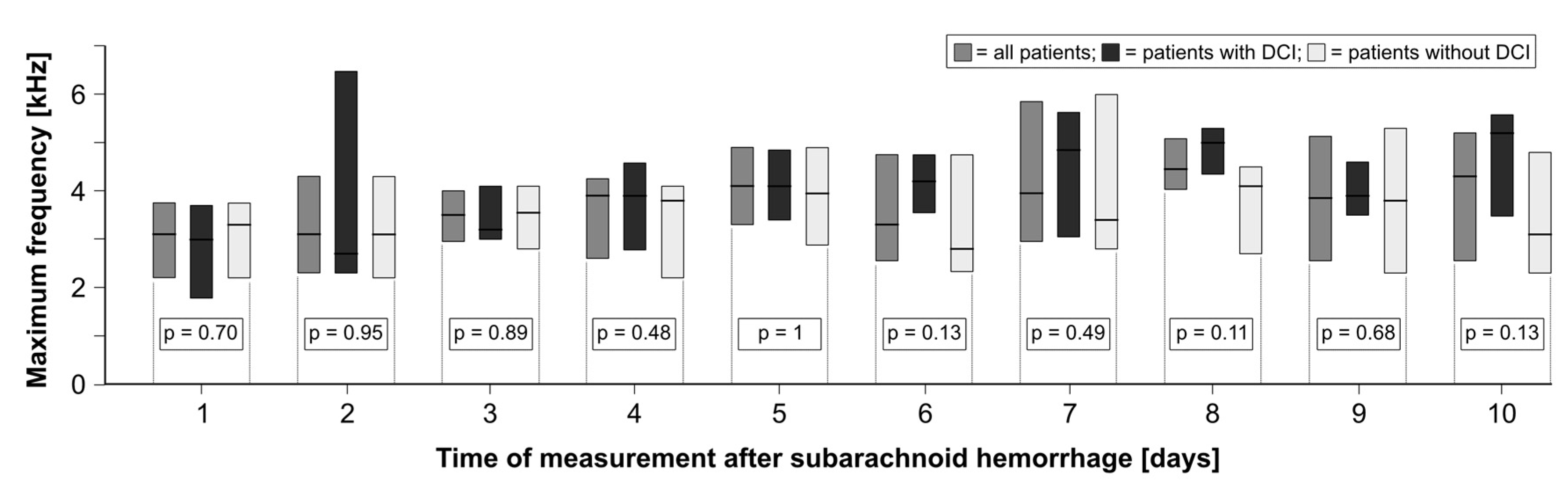
- In contrast, despite expected increases in TCD frequencies during the disease course, this standard diagnostic evaluation for detecting clinically relevant vasospasms did not reveal significant associations with DCI development in our cohort.
- Finally, although the interpretability may be limited by the small sample size, absent differences in premedication as well as the therapeutic regimen on day 5 indicate that potential iatrogenic confounders may not account for the observed autonomic dysregulation in SAH patients who develop DCI.
4.2. Autonomic Dysfunction, Development of DCI, and Functional Outcome—Pathophysiological Context
4.3. TCD—Recommended Monitoring Technique with Tenuous Benefits
4.4. Therapeutic Regimen, Rescue Strategies, Clinical Outcome, and Future Directions
4.5. Limitations
4.6. Conclusions
Author Contributions
Funding
Institutional Review Board Statement
Informed Consent Statement
Data Availability Statement
Conflicts of Interest
Abbreviations
| SAH | subarachnoid hemorrhage |
| EBI | early brain injury |
| SBI | secondary brain injury |
| DCI | delayed cerebral ischemia |
| TCD | transcranial Doppler ultrasound |
| CT | computed tomography |
| HRV | heart rate variability |
| CAA | cerebral amyloid angiopathy |
| IMC | intermediate care unit |
| ICU | intensive care unit |
| mRS | modified Rankin scale |
| LTFU | lost to follow-up |
| IVH | intraventricular hemorrhage |
| TIA | transient ischemic attack |
| ACE | angiotensin-converting enzyme |
| ARB | angiotensin II receptor blockers |
| WFNS | World Federation of Neurosurgical Societies |
| NIHSS | National Institutes of Health stroke scale |
| mFS | modified Fisher scale |
| LOS | length of stay |
| RRI | R–R interval |
| BP | blood pressure |
| SBP | systolic blood pressure |
| DBP | diastolic blood pressure |
| SD | standard deviation |
| RMSSD | root mean square of successive difference |
| CV | coefficient of variation |
| TRS | trigonometric regressive spectral analysis |
| LF | low frequency |
| HF | high frequency |
| nu | normalized |
| Max. | maximum |
| TTM | target temperature monitoring |
| AUC | area under the curve |
References
- Claassen, J.; Park, S. Spontaneous subarachnoid haemorrhage. Lancet 2022, 400, 846–862. [Google Scholar] [CrossRef] [Scilit] [PubMed] [PubMed Central]
- Neifert, S.N.; Chapman, E.K.; Martini, M.L.; Shuman, W.H.; Schupper, A.J.; Oermann, E.K.; Mocco, J.; Macdonald, R.L. Aneurysmal Subarachnoid Hemorrhage: The Last Decade. Transl. Stroke Res. 2021, 12, 428–446. [Google Scholar] [CrossRef] [Scilit] [PubMed]
- Vergouwen, M.D.I.; Ramos-Pachon, A.; Terecoasa, E.O.; Willett, N.; Cuadrado-Godia, E.; Gaberel, T.; Gizewski, E.R.; Helbok, R.; Ho, L.; Hugelshofer, M.; et al. European Stroke Organisation (ESO), European Association of Neurosurgical Societies (EANS) and European Society for Minimally Invasive Neurological Therapy (ESMINT) guideline on aneurysmal subarachnoid haemorrhage. Eur. Stroke J. 2026, 11, aakag043. [Google Scholar] [CrossRef] [Scilit] [PubMed] [PubMed Central]
- Hoh, B.L.; Ko, N.U.; Amin-Hanjani, S.; Chou, S.H.-Y.; Cruz-Flores, S.; Dangayach, N.S.; Derdeyn, C.P.; Du, R.; Hänggi, D.; Hetts, S.W.; et al. 2023 Guideline for the Management of Patients with Aneurysmal Subarachnoid Hemorrhage: A Guideline from the American Heart Association/American Stroke Association. Stroke 2023, 54, e314–e370. [Google Scholar] [CrossRef] [Scilit] [PubMed]
- Zirpe, K.G.; Tiwari, A.M.; Kulkarni, A.P.; Govil, D.; Dixit, S.B.; Munjal, M.; Sinha, S.; Samavedam, S.; Singh, Y.P.; Kuragayala, S.D.; et al. Adverse Events during Intrahospital Transport of Critically Ill Patients: A Multicenter, Prospective, Observational Study (I-TOUCH Study). Indian J. Crit. Care Med. 2023, 27, 635–641. [Google Scholar] [CrossRef] [Scilit] [PubMed] [PubMed Central]
- Chen, Y.; Li, G.; Chen, X.; Wang, D.; Fang, W.; Kang, D.; Ding, C. Non-cerebral vasospasm factors and cerebral vasospasm predict delayed cerebral ischemia after aneurysmal subarachnoid hemorrhage. Chin. Med. J. 2021, 135, 222–224. [Google Scholar] [CrossRef] [Scilit] [PubMed] [PubMed Central]
- Johnston, B.W.; Barrett-Jolley, R.; Krige, A.; Welters, I.D. Heart rate variability: Measurement and emerging use in critical care medicine. J. Intensive Care Soc. 2020, 21, 148–157. [Google Scholar] [CrossRef] [Scilit] [PubMed] [PubMed Central]
- Agrawal, S.; Nijs, K.; Subramaniam, S.; Englesakis, M.; Venkatraghavan, L.; Chowdhury, T. Predictor role of heart rate variability in subarachnoid hemorrhage: A systematic review. J. Clin. Monit. Comput. 2024, 38, 177–185. [Google Scholar] [CrossRef] [Scilit] [PubMed]
- Borutta, M.C.; Gerner, S.T.; Moeser, P.; Hoelter, P.; Engelhorn, T.; Doerfler, A.; Huttner, H.B.; Schwab, S.; Kuramatsu, J.B.; Koehn, J. Correlation between clinical severity and extent of autonomic cardiovascular impairment in the acute phase of subarachnoid hemorrhage. J. Neurol. 2022, 269, 5541–5552. [Google Scholar] [CrossRef] [Scilit] [PubMed] [PubMed Central]
- Aslid, R.; Markwalder, T.M.; Nornes, H. Noninvasive transcranial Doppler ultrasound recording of flow velocity in basal cerebral arteries. J. Neurosurg. 1982, 57, 769–774. [Google Scholar] [CrossRef] [Scilit] [PubMed]
- Loomis, A.L.; Chakko, M.N. Doppler Trans-Cranial Assessment, Protocols, and Interpretation. In StatPearls [Internet]; StatPearls Publishing: Treasure Island, FL, USA, 2023. [Google Scholar]
- Marshall, S.A.; Nyquist, P.; Ziai, W.C. The role of transcranial Doppler ultrasonography in the diagnosis and management of vasospasm after aneurysmal subarachnoid hemorrhage. Neurosurg. Clin. N. Am. 2010, 21, 291–303. [Google Scholar] [CrossRef] [Scilit] [PubMed]
- Hilz, M.J.; Dütsch, M. Quantitative studies of autonomic function. Muscle Nerve 2006, 33, 6–20. [Google Scholar] [PubMed]
- Task Force of the European Society of Cardiology the North American Society of Pacing and Electrophysiology. Heart rate variability: Standards of measurement, physiological inter pretation and clinical use. Circulation 1996, 93, 1043–1065. [Google Scholar] [PubMed]
- Marino, L.; Badenes, R.; Bilotta, F. Heart Rate Variability for Outcome Prediction in Intracerebral and Subarachnoid Hemorrhage: A Systematic Review. J. Clin. Med. 2023, 12, 4355. [Google Scholar] [CrossRef] [Scilit] [PubMed] [PubMed Central]
- Muraoka, S.; Kumagai, Y.; Koketsu, N.; Araki, Y.; Saito, R. Paroxysmal Sympathetic Hyperactivity in Stroke. World Neurosurg. 2023, 178, 28–36. [Google Scholar] [CrossRef] [Scilit] [PubMed]
- Hasegawa, Y.; Uchikawa, H.; Kajiwara, S.; Morioka, M. Central sympathetic nerve activation in subarachnoid hemorrhage. J. Neurochem. 2022, 160, 34–50. [Google Scholar] [CrossRef] [Scilit] [PubMed]
- Aydin, M.D.; Kanat, A.; Sahin, B.; Sahin, M.H.; Ergene, S.; Demirtas, R. New experimental finding of dangerous autonomic ganglia changes in cardiac injury following subarachnoid hemorrhage; a reciprocal culprit-victim relationship between the brain and heart. Int. J. Neurosci. 2024, 134, 91–102. [Google Scholar] [CrossRef] [Scilit] [PubMed]
- Uryga, A.; Nasr, N.; Kasprowicz, M.; Woźniak, J.; Goździk, W.; Burzyńska, M. Changes in autonomic nervous system during cerebral desaturation episodes in aneurysmal subarachnoid hemorrhage. Auton. Neurosci. 2022, 239, 102968. [Google Scholar] [CrossRef] [Scilit] [PubMed]
- Allen, W.L.; Merchant, K.S.; Martin, A.K.; Gruenbaum, S.E.; Gruenbaum, B.F. The neurocardiac axis in acute intracranial stress. J. Clin. Anesth. 2026, 109, 112105. [Google Scholar] [CrossRef] [Scilit] [PubMed]
- Du, Y.; Demillard, L.J.; Ren, J. Catecholamine-induced cardiotoxicity: A critical element in the pathophysiology of stroke-induced heart injury. Life Sci. 2021, 287, 120106. [Google Scholar] [CrossRef] [Scilit] [PubMed]
- Megjhani, M.; Kaffashi, F.; Terilli, K.; Alkhachroum, A.; Esmaeili, B.; Doyle, K.W.; Murthy, S.; Velazquez, A.G.; Connolly, E.S., Jr.; Roh, D.J.; et al. Heart Rate Variability as a Biomarker of Neurocardiogenic Injury After Subarachnoid Hemorrhage. Neurocrit. Care 2020, 32, 162–171. [Google Scholar] [CrossRef] [Scilit] [PubMed] [PubMed Central]
- Bjerkne Wenneberg, S.; Löwhagen Hendén, P.M.; Oras, J.; Naredi, S.; Block, L.; Ljungqvist, J.; Odenstedt Hergès, H. Heart rate variability monitoring for the detection of delayed cerebral ischemia after aneurysmal subarachnoid hemorrhage. Acta Anaesthesiol. Scand. 2020, 64, 945–952. [Google Scholar] [CrossRef] [Scilit] [PubMed]
- Wang, H.P.; Chen, C.C.; Lee, C.C.; Chen, C.T.; Chang, T.W.; Yeap, M.C.; Liu, Y.T.; Hsieh, P.C.; Wu, M.H.; Liu, Z.H.; et al. Using a continuous electrocardiographic patch with heart rhythm analysis in the subacute stage of aneurysmal subarachnoid hemorrhage: The feasibility verification. Clin. Neurol. Neurosurg. 2023, 228, 107687. [Google Scholar] [CrossRef] [Scilit] [PubMed]
- Odenstedt Hergès, H.; Vithal, R.; El-Merhi, A.; Naredi, S.; Staron, M.; Block, L. Machine learning analysis of heart rate variability to detect delayed cerebral ischemia in subarachnoid hemorrhage. Acta Neurol. Scand. 2022, 145, 151–159. [Google Scholar] [CrossRef] [Scilit] [PubMed]
- Robba, C.; Busl, K.M.; Claassen, J.; Diringer, M.N.; Helbok, R.; Park, S.; Rabinstein, A.; Treggiari, M.; Vergouwen, M.D.I.; Citerio, G. Contemporary management of aneurysmal subarachnoid haemorrhage. An update for the intensivist. Intensive Care Med. 2024, 50, 646–664. [Google Scholar] [CrossRef] [Scilit] [PubMed] [PubMed Central]
- Snider, S.B.; Migdady, I.; LaRose, S.L.; Mckeown, M.E.; Regenhardt, R.W.; Lai, P.M.R.; Vaitkevicius, H.; Du, R. Transcranial-Doppler-Measured Vasospasm Severity is Associated with Delayed Cerebral Infarction After Subarachnoid Hemorrhage. Neurocrit. Care 2022, 36, 815–821. [Google Scholar] [CrossRef] [Scilit] [PubMed]
- Kelly, P.D.; Yengo-Kahn, A.M.; Tang, A.R.; Jonathan, S.V.; Reynolds, R.A.; Ye, F.; Zhao, Z.; Froehler, M.T.; Fusco, M.R.; Morone, P.J.; et al. Conditional Vasospasm-Free Survival Following Aneurysmal Subarachnoid Hemorrhage. Neurocrit. Care 2022, 37, 81–90. [Google Scholar] [CrossRef] [Scilit] [PubMed] [PubMed Central]
- Lauzier, D.C.; Jayaraman, K.; Yuan, J.Y.; Diwan, D.; Vellimana, A.K.; Osbun, J.W.; Chatterjee, A.R.; Athiraman, U.; Dhar, R.; Zipfel, G.J. Early Brain Injury After Subarachnoid Hemorrhage: Incidence and Mechanisms. Stroke 2023, 54, 1426–1440. [Google Scholar] [CrossRef] [Scilit] [PubMed] [PubMed Central]
- Yuan, J.Y.; Chen, Y.; Jayaraman, K.; Kumar, A.; Zlepper, Z.; Allen, M.L.; Athiraman, U.; Osbun, J.; Zipfel, G.; Dhar, R. Automated Quantification of Compartmental Blood Volumes Enables Prediction of Delayed Cerebral Ischemia and Outcomes After Aneurysmal Subarachnoid Hemorrhage. World Neurosurg. 2023, 170, e214–e222. [Google Scholar] [CrossRef] [Scilit] [PubMed] [PubMed Central]
- Akeret, K.; Buzzi, R.M.; Schaer, C.A.; Thomson, B.R.; Vallelian, F.; Wang, S.; Willms, J.; Sebök, M.; Held, U.; Deuel, J.W.; et al. Cerebrospinal fluid hemoglobin drives subarachnoid hemorrhage-related secondary brain injury. J. Cereb. Blood Flow Metab. 2021, 41, 3000–3015. [Google Scholar] [CrossRef] [Scilit] [PubMed] [PubMed Central]
- Helbok, R.; Rass, V.; Kofler, M.; Talasz, H.; Schiefecker, A.; Gaasch, M.; Scherfler, C.; Pfausler, B.; Thomé, C.; Beer, R.; et al. Intracerebral Iron Accumulation may be Associated with Secondary Brain Injury in Patients with Poor Grade Subarachnoid Hemorrhage. Neurocrit. Care 2022, 36, 171–179. [Google Scholar] [CrossRef] [Scilit] [PubMed] [PubMed Central]
- Heinz, R.; Brandenburg, S.; Nieminen-Kelhä, M.; Kremenetskaia, I.; Boehm-Sturm, P.; Vajkoczy, P.; Schneider, U.C. Microglia as target for anti-inflammatory approaches to prevent secondary brain injury after subarachnoid hemorrhage (SAH). J. Neuroinflamm. 2021, 18, 36. [Google Scholar] [CrossRef] [Scilit] [PubMed] [PubMed Central]
- Mehra, A.; Gomez, F.; Bischof, H.; Diedrich, D.; Laudanski, K. Cortical Spreading Depolarization and Delayed Cerebral Ischemia; Rethinking Secondary Neurological Injury in Subarachnoid Hemorrhage. Int. J. Mol. Sci. 2023, 24, 9883. [Google Scholar] [CrossRef] [Scilit] [PubMed] [PubMed Central]
- Wolf, S.; Mielke, D.; Barner, C.; Malinova, V.; Kerz, T.; Wostrack, M.; Czorlich, P.; Salih, F.; Engel, D.C.; Ehlert, A.; et al. Effectiveness of Lumbar Cerebrospinal Fluid Drain Among Patients with Aneurysmal Subarachnoid Hemorrhage: A Randomized Clinical Trial. JAMA Neurol. 2023, 80, 833–842. [Google Scholar] [CrossRef] [Scilit] [PubMed]
- Lee, K.S.; Chari, A.; Motiwala, M.; Khan, N.R.; Arthur, A.S.; Lawton, M.T. Effectiveness of Cerebrospinal Fluid Lumbar Drainage Among Patients with Aneurysmal Subarachnoid Hemorrhage: An Updated Systematic Review and Meta-Analysis. World Neurosurg. 2024, 183, 246–253.e12. [Google Scholar] [CrossRef] [Scilit] [PubMed]
- Schlaeppi, J.A.; Affentranger, L.; Bervini, D.; Z’Graggen, W.J.; Raabe, A.; Pollo, C. Electrical Stimulation for Cerebral Vasospasm After Subarachnoid Hemorrhage: A Systematic Review. Neuromodulation 2022, 25, 1227–1239. [Google Scholar] [CrossRef] [Scilit] [PubMed]
- Brenner, L.B.O.; Batista, S.; Prestes, M.Z.; Bocanegra-Becerra, J.E.; Rabelo, N.N.; Bertani, R.; Welling, L.C.; Figueiredo, E.G. Stellate Ganglion Block in Subarachnoid Hemorrhage: A Promising Protective Measure Against Vasospasm? World Neurosurg. 2024, 182, 124–131. [Google Scholar] [CrossRef] [Scilit] [PubMed]
- Wendel, C.; Oberhauser, C.; Schiff, J.; Henkes, H.; Ganslandt, O. Stellate Ganglion Block and Intraarterial Spasmolysis in Patients with Cerebral Vasospasm: A Retrospective Cohort Study. Neurocrit. Care 2024, 40, 603–611. [Google Scholar] [CrossRef] [Scilit] [PubMed] [PubMed Central]
- Takemoto, Y.; Hasegawa, Y.; Hayashi, K.; Cao, C.; Hamasaki, T.; Kawano, T.; Mukasa, A.; Kim-Mitsuyama, S. The Stabilization of Central Sympathetic Nerve Activation by Renal Denervation Prevents Cerebral Vasospasm after Subarachnoid Hemorrhage in Rats. Transl. Stroke Res. 2020, 11, 528–540. [Google Scholar] [CrossRef] [Scilit] [PubMed]



| All Patients (n = 53) | DCI (n = 17) | No DCI (n = 36) | p-Values (DCI vs. No DCI) | |
|---|---|---|---|---|
| Age [years], Median (IQR) | 54 (49.0–62.5) | 54 (49.5–66.5) | 53 (48.5–60.8) | 0.36 |
| Female sex, n (%) | 28 (52.8) | 8 (47.1) | 20 (55.6) | 0.78 |
| Prior comorbidities | ||||
| Arterial hypertension, n (%) | 28 (52.8) | 12 (70.6) | 16 (44.4) | 0.14 |
| Diabetes mellitus, n (%) | 8 (15.1) | 3 (17.6) | 5 (13.9) | 1 |
| Dyslipidemia, n (%) | 8 (15.1) | 3 (17.6) | 5 (13.9) | 1 |
| Prior ischemic stroke/TIA, n (%) | 1 (1.9) | 1 (5.9) | 0 (0.0) | 0.32 |
| Prior hemorrhagic stroke, n (%) | 0 (0.0) | 0 (0.0) | 0 (0.0) | 1 |
| Congestive heart failure, n (%) | 4 (7.5) | 2 (11.8) | 2 (5.6) | 0.59 |
| Premedication | ||||
| Antihypertensive drugs, n (%) | 18 (34.0) | 6 (35.3) | 12 (33.3) | 0.86 |
| - ACE inhibitor/ARB, n (%) | 14 (26.4) | 4 (23.5) | 10 (27.8) | 1 |
| - Betablocker, n (%) | 7 (13.2) | 2 (11.8) | 5 (13.9) | 1 |
| - Diuretic, n (%) | 11 (20.8) | 4 (23.5) | 7 (19.4) | 1 |
| - Calcium channel blocker, n (%) | 3 (5.7) | 1 (5.9) | 2 (5.6) | 1 |
| Antiplatelet drug, n (%) | 2 (3.8) | 1 (5.9) | 1 (2.8) | 1 |
| Anticoagulation, n (%) | 7 (13.2) | 3 (17.6) | 4 (11.1) | 0.67 |
| Clinical parameters | ||||
| Hunt & Hess score, Median (IQR) | 3 (1–4) | 4 (2–4) | 3 (1–4) | 0.43 |
| NIHSS, Median (IQR) | 12 (0–38) | 38 (0–38) | 12 (0–38) | 0.35 |
| WFNS scale, Median (IQR) | 3 (1–5) | 4 (1–5) | 3 (1–5) | 0.41 |
| Pre-mRS, Median (IQR) | 0 (0–0) | 0 (0–0) | 0 (0–0) | 0.97 |
| Mechanical ventilation on day 1, n (%) | 34 (64.2) | 13 (76.5) | 21 (58.3) | 0.24 |
| Radiological parameters | ||||
| mFS, Median (IQR) | 4 (3–4) | 4 (3–4) | 4 (3–4) | 0.59 |
| Cerebral aneurysm, n (%) | 39 (73.6) | 14 (82.4) | 25 (69.4) | 0.51 |
| - Anterior circulation, n (%) | 28 (52.8) | 12 (70.6) | 16 (44.4) | 0.14 |
| - Posterior circulation, n (%) | 11 (20.8) | 5 (29.4) | 6 (16.7) | 0.47 |
| Intracerebral hemorrhage, n (%) | 14 (26.4) | 9 (52.9) | 5 (13.9) | * <0.01 |
| - Right hemisphere, n (%) | 5 (9.4) | 3 (17.6) | 2 (5.6) | 0.31 |
| - Left hemisphere, n (%) | 8 (15.1) | 5 (29.4) | 3 (8.3) | 0.09 |
| - Corpus callosum, n (%) | 1 (1.9) | 1 (5.9) | 0 (0.0) | 0.32 |
| Intraventricular hemorrhage, n (%) | 30 (56.6) | 9 (52,9) | 21 (58.3) | 0.77 |
| Acute occluding hydrocephalus, n (%) | 32 (60.4) | 11 (64.7) | 21 (58.3) | 0.88 |
| Outcome parameters | ||||
| LOS [days], Median (IQR) | 17 (11–23) | 17 (12.5–23.5) | 17 (10.3–23.0) | 0.98 |
| In-hospital death, n (%) | 6 (11.3) | 2 (11.8) | 4 (11.1) | 1 |
| mRS after 1 to 3 months, Median (IQR) | 1 (1–4) | 3 (1–5) | 1 (1–4) | 0.52 |
| mRS after 3 to 6 months, Median (IQR) | 1 (0–4) | 1 (0–6) | 1 (0–3) | 0.26 |
| mRS after 3 to 9 months, Median (IQR) | 1 (0–3) | 1 (0–5.3) | 1 (0–3) | 0.44 |
| All Patients (n = 53) | Favorable Outcome (mRS ≤ 3; n = 33) | Unfavorable Outcome (mRS > 3; n = 15) | p-Values (mRS ≤ 3 vs. mRS > 3) | |
|---|---|---|---|---|
| Age [years], Median (IQR) | 54.0 (50.0–62.8) | 53.0 (47.0–62.0) | 57.0 (53.0–66.0) | 0.09 |
| Female sex, n (%) | 25 (52.1) | 18 (54.5) | 7 (46.7) | 0.84 |
| Prior comorbidities | ||||
| Arterial hypertension, n (%) | 25 (52.1) | 16 (48.5) | 9 (60.0) | 0.67 |
| Diabetes mellitus, n (%) | 7 (14.6) | 5 (15.2) | 2 (13.3) | 1 |
| Dyslipidemia, n (%) | 8 (16.7) | 6 (18.2) | 2 (13.3) | 1 |
| Prior ischemic stroke/TIA, n (%) | 1 (2.1) | 0 | 1 (6.7) | 0.31 |
| Prior hemorrhagic stroke, n (%) | 0 | 0 | 0 | 1 |
| Congestive heart failure, n (%) | 4 (8.3) | 2 (6.1) | 2 (13.3) | 0.58 |
| Premedication | ||||
| Antihypertensive drugs, n (%) | 16 (33.3) | 8 (24.2) | 8 (53.3) | 0.10 |
| - ACE inhibitor/ARB, n (%) | 12 (25) | 8 (24.2) | 4 (26.7) | 1 |
| - Betablocker, n (%) | 7 (14.6) | 6 (18.2) | 1 (6.7) | 0.41 |
| - Diuretic, n (%) | 9 (18.8) | 4 (12.1) | 5 (33.3) | 0.11 |
| - Calcium channel blocker, n (%) | 3 (6.3) | 2 (6.1) | 1 (6.7) | 1 |
| Antiplatelet drug, n (%) | 2 (4.2) | 1 (3.0) | 1 (6.7) | 1 |
| Anticoagulation, n (%) | 6 (12.5) | 4 (12.1) | 2 (13.3) | 1 |
| Clinical parameters | ||||
| Hunt & Hess score, Median (IQR) | 3 (1–4) | 2 (1–4) | 4 (3–5) | * <0.01 |
| NIHSS, Median (IQR) | 2 (0–38) | 0 (0–3) | 38 (38) | * <0.01 |
| WFNS scale, Median (IQR) | 3 (1–5) | 1 (1–4) | 5 (4–5) | * <0.01 |
| Pre-mRS, Median (IQR) | 0 (0–0) | 0 (0–0) | 0 (0–0) | 0.14 |
| Mechanical ventilation on day 1, n (%) | 32 (66.7) | 17 (51.5) | 15 (100) | * <0.01 |
| Radiological parameters | ||||
| mFS, Median (IQR) | 4 (3–4) | 4 (3–4) | 4 (3–4) | 0.16 |
| Cerebral aneurysm, n (%) | 37 (77.1) | 26 (78.8) | 11 (73.3) | 0.72 |
| - Anterior circulation, n (%) | 28 (52.8) | 19 (57.6) | 8 (53.3) | 1 |
| - Posterior circulation, n (%) | 11 (20.8) | 7 (21.2) | 3 (20.0) | 1 |
| Intracerebral hemorrhage, n (%) | 14 (29.2) | 5 (15.2) | 9 (60.0) | * <0.01 |
| - Right hemisphere, n (%) | 5 (9.4) | 2 (6.1) | 2 (13.3) | 0.58 |
| - Left hemisphere, n (%) | 8 (15.1) | 2 (6.1) | 6 (40.0) | * <0.01 |
| - Corpus callosum, n (%) | 1 (1.9) | 1 (3.0) | 0 | 1 |
| Intraventricular hemorrhage, n (%) | 27 (56.3) | 20 (60.6) | 7 (46.7) | 0.55 |
| Acute occluding hydrocephalus, n (%) | 29 (60.4) | 17 (51.5) | 12 (80.0) | 0.11 |
| DCI, n (%) | 15 (31.3) | 10 (30.3) | 5 (33.3) | 0.73 |
| Outcome parameters | ||||
| LOS [days], Median (IQR) | 18 (11–23) | 18 (11–24) | 16 (4–23) | 0.56 |
| In-hospital death, n (%) | 6 (12.5) | 0 | 6 (40.0) | * <0.01 |
| All Patients (n = 53) | DCI (n = 17) | No DCI (n = 36) | p-Values (DCI vs. No DCI) | |
|---|---|---|---|---|
| Max. frequencies day 1, Median (IQR) | 3.10 (2.20–3.75) | 3.00 (1.78–3.70) | 3.30 (2.20–3.75) | 0.70 |
| Max. frequencies day 2, Median (IQR) | 3.10 (2.30–4.30) | 2.70 (2.30–6.48) | 3.10 (2.20–4.30) | 0.95 |
| Max. frequencies day 3, Median (IQR) | 3.50 (2.95–4.00) | 3.20 (3.00–4.10) | 3.55 (2.80–4.08) | 0.89 |
| Max. frequencies day 4, Median (IQR) | 3.90 (2.60–4.25) | 3.90 (2.78–4.58) | 3.80 (2.20–4.10) | 0.45 |
| Max. frequencies day 5, Median (IQR) | 4.10 (3.30–4.90) | 4.10 (3.40–4.85) | 3.95 (2.88–4.90) | 1 |
| Max. frequencies day 6, Median (IQR) | 3.30 (2.55–4.75) | 4.20 (3.55–4.75) | 2.80 (2.33–4.75) | 0.13 |
| Max. frequencies day 7, Median (IQR) | 3.95 (2.95–5.85) | 4.85 (3.05–5.63) | 3.40 (2.80–6.00) | 0.49 |
| Max. frequencies day 8, Median (IQR) | 4.45 (4.03–5.08) | 5.00 (4.35–5.30) | 4.10 (2.70–4.50) | 0.11 |
| Max. frequencies day 9, Median (IQR) | 3.85 (2.55–5.13) | 3.90 (3.50–4.60) | 3.80 (2.30–5.30) | 0.68 |
| Max. frequencies day 10, Median (IQR) | 4.30 (2.48–5.20) | 5.20 (3.48–5.58) | 3.10 (2.30–4.80) | 0.13 |
| Location of max. frequencies, n (%) | ||||
| Anterior cerebral artery | 11 (20.8) | 4 (23.5) | 7 (19.4) | 0.60 |
| Middle cerebral artery | 22 (41.5) | 10 (58.8) | 12 (33.3) | 0.18 |
| Carotid siphon | 12 (22.6) | 2 (11.8) | 10 (27.8) | 0.11 |
| Posterior cerebral artery | 0 | 0 | 0 | 1 |
| Basilar artery | 2 (3.8) | 1 (5.9) | 1 (2.8) | 0.59 |
| Vertebral artery | 1 (1.9) | 0 | 1 (2.8) | 0.65 |
| All Patients (n = 53) | DCI (n = 17) | No DCI (n = 36) | p-Values (DCI vs. No DCI) | |
|---|---|---|---|---|
| DAY 1/INITIAL ASSESSMENT (within 24 h) | ||||
| Biosignals, median (IQR) | ||||
| Systolic blood pressure [mmHg] | 132.6 (116.5–147.1) | 116.8 (109.3–138.3) | 135.1 (125.3–148.5) | 0.17 |
| Diastolic blood pressure [mmHg] | 66.1 (58.4–73.3) | 63.0 (58.7–70.4) | 66.6 (55.6–75.8) | 0.72 |
| Heart rate [min−1] | 940.1 (765.8–1054.3) | 932.6 (754.1–1040.9) | 944.0 (758.9–1067.8) | 0.84 |
| Sympathetic modulation, median (IQR) | ||||
| RRI-LF powers [ms2] | 163.6 (15.6–893.6) | 163.6 (14.3–338.6) | 156.8 (17.1–1328.2) | 0.65 |
| RRI-LF(nu) powers | 0.5 (0.4–0.7) | 0.4 (0.4–0.6) | 0.6 (0.4–0.8) | 0.15 |
| SBP-LF powers [mmHg2] | 4.6 (1.3–15.7) | 2.9 (1.2–19.6) | 4.8 (1.3–26.3) | 0.91 |
| Parasympathetic modulation, median (IQR) | ||||
| RRI-RMSSD [ms] | 19.3 (8.9–47.8) | 26.1 (17.9–38.2) | 15.6 (7.8–60.1) | 0.34 |
| RRI-HF powers [ms2] | 80.4 (17.5–429.0) | 157.6 (36.5–282.4) | 79.0 (17.4–1219.8) | 0.77 |
| RRI-HF(nu) powers | 0.5 (0.3–0.6) | 0.6 (0.4–0.7) | 0.5 (0.2–0.6) | 0.15 |
| Total autonomic modulation, median (IQR) | ||||
| RRI-SD [ms] | 23.0 (10.6–43.6) | 24.3 (14.3–31.5) | 20.2 (9.9–50.6) | 0.84 |
| RRI-CV [%] | 2.4 (1.2–5.2) | 3.0 (1.4–4.0) | 2.2 (1.2–5.9) | 0.87 |
| RRI-total powers [ms2] | 237.3 (66.5–1458.8) | 360.2 (57.7–713.7) | 232.6 (60.9–2665.1) | 0.91 |
| Sympathovagal balance, median (IQR) | ||||
| RRI-LF/HF ratio | 1.5 (0.8–2.5) | 0.9 (0.4–2.0) | 1.6 (0.9–3.5) | 0.20 |
| DAY 5 | ||||
| Biosignals, median (IQR) | ||||
| Systolic blood pressure [mmHg] | 138.8 (132.2–149.9) | 132.6 (125.5–144.8) | 141.4 (135.4–150.5) | 0.12 |
| Diastolic blood pressure [mmHg] | 70.2 (61.4–75.5) | 70.2 (57.7–77.2) | 70.9 (65.3–74.8) | 1 |
| Heart rate [min−1] | 882.2 (744.3–1030.6) | 829.6 (717.8–1006.2) | 883.7 (740.3–1096.1) | 0.52 |
| Sympathetic modulation, median (IQR) | ||||
| RRI-LF powers [ms2] | 207.1 (8.4–989.7) | 9.3 (3.8–278.8) | 587.3 (87.9–1210.8) | * 0.01 |
| RRI-LF(nu) powers | 0.6 (0.4–0.7) | 0.5 (0.3–0.6) | 0.6 (0.5–0.7) | 0.09 |
| SBP-LF powers [mmHg2] | 5.9 (3.3–18.0) | 3.4 (1.9–9.6) | 9.1 (4.5–51.5) | * 0.04 |
| Parasympathetic modulation, median (IQR) | ||||
| RRI-RMSSD [ms] | 18.9 (6.1–37.8) | 10.3 (4.6–19.6) | 27.0 (11.0–40.3) | 0.07 |
| RRI-HF powers [ms2] | 83.3 (7.0–575.8) | 20.3 (6.2–145.3) | 215.5 (28.3–612.5) | 0.07 |
| RRI-HF(nu) powers | 0.4 (0.3–0.6) | 0.5 (0.4–0.7) | 0.4 (0.3–0.5) | 0.09 |
| Total autonomic modulation, median (IQR) | ||||
| RRI-SD [ms] | 18.0 (4.9–46.8) | 9.6 (3.7–24.2) | 34.1 (14.7–53.6) | * 0.01 |
| RRI-CV [%] | 1.7 (0.6–5.3) | 1.0 (0.4–0.5) | 3.9 (1.2–5.6) | * 0.01 |
| RRI-total powers [ms2] | 259.3 (14.4–1565.5) | 29.5 (9.9–424.1) | 1035.9 (151.9–1898.1) | * <0.01 |
| Sympathovagal balance, median (IQR) | ||||
| RRI-LF/HF ratio | 1.4 (0.8–2.0) | 1.0 (0.5–1.4) | 1.5 (1.2–2.4) | 0.07 |
| All Patients (n = 53) | DCI (n = 17) | No DCI (n = 36) | p-Values (DCI vs. No DCI) | |
|---|---|---|---|---|
| Nimodipine, n (%) | 53 (100) | 17 (100) | 36 (100) | 1 |
| Vasopressor therapy, n (%) | 27 (50.9) | 11 (64.7) | 16 (44.4) | 0.24 |
| Norepinephrine | 27 (50.9) | 11 (64.7) | 16 (44.4) | 0.24 |
| Other catecholamine | 3 (5.7) | 2 (11.8) | 1 (2.8) | 0.24 |
| Antihypertensivedrugs, n (%) | 11 (20.8) | 1 (5.9) | 10 (27.8) | 0.08 |
| Continuous infusion (syringe pump) | 8 (15.1) | 1 (5.9) | 7 (19.4) | 0.25 |
| - Alpha-1 adrenoceptor antagonist | 8 (15.1) | 1 (5.9) | 7 (19.4) | 0.25 |
| - Alpha-2 adrenoceptor agonist | 1 (1.9) | 0 (0.0) | 1 (2.8) | 1 |
| - Dihydralazine | 3 (5.7) | 0 (0.0) | 3 (8.3) | 0.54 |
| Oral drug therapy | 6 (11.3) | 1 (5.9) | 5 (13.9) | 0.65 |
| - ACE inhibitor/ARB | 6 (11.3) | 1 (5.9) | 5 (13.9) | 0.65 |
| - Betablocker | 5 (9.4) | 1 (5.9) | 4 (11.1) | 0.66 |
| - Diuretic | 2 (3.8) | 0 (0.0) | 2 (5.6) | 0.56 |
| - Calcium channel blocker | 2 (3.8) | 0 (0.0) | 2 (5.6) | 0.56 |
| Analgesic drugs, n (%) | 43 (81.1) | 15 (88.2) | 28 (77.8) | 0.47 |
| Opioids | 43 (81.1) | 15 (88.2) | 28 (77.8) | 0.47 |
| Non-opioids | 24 (45.3) | 7 (41.2) | 17 (47.2) | 0.77 |
| Sedative drugs, n (%) | 34 (64.2) | 13 (76.5) | 21 (58.3) | 0.23 |
| Midazolam | 30 (56.6) | 11 (64.7) | 19 (35.8) | 0.55 |
| Esketamine | 21 (39.6) | 8 (47.1) | 13 (36.1) | 0.55 |
| Propofol | 7 (13.2) | 4 (23.5) | 3 (8.3) | 0.19 |
| Mechanical Ventilation, n (%) | 35 (66.0) | 13 (76.5) | 22 (61.1) | 0.36 |
| All Patients (n = 53) | DCI (n = 17) | No DCI (n = 36) | p-Values (DCI vs. No DCI) | |
|---|---|---|---|---|
| Cerebrospinal fluid drainage, n (%) | 40 (75.5) | 15 (88.2) | 25 (69.4) | 0.18 |
| External ventricular drain | 32 (60.4) | 12 (70.6) | 20 (55.6) | 0.46 |
| Lumbar drain | 30 (56.6) | 12 (70.6) | 18 (50.0) | 0.27 |
| Intraventricular thrombolysis, n (%) | 22 (42.5) | 9 (52.9) | 13 (36.1) | 0.39 |
| Intra-arterial vasospasmolysis, n (%) | 9 (17.0) | 6 (35.3) | 3 (8.3) | * 0.02 |
| Osmotic active therapy, n (%) | 8 (15.1) | 7 (41.2) | 1 (2.8) | * <0.01 |
| Mannitol | 5 (9.4) | 4 (23.5) | 1 (2.8) | * 0.03 |
| Hypertonic saline | 7 (13.2) | 6 (35.3) | 1 (2.8) | * <0.01 |
| TTM to apply normothermia, n (%) | 12 (22.6) | 6 (35.3) | 6 (16.7) | 0.15 |
| Mechanical Ventilation, n (%) | 35 (66.0) | 13 (76.5) | 22 (61.1) | 0.36 |
| All Patients (n = 48) | Favorable Outcome (mRS ≤ 3; n = 33) | Unfavorable Outcome (mRS > 3; n = 15) | p-Values (mRS ≤ 3 vs. mRS > 3) | |
|---|---|---|---|---|
| DAY 5 | ||||
| Biosignals, median (IQR) | ||||
| Systolic blood pressure [mmHg] | 139.0 (130.8–150.0) | 139.3 (127.9–151.0) | 136.6 (132.0–147.4) | 0.80 |
| Diastolic blood pressure [mmHg] | 69.8 (60.3–76.1) | 72.2 (65.9–77.2) | 57.7 (50.3–73.5) | 0.12 |
| Heart rate [min−1] | 883.4 (740.3–1032.2) | 883.4 (732.3–1025.5) | 888.6 (765.9–1126.9) | 1 |
| Sympathetic modulation, median (IQR) | ||||
| RRI-LF powers [ms2] | 172.4 (8.0–1004.8) | 286.1 (35.6–1034.9) | 5.3 (0.8–375.8) | 0.07 |
| RRI-LF(nu) powers | 0.6 (0.4–0.7) | 0.6 (0.5–0.7) | 0.4 (0.2–0.6) | * 0.03 |
| SBP-LF powers [mmHg2] | 5.4 (3.1–19.4) | 8.1 (4.4–44.4) | 3.7 (1.7–8.9) | 0.12 |
| Parasympathetic modulation, median (IQR) | ||||
| RRI-RMSSD [ms] | 18.2 (5.8–33.7) | 20.5 (13.2–38.0) | 6.9 (4.5–30.5) | 0.18 |
| RRI-HF powers [ms2] | 63.0 (6.9–559.3) | 138.7 (2.1–570.3) | 7.9 (5.5–294.5) | 0.20 |
| RRI-HF(nu) powers | 0.4 (0.3–0.6) | 0.4 (0.3–0.5) | 0.6 (0.4–0.8) | * 0.03 |
| Total autonomic modulation, median (IQR) | ||||
| RRI-SD [ms] | 18.0 (4.7–44.2) | 24.6 (11.8–46.0) | 4.9 (3.0–23.5) | 0.12 |
| RRI-CV [%] | 1.7 (0.6–4.7) | 2.7 (1.5–5.3) | 0.7 (0.4–1.9) | 0.05 |
| RRI-total powers [ms2] | 254.5 (13.8–1593.5) | 501.6 (71.6–1649.7) | 13.0 (6.7–670.3) | 0.08 |
| Sympathovagal balance, median (IQR) | ||||
| RRI-LF/HF ratio | 1.4 (0.7–2.0) | 1.5 (1.1–2.7) | 0.7 (0.2–1.4) | * 0.03 |
Disclaimer/Publisher’s Note: The statements, opinions and data contained in all publications are solely those of the individual author(s) and contributor(s) and not of MDPI and/or the editor(s). MDPI and/or the editor(s) disclaim responsibility for any injury to people or property resulting from any ideas, methods, instructions or products referred to in the content. |
© 2026 by the authors. Licensee MDPI, Basel, Switzerland. This article is an open access article distributed under the terms and conditions of the Creative Commons Attribution (CC BY) license.
Share and Cite
Borutta, M.C.; Vetter, C.; Kraemer, F.; Gerner, S.T.; Macha, K.; Singer, L.; Engelhorn, T.; Doerfler, A.; Schwab, S.; Koehn, J. Associations Between Delayed Cerebral Ischemia in Spontaneous Subarachnoid Hemorrhage and Dysfunction of Autonomic Cardiovascular Modulation Compared to Transcranial Doppler Ultrasound Findings. Diagnostics 2026, 16, 2125. https://doi.org/10.3390/diagnostics16132125
Borutta MC, Vetter C, Kraemer F, Gerner ST, Macha K, Singer L, Engelhorn T, Doerfler A, Schwab S, Koehn J. Associations Between Delayed Cerebral Ischemia in Spontaneous Subarachnoid Hemorrhage and Dysfunction of Autonomic Cardiovascular Modulation Compared to Transcranial Doppler Ultrasound Findings. Diagnostics. 2026; 16(13):2125. https://doi.org/10.3390/diagnostics16132125
Chicago/Turabian StyleBorutta, Matthias C., Chiara Vetter, Florian Kraemer, Stefan T. Gerner, Kosmas Macha, Ludwig Singer, Tobias Engelhorn, Arnd Doerfler, Stefan Schwab, and Julia Koehn. 2026. "Associations Between Delayed Cerebral Ischemia in Spontaneous Subarachnoid Hemorrhage and Dysfunction of Autonomic Cardiovascular Modulation Compared to Transcranial Doppler Ultrasound Findings" Diagnostics 16, no. 13: 2125. https://doi.org/10.3390/diagnostics16132125
APA StyleBorutta, M. C., Vetter, C., Kraemer, F., Gerner, S. T., Macha, K., Singer, L., Engelhorn, T., Doerfler, A., Schwab, S., & Koehn, J. (2026). Associations Between Delayed Cerebral Ischemia in Spontaneous Subarachnoid Hemorrhage and Dysfunction of Autonomic Cardiovascular Modulation Compared to Transcranial Doppler Ultrasound Findings. Diagnostics, 16(13), 2125. https://doi.org/10.3390/diagnostics16132125