


Abstract
Introduction: Invasive prenatal testing with chromosomal microarray analysis after first-trimester screening is a relevant option but there is still debate regarding the indications. Therefore, we evaluated the prevalence of numerical chromosomal aberrations detected by classic karyotype and clinically relevant copy number variants (CNVs) in prenatal samples using array comparative genomic hybridization (aCGH) stratified to NT thickness: <the 95th percentile, the 95th percentile–2.9 mm, 3.0–3.4 mm, 3.5–3.9 mm, 4.0–4.5 mm, and >4.5 mm, and by the presence/absence of associated structural anomalies detected by ultrasonography. Materials and Methods: Retrospective cohort study carried out at two tertiary Polish centers for prenatal diagnosis (national healthcare system) in central and south regions from January 2018 to December 2021. A total of 1746 prenatal samples were received. Indications for invasive prenatal testing included high risk of Down syndrome in the first-trimester combined test (n = 1484) and advanced maternal age (n = 69), and, in 193 cases, other reasons, such as parental request, family history of congenital defects, and genetic mutation carrier, were given. DNA was extracted directly from amniotic fluid (n = 1582) cells and chorionic villus samples (n = 164), and examined with classic karyotype and aCGH. Results: Of the entire cohort of 1746 fetuses, classical karyotype revealed numerical chromosomal aberrations in 334 fetuses (19.1%), and aCGH detected CNV in 5% (n = 87). The frequency of numerical chromosomal aberrations increased with NT thickness from 5.9% for fetuses with NT < p95th to 43.3% for those with NT > 4.5 mm. The highest rate of numerical aberrations was observed in fetuses with NT > 4.5 mm having at least one structural anomaly (50.2%). CNVs stratified by NT thickness were detected in 2.9%, 2.9%, 3.5%, 4.3%, 12.2%, and 9.0% of fetuses with NT < 95th percentile, 95th percentile–2.9 mm, 3.0–3.4 mm, 3.5–3.9 mm, 4.0–4.5 mm, and >4.5 mm, respectively. After exclusion of fetuses with structural anomalies and numerical aberrations, aCGH revealed CNVs in 2.0% of fetuses with NT < 95th percentile, 1.5% with NTp95–2.9 mm, 1.3% with NT 3.0–3.4 mm, 5.4% with NT 3.5–3.9 mm, 19.0% with NT 4.0–4.5 mm, and 14.8% with NT > 4.5 mm. Conclusions: In conclusion, our study indicates that performing aCGH in samples referred to invasive prenatal testing after first-trimester screening provides additional clinically valuable information over conventional karyotyping, even in cases with normal NT and anatomy.
1. Introduction
Screening for aneuploidy between 11 and 13+6 weeks of gestation has been a standard of prenatal care since the 1990s [1], and nuchal translucency (NT) is the single most effective ultrasound marker of chromosomal defects [2]. Valid values for NT depend on crown–rump length (CRL) and are those between the 5th and 95th percentiles [2]. The 95th percentile of NT increases linearly with CRL from 2.1 mm at a CRL of 45 mm to 2.7 mm for a CRL of 84 mm, whereas the 99th percentile does not change with CRL, and it is ~3.5 mm [2]. Studies have shown that increased NT alone or in combination with other ultrasound or serum biochemical markers is an effective tool for identifying trisomy 21. Moreover, NT may indicate a wide range of other genetic syndromes, major structural defects, risk of fetal death, miscarriage, and/or neurodevelopmental disorders [2,3,4,5]. Thus, there is consensus that NT ≥ the 99th percentile (≥3.5 mm) and/or the presence of additional structural defects are the indications for invasive prenatal testing [6,7,8,9,10]. However, there is still a debate regarding the management of NT values between the 95th and 99th percentile for gestational age and/or the so-called intermediate risk in the combined test. Currently, the prevailing opinion is that in the absence of a structural defect, it is an indication for cell-free DNA (cfDNA) analysis [9,11]. Undoubtedly, cfDNA is a highly effective test for trisomy 21 screening [12,13]; however, analysis of cfDNA does not show many other chromosomal aberrations of the fetus, such as microdeletions or microduplications, commonly referred to as copy number variants (CNVs) [14,15]. These submicroscopic aberrations are not detected in the classic karyotype but by chromosomal microarray analysis (CMA), which includes single nucleotide polymorphism (SNP) array and array comparative genomic hybridization (aCGH), the most commonly used chromosome microarray analysis. CMA identifies pathogenic chromosomal aberrations with more than 100 times greater resolution than classical karyotype testing [16]. Thus, currently, aCGH is recommended as a first-line genetic test for the prenatal evaluation of fetuses with structural anomalies observed by ultrasound and/or NT ≥ 3.5 mm [17,18]. CNVs can also have a major impact on the child’s neurological development; therefore, the prenatal assessment of the presence of pathogenic CNVs can have a significant impact on prognosis, the decision-making process of the parents, and health care planning. This information can also be helpful to parents in terms of enabling them to make an informed decision of whether to undergo invasive or noninvasive prenatal genetic testing.
Therefore, the aim of this study was to comprehensively evaluate the prevalence of copy number variants (CNVs) in prenatal samples using array comparative genomic hybridization (aCGH) stratified to NT thickness and presence/absence of structural anomalies.
2. Materials and Methods
This was a retrospective cohort study carried out at tertiary Polish centers for the prenatal diagnosis and management of fetal and neonatal pathology—the Department of Obstetrics and Perinatology in Cracow, the Department of Medical Genetics, Jagiellonian University Medical College in Cracow, and Department of Medical Genetics, Institute of Mother and Child in Warsaw. These centers have been conducting screening for aneuploidy for patients from central, southern, and eastern Poland since 2004, established by the Polish National Health Fund.
Databases were searched for cases undergoing invasive prenatal testing with known NT values after first-trimester screening between January 2018 and December 2021, where classical karyotype and aCGH were offered simultaneously. A total of 1746 samples meeting the inclusion criteria were recorded during the analyzed period.
First-trimester screening was carried out following Polish and international guidelines [19,20]. NT measurements were conducted according to the Fetal Medicine Foundation protocol [2] by physicians certified by the Fetal Medicine Foundation and Polish Society of Obstetrics and Gynecology.
The Polish protocol for first-trimester screening for aneuploidy is based on combined testing [7,10,19]. The individual risk for trisomy 21, 18, or 13 is based on maternal age, the fetal nuchal translucency (NT) value, and maternal serum markers, namely, free beta-hCG and PAPP-A. Other ultrasound parameters, such as the fetal nasal bone, tricuspid valve flow, blood flow in the ductus venosus, and fetal anatomy, were also assessed. NT ≥ 99th percentile (3.5 mm) and/or detection of structural anomalies are indications for invasive prenatal testing [10,19]. According to the Polish screening protocol [10], NT ≥ 3.5 mm is also an indication for referral to a tertiary-care center for further investigation in the form of additional ultrasound scans and echocardiography.
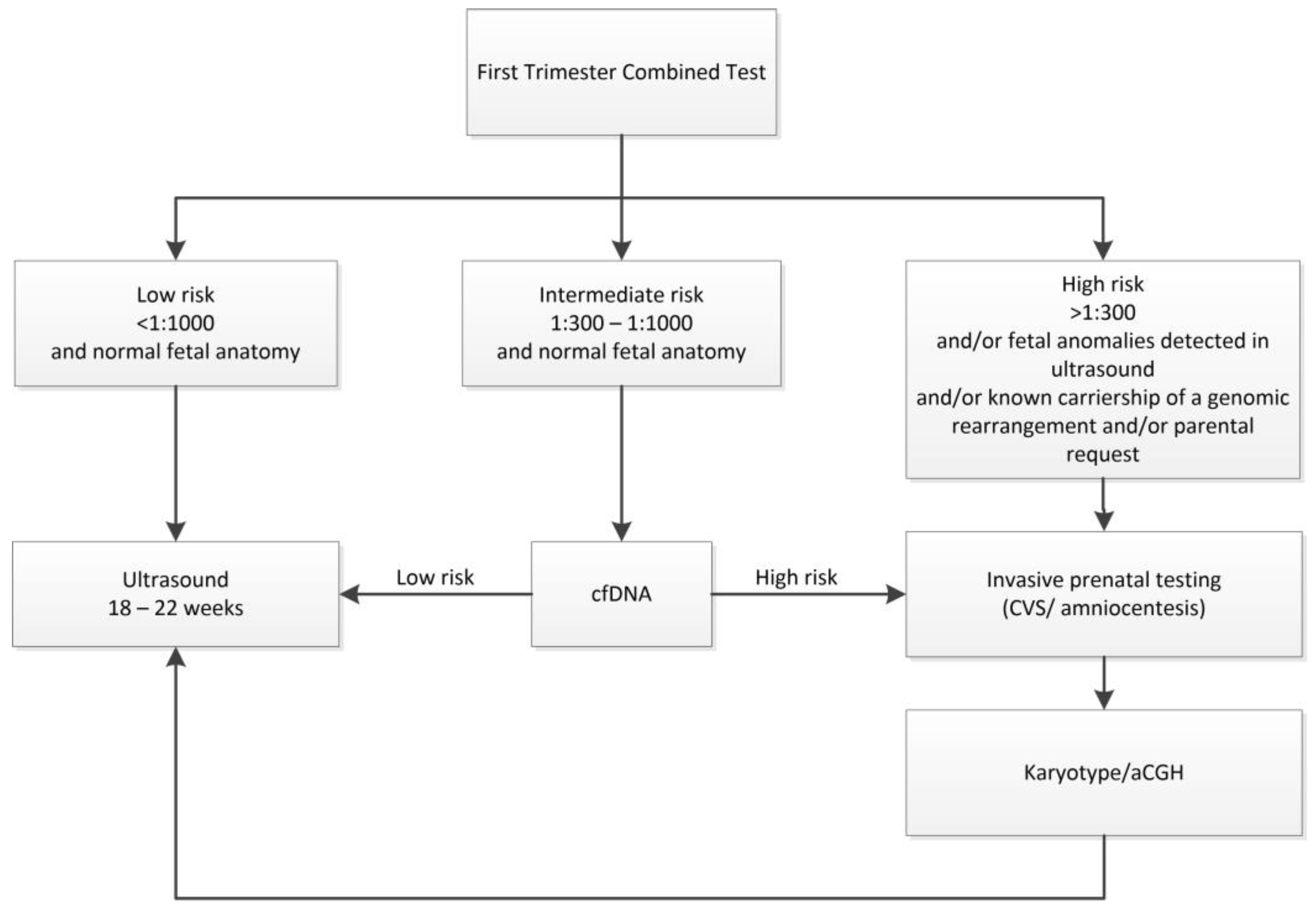
When NT thickness is above the 95th percentile but below 3.5 mm, the recommendation for invasive testing is based on risk calculated by the combined test (CT). The combined test high-risk cut-off used in Poland is >1:300, and in such cases, invasive testing is recommended. In cases with intermediate risk based on combined testing (1/300 to 1/1000) and without associated structural anomalies, the Polish guidelines recommended noninvasive prenatal testing with cfDNA tests, except in cases when the parents opt for prenatal invasive testing. In addition, in Poland, the provision of information about all diagnostic possibilities to patients is required, and therefore, each patient, regardless of the risk, is informed about the possibility of cfDNA as a screening method; however, these tests are not refunded by the Polish National Health Fund.
Fetuses with CT risk lower than 1/1000 are referred for second-trimester screening based on ultrasound [10]. Polish recommendations offer invasive testing not only for those patients with a positive screening result, but also in cases of a known carriership of a genomic rearrangement or an early onset genetic disease, pathogenic copy number variant (CNV) in previous pregnancy, and carriership of a pathogenic CNV or a balanced translocation in a parent, as well as in cases of parental anxiety (Figure 1).
Figure 1.
Prenatal diagnosis algorithm according to the Polish recommendations.
Depending on the indication and gestational age, genetic testing was performed on chorionic villus samples (n = 164) or amniotic fluid (n = 1582).
The frequency of chromosomal aberrations was estimated for the following nuchal translucency categories: below the 95th percentile for CRL, between the 95th percentile for CRL and 2.9 mm, 3.0–3.4 mm, 3.5–3.9 mm, 4.0–4.5 mm, and >4.5 mm. The analyses were also conducted in each category in relation to the fetuses, where the anatomy assessed by ultrasound in the first and early second trimesters was normal or not.
Informed consent for genetic studies was obtained from all pregnant women, and pre-test and post-test counseling, including provision of information about the risks associated with the procedure and the diagnostic techniques involved, was performed by trained clinical geneticists. Those who chose to participate provided written informed consent after the discussion of the potential advantages and risks of chromosomal microarray testing, including the possibility of identifying variants of uncertain clinical significance (VOUSs) and genetic variants in the fetus that cause adult-onset disorders.
2.1. Genetic Testing
Array Comparative Genomic Hybridization Analysis and Interpretation
Genetic testing with the aCGH procedure was performed at the Department of Medical Genetics, Jagiellonian University Medical College, and the Department of Medical Genetics, Institute of Mother and Child, Warsaw, as previously described in detail [21,22] following amniocentesis (n = 1582) or chorionic villus sampling (n = 164).
Unprocessed amniotic fluid (~10 mL) or chorionic villi were used for genomic DNA extraction using the Genomic Mini AX Body Fluids kit (A&A Biotechnology, GdańskPoland) or QuickGene DNA tissue kit S (Kurabo Industries, Osaka, Japan). DNA concentration and purity were determined using a NanoDrop 1000 spectrophotometer (Thermo Scientific, Waltham, MA, USA). aCGH analysis was performed with 8 × 60 K oligonucleotide microarrays (Agilent, Santa Clara US or Oxford Gene Technology, Cytosure, Oxfordshire, UK) following the manufacturer’s protocol.
The results were reported in a specific consultation for post-test genetic counseling, where the results and implications crucial to support informed decision-making were explained to the women.
Results of a pathogenic or potentially pathogenic nature were revealed to patients, with subsequent comprehensive genetic counseling given. Additionally, in cases with fetal pathogenic CNVs, parents were encouraged to undergo aCGH to verify if the fetal CNVs were inherited or appeared de novo.
The aCGH procedure and interpretation were conducted as described previously [21,22]. The reported CNVs were classified according to European standards [21]. A newly identified CNV was considered a relevant chromosomal aberration if it was detected in a minimum of 3 consecutive oligonucleotide microarray probes. Aberrations were filtered up to a minimal size of 1 Mb for deletions and duplications. However, CNVs smaller than 1 Mb were included for further analysis if they were clearly pathogenic or potentially pathogenic based on data from the following databases: ClinVar (Clinically Relevant Variation Database; https://www.ncbi.nlm.nih.gov/clinvar/ (accessed on 5 May 2024)); DECIPHER (Database of Chromosomal Imbalance and Phenotype in Humans Using Ensembl Resource, http://decipher.sanger.ac.uk/ (accessed on 28 April 2024)); databases supported by data from OMIM (Online Mendelian Inheritance in Man: http://www.ncbi.nlm.nih.gov/omim (accessed on 28 April 2024)); the UniQue databases; the Database of Genomic Variants (DGV, http://dgv.tcag.ca/dgv/ (accessed on 29 April 2024)); Clinical Genome Resource (ClinGen, https://www.clinicalgenome.org/ (accessed on 5 May 2024)); and PubMed (https://www.ncbi.nlm.nih.gov/pubmed/ (accessed on 5 May 2024)). The CNVs were assigned the following interpretations according to international guidelines [18,23,24,25]: (1) pathogenic or likely pathogenic, (2) variants of uncertain significance (VUSs), and (3) benign and likely benign. In our study, we focused on pathogenic/likely pathogenic CNVs.
This study was approved by the Medical Ethical Committee of the Medical College Jagiellonian University of Kraków (KBE1072.6120.248.2021).
2.2. Statistical Analysis
All data were recorded in an electronic database. Any direct identifiers were removed and replaced by a code. Patient characteristics are described as means with standard deviation for normally distributed numerical data and as percentages for categorical variables. Difference in maternal age was analyzed by the Student t-test. Chi-square and Fisher’s exact tests were used for comparisons of categorical variables. In all analyses, p values < 0.05 were considered statistically significant. SPSS 19.0 software was used for statistical analysis.
3. Results
In 1484 (84.9%) cases, invasive prenatal diagnosis was indicated because of abnormal results from combined first-trimester screening. In 69 (3.9%) cases, invasive tests were indicated because of advanced maternal age, and in 193 (11.2%) cases, other reasons, such as parental request, family history of congenital defects, and genetic mutation carrier, were given.
In the entire cohort of 1746 fetuses who underwent invasive prenatal testing, classical karyotype testing revealed numerical chromosomal aberrations in 334 fetuses (19.1%), and aCGH detected clinically significant CNVs in 87 (5%) additional cases (Table 1). The most common numerical aberration was trisomy 21 (n = 174), and 22q11.2 microdeletion was the most common CNV, occurring in 21 cases. The sizes of CNVs ranged from 0.01 Mb to 93.4 Mb (Table 2). In 608 fetuses, at least one structural anomaly was detected by ultrasound. The most common anomalies were cardiovascular defects, which occurred in 210 (12.0%) cases. Among the ultrasound markers assessed in the first trimester, the most common finding was nasal bone hypoplasia, which was detected in 10.1% (177/1746) of fetuses (Table 1).
Table 1.
Nuchal translucency (NT) and associated ultrasound and genetic findings among 1746 fetuses undergoing invasive prenatal testing.
Table 2.
Pathogenic or likely pathogenic copy number variants identified by array comparative genomic hybridization (aCGH).
In 754 (43.2%) cases, NT was below the 95th percentile, and in 992 (56.8%) cases, NT was ≥95th percentile (Table 1). In the subgroup of fetuses with NT < p95, numerical chromosomal aberrations were detected in 45 cases (5.9%) (Table 1) and CNVs in 22 (2.9%) cases (Table 1). In 39.8% of cases, there was one or more additional structural anomalies, and the most common ultrasound findings were cardiovascular anomalies (13.5%), followed by nasal bone hypoplasia (11.7%) and skeletal anomalies (8.2%) (Table 1). In 454 (60.2%) fetuses with NT < the 95th percentile and without associated structural anomalies detectable during first- or early second-trimester ultrasound screening, chromosomal aberrations were revealed in 5.3% (Table 1), with trisomy 21 and structural CNVs occurring with the same frequency of 1.9%.
In 992 (56.8%) patients, NT was ≥p95 (Table 1), with 621 (35.5%) fetuses having NT ≥ 3.5 mm. Numerical chromosomal aberrations were detected in 29.1% of fetuses with NT ≥ p95, and CNV in 6.5% of cases (Table 1). The most common numerical aberration was trisomy 21 (15.8%), followed by Turner syndrome (5.7%) and trisomy 18 (4.8%) (Table 1). The three most common trisomies accounted for 63.3% of all pathogenic chromosomal aberrations. In the cohort of fetuses with NT ≥ p95 and after excluding numerical aberrations, CNVs were present in 9.2% of fetuses (65/703). 22q21 microdeletions/microduplications were the most common among CNVs (21.5%; 14/65) (Table 2). In 684 cases (68.9%) of fetuses with NT ≥ p95th, no associated structural anomalies were noted, and 31.1% had at least one associated structural defect. The most common were cardiovascular anomalies (10.8%), followed by hydrops (8.1%). Similar to the group of fetuses with NT < p95th, the most common first-trimester ultrasound marker was NB hypoplasia (8.9%) (Table 1).
In the subgroup of fetuses with NT ≥ 3.5 mm, pathogenic chromosomal aberrations were present in 44.7% (278/621). In 36.2% (225/621) of cases, karyotype revealed numerical chromosomal aberrations and aCGH detected CNVs in 8.5% (53/621) of cases.
Considering the ranges of NT values, the frequency of numerical chromosomal aberrations increased with NT thickness from 5.9% for fetuses with NT < p95th to 43.3% for those with NT > 4.5mm (Table 2). Regarding CNV incidence, no significant differences were observed between fetuses with NT < p95 and those with NT values up to 3.5mm (Table 2). The highest percentage of CNVs was observed in the group of fetuses with NT 4.0–4.5mm (12.2%) (Table 3).
Table 3.
Frequency of chromosomal aberrations in classical karyotype and CNVs detected by aCGH depending on the nuchal translucency (NT) value.
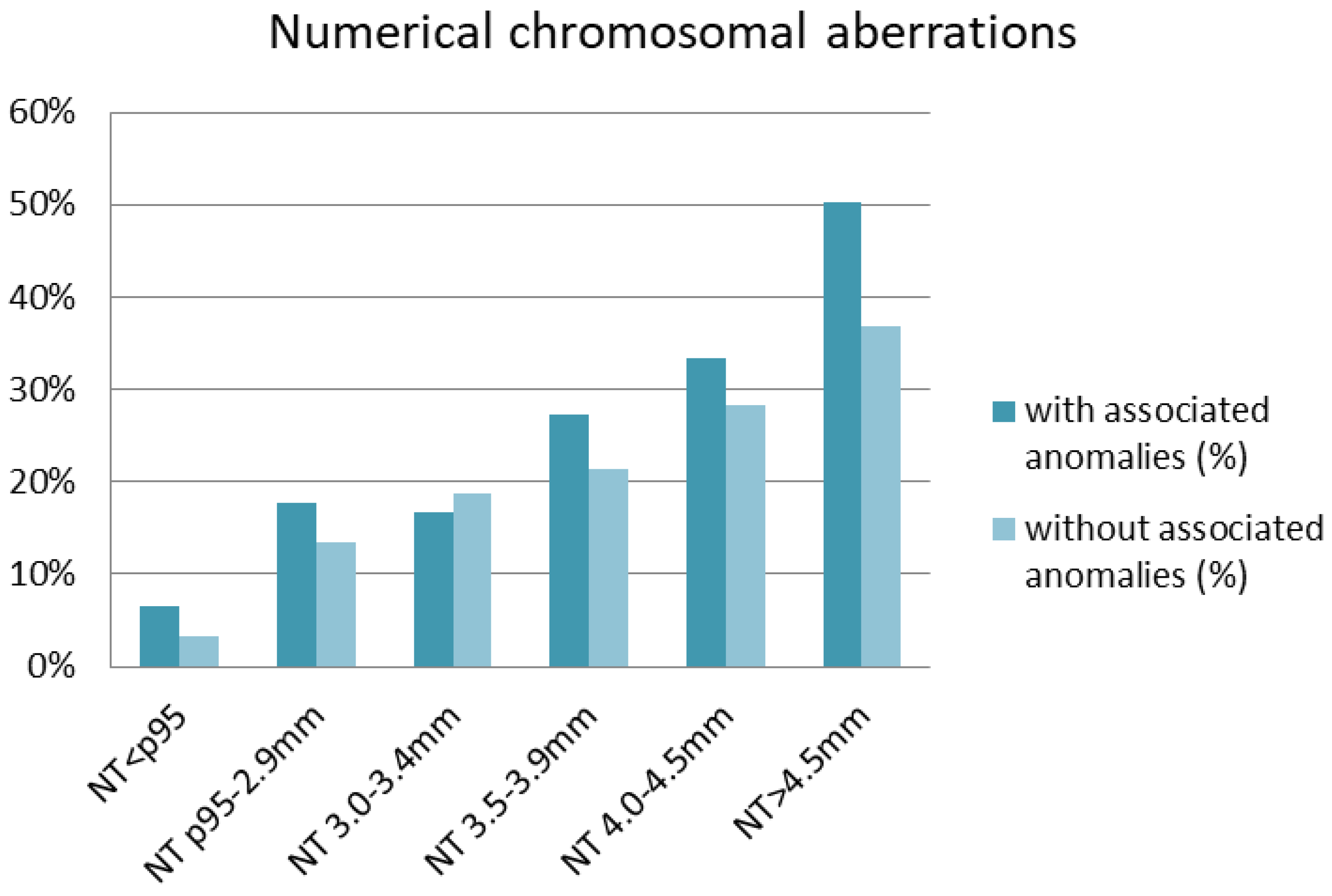
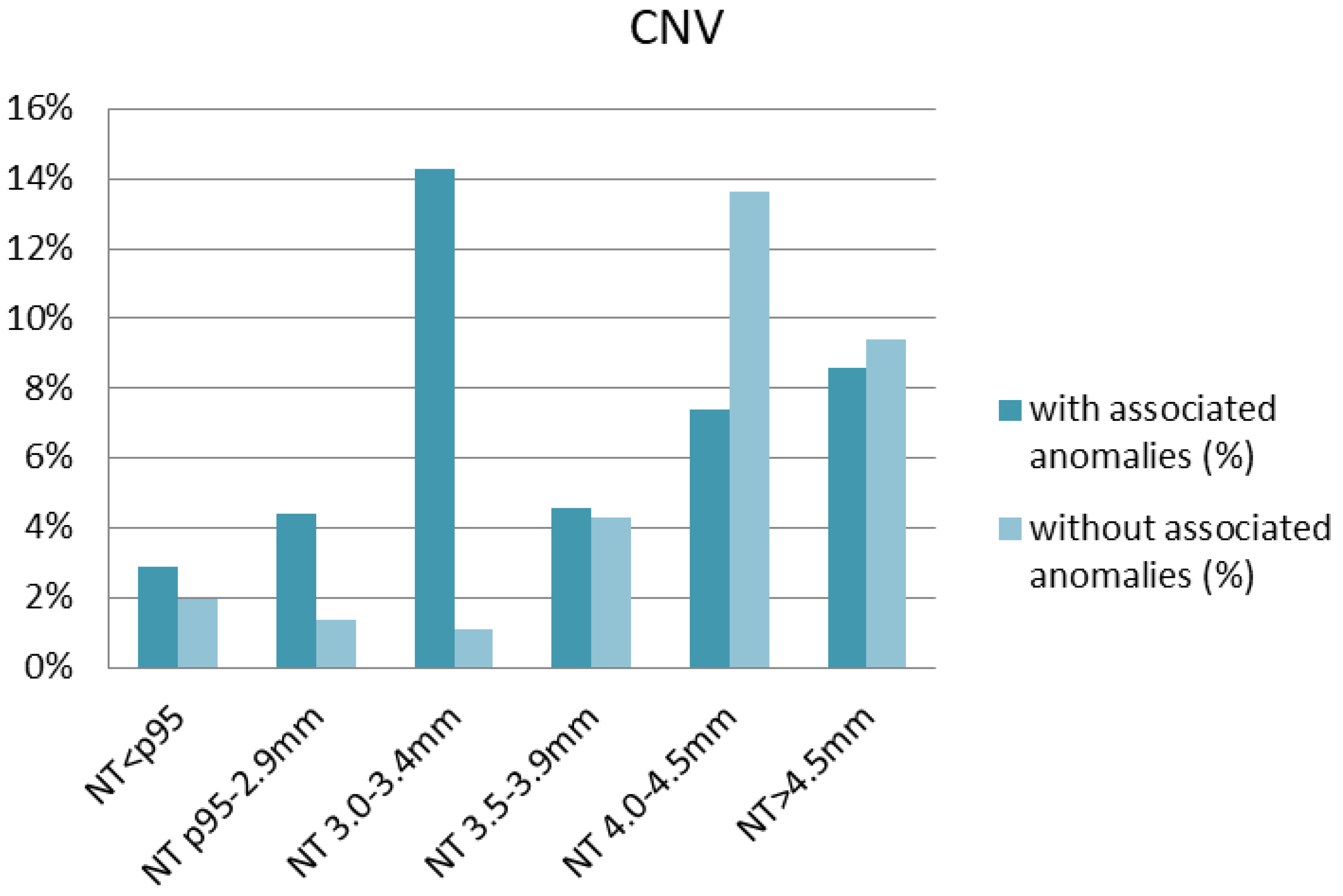
The studied cohort was also stratified according to NT thickness and the presence or absence of accompanying anomalies (Table 4). Both in the absence and presence of accompanying anomalies, the frequency of detected numerical aberrations increased with NT thickness (Figure 2). The highest rate of numerical aberrations was observed in fetuses with NT > 4.5 mm having at least one structural anomaly (50.2%). With regard to CNVs, no significant differences in the frequency were observed between fetuses with NT < p95 and those with NT p95-2.9 mm (Table 4). CNVs occurred most frequently in fetuses with NT 3.0–3.4 mm (14.3%) in which additional structural anomalies were diagnosed, followed by fetuses with NT 4.0–4.5 mm and without accompanying anomalies (13.6%) (Figure 3). Interestingly, in fetuses with a normal karyotype and NT < p95th, microarray analysis revealed clinically relevant deletions or duplications in 4.8% with a structural anomaly and in 2.0% of those without structural defects (Table 4). After excluding cases with structural anomalies and numerical aberrations, CNVs occurred in 1.3% of fetuses with NT p95th-3.4 mm and in 13.0% with NT ≥ 3.5 mm.
Table 4.
Detected chromosomal aberrations depending on the nuchal translucency (NT) value and associated anomalies.
Figure 2.
Distribution of numerical chromosomal aberrations depending on nuchal translucency (NT) thickness.
Figure 3.
Distribution of copy number variants (CNVs) depending on the nuchal translucency (NT) thickness.
4. Discussion
This was a retrospective study analyzing the results of classical karyotype and aCGH depending on NT thickness and the occurrence/absence of associated defects.
In the entire cohort of 1746 fetuses studied, 19.1% of numerical aberrations were detected and aCGH additionally revealed CNVs in 5% of fetuses.
The frequency of numerical aberrations increased with NT thickness and in the case of additional defects. In the subgroup of fetuses with NT > 4.5 mm and at least one anomaly, numerical chromosomal aberration was detected in 50.2% cases. Clinically significant microdeletions/microduplications were present in each analyzed subgroup, with the highest percentage in the group of fetuses with NT 3.0–3.4 mm having at least one structural anomaly.
In recent years, the association between increased NT and pathogenic CNVs has been extensively explored in the literature [9]. However, there is still a debate about the NT value at which we should refer patients for invasive diagnostics or recommend cfDNA testing, especially when increased NT is not accompanied by structural abnormalities or there is increased risk of aneuploidy after the first-trimester combined test [12,26]. It is common for physicians conducting first-trimester screening for aneuploidy to have doubts about the choice regarding next steps in the diagnostic path for patient with a NT between the 95th percentile and 3.5 mm, and those with NT within the normal range but the combined test indicates a high risk of aneuploidy. We believe that the results of our research will help to make decisions about further diagnostic steps.
Currently, NT ≥ 3.5 mm is an undisputed indication for invasive prenatal testing. However, the percentage of detected chromosomal aberrations varies between studied cohorts. The rate of chromosomal abnormalities in fetuses with NT ≥ 3.5 mm (44.7%) was higher in our study than that reported by Srebniak et al. (38%) [14] but similar to that reported by Bardi et al. (43%) [27]. It was reported in the literature that pathogenic CNVs can be detected by CMA in 0–15% of fetuses with NT ≥ 3.5 mm depending on the selected or unselected nature of the studied cohorts [9,15,28,29,30]. In our study, pathogenic CNVs were detected in 8.5% of cases with NT ≥ 3.5 mm in the unselected population and in 13.0% of cases with isolated NT ≥ 3.5 mm where karyotype and anatomy were assessed as normal. These results are similar to those reported by Tanner et al. (13.7%) [31] and by Coello-Cahuao et al. (12%) [32]. However, in other studies [9,27], the frequency of pathogenic CNVs among fetuses with normal karyotype and isolated increased NT ≥ 3.5 mm was reported to be between 3% and 9%. In our study, the frequency was also higher than that in another French multicenter project [33], where it was 2.7%, but in the French study, fetuses between 11 and 28 weeks of gestation were included. It could be supposed that in our study, we did not observe all structural anomalies, as the study was conducted during the first- and early second-trimester. Therefore, when giving a consultation, the duration of pregnancy should be considered. It can be assumed that some structural abnormalities may appear in later stages of pregnancy; on the other hand, in some cases of fetal structural and/or genetic defects, stillbirth may occur in the following weeks.
Recently, a cut-off of 3.0 mm was proposed as an indication for CMA [11,34]. In previous studies where fetuses with NT between 3.0 and 3.4 mm were included, the prevalence of CNVs varied between 0.4 and 2.1% [34,35,36]. In our cohort, it was 4.3% in an unselected cohort with a normal karyotype and in 1.3% of fetuses with a normal karyotype and normal anatomy.
Moreover, in our study, 21.5% of chromosomal aberrations were found in pregnancies with NT values between the 95th and 99th percentiles, which is similar to the observation by Bardi et al. [27]. On the other hand, in our study, CNVs were present in 1.4% of fetuses with NT p95th–99th percentile with normal karyotypes, and in previous studies, this prevalence was estimated to be between 1.7% and 2.6% [37,38].
In our study, chromosomal aberrations were diagnosed in 35.7% of the fetuses with NT ≥ 95th percentile, which is higher than previously reported [27]. A total of 22.5% of fetuses with NT above the 95th percentile had trisomy 21, 13, or 18, which is similar to the finding by Bardi et al. [27], where these three common trisomies were reported in 23.9% of fetuses.
It was shown that in the absence of aneuploidy, fetuses with increased NT have an increased risk of other fetal defects, particularly cardiac and skeletal abnormalities [5,22].
In our study, 29.6% of fetuses with NT p95th–99th percentiles and 31.9% of fetuses with NT above the 99th percentile were found to have at least one structural anomaly, and in the study by Bardi et al. [27], the percentages were 21.3% and 62%, respectively.
In their prospective cohort study, Wapner et al. [16] reported that in pregnancies with fetal structural anomalies and a normal karyotype, the incremental diagnostic yield with CMA was approximately 6% above what a karyotype could detect. These authors [16] also reported a 1.7% diagnostic yield of CMA in samples with normal conventional karyotype when the indication was advanced maternal age or positive screening results. In our observation, CNVs were detected in 2.0% of fetuses with NT < the 95th percentile where karyotype and anatomy were normal. However, a recent meta-analysis [39] indicated that 0.84% of fetuses that had invasive testing because of advanced maternal age or anxiety carried a pathogenic clinically significant submicroscopic aberration, and that pregnant women under 36 years of age have a higher risk for submicroscopic pathogenic aberrations than for trisomy 21.
Moreover, it should be noted that, according to the last study [40], even when the karyotype and microarray are normal, there is an increased residual risk of morbidity-related outcomes compared with the general population, particularly if NT is greater than 6 mm.
Strengths and Limitations
The strength of this study is that it was a two-center cohort study that included a representative study population due to publicly funded prenatal care. This allowed us to evaluate the frequency of chromosomal aberrations and CNVs in subgroups with different NT thickness, which may be helpful for clinicians to choose the most appropriate test. The limitation is that it was a retrospective study, and we did not have a long follow-up, but only the data from the first- and early second-trimester scans. Therefore, we did not analyze all pregnancy outcomes, and we therefore cannot estimate the actual impact of the results on the outcome. Second, in some cases, it is possible that some genetic disorders might have been detected if other genetic methods had been used.
5. Conclusions
In fetuses undergoing invasive prenatal testing because of increased NT and/or increased risk after first-trimester screening, microarray analysis should be considered as the method of choice, as it allows the detection of submicroscopic anomalies that are not detected by karyotyping alone, even in the absence of structural abnormalities. Increased NT ≥ 3.5 mm is a strong indication for genetic testing with the aCGH technique. However, according to our results, in cases with NT <the 99th percentile and normal anatomy during the first trimester, aCGH can detect pathogenic CNVs in a significant percentage of cases.
Therefore, invasive testing with aCGH should be discussed with parents choosing invasive procedures.
Key Message: Performing aCGH in fetuses referred for invasive prenatal testing after first-trimester screening provides additional, clinically valuable information compared to conventional karyotyping, even in the presence of normal anatomy. This information can be helpful to parents, enabling them to make an informed decision about which diagnostic path to take.
Author Contributions
Conceptualization, A.W. and N.B.; Data curation, A.W., K.K., K.S., A.M.-T. and N.B.; Formal analysis, A.W., K.K., K.S. and N.B.; Funding acquisition, A.W. and H.H.; Investigation, A.W. and W.W.; Methodology, A.W., M.B.-M., K.K. and N.B.; Project administration, A.W. and W.W.; Resources, A.W., K.K., K.S, A.M.-T., M.B.-M., H.H. and N.B.; Software, W.W.; Supervision, H.H. and M.B.-M.; Validation, A.W, K.K. and K.S.; Visualization, A.W. and W.W.; Writing—original draft, A.W., M.B.-M. and N.B.; Writing—review and editing, A.W., W.W., A.M.-T., M.B.-M. and N.B. All authors have read and agreed to the published version of the manuscript.
Funding
The APC was funded by institutional academic funds allocated to the authors, provided by the Medical College Jagiellonian University.
Institutional Review Board Statement
This study was conducted in accordance with the Declaration of Helsinki and approved by the Medical Ethical Committee of the Medical College Jagiellonian University of Kraków (KBE1072.6120.248.2021).
Informed Consent Statement
Informed consent was obtained from the patients involved in the study. A informed consent form has been provided as requested.
Data Availability Statement
The raw data supporting the conclusions of this article will be made available by the authors upon request.
Conflicts of Interest
The authors have stated explicitly that there are no conflicts of interest in connection with this article. The authors have no relevant financial or nonfinancial interests to disclose.
Abbreviations
aCGH: array comparative genomic hybridization; cfDNA, cell-free DNA; CNV, copy number variant; CRL, crown–rump length; NT, nuchal translucency.
References
- Nicolaides, K.H.; Azar, G.; Byrne, D.; Mansur, C.; Marks, K. Fetal nuchal translucency: Ultrasound screening for chromosomal defects in first trimester of pregnancy. BMJ 1992, 304, 867–869. [Google Scholar] [CrossRef] [Scilit] [PubMed]
- Snijders, R.J.; Noble, P.; Sebire, N.; Souka, A.; Nicolaides, K.H. UK multicentre project on assessment of risk of trisomy 21 by maternal age and fetal nuchal translucency thickness at 10–14 weeks of gestation. Fetal Medicine Foundation First Trimester Screening Group. Lancet 1998, 352, 343–346. [Google Scholar] [CrossRef] [Scilit] [PubMed]
- Souka, A.P.; Von Kaisenberg, C.S.; Hyett, J.A.; Sonek, J.D.; Nicolaides, K.H. Increased nuchal translucency with normal karyotype. Am. J. Obs. Gynecol. 2005, 192, 1005–1021. [Google Scholar] [CrossRef] [Scilit] [PubMed]
- Hellmuth, S.G.; Pedersen, L.H.; Miltoft, C.B.; Petersen, O.B.; Kjaergaard, S.; Ekelund, C.; Tabor, A. Increased nuchal translucency thickness and risk of neurodevelopmental disorders. Ultrasound Obs. Gynecol. 2017, 49, 592–598. [Google Scholar] [CrossRef] [Scilit] [PubMed]
- Baer, R.J.; Norton, M.E.; Shaw, G.M.; Flessel, M.C.; Goldman, S.; Currier, R.J.; Jelliffe-Pawlowski, L.L. Risk of selected structural abnormalities in infants after increased nuchal translucency measurement. Am. J. Obstet. Gynecol. 2014, 211, 675.e1–675.e19. [Google Scholar] [CrossRef] [Scilit] [PubMed]
- Wright, D.; Kagan, K.O.; Molina, F.S.; Gazzoni, A.; Nicolaides, K.H. A mixture model of nuchal translucency thickness in screening for chromosomal defects. Ultrasound Obs. Gynecol. 2008, 31, 376–383. [Google Scholar] [CrossRef] [Scilit] [PubMed]
- Kagan, K.O.; Wright, D.; Valencia, C.; Maiz, N.; Nicolaides, K.H. Screening for trisomies 21, 18 and 13 by maternal age, fetal nuchal translucency, fetal heart rate, free beta-hCG and pregnancy-associated plasma protein-A. Hum. Reprod. 2008, 23, 1968–1975. [Google Scholar] [CrossRef] [Scilit]
- Nicolaides, K.H. Screening for fetal aneuploidies at 11 to 13 weeks. Prenat. Diagn. 2011, 31, 7–15. [Google Scholar] [CrossRef] [Scilit]
- Grande, M.; Jansen, F.A.R.; Blumenfeld, Y.J.; Fisher, A.; Odibo, A.O.; Haak, M.C.; Borrell, A. Genomic microarray in fetuses with increased nuchal translucency and normal karyotype: A systematic review and meta-analysis. Ultrasound Obs. Gynecol. 2015, 46, 650–658. [Google Scholar] [CrossRef] [Scilit] [PubMed]
- Polish Gynaecological Society guideline on prenatal diagnosis. Ginekol. Pol. 2009, 80, 390–393.
- Maya, I.; Yacobson, S.; Kahana, S.; Yeshaya, J.; Tenne, T.; Agmon-Fishman, I.; Cohen-Vig, L.; Shohat, M.; Basel-Vanagaite, L.; Sharony, R. Cut-off value of nuchal translucency as indication for chromosomal microarray analysis. Ultrasound Obs. Gynecol. 2017, 50, 332–335. [Google Scholar] [CrossRef] [Scilit] [PubMed]
- Kagan, K.O.; Sonek, J.; Wagner, P.; Hoopmann, M. Principles of first trimester screening in the age of non-invasive prenatal diagnosis: Screening for chromosomal abnormalities. Arch. Gynecol. Obs. 2017, 296, 645–651. [Google Scholar] [CrossRef] [Scilit]
- van der Meij, K.R.M.; Sistermans, E.A.; Macville, M.V.E.; Stevens, S.J.C.; Bax, C.J.; Bekker, M.N.; Bilardo, C.M.; Boon, E.M.J.; Boter, M.; Diderich, K.E.M.; et al. TRIDENT-2: National implementation of genome-wide non-invasive prenatal testing as a first-tier screening test in the Netherlands. Am. J. Hum. Genet. 2019, 105, 1091–1101. [Google Scholar] [CrossRef] [Scilit]
- Srebniak, M.I.; de Wit, M.C.; Diderich, K.E.; Govaerts, L.C.; Joosten, M.; Knapen, M.F.; Bos, M.J.; Looye-Bruinsma, G.A.; Koningen, M.; Go, A.T.; et al. Enlarged NT (≥3.5 mm) in the first trimester—Not all chromosome aberrations can be detected by NIPT. Mol. Cytogenet. 2016, 9, 69. [Google Scholar] [CrossRef] [Scilit]
- Huang, J.; Poon, L.C.; Akolekar, R.; Choy, K.W.; Leung, T.Y.; Nicolaides, K.H. Is high fetal nuchal translucency associated with submicroscopic chromosomal abnormalities on array CGH? Ultrasound Obs. Gynecol. 2014, 43, 620–624. [Google Scholar] [CrossRef] [Scilit]
- Wapner, R.J.; Martin, C.L.; Levy, B.; Ballif, B.C.; Eng, C.M.; Zachary, J.M.; Savage, M.; Platt, L.D.; Saltzman, D.; Grobman, W.A.; et al. Chromosomal microarray versus karyotyping for prenatal diagnosis. N. Engl. J. Med. 2012, 367, 2175–2184. [Google Scholar] [CrossRef] [Scilit]
- Committee on Genetics and the Society for Maternal-Fetal Medicine. Committee Opinion No.682: Microarrays and Next-Generation Sequencing Technology: The Use of Advanced Genetic Diagnostic Tools in Obstetrics and Gynecology. Obs. Gynecol. 2016, 128, e262–e268. [Google Scholar] [CrossRef] [Scilit] [PubMed]
- Silva, M.; de Leeuw, N.; Mann, K.; Schuring-Blom, H.; Morgan, S.; Giardino, D.; Rack, K.; Hastings, R. European guidelines for constitutional cytogenomic analysis. Eur. J. Hum. Genet. 2019, 27, 1–16. [Google Scholar] [CrossRef] [Scilit] [PubMed]
- Polish Gynecological Society—Ultrasound Section Guidelines on ultrasound screening in uncomplicated pregnancy (2 December 2011). Ginekol. Pol. 2012, 83, 309–315.
- Salomon, L.J.; Alfirevic, Z.; Bilardo, C.M.; Chalouhi, G.E.; Ghi, T.; Kagan, K.O.; Lau, T.K.; Papageorghiou, A.T.; Raine-Fenning, N.J.; Stirnemann, J.; et al. ISUOG practice guidelines: Performance of first-trimester fetal ultrasound scan. Ultrasound Obstet. Gynecol. 2013, 41, 102–113. [Google Scholar]
- Kowalczyk, K.; Bartnik-Głaska, M.; Smyk, M.; Plaskota, I.; Bernaciak, J.; Kędzior, M.; Wiśniowiecka-Kowalnik, B.; Jakubów-Durska, K.; Braun-Walicka, N.; Barczyk, A.; et al. Prenatal Diagnosis by Array Comparative Genomic Hybridization in Fetuses with Cardiac Abnormalities. Genes 2021, 12, 2021. [Google Scholar] [CrossRef] [Scilit]
- Wójtowicz, A.; Madetko-Talowska, A.; Wójtowicz, W.; Szewczyk, K.; Huras, H.; Bik-Multanowski, M. Cardiovascular Anomalies among 1005 Fetuses Referred to Invasive Prenatal Testing-A Comprehensive Cohort Study of Associated Chromosomal Aberrations. Int. J. Environ. Res. Public Health 2022, 19, 10019. [Google Scholar] [CrossRef] [Scilit]
- Claustres, M.; Kožich, V.; Dequeker, E.; Fowler, B.; Hehir-Kwa, J.; Miller, K.; Oosterwijk, C.; Peterlin, B.; van Ravenswaaij-Arts, C.; Zimmermann, U.; et al. Recommendations for reporting results of diagnostic genetic testing (biochemical, cytogenetic and molecular genetic). Eur. J. Hum. Gen. 2013, 22, 160–170. [Google Scholar] [CrossRef] [Scilit]
- Nowakowska, B. Clinical interpretation of copy number variants in the human genome. J. Appl. Gen. 2017, 58, 449–457. [Google Scholar] [CrossRef] [Scilit] [PubMed]
- Cheng, S.; Chan, K.; Leung, K.; Au, P.; Tam, W.; Li, S.; Luk, H.; Kan, A.; Chung, B.; Lo, I.; et al. Experience of chromosomal microarray applied in prenatal and postnatal settings in Hong Kong. Am. J. Med. Gen. Part. C Semin. Med. Genet. 2019, 181, 196–207. [Google Scholar] [CrossRef] [Scilit]
- Johnson, K.; Kelley, J.; Saxton, V.; Walker, S.P.; Hui, L. Declining invasive prenatal diagnostic procedures: A comparison of tertiary hospital and national data from 2012 to 2015. Aust. N. Z. J. Obs. Gynaecol. 2017, 57, 152–156. [Google Scholar] [CrossRef] [Scilit]
- Bardi, F.; Bosschieter, P.; Verheij, J.; Go, A.; Haak, M.; Bekker, M.; Sikkel, E.; Coumans, A.; Pajkrt, E.; Bilardo, C. Is there still a role for nuchal translucency measurement in the changing paradigm of first trimester screening? Prenat. Diagn. 2020, 40, 197–205. [Google Scholar] [CrossRef] [Scilit]
- Shaffer, L.G.; Rosenfeld, J.A.; Dabell, M.P.; Coppinger, J.; Bandholz, A.M.; Ellison, J.W.; Ravnan, J.B.; Torchia, B.S.; Ballif, B.C.; Fisher, A.J. Detection rates of clinically significant genomic alterations by microarray analysis for specific anomalies detected by ultrasound. Prenat. Diagn. 2012, 32, 986–995. [Google Scholar] [CrossRef] [Scilit]
- Scott, F.; Murphy, K.; Carey, L.; Greville, W.; Mansfield, N.; Barahona, P.; Robertson, R.; McLennan, A. Prenatal diagnosis using combined quantitative fluorescent polymerase chain reaction and array comparative genomic hybridization analysis as a first-line test: Results from over 1000 consecutive cases. Ultrasound Obs. Gynecol 2013, 41, 500–507. [Google Scholar] [CrossRef] [Scilit]
- Hillman, S.C.; McMullan, D.J.; Hall, G.; Togneri, F.S.; James, N.; Maher, E.J.; Meller, C.H.; Williams, D.; Wapner, R.J.; Maher, E.R.; et al. Use of prenatal chromosomal microarray: Prospective cohort study and systematic review and meta-analysis. Ultrasound Obs. Gynecol. 2013, 41, 610–620. [Google Scholar] [CrossRef] [Scilit]
- Tanner, L.M.; Alitalo, T.; Stefanovic, V. Prenatal array comparative genomic hybridization in a well-defined cohort of high-risk pregnancies. A 3-year implementation results in a public tertiary academic referral hospital. Prenat. Diagn. 2021, 41, 422–433. [Google Scholar] [CrossRef] [Scilit]
- Coello-Cahuao, E.; Sánchez-Durán, M.Á.; Calero, I.; Higueras, M.T.; García, M.A.; Rodó, C.; Maiz, N.; Plaja Rustein, A.; Castells-Sarret, N.; Mediano-Vizuete, C.; et al. Array study in fetuses with nuchal translucency above the 95th percentile: A 4-year observational single-centre study. Arch. Gynecol. Obs. 2023, 307, 285–292. [Google Scholar] [CrossRef] [Scilit]
- Egloff, M.; Hervé, B.; Quibel, T.; Jaillard, S.; Le Bouar, G.; Uguen, K.; Saliou, A.H.; Valduga, M.; Perdriolle, E.; Coutton, C.; et al. Diagnostic yield of chromosomal microarray analysis in fetuses with isolated increased nuchal translucency: A French multicenter study. Ultrasound Obs. Gynecol. 2018, 52, 715–721. [Google Scholar] [CrossRef] [Scilit]
- Petersen, O.B.; Smith, E.; Van Opstal, D.; Polak, M.; Knapen, M.F.C.M.; Diderich, K.E.M.; Bilardo, C.M.; Arends, L.R.; Vogel, I.; Srebniak, M.I. Nuchal translucency of 3.0–3.4 mm an indication for NIPT or microarray? Cohort analysis and literature review. Acta Obs. Gynecol. Scand. 2020, 99, 765–774. [Google Scholar] [CrossRef] [Scilit]
- Hui, L.; Pynaker, C.; Bonacquisto, L.; Lindquist, A.; Poulton, A.; Kluckow, E.; Hutchinson, B.; Norris, F.; Pertile, M.D.; Gugasyan, L.; et al. Reexamining the optimal nuchal translucency cutoff for diagnostic testing in the cell-free DNA and microarray era: Results from the Victorian Perinatal Record Linkage study. Am. J. Obs. Gynecol. 2021, 225, 527.e1–527.e12. [Google Scholar] [CrossRef] [Scilit]
- Sagi-Dain, L.; Singer, A.; Ben Shachar, S.; Josefsberg Ben Yehoshua, S.; Feingold-Zadok, M.; Greenbaum, L.; Maya, I. Risk of Clinically Significant Chromosomal Microarray Analysis Findings in Fetuses with Nuchal Translucency From 3.0 mm through 3.4 mm. Obs. Gynecol. 2021, 137, 126–131. [Google Scholar] [CrossRef] [Scilit]
- Su, L.; Huang, H.; An, G.; Cai, M.; Wu, X.; Li, Y.; Xie, X.; Lin, Y.; Wang, M.; Xu, L. Clinical application of chromosomal microarray analysis in fetuses with increased nuchal translucency and normal karyotype. Mol. Genet. Genom. Med. 2019, 7, e811. [Google Scholar] [CrossRef] [Scilit]
- Xue, S.; Yan, H.; Chen, J.; Li, N.; Wang, J.; Liu, Y.; Zhang, H.; Li, S.; Zhang, W.; Chen, D.; et al. Genetic examination for fetuses with increased fetal nuchal translucency by genomic technology. Cytogenet. Genome Res. 2020, 160, 57–62. [Google Scholar] [CrossRef] [Scilit]
- Srebniak, M.I.; Joosten, M.; Knapen, M.F.C.M.; Arends, L.R.; Polak, M.; van Veen, S.; Go, A.T.J.I.; Van Opstal, D. Frequency of submicroscopic chromosomal aberrations in pregnancies without increased risk for structural chromosomal aberrations: Systematic review and meta-analysis. Ultrasound Obs. Gynecol. 2018, 51, 445–452. [Google Scholar] [CrossRef] [Scilit] [PubMed]
- Spataro, E.; Cordisco, A.; Luchi, C.; Filardi, G.R.; Masini, G.; Pasquini, L. Increased nuchal translucency with normal karyotype and genomic microarray analysis: A multicenter observational study. Int. J. Gynaecol. Obs. 2023, 161, 1040–1045. [Google Scholar] [CrossRef] [Scilit]




Disclaimer/Publisher’s Note: The statements, opinions and data contained in all publications are solely those of the individual author(s) and contributor(s) and not of MDPI and/or the editor(s). MDPI and/or the editor(s) disclaim responsibility for any injury to people or property resulting from any ideas, methods, instructions or products referred to in the content. |
© 2024 by the authors. Licensee MDPI, Basel, Switzerland. This article is an open access article distributed under the terms and conditions of the Creative Commons Attribution (CC BY) license (https://creativecommons.org/licenses/by/4.0/).