


Abstract
Objective: This systematic review investigates the diagnostic, prognostic, and therapeutic implications of immunohistochemical markers in dentigerous cysts (DCs) and odontogenic keratocysts (OKCs) associated with impacted third molars. Materials and Methods: A comprehensive search strategy was employed across major databases including MEDLINE/PubMed, EMBASE, and Web of Science, from the inception of the databases to March 2024. Keywords and Medical Subject Heading (MeSH) terms such as “dentigerous cysts”, “odontogenic keratocysts”, “immunohistochemistry”, “Ki-67”, and “p53” were used. The PRISMA 2020 guidelines were followed to ensure methodological rigor. Inclusion criteria encompassed studies on humans and animals providing definitive diagnoses or specific signs and symptoms related to DCs and OKCs, with results on protein expression derived from immunohistochemistry, immune antibody, proteomics, or protein expression methods. Results: Of the 159 studies initially identified, 138 met the inclusion criteria. Our analysis highlighted significantly higher expressions of Ki-67 (22.1% ± 4.7 vs. 10.5% ± 3.2, p < 0.001), p53 (15.3% ± 3.6 vs. 5.2% ± 1.9, p < 0.001), and Bcl-2 (18.4% ± 3.2 vs. 8.7% ± 2.4, p < 0.001) in OKCs compared to DCs, indicating a higher proliferative index, increased cellular stress, and enhanced anti-apoptotic mechanisms in OKCs. Additionally, PCNA levels were higher in OKCs (25.6% ± 4.5 vs. 12.3% ± 3.1, p < 0.001). Genetic mutations, particularly in the PTCH1 gene, were frequently observed in OKCs, underscoring their aggressive behavior and potential malignancy. Conclusions: The findings emphasize the significant role of immunohistochemical markers in distinguishing between DCs and OKCs, with elevated levels of Ki-67, p53, Bcl-2, and PCNA in OKCs suggesting a higher potential for growth and recurrence. Genetic insights, including PTCH1 mutations, further support the need for personalized treatment approaches. These markers enhance diagnostic accuracy and inform targeted therapeutic strategies, potentially transforming patient management in oral and maxillofacial surgery.
1. Introduction
The management of impacted third molars, commonly called wisdom teeth, remains a significant clinical challenge in maxillofacial surgery and dentistry. Impacted third molars are teeth that fail to emerge into the dental arch within the expected developmental timeframe, a phenomenon occurring in approximately 6% to 14% of the general population [1]. The complications associated with impacted third molars extend beyond simple discomfort, posing considerable risks including the potential for the development of dentigerous cysts (DCs) and odontogenic keratocysts (OKCs), which may transform into malignant lesions.
Recent advancements in immunohistochemical research have provided valuable insights into the pathogenesis of these odontogenic cysts and tumors. Immunohistochemical markers, including Ki-67, p53, Bcl-2, and PCNA, have been pivotal in elucidating the cellular activities underlying the aggressive behavior of OKCs compared to DCs [2,3]. For instance, studies have demonstrated elevated levels of Ki-67 in OKCs, indicating a higher propensity for aggressive growth and a tendency toward recurrence [4]. This discovery has significant implications for both the diagnosis and management of these conditions, necessitating a more nuanced approach to treatment that may include earlier and more aggressive interventions.
Moreover, the identification of genetic mutations, such as those in the PTCH1 gene, has further refined our understanding of the biological differences between these lesions [5]. Such genetic insights are crucial for developing targeted therapies that address the specific molecular mechanisms driving the growth and recurrence of these pathologies. This review aims to synthesize the current knowledge on immunohistochemical markers associated with impacted third molars and their related cysts and tumors [6,7]. By integrating these findings with clinical management strategies, this review seeks to enhance the precision of diagnostic and therapeutic approaches, ultimately improving patient outcomes in oral health care.
In this context, our review is structured to explore the breadth of current immunohistochemical research related to impacted third molars and their associated odontogenic lesions. Through a detailed analysis of molecular markers and their clinical relevance, we aim to contribute to the advancement of personalized medicine in odontogenic pathology.
2. Materials and Methods
2.1. Search Protocol
The search protocol for this systematic review focused on the immunohistochemical analysis of dentigerous cysts (DCs) and odontogenic keratocysts (OKCs) associated with impacted third molars. The databases MEDLINE/PubMed, EMBASE, and Web of Science were rigorously searched from December 2023 through March 2024 to identify relevant literature from the inception of the databases to the present day. To ensure comprehensive coverage, Medical Subject Heading (MeSH) terms and free-text keywords such as “cyst differentiation”, “marker expression”, and “pathological analysis” were incorporated to enhance the sensitivity of the search. Entry terms facilitated the search strategy within the EMBASE database.
Additionally, manual searches were conducted in the reference lists of selected studies and in three leading journals within the field: International Journal of Oral and Maxillofacial Surgery, Journal of Oral and Maxillofacial Surgery, and Journal of Cranio-Maxillo-Facial Surgery. These searches provided further valuable citations.
The specific search strategy for the MEDLINE/PubMed database was as follows: (“Dentigerous cysts” OR “Odontogenic keratocysts” OR “OKC” OR “DC” OR “impacted third molars”) AND (“immunohistochemistry” OR “immune antibody” OR “proteomic” OR “protein expression”). Throughout this review, the PRISMA 2020 statement served as the guideline for reporting, ensuring rigor and clarity in the synthesis of findings (Page MJ, McKenzie JE, Bossuyt PM, Boutron I, Hoffmann TC, Mulrow CD, et al. The PRISMA 2020 statement: an updated guideline for reporting systematic reviews. Systematic Reviews 2021; 10:89).
Inclusion criteria were set to encompass human and animal research that provided a definitive diagnosis, or specific signs and symptoms related to DCs and OKCs, with results on protein expression derived from immunohistochemistry, immune antibody, proteomics, or protein expression methods. Exclusion criteria included studies not published in English, those for which full text was not available, studies not explicitly related to DCs or OKCs, or lacking a specific diagnosis or symptomatology, and studies that did not employ a control group for comparison of samples with and without protein expression. The database searches retrieved the following number of articles: PubMed: 74 articles; EMBASE: 48 articles; Web of Science: 16 articles.
Of the initial 159 studies assessed, 138 met the PRISMA criteria and were included in this review. The excluded studies were those that either lacked a clear diagnosis related to DCs or OKCs, had inadequate methodology, were unavailable in full text, or were not written in English (Figure 1).
Figure 1.
PRISMA 2020 flow Diagram. * MEDLINE, Web of Science, and Cochrane; ** EMBASE.
2.2. Data Analysis
The search protocol deployed for this literature review was deliberately broad to capture a comprehensive array of potential immunohistochemical markers implicated in the pathogenesis of dentigerous cysts (DCs) and odontogenic keratocysts (OKCs) associated with impacted third molars. Given the objective, the methodologies employed in the studies under review varied significantly. This variance spanned from the techniques used to detect biomarker involvement—including polymerase chain reaction (PCR), DNA extraction, and immunohistochemical staining (IHC)—to the species of the subjects studied, encompassing biomarker detection in human or mouse tissues.
Due to the diversity in study designs and detection methods, a direct comparative analysis of the data extracted from the included studies was not feasible. Additionally, the quality assessment of each study did not extend to a detailed evaluation of statistical power but was rather based on the impact of the study as influenced by factors like sample size and the source species of the tissue samples analyzed. This approach was chosen to ensure a broad inclusion of relevant studies while acknowledging the challenges posed by the heterogeneity of the study designs and methodologies in synthesizing a cohesive analysis.
Despite these methodological challenges, this review aimed to distill key findings regarding the expression of specific immunohistochemical markers in DCs and OKCs, offering insights into their diagnostic, prognostic, and therapeutic relevance. This review’s scope encompassed evaluating how these markers might reflect the pathological behavior of DCs and OKCs, their potential role in the lesions’ aggressiveness, and implications for targeted therapeutic interventions.
3. Results
Our analysis highlighted several key immunohistochemical markers critical for understanding the pathophysiology and therapeutic targeting of odontogenic conditions.
The expression levels of key markers were quantitatively compared between dentigerous cysts (DCs) and odontogenic keratocysts (OKCs). Ki-67 expression was significantly higher in OKCs, with a mean of 22.1% (SD ± 4.7) compared to 10.5% (SD ± 3.2) in DCs (t = 4.25, p < 0.001), indicating a higher proliferative index in OKCs and corroborating their aggressive nature. Similarly, p53 showed elevated levels in OKCs, with a mean of 15.3% (SD ± 3.6) versus 5.2% (SD ± 1.9) in DCs (t = 5.67, p < 0.001), suggesting increased cellular stress and mutation accumulation in OKCs [8].
Bcl-2 expression was also higher in OKCs, with mean levels of 18.4% (SD ± 3.2) compared to 8.7% (SD ± 2.4) in DCs, showing a significant difference (t = 4.98, p < 0.001). This higher expression indicates enhanced anti-apoptotic mechanisms in OKCs [9]. Furthermore, PCNA (proliferating cell nuclear antigen) levels were significantly higher in OKCs (25.6%, SD ± 4.5) compared to DCs (12.3%, SD ± 3.1) (t = 5.82, p < 0.001), indicating a higher proliferative rate in OKCs [10].
Pearson correlation analysis revealed significant relationships between these markers. Ki-67 and p53 showed a strong positive correlation (r = 0.68, p < 0.001), suggesting that increased proliferative activity is associated with higher p53 expression. Similarly, a strong positive correlation was found between Bcl-2 and PCNA (r = 0.72, p < 0.001), indicating linked proliferative and anti-apoptotic activities. Although the correlation between p53 and Bax was negative (r = −0.100), it was not statistically significant, indicating complex interactions between pro-apoptotic and anti-apoptotic factors [11].
Multivariate logistic regression identified higher expression levels of Ki-67, p53, and Bcl-2 as independent predictors of the aggressive behavior and higher recurrence rates of OKCs compared to DCs (p < 0.05 for all markers). This underscores the distinct biological profile of OKCs, characterized by heightened proliferative and anti-apoptotic activity [12].
Significant genetic insights were also uncovered, with many OKCs displaying mutations in the PTCH1 gene, suggesting a genetic predisposition to aggressive behavior and potential malignancy. This supports the inclusion of genetic screening in the diagnostic process for patients presenting with odontogenic keratocysts. Additionally, alterations in the SHH (Sonic Hedgehog) pathway were commonly associated with OKCs, implicating it in their pathogenesis and suggesting potential therapeutic targets [13].
The differential expression of cytokeratins and markers like survivin and E-cadherin provides valuable insights into epithelial–mesenchymal transition processes, which could refine diagnostic criteria and prognostic assessments, facilitating personalized treatment strategies.
An analysis of current treatment strategies revealed varying degrees of success, with approaches like enucleation combined with adjunct therapies showing promise in reducing recurrence rates. The integration of immunohistochemical data is influencing treatment protocols, suggesting more aggressive or targeted approaches based on specific marker expression.
In conclusion, the findings underscore the significance of immunohistochemical markers in understanding the biological behavior of DCs and OKCs. These insights enhance diagnostic accuracy and facilitate the development of effective, personalized therapeutic strategies, potentially transforming patient management in oral and maxillofacial surgery.
4. Discussion
4.1. Pathophysiology and Molecular Basis
The reclassification of odontogenic keratocysts (OKCs) to keratocystic odontogenic tumors (KCOTs) by the World Health Organization marks a significant advancement in our understanding of these lesions. This change highlights their invasive characteristics, unique histological features, and genetic bases [14]. Central to this reclassification is the identification of mutations in the PTCH1 gene, a critical component of the Sonic Hedgehog (SHH) signaling pathway, which plays a vital role in cell development and differentiation [14].
The SHH pathway’s importance in craniofacial development is well established, with its dysregulation linked to conditions such as nevoid basal cell carcinoma syndrome (NBCCS) [15]. Anomalies in this pathway can lead to altered palatogenesis and tooth formation, emphasizing its essential role in normal facial and dental growth. Advances in genetic research have shed light on missense mutations in the PTCH1 gene across various odontogenic conditions, highlighting a direct connection to the aggressive and recurrent nature of KCOTs [15,16]. These insights have opened avenues for targeted therapeutic approaches, such as the use of inhibitors like vismodegib, which has been shown to significantly reduce the size and recurrence rates of these aggressive tumors [16].
The study by Madras and Lapointe (2008) provides an essential review of KCOTs, particularly focusing on their aggressive nature and the implications of their reclassification from cysts to tumors [14]. Their findings on the recurrence rates associated with various treatment modalities are particularly revealing:
- Enucleation and Curettage showed a recurrence rate of 30%.
- Enucleation with Carnoy’s solution and marsupialization followed by enucleation/cystectomy demonstrated substantially lower recurrence rates, around 9–10% and 9–14%, respectively.
- Resection, the most definitive treatment, showed a recurrence rate of 0%.
These findings highlight the necessity for aggressive treatment strategies and are in harmony with molecular insights suggesting that targeting the SHH pathway could provide less invasive and more effective treatment options in the future. Understanding the intricate relationship between genetic mutations in the PTCH1 gene and their impact on lesion development and behavior is crucial for advancing diagnosis, management, and the development of targeted treatments. This knowledge plays a pivotal role in the evolution of precision medicine strategies that tailor treatments based on specific genetic profiles, potentially enhancing patient outcomes.
In summary, exploring the genetic and molecular framework of odontogenic lesions, with a focus on the SHH pathway and PTCH1 mutations, offers a comprehensive understanding of these conditions. It paves the way for the development of effective, targeted treatment options, ushering in a new era of personalized care characterized by enhanced treatments and outcomes. The continuous integration of these insights into clinical practice is vital to transforming the treatment landscape for odontogenic lesions, ensuring that therapeutic discoveries are swiftly translated into clinical benefits (Table 1).
Table 1.
Pathophysiology and molecular basis (PTCH1, Sonic Hedgehog, NBCCS).
4.2. Genetic and Molecular Alterations
Our review delved deeply into the genetic foundations and molecular dynamics influencing the pathogenesis of odontogenic lesions, such as ameloblastomas (ABs), adenomatoid odontogenic tumors (AOTs), and odontogenic keratocysts (OKCs). A significant focus was on the genetic mutations impacting the Sonic Hedgehog (SHH) pathway and the PTCH1 gene, which are crucially linked to the development, aggressive behavior, and response to treatment of these lesions [17,18].
The SHH pathway, critical for tissue regulation and development, has been shown to be disrupted in the invasive nature of ABs and OKCs, highlighting the potential for targeting this pathway as an effective therapeutic strategy [17,18]. Inactivating mutations in the PTCH1 gene, prevalent in keratocystic odontogenic tumors (KCOTs), directly relate to the lesions’ aggressiveness and offer promising targets for novel treatments [19,20].
Identification of these genetic alterations has significantly advanced diagnostic and prognostic techniques, facilitating the development of personalized treatment plans. Biomarkers such as PTCH1 now guide clinical decision-making, demonstrating how genetic discoveries are directly applied to enhance patient care. For instance, the detection of PTCH1 mutations in patients can lead to the adoption of SHH pathway inhibitors as part of the treatment regimen, enhancing the efficacy of treatments tailored to specific genetic profiles [21].
Advances in techniques such as whole exome sequencing have enabled the differentiation of odontogenic diseases and the customization of treatment based on the genetic characteristics of each lesion, marking a significant progression towards precision medicine. This shift is promoting more effective, targeted, and patient-centered management.
Supporting evidence from Rodrigues et al. (2022) highlights the significance of SHH pathway components in epithelial odontogenic lesions, showing differential expression of SHH, SMO, and GLI-1 proteins across various odontogenic tumors, reinforcing the therapeutic potential of these pathways [17]. Similarly, Stojanov et al. (2020) identified biallelic PTCH1 inactivation as a dominant genomic change in sporadic keratocystic odontogenic tumors, supporting the classification of KCOTs as neoplasms with cystic growth and underscoring the importance of SHH pathway inhibitors in their treatment [18].
Further studies, like those by Grachtchouk et al. (2006) and Zhai et al. (2019), have demonstrated that odontogenic keratocysts in both mice and humans are associated with deregulated Hedgehog signaling due to PTCH1 mutations, suggesting that targeting the Hh signaling pathway could be a potential therapeutic approach for treating OKCs. Specifically, Zhai et al. showed that the SHH pathway inhibitor GDC-0449 effectively inhibits SHH signaling and cell proliferation in an in vitro isogenic cellular model simulating odontogenic keratocysts with a PTCH1 mutation, highlighting the therapeutic potential of SHH pathway inhibitors [19,22].
In conclusion, a deeper understanding of genetic mutations and molecular alterations within the SHH pathway and PTCH1 gene enriches our comprehension of the pathophysiology of odontogenic lesions. This knowledge not only opens the door to targeted therapies but also heralds a new era of personalized care for patients, characterized by improved treatments and outcomes. The ongoing integration of these insights into clinical practice continues to transform the landscape of treatment for odontogenic lesions, ensuring that new therapeutic discoveries are translated into clinical benefits (Table 2).
Table 2.
Genetic and molecular changes (Sonic Hedgehog, PTCH1).
4.3. Cell Adhesion, Proliferation, and Apoptosis Markers
Cell adhesion, proliferation, and apoptosis markers such as Bcl-2, PCNA, p53, and Ki-67 play pivotal roles in the pathogenesis of odontogenic lesions, including dentigerous cysts (DCs), radicular cysts (RCs), and odontogenic keratocysts (OKCs) [33,34,35,36]. The expression of these markers provides critical insights into the biological behaviors of these lesions and their implications for diagnosis, prognosis, and therapy.
Elevated expressions of Bcl-2 and Ki-67 are associated with the aggressiveness and likelihood of recurrence in these lesions [33,34]. Similarly, increased p53 expression is linked to greater cell proliferation and aggressiveness [35,36]. The variation in the expression of these biomarkers across different odontogenic lesions offers essential diagnostic and prognostic information, aiding in their differentiation and management.
For instance, increased levels of Ki-67 in OKCs often lead clinicians to opt for more aggressive surgical interventions and closer follow-up schedules, integrating marker profiles into personalized treatment plans. Furthermore, the presence of Bcl-2 in recurrent lesions has prompted research into adjuvant therapies that could inhibit this protein to reduce recurrence rates, directly impacting treatment protocols [33]. This demonstrates how the practical application of these biomarker insights is integrated into therapeutic strategies, enhancing the efficacy of treatments tailored to specific genetic profiles [33,34].
Additionally, changes in cell adhesion markers, such as the downregulation of E-cadherin and upregulation of N-cadherin, suggest epithelial–mesenchymal transition (EMT) in KCOTs, presenting potential therapeutic targets to control lesion progression and recurrence [34]. These changes in cellular behavior not only inform on the potential aggressiveness of the lesions but also guide the development of targeted interventions aimed at mitigating invasive growth and improving surgical outcomes.
In essence, the analysis of cell adhesion, proliferation, and apoptosis markers not only enriches our understanding of the pathogenesis of these conditions but also identifies key diagnostic and therapeutic targets. These insights are invaluable for the development of tailored treatment strategies and underscore the importance of ongoing research to find innovative management approaches for odontogenic lesions, with the goal of improving patient outcomes by addressing the molecular basis of these conditions [33,34,35,36] (Table 3).
Table 3.
Cell adhesion, proliferation, and apoptosis markers (Bcl-2, PCNA, p53, Ki-67).
4.4. Matrix Metalloproteinases (MMPs) and Their Role
Matrix metalloproteinases (MMPs), particularly MMP-2 and MMP-9, play crucial roles in the development and progression of odontogenic lesions such as dentigerous cysts (DCs) and odontogenic keratocysts (OKCs) [71,72,73]. These enzymes are instrumental in the breakdown of extracellular matrix components, contributing to the rapid growth and potential recurrence of these cysts and tumors by enhancing their invasiveness. Genetic studies have linked specific gene variations of MMPs to the aggressive nature of lesions such as ameloblastomas and keratocystic odontogenic tumors (KCOTs), suggesting the potential for therapies targeting these genetic traits [72]. Additionally, the presence of MMP-7 and MMP-9 has been associated with more aggressive behavior in keratocysts related to nevoid basal cell carcinoma syndrome (NBCCS), indicating these enzymes as potential markers for distinguishing between syndromic and non-syndromic lesions [73].
The examination of MMPs in lesions like DCs and OKCs not only deepens our understanding of these conditions but also reveals how these enzymes contribute to their pathology, leading to the development of targeted treatments based on the lesions’ molecular and genetic characteristics. For instance, studies have specifically linked MMP-9 to the aggressive behavior of odontogenic keratocysts, suggesting that MMP inhibitors could serve as effective therapeutic agents. Clinical case reports have demonstrated that the local application of MMP inhibitors can significantly reduce the invasiveness of these lesions, supporting their use as adjunct therapies alongside conventional surgical methods [74,75]. This practical application highlights how understanding MMP activity can lead to more targeted and effective treatment approaches, demonstrating the direct impact of molecular insights on improving clinical outcomes.
Recent studies have provided significant insights into the role of MMPs in odontogenic lesions. Ortiz-García et al. analyzed the expression levels and proteolytic activities of MMP-2 and MMP-9 in various odontogenic lesions, finding that both enzymes showed higher proteolytic activity in cystic and tumor lesions compared to dental follicles, highlighting their role in the growth and development of these lesions [71]. Aloka et al. conducted a pilot study on the gene polymorphisms of MMP-2 and MMP-9 in aggressive and nonaggressive odontogenic lesions. They found significant associations between specific polymorphisms and the aggressiveness of lesions such as ameloblastomas and KCOTs, indicating that these genetic traits could guide the development of targeted therapies [72]. Furthermore, Loreto et al. examined the expression of MMP-7 and MMP-9 in NBCCS-related, recurrent, and sporadic keratocysts. Their findings suggested that higher expressions of these MMPs in NBCCS-OKCs correlate with the more aggressive and recurrent nature of these lesions, emphasizing the potential of MMPs as therapeutic targets [73].
The practical applications of these findings are significant. The use of MMP inhibitors as adjunct therapies has been shown to reduce the invasiveness of odontogenic lesions, offering a less invasive alternative to conventional surgical methods. This integration of molecular insights into clinical practice can enhance the efficacy of treatments tailored to specific genetic profiles, ultimately improving patient outcomes [74,75].
In summary, MMPs offer key insights into the biological processes underlying odontogenic lesions. This knowledge not only aids in diagnosis but also informs the development of targeted therapeutic interventions, promising new, more effective ways to manage these conditions and improve patient outcomes [71,72,73,74,75] (Table 4).
Table 4.
Matrix metalloproteinases (MMP-2, MMP-7, and MMP-9).
4.5. Cytokeratins and Other Markers
Cytokeratins (CKs) and markers such as survivin, E-cadherin, CD138, and CD38 are critical for understanding the development, behavior, and diagnosis of odontogenic lesions, including cysts and tumors. The expression patterns of these markers provide valuable information about the biological behavior of these lesions, influencing their management and prognosis [6,78,79,80].
The presence of these markers in specific lesions such as central adenoid basal (CAB), keratocystic odontogenic tumor (KCOT), dentigerous cyst (DC), and radicular cyst (RC) is crucial for accurate diagnosis and assessment of aggressiveness. For instance, increased survivin expression, typically associated with tumor survival and resistance to apoptosis, has been targeted in recent therapeutic trials with survivin inhibitors, showcasing a direct clinical application of these biomarkers in enhancing treatment efficacy. This emphasizes how differential expression of markers like survivin can inform treatment choices and potentially improve clinical outcomes by targeting specific molecular pathways involved in lesion survival and growth [6,78].
Research into markers like syndecan-1 (CD138) and CD56 (NCAM) has also revealed their roles in tumor development and their potential to help distinguish between cystic and tumorous odontogenic lesions. Studies have shown strong CD138 expression in KCOTs and dentigerous cysts, aiding in differentiating these from other lesions. Additionally, CD56 has been noted for its aberrant expression in KCOTs, particularly in syndromic cases, helping to differentiate these from orthokeratinized odontogenic cysts (OOCs) and other similar lesions [79,80].
Investigations into CK expression have emphasized its significance in differentiating between various odontogenic lesions, aiding in the identification of their histopathological features and suggesting different underlying causes for conditions such as OOCs and epithelial dysplasia cysts (EDCs). For example, CK10 and CK19 expression patterns have been useful in distinguishing OOCs from epidermoid cysts (EDCs) and odontogenic keratocysts (OKCs), which is crucial for accurate diagnosis and management. Furthermore, the expression of CK14 and CK18 has been explored in various lesions, revealing differences that help understand their pathogenesis and behavior [81,82,83].
The analysis of these markers not only enriches diagnostic capabilities but also points toward potential new treatments, enhancing the ability to predict and manage the outcomes of odontogenic cysts and tumors more effectively. Understanding these markers’ roles in lesion pathophysiology aids clinicians in tailoring therapeutic approaches based on specific diagnostic and prognostic data, ultimately leading to more effective and personalized patient care [80,83].
In conclusion, the study of CKs, survivin, E-cadherin, CD138, and CD38 provides a deeper understanding of the molecular mechanisms underlying odontogenic lesions. This knowledge is instrumental in developing more accurate diagnostic tools and effective therapeutic strategies, leading to improved patient outcomes in the management of odontogenic cysts and tumors [6,78,79,80,81,82,83] (Table 5).
Table 5.
Cytokeratins (CK7, CK10, CK13, CK14, CK17, CK18, CK19, survivin, E-cadherin, CD138, CD56, and CD38).
4.6. Ki-67 and Other Proliferative Markers
Proliferative markers, particularly Ki-67, are instrumental in understanding the growth behavior, aggressiveness, and recurrence likelihood of odontogenic lesions such as odontogenic keratocysts (OKCs), dentigerous cysts (DCs), ameloblastomas (ABs), and unicystic ameloblastomas (UAs) [93,94]. Ki-67, a marker indicating cellular proliferation, shows notably higher levels in OKCs compared to DCs, suggesting a more aggressive growth pattern and a greater propensity for recurrence [56].
The interaction of Ki-67 with other markers like p63 and MCM3 provides deeper insights into the complex biology of these lesions, enabling clinicians to better predict their behavior and tailor treatment approaches accordingly. For instance, p63, which is associated with the regulation of epithelial cell proliferation and differentiation, shows higher expression in OKCs and ABs compared to DCs, correlating with their more aggressive behavior [95,96]. MCM3, another proliferation marker, also demonstrates higher expression in OKCs and ABs, further supporting their higher proliferative activity compared to DCs [97].
Elevated Ki-67 levels, particularly in OKCs and ABs, signal a higher risk of recurrence, guiding clinicians towards more aggressive management strategies, from surgical resections to closer post-operative monitoring. The incorporation of Ki-67 staining in routine diagnostic procedures has improved the stratification of recurrence risk, enabling clinicians to tailor follow-up intervals and treatment intensities based on individual patient profiles, thereby optimizing clinical outcomes [98,99]. For example, studies have shown that OKCs exhibit a significantly higher cellular proliferation index in the suprabasal layers compared to DCs, indicating their potential for more aggressive behavior and higher recurrence rates [56,94].
Additionally, research indicates that the expression of Ki-67 and MCM3 in different odontogenic lesions not only reflects their proliferative capacity but also aids in distinguishing between more and less aggressive types. For example, the mean Ki-67 labeling index in ameloblastomas is significantly higher than in DCs, highlighting the neoplastic nature of ameloblastomas compared to the more benign behavior of DCs [100]. Furthermore, the positive correlation between Ki-67 and p53 expression in OKCs and DCs underscores the role of these markers in understanding the pathogenesis and biological behavior of these lesions [101].
To modify and personalize therapy based on these markers, non-surgical approaches such as targeted therapies could be explored. For instance, lesions with high Ki-67 expression might benefit from treatments that inhibit cellular proliferation. The development of targeted inhibitors against specific pathways involved in cell proliferation, such as p63 or MCM3, could provide alternative or adjunctive treatments to traditional surgical methods. Additionally, personalized follow-up schedules based on Ki-67 levels could improve patient outcomes by ensuring timely intervention for recurrent lesions.
In summary, the evaluation of proliferative markers like Ki-67 marks a significant advancement in the field of oral health care. These markers provide crucial insights into how odontogenic lesions develop and respond to treatments, improving the prediction of outcomes and enabling more effective planning and execution of therapeutic strategies. Ongoing research into these markers is vital for refining management approaches and achieving better patient care outcomes [56,93,94,95,96,97,98,99,100,101] (Table 6).
Table 6.
Proliferative markers (Ki-67, maspin, CD138, syndecan-1, p67, MCM-2, MCM-3, EGF, CD34, and COX-2).
4.7. Therapeutic Insights and Surgical Management
Understanding the molecular and biochemical underpinnings of odontogenic lesions, particularly odontogenic keratocysts (OKCs), has significantly improved their therapeutic management and surgical outcomes. Insights into the biochemical behavior of these lesions have led to the refinement of surgical techniques and the development of treatments tailored to their specific pathophysiological features.
Marsupialization, a pre-surgical technique used for OKCs, not only reduces the size of the lesion but also induces biochemical changes within the cyst, such as increased Slug expression [127]. These changes are associated with fibrosis of the cyst wall, which facilitates easier surgical removal and reduces the likelihood of aggressive recurrence. The biochemical insights gained from studying odontogenic lesions have led to significant improvements in surgical management. Techniques like marsupialization, which have been shown to alter biochemical markers within the cyst, are now routinely used to prepare lesions for less invasive surgery, reducing the risk of recurrence. The use of pre-surgical marsupialization based on biochemical marker changes exemplifies how molecular insights are integrated into surgical planning, enhancing therapeutic outcomes by modifying the biological behavior of lesions before more definitive surgical interventions.
These therapeutic insights emphasize the importance of considering the biological behavior of lesions in surgical planning, moving beyond mere removal to positively influencing the lesion’s biochemical environment. Utilizing biochemical markers like Slug in surgical planning allows for more customized and effective interventions, aiming to minimize the risk of recurrence and enhance overall treatment outcomes.
Research into the effects of marsupialization has shown that this procedure significantly increases epithelial thickness and collagen production within the cyst wall [127]. These changes are crucial for reducing the size and aggressiveness of OKCs, facilitating their surgical management. Furthermore, the study by Baris et al. highlighted that marsupialization leads to a significant reduction in the radiographic size of OKCs and an increase in fibrosis, which are key factors in preventing recurrence [127].
The potential for new therapeutic targets based on the molecular and biochemical profiles of odontogenic lesions points towards an era of targeted, specific treatments. For instance, targeting pathways involved in epithelial–mesenchymal transition (EMT) and inflammation could provide new avenues for therapy. The increased expression of Slug post-marsupialization indicates its role in EMT and fibrosis, suggesting that therapies targeting Slug could enhance the efficacy of marsupialization and other surgical interventions.
The advancements in understanding the molecular and biochemical behavior of odontogenic lesions have significant implications for personalized therapy. By integrating molecular insights into surgical planning and postoperative management, clinicians can tailor interventions to the specific characteristics of each lesion, improving patient outcomes. The use of targeted therapies alongside traditional surgical methods could further reduce recurrence rates and enhance the overall effectiveness of treatment.
In summary, the study of the molecular and biochemical aspects of odontogenic lesions, particularly OKCs, has led to significant advancements in their therapeutic management. The integration of biochemical markers into surgical planning and the development of targeted therapies promise to improve patient outcomes by aligning treatment strategies with the underlying causes of lesion behavior [127] (Table 7).
Table 7.
Therapeutic and surgical management (OKCs).
4.8. Emerging Markers and Therapeutic Targets
The treatment landscape for odontogenic lesions, such as odontogenic keratocyst (OKC), adenomatoid odontogenic tumor (AOT), and ameloblastoma (AB), is evolving rapidly due to new discoveries in molecular markers and therapeutic targets like survivin, EGFR, BMP4, FOXN1, and paxillin [128,129,130,131]. These markers are shifting treatment paradigms from traditional surgical interventions to innovative, targeted therapies that address the underlying molecular and genetic drivers of these lesions.
Research into these molecular pathways and genetic mutations has unveiled new therapeutic opportunities. For instance, the roles of molecules such as survivin and EGFR suggest novel approaches for managing lesion growth. Studies have demonstrated that survivin expression is highest in ameloblastoma, followed by OKC, AOT, and reduced enamel epithelium, suggesting that survivin plays a role in inhibiting apoptosis and influencing the biological behavior of these lesions [128]. Similarly, EGFR and survivin have been shown to play crucial roles in the pathogenesis of ameloblastoma, OKC, and calcifying odontogenic cyst, highlighting the potential of targeting these markers in therapeutic approaches [129].
Differences in BMP4 and FOXN1 expression are opening new diagnostic and treatment avenues, potentially allowing for the modulation of cellular behaviors within lesions. Higher expression of BMP4 and FOXN1 in orthokeratinized odontogenic cysts (OOCs) compared to OKCs suggests a higher level of activation of pathways involved in more mature epithelial differentiation in OOCs, potentially contributing to their more benign behavior [130]. This distinction could aid in differential diagnosis and guide targeted therapeutic strategies.
The discovery of new molecular markers and therapeutic targets is transforming the treatment landscape for odontogenic lesions. The identification of EMT-related markers such as Snail and Slug in odontogenic cysts has prompted the exploration of EMT inhibitors as potential therapeutic options, aiming to prevent the invasive progression of these lesions. Significant expression of EMT markers like Snail and Slug in keratocystic odontogenic tumors (KOTs) suggests their role in EMT induction and potential as targets for therapeutic intervention [132].
The exploration of markers related to cell growth, apoptosis, and EMT is leading to therapies that directly target these cellular processes. Such targeted approaches are part of a broader shift towards precision medicine in the treatment of odontogenic lesions, aiming for more effective management with fewer adverse effects and more personalized treatment plans. Differential protein expressions in peripheral ameloblastoma and oral basal cell carcinoma have been shown to aid in accurate classification and tailored treatments [133].
Understanding the molecular and biochemical underpinnings of odontogenic lesions, particularly OKCs, has significantly improved their therapeutic management and surgical outcomes. Insights into the biochemical behavior of these lesions have led to the refinement of surgical techniques and the development of treatments tailored to their specific pathophysiological features. For example, marsupialization, a pre-surgical technique used for OKCs, not only reduces the size of the lesion but also induces biochemical changes within the cyst, such as increased Slug expression [127]. These changes are associated with fibrosis of the cyst wall, which facilitates easier surgical removal and reduces the likelihood of aggressive recurrence. Marsupialization significantly reduces the size of OKCs and increases epithelial thickness and collagenization, suggesting fibrosis and cyst wall strengthening, thus supporting its use as an effective treatment for reducing OKC size and potential recurrence [127].
The potential for new therapeutic targets based on the molecular and biochemical profiles of odontogenic lesions points towards an era of targeted, specific treatments. These advancements promise to align therapeutic strategies more closely with the underlying causes of a lesion’s behavior, enhancing the effectiveness of interventions and leading to better patient outcomes [128,129,130,131].
In summary, the evaluation and incorporation of molecular and biochemical markers in the management of odontogenic lesions represents a significant advancement in the field. These markers provide crucial insights into how these lesions develop and respond to treatments, improving the prediction of outcomes and enabling more effective planning and execution of therapeutic strategies. Ongoing research into these markers is vital for refining management approaches and achieving better patient care outcomes (Table 8).
Table 8.
Markers and therapeutic targets (OKC, AOT, AB, Survivin, EGFR, BMP4, FOXN1, and paxillin).
5. Conclusions
This systematic review has comprehensively explored the roles of immunohistochemical markers in dentigerous cysts (DCs) and odontogenic keratocysts (OKCs) associated with impacted third molars. By synthesizing data from 138 articles, this review highlights the diagnostic, prognostic, and therapeutic importance of markers such as Ki-67, p53, Bcl-2, and PCNA. These markers have proven instrumental in predicting aggressive behavior and guiding management strategies for OKCs, which are prone to aggressive growth and recurrence.
The findings indicate that the elevated expressions of Ki-67 and p53 in OKCs are particularly significant, suggesting that these markers can critically inform clinical decisions regarding the timing and extent of surgical interventions. Additionally, the identification of PTCH1 gene mutations and alterations in the SHH pathway presents promising targets for developing novel therapeutic approaches, potentially leading to more effective treatments tailored to the genetic profiles of individual lesions.
However, this review acknowledges several limitations, including the heterogeneity of the study designs, sample sizes, and methodologies used, which may affect the generalizability of the findings. Most studies were limited by small sample sizes and the retrospective nature of data collection, which can introduce bias and limit the applicability of the results to a broader population. Furthermore, the predominance of research from high-resource settings may not accurately represent the global burden and characteristics of these conditions.
To address these limitations, future research should focus on conducting large-scale, multicentric prospective studies that include diverse populations to enhance the external validity of the findings. There is also a pressing need for longitudinal studies to assess the long-term outcomes of different therapeutic interventions and their impact on the patient’s quality of life. Exploring the molecular mechanisms driving the expression of these immunohistochemical markers could uncover additional therapeutic targets. Furthermore, the development of non-invasive diagnostic tools based on these markers could revolutionize the early detection and management of DCs and OKCs, offering substantial improvements in patient care.
In summary, while this review makes significant strides toward understanding the complex pathology of odontogenic cysts and tumors, it also underscores the crucial need for continued research and innovation in this field. Ensuring that future studies address the identified limitations will be essential for producing findings that are robust, replicable, and applicable to diverse patient populations. By integrating immunohistochemical data into clinical practice, clinicians can optimize therapeutic outcomes and reduce the recurrence rates of these potentially aggressive conditions, ultimately advancing patient care in oral and maxillofacial surgery.
Author Contributions
Conceptualization, L.E.A. and S.Z.; methodology, L.E.A. and S.Z.; software, L.E.A.; validation, L.E.A., D.L., D.B. and O.K.; formal analysis L.E.A.; investigation, L.E.A. and S.Z.; resources, L.E.A.; data curation, L.E.A.; writing—original draft preparation, L.E.A. and S.Z.; writing—review and editing, S.Z.; visualization, D.B.; supervision, L.E.A.; project administration, L.E.A. All authors have read and agreed to the published version of the manuscript.
Funding
This research received no external funding.
Institutional Review Board Statement
Not applicable.
Data Availability Statement
Data can be found on PubMed.
Acknowledgments
We would like to acknowledge the Marquette University School of Dentistry for their support with this research.
Conflicts of Interest
The authors declare no conflicts of interest.
References
- Vigneswaran, A.T.; Shilpa, S. The incidence of cysts and tumors associated with impacted third molars. J. Pharm. Bioallied Sci. 2015, 7 (Suppl. S1), S251–S254. [Google Scholar] [CrossRef] [Scilit] [PubMed]
- Passi, D.; Singh, G.; Dutta, S.; Srivastava, D.; Chandra, L.; Mishra, S.; Srivastava, A.; Dubey, M. Study of pattern and prevalence of mandibular impacted third molar among Delhi-National Capital Region population with newer proposed classification of mandibular impacted third molar: A retrospective study. Natl. J. Maxillofac. Surg. 2019, 10, 59–67. [Google Scholar] [PubMed]
- Miranda da Rosa, F.; Oliveira, M.G.; Palmeira da Silva, V.; Rados, P.V.; Sant’Ana Filho, M. Relationship between the positions of impacted third molars and the presence of dentigerous cysts. Gen. Dent. 2015, 63, 43–46. [Google Scholar] [PubMed]
- Mehta, D.N.; Thakkar, V.C.; Mandviya, P.; Jadav, B.; Goswami, R.; Chavda, R. Dermatoglyphic Patterns in Patients Having Impacted and Erupted Third Molars—A Comparative Study. J. Pharm. Bioallied Sci. 2023, 15 (Suppl. S2), S1142–S1144. [Google Scholar] [CrossRef] [Scilit] [PubMed]
- Li, K.; Xu, W.; Zhou, T.; Chen, J.; He, Y. The radiological and histological investigation of the dental follicle of asymptomatic impacted mandibular third molars. BMC Oral Health 2022, 22, 642. [Google Scholar] [CrossRef] [Scilit]
- Özcan, A.; Yavan, İ.; Günhan, Ö. Immunohistochemical characteristics of cystic odontogenic lesions: A comparative study. Turk Patoloji Derg. 2015, 31, 104–110. [Google Scholar] [PubMed]
- Hunter, K.D.; Speight, P.M. The diagnostic usefulness of immunohistochemistry for odontogenic lesions. Head Neck Pathol. 2014, 8, 392–399. [Google Scholar] [CrossRef] [Scilit]
- Slusarenko da Silva, Y.; Stoelinga, P.J.W.; Grillo, R.; da Graça Naclério-Homem, M. Cyst or Tumor? A systematic review and meta-analysis on the expression of p53 marker in Odontogenic Keratocysts. J. Cranio-Maxillo-Facial Surg. 2021, 49, 1101–1106. [Google Scholar] [CrossRef] [Scilit] [PubMed]
- Naz, I.; Mahmood, M.K.; Nagi, A.H. Expression of Bcl-2 in Primary and Recurrent Odontogenic Keratocysts in Comparison with Other Odontogenic Lesions. Asian Pac. J. Cancer Prev. 2015, 16, 6289–6292. [Google Scholar] [CrossRef] [Scilit]
- Rahman, F.; Bhargava, A.; Tippu, S.R.; Kalra, M.; Bhargava, N.; Kaur, I.; Srivastava, S. Analysis of the immunoexpression of Ki-67 and Bcl-2 in the pericoronal tissues of impacted teeth, dentigerous cysts and gingiva using software image analysis. Dent. Res. J. 2013, 10, 31–37. [Google Scholar]
- Tenório, J.R.; Santana, T.; Queiroz, S.I.; de Oliveira, D.H.; Queiroz, L.M. Apoptosis and cell cycle aberrations in epithelial odontogenic lesions: An evidence by the expression of p53, Bcl-2 and Bax. Med. Oral Patol. Oral Cir. Buccal 2018, 23, e120–e125. [Google Scholar] [CrossRef] [Scilit] [PubMed]
- Seyedmajidi, M.; Nafarzadeh, S.; Siadati, S.; Shafaee, S.; Bijani, A.; Keshmiri, N. p53 and PCNA Expression in Keratocystic Odontogenic Tumors Compared with Selected Odontogenic Cysts. Int. J. Mol. Cell. Med. 2013, 2, 185–193. [Google Scholar] [PubMed]
- Singh, A.; Astekar, M.S.; Sapra, G.; Agarwal, A.; Murari, A. Immunohistochemical expression of paxillin in ameloblastoma and odontogenic keratocyst: An observational study. J. Oral Maxillofac. Pathol. 2023, 27, 727–734. [Google Scholar] [CrossRef] [Scilit] [PubMed]
- Madras, J.; Lapointe, H. Keratocystic odontogenic tumour: Reclassification of the odontogenic keratocyst from cyst to tumour. J. Can. Dent. Assoc. 2008, 74, 165–165h. [Google Scholar] [PubMed]
- Cobourne, M.T.; Xavier, G.M.; Depew, M.; Hagan, L.; Sealby, J.; Webster, Z.; Sharpe, P.T. Sonic hedgehog signalling inhibits palatogenesis and arrests tooth development in a mouse model of the nevoid basal cell carcinoma syndrome. Dev. Biol. 2009, 331, 38–49. [Google Scholar] [CrossRef] [Scilit] [PubMed]
- Shimura, M.; Nakashiro, K.I.; Sawatani, Y.; Hasegawa, T.; Kamimura, R.; Izumi, S.; Komiyama, Y.; Fukumoto, C.; Yagisawa, S.; Yaguchi, E.; et al. Whole Exome Sequencing of SMO, BRAF, PTCH1 and GNAS in Odontogenic Diseases. In Vivo 2020, 34, 3233–3240. [Google Scholar] [CrossRef] [Scilit]
- Rodrigues, K.S.; Santos, H.B.P.; Morais, E.F.; Freitas, R.A. Immunohistochemical analysis of SHH, SMO and GLI-1 proteins in epithelial odontogenic lesions. Braz. Dent. J. 2022, 33, 91–99. [Google Scholar] [CrossRef] [Scilit] [PubMed]
- Stojanov, I.J.; Schaefer, I.M.; Menon, R.S.; Wasman, J.; Gokozan, H.N.; Garcia, E.P.; Baur, D.A.; Woo, S.B.; Sholl, L.M. Biallelic PTCH1 Inactivation Is a Dominant Genomic Change in Sporadic Keratocystic Odontogenic Tumors. Am. J. Surg. Pathol. 2020, 44, 553–560. [Google Scholar] [CrossRef] [Scilit]
- Zhai, J.; Zhang, H.; Zhang, J.; Zhang, R.; Hong, Y.; Qu, J.; Chen, F.; Li, T. Effect of the sonic hedgehog inhibitor GDC-0449 on an in vitro isogenic cellular model simulating odontogenic keratocysts. Int. J. Oral Sci. 2019, 11, 4. [Google Scholar] [CrossRef] [Scilit]
- Ren, C.; Amm, H.M.; DeVilliers, P.; Wu, Y.; Deatherage, J.R.; Liu, Z.; MacDougall, M. Targeting the sonic hedgehog pathway in keratocystic odontogenic tumor. J. Biol. Chem. 2012, 287, 27117–27125. [Google Scholar] [CrossRef] [Scilit]
- Yagyuu, T.; Kirita, T.; Sasahira, T.; Moriwaka, Y.; Yamamoto, K.; Kuniyasu, H. Recurrence of keratocystic odontogenic tumor: Clinicopathological features and immunohistochemical study of the Hedgehog signaling pathway. Pathobiol. J. Immunopathol. Mol. Cell. Biol. 2008, 75, 171–176. [Google Scholar] [CrossRef] [Scilit] [PubMed]
- Grachtchouk, M.; Liu, J.; Wang, A.; Wei, L.; Bichakjian, C.K.; Garlick, J.; Paulino, A.F.; Giordano, T.; Dlugosz, A.A. Odontogenic keratocysts arise from quiescent epithelial rests and are associated with deregulated hedgehog signaling in mice and humans. Am. J. Pathol. 2006, 169, 806–814. [Google Scholar] [CrossRef] [Scilit] [PubMed]
- Hasegawa, D.; Ochiai-Shino, H.; Onodera, S.; Nakamura, T.; Saito, A.; Onda, T.; Watanabe, K.; Nishimura, K.; Ohtaka, M.; Nakanishi, M.; et al. Gorlin syndrome-derived induced pluripotent stem cells are hypersensitive to hedgehog-mediated osteogenic induction. PLoS ONE 2017, 12, e0186879. [Google Scholar] [CrossRef] [Scilit] [PubMed]
- Kesireddy, M.; Mendiola, V.L.; Jana, B.; Patel, S. Long-term Response to Vismodegib in a Patient with Gorlin-Goltz Syndrome: A Case Report and Review of Pathological Mechanisms Involved. Cureus 2019, 11, e5383. [Google Scholar] [CrossRef] [Scilit] [PubMed]
- Wang, Y.J.; Zhang, J.Y.; Dong, Q.; Li, T.J. Orthokeratinized odontogenic cysts: A clinicopathologic study of 159 cases and molecular evidence for the absence of PTCH1 mutations. J. Oral Pathol. Med. Off. Publ. Int. Assoc. Oral Pathol. Am. Acad. Oral Pathol. 2022, 51, 659–665. [Google Scholar] [CrossRef] [Scilit] [PubMed]
- Pan, S.; Xu, L.L.; Sun, L.S.; Li, T.J. Identification of known and novel PTCH mutations in both syndromic and non-syndromic keratocystic odontogenic tumors. Int. J. Oral Sci. 2009, 1, 34–38. [Google Scholar] [CrossRef] [Scilit] [PubMed]
- Hellani, A.; Baghdadi, H.; Dabbour, N.; Almassri, N.; Abu-Amero, K.K. A novel PTCH1 germline mutation distinguishes basal cell carcinoma from basaloid follicular hamartoma: A case report. J. Med. Case Rep. 2009, 3, 52. [Google Scholar] [CrossRef] [Scilit] [PubMed]
- Asevedo Campos de Resende, T.; de Fátima Bernardes, V.; Carolina da Silva, J.; De Marco, L.A.; Santiago Gomez, R.; Cavalieri Gomes, C.; Gonçalves Diniz, M. Loss of heterozygosity of MIR15A/MIR16-1, negative regulators of the antiapoptotic gene BCL2, is not common in odontogenic keratocysts. Oral Surg. Oral Med. Oral Pathol. Oral Radiol. 2018, 125, 313–316. [Google Scholar] [CrossRef] [Scilit] [PubMed]
- Hong, Y.Y.; Yu, F.Y.; Qu, J.F.; Chen, F.; Li, T.J. Fibroblasts regulate variable aggressiveness of syndromic keratocystic and non-syndromic odontogenic tumors. J. Dent. Res. 2014, 93, 904–910. [Google Scholar] [CrossRef] [Scilit]
- Shimada, Y.; Katsube, K.; Kabasawa, Y.; Morita, K.; Omura, K.; Yamaguchi, A.; Sakamoto, K. Integrated genotypic analysis of hedgehog-related genes identifies subgroups of keratocystic odontogenic tumor with distinct clinicopathological features. PLoS ONE 2013, 8, e70995. [Google Scholar] [CrossRef] [Scilit]
- Pastorino, L.; Pollio, A.; Pellacani, G.; Guarneri, C.; Ghiorzo, P.; Longo, C.; Bruno, W.; Giusti, F.; Bassoli, S.; Bianchi-Scarrà, G.; et al. Novel PTCH1 mutations in patients with keratocystic odontogenic tumors screened for nevoid basal cell carcinoma (NBCC) syndrome. PLoS ONE 2012, 7, e43827. [Google Scholar] [CrossRef] [Scilit] [PubMed]
- Kaibuchi-Ando, K.; Takeichi, T.; Ito, Y.; Takeuchi, S.; Yamashita, Y.; Yamada, M.; Muro, Y.; Ogi, T.; Akiyama, M. Odontogenic keratocysts are an important clue for diagnosing basal cell nevus syndrome. Nagoya J. Med. Sci. 2021, 83, 393–396. [Google Scholar] [PubMed]
- Friedlander, L.T.; Hussani, H.; Cullinan, M.P.; Seymour, G.J.; De Silva, R.K.; De Silva, H.; Cameron, C.; Rich, A.M. VEGF and VEGFR2 in dentigerous cysts associated with impacted third molars. Pathology 2015, 47, 446–451. [Google Scholar] [CrossRef] [Scilit] [PubMed]
- Zhong, W.Q.; Chen, G.; Zhang, W.; Ren, J.G.; Wu, Z.X.; Zhao, Y.; Liu, B.; Zhao, Y.F. Epithelial-mesenchymal transition in keratocystic odontogenic tumor: Possible role in locally aggressive behavior. BioMed Res. Int. 2015, 2015, 168089. [Google Scholar] [CrossRef] [Scilit] [PubMed]
- Escobar, E.; Gómez-Valenzuela, F.; Peñafiel, C.; Chimenos-Küstner, E.; Pérez-Tomás, R. Aberrant immunoexpression of p53 tumour-suppressor and Bcl-2 family proteins (Bcl-2 and Bax) in ameloblastomas and odontogenic keratocysts. J. Clin. Exp. Dent. 2023, 15, e125–e134. [Google Scholar] [PubMed]
- Gadbail, A.R.; Chaudhary, M.; Patil, S.; Gawande, M. Actual Proliferating Index and p53 protein expression as prognostic marker in odontogenic cysts. Oral Dis. 2009, 15, 490–498. [Google Scholar] [CrossRef] [Scilit] [PubMed]
- Ruiz, P.A.; Toledo, O.A.; Nonaka, C.F.; Pinto, L.P.; Souza, L.B. Immunohistochemical expression of vascular endothelial growth factor and matrix metalloproteinase-9 in radicular and residual radicular cysts. J. Appl. Oral Sci. Rev. FOB 2010, 18, 613–620. [Google Scholar] [CrossRef] [Scilit] [PubMed]
- Pereira, T.; Shetty, S.J.; Punjabi, V.; Vidhale, R.G.; Gotmare, S.S.; Kamath, P. Immunohistochemical expression of SOX2 in OKC and ameloblastoma: A comparative study. J. Oral Maxillofac. Pathol. 2023, 27, 685–692. [Google Scholar] [CrossRef] [Scilit] [PubMed]
- Mukhopadhyay, A.; Panda, A.; Mishra, P.; Chowdhary, G.; Mohanty, A.; Sahoo, P.D. Comparative immunohistochemical analysis of WT-1, Syndecan and Snail in Ameloblastoma and odontogenic keratocyst: A retrospective study. J. Oral Maxillofac. Pathol. 2023, 27, 295–301. [Google Scholar] [PubMed]
- Silva, B.S.; Silva, L.R.; Lima, K.L.; Dos Santos, A.C.; Oliveira, A.C.; Dezzen-Gomide, A.C.; Batista, A.C.; Yamamoto-Silva, F.P. SOX2 and BCL-2 Expressions in Odontogenic Keratocyst and Ameloblastoma. Med. Oral Patol. Oral Cir. Bucal 2020, 25, e283–e290. [Google Scholar] [CrossRef] [Scilit]
- Soluk Tekkeşın, M.; Mutlu, S.; Olgaç, V. Expressions of bax, bcl-2 and Ki-67 in odontogenic keratocysts (Keratocystic Odontogenic Tumor) in comparison with ameloblastomas and radicular cysts. Turk Patoloji Derg. 2012, 28, 49–55. [Google Scholar] [PubMed]
- Kaczmarzyk, T.; Kisielowski, K.; Koszowski, R.; Rynkiewicz, M.; Gawełek, E.; Babiuch, K.; Bednarczyk, A.; Drozdzowska, B. Investigation of clinicopathological parameters and expression of COX-2, bcl-2, PCNA, and p53 in primary and recurrent sporadic odontogenic keratocysts. Clin. Oral Investig. 2018, 22, 3097–3106. [Google Scholar] [CrossRef] [Scilit] [PubMed]
- Kisielowski, K.; Drozdzowska, B.; Szuta, M.; Kaczmarzyk, T. Prognostic relevance of clinicopathological factors in sporadic and syndromic odontogenic keratocysts: A comparative study. Adv. Clin. Exp. Med. 2023, 32, 245–259. [Google Scholar] [CrossRef] [Scilit] [PubMed]
- Byun, J.H.; Kang, Y.H.; Choi, M.J.; Park, B.W. Expansile keratocystic odontogenic tumor in the maxilla: Immunohistochemical studies and review of literature. J. Korean Assoc. Oral Maxillofac. Surg. 2013, 39, 182–187. [Google Scholar] [CrossRef] [Scilit] [PubMed]
- Edamatsu, M.; Kumamoto, H.; Ooya, K.; Echigo, S. Apoptosis-related factors in the epithelial components of dental follicles and dentigerous cysts associated with impacted third molars of the mandible. Oral Surg. Oral Med. Oral Pathol. Oral Radiol. Endod. 2005, 99, 17–23. [Google Scholar] [CrossRef] [Scilit]
- Razavi, S.M.; Torabinia, N.; Mohajeri, M.R.; Shahriyary, S.; Ghalegolab, S.; Nouri, S. Expression of Bcl-2 and epithelial growth factor receptor proteins in keratocystic odontogenic tumor in comparison with dentigerous cyst and ameloblastoma. Dent. Res. J. 2015, 12, 342–347. [Google Scholar]
- Sreedhar, G.; Raju, M.V.; Metta, K.K.; Manjunath, S.; Shetty, S.; Agarwal, R.K. Immunohistochemical analysis of factors related to apoptosis and cellular proliferation in relation to inflammation in dentigerous and odontogenic keratocyst. J. Nat. Sci. Biol. Med. 2014, 5, 112–115. [Google Scholar] [CrossRef] [Scilit]
- Villalba, L.; Stolbizer, F.; Blasco, F.; Mauriño, N.R.; Piloni, M.J.; Keszler, A. Pericoronal follicles of asymptomatic impacted teeth: A radiographic, histomorphologic, and immunohistochemical study. Int. J. Dent. 2012, 2012, 935310. [Google Scholar] [CrossRef] [Scilit]
- Nimmanagoti, R.; Nandan, S.; Kulkarni, P.G.; Reddy, S.P.; Keerthi, M.; Pupala, G. Protein 53, B-Cell Lymphoma-2, Cyclooxygenase-2, and CD105 Reactivity in Keratocystic Odontogenic Tumors: An Immunohistochemical Analysis. Int. J. Appl. Basic Med. Res. 2019, 9, 27–31. [Google Scholar]
- Phull, K.; Metgud, R.; Patel, S. A study of the distribution of B-cell lymphoma/leukemia-2 in odontogenic cyst and tumors: Histochemical study. J. Cancer Res. Ther. 2017, 13, 570–575. [Google Scholar]
- Sindura, C.; Babu, C.; Mysorekar, V.; Kumar, V. Study of immunohistochemical demonstration of Bcl-2 protein in ameloblastoma and keratocystic odontogenic tumor. J. Oral Maxillofac. Pathol. 2013, 17, 176–180. [Google Scholar] [CrossRef] [Scilit]
- Cserni, D.; Zombori, T.; Vörös, A.; Stájer, A.; Rimovszki, A.; Daru, K.; Baráth, Z.; Cserni, G. A Clinicopathological Approach to Odontogenic Cysts: The Role of Cytokeratin 17 and bcl2 Immunohistochemistry in Identifying Odontogenic Keratocysts. Pathol. Oncol. Res. 2020, 26, 2613–2620. [Google Scholar] [CrossRef] [Scilit] [PubMed]
- Shetty, D.C.; Urs, A.B.; Godhi, S.; Gupta, S. Classifying odontogenic keratocysts as benign cystic neoplasms: A molecular insight into its aggressiveness. J. Maxillofac. Oral Surg. 2010, 9, 30–34. [Google Scholar] [CrossRef] [Scilit] [PubMed]
- González-Moles, M.A.; Mosqueda-Taylor, A.; Delgado-Rodríguez, M.; Martínez-Mata, G.; Gil-Montoya, J.A.; Díaz-Franco, M.A.; Bravo-Pérez, J.J.; M-González, N. Analysis of p53 protein by PAb240, Ki-67 expression and human papillomavirus DNA detection in different types of odontogenic keratocyst. Anticancer Res. 2006, 26, 175–181. [Google Scholar] [PubMed]
- de Oliveira, M.G.; Lauxen, I.d.S.; Chaves, A.C.; Rados, P.V.; Sant’Ana Filho, M. Immunohistochemical analysis of the patterns of p53 and PCNA expression in odontogenic cystic lesions. Med. Oral Patol. Oral Cir. Buccal 2008, 13, E275–E280. [Google Scholar]
- Gaballah, E.T.; Tawfik, M.A. Immunohistochemical analysis of P53 protein in odontogenic cysts. Saudi Dent. J. 2010, 22, 167–170. [Google Scholar] [CrossRef] [Scilit] [PubMed]
- Chandrangsu, S.; Sappayatosok, K. p53, p63 and p73 expression and angiogenesis in keratocystic odontogenic tumors. J. Clin. Exp. Dent. 2016, 8, e505–e511. [Google Scholar] [CrossRef] [Scilit]
- Khan, A.A.; Qahtani, S.A.; Dawasaz, A.A.; Saquib, S.A.; Asif, S.M.; Ishfaq, M.; Kota, M.Z.; Ibrahim, M. Management of an extensive odontogenic keratocyst: A rare case report with 10-year follow-up. Medicine 2019, 98, e17987. [Google Scholar] [CrossRef] [Scilit]
- Kadashetti, V.; Patil, N.; Datkhile, K.; Kanetakar, S.; Shivakumar, K.M. Analysis of expression of p53, p63 and proliferating cell nuclear antigen proteins in odontogenic keratocyst: An immunohistochemical study. J. Oral Maxillofac. Pathol. 2020, 24, 273–278. [Google Scholar] [CrossRef] [Scilit]
- Yanatatsaneejit, P.; Boonsrang, A.; Mutirangura, A.; Patel, V.; Kitkumthorn, N. P53 polymorphism at codon 72 is associated with keratocystic odontogenic tumors in the Thai population. Asian Pac. J. Cancer Prev. 2015, 16, 1997–2001. [Google Scholar] [CrossRef] [Scilit]
- Varsha, B.; Gharat, A.L.; Nagamalini, B.; Jyothsna, M.; Mothkur, S.T.; Swaminathan, U. Evaluation and comparison of expression of p63 in odontogenic keratocyst, solid ameloblastoma and unicystic ameloblastoma. J. Oral Maxillofac. Pathol. 2014, 18, 223–228. [Google Scholar] [CrossRef] [Scilit] [PubMed]
- Sajeevan, T.P.; Saraswathi, T.R.; Ranganathan, K.; Joshua, E.; Rao, U.D. Immunohistochemical study of p53 and proliferating cell nuclear antigen expression in odontogenic keratocyst and periapical cyst. J. Pharm. Bioallied Sci. 2014, 6 (Suppl. S1), S52–S57. [Google Scholar] [CrossRef] [Scilit] [PubMed]
- Razavi, S.M.; Khalesi, S.; Torabinia, N. Investigation of clinicopathological parameters alongside with p53 expression in primary and recurrent keratocysticodontogenic tumours. Malays. J. Pathol. 2014, 36, 105–113. [Google Scholar] [PubMed]
- Chandrashekar, C.; Patel, P.; Thennavan, A.; Radhakrishnan, R. Odontogenic keratocyst: Analysis of recurrence by AgNOR, p53 and MDM2 profiling. J. Oral Maxillofac. Pathol. 2020, 24, 184–185. [Google Scholar]
- Deyhimi, P.; Hashemzade, Z. Comparative study of TGF-alpha and P53 markers’ expression in odontogenic keratocyst and orthokeratinaized odontogenic cyst. Dent. Res. J. 2012, 9 (Suppl. S1), S39–S44. [Google Scholar]
- Ogden, G.R.; Chisholm, D.M.; Kiddie, R.A.; Lane, D.P. p53 protein in odontogenic cysts: Increased expression in some odontogenic keratocysts. J. Clin. Pathol. 1992, 45, 1007–1010. [Google Scholar] [CrossRef] [Scilit] [PubMed]
- Aldahash, F. Systematic review and meta-analysis of the expression of p53 in the odontogenic lesions. J. Oral Maxillofac. Pathol. JOMFP 2023, 27, 168–172. [Google Scholar] [CrossRef] [Scilit] [PubMed]
- Gupta, R.; Chaudhary, M.; Patil, S.; Fating, C.; Hande, A.; Suryawanshi, H. Expression of p63 in tooth germ, dentigerous cyst and ameloblastoma. J. Oral Maxillofac. Pathol. 2019, 23, 43–48. [Google Scholar] [CrossRef] [Scilit]
- Akshatha, B.K.; Karuppiah, K.; Manjunath, G.S.; Kumarswamy, J.; Papaiah, L.; Rao, J. Immunohistochemical evaluation of inducible nitric oxide synthase in the epithelial lining of odontogenic cysts: A qualitative and quantitative analysis. J. Oral Maxillofac. Pathol. 2017, 21, 375–381. [Google Scholar]
- Fatemeh, M.; Sepideh, A.; Sara, B.S.; Nazanin, M. P53 Protein Expression in Dental Follicle, Dentigerous Cyst, Odontogenic Keratocyst, and Inflammatory Subtypes of Cysts: An Immunohistochemical Study. Oman Med. J. 2017, 32, 227–232. [Google Scholar] [CrossRef] [Scilit]
- Ortiz-García, J.Z.; Munguía-Robledo, S.; Estrada-Orozco, J.J.; Licéaga-Escalera, C.; Rodríguez, M.A. Expression level and proteolytic activity of MMP-2 and MMP-9 in dental follicles, dentigerous cysts, odontogenic keratocysts and unicystic ameloblastomas. J. Oral Biol. Craniofacial Res. 2022, 12, 339–342. [Google Scholar] [CrossRef] [Scilit] [PubMed]
- Aloka, D.; Padmakumar, S.K.; Sathyan, S.; Sebastian, M.; Banerjee, M.; Beena, V.T. Association of matrix metalloproteinase 2 and matrix metalloproteinase 9 gene polymorphism in aggressive and nonaggressive odontogenic lesions: A pilot study. J. Oral Maxillofac. Pathol. 2019, 23, 158. [Google Scholar] [CrossRef] [Scilit] [PubMed]
- Loreto, C.; Polizzi, A.; Filetti, V.; Pannone, G.; Dos Santos, J.N.; Venezia, P.; Leonardi, R.; Isola, G. Expression of Matrix Metalloproteinases 7 and 9, Desmin, Alpha-Smooth Muscle Actin and Caldesmon, in Odontogenic Keratocyst Associated with NBCCS, Recurrent and Sporadic Keratocysts. Biomolecules 2022, 12, 775. [Google Scholar] [CrossRef] [Scilit] [PubMed]
- Suojanen, J.; Lehtonen, N.; Färkkilä, E.; Hietanen, J.; Teronen, O.; Sorsa, T.; Hagström, J. Common Matrix Metalloproteinases (MMP-8, -9, -25, and -26) Cannot Explain Dentigerous Cyst Expansion. J. Clin. Diagn. Res. 2014, 8, ZC82–ZC85. [Google Scholar] [PubMed]
- Kuźniarz, K.; Luchowska-Kocot, D.; Tomaszewski, T.; Kurzepa, J. Role of matrix metalloproteinases and their tissue inhibitors in the pathological mechanisms underlying maxillofacial cystic lesions. Biomed. Rep. 2021, 15, 65. [Google Scholar] [CrossRef] [Scilit] [PubMed]
- de Andrade Santos, P.P.; de Aquino, A.R.; Oliveira Barreto, A.; de Almeida Freitas, R.; Galvão, H.C.; de Souza, L.B. Immunohistochemical expression of nuclear factor κB, matrix metalloproteinase 9, and endoglin (CD105) in odontogenic keratocysts, dentigerous cysts, and radicular cysts. Oral Surg. Oral Med. Oral Pathol. Oral Radiol. Endod. 2011, 112, 476–483. [Google Scholar] [CrossRef] [Scilit] [PubMed]
- Ribeiro, B.F.; Ferreira de Araújo, C.R.; dos Santos, B.R.; de Almeida Freitas, R. Immunohistochemical expression of matrix metalloproteinases 1, 2, 7, 9, and 26 in the calcifying cystic odontogenic tumor. Oral Surg. Oral Med. Oral Pathol. Oral Radiol. Endod. 2011, 112, 609–615. [Google Scholar] [CrossRef] [Scilit] [PubMed]
- Etemad-Moghadam, S.; Alaeddini, M. A comparative study of syndecan-1 expression in different odontogenic tumors. J. Oral Biol. Craniofacial Res. 2017, 7, 23–26. [Google Scholar] [CrossRef] [Scilit] [PubMed]
- Vera-Sirera, B.; Forner-Navarro, L.; Vera-Sempere, F. NCAM (CD56) expression in keratin-producing odontogenic cysts: Aberrant expression in KCOT. Head Face Med. 2015, 11, 3. [Google Scholar] [CrossRef] [Scilit] [PubMed]
- Jaafari-Ashkavandi, Z.; Dehghani-Nazhvani, A.; Razmjouyi, F. CD56 Expression in Odontogenic Cysts and Tumors. J. Dent. Res. Dent. Clin. Dent. Prospect. 2014, 8, 240–245. [Google Scholar]
- Padmapriya, V.M.; Kavitha, B.; Sivapathasundram, B.; Nagaraj, J. Comparison of cytokeratin expressions among orthokeratinized odontogenic cysts, epidermoid cysts and odontogenic keratocysts: An immunohistochemical study. J. Oral Maxillofac. Pathol. 2020, 24, 472–478. [Google Scholar] [CrossRef] [Scilit]
- Sheethal, H.S.; Rao, K.; Umadevi, H.S.; Chauhan, K. Odontogenic keratocyst arising in the maxillary sinus: A rare case report. J. Oral Maxillofac. Pathol. 2019, 23 (Suppl. S1), 74–77. [Google Scholar] [CrossRef] [Scilit]
- Hoshino, M.; Inoue, H.; Kikuchi, K.; Miyazaki, Y.; Yoshino, A.; Hara, H.; Terui, T.; Kusama, K.; Sakashita, H. Comparative study of cytokeratin and langerin expression in keratinized cystic lesions of the oral and maxillofacial regions. J. Oral Sci. 2015, 57, 287–294. [Google Scholar] [CrossRef] [Scilit]
- Al-Otaibi, O.; Khounganian, R.; Anil, S.; Rajendran, R. Syndecan-1 (CD 138) surface expression marks cell type and differentiation in ameloblastoma, keratocystic odontogenic tumor, and dentigerous cyst. J. Oral Pathol. Med. 2013, 42, 186–193. [Google Scholar] [CrossRef] [Scilit] [PubMed]
- Krishnan, R.P.; Pandiar, D.; Sagar, S. Immunohistochemical Expression of CK14 and Bcl-2 in Odontogenic Keratocyst and Its Variants. Appl. Immunohistochem. Mol. Morphol. 2024, 32, 151–156. [Google Scholar] [CrossRef] [Scilit] [PubMed]
- Yamamoto, K.; Matsusue, Y.; Kurihara, M.; Takahashi, Y.; Kirita, T. A keratocyst in the buccal mucosa with the features of keratocystic odontogenic tumor. Open Dent. J. 2013, 7, 152–156. [Google Scholar] [CrossRef] [Scilit] [PubMed]
- Kureel, K.; Urs, A.B.; Augustine, J. Cytokeratin and fibronectin expression in orthokeratinized odontogenic cyst: A comparative immunohistochemical study. J. Oral Maxillofac. Pathol. 2019, 23, 65–72. [Google Scholar]
- Bhakhar, V.P.; Shah, V.S.; Ghanchi, M.J.; Gosavi, S.S.; Srivastava, H.M.; Pachore, N.J. A Comparative Analysis of Cytokeratin 18 and 19 Expressions in Odontogenic Keratocyst, Dentigerous Cyst and Radicular Cyst with a Review of Literature. J. Clin. Diagn. Res. 2016, 10, ZC85–ZC89. [Google Scholar] [CrossRef] [Scilit]
- Shruthi, D.K.; Shivakumar, M.C.; Tegginamani, A.S.; Karthik, B.; Chetan, B.I. Cytokeratin 14 and cytokeratin 18 expressions in reduced enamel epithelium and dentigerous cyst: Possible role in oncofetal transformation and histogenesis- of follicular type of adenomatoid odontogenic tumor. J. Oral Maxillofac. Pathol. 2014, 18, 365–371. [Google Scholar] [CrossRef] [Scilit]
- Swetha, P.; Ramesh, K.; Madhavan, N.; Veeravarmal, V.; Sameera, A. Expression of inducible nitric oxide synthase in the epithelial linings of odontogenic keratocyst, dentigerous cyst and radicular cyst: A pathological insight. Ann. Med. Health Sci. Res. 2014, 4, 583–589. [Google Scholar] [CrossRef] [Scilit]
- Sudhakara, M.; Rudrayya, S.P.; Vanaki, S.S.; Bhullar, R.K.; Shivakumar, M.S.; Hosur, M. Expression of CK14 and vimentin in adenomatoid odontogenic tumor and dentigerous cyst. J. Oral Maxillofac. Pathol. 2016, 20, 369–376. [Google Scholar] [CrossRef] [Scilit] [PubMed]
- Saluja, P.; Arora, M.; Dave, A.; Shetty, V.P.; Khurana, C.; Madan, A.; Rai, R.; Katiyar, A. Role of Cytokeratin-7 in the pathogenesis of odontogenic cysts—An immunohistochemical study. Med. Pharm. Rep. 2019, 92, 282–287. [Google Scholar] [CrossRef] [Scilit] [PubMed]
- Portes, J.; Cunha, K.S.G.; da Silva, L.E.; da Silva, A.K.F.; Conde, D.C.; Silva Junior, A. Computerized Evaluation of the Immunoexpression of Ki-67 Protein in Odontogenic Keratocyst and Dentigerous Cyst. Head Neck Pathol. 2020, 14, 598–605. [Google Scholar] [CrossRef] [Scilit] [PubMed]
- Hammad, H.M.; Nagrash, O.M.; Safadi, R.A. Maspin, Syndecan-1, and Ki-67 in the Odontogenic Keratocyst: An Immunohistochemical Analysis. Int. J. Dent. 2020, 2020, 7041520. [Google Scholar] [CrossRef] [Scilit]
- Alsaegh, M.A.; Altaie, A.M.; Zhu, S. p63 Expression and its Relation to Epithelial Cells Proliferation in Dentigerous Cyst, Odontogenic Keratocyst, and Ameloblastoma. Pathol. Oncol. Res. 2020, 26, 1175–1182. [Google Scholar] [CrossRef] [Scilit] [PubMed]
- Jaafari-Ashkavandi, Z.; Geramizadeh, B.; Ranjbar, M.A. P63 and Ki-67 Expression in Dentigerous Cyst and Ameloblastomas. J. Dent. 2015, 16, 323–328. [Google Scholar]
- Jaafari-Ashkavandi, Z.; Mehranmehr, F.; Roosta, E. MCM3 and Ki67 proliferation markers in odontogenic cysts and ameloblastoma. J. Oral Biol. Craniofacial Res. 2019, 9, 47–50. [Google Scholar] [CrossRef] [Scilit] [PubMed]
- Brito-Mendoza, L.; Bologna-Molina, R.; Irigoyen-Camacho, M.E.; Martinez, G.; Sánchez-Romero, C.; Mosqueda-Taylor, A. A Comparison of Ki67, Syndecan-1 (CD138), and Molecular RANK, RANKL, and OPG Triad Expression in Odontogenic Keratocyts, Unicystic Ameloblastoma, and Dentigerous Cysts. Dis. Mrk. 2018, 2018, 7048531. [Google Scholar] [CrossRef] [Scilit]
- Modi, T.G.; Chalishazar, M.; Kumar, M. Expression of Ki-67 in odontogenic cysts: A comparative study between odontogenic keratocysts, radicular cysts and dentigerous cysts. J. Oral Maxillofac. Pathol. 2018, 22, 146. [Google Scholar] [CrossRef] [Scilit]
- Nafarzadeh, S.; Seyedmajidi, M.; Jafari, S.; Bijani, A.; Rostami-Sarokolaei, A. A comparative study of PCNA and Ki-67 expression in dental follicle, dentigerous cyst, unicystic ameloblastoma and ameloblastoma. Int. J. Mol. Cell. Med. 2013, 2, 27–33. [Google Scholar]
- Coşarcă, A.S.; Mocan, S.L.; Păcurar, M.; Fülöp, E.; Ormenişan, A. The evaluation of Ki67, p53, MCM3 and PCNA immunoexpressions at the level of the dental follicle of impacted teeth, dentigerous cysts and keratocystic odontogenic tumors. Rom. J. Morphol. Embryol. = Rev. Roum. Morphol. Embryol. 2016, 57, 407–412. [Google Scholar]
- Alsaegh, M.A.; Miyashita, H.; Taniguchi, T.; Zhu, S.R. Odontogenic epithelial proliferation is correlated with COX-2 expression in dentigerous cyst and ameloblastoma. Exp. Ther. Med. 2017, 13, 247–253. [Google Scholar] [CrossRef] [Scilit] [PubMed]
- Güler, N.; Comunoğlu, N.; Cabbar, F. Ki-67 and MCM-2 in dental follicle and odontogenic cysts: The effects of inflammation on proliferative markers. Sci. World J. 2012, 2012, 946060. [Google Scholar] [CrossRef] [Scilit] [PubMed]
- Kim, D.K.; Ahn, S.G.; Kim, J.; Yoon, J.H. Comparative Ki-67 expression and apoptosis in the odontogenic keratocyst associated with or without an impacted tooth in addition to unilocular and multilocular varieties. Yonsei Med. J. 2003, 44, 841–846. [Google Scholar] [CrossRef] [Scilit] [PubMed]
- de Oliveira, M.G.; Lauxen, I.d.S.; Chaves, A.C.; Rados, P.V.; Sant’Ana Filho, M. Odontogenic epithelium: Immunolabeling of Ki-67, EGFR and survivin in pericoronal follicles, dentigerous cysts and keratocystic odontogenic tumors. Head Neck Pathol. 2011, 5, 1–7. [Google Scholar] [CrossRef] [Scilit] [PubMed]
- Nadalin, M.R.; Fregnani, E.R.; Silva-Sousa, Y.T.; Perez, D.E. Syndecan-1 (CD138) and Ki-67 expression in odontogenic cystic lesions. Braz. Dent. J. 2011, 22, 223–229. [Google Scholar] [CrossRef] [Scilit] [PubMed]
- Naruse, T.; Yamashita, K.; Yanamoto, S.; Rokutanda, S.; Matsushita, Y.; Sakamoto, Y.; Sakamoto, H.; Ikeda, H.; Ikeda, T.; Asahina, I.; et al. Histopathological and immunohistochemical study in keratocystic odontogenic tumors: Predictive factors of recurrence. Oncol. Lett. 2017, 13, 3487–3493. [Google Scholar] [CrossRef] [Scilit] [PubMed]
- Selvi, F.; Tekkesin, M.S.; Cakarer, S.; Isler, S.C.; Keskin, C. Keratocystic odontogenic tumors: Predictive factors of recurrence by Ki-67 and AgNOR labelling. Int. J. Med. Sci. 2012, 9, 262–268. [Google Scholar] [CrossRef] [Scilit] [PubMed]
- Ba, K.; Li, X.; Wang, H.; Liu, Y.; Zheng, G.; Yang, Z.; Li, M.; Shimizutani, K.; Koseki, T. Correlation between imaging features and epithelial cell proliferation in keratocystic odontogenic tumour. Dento Maxillo Facial Radiol. 2010, 39, 368–374. [Google Scholar]
- Mendes, R.A.; Carvalho, J.F.; van der Waal, I. A comparative immunohistochemical analysis of COX-2, p53, and Ki-67 expression in keratocystic odontogenic tumors. Oral Surg. Oral Med. Oral Pathol. Oral Radiol. Endod. 2011, 111, 333–339. [Google Scholar] [CrossRef] [Scilit]
- Kuroyanagi, N.; Sakuma, H.; Miyabe, S.; Machida, J.; Kaetsu, A.; Yokoi, M.; Maeda, H.; Warnakulasuriya, S.; Nagao, T.; Shimozato, K. Prognostic factors for keratocystic odontogenic tumor (odontogenic keratocyst): Analysis of clinico-pathologic and immunohistochemical findings in cysts treated by enucleation. J. Oral Pathol. Med. 2009, 38, 386–392. [Google Scholar] [CrossRef] [Scilit] [PubMed]
- Ono, S.; Hirose, K.; Sukegawa, S.; Nakamura, S.; Motooka, D.; Iwamoto, Y.; Hori, Y.; Oya, K.; Fukuda, Y.; Toyosawa, S. Multiple orthokeratinized odontogenic cysts: Clinical, pathological, and genetic characteristics. Diagn. Pathol. 2022, 17, 82. [Google Scholar] [CrossRef] [Scilit] [PubMed]
- Park, S.; Jung, H.S.; Jung, Y.S.; Nam, W.; Cha, J.Y.; Jung, H.D. Changes in Cellular Regulatory Factors before and after Decompression of Odontogenic Keratocysts. J. Clin. Med. 2020, 10, 30. [Google Scholar] [CrossRef] [Scilit] [PubMed]
- Jabbarzadeh, M.; Hamblin, M.R.; Pournaghi-Azar, F.; Vakili Saatloo, M.; Kouhsoltani, M.; Vahed, N. Ki-67 expression as a diagnostic biomarker in odontogenic cysts and tumors: A systematic review and meta-analysis. J. Dent. Res. Dent. Clin. Dent. Prospect. 2021, 15, 66–75. [Google Scholar] [CrossRef] [Scilit]
- Bhola, R.; Narwal, A.; Kamboj, M.; Devi, A. Immunohistochemical Comparison of Ki-67 and MCM-3 in Odontogenic Cysts: An Observational Study. Appl. Immunohistochem. Mol. Morphol. 2024, 32, 111–116. [Google Scholar] [CrossRef] [Scilit]
- Embaló, B.; Parize, H.N.; Rivero, E.R.C. Evaluation of cell proliferation in cystic lesions associated with impacted third molars. Microsc. Res. Tech. 2018, 81, 1241–1245. [Google Scholar] [CrossRef] [Scilit] [PubMed]
- Kucukkolbasi, H.; Esen, A.; Erinanc, O.H. Immunohistochemical analysis of Ki-67 in dental follicle of asymptomatic impacted third molars. J. Oral Maxillofac. Pathol. 2014, 18, 189–193. [Google Scholar] [PubMed]
- Cimadon, N.; Lauxen, I.S.; Carrard, V.C.; Sant’Ana Filho, M.; Rados, P.V.; Oliveira, M.G. Analysis of the proliferative potential of odontogenic epithelial cells of pericoronal follicles. J. Contemp. Dent. Pract. 2014, 15, 761–765. [Google Scholar] [CrossRef] [Scilit] [PubMed]
- Chaturvedi, T.P.; Gupta, K.; Agrawal, R.; Naveen Kumar, P.G.; Gupta, J. Immunohistochemical expression of Ki-67 and Glypican-3 to distinguish aggressive from nonaggressive benign odontogenic tumors. J. Cancer Res. Ther. 2022, 18 (Suppl. S2), S205–S209. [Google Scholar] [PubMed]
- Yamasaki, S.; Shintani, T.; Ando, T.; Miyauchi, M.; Yanamoto, S. Transformation of an odontogenic keratocyst into a solid variant of odontogenic keratocyst/keratoameloblastoma during long-term follow-up: A case report. Mol. Med. Rep. 2024, 29, 44. [Google Scholar] [CrossRef] [Scilit]
- Dong, Q.; Pan, S.; Sun, L.S.; Li, T.J. Orthokeratinized odontogenic cyst: A clinicopathologic study of 61 cases. Arch. Pathol. Lab. Med. 2010, 134, 271–275. [Google Scholar] [CrossRef] [Scilit] [PubMed]
- Mustansir-Ul-Hassnain, S.; Chandavarkar, V.; Mishra, M.N.; Patil, P.M.; Bhargava, D.; Sharma, R. Histopathologic and immunohistochemical findings of odontogenic jaw cysts treated by decompression technique. J. Oral Maxillofac. Pathol. 2021, 25, 272–278. [Google Scholar] [PubMed]
- Trujillo-González, D.; Villarroel-Dorrego, M.; Toro, R.; Vigil, G.; Pereira-Prado, V.; Bologna-Molina, R. Decompression induces inflammation but do not modify cell proliferation and apoptosis in odontogenic keratocyst. J. Clin. Exp. Dent. 2022, 14, e100–e106. [Google Scholar] [CrossRef] [Scilit] [PubMed]
- Zhou, Q.; Xu, L.; Li, H.; Xia, R.H. Orthokeratinized odontogenic cyst (OOC): Clinicopathological and radiological features of a series of 48 cases. Pathol. Res. Pract. 2022, 236, 153969. [Google Scholar] [CrossRef] [Scilit] [PubMed]
- Orikpete, E.V.; Omoregie, O.F.; Ojo, M.A. Proliferative and anti-apoptotic indices of unicystic ameloblastoma, odontogenic keratocyst, dentigerous cyst and radicular cyst. J. Oral Maxillofac. Pathol. 2020, 24, 399. [Google Scholar] [CrossRef] [Scilit] [PubMed]
- Lafuente-Ibáñez de Mendoza, I.; Aguirre-Urizar, J.M.; Villatoro-Ugalde, V.; Magaña-Quiñones, J.J.; Lana-Ojeda, J.; Mosqueda-Taylor, A. Peripheral odontogenic keratocyst: Clinicopathological and immunohistochemical characterization. Oral Dis. 2022, 28, 1198–1206. [Google Scholar] [CrossRef] [Scilit] [PubMed]
- Baris, E.; Secen, A.; Karabulut, S.; Gultekin, S.E. Investigation of the effects of marsupialization on histomorphological and immunohistochemical markers of odontogenic keratocysts. Niger. J. Clin. Pract. 2022, 25, 1548–1556. [Google Scholar] [CrossRef] [Scilit] [PubMed]
- Latha, H.A.; Prakash, A.R.; Kanth, M.R.; Reddy, A.V.S.; Sreenath, G.; Vidya, K.S. Expression of anti—Apoptotic survivin in odontogenic keratocyst, adenomatoid odontogenic tumor and ameloblastoma. J. Oral Maxillofac. Pathol. 2023, 27, 601. [Google Scholar]
- Baddireddy, S.M.; Manyam, R.; Thomas, D.C. Expression of EGFR and survivin in ameloblastoma, odontogenic keratocyst and calcifying odontogenic cyst—An immunohistochemical study. J. Oral Maxillofac. Pathol. 2023, 27, 424. [Google Scholar] [PubMed]
- Thermos, G.; Piperi, E.; Tosios, K.I.; Nikitakis, N.G. Expression of BMP4 and FOXN1 in orthokeratinized odontogenic cyst compared to odontogenic keratocyst suggests an epidermal phenotype. Biotech. Histochem. 2022, 97, 584–592. [Google Scholar] [CrossRef] [Scilit]
- Zhang, L.Z.; Man, Q.W.; Liu, J.Y.; Zhong, W.Q.; Zheng, Y.Y.; Zhao, Y.F.; Liu, B. Overexpression of Fra-1, c-Jun and c-Fos in odontogenic keratocysts: Potential correlation with proliferative and anti-apoptotic activity. Histopathology 2018, 73, 933–942. [Google Scholar] [CrossRef] [Scilit] [PubMed]
- Porto, L.P.; dos Santos, J.N.; Ramalho, L.M.; Figueiredo, A.L.; Carneiro Júnior, B.; Gurgel, C.A.; Paiva, K.B.; Xavier, F.C. E-cadherin regulators are differentially expressed in the epithelium and stroma of keratocystic odontogenic tumors. J. Oral Pathol. Med. 2016, 45, 302–311. [Google Scholar] [CrossRef] [Scilit] [PubMed]
- Kim, Y.S.; Lee, S.K. Different Protein Expressions between Peripheral Ameloblastoma and Oral Basal Cell Carcinoma Occurred at the Same Mandibular Molar Area. Korean J. Pathol. 2014, 48, 151–158. [Google Scholar] [CrossRef] [Scilit] [PubMed]
- Pinheiro, J.C.; de Carvalho, C.H.P.; Galvão, H.C.; Pereira Pinto, L.; de Souza, L.B.; de Andrade Santos, P.P. Relationship between mast cells and E-cadherin in odontogenic keratocysts and radicular cysts. Clin. Oral Investig. 2020, 24, 181–191. [Google Scholar] [CrossRef] [Scilit]
- Cesinaro, A.M.; Burtini, G.; Maiorana, A.; Rossi, G.; Migaldi, M. Expression of calretinin in odontogenic keratocysts and basal cell carcinomas: A study of sporadic and Gorlin-Goltz syndrome-related cases. Ann. Diagn. Pathol. 2020, 45, 151472. [Google Scholar] [CrossRef] [Scilit] [PubMed]
- Galvão, H.C.; Gordón-Núñez, M.A.; de Amorim, R.F.; de Almeida Freitas, R.; de Souza, L.B. Immunohistochemical expression of protein 53, murine double minute 2, B-cell lymphoma 2, and proliferating cell nuclear antigen in odontogenic cysts and keratocystic odontogenic tumor. Indian J. Dent. Res. Off. Publ. Indian Soc. Dent. Res. 2013, 24, 369–374. [Google Scholar] [CrossRef] [Scilit]
- Ghafouri-Fard, S.; Atarbashi-Moghadam, S.; Taheri, M. Genetic factors in the pathogenesis of ameloblastoma, dentigerous cyst and odontogenic keratocyst. Gene 2021, 771, 145369. [Google Scholar] [CrossRef] [Scilit]
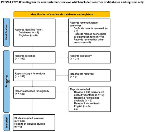
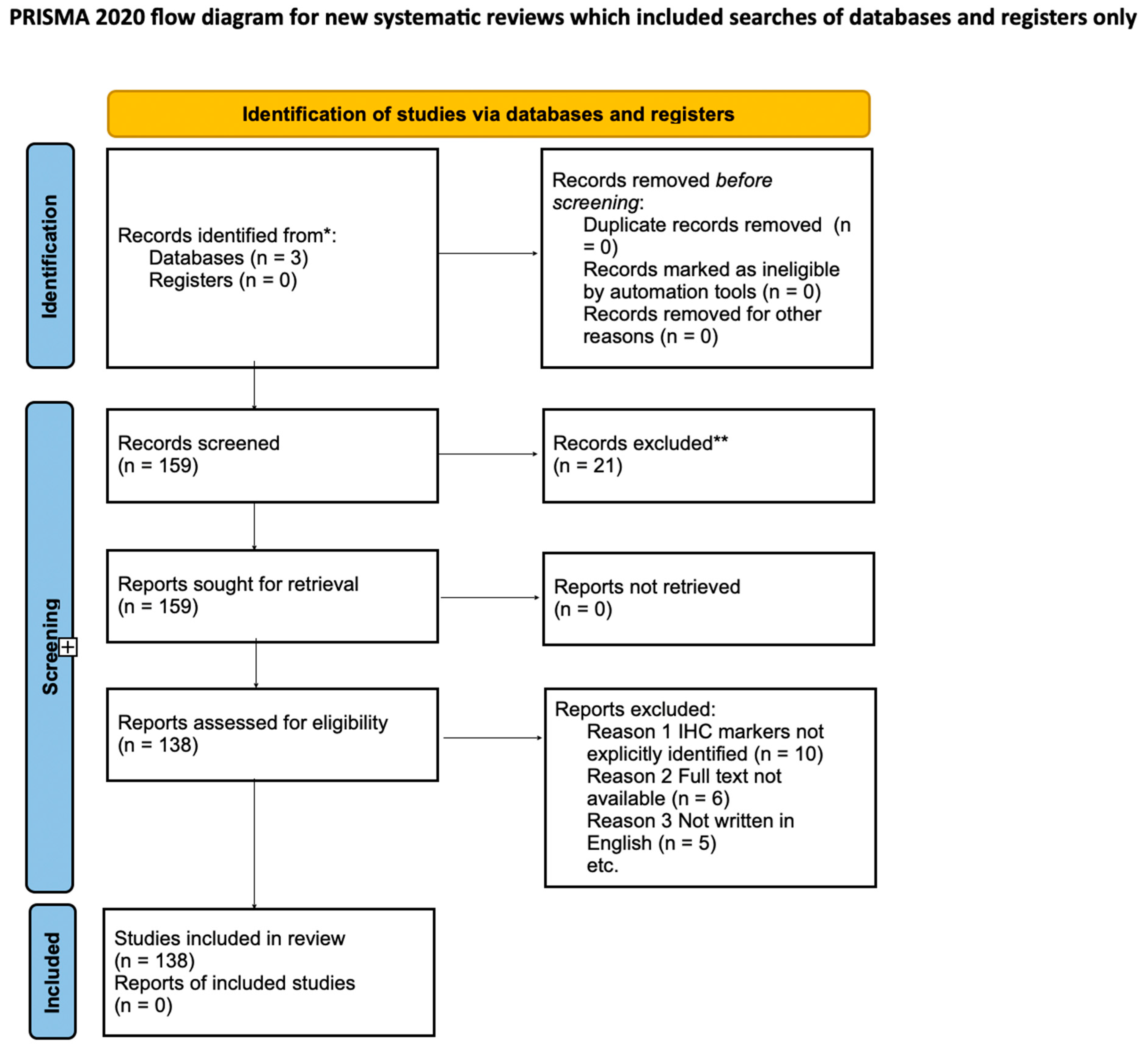

Disclaimer/Publisher’s Note: The statements, opinions and data contained in all publications are solely those of the individual author(s) and contributor(s) and not of MDPI and/or the editor(s). MDPI and/or the editor(s) disclaim responsibility for any injury to people or property resulting from any ideas, methods, instructions or products referred to in the content. |
© 2024 by the authors. Licensee MDPI, Basel, Switzerland. This article is an open access article distributed under the terms and conditions of the Creative Commons Attribution (CC BY) license (https://creativecommons.org/licenses/by/4.0/).