The Calculation and Evaluation of an Ultrasound-Estimated Fat Fraction in Non-Alcoholic Fatty Liver Disease and Metabolic-Associated Fatty Liver Disease
 , , ,
, , ,
Abstract
1. Introduction
2. Materials and Methods
2.1. Patient Selection
2.2. Ultrasound
2.3. Magnetic Resonance Imaging
2.4. Statistical Analysis
3. Results
3.1. Demographics and Characteristics of Patient Groups
3.2. Regression Models
3.3. Non-Linear Least Squares Models
3.4. Correlation between UEFF and MRI-PDFF
3.5. Comparison of UEFF between Steatosis Grades
3.6. Diagnosis of Different Grades of Steatosis with UEFF
4. Discussion
5. Conclusions
Author Contributions
Funding
Institutional Review Board Statement
Informed Consent Statement
Data Availability Statement
Acknowledgments
Conflicts of Interest
References
- Younossi, Z.M.; Golabi, P.; Paik, J.M.; Henry, A.; Van Dongen, C.; Henry, L. The Global Epidemiology of Nonalcoholic Fatty Liver Disease (NAFLD) and Nonalcoholic Steatohepatitis (NASH): A Systematic Review. Hepatology 2023, 77, 1335–1347. [Google Scholar] [CrossRef] [Scilit] [PubMed]
- European Association for the Study of the Liver (EASL); European Association for the Study of Diabetes (EASD); European Association for the Study of Obesity (EASO). EASL-EASD-EASO Clinical Practice Guidelines for the Management of Non-Alcoholic Fatty Liver Disease. J. Hepatol. 2016, 64, 1388–1402. [Google Scholar] [CrossRef] [Scilit] [PubMed]
- Eslam, M.; Newsome, P.N.; Sarin, S.K.; Anstee, Q.M.; Targher, G.; Romero-Gomez, M.; Zelber-Sagi, S.; Wai-Sun Wong, V.; Dufour, J.-F.; Schattenberg, J.M.; et al. A New Definition for Metabolic Dysfunction-Associated Fatty Liver Disease: An International Expert Consensus Statement. J. Hepatol. 2020, 73, 202–209. [Google Scholar] [CrossRef] [Scilit]
- Nalbantoglu, I.L.K.; Brunt, E.M. Role of Liver Biopsy in Nonalcoholic Fatty Liver Disease. World J. Gastroenterol. 2014, 20, 9026–9037. [Google Scholar] [CrossRef] [Scilit] [PubMed]
- Gu, J.; Liu, S.; Du, S.; Zhang, Q.; Xiao, J.; Dong, Q.; Xin, Y. Diagnostic Value of MRI-PDFF for Hepatic Steatosis in Patients with Non-Alcoholic Fatty Liver Disease: A Meta-Analysis. Eur. Radiol. 2019, 29, 3564–3573. [Google Scholar] [CrossRef] [Scilit] [PubMed]
- Ferraioli, G.; Berzigotti, A.; Barr, R.G.; Choi, B.I.; Cui, X.W.; Dong, Y.; Gilja, O.H.; Lee, J.Y.; Lee, D.H.; Moriyasu, F.; et al. Quantification of Liver Fat Content with Ultrasound: A WFUMB Position Paper. Ultrasound Med. Biol. 2021, 47, 2803–2820. [Google Scholar] [CrossRef] [Scilit]
- Zeng, K.-Y.; Bao, W.-Y.-G.; Wang, Y.-H.; Liao, M.; Yang, J.; Huang, J.-Y.; Lu, Q. Non-Invasive Evaluation of Liver Steatosis with Imaging Modalities: New Techniques and Applications. World J. Gastroenterol. 2023, 29, 2534–2550. [Google Scholar] [CrossRef] [Scilit]
- Zsombor, Z.; Rónaszéki, A.D.; Csongrády, B.; Stollmayer, R.; Budai, B.K.; Folhoffer, A.; Kalina, I.; Győri, G.; Bérczi, V.; Maurovich-Horvat, P.; et al. Evaluation of Artificial Intelligence-Calculated Hepatorenal Index for Diagnosing Mild and Moderate Hepatic Steatosis in Non-Alcoholic Fatty Liver Disease. Medicina 2023, 59, 469. [Google Scholar] [CrossRef] [Scilit]
- Jeon, S.K.; Lee, J.M.; Joo, I.; Park, S.J. Quantitative Ultrasound Radiofrequency Data Analysis for the Assessment of Hepatic Steatosis in Nonalcoholic Fatty Liver Disease Using Magnetic Resonance Imaging Proton Density Fat Fraction as the Reference Standard. Korean J. Radiol. 2021, 22, 1077–1086. [Google Scholar] [CrossRef] [Scilit]
- Rónaszéki, A.D.; Budai, B.K.; Csongrády, B.; Stollmayer, R.; Hagymási, K.; Werling, K.; Fodor, T.; Folhoffer, A.; Kalina, I.; Győri, G.; et al. Tissue Attenuation Imaging and Tissue Scatter Imaging for Quantitative Ultrasound Evaluation of Hepatic Steatosis. Medicine 2022, 101, e29708. [Google Scholar] [CrossRef] [Scilit]
- Şendur, H.N.; Özdemir Kalkan, D.; Cerit, M.N.; Kalkan, G.; Şendur, A.B.; Özhan Oktar, S. Hepatic Fat Quantification With Novel Ultrasound Based Techniques: A Diagnostic Performance Study Using Magnetic Resonance Imaging Proton Density Fat Fraction as Reference Standard. Can. Assoc. Radiol. J. 2023, 74, 362–369. [Google Scholar] [CrossRef] [Scilit] [PubMed]
- Ferraioli, G.; Kumar, V.; Ozturk, A.; Nam, K.; de Korte, C.L.; Barr, R.G. US Attenuation for Liver Fat Quantification: An AIUM-RSNA QIBA Pulse-Echo Quantitative Ultrasound Initiative. Radiology 2022, 302, 495–506. [Google Scholar] [CrossRef] [Scilit] [PubMed]
- Wear, K.A.; Han, A.; Rubin, J.M.; Gao, J.; Lavarello, R.; Cloutier, G.; Bamber, J.; Tuthill, T. US Backscatter for Liver Fat Quantification: An AIUM-RSNA QIBA Pulse-Echo Quantitative Ultrasound Initiative. Radiology 2022, 305, 526–537. [Google Scholar] [CrossRef] [Scilit] [PubMed]
- Jeon, S.K.; Joo, I.; Kim, S.Y.; Jang, J.K.; Park, J.; Park, H.S.; Lee, E.S.; Lee, J.M. Quantitative Ultrasound Radiofrequency Data Analysis for the Assessment of Hepatic Steatosis Using the Controlled Attenuation Parameter as a Reference Standard. Ultrasonography 2021, 40, 136–146. [Google Scholar] [CrossRef] [Scilit] [PubMed]
- Abbey, C.K.; Nguyen, N.Q.; Insana, M.F. Effects of Frequency and Bandwidth on Diagnostic Information Transfer in Ultrasonic B-Mode Imaging. IEEE Trans. Ultrason. Ferroelectr. Freq. Control 2012, 59, 1115–1126. [Google Scholar] [CrossRef] [Scilit] [PubMed]
- Ferraioli, G.; Raimondi, A.; Maiocchi, L.; De Silvestri, A.; Poma, G.; Kumar, V.; Barr, R.G. Liver Fat Quantification With Ultrasound: Depth Dependence of Attenuation Coefficient. J. Ultrasound Med. 2023, 42, 2247–2255. [Google Scholar] [CrossRef] [Scilit]
- Labyed, Y.; Milkowski, A. Novel Method for Ultrasound-Derived Fat Fraction Using an Integrated Phantom. J. Ultrasound Med. 2020, 39, 2427–2438. [Google Scholar] [CrossRef] [Scilit]
- Han, A.; Zhang, Y.N.; Boehringer, A.S.; Montes, V.; Andre, M.P.; Erdman, J.W.; Loomba, R.; Valasek, M.A.; Sirlin, C.B.; O’Brien, W.D. Assessment of Hepatic Steatosis in Nonalcoholic Fatty Liver Disease by Using Quantitative US. Radiology 2020, 295, 106–113. [Google Scholar] [CrossRef] [Scilit]
- Kaposi, P.N.; Unger, Z.; Fejér, B.; Kucsa, A.; Tóth, A.; Folhoffer, A.; Szalay, F.; Bérczi, V. Interobserver Agreement and Diagnostic Accuracy of Shearwave Elastography for the Staging of Hepatitis C Virus-Associated Liver Fibrosis. J. Clin. Ultrasound 2019, 48, 67–74. [Google Scholar] [CrossRef] [Scilit] [PubMed]
- Barr, R.G.; Wilson, S.R.; Rubens, D.; Garcia-Tsao, G.; Ferraioli, G. Update to the Society of Radiologists in Ultrasound Liver Elastography Consensus Statement. Radiology 2020, 296, 263–274. [Google Scholar] [CrossRef] [Scilit] [PubMed]
- Orcel, T.; Chau, H.T.; Turlin, B.; Chaigneau, J.; Bannier, E.; Otal, P.; Frampas, E.; Leguen, A.; Boulic, A.; Saint-Jalmes, H.; et al. Evaluation of Proton Density Fat Fraction (PDFF) Obtained from a Vendor-Neutral MRI Sequence and MRQuantif Software. Eur. Radiol. 2023; Epub ahead of print. [CrossRef] [Scilit]
- Hamilton, G.; Yokoo, T.; Bydder, M.; Cruite, I.; Schroeder, M.E.; Sirlin, C.B.; Middleton, M.S. In Vivo Characterization of the Liver Fat 1H MR Spectrum. NMR Biomed. 2011, 24, 784–790. [Google Scholar] [CrossRef] [Scilit] [PubMed]
- Jung, J.; Han, A.; Madamba, E.; Bettencourt, R.; Loomba, R.R.; Boehringer, A.S.; Andre, M.P.; Erdman, J.W.; O’Brien, W.D.; Fowler, K.J.; et al. Direct Comparison of Quantitative US versus Controlled Attenuation Parameter for Liver Fat Assessment Using MRI Proton Density Fat Fraction as the Reference Standard in Patients Suspected of Having NAFLD. Radiology 2022, 304, 75–82. [Google Scholar] [CrossRef] [Scilit] [PubMed]
- Han, A.; Byra, M.; Heba, E.; Andre, M.P.; Erdman, J.W.; Loomba, R.; Sirlin, C.B.; O’Brien, W.D. Noninvasive Diagnosis of Nonalcoholic Fatty Liver Disease and Quantification of Liver Fat with Radiofrequency Ultrasound Data Using One-Dimensional Convolutional Neural Networks. Radiology 2020, 295, 342–350. [Google Scholar] [CrossRef] [Scilit] [PubMed]
- Jeon, S.K.; Lee, J.M.; Joo, I.; Yoon, J.H.; Lee, G. Two-Dimensional Convolutional Neural Network Using Quantitative US for Noninvasive Assessment of Hepatic Steatosis in NAFLD. Radiology 2023, 307, e221510. [Google Scholar] [CrossRef] [Scilit] [PubMed]
- An, Z.; Liu, Q.; Zeng, W.; Wang, Y.; Zhang, Q.; Pei, H.; Xin, X.; Yang, S.; Lu, F.; Zhao, Y.; et al. Relationship between Controlled Attenuated Parameter and Magnetic Resonance Imaging-Proton Density Fat Fraction for Evaluating Hepatic Steatosis in Patients with NAFLD. Hepatol. Commun. 2022, 6, 1975–1986. [Google Scholar] [CrossRef] [Scilit] [PubMed]
- Caussy, C.; Alquiraish, M.H.; Nguyen, P.; Hernandez, C.; Cepin, S.; Fortney, L.E.; Ajmera, V.; Bettencourt, R.; Collier, S.; Hooker, J.; et al. Optimal Threshold of Controlled Attenuation Parameter with MRI-PDFF as the Gold Standard for the Detection of Hepatic Steatosis. Hepatology 2018, 67, 1348–1359. [Google Scholar] [CrossRef] [Scilit]
- Tamaki, N.; Ajmera, V.; Loomba, R. Non-Invasive Methods for Imaging Hepatic Steatosis and Their Clinical Importance in NAFLD. Nat. Rev. Endocrinol. 2022, 18, 55–66. [Google Scholar] [CrossRef] [Scilit]


| NAFLD (n = 90) | MAFLD (n = 51) | |
|---|---|---|
| Sex | ||
| female (%) | 44 (48.9%) | 26 (51.0%) |
| male (%) | 46 (51.1%) | 25 (49.0%) |
| Age (years) | ||
| mean (±SD) | 55.1 (13.3) | 54.3 (13.4) |
| range | 23–78 | 21–82 |
| BMI (kg/m2) | ||
| mean (±SD) | 29.3 (4.4) | 26.6 (3.9) |
| range | 18.6–45.3 | 19.7–35.3 |
| T2DM (%) | 24 (35.3%) | 1 (2.7%) |
| Etiology of chronic liver disease (%) | ||
| AIH | 5 (9.8%) | |
| alcoholic | 3 (5.9%) | |
| chr. HBV | 3 (5.9%) | |
| chr. HCV | 7 (13.7%) | |
| hemochr. | 6 (11.8%) | |
| hepatotoxic med. | 15 (29.4%) | |
| PBC/PSC | 8 (15.7%) | |
| Wilson’s disease | 4 (7.8%) | |
| none | 91 (100%) | |
| NAFLD (n = 90) | MAFLD (n = 51) | |
|---|---|---|
| AC (dB/cm/Mhz) | ||
| mean ± SD | 0.83 ± 0.15 | 0.73 ± 0.17 |
| range | 0.55–1.22 | 0.49–1.26 |
| BSC-D (arbitrary unit) | ||
| mean ± SD | 100.54 ± 9.6 | 92.68 ± 11.87 |
| range | 60–116.62 | 62.52–110.38 |
| LS (kPa) | ||
| mean ± SD | 7.24 ± 3.2 | 11.26 ± 8.9 |
| range | 3.2–21.8 | 3.9–39.7 |
| MRI-PDFF (%) | ||
| mean ± SD | 12.1 ± 8.86 | 7.8 ± 7.7 |
| range | 0.2–44.6 | 0.4–30.1 |
| UEFF (%) 1 | ||
| mean ± SD | 12.06 ± 7.23 | 7.08 ± 7.86 |
| range | −1.25–35.3 | −1.99–37.03 |
| Steatosis grade 2 | ||
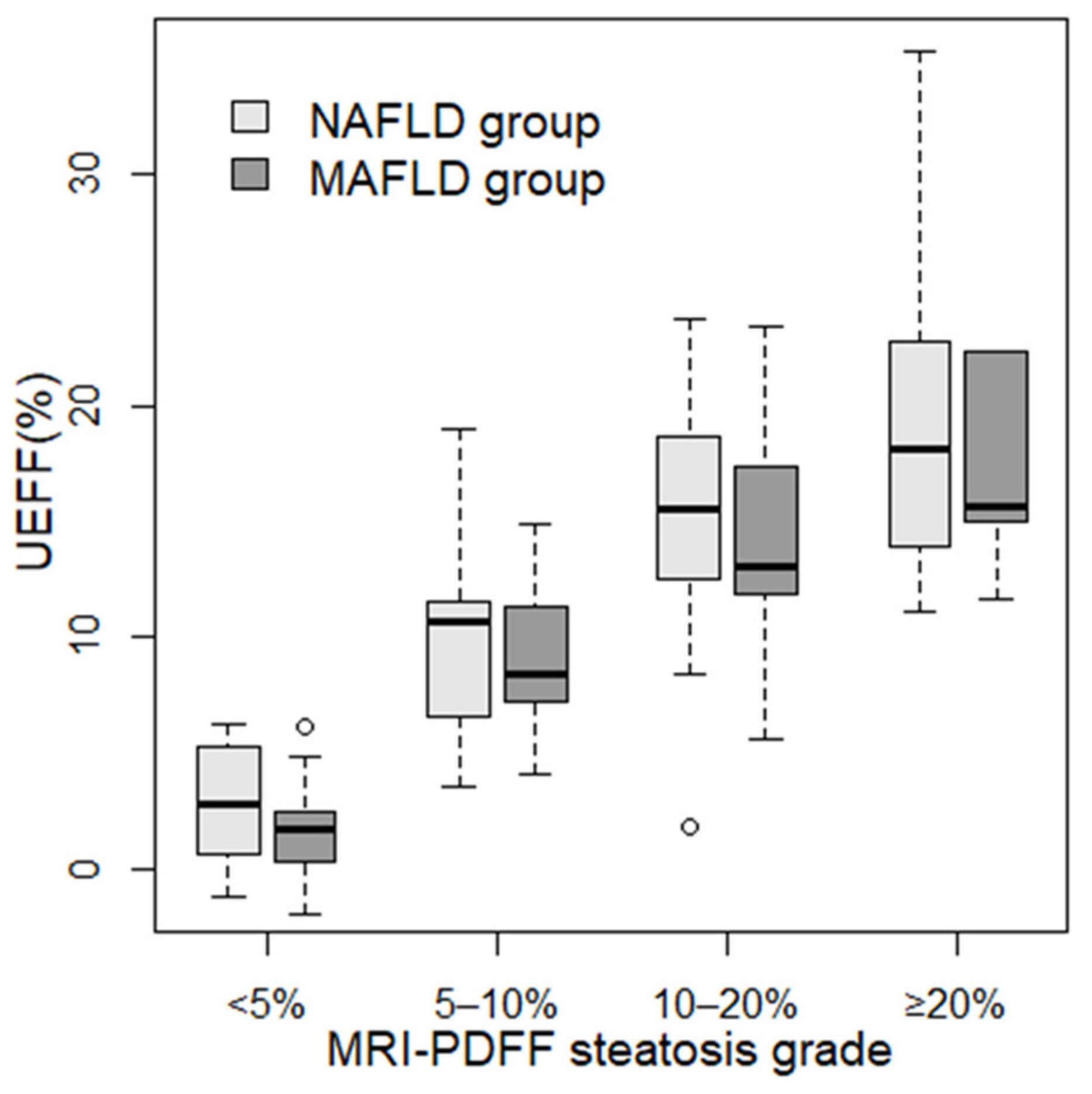
| <5% | 19 (21.1%) | 28 (54.9%) |
| 5–10% | 22 (24.4%) | 8 (15.7%) |
| 10–20% | 34 (37.8%) | 10 (19.6%) |
| ≥20% | 15 (16.7%) | 5 (9.8%) |
| Fibrosis grade 3 | ||
| F0/F1 | 12 (13.3%) | 5 (9.8%) |
| F2 | 64 (71.1%) | 25 (49.1%) |
| F3 | 8 (8.9%) | 12 (23.5%) |
| F4 | 6 (6.7%) | 9 (17.6%) |
| Univariable Analysis | Multivariable Analysis | |||||||
|---|---|---|---|---|---|---|---|---|
| adj. R2 | F Stat. | ß | p-Value | adj. R2 | F Stat. | ß | p-Value | |
| NAFLD | ||||||||
| AC | 0.504 | 91.47 | 43.04 | <0.001 | 0.485 | 18.65 | 36.16 | <0.001 |
| BSC-D | 0.31 | 14.01 | 0.52 | <0.001 | 0.485 | 18.65 | 0.18 | 0.118 |
| LS | 0.0009 | 1.08 | 0.3 | 0.3 | 0.485 | 18.65 | 0.06 | 0.781 |
| BMI | 0.078 | 7.36 | 0.63 | 0.008 | 0.485 | 18.65 | 0.07 | 0.696 |
| MAFLD | ||||||||
| AC | 0.748 | 149.2 | 40.19 | <0.001 | 0.775 | 34.49 | 44.82 | <0.001 |
| BSC-D | 0.235 | 16.38 | 0.32 | <0.001 | 0.775 | 34.49 | 0.07 | 0.298 |
| LS | 0.049 | 3.57 | −0.22 | 0.065 | 0.775 | 34.49 | 0.05 | 0.521 |
| BMI | 0.23 | 12.65 | 1.07 | 0.001 | 0.775 | 34.49 | −0.21 | 0.38 |
| NAFLD Group 1 | MAFLD Group 2 | |||||||
|---|---|---|---|---|---|---|---|---|
| Model | adj. R2 | RMSE | Cor. Coef. | p-Value | adj. R2 | RMSE | Cor. Coef. | p-Value |
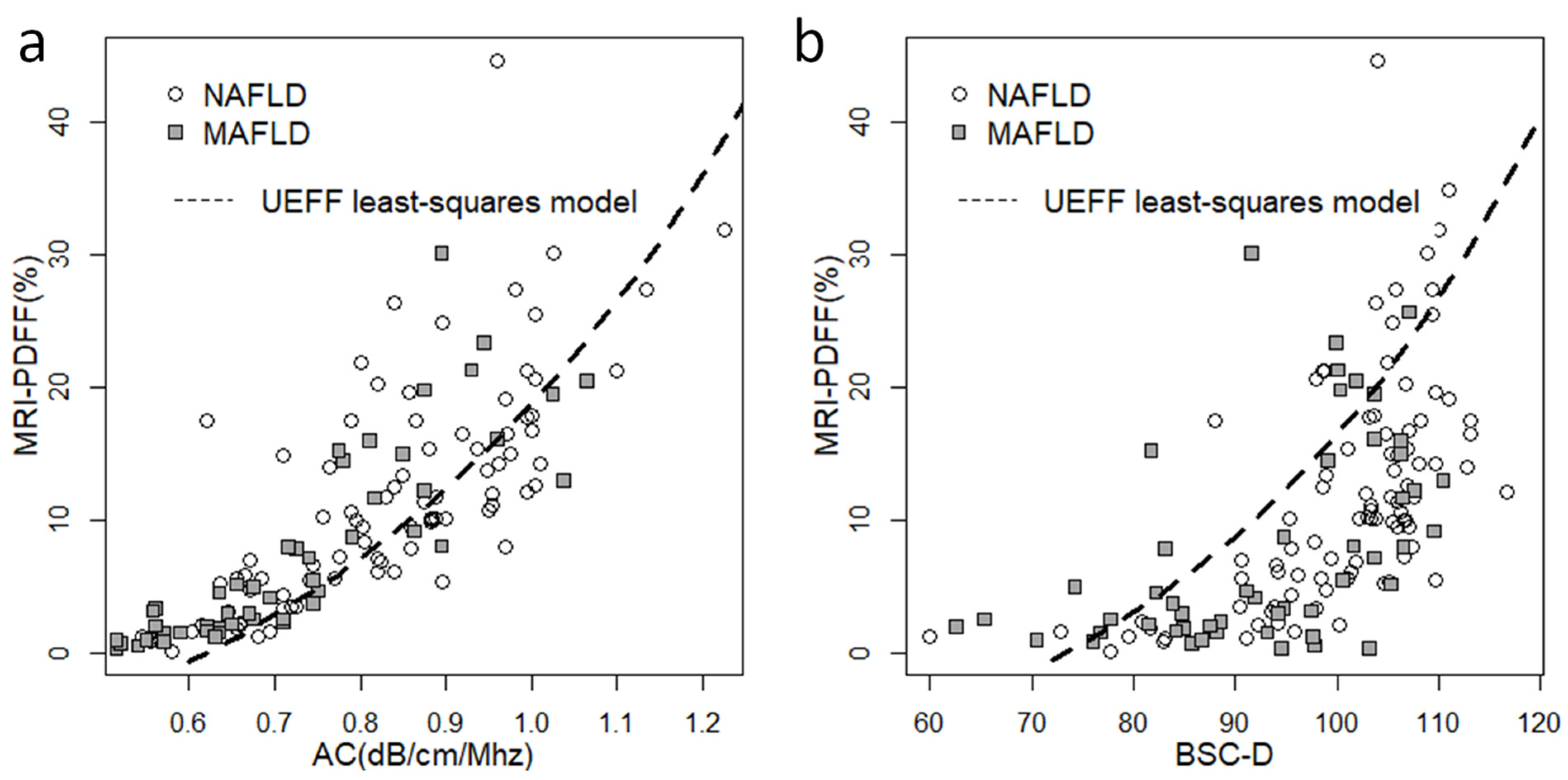
| AC | 0.498 ± 0.046 | 6.415 ± 0.561 | 0.716 0.6–0.8 | <0.001 | 0.733 | 3.911 | 0.859 0.76–0.92 | <0.001 |
| BSC-D | 0.318 ± 0.075 | 7.687 ± 0.696 | 0.581 0.43–0.7 | <0.001 | 0.253 | 6.608 | 0.518 0.28–0.69 | <0.001 |
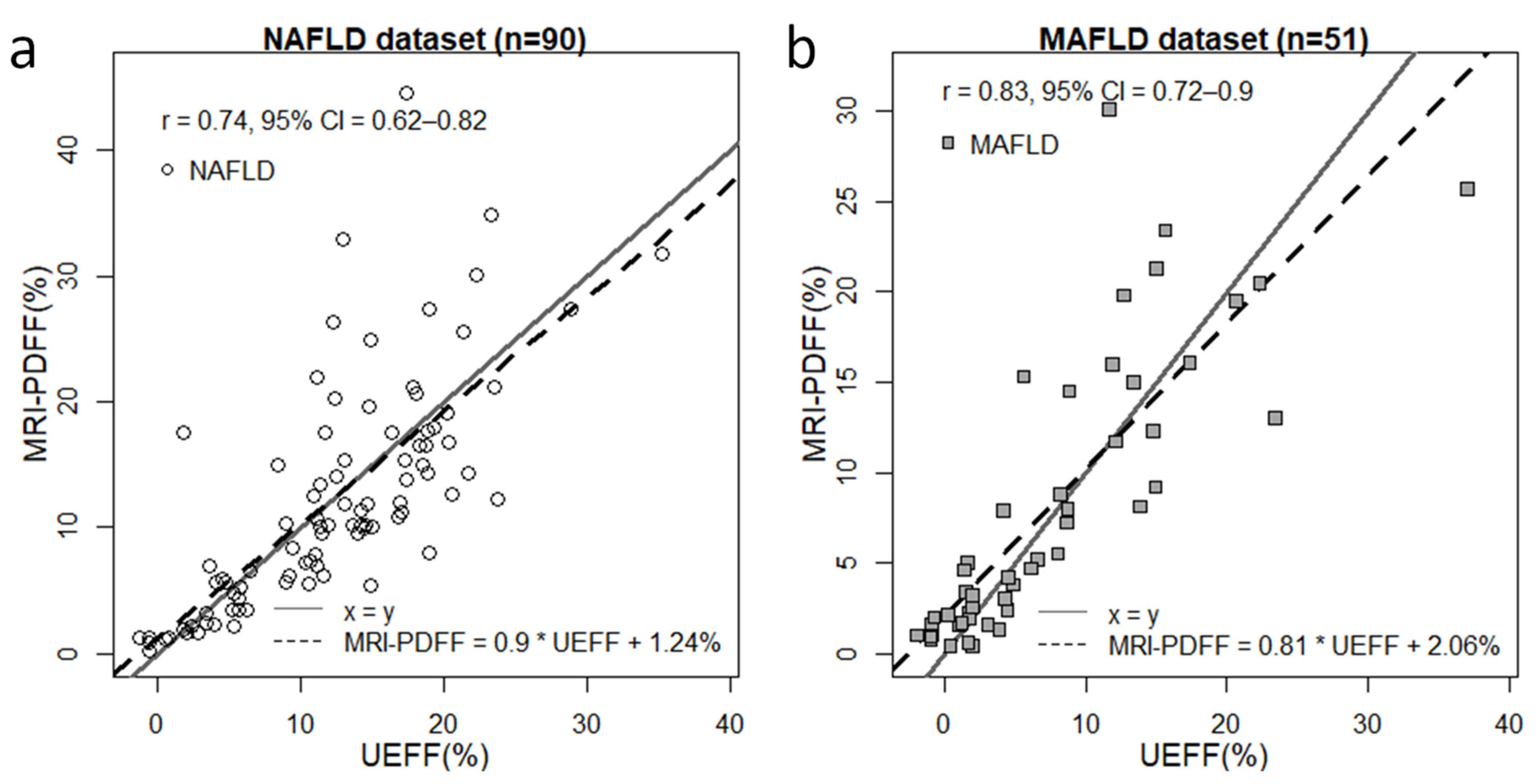
| AC + BSC-D | 0.446 ± 0.152 | 7.347 ± 3.105 | 0.735 0.62–0.82 | <0.001 | 0.673 | 4.574 | 0.828 0.72–0.9 | <0.001 |
| <5% vs. ≥5% MRI-PDFF | 10% vs. ≥10% MRI-PDFF | |||
|---|---|---|---|---|
| NAFLD Group | MAFLD Group | NAFLD Group | MAFLD Group | |
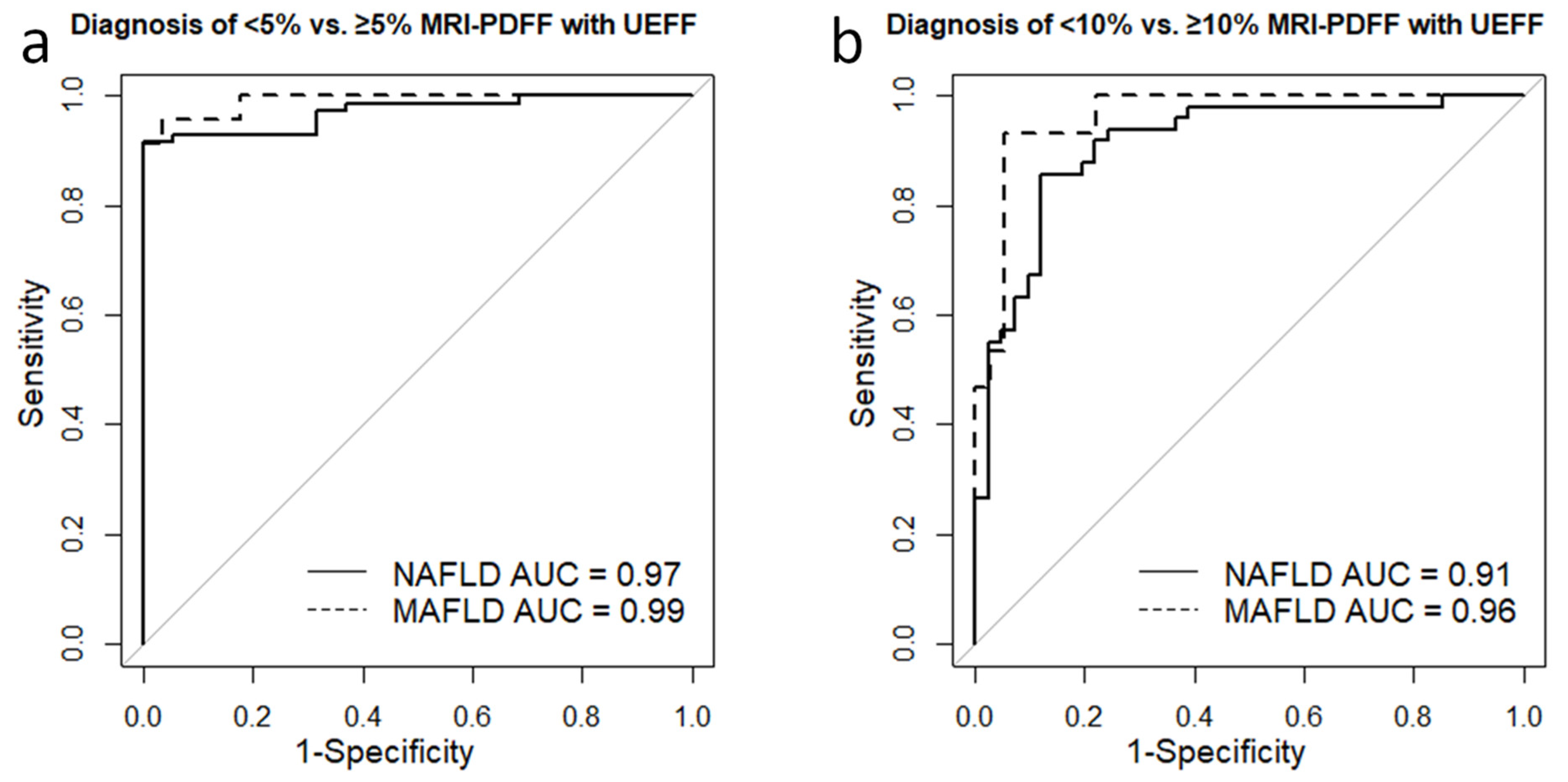
| Cutoff | 6.36% | 5.22% | 11.64% | 8.74% |
| AUC | 0.97 | 0.99 | 0.91 | 0.96 |
| SN | 0.92 | 0.96 | 0.86 | 0.93 |
| SP | 1 | 0.96 | 0.88 | 0.94 |
| PPV | 1 | 0.96 | 0.89 | 0.88 |
| NPV | 0.76 | 0.96 | 0.84 | 0.97 |
| TA | 0.93 | 0.96 | 0.87 | 0.94 |
| Power | 1 | 1 | 1 | 1 |
Disclaimer/Publisher’s Note: The statements, opinions and data contained in all publications are solely those of the individual author(s) and contributor(s) and not of MDPI and/or the editor(s). MDPI and/or the editor(s) disclaim responsibility for any injury to people or property resulting from any ideas, methods, instructions or products referred to in the content. |
© 2023 by the authors. Licensee MDPI, Basel, Switzerland. This article is an open access article distributed under the terms and conditions of the Creative Commons Attribution (CC BY) license (https://creativecommons.org/licenses/by/4.0/).
Share and Cite
Kaposi, P.N.; Zsombor, Z.; Rónaszéki, A.D.; Budai, B.K.; Csongrády, B.; Stollmayer, R.; Kalina, I.; Győri, G.; Bérczi, V.; Werling, K.; et al. The Calculation and Evaluation of an Ultrasound-Estimated Fat Fraction in Non-Alcoholic Fatty Liver Disease and Metabolic-Associated Fatty Liver Disease. Diagnostics 2023, 13, 3353. https://doi.org/10.3390/diagnostics13213353
Kaposi PN, Zsombor Z, Rónaszéki AD, Budai BK, Csongrády B, Stollmayer R, Kalina I, Győri G, Bérczi V, Werling K, et al. The Calculation and Evaluation of an Ultrasound-Estimated Fat Fraction in Non-Alcoholic Fatty Liver Disease and Metabolic-Associated Fatty Liver Disease. Diagnostics. 2023; 13(21):3353. https://doi.org/10.3390/diagnostics13213353
Chicago/Turabian StyleKaposi, Pál Novák, Zita Zsombor, Aladár D. Rónaszéki, Bettina K. Budai, Barbara Csongrády, Róbert Stollmayer, Ildikó Kalina, Gabriella Győri, Viktor Bérczi, Klára Werling, and et al. 2023. "The Calculation and Evaluation of an Ultrasound-Estimated Fat Fraction in Non-Alcoholic Fatty Liver Disease and Metabolic-Associated Fatty Liver Disease" Diagnostics 13, no. 21: 3353. https://doi.org/10.3390/diagnostics13213353
APA StyleKaposi, P. N., Zsombor, Z., Rónaszéki, A. D., Budai, B. K., Csongrády, B., Stollmayer, R., Kalina, I., Győri, G., Bérczi, V., Werling, K., Maurovich-Horvat, P., Folhoffer, A., & Hagymási, K. (2023). The Calculation and Evaluation of an Ultrasound-Estimated Fat Fraction in Non-Alcoholic Fatty Liver Disease and Metabolic-Associated Fatty Liver Disease. Diagnostics, 13(21), 3353. https://doi.org/10.3390/diagnostics13213353