


Abstract
Arginase-1 catalyzes the conversion of arginine to ornithine and urea. Because of its predominant expression in hepatocytes, it serves as a marker for hepatocellular carcinoma, although other tumor entities can also express arginase-1. To comprehensively determine arginase-1 expression in normal and neoplastic tissues, tissue microarrays containing 14,912 samples from 117 different tumor types and 608 samples of 76 different normal tissue types were analyzed by immunohistochemistry. In normal tissues, arginase-1 was expressed in the liver, the granular layer of the epidermis, and in granulocytes. Among tumors, a nuclear and cytoplasmic arginase-1 immunostaining was predominantly observed in hepatocellular carcinoma, where 96% of 49 cancers were at least moderately positive. Although 22 additional tumor categories showed occasional arginase immunostaining, strong staining was exceedingly rare in these entities. Staining of a few tumor cells was observed in squamous cell carcinomas of various sites. Staining typically involved maturing cells with the beginning of keratinization in these tumors and was significantly associated with a low grade in 635 squamous cell carcinomas of various sites (p = 0.003). Teratoma, urothelial carcinoma and pleomorphic adenomas sometimes also showed arginase expression in areas with squamous differentiation. In summary, arginase-1 immunohistochemistry is highly sensitive and specific for hepatocellular carcinoma if weak and focal staining is disregarded.
1. Introduction
Arginase-1 is encoded by the ARG1 gene located at 6q23. It acts as a cytosolic manganese-dependent enzyme that catalyzes the conversion of arginine to ornithine and urea in the final step of the urea cycle [1,2,3,4]. Among normal tissues, it is predominantly expressed in hepatocytes and inflammatory cells. Because arginase-1 expression is usually retained in hepatocellular carcinoma, a cancer derived from hepatocytes, the immunohistochemical detection of arginase expression is commonly used to support the difficult distinction of hepatocellular carcinoma from cholangiocellular carcinoma and metastases to the liver [5]. This procedure is supported by more than 20 studies demonstrating arginase-1 expression in 80–100% of hepatocellular carcinomas [6,7,8,9,10,11,12,13,14,15,16,17,18,19,20,21,22,23,24,25,26].
Multiple studies have suggested that arginase-1 expression of tumor cells is largely absent in other important tumor types, such as renal cell carcinomas [8,25], ductal adenocarcinoma of the pancreas [8,21], gastric adenocarcinoma [25], esophageal adenocarcinoma [25], adenocarcinoma of the lung [25], and in lobular breast cancer [25]. The extent to which arginase-1 expression is specific for hepatocellular carcinoma is still unclear, however. Studies have demonstrated arginase-1 expression in 0–7% of prostate cancer [25,26,27], 6% of adenocarcinomas of the ampulla Vateri [28], 84% of squamous cell carcinoma of the oral cavity and the larynx [14], and “high” expression in 47% of the 79 analyzed invasive breast carcinomas of no special type (NST) [29]. In one study, multiple soft tissue tumors were described to show arginase-1 expression in up to 100% of cases [30]. The published arginase-1 positivity rates are also markedly variable in tumor entities that are often seen in the liver such as cholangiocarcinoma (positivity described in 0–68% of cases, [8,19,24,25,26,31], breast cancer NST (0–47%), [8,18,26,29]), colorectal adenocarcinoma (0–100%) [8,26,32,33] and even hepatocellular carcinoma 45–100%, [6,8,9,11,12,22,26]. This data variability is most likely due to the use of different antibodies, staining protocols and criteria for staining interpretation in the respective studies.
To better understand the prevalence and diagnostic utility of arginase-1 expression in cancer, a comprehensive study analyzing large numbers of neoplastic and non-neoplastic tissues under highly standardized conditions is desirable. For this purpose, arginase-1 expression was analyzed in more than 14,000 tumor tissue samples from 117 different tumor types and subtypes as well as 76 non-neoplastic tissue categories by immunohistochemistry (IHC) in a tissue microarray (TMA) format in this study.
2. Materials and Methods
Tissue Microarrays (TMAs). Our normal tissue TMA was composed of 8 samples from 8 different donors for each of the 76 different normal tissue types (608 samples on one slide). The cancer TMAs contained a total of 14,912 primary tumors from 117 tumor types and subtypes. The composition of normal and tumor TMAs is described in the results section. All samples were obtained from the archives of the Institutes of Pathology, University Hospital of Hamburg, Germany, the Institute of Pathology, Clinical Center Osnabrueck, Germany, and Department of Pathology, Academic Hospital Fuerth, Germany. Tissues were fixed in 4% buffered formalin and then embedded in paraffin. The TMA manufacturing process was described earlier in detail [34,35,36]. In brief, one tissue spot (diameter: 0.6 mm) was transferred from a cancer containing donor block to an empty recipient paraffin block. The use of archived remnants of diagnostic tissues for TMA manufacturing, their analysis for research purposes, and patient data were conducted according to local laws (HmbKHG, §12) and the analysis had been approved by the local ethics committee (Ethics commission Hamburg, WF-049/09). All work has been carried out in compliance with the Helsinki Declaration.
Immunohistochemistry (IHC). Freshly cut TMA sections were all immunostained on one day and in one experiment. Slides were deparaffinized and exposed to heat-induced antigen retrieval for 5 min in an autoclave at 121 °C in a pH 7.8 TRIS-EDTA-citrate buffer. The primary antibody specific to arginase-1 (rabbit recombinant, MSVA-511R, MS Validated Antibodies, GmbH, Hamburg, Germany) was applied at 37 °C for 60 min at a dilution of 1: 150 in antibody diluent from Agilent/Dako #S080938. The bound antibody was then visualized using the EnVision Kit (Agilent/Dako #K5007) according to the manufacturer’s directions. For tumor tissues, the percentage of positive neoplastic cells was estimated, and the staining intensity was semi-quantitatively recorded (0, 1+, 2+, 3+). For statistical analyses, the staining results were categorized into four groups. Tumors without any staining were considered negative. Tumors with 1+ staining intensity in ≤70% of cells or 2+ intensity in ≤30% of cells were considered weakly positive. Tumors with 1+ staining intensity in >70% of cells, 2+ intensity in 31–70%, or 3+ intensity in ≤30% were considered moderately positive. Tumors with 2+ intensity in >70% or 3+ intensity in >30% of cells were considered strongly positive. For antibody validation, the normal tissue TMA was also stained with a second anti-arginase-1 antibody (Cell Marque clone SP156, Cat. # 380R-18) for 20 min, at a dilution of 1:6.25 in a Dako Link48 autostainer after Flex-high antigen retrieval.
Statistics. Statistical calculations were performed with JMP 14 software (SAS Institute Inc., Cary, NC 27513, USA). The chi²-test was performed to search for associations between arginase immunostaining and a tumor phenotype in squamous cell cancers.
3. Results
3.1. Technical Issues
A total of 12,047 (81%) of 14,912 tumor samples were interpretable in our TMA analysis. Non-interpretable samples demonstrated a lack of unequivocal tumor cells or loss of tissue location during technical procedures. Sufficient numbers of samples of each normal tissue type were evaluable.
3.2. Staining Pattern in Normal Tissues
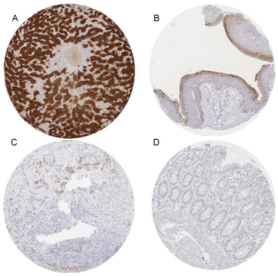
The Arginase-1 immunostaining was typically cytoplasmic and nuclear. By far the strongest Arginase-1 immunostaining was seen in hepatocytes. Moderate to strong cytoplasmic and nuclear arginase immunostaining also occurred in the granular cell layer of keratinizing squamous epithelium of the skin. Moderate staining occurred in granulocytes and its precursor cells in the bone marrow. A weak to moderate Arginase-1 positivity was seen in a fraction of the decidua cells. Representative images are shown in Figure 1. Arginase-1 immunostaining was not observed in any other epithelial cells from the gastrointestinal tract, urothelium, non-keratinizing squamous epithelia, pancreas, salivary glands, thyroid, parathyroid gland, adenohypophysis, adrenal gland, prostate, epididymis, testis, seminal vesicle, endometrium, endocervix, fallopian tube, kidney, respiratory epithelium, lung, placenta, various types of muscle cells, myometrium, lymphatic organs, endothelium, brain and neurohypophysis. All positive stainings were also confirmed by the use of a second independent antibody (Cell Marque clone SP156, Supplementary Materials Figure S1).
Figure 1.
Arginase-1 immunostaining in normal cells. The panels show a strong cytoplasmic and nuclear arginase-1 positivity of hepatocytes (A). Hepatocyte staining is strong enough that some staining is also seen in the adjacent stroma (contamination artifact). A weak to moderate cytoplasmic and nuclear arginase-1 immunostaining occurs in the granular cell layer of the keratinizing squamous epithelium of the skin (B) while staining is weak and cytoplasmic in granulocytes infiltrating an arginase-1 negative cholangiocellular carcinoma (C). Arginase-1 immunostaining is absent in colon epithelium (D). Magnification 100×, TMA spot size 600 µm.
3.3. Arginase in Cancer
Arginase expression was predominantly observed in hepatocellular carcinoma, where 88% of 49 tumors showed a strong arginase positivity and 96% a moderate arginase positivity independently from the tumor stage (p = 0.4132). A characteristic nuclear and cytoplasmic staining was typically seen in these tumors. Although 22 additional tumor categories showed arginase immunostaining in a much smaller fraction of cases, strong and even moderate arginase-1 staining was exceedingly rare in these entities (Table 1).
Table 1.
Arginase-1 immunostaining in human tumors.
The focal staining of a few tumor cells was observed in squamous cell carcinomas for various sites, where it was significantly associated with a low histological tumor grade (p = 0.003, Table 2).
Table 2.
Arginase-1 immunostaining and tumor phenotype in 635 squamous cell carcinomas (SQCC) of various origins, including SQCC of the floor of the mouth (n = 99), pharynx (n = 40), larynx (n = 86), cervix (n = 116), vagina (n = 36), vulva (n = 106), penis (n = 59), skin (n = 45), and anal canal (n = 48).
Arginase-1 staining was unrelated to HPV-status, however (Table 3).
Table 3.
Arginase-1 immunostaining and HPV status in squamous cell carcinomas.
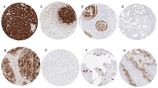
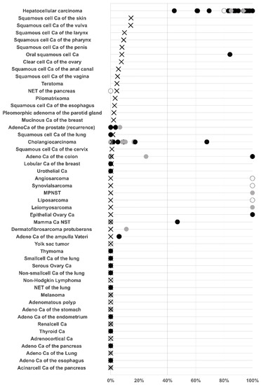
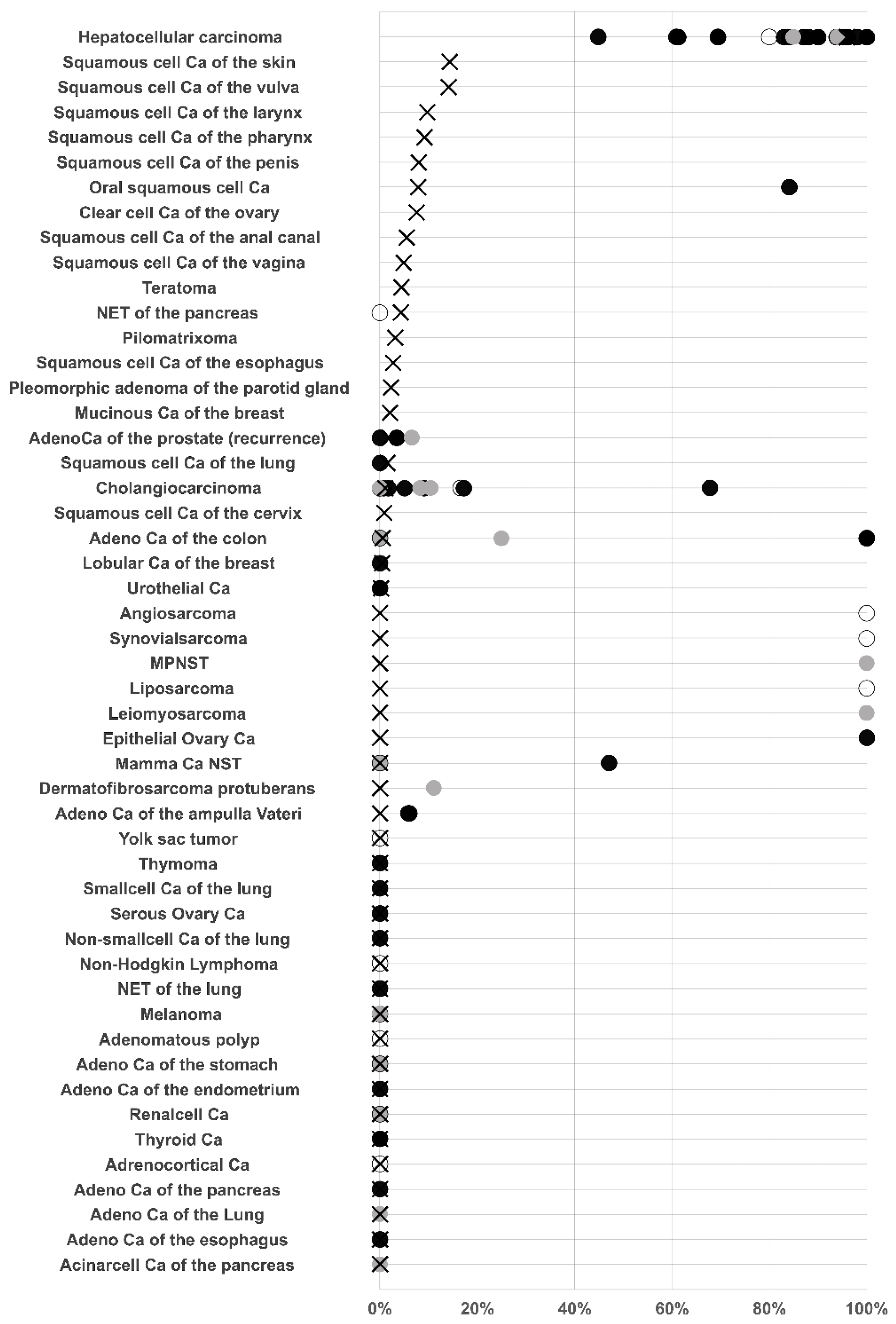
Similarly to arginase expression of normal squamous epithelium, arginase positivity typically involved maturing cells at the beginning of keratinization in these tumors. Rare positive cases of teratoma, urothelial carcinoma and pleomorphic adenomas also showed arginase expression in areas with squamous differentiation. Other tumor entities with occasional and mostly low-level arginase-1 immunostaining included clear cell carcinomas of the ovary, neuroendocrine tumors of the pancreas, mucinous and lobular carcinoma of the breast, cholangiocarcinoma, and colorectal adenocarcinoma. In rare case of arginase positive colorectal and mucinous breast carcinomas, arginase staining predominated in the intratumoral mucus (breast) or mucin producing goblet cells (colon). Representative images of arginase immunostaining in cancers are presented in Figure 2. A ranking order of arginase-1 positive and strongly positive cases in combination, with a summary of data from comparable studies, is provided in Figure 3.
Figure 2.
Arginase-1 immunostaining in cancer. The panels show examples of a strong (A) and a more variable, moderate to strong (B) nuclear and cytoplasmic arginase-1 staining in hepatocellular carcinomas. A weak and purely cytoplasmic arginase-1 staining in a cholangiocellular carcinoma (C) which is particularly seen in cells adjacent to strongly positive normal hepatocytes may reflect a “contamination” artifact. In another cholangiocellular carcinoma, tumor cells are arginase-1 negative, and staining is limited to tumor-associated granulocytes (D). A focal arginase-1 immunostaining is seen in keratinizing cells of a pharyngeal squamous cell carcinoma (E), a clear cell carcinoma of the ovary (F), and a Gleason 4 + 4 = 8 adenocarcinoma of the prostate (G). In a colorectal adenocarcinoma arginase-1 staining occurs in goblet cells and tumor-associated mucins (H). Magnification 100×, TMA spot size 600 µm.
Figure 3.
Graphical comparison of Arginase data from this study (x) in comparison with the previous literature (dots). Open: n = 1–10, grey: n = 11–25, black: n > 25. For comparison purposes, studies that did not differentiate between different tumor subtypes were marked with black dots and the overall positivity rate was applied to the different tumor subtypes present in our tumor microarrays. All studies are referred to in the reference list.
4. Discussion
Given the large scale of our study, we placed emphasis on the thorough validation of our assay. The International Working Group for Antibody Validation (IWGAV) proposed that antibody validation for immunohistochemistry on formalin fixed tissues should include either a comparison of the findings obtained by two different independent antibodies, or a comparison with expression data obtained by another independent method [37]. To ensure that any antibody cross reactivity would be detected in our validation experiment, a broad range of different normal tissue categories were included in the analysis and the immunohistochemical staining results were not only compared with a second independent antibody, but also with RNA expression data derived from three independent RNA screening studies, including the Human Protein Atlas (HPA) RNA-seq tissue dataset [38], the FANTOM5 project [39,40], and the Genotype-Tissue Expression (GTEx) project [41]. The 76 different normal tissues that were selected for this experiment are likely to contain the majority of proteins occurring in the cells of adult humans. Therefore, we consider it likely that undesired antibody cross-reactivity can be detected with high certainty. A specific antibody reactivity in our experimental set-up is supported by the detection of significant arginase-1 immunostaining in all organs, with documented arginase-1 RNA expression (liver, skin, bone marrow, and granulocytes). The fact that RNA expression had not previously been documented for decidua cells is not surprising, given the paucity of these cells in mature placenta tissue samples that were systematically screened for RNA expression. True arginase-1 expression in decidua cells is, however, supported by the comparison with the independent antibody Cell Marque clone SP156, which also confirmed liver, skin, bone marrow and granulocyte staining (Supplementary Materials Figure S1).
A successful analysis of 12,047 cancers from 117 different tumor entities revealed a pattern of expression for arginase-1 that strongly correlated with the findings in normal tissues. Additionally, Arginase-1 expression was commonly seen in hepatocellular carcinomas, maturing/keratinizing zones of squamous cell carcinomas and in tumor infiltrating granulocytes. These findings greatly support the use of using arginase-1 immunohistochemistry for corroborating a suspected diagnosis of hepatocellular carcinoma. That 88% of 49 successfully analyzed HCCs showed a strong arginase-1 immunostaining and 96% showed a moderate staining fits well with earlier data. Of the 21 studies analyzing arginase-1 in hepatocellular carcinoma 9 described positivity rates of >90% [10,13,16,17,18,19,23,25]. That some studies also reported arginase-1 positivity rates of 44.9% [22], 80% [15] and 84% [14] reflects the inherent issue of incomplete reproducibility of immunostaining as long as the associated reagents and protocols are not standardized. Most of the previous studies agree that the few arginase-1 negative hepatocellular carcinomas are often poorly differentiated [8,12,22,26].
It is of note that the experimental set-up of this study resulted in some diffuse weak to moderate staining of stroma as well as of tumor tissue adjacent to strongly arginase-1 positive normal liver cells. This staining is likely to represent a contamination artifact due to the potential diffusion of abundant arginase-1 from the hepatocytes to adjacent tissue. Such a diffusion of abundant proteins may be facilitated by tissue damage, which can, for example, be caused by prolonged tissue ischemia before fixation occurs. Comparable artifacts occur, for example, in the thyroid, where some thyroglobulin immunostaining of medullary carcinomas can be observed in areas adjacent to normal follicles that contain abundant thyroglobulin [42]. The one arginase-1 positive cholangiocellular carcinoma of the liver showed a weak cytoplasmic staining for approximately 25% of the cells, which we accepted as “arginase-1 positive” because of the absence of strongly positive liver cells in this sample. If we had disregarded a weak to moderate arginase-1 immunostaining limited to the cytoplasm (non-nuclear) in samples from the liver, we would not have recorded any arginase-1 positivity in cholangiocellular carcinomas. In addition, our study did not reveal signs of intratumoral heterogeneity of arginase staining, although this aspect was not systematically addressed. It seems possible that a variable interpretation of such findings has contributed to the high variability of published data on arginase-1 positivity in cholangiocarcinoma, ranging from 0% [8] to 68% [31]. The pattern of arginase-1 immunostaining in squamous cell carcinomas closely resembled the findings in the normal keratinizing squamous epithelium. Arginase-1 positivity is focal in squamous cell carcinomas and tightly linked to a distinct maturation stage of the epithelium which is comparable to the granular layer of the normal squamous epithelium. As keratinization represents a feature of more mature squamous epithelium, it is not surprising that arginase-1 positivity was statistically linked to well-differentiated squamous cell carcinomas in this study.
Arginase-1 positivity was only very rarely observed in non-hepatocellular, non-squamous cell carcinomas. In some of these cases, arginase-1 positivity occurred in areas of squamous differentiation (urothelial carcinoma, pleomorphic adenoma) or in the keratinizing squamous epithelium of a testicular teratoma. That a focal—mostly weak—arginase-1 immunostaining could also occasionally be seen in mucus producing cells of colorectal adenocarcinomas, clear cell carcinoma of the ovary, mucinous and lobular breast cancer may reflect the fact that genes without a cancer promoting function may be randomly activated in cancer cells [43,44].
In summary, these data show that strong nuclear and cytoplasmic arginase-1 immunostaining is largely specific for tumors of hepatocellular origin. The most significant issue to consider from a diagnostic point of view is the possibility of a contamination artifact in non-arginase-1 expressing tumor cells, adjacent to strongly arginase-1 positive normal liver cells. Furthermore, Arginase-1 positivity is also common in squamous cell carcinomas but limited to areas at the beginning of keratinization in these tumors.
Supplementary Materials
The following are available online at https://www.mdpi.com/article/10.3390/diagnostics11122351/s1, Supplementary Figure S1: Antibody validation by comparison of antibodies. The panels show a complete concordance of staining results obtained by two independent arginase-1 antibodies. Using MSVA-511R, there was a strong nuclear and cytoplasmic staining of hepatocytes (A) and weak to moderate nuclear and cytoplasmic staining of the granular layer of keratinizing squamous epithelium of the skin (B,C). Using Cell Marque clone SP156, nearly identical staining is seen in hepatocytes (D), and the skin (E,F). The images (A–C) and (D–F) were taken from consecutive tissue sections. Magnification 100×, TMA spot size 600 µm.
Author Contributions
M.L., R.S., G.S., R.U. contributed to conception, design, data collection, data analysis and manuscript writing. E.G., A.M., F.B., D.H., S.W., A.H.M., E.B., T.S.C., W.W., C.B., P.L., T.K., S.S., D.D. participated in pathology data analysis and data interpretation. A.H., V.R., C.F., R.U., N.G., F.J. and S.M. immunohistochemistry analysis. A.H.M., T.K. conception and design, collection of samples. C.H.-M. and R.S. performed statistical analysis. M.L., R.U., R.S., G.S. study supervision. All authors agree to be accountable for the content of the work. All authors have read and agreed to the published version of the manuscript.
Funding
This research received no external funding.
Institutional Review Board Statement
The usage of archived diagnostic left-over tissues for manufacturing of TMAs and their analysis for research purposes as well as patient data analysis has been approved by local laws (HmbKHG, §12,1) and by the local ethics committee (Ethics commission Hamburg, WF-049/09). All work was carried out in compliance with the Helsinki Declaration.
Informed Consent Statement
Patient consent was waived due to local laws (HmbKHG, §12,1) that permit research with anonymized diagnostic left-over tissue samples.
Data Availability Statement
Raw data are available upon reasonable request. All data relevant to the study are included in the article.
Acknowledgments
We are grateful to Melanie Witt, Inge Brandt, Maren Eisenberg, and Sünje Seekamp for excellent technical assistance.
Conflicts of Interest
The arginase-1 antibody (rabbit recombinant, MSVA-511R) was provided from MS Validated Antibodies GmbH (owned by a family member of GS).
References
- Clemente, G.S.; van Waarde, A.; Antunes, I.F.; Domling, A.; Elsinga, P.H. Arginase as a Potential Biomarker of Disease Progression: A Molecular Imaging Perspective. Int. J. Mol. Sci. 2020, 21, 5291. [Google Scholar] [CrossRef] [Scilit] [PubMed]
- Di Costanzo, L.; Moulin, M.; Haertlein, M.; Meilleur, F.; Christianson, D.W. Expression, purification, assay, and crystal structure of perdeuterated human arginase I. Arch. Biochem. Biophys. 2007, 465, 82–89. [Google Scholar] [CrossRef] [Scilit]
- Christianson, D.W. Arginase: Structure, mechanism, and physiological role in male and female sexual arousal. Acc. Chem. Res. 2005, 38, 191–201. [Google Scholar] [CrossRef] [Scilit] [PubMed]
- Nguyen, J.; Charmley, P.; Grody, W.W.; Cederbaum, S.D.; King, M.C.; Gatti, R.A. Genetic linkage group (ARG1-D6S33-MYB) on chromosome 6q containing the arginase-1 and MYB genes. Cytogenet. Cell Genet. 1990, 54, 95–96. [Google Scholar] [CrossRef] [Scilit]
- Takahashi, Y.; Dungubat, E.; Kusano, H.; Ganbat, D.; Tomita, Y.; Odgerel, S.; Fukusato, T. Application of Immunohistochemistry in the Pathological Diagnosis of Liver Tumors. Int. J. Mol. Sci. 2021, 22, 5780. [Google Scholar] [CrossRef] [Scilit]
- Lu, S.X.; Huang, Y.H.; Liu, L.L.; Zhang, C.Z.; Yang, X.; Yang, Y.Z.; Shao, C.K.; Li, J.M.; Xie, D.; Zhang, X.; et al. alpha-Fetoprotein mRNA in situ hybridisation is a highly specific marker of hepatocellular carcinoma: A multi-centre study. Br. J. Cancer 2021, 124, 1988–1996. [Google Scholar] [CrossRef] [Scilit]
- Moudi, B.; Mahmoudzadeh-Sagheb, H.; Heidari, Z. Hepatocyte paraffin 1 and arginase-1 are effective panel of markers in HBV-related HCC diagnosis in fine-needle aspiration specimens. BMC Res. Notes 2020, 13, 388. [Google Scholar] [CrossRef] [Scilit]
- Obiorah, I.E.; Chahine, J.; Park, B.U.; Ko, K.; deGuzman, J.; Kallakury, B. Well differentiated arginase-1 negative hepatocellular carcinoma. Transl. Gastroenterol. Hepatol. 2019, 4, 66. [Google Scholar] [CrossRef] [Scilit] [PubMed]
- Labib, O.H.; Harb, O.A.; Khalil, O.H.; Baiomy, T.A.; Gertallah, L.M.; Ahmed, R.Z. The Diagnostic Value of Arginase-1, FTCD, and MOC-31 Expression in Early Detection of Hepatocellular Carcinoma (HCC) and in Differentiation Between HCC and Metastatic Adenocarcinoma to the Liver. J. Gastrointest. Cancer 2020, 51, 88–101. [Google Scholar] [CrossRef] [Scilit] [PubMed]
- You, J.; Chen, W.; Chen, J.; Zheng, Q.; Dong, J.; Zhu, Y. The Oncogenic Role of ARG1 in Progression and Metastasis of Hepatocellular Carcinoma. Biomed. Res. Int. 2018, 2018, 2109865. [Google Scholar] [CrossRef] [Scilit]
- Zhao, C.L.; Hui, Y.; Wang, L.J.; Yang, D.; Yakirevich, E.; Mangray, S.; Huang, C.K.; Lu, S. Alanine-glyoxylate aminotransferase 1 (AGXT1) is a novel marker for hepatocellular carcinomas. Hum. Pathol. 2018, 80, 76–81. [Google Scholar] [CrossRef] [Scilit]
- Clark, I.; Shah, S.S.; Moreira, R.; Graham, R.P.; Wu, T.T.; Torbenson, M.S.; Chandan, V. A subset of well-differentiated hepatocellular carcinomas are Arginase-1 negative. Hum. Pathol. 2017, 69, 90–95. [Google Scholar] [CrossRef] [Scilit] [PubMed]
- Fujikura, K.; Yamasaki, T.; Otani, K.; Kanzawa, M.; Fukumoto, T.; Ku, Y.; Hirose, T.; Itoh, T.; Zen, Y. BSEP and MDR3: Useful Immunohistochemical Markers to Discriminate Hepatocellular Carcinomas From Intrahepatic Cholangiocarcinomas and Hepatoid Carcinomas. Am. J. Surg. Pathol. 2016, 40, 689–696. [Google Scholar] [CrossRef] [Scilit]
- Srivastava, S.; Ghosh, S.K. Modulation of L-Arginine-Arginase Metabolic Pathway Enzymes: Immunocytochemistry and mRNA Expression in Peripheral Blood and Tissue Levels in Head and Neck Squamous Cell Carcinomas in North East India. Asian Pac. J. Cancer Prev. 2015, 16, 7031–7038. [Google Scholar] [CrossRef] [Scilit]
- Salleng, K.J.; Revetta, F.L.; Deane, N.G.; Washington, M.K. The Applicability of a Human Immunohistochemical Panel to Mouse Models of Hepatocellular Neoplasia. Comp. Med. 2015, 65, 398–408. [Google Scholar]
- Geramizadeh, B.; Seirfar, N. Diagnostic Value of Arginase-1 and Glypican-3 in Differential Diagnosis of Hepatocellular Carcinoma, Cholangiocarcinoma and Metastatic Carcinoma of Liver. Hepat. Mon. 2015, 15, e30336. [Google Scholar] [CrossRef] [Scilit]
- Nguyen, T.; Phillips, D.; Jain, D.; Torbenson, M.; Wu, T.T.; Yeh, M.M.; Kakar, S. Comparison of 5 Immunohistochemical Markers of Hepatocellular Differentiation for the Diagnosis of Hepatocellular Carcinoma. Arch. Pathol. Lab. Med. 2015, 139, 1028–1034. [Google Scholar] [CrossRef] [Scilit]
- Sang, W.; Zhang, W.; Cui, W.; Li, X.; Abulajiang, G.; Li, Q. Arginase-1 is a more sensitive marker than HepPar-1 and AFP in differential diagnosis of hepatocellular carcinoma from nonhepatocellular carcinoma. Tumour Biol. 2015, 36, 3881–3886. [Google Scholar] [CrossRef] [Scilit]
- Lagana, S.M.; Salomao, M.; Remotti, H.E.; Knisely, A.S.; Moreira, R.K. Bile salt export pump: A sensitive and specific immunohistochemical marker of hepatocellular carcinoma. Histopathology 2015, 66, 598–602. [Google Scholar] [CrossRef] [Scilit]
- Shahid, M.; Mubeen, A.; Tse, J.; Kakar, S.; Bateman, A.C.; Borger, D.; Rivera, M.N.; Ting, D.T.; Deshpande, V. Branched chain in situ hybridization for albumin as a marker of hepatocellular differentiation: Evaluation of manual and automated in situ hybridization platforms. Am. J. Surg. Pathol. 2015, 39, 25–34. [Google Scholar] [CrossRef] [Scilit]
- Fatima, N.; Cohen, C.; Siddiqui, M.T. Arginase-1: A highly specific marker separating pancreatic adenocarcinoma from hepatocellular carcinoma. Acta Cytol. 2014, 58, 83–88. [Google Scholar] [CrossRef] [Scilit]
- Ye, Y.; Huang, A.; Huang, C.; Liu, J.; Wang, B.; Lin, K.; Chen, Q.; Zeng, Y.; Chen, H.; Tao, X.; et al. Comparative mitochondrial proteomic analysis of hepatocellular carcinoma from patients. Proteom. Clin. Appl. 2013, 7, 403–415. [Google Scholar] [CrossRef] [Scilit]
- Krings, G.; Ramachandran, R.; Jain, D.; Wu, T.T.; Yeh, M.M.; Torbenson, M.; Kakar, S. Immunohistochemical pitfalls and the importance of glypican 3 and arginase in the diagnosis of scirrhous hepatocellular carcinoma. Mod. Pathol. 2013, 26, 782–791. [Google Scholar] [CrossRef] [Scilit]
- Radwan, N.A.; Ahmed, N.S. The diagnostic value of arginase-1 immunostaining in differentiating hepatocellular carcinoma from metastatic carcinoma and cholangiocarcinoma as compared to HepPar-1. Diagn. Pathol. 2012, 7, 149. [Google Scholar] [CrossRef] [Scilit]
- Timek, D.T.; Shi, J.; Liu, H.; Lin, F. Arginase-1, HepPar-1, and Glypican-3 are the most effective panel of markers in distinguishing hepatocellular carcinoma from metastatic tumor on fine-needle aspiration specimens. Am. J. Clin. Pathol. 2012, 138, 203–210. [Google Scholar] [CrossRef] [Scilit]
- Yan, B.C.; Gong, C.; Song, J.; Krausz, T.; Tretiakova, M.; Hyjek, E.; Al-Ahmadie, H.; Alves, V.; Xiao, S.Y.; Anders, R.A.; et al. Arginase-1: A new immunohistochemical marker of hepatocytes and hepatocellular neoplasms. Am. J. Surg. Pathol. 2010, 34, 1147–1154. [Google Scholar] [CrossRef] [Scilit]
- Giedl, J.; Buttner-Herold, M.; Wach, S.; Wullich, B.; Hartmann, A.; Agaimy, A. Hepatocyte differentiation markers in adenocarcinoma of the prostate: Hepatocyte paraffin 1 but not arginase-1 is specifically expressed in a subset of prostatic adenocarcinoma. Hum. Pathol. 2016, 55, 101–107. [Google Scholar] [CrossRef] [Scilit] [PubMed]
- Lagana, S.; Hsiao, S.; Bao, F.; Sepulveda, A.; Moreira, R.; Lefkowitch, J.; Remotti, H. HepPar-1 and Arginase-1 Immunohistochemistry in Adenocarcinoma of the Small Intestine and Ampullary Region. Arch. Pathol. Lab. Med. 2015, 139, 791–795. [Google Scholar] [CrossRef] [Scilit]
- Ming, Z.; Zou, Z.; Cai, K.; Xu, Y.I.; Chen, X.; Yi, W.; Luo, J.; Luo, Z. ARG1 functions as a tumor suppressor in breast cancer. Acta Biochim. Biophys. Sin. 2020, 52, 1257–1264. [Google Scholar] [CrossRef] [Scilit]
- Yan, X.; Takahara, M.; Xie, L.; Gondo, C.; Setsu, N.; Oda, Y.; Takeuchi, S.; Tu, Y.; Moroi, Y.; Furue, M. Arginine metabolism in soft tissue sarcoma. J. Dermatol. Sci. 2011, 61, 211–215. [Google Scholar] [CrossRef] [Scilit]
- Iida, H.; Hata, M.; Kakuno, A.; Hirano, H.; Yamanegi, K.; Yamada, N.; Ohyama, H.; Terada, N.; Yasui, C.; Yamanaka, N.; et al. Expression of hepatocyte markers in mass-forming peripheral and periductal-infiltrating hilar intrahepatic cholangiocarcinomas. Oncol. Lett. 2011, 2, 1041–1046. [Google Scholar] [CrossRef] [Scilit] [PubMed]
- Ma, Z.; Lian, J.; Yang, M.; Wuyang, J.; Zhao, C.; Chen, W.; Liu, C.; Zhao, Q.; Lou, C.; Han, J.; et al. Overexpression of Arginase-1 is an indicator of poor prognosis in patients with colorectal cancer. Pathol. Res. Pract. 2019, 215, 152383. [Google Scholar] [CrossRef] [Scilit]
- Ren, F.; Weng, W.; Zhang, Q.; Tan, C.; Xu, M.; Zhang, M.; Wang, L.; Sheng, W.; Ni, S.; Huang, D. Clinicopathological features and prognosis of AFP-producing colorectal cancer: A single-center analysis of 20 cases. Cancer Manag. Res. 2019, 11, 4557–4567. [Google Scholar] [CrossRef] [Scilit] [PubMed]
- Dancau, A.M.; Simon, R.; Mirlacher, M.; Sauter, G. Tissue microarrays. Methods Mol. Biol 2016, 1381, 53–65. [Google Scholar] [CrossRef] [Scilit]
- Mirlacher, M.; Simon, R. Recipient block tma technique. Methods Mol. Biol. 2010, 664, 37–44. [Google Scholar] [CrossRef] [Scilit] [PubMed]
- Kononen, J.; Bubendorf, L.; Kallioniemi, A.; Barlund, M.; Schraml, P.; Leighton, S.; Torhorst, J.; Mihatsch, M.J.; Sauter, G.; Kallioniemi, O.P. Tissue microarrays for high-throughput molecular profiling of tumor specimens. Nat. Med. 1998, 4, 844–847. [Google Scholar] [CrossRef] [Scilit] [PubMed]
- Uhlen, M.; Bandrowski, A.; Carr, S.; Edwards, A.; Ellenberg, J.; Lundberg, E.; Rimm, D.L.; Rodriguez, H.; Hiltke, T.; Snyder, M.; et al. A proposal for validation of antibodies. Nat. Methods 2016, 13, 823–827. [Google Scholar] [CrossRef] [Scilit] [PubMed]
- Uhlen, M.; Fagerberg, L.; Hallstrom, B.M.; Lindskog, C.; Oksvold, P.; Mardinoglu, A.; Sivertsson, A.; Kampf, C.; Sjostedt, E.; Asplund, A.; et al. Tissue-based map of the human proteome. Science 2015, 347, 1260419. [Google Scholar] [CrossRef] [Scilit]
- Lizio, M.; Abugessaisa, I.; Noguchi, S.; Kondo, A.; Hasegawa, A.; Hon, C.C.; de Hoon, M.; Severin, J.; Oki, S.; Hayashizaki, Y.; et al. Update of the fantom web resource: Expansion to provide additional transcriptome atlases. Nucleic Acids Res. 2019, 47, D752–D758. [Google Scholar] [CrossRef] [Scilit]
- Lizio, M.; Harshbarger, J.; Shimoji, H.; Severin, J.; Kasukawa, T.; Sahin, S.; Abugessaisa, I.; Fukuda, S.; Hori, F.; Ishikawa-Kato, S.; et al. Gateways to the fantom5 promoter level mammalian expression atlas. Genome Biol. 2015, 16, 22. [Google Scholar] [CrossRef] [Scilit]
- Consortium, G.T. The genotype-tissue expression (gtex) project. Nat. Genet 2013, 45, 580–585. [Google Scholar] [CrossRef] [Scilit]
- Steurer, S.; Schneider, J.; Buscheck, F.; Luebke, A.M.; Kluth, M.; Hube-Magg, C.; Hinsch, A.; Hoflmayer, D.; Weidemann, S.; Fraune, C.; et al. Immunohistochemically detectable thyroglobulin expression in extrathyroidal cancer is 100% specific for thyroidal tumor origin. Ann. Diagn Pathol. 2021, 54, 151793. [Google Scholar] [CrossRef] [Scilit]
- Castel, P.; Rauen, K.A.; McCormick, F. The duality of human oncoproteins: Drivers of cancer and congenital disorders. Nat. Rev. Cancer 2020, 20, 383–397. [Google Scholar] [CrossRef] [Scilit]
- Pon, J.R.; Marra, M.A. Driver and passenger mutations in cancer. Annu. Rev. Pathol. 2015, 10, 25–50. [Google Scholar] [CrossRef] [Scilit]

Publisher’s Note: MDPI stays neutral with regard to jurisdictional claims in published maps and institutional affiliations. |
© 2021 by the authors. Licensee MDPI, Basel, Switzerland. This article is an open access article distributed under the terms and conditions of the Creative Commons Attribution (CC BY) license (https://creativecommons.org/licenses/by/4.0/).