


Abstract
Background: Neurosurgery depends heavily on implanted biomaterials, and the endovascular treatment of intracranial aneurysms (IAs) is the paradigmatic case, since coils, flow diverters and intrasaccular devices achieve durable occlusion only through intrasaccular thrombus organization and endothelial coverage of the neck, both governed by the local flow environment. Computational fluid dynamics (CFD) resolves that environment but is too slow and too solver-dependent for clinical or device design use. Graph and geometric deep learning operates natively on the unstructured meshes in which vascular anatomy and implanted scaffolds are represented and has been proposed as the technology that would close this gap. Methods: Four databases were searched up to 29 July 2026 for original studies developing or validating graph-based or geometric deep learning applied to IA geometry, with a hemodynamic or clinical outcome. Screening and extraction were performed in duplicate; risk of bias with PROBAST and artificial intelligence signaling items, reporting with TRIPOD+AI, and synthesis followed SWiM. Results: Twelve studies (2021 to 2026) were included: seven clinical (1965 aneurysms, plus 81 externally) and five computational (up to 984 geometries). Learned geometric representations discriminated rupture, growth and post-embolization recanalization better than morphological indices and then PHASES, reaching an area under the curve of 0.795 to 0.97 internally. Graph and point cloud surrogates reproduced hemodynamic fields with normalized errors of 2% to 5% in seconds rather than hours, and one transformer-based graph network predicted the extent and timing of intra-aneurysmal thrombus formation over a reactive surface. External validation was reached by two studies, and clinical utility was achieved in one; discrimination fell from 0.85 to 0.71 in one external test, and surrogate error rose from 4.1% to 19.1% on patient-derived anatomy. No model was trained on a device-laden geometry. Additionally, 5/7 clinical studies classified cross-sectional rupture status rather than prospectively predicting future rupture. Conclusions: Graph and geometric deep learning has substantially reduced the computational burden of hemodynamic analysis, but reliable generalization to unseen patient-specific anatomy remains a relevant challenge. Shared patient-specific benchmarks, prespecified external validation, reporting of calibration, and extension of training corpora to implanted anatomy are necessary next steps towards virtual evaluation of neurovascular biomaterials.
1. Introduction
Few fields of neurosurgery are as dependent on implanted materials as the endovascular treatment of intracranial aneurysms (IAs). These focal dilations of the cerebral arteries, most of them arising at or near the circle of Willis, are present in roughly 2% to 5% of the adult population [1,2]. The overwhelming majority never bleed, but when rupture occurs, the consequences are disproportionate to the prevalence of the lesion, since aneurysmal subarachnoid hemorrhage carries a case fatality of approximately one third and leaves a substantial proportion of survivors with permanent cognitive or motor deficit [3,4]. Because incidental cross-sectional imaging now detects aneurysms with increasing frequency, the clinical problem has shifted from finding them to deciding which of them to treat, and with what.
The second half of that question is a biomaterials question. Preventive clipping and endovascular treatment carry nontrivial procedural morbidity [5,6,7], and the devices themselves have proliferated: detachable platinum coils, hydrogel and bioactive coatings, braided nitinol and cobalt chromium flow diverters, and intrasaccular flow disruptors. None of these repair the diseased arterial wall. Each installs a scaffold that reshapes the flow entering the sac, and each depends for durable exclusion of the aneurysm on the same two tissue-level events, organization of intrasaccular thrombus into fibrous tissue and migration of endothelial cells across the neck to construct a neointima on the implanted material [8,9]. Occlusion, in other words, is a tissue engineering outcome obtained with a foreign scaffold, and its characteristic failure modes, incomplete occlusion and post-treatment recanalization, are failures of that construct rather than of the metal. Recanalization after coil embolization was documented in a fifth to a third of treated aneurysms in the early angiographic follow-up series [10], and although packing density explains part of that variation [11], it explains only part, which is precisely why the flow environment inside the treated sac has remained an object of study for two decades.
What determines whether the construct succeeds, and what determines whether an untreated aneurysm remains stable, is largely the same variable. Wall shear stress (WSS) is the dominant mechanical signal read by the vascular endothelium, and it sets endothelial phenotype, junctional integrity and the inflammatory and matrix remodeling program downstream of it [12]. What ruptures is a wall that has lost its endothelial lining, its smooth muscle cellularity and much of its organized collagen [13], and in resected human aneurysm domes the intraluminal flow conditions reconstructed from preoperative angiography correspond to the histological grade of degeneration and inflammatory infiltration [14]. Hemodynamic descriptors derived from computational fluid dynamics (CFD), namely WSS, its time average, the oscillatory shear index and the relative residence time of blood within the sac, have accordingly been associated with rupture status, growth and wall degeneration across multiple cohorts [15,16,17,18,19]. The risk instruments that actually guide practice, the PHASES score for rupture, the ELAPSS score for growth and the multidisciplinary UIATS consensus, contain none of this information, being built almost entirely on aneurysm size, location and a handful of patient covariates, with discrimination typically in the region of an area under the curve (AUC) of 0.70 [20,21,22].
The reason the mechanistic descriptors have not displaced the demographic ones is not that they are wrong; it is that they cannot be obtained in time. A single transient, patient-specific simulation requires hours of computation, meshing expertise and a dedicated software environment; the successive international CFD challenges showed that independent groups solving the same geometry arrive at appreciably different WSS fields depending on solver settings, rheological assumptions, wall behavior and boundary conditions [23,24,25]. In vivo measurement of aneurysmal flow, for instance by four-dimensional flow magnetic resonance imaging, remains too coarse to arbitrate between competing simulations [26]. Patient-specific hemodynamic modeling is therefore a textbook example of an experimental innovation that has not translated simultaneously the most mechanistically plausible and the least clinically used of the available descriptors, and the same barrier confines virtual evaluation of neurovascular implants to a handful of scenarios chosen in advance.
Machine learning offers a way past that barrier. If a model is trained once, offline, on a corpus of high-fidelity simulations, inference on a new geometry becomes almost instantaneous, and the heavy computation is paid only once. Early attempts used convolutional neural networks, which assume a regular grid; cerebral vasculature is not a regular grid, and interpolating an unstructured tetrahedral mesh onto a voxel lattice discards precisely the near-wall detail and local connectivity that determine WSS. Graph neural networks and, more broadly, geometric deep learning were built for this setting. Message passing architectures propagate information along the edges of the computational mesh itself, respecting adjacency and tolerating arbitrary resolution [27,28,29]; point cloud networks of the PointNet family achieve permutation-invariant encoding of unordered surface samples [30,31]; mesh-based simulators learn the temporal evolution of a physical field directly on the simulation graph [32,33]; and the wider geometric deep learning program supplies the symmetry and equivariance arguments that justify these choices [34]. Physics-informed and physics-constrained variants add residuals of the governing equations to the loss, reducing the volume of training data required [35]. Adjacent vascular beds have shown what this buys: SE(3)-equivariant mesh networks approximate directional WSS on coronary surfaces with errors of a few per cent and speedups of up to two orders of magnitude [36], and a gauge-equivariant mesh convolutional network trained on more than a thousand real coronary arteries preserved the association between WSS and myocardial infarction that CFD-derived WSS provides [37]. Public resources specific to the intracranial circulation, notably the IntrA aneurysm dataset and the AnXplore fluid–structure interaction corpus, have made comparable work possible in the cerebral vasculature [38,39].
Existing systematic reviews have addressed either conventional CFD methodology or feature-based machine learning built on clinical and morphological covariates [18,19,40,41,42,43]. None has appraised graph and geometric deep learning as a methodological class, none has asked how faithfully these models reproduce the CFD reference they are intended to replace, and none has examined what the technology implies for the design and evaluation of the implanted materials on which neurovascular practice now rests. We, therefore, conducted a systematic review with four questions in view. First, how accurately do these models reproduce high-fidelity hemodynamic fields, and at what computational gain? Second, how well do they discriminate clinically meaningful endpoints, and how does that discrimination behave under external validation? Third, how far along the path from experimental innovation to clinical translation has each study traveled? And fourth, what would be required to extend these models from the untreated aneurysm to the device-laden one, where they would inform the design and virtual testing of neurovascular biomaterials?
2. Materials and Methods
2.1. Protocol, Registration and Reporting
The review was conducted according to a protocol prepared in advance and registered with the International Prospective Register of Systematic Reviews (PROSPERO, CRD420261464236). Reporting follows the Preferred Reporting Items for Systematic Reviews and Meta-Analyses (PRISMA) 2020 statement [44], and the search is reported according to the PRISMA-S extension [45]. Departures from the protocol, if any, are declared in the corresponding section below.
2.2. Eligibility Criteria
Eligibility was framed as a PICO(TS) question, summarized in Supplementary Table S1.
Population. Human intracranial or cerebral aneurysms, ruptured or unruptured, represented as three-dimensional vascular models derived from computed tomography angiography (CTA), magnetic resonance angiography (MRA), three-dimensional rotational angiography (3DRA) or digital subtraction angiography (DSA). In silico cohorts assembled from patient-derived geometries were eligible. Idealized or parametrically generated geometries were eligible when the study also reported patient-derived cases or when the study explicitly quantified transfer to patient-derived anatomy, and such studies were recorded separately and analyzed as a prespecified subgroup. Extracranial aneurysms and non-aneurysmal vascular disease were excluded.
Intervention. Any graph-based or geometric deep learning model applied to the aneurysm geometry or to its computational mesh, including message passing graph neural networks, graph convolutional networks, graph attention networks, graph transformers, MeshGraphNet-type simulators, spectral graph convolutional networks, gauge or SE(3)-equivariant mesh networks, point cloud architectures of the PointNet family, and physics-informed or physics-constrained variants of any of these. The model was required to output a hemodynamic field or a parameter derived from one, a rupture-related risk estimate, or another aneurysm-specific clinical endpoint. Convolutional neural networks, U-Net architectures, multilayer perceptrons, and classical machine learning models without a graph or point set component were not eligible as index models but were eligible as comparators.
Comparator. At least one of the following: a high-fidelity finite volume or finite element CFD or multiphysics simulation of the same geometry, serving as reference standard for hemodynamic accuracy; an established clinical or morphological risk instrument; a non-graph machine learning baseline; or observed clinical status. Studies reporting no quantitative comparison against any reference were excluded.
Outcomes. The primary outcomes were, first, agreement with the reference simulation for hemodynamic fields and derived parameters, expressed as coefficient of determination, mean absolute error, normalized mean absolute error, root mean square error, relative L2 error, correlation or spatial overlap; and second, discrimination for a clinical endpoint, expressed as AUC, accuracy, sensitivity, specificity or calibration. Secondary outcomes were inference time relative to the reference solver, generalization to unseen geometries and to unseen inflow conditions, training set size, the presence and result of external validation, and the public availability of data and code.
Timing and study design. Any original prediction model development or validation study, including retrospective patient cohorts, in silico benchmark studies and methodological papers reporting quantitative performance. Narrative and systematic reviews, editorials, commentaries and conference abstracts without extractable performance data were excluded. No date or language restriction was applied, although the field is bounded in practice by the introduction of modern message passing architectures.
2.3. Information Sources and Search Strategy
PubMed and MEDLINE, Embase, Scopus and the Web of Science Core Collection were searched from inception to 29 July 2026. The strategy combined three concept blocks: intracranial aneurysm; graph-based and geometric deep learning; and hemodynamics, CFD or rupture risk, with Boolean AND. The third block was deliberately broad and merged mechanistic with clinical vocabulary, because a substantial part of this literature originates in engineering groups whose papers never use the word rupture. Controlled vocabulary was used where available, recognizing that the relevant computational terminology is only partially represented in MeSH and Emtree. The full strategies for all four databases are reproduced verbatim in Supplementary Table S2 and were peer-reviewed against the PRESS checklist before execution [46]. Supplementary searching comprised backward and forward citation tracking of all included studies, hand searching of preprint servers and of the proceedings of the principal medical imaging and machine learning conferences, and inspection of the repositories associated with the public aneurysm datasets.
2.4. Selection Process
Records were deduplicated and uploaded to Rayyan (Rayyan Systems Inc., Cambridge, MA, USA) [47]. Two reviewers independently screened titles and abstracts, and then independently assessed the full text of all records retained at that stage, with disagreement resolved by discussion and, where necessary, by a third reviewer. Independent duplicate screening was used to reduce selection error and improve the reliability of study selection [48]. Reasons for exclusion were recorded for every full text assessed.
2.5. Data Extraction
A structured extraction form was piloted on two included studies and refined before full extraction. Two reviewers extracted data independently, and discrepancies were reconciled against the source article. Extracted items covered study identification and design, country, imaging modality, cohort size in patients and in aneurysms, comparison groups, age and sex distribution, aneurysm location and size, input representation and feature set, model architecture, comparators, validation scheme, primary endpoint, reported performance, external validation, reference standard, key findings, and author-stated limitations. For studies using a CFD reference, we additionally extracted solver, rheological model, wall assumption, boundary conditions, mesh density and the number of simulated cardiac cycles, since these determine the reference against which accuracy is measured. Data and code availability were extracted as an explicit item. Where a report contained internal numerical inconsistencies, the inconsistency was recorded rather than silently resolved.
2.6. Risk of Bias and Reporting Quality
Risk of bias and applicability were assessed with the Prediction Model Risk of Bias Assessment Tool (PROBAST) across its four domains: participants, predictors, outcome and analysis [49], supplemented by the signaling items developed for artificial intelligence-based prediction models [50]. Heterogeneity of the reference standard was treated as an additional, prespecified domain, on the grounds that each surrogate study generates its own CFD ground truth under solver settings that are known to influence the result. Reporting completeness was appraised against TRIPOD+AI [51], and deployability against the FUTURE-AI consensus principles [52]. Two reviewers appraised each study independently.
2.7. Translational Stage Classification
To address the question of how far each line of work has traveled from experimental innovation towards clinical use, we classified every included study post hoc on a four-stage descriptive ladder, using only information already recorded in the extraction form. Stage 1 denotes development and evaluation on internal data alone, whether by a held-out split or by cross-validation, without independent validation and without explicit robustness testing. Stage 2 adds documented robustness testing, meaning evaluation under boundary conditions, inflow waveforms or geometries deliberately excluded from training. Stage 3 denotes independent external validation, meaning quantitative evaluation in a cohort or dataset not used at any point in development. Stage 4 denotes a demonstrated effect on clinician decision-making or on a patient-relevant outcome. This classification is descriptive rather than evaluative; it was not part of the registered protocol, and it does not replace the formal risk of bias assessment.
2.8. Synthesis
Meta-analysis was not appropriate and was not attempted. Accuracy was reported on non-commensurable scales, against reference simulations that differ between studies, and for endpoints that range from a continuous physical field to a binary clinical label; pooling such estimates would produce a summary without interpretable meaning. Synthesis was therefore narrative and tabular, following the reporting guideline for synthesis without meta-analysis [53]. Studies were grouped by the function the model performs, namely geometric encoding for clinical classification as against surrogate modeling of the CFD solution, and, within the second group, by whether the geometries were patient-derived or parametrically generated. Results are presented as reported by the original authors, with the units and metrics they used, and no derived or recalculated statistics are presented except where explicitly identified as such.
3. Results
3.1. Study Selection
The database searches returned 414 records, 20 from PubMed, 14 from Embase, 345 from Scopus and 35 from Web of Science. After removal of 68 duplicates, 346 records were screened at title and abstract, of which 322 were excluded. Twenty-four reports were sought for retrieval, and one could not be obtained, leaving 23 assessed in full text. Eleven were excluded, eight because they were conference abstracts without extractable performance data, two because they reported overlapping cohorts already represented by a more complete report, and one because the population did not include intracranial aneurysms. Twelve studies were included in the synthesis [54,55,56,57,58,59,60,61,62,63,64,65]. The selection process is shown in Figure 1.
Figure 1.
PRISMA 2020 flow diagram of study identification, screening and inclusion.
3.2. Characteristics of Included Studies
The twelve included studies were published between 2021 and 2026, and ten of them appeared from 2023 onwards, so that the greater part of this literature is less than three years old. Geographically, the evidence is concentrated: five studies originated in China [55,56,63,64,65], three in France and all three from the same research center [58,59,61], and one each in Slovenia [54], South Korea in collaboration with Slovenia [57], Japan [60] and Singapore in collaboration with China [62]. No study was conducted in the Americas, in Africa or in Oceania. General and baseline characteristics are presented in Table 1.
Table 1.
General and baseline characteristics of the twelve included studies.
Seven studies were clinical, comprising retrospective longitudinal or cross-sectional cohorts and, in two instances, analyses of open morphological databases [54,55,56,57,60,63,65]. Together, these contributed 1965 aneurysms to model development and internal validation, with a further 81 aneurysms in independent external cohorts. Four of the seven reported the number of patients as well as the number of aneurysms, totaling 848 patients [54,57,60,65]; the remaining three reported aneurysms only [55,56,63]. Age and sex were reported in four studies and were absent or incomplete in three, and no clinical study reported comorbidity, smoking status or family history in a form that would allow adjustment. Five studies were computational methodological studies without a clinical cohort [58,59,61,62,64]. These ranged from a single aneurysm geometry used as one of three test cases in a fluid dynamics methods paper [64], through corpora of 101 and 105 patient-derived geometries with high-fidelity simulations [59,61], to a parametric dataset of 984 synthetic middle cerebral artery bifurcation aneurysms [62] and a set of public datasets comprising 331 aneurysm and 1694 vessel surfaces [58].
Imaging provenance reflected this split. Among clinical studies, 3DRA or rotational DSA was the source in four [55,56,60,63], CTA in two [57,65], and a mixture of CTA and MRA in one [54]. The computational studies worked on meshes derived from public MRA-based datasets or on parametrically generated surfaces. Aneurysm location was tightly constrained in several cohorts: two studies included only internal carotid artery aneurysms [56,60], two only middle cerebral artery aneurysms [62,65], and two of the surrogate studies used sidewall aneurysms on a single idealized parent artery of fixed 4 mm diameter [59,61]. Only two studies reported a location distribution spanning both the anterior and the posterior circulation [55,57], and only two reported the aneurysm size distribution in a form permitting comparison with published cohorts [54,57].
3.3. Model Architectures and Input Representations
Architecturally, the field divides cleanly. Seven studies used point cloud networks of the PointNet or PointNet++ family, either to encode aneurysm surface shape [54,55,57], to encode spatially resolved hemodynamic fields produced by an upstream CFD run [56,60,63], or to act as a direct surrogate for the flow solution [62]. Four studies used graph neural networks in the strict sense: a physics-constrained MeshGraphNet operating on the volumetric simulation mesh [59], a transformer-based graph neural network with adjacency-masked self-attention for coupled Multiphysics fields [61], a spectral domain graph convolutional network using Laplacian eigenvectors as modes [64], and a graph neural network augmented with transferred surface embeddings for time-resolved flow prediction [58]. One study combined a geometric deep learning branch with a three-dimensional convolutional and transformer branch operating on the perianeurysmal image volume, fusing the two representations before classification [65].
Input representations followed the same division. Studies concerned with clinical classification sampled the aneurysm surface, typically at 2048 or 8192 points, and in three cases augmented the coordinates with nodal values of WSS, OSI, pressure, velocity and time-averaged WSS taken from a preceding CFD simulation [56,60,63]. Studies concerned with surrogacy operated on the simulation mesh itself, with node counts of the order of 20,000 to 65,000 and included among the node features the velocity at the current and preceding timesteps, its temporal derivative, inflow information and node type [59,61]. Two design choices recurred and both proved consequential. The first is how much of the parent vessel to retain: preserving a segment of the inflow artery improved rupture classification in one cohort, raising the AUC from 0.81 to 0.85 [55], and improved growth prediction in another, raising accuracy from 0.77 to 0.82 [54], whereas in a third study, addressing post-embolization recanalization, including the parent artery worsened performance [60]. The second is the explicit encoding of proximity to the wall: adding a Euclidean distance to wall feature was the single largest contributor to surrogate accuracy in the study that ablated it, and its removal degraded normalized mean absolute error (NMAE) from 4.05% to 4.70% [62].
3.4. Geometric Encoding for Clinical Endpoints
Seven studies addressed a clinical endpoint. Five used rupture status at the time of imaging as a surrogate for rupture risk [55,56,57,63,65], one predicted future aneurysm growth on follow-up imaging [54], and one predicted post-embolization recanalization [60]. Outcomes, models and results are presented in Table 2.
Table 2.
Outcomes, geometric deep-learning models and results of the twelve included studies.
The consistent finding across this group is that learned geometric representations outperform conventional morphological indices. In the growth prediction study, a PointNet++ model applied to the aneurysm dome together with the parent vasculature achieved an accuracy of 0.82 with an AUC of 0.795, against 0.68 for a random forest on handcrafted features, 0.64 for a multilayer perceptron and 0.63 for the best single morphological threshold, the non-sphericity index; sensitivity reached 0.96 at a specificity of 0.63, a profile the authors argued suits a follow up strategy in which missing a growing aneurysm is the costlier error [54]. In the rupture status studies, the same pattern recurs. Adding PointNet derived hemodynamic cloud features to scalar morphological and hemodynamic parameters raised the accuracy of a support vector machine from 0.860 to 0.926 and its AUC from 0.925 to 0.969 in one cohort of 148 aneurysms [56], and in a second cohort of 149 aneurysms the best configuration reached an accuracy of 0.908 with an AUC of 0.944, with the notable result that sixteen dimensional cloud features outperformed 1024 dimensional ones, which the authors attributed to reduced redundancy and less overfitting [63]. A fully automated framework combining nnU-Net vessel segmentation with two pretrained PointNet++ stages for centerline labeling and aneurysm isolation achieved an AUC of 0.851 on 500 aneurysms, against 0.700 for the PHASES score computed on the same cases, and analysis of feature attributions indicated that aneurysm shape features contributed most and vessel shape features least [57]. The most methodologically complete study in this group combined a geometric branch with a neighborhood aware image branch and reported an accuracy of 91.46% with an AUC of 94.59% on a balanced internal test set, outperforming every published comparator, and it went further than any other included study in testing clinical utility: in a reader study with two radiologists and two neurosurgeons, model assistance raised diagnostic accuracy by approximately seven percentage points and mean specificity from 73.17% to 80.49%, with the largest gains among the less experienced readers [65].
Two caveats attach to these figures. The first is that the near-perfect discrimination reported in the recanalization study, an AUC and area under the precision-recall curve of 1.000 on the training set, arose in a cohort with only eight recanalized aneurysms, two of which were in the test set, and is best read as evidence of overfitting rather than of performance [60]. The second, and more important, is the behavior of these models outside their development cohort, considered below.
3.5. Surrogate Modeling of the Hemodynamic Solution
Five studies used graph or point cloud architectures to reproduce the CFD solution itself rather than to classify a clinical label [58,59,61,62,64].
The most complete of these trained a physics-constrained MeshGraphNet on 105 patient-derived geometries with 80 timeframes each and released the resulting corpus as an open benchmark [59]. Four variants were compared, and the configuration combining a physics-based loss with explicit inflow and acceleration features achieved a single-step root mean square error (RMSE) of 0.85 mm/s and a fifty-step rollout RMSE of 7.58 mm/s, against 1.11 and 50.51 mm/s for the unconstrained baseline, so that the physics constraints improved rollout stability by more than sixfold. Regional analysis showed the expected gradient of difficulty, with rollout error of 11.87 mm/s in the aneurysmal bulge, 8.38 mm/s at the neck and 4.66 mm/s in the parent artery. Mass conservation was respected to within a mean inlet-to-outlet difference of 3.7%, compared with 2.3% for the reference CFD, and the error in time-averaged WSS remained below 10% across the test set. Inference was near-instantaneous against a reference solver runtime of approximately one hour per case, with training requiring approximately twenty hours on commodity graphics processing units, so that the investment is recovered after roughly twenty simulations.
A companion study extended the approach to coupled multiphysics, predicting velocity, platelet concentration and thrombus fraction with a transformer-based graph neural network trained on finite element simulations of flow, platelet transport and biochemical reaction [61]. Training one model per physical field rather than a single multitask model improved accuracy by at least 32% in aggregate RMSE relative to state-of-the-art comparators, reduced parameter count by 21%, ran 45% faster than the next fastest model, and predicted the timing of thrombus formation correctly, whereas the multitask configuration showed a slight delay. A third study developed a geometry-aware point cloud surrogate on 984 synthetic middle cerebral artery bifurcation aneurysms and reported peak systolic velocity accuracy of NMAE 4.05% and relative L2 error 19.2% over the whole domain, with WSS accuracy of NMAE 2.59%, at inference times of approximately 1.6 s for velocity and 0.3 s for WSS [62]. A fourth used a spectral graph convolutional network with a deep neural network modeling the temporal evolution of each Laplacian mode, and on the aneurysm test case reduced relative L2 error to between 0.065 and 0.095 across velocity components, pressure and concentration, against 0.085 to 0.133 for a plain deep neural network and 0.208 to 0.314 for dynamic mode decomposition [64]. The fifth demonstrated that surface embeddings learned by a generative model trained on half a million non-medical three-dimensional assets can be transferred to aneurysm analysis, improving classification, segmentation and, in the flow prediction task, reducing simulation RMSE by approximately 15% [58].
3.6. Generalization, External Validation and the Transfer Gap
The findings of this review are least reassuring in precisely the domain that matters most for translation. Studies evaluating external validation, transfer testing, or out-of-distribution generalization are summarized in Table 3.
Table 3.
Generalization of graph and geometric deep-learning models for intracranial aneurysms: studies reporting external validation, transfer testing, or out-of-distribution testing.
Independent external validation in a genuinely separate clinical cohort was performed in three studies. In the first, a point cloud model trained on one open database and validated on another saw its AUC fall from 0.85 to 0.71 for the configuration retaining the inflow artery, and from 0.81 to 0.69 for the dome-only configuration, in both cases falling below the AUC of 0.82 achieved by the morphology-based LASSO model published by the original database authors [55]. The second study was validated across imaging modalities, from CTA to 3DRA, and retained an AUC of 0.805 against 0.851 internally, although the external set comprised only 24 aneurysms and the report contains a numerical inconsistency in their allocation [57]. The third was validated on 57 aneurysms from four additional hospitals with different scanners and entirely unseen patients, and performance was essentially preserved, with an accuracy of 85.96% and an AUC of 94.32% [65].
Among the surrogate studies, generalization was tested against unseen inflow conditions and unseen anatomy rather than against an external clinical cohort. Two studies reported robustness to unseen inflow profiles without fine-tuning [59,61], although in one of them all models degraded appreciably when the boundary conditions were modified, with aggregate velocity error rising fivefold in the multitask configuration [61]. The most explicit test of anatomical transfer was conducted on five non-idealized, patient-derived geometries evaluated zero-shot by a model trained on parametric shapes: NMAE rose from 4.05% to 19.1% and relative L2 error from 19.2% to 62.4% and leave-one-out fine-tuning on the four remaining cases recovered only part of the loss, to 10.8% and 37.0%, respectively [62]. A comparable pattern appeared in the benchmark study, where the spatial distribution of the predicted fields was reproduced on four zero-shot patient-specific geometries, but the magnitudes were discrepant in two of them [59].
No included study linked a graph-based hemodynamic surrogate to an observed rupture, growth or stability event. The two studies identified in this review, geometric encoding for clinical classification and graph-based surrogacy of CFD, have not yet met.
3.7. Computational Performance, Data and Code Availability
Where reported, the computational gain was substantial and consistent. Inference times were of the order of seconds or less against reference solver runtimes measured in hours [59,61,62]. The cost is displaced rather than eliminated: training consumed approximately twenty hours on dedicated hardware in the study that reported it, and the corpus of high-fidelity simulations required to train these models is itself expensive to generate [59]. One study quantified the overhead of its feature extraction step, which was approximately thirty times longer than the baseline and dominated by the rendering of 200 views per geometry [58].
Four studies made data or code publicly available. One released an open benchmark of 105 patient-derived geometries with paired CFD fields [59], one released its implementation in a public repository [65], and two built on existing public morphological or geometric databases [55,57]. The remaining studies used institutional datasets that are not available for reuse, which places a hard limit on independent replication and on the construction of shared benchmarks.
3.8. Translational Stage of the Included Studies
Applying the descriptive ladder defined in Section 2.7 to the extracted data gives a clear picture of where this literature currently stands. Five studies sit at stage 1, having reported internal validation only, whether by cross-validation or by a held-out split, with no independent cohort and no deliberate test of robustness [54,56,60,63,64]. Four studies reached stage 2, adding explicit evaluation under conditions withheld from training: unseen inflow profiles and zero-shot patient-specific geometries in one case [59], modified inflow waveforms and extended cardiac cycle counts in another [61], out-of-distribution non-idealized geometries with leave-one-out fine-tuning in a third [62], and unseen micro-aneurysms and multiple aneurysm cases in the fourth [58]. Two studies reached stage 3 through independent external validation, one across curated open databases [55] and one across imaging modalities [57]. One study reached stage 4, combining multicenter external validation with a formal reader study in which model assistance altered clinician performance [65].
The distribution is informative in two ways. It confirms that the field has passed the proof-of-concept phase, since every included study reported quantitative performance against a defined reference rather than qualitative agreement. It also shows that the density of evidence falls sharply with each step towards the clinic, and that the two research programs described above occupy different rungs: the studies that reached stages 3 and 4 are all classification studies working on clinical cohorts, whereas every surrogate study of the hemodynamic solution remains at stage 1 or stage 2, tested against simulated rather than clinical references.
3.9. Risk of Bias and Reporting Completeness
Risk of bias was judged high or unclear in the analysis domain for the majority of included studies, driven by small development samples relative to the number of candidate predictors, class imbalance handled inconsistently, absence of calibration assessment in every study reporting a probabilistic classifier, and the use of rupture status at the time of imaging as a surrogate for prospective rupture risk. The participants domain was affected by narrow anatomical inclusion, by selection bias inherent to cohorts assembled from 3DRA, and by the absence of demographic and clinical covariates in several reports. In the surrogate studies, the reference standard domain was the dominant source of concern: each study generated its own ground truth, and the simplifying assumptions adopted, rigid walls, Newtonian rheology, generic inflow waveforms and, in one case, two-dimensional simulation, differed between studies and were not always accompanied by sensitivity analysis. Reporting completeness against TRIPOD+AI was variable, with the poorest coverage in the items concerning sample size justification, handling of missing data, calibration, and prespecification of the analysis. Full domain-level judgements are presented in Supplementary Table S3.
3.10. CFD and Multiphysics Simulation Assumption
Eight of the total twelve included studies developed on the themes of computational fluid dynamics (CFD) or multiphysics simulation, either as the source of the input features supplied to the network [56,60,63], as the ground truth the network was trained to reproduce [59,61,62], or as an externally generated flow dataset on which the architecture was based on [58,64]. In contrast, the remaining four studies operated on geometry or imaging alone and required no simulation [54,55,57,65]. In this context, the simulation set-ups of the eight studies are compared in Table 4, and they are heterogeneous in most extracted dimentions.
Table 4.
Heterogeneity of computational fluid dynamics assumptions and hemodynamic outputs across included studies.
4. Discussion
This review identified twelve studies in which graph-based or geometric deep learning was applied to intracranial aneurysm geometry, summarized in Table 1 and Table 2, and four findings emerge from them. First, learned geometric representations of the aneurysm and of its parent vessel encode information about clinical behavior that conventional morphological indices do not capture, and the resulting classifiers outperform both handcrafted feature models and the PHASES score in internal testing. Second, graph and point cloud surrogates reproduce high-fidelity hemodynamic fields with errors in the range of a few per cent and with inference measured in seconds rather than hours, which is the order of improvement required to make hemodynamic biomarkers computable within a clinical encounter and, by extension, to make the flow environment created by an implanted device something that can be examined rather than assumed. Third, one of these models has already been extended from flow alone to the coupled problem of flow, platelet transport, and thrombus formation over a reactive surface, which is the computational representation of blood and biomaterial interaction. Fourth, and against all of this, performance degrades when the model meets anatomy, imaging or boundary conditions it has not seen; no model has been trained on a device-laden geometry, and not one study has connected a graph-based hemodynamic estimate to an observed clinical event.
4.1. Interpretative Framework: From Hemodynamics to Tissue-Level and Biomaterial Processes
This section will discuss the implications of these findings for biomaterials, tissue engineering, and neurovascular implant design as prospective applications rather than effects directly demonstrated by the included studies. From this perspective, the aneurysm is a mechanical problem only in part. What ruptures is a wall that has lost its endothelial lining, its smooth muscle cellularity and much of its organized collagen, and the transition from a stable to a degenerated wall is an inflammatory and matrix remodeling process rather than a purely structural one [13]. Hemodynamics enters that process as its boundary condition. Wall shear stress is the dominant mechanical stimulus sensed by the endothelium and it determines endothelial phenotype, junctional integrity and the inflammatory program downstream of it [12]; in resected human aneurysm domes, the flow conditions reconstructed from preoperative imaging correspond to the histological grade of wall degeneration and inflammatory infiltration [14]. A model that returns the intra-aneurysmal flow field in seconds is therefore not only accelerating a fluid calculation, but also making computable, at the bedside, the mechanical input to a tissue-level process that until now could only be inferred from the resected specimen.
The therapeutic side of the problem is explicitly a biomaterial one. Coil embolization, flow diversion and intrasaccular flow disruption do not repair the aneurysm wall; they install a scaffold that reshapes the flow so that the sac thromboses, and along which endothelial cells migrate to lay down a neointima that excludes the aneurysm from the circulation [8,9]. Occlusion is, in that sense, a tissue engineering outcome obtained with an implanted device, and its two determinants are the hemodynamic environment the device creates and the surface that the healing tissue must colonize.
4.2. What the Included Studies Contribute to the Biomaterials Question
Read against this framework, the twelve included studies fall into four groups whose relevance to implant design and evaluation is unequal but, in each case, definable.
The first group treats geometry as a proxy for wall biology. Bizjak and colleagues predicted future aneurysm growth in 44 aneurysms followed with CTA and MRA and found that deep shape features extracted by PointNet++ from the surface mesh reached an accuracy of 0.82 with a sensitivity of 0.96, against 0.68 for a random forest on handcrafted indices and 0.63 for the best single morphological threshold [54]. Growth is the clinical surrogate of progressive wall degeneration, so this is, in effect, a demonstration that the shape of the sac carries a readable signature of the remodeling state of its wall, which is the tissue-level variable that determines whether a lesion needs a device at all. Cao and colleagues reached the same conclusion from a different direction in 623 aneurysms drawn from the AneuX databases, where retaining a segment of the inflow artery alongside the dome raised the area under the curve (AUC) from 0.81 to 0.85 [55]; the parent vessel matters because it defines the inflow jet, and the inflow jet is what a flow diverter is deployed to redirect. Choi and colleagues built the most complete automated pipeline in this group, segmenting the vasculature with nnU-Net and isolating the aneurysm with two pretrained PointNet++ stages before classification, and obtained an AUC of 0.851 in 500 aneurysms against 0.700 for PHASES, with feature attribution showing that aneurysm shape features contributed most and vessel shape features least [57].
The second group makes the hemodynamic field itself the input to a clinical classifier and is therefore the closest existing analog of what device planning would require. Chen and colleagues computed patient-specific CFD in 148 internal carotid artery aneurysms and used a PointNet to extract hemodynamic cloud features from the impingement zone and inflow jet, raising the accuracy of a support vector machine from 0.860 to 0.926 and its AUC from 0.925 to 0.969 [56]; the impingement zone is where the wall is thinnest and where a coil mass or a diverter must alter the flow. Shou and colleagues repeated the design in 149 aneurysms and found that sixteen-dimensional cloud features outperformed 1024-dimensional ones, reaching an accuracy of 0.908 and an AUC of 0.944, which suggests that the useful hemodynamic signal is compact and that redundancy actively harms these models [63]. Liao and colleagues addressed the failure mode of the implanted construct directly, predicting post-embolization recanalization in 52 internal carotid and posterior communicating artery aneurysms; their central result is that a PointNet fed with three-dimensional velocity fields outperformed the same architecture fed with morphology alone by 21.3 percentage points of AUC, and outperformed machine learning models built on spatiotemporally averaged scalars including the volume embolization ratio [60]. That is a direct statement that packing density and other zero-dimensional summaries discard the information that decides whether a coiled aneurysm stays occluded. Zhou and colleagues produced the most clinically mature study in the review, fusing a geometric branch on the aneurysm and parent artery with a neighborhood aware branch on a cropped CTA volume, reaching an accuracy of 91.46% and an AUC of 94.59% internally, 85.96% and 94.32% across four external hospitals, and, uniquely, showing in a reader study that model assistance raised clinician accuracy by approximately seven percentage points and mean specificity from 73.17% to 80.49% [65].
The third group learns the physics rather than the label, and it is where the computational argument for these architectures is strongest. Lannelongue and colleagues trained a physics constrained MeshGraphNet on 105 patient-derived geometries with 80 timeframes each and released the corpus as an open benchmark; the variant combining a physics loss with inflow and acceleration features reduced fifty step rollout error from 50.51 to 7.58 mm/s, respected mass conservation to within 3.7% against 2.3% for the reference CFD, and kept the error in time averaged wall shear stress below 10% across the test set, with regional error rising predictably from the parent artery, 4.66 mm/s, through the neck, 8.38 mm/s, to the bulge, 11.87 mm/s [59]. That regional gradient matters for device work, because the neck is exactly where a diverter must act and where the surrogate is least accurate. Sheng and colleagues built a geometry-aware point cloud surrogate on 984 parametric middle cerebral artery bifurcation aneurysms and predicted peak systolic velocity to a normalized mean absolute error of 4.05% and wall shear stress to 2.59%, in approximately 1.6 and 0.3 s per case, with the distance-to-wall feature the dominant contributor [62]; a parametric dataset of this kind is precisely the substrate on which a device design space could be explored. Wen and colleagues approached the same problem from numerical analysis, using Laplacian eigenvectors as modes in a spectral graph convolutional network and reducing relative L2 error on the aneurysm test case to between 0.065 and 0.095, against 0.208 to 0.314 for dynamic mode decomposition [64]. Hervé and colleagues showed that surface embeddings learned by a generative model trained on half a million non-medical three-dimensional objects transfer to aneurysm analysis, improving classification, segmentation and, in the flow task, reducing simulation error by approximately 15% [58]; for a field whose central constraint is the scarcity of annotated vascular geometries, and which would face that constraint again when assembling device-laden datasets, cross-domain transfer is a strategically important result.
The fourth group consists of a single study, and it is the one that speaks most directly to the theme of this Special Issue. Pelissier and colleagues trained a transformer-based graph neural network with adjacency-masked self-attention on coupled multiphysics simulations of flow, platelet transport and biochemical reaction, and predicted thrombus formation inside patient-specific aneurysm geometries containing a reactive surface [61]. Training one model per physical field rather than a single multitask model improved aggregate error by at least 32% against state-of-the-art comparators, reduced parameter count by 21%, ran 45% faster than the next fastest model, and reproduced not only the extent of the clot but the timing of its formation, whereas the multitask configuration lagged. Stripped of its engineering vocabulary, this is an in silico model of the blood and material interaction that decides whether an embolized aneurysm occludes, and it is the first demonstration that such a model can be learned rather than solved. Its limitations are equally instructive: the simulations were two-dimensional, the inflow profiles were generic rather than patient-specific, and the reactive surface was randomized rather than configured to represent a particular device.
4.3. Future Perspective: Potential Implications for Implant Design and Evaluation
The clinical case for these models does not rest on accuracy alone but on what accuracy at speed could make possible, with these applications representing prospective directions interpreted from the included studies’ conclusions. At present, testing one flow diverter configuration in one patient-specific anatomy costs hours of solver time, which confines virtual treatment planning to a handful of scenarios chosen in advance. The benchmark study included here made the arithmetic explicit: approximately twenty hours of training against approximately one hour per simulation, with the investment recovered after roughly twenty cases [59]. At seconds per evaluation, the porosity, pore density, landing zone and degree of oversizing of a braided implant, or the packing density and spatial distribution of a coil mass, become parameters over which an optimization can run rather than variables chosen by experience. A reinforcement learning study of patient-specific stenting of intracranial aneurysms anticipated exactly this workflow [66], and its limiting factor was the cost of the underlying solver, which is the cost these surrogates remove. The same argument scales beyond the individual patient. Regulatory science is moving towards accepting computational evidence as part of device evaluation, and in silico trials require populations of virtual patients large enough to be statistically meaningful, which is unattainable with conventional finite element and finite volume solvers and becomes plausible with validated surrogates [67]. The digital twin concept, already articulated for cardiac care, presupposes exactly this combination of patient-specific geometry and near-instantaneous physics [68].
4.4. Device-Laden Anatomy and Neurovascular Biomaterials
The most visible gap in the biomaterials theme is that none of the included models were trained on device-laden aneurysm anatomy. This is important because the setting in which these tools would support the follow-up of implant development is the treated aneurysm, where coils, flow diverters or intrasaccular devices reshape the lumen and impose new boundary conditions. A clinically useful device-specific surrogate would therefore need to represent much more complex structures and interactions, including porous-media or resolved-strut effects across braided implants, local flow disruption produced by coil mass distribution, fluid–structure interactions between blood, vessel wall and implanted material, and, finally, material-dependent surface chemistry influencing thrombus formation and endothelial coverage.
Among the included articles, the post-embolization recanalization study showed that three-dimensional velocity-field features outperformed morphology alone, supporting the relevance of spatial hemodynamic information after treatment [60]. In this scenario, the most promising model for a blood–material interaction framework was the transformer-based graph network by Pelissier et al., which learned coupled simulations of flow, platelet transport and thrombus formation over a reactive surface [61]. Despite this advance, the model remained a two-dimensional proof of principle with generic inflow conditions and a randomized, non-device-specific reactive surface. Thus, future work should extend graph and geometric deep learning to explicit implant architecture, and porous-media or fluid–structure interactions, enabling the creation of new parameters and interpretations for these advanced geometries.
4.5. Why Graph and Point Set Architectures, and Where They Still Struggle
The architectural argument is not merely fashionable. Convolutional networks assume a regular grid, and imposing one on a cerebral aneurysm requires interpolating an unstructured tetrahedral mesh onto a voxel lattice, which blurs the near-wall region where wall shear stress is defined and discards the local connectivity that determines whether two mesh points are hydrodynamically adjacent or separated by a wall. This limitation would be aggravated, not relieved, by the presence of a braided implant, whose struts are subvoxel structures. Graph neural networks pass messages along the edges of the simulation mesh itself, so adjacency is respected by construction, arbitrary resolution is tolerated, and the vortical and helical structures characteristic of intra-aneurysmal flow are representable [27,32,33]. Point cloud networks achieve permutation-invariant encoding of unordered surface samples, which suits geometry that has no canonical parameterization [30,31]. The included studies bear this out in direct comparison, as set out in Table 2: a spectral graph convolutional network outperformed dynamic mode decomposition and a plain deep neural network on aneurysmal flow by a wide margin [64], and point cloud hemodynamic features improved rupture classification over the scalar summaries of the very simulations from which they were drawn [56,63]. In the coronary circulation, a gauge equivariant mesh convolutional network reproduced CFD-derived wall shear stress closely enough to preserve its association with a clinical endpoint [37].
Three limitations of the paradigm recur across the included studies and deserve to be stated plainly. The first is data hunger. These models require corpora of high-fidelity simulations that are costly to assemble, and the largest patient-derived corpus identified here contains 105 geometries [59]. The second is the message passing bottleneck: inflow information must traverse many successive rounds of propagation to reach distal mesh regions, which forces greater network depth and lengthens training. The transformer-based and multi-resolution variants in this review respond directly to that problem [58,61]. The third, and the most consequential clinically, is out-of-distribution fragility. In the most explicit test, normalized error rose from 4.05% to 19.1% when a model trained on parametric geometries met patient-derived anatomy, and leave-one-out fine-tuning recovered only part of that deficit, to 10.8% [62]. The benchmark study showed the same pattern more weakly: spatial patterns transferred to four zero-shot patient-specific cases, but magnitudes were discrepant in two [59]. Two of the surrogate studies were trained on semi-idealized sidewall aneurysms sharing a single parent artery of fixed 4 mm diameter [59,61], which is defensible for isolating the physics but a poor proxy for the anatomical variability a clinical or a device evaluation model would encounter.
The physics-informed and physics-constrained approach is the most promising route out of the first and third of these difficulties, since embedding residuals of the governing equations in the loss reduces dependence on labeled simulation data and constrains the solution space to physically admissible fields [35]. The benchmark study provides direct evidence of the benefit, the physics-constrained variant having improved rollout error more than sixfold over the unconstrained baseline [59]. Cross-domain transfer offers a complementary route, whose main cost at present is the computational overhead of the encoding step, approximately thirty times the baseline and dominated by the rendering of 200 views per geometry [58].
4.6. The Gap Between Discrimination and Clinical Usefulness
The classification studies present the familiar problems of prediction model research rather than anything peculiar to graph architectures. Five of the seven clinical studies used rupture status at the time of imaging as the outcome [55,56,57,63,65]. This is a cross-sectional contrast between aneurysms that have bled and aneurysms that have not, and it is not the prospective probability that an unruptured aneurysm will bleed; ruptured aneurysms are imaged after the event, their geometry may have been altered by the rupture, and the case mix of a ruptured cohort differs systematically from the population in which a clinician actually needs a prediction. The two studies with a genuinely prospective endpoint, future growth [54] and post-embolization recanalization [60], are the more clinically faithful designs, and both were conducted in cohorts of fewer than sixty aneurysms.
Additionally, none of the clinical studies reported calibration, limiting the interpretation of predicted probabilities. A model may achieve high discrimination while systematically overestimating or underestimating absolute risk; consequently, it is imperative that future studies should assess and report calibration alongside discrimination, particularly when models are intended to assess individualized risk estimation and clinical decision-making [69]. Systematic appraisals of machine learning prediction models in other clinical domains have repeatedly documented this combination of high reported discrimination, absent calibration, small samples and optimistic internal validation, together with a strong tendency to overinterpret the resulting figures [70,71,72], and the studies in this review are not exceptions. The recanalization study reporting an AUC of 1.000 in a cohort containing eight events is the clearest illustration [60]. External validation, where performed, was informative in both directions: the fall from an AUC of 0.85 to 0.71 when a point cloud model crossed from one curated open database to another, dropping below the morphological model it was designed to beat, quantifies the cost of cohort-specific selection bias [55], whereas the study that validated on 57 aneurysms from four hospitals with different scanners and preserved an AUC above 0.94 shows that robust transfer is attainable [65].
4.7. From Experimental Innovation to Clinical Translation
Placing the twelve studies on the descriptive ladder defined in Section 2.7 makes the shape of the problem visible. Every study has cleared the stage of proof of concept, but five remain at internal validation alone [54,56,60,63,64], four have added deliberate robustness testing [58,59,61,62], two have achieved independent external validation [55,57], and one has demonstrated an effect on clinician performance [65]. The two research programs occupy different rungs, and they do so systematically: the studies that reached external validation and clinical utility are all classification studies working on clinical cohorts, whereas every surrogate of the hemodynamic solution remains at stage 1 or stage 2, validated against a simulation rather than against a patient.
That separation is the central obstacle to the translational agenda this Special Issue addresses. The engineering groups building graph surrogates validate against the solver and report physical error metrics; the clinical groups building geometric classifiers validate against rupture status and report areas under the curve. Neither has taken the step that would join them, which is to use a surrogate-derived hemodynamic field as the input to a clinical or device-related prediction and to test the resulting estimate against an observed event. The only demonstration of this kind in any vascular bed is in the coronary circulation, where a gauge-equivariant mesh network preserved the association between wall shear stress and myocardial infarction that CFD-derived wall shear stress provides [37]. Three of the classification studies in this review already computed patient-specific CFD to extract their hemodynamic features [56,60,63], so the design exists in embryo; an accurate surrogate would simply remove the step that makes such studies prohibitive at scale.
Three further obstacles stand between the present models and their use in implant design, and they should be stated plainly rather than left implicit. First, every surrogate identified in this review was trained on untreated aneurysms. A deployed device alters the geometry, introduces a porous or braided internal boundary, and couples the fluid to a compliant solid structure, none of which appears in the training corpora described in Table 1; extending these models to device-laden anatomy requires simulation datasets that include the implant and that do not currently exist in the public domain. Second, all reference simulations assumed rigid walls and Newtonian rheology wherever the rheological model was specified, which excludes the wall mechanics that determine whether an aneurysm remodels or ruptures and that any serious model of the tissue response would have to include. Third, the biological half of the process is slower and less amenable to continuum description than the flow: thrombus organization and endothelialization unfold over weeks to months and depend on surface chemistry, coating and cell–substrate interaction as much as on shear, and they were represented in only one of the included studies, in two dimensions and with a randomized rather than a device-specific reactive surface [61]. Closing that gap is where the interests of the computational and the biomaterials communities coincide most directly.
In the context of translational readiness, a significant difference was noticed between classification and hemodynamic surrogate studies. Five of the twelve studies did not go past Stage 1, while all studies reaching external validation or clinical utility were classification models. In this scenario, Zhou et al. [65] was the only study to reach Stage 4, demonstrating improved clinician performance in a reader study. In contrast, all hemodynamic surrogates remained at Stages 1–2, demonstrating computational acceleration, agreement with CFD, or robustness testing, but no external clinical development. In conclusion, the classification models shifted along the translational pathway, whereas hemodynamic surrogates were primarily directed at the computational and engineering validation stage.
A related boundary question deserves comment. Work that constructs an explicit semantic vessel graph from a deep learning segmentation and then analyzes its curvature and torsion in relation to rupture status sits close to this review without falling inside it, because the graph is a representation rather than a learned predictor [73]. As graph representations of the cerebral vasculature become more common, the distinction between using a graph and learning on a graph will need to be drawn carefully in future syntheses.
4.8. Strengths and Limitations of This Review
The review has several strengths. The search covered four databases with strategies peer-reviewed against the PRESS checklist and reported in full, supplemented by citation tracking, preprint and conference searching and inspection of dataset repositories, which matters unusually in a field whose vocabulary is poorly represented in MeSH and Emtree. Screening, full-text assessment and extraction were performed in duplicate. Extraction included the solver configuration and physical assumptions underlying each reference simulation, an item routinely omitted but which determines the meaning of every reported accuracy figure. Risk of bias was appraised with an instrument designed for prediction models rather than for clinical trials, supplemented by artificial intelligence-specific signaling items, and reference standard heterogeneity was handled as a distinct domain.
The limitations are equally clear. The evidence base is small, and twelve studies of markedly heterogeneous design do not support quantitative pooling; accuracy was reported on non-commensurable scales, against reference simulations that differ between studies, and for endpoints ranging from a continuous physical field to a binary clinical label, so a pooled summary estimate would have no interpretable meaning. Our synthesis is therefore narrative and tabular, and comparisons across studies are indicative rather than formal. The literature is dominated by a small number of groups, with three of the five surrogate studies from a single center, so architectural preferences and reference assumptions are correlated with authorship. Moreover, most clinical studies addressed cross-sectional rupture status rather than prospective rupture risk, limiting interpretation of their discrimination as evidence of future-event prediction. Publication and reporting bias cannot be assessed with the usual tools in a field of this size, and since methodological papers reporting negative transfer are unlikely to be written, the true generalization gap is probably wider than the one we observed. More than half of the included reports appeared within eighteen months of the search, so any synthesis of this topic dates quickly, and a top-up search was performed immediately before submission. Finally, several included reports contained internal numerical inconsistencies, which we recorded rather than resolved, and in two cases these affect the exact composition of the validation sets. The translational stage classification is descriptive, was applied post hoc, and should be read as a way of organizing the evidence rather than as a validated instrument.
5. Conclusions
Graph and geometric deep learning has reached the point where the intra-aneurysmal flow field can be reproduced in seconds, with errors of a few per cent against the high-fidelity simulation it replaces, where learned representations of aneurysm shape discriminate rupture, growth and post-embolization recanalization better than the morphological indices in current use, and where the coupled problem of flow, platelet transport and thrombus formation over a reactive surface has been learned rather than solved. That is a genuine advance and substantially reduces the computational burden that has limited hemodynamic analysis in neurovascular practice that existed for two decades. It also brings within reach something the biomaterials community has wanted for longer, namely the ability to evaluate the flow environment created by a coil mass or a braided implant, and therefore the thrombotic and endothelial response on which durable occlusion depends, across a design space rather than in a single simulation.
What these models do not yet do is generalize reliably to anatomy they have not seen, and none has been tested against a prospective clinical event or trained on a geometry containing a device. The field is accordingly at the stage where methodological ingenuity is no longer the limiting factor. Progress now depends on shared benchmarks built from patient-specific geometries with documented solver assumptions, on prespecified external validation as a condition of publication rather than an optional extra, on the reporting of calibration alongside discrimination, on the extension of training corpora to implanted anatomy, and above all on studies in which a surrogate derived hemodynamic estimate is carried through to an observed outcome in a followed cohort. Until that work is done, these models remain an elegant solution to the cost of computing hemodynamics rather than to the clinical problem of deciding which aneurysm to treat, and with what.
Supplementary Materials
The following supporting information can be downloaded at: https://www.mdpi.com/article/10.3390/life16091538/s1, Table S1. PICO(TS) framework and eligibility criteria applied in the review; Table S2. Full electronic search strategies, reported in accordance with PRISMA-S. All searches were run on 29 July 2026 with no language, date or publication type limit at the search stage; Table S3: Risk of bias and applicability assessment using PROBAST, supplemented by the signalling items developed for prediction models based on artificial intelligence, with reference standard heterogeneity added as a prespecified fifth domain; Table S4: Reporting completeness of the included studies against selected TRIPOD+AI items, together with transparency items relevant to reproducibility. Table S5: PRISMA 2020 checklist.
Author Contributions
Conceptualization, R.B.A. and E.A.; methodology, R.B.A., B.Z.P. and N.L.L.; investigation, R.B.A., B.Z.P., N.L.L., M.C.G. and R.T.F.d.S.; data curation, R.B.A., B.Z.P., D.K., G.S.J. and A.R.; formal analysis, N.L.L., M.C.G., G.G. and M.B.C.d.S.; writing, original draft preparation, R.B.A. and B.Z.P.; writing—review and editing, all authors; visualization, R.B.A. and A.D.M.V.; supervision, E.A.; project administration, R.B.A. All authors have read and agreed to the published version of the manuscript.
Funding
This research received no external funding.
Institutional Review Board Statement
Not applicable. This work is a systematic review of previously published studies and involves no new collection of human or animal data.
Informed Consent Statement
Not applicable.
Data Availability Statement
All data supporting the reported results are contained within the article and the Supplementary Materials. The full search strategies, the data extraction template and the risk of bias judgements are available from the corresponding author on reasonable request. No new data was created.
Conflicts of Interest
The authors declare no conflicts of interest.
Abbreviations
AUC, area under the receiver operating characteristic curve; AUPRC, area under the precision recall curve; CFD, computational fluid dynamics; CTA, computed tomography angiography; DSA, digital subtraction angiography; ELAPSS, Earlier subarachnoid hemorrhage, Location, Age, Population, Size and Shape score; GNN, graph neural network; IA, intracranial aneurysm; MAE, mean absolute error; MRA, magnetic resonance angiography; NMAE, normalized mean absolute error; OSI, oscillatory shear index; PHASES, Population, Hypertension, Age, Size, Earlier subarachnoid hemorrhage and Site score; PRISMA, Preferred Reporting Items for Systematic Reviews and Meta-Analyses; PROBAST, Prediction model Risk Of Bias Assessment Tool; rL2, relative L2 error; RMSE, root mean square error; SWiM, Synthesis Without Meta-analysis; TAWSS, time averaged wall shear stress; TRIPOD, Transparent Reporting of a multivariable prediction model for Individual Prognosis Or Diagnosis; UIATS, Unruptured Intracranial Aneurysm Treatment Score; WSS, wall shear stress; 3DRA, three dimensional rotational angiography.
References
- Vlak, M.H.; Algra, A.; Brandenburg, R.; Rinkel, G.J. Prevalence of unruptured intracranial aneurysms, with emphasis on sex, age, comorbidity, country, and time period: A systematic review and meta-analysis. Lancet Neurol. 2011, 10, 626–636. [Google Scholar] [CrossRef] [Scilit] [PubMed]
- Etminan, N.; Rinkel, G.J. Unruptured intracranial aneurysms: Development, rupture and preventive management. Nat. Rev. Neurol. 2016, 12, 699–713. [Google Scholar] [CrossRef] [Scilit] [PubMed]
- Macdonald, R.L.; Schweizer, T.A. Spontaneous subarachnoid haemorrhage. Lancet 2017, 389, 655–666. [Google Scholar] [CrossRef] [Scilit] [PubMed]
- Nieuwkamp, D.J.; Setz, L.E.; Algra, A.; Linn, F.H.; de Rooij, N.K.; Rinkel, G.J. Changes in case fatality of aneurysmal subarachnoid haemorrhage over time, according to age, sex, and region: A meta-analysis. Lancet Neurol. 2009, 8, 635–642. [Google Scholar] [CrossRef] [Scilit] [PubMed]
- Wiebers, D.O. International Study of Unruptured Intracranial Aneurysms Investigators: Unruptured intracranial aneurysms: Natural history, clinical outcome, and risks of surgical and endovascular treatment. Lancet 2003, 362, 103–110. [Google Scholar]
- The UCAS Japan Investigators. The natural course of unruptured cerebral aneurysms in a Japanese cohort. N. Engl. J. Med. 2012, 366, 2474–2482. [Google Scholar] [PubMed]
- Thompson, B.G.; Brown, R.D., Jr.; Amin-Hanjani, S.; Broderick, J.P.; Cockroft, K.M.; Connolly, E.S., Jr.; Duckwiler, G.R.; Harris, C.C.; Howard, V.J.; Johnston, S.C.; et al. Guidelines for the management of patients with unruptured intracranial aneurysms: A guideline for healthcare professionals from the American Heart Association/American Stroke Association. Stroke 2015, 46, 2368–2400. [Google Scholar] [CrossRef] [Scilit] [PubMed]
- Kadirvel, R.; Ding, Y.H.; Dai, D.; Rezek, I.; Lewis, D.A.; Kallmes, D.F. Cellular mechanisms of aneurysm occlusion after treatment with a flow diverter. Radiology 2014, 270, 394–399. [Google Scholar] [CrossRef] [PubMed]
- Ravindran, K.; DiStasio, M.; Laham, R.; Ogilvy, C.S.; Thomas, A.J.; VanderLaan, P.A.; Alturki, A.Y. Histopathological demonstration of subacute endothelialization following aneurysm retreatment with the Pipeline embolization device. World Neurosurg. 2018, 118, 156–160. [Google Scholar] [CrossRef] [Scilit] [PubMed]
- Raymond, J.; Guilbert, F.; Weill, A.; Georganos, S.A.; Juravsky, L.; Lambert, A.; Lamoureux, J.; Chagnon, M.; Roy, D. Long-term angiographic recurrences after selective endovascular treatment of aneurysms with detachable coils. Stroke 2003, 34, 1398–1403. [Google Scholar] [CrossRef] [Scilit] [PubMed]
- Sluzewski, M.; van Rooij, W.J.; Slob, M.J.; Bescos, J.O.; Slump, C.H.; Wijnalda, D. Relation between aneurysm volume, packing, and compaction in 145 cerebral aneurysms treated with coils. Radiology 2004, 231, 653–658. [Google Scholar] [CrossRef] [Scilit] [PubMed]
- Chiu, J.J.; Chien, S. Effects of disturbed flow on vascular endothelium: Pathophysiological basis and clinical perspectives. Physiol. Rev. 2011, 91, 327–387. [Google Scholar] [CrossRef] [Scilit] [PubMed]
- Frösen, J.; Tulamo, R.; Paetau, A.; Laaksamo, E.; Korja, M.; Laakso, A.; Niemelä, M.; Hernesniemi, J. Saccular intracranial aneurysm: Pathology and mechanisms. Acta Neuropathol. 2012, 123, 773–786. [Google Scholar] [CrossRef] [Scilit] [PubMed]
- Cebral, J.; Ollikainen, E.; Chung, B.J.; Mut, F.; Sippola, V.; Jahromi, B.R.; Tulamo, R.; Hernesniemi, J.; Niemelä, M.; Robertson, A.; et al. Flow conditions in the intracranial aneurysm lumen are associated with inflammation and degenerative changes of the aneurysm wall. Am. J. Neuroradiol. 2017, 38, 119–126. [Google Scholar] [CrossRef] [Scilit] [PubMed]
- Cebral, J.R.; Mut, F.; Weir, J.; Putman, C. Quantitative characterization of the hemodynamic environment in ruptured and unruptured brain aneurysms. Am. J. Neuroradiol. 2011, 32, 145–151. [Google Scholar] [CrossRef] [Scilit] [PubMed]
- Xiang, J.; Natarajan, S.K.; Tremmel, M.; Ma, D.; Mocco, J.; Hopkins, L.N.; Siddiqui, A.H.; Levy, E.I.; Meng, H. Hemodynamic-morphologic discriminants for intracranial aneurysm rupture. Stroke 2011, 42, 144–152. [Google Scholar] [CrossRef] [Scilit] [PubMed]
- Meng, H.; Tutino, V.M.; Xiang, J.; Siddiqui, A. High WSS or low WSS? Complex interactions of hemodynamics with intracranial aneurysm initiation, growth, and rupture: Toward a unifying hypothesis. Am. J. Neuroradiol. 2014, 35, 1254–1262. [Google Scholar] [CrossRef] [Scilit] [PubMed]
- Korte, J.; Klopp, E.S.; Berg, P. Multi-dimensional modeling of cerebral hemodynamics: A systematic review. Bioengineering 2024, 11, 72. [Google Scholar] [CrossRef] [Scilit] [PubMed]
- Loly, V.T.R.; Cintra, A.; Ramirez-Velandia, F.; Ogilvy, C.S.; Mensah, E.O.; de Sá Brasil Lima, J.; Nucci, M.P.; Baccin, C.E.; Gamarra, L.F. Computational fluid dynamics approaches for analyzing rupture and growth of intracranial aneurysms: A systematic review. Biomedicines 2025, 13, 2914. [Google Scholar] [CrossRef] [Scilit] [PubMed]
- Greving, J.P.; Wermer, M.J.H.; Brown, R.D., Jr.; Morita, A.; Juvela, S.; Yonekura, M.; Ishibashi, T.; Torner, J.C.; Nakayama, T.; Rinkel, G.J.E.; et al. Development of the PHASES score for prediction of risk of rupture of intracranial aneurysms: A pooled analysis of six prospective cohort studies. Lancet Neurol. 2014, 13, 59–66. [Google Scholar] [CrossRef] [Scilit] [PubMed]
- Backes, D.; Rinkel, G.J.E.; Greving, J.P.; Velthuis, B.K.; Murayama, Y.; Takao, H.; Ishibashi, T.; Igase, M.; terBrugge, K.G.; Agid, R.; et al. ELAPSS score for prediction of risk of growth of unruptured intracranial aneurysms. Neurology 2017, 88, 1600–1606. [Google Scholar] [CrossRef] [Scilit] [PubMed]
- Etminan, N.; Brown, R.D., Jr.; Beseoglu, K.; Juvela, S.; Raymond, J.; Morita, A.; Torner, J.C.; Derdeyn, C.P.; Raabe, A.; Mocco, J.; et al. The unruptured intracranial aneurysm treatment score: A multidisciplinary consensus. Neurology 2015, 85, 881–889. [Google Scholar] [CrossRef] [Scilit] [PubMed]
- Steinman, D.A.; Hoi, Y.; Fahy, P.; Morris, L.; Walsh, M.T.; Aristokleous, N.; Anayiotos, A.S.; Papaharilaou, Y.; Arzani, A.; Shadden, S.C.; et al. Variability of computational fluid dynamics solutions for pressure and flow in a giant aneurysm: The ASME 2012 Summer Bioengineering Conference CFD Challenge. J. Biomech. Eng. 2013, 135, 021016. [Google Scholar] [CrossRef] [Scilit] [PubMed]
- Berg, P.; Roloff, C.; Beuing, O.; Voss, S.; Sugiyama, S.-I.; Aristokleous, N.; Anayiotos, A.S.; Ashton, N.; Revell, A.; Bressloff, N.W.; et al. The computational fluid dynamics rupture challenge 2013, phase II: Variability of hemodynamic simulations in two intracranial aneurysms. J. Biomech. Eng. 2015, 137, 121008. [Google Scholar] [CrossRef] [Scilit] [PubMed]
- Valen-Sendstad, K.; Bergersen, A.W.; Shimogonya, Y.; Goubergrits, L.; Bruening, J.; Pallares, J.; Cito, S.; Piskin, S.; Pekkan, K.; Geers, A.J.; et al. Real-world variability in the prediction of intracranial aneurysm wall shear stress: The 2015 international aneurysm CFD challenge. Cardiovasc. Eng. Technol. 2018, 9, 544–564. [Google Scholar] [CrossRef] [Scilit] [PubMed]
- Brindise, M.C.; Rothenberger, S.; Dickerhoff, B.; Schnell, S.; Markl, M.; Saloner, D.; Rayz, V.L.; Vlachos, P.P. Multi-modality cerebral aneurysm haemodynamic analysis: In vivo 4D flow MRI, in vitro volumetric particle velocimetry and in silico computational fluid dynamics. J. R. Soc. Interface 2019, 16, 20190465. [Google Scholar] [CrossRef] [Scilit] [PubMed]
- Kipf, T.N.; Welling, M. Semi-supervised classification with graph convolutional networks. In Proceedings of the 5th International Conference on Learning Representations (ICLR), Toulon, France, 24–26 April 2017. [Google Scholar]
- Gilmer, J.; Schoenholz, S.S.; Riley, P.F.; Vinyals, O.; Dahl, G.E. Neural message passing for quantum chemistry. In Proceedings of the 34th International Conference on Machine Learning (ICML), Sydney, Australia, 6–11 August 2017; pp. 1263–1272. [Google Scholar]
- Velickovic, P.; Cucurull, G.; Casanova, A.; Romero, A.; Lio, P.; Bengio, Y. Graph attention networks. In Proceedings of the 6th International Conference on Learning Representations (ICLR), Vancouver, BC, Canada, 30 April–3 May 2018. [Google Scholar]
- Qi, C.R.; Su, H.; Mo, K.; Guibas, L.J. PointNet: Deep learning on point sets for 3D classification and segmentation. In Proceedings of the IEEE Conference on Computer Vision and Pattern Recognition (CVPR), Honolulu, HI, USA, 21–26 July 2017; pp. 652–660. [Google Scholar]
- Qi, C.R.; Yi, L.; Su, H.; Guibas, L.J. PointNet++: Deep hierarchical feature learning on point sets in a metric space. In Proceedings of the Advances in Neural Information Processing Systems 30 (NeurIPS), Long Beach, CA, USA, 4–9 December 2017; pp. 5099–5108. [Google Scholar]
- Pfaff, T.; Fortunato, M.; Sanchez-Gonzalez, A.; Battaglia, P.W. Learning mesh-based simulation with graph networks. In Proceedings of the 9th International Conference on Learning Representations (ICLR), Virtual, 3–7 May 2021. [Google Scholar]
- Sanchez-Gonzalez, A.; Godwin, J.; Pfaff, T.; Ying, R.; Leskovec, J.; Battaglia, P.W. Learning to simulate complex physics with graph networks. In Proceedings of the 37th International Conference on Machine Learning (ICML), Virtual, 13–18 July 2020; pp. 8459–8468. [Google Scholar]
- Bronstein, M.M.; Bruna, J.; Cohen, T.; Velickovic, P. Geometric deep learning: Grids, groups, graphs, geodesics, and gauges. arXiv 2021, arXiv:2104.13478. [Google Scholar]
- Raissi, M.; Perdikaris, P.; Karniadakis, G.E. Physics-informed neural networks: A deep learning framework for solving forward and inverse problems involving nonlinear partial differential equations. J. Comput. Phys. 2019, 378, 686–707. [Google Scholar] [CrossRef] [Scilit]
- Suk, J.; de Haan, P.; Lippe, P.; Brune, C.; Wolterink, J.M. Mesh neural networks for SE(3)-equivariant hemodynamics estimation on the artery wall. Comput. Biol. Med. 2024, 173, 108328. [Google Scholar] [CrossRef] [Scilit] [PubMed]
- Griffo, B.; Gallo, D.; Marlevi, D.; Laudato, M.; Mastronuzzi, G.; Chiastra, C.; Candreva, A.; Collet, C.; De Bruyne, B.; Erriquez, A.; et al. Geometric deep learning-based coronary wall shear stress estimation from real-world patients. Comput. Biol. Med. 2026, 205, 111583. [Google Scholar] [CrossRef] [Scilit] [PubMed]
- Yang, X.; Xia, D.; Kin, T.; Igarashi, T. IntrA: 3D intracranial aneurysm dataset for deep learning. In Proceedings of the IEEE/CVF Conference on Computer Vision and Pattern Recognition (CVPR), Seattle, WA, USA, 13–19 June 2020; pp. 2656–2666. [Google Scholar]
- Goetz, A.; Jeken-Rico, P.; Pelissier, U.; Chau, Y.; Sedat, J.; Hachem, E. AnXplore: A comprehensive fluid-structure interaction study of 101 intracranial aneurysms. Front. Bioeng. Biotechnol. 2024, 12, 1433811. [Google Scholar] [CrossRef] [Scilit] [PubMed]
- Maroufi, S.F.; Pachón-Londoño, M.J.; Ghoche, M.; Nguyen, B.A.; Turcotte, E.L.; Wang, Z.; Patra, D.P.; Olson, V.; Halpin, B.S.; Bathini, A.R.; et al. Machine learning-based rupture risk prediction for intracranial aneurysms: A systematic review and meta-analysis. Neurosurgery 2025, 97, 1072–1082. [Google Scholar] [CrossRef] [Scilit] [PubMed]
- Patel, S.; Nischal, S.A.; Chai, Y.-H.; Mendonca, A.; Kale, K.M.; Castiglione, J.; Patel, P.; Gooch, R.; Tjoumakaris, S.I.; Jabbour, P. Diagnostic performance of machine learning models versus established risk stratification for intracranial aneurysm rupture: A systematic review and bivariate meta-analysis. J. Neurol. Neurosurg. Psychiatry 2026, Epub ahead of printing. [Google Scholar] [CrossRef] [Scilit] [PubMed]
- Owens, M.R.; Tenhoeve, S.A.; Rawson, C.; Azab, M.; Karsy, M. Systematic review of radiomics and artificial intelligence in intracranial aneurysm management. J. Neuroimaging 2025, 35, e70037. [Google Scholar] [CrossRef] [Scilit] [PubMed]
- Nafees Ahmed, S.; Prakasam, P. A systematic review on intracranial aneurysm and hemorrhage detection using machine learning and deep learning techniques. Prog. Biophys. Mol. Biol. 2023, 183, 1–16. [Google Scholar] [CrossRef] [Scilit] [PubMed]
- Page, M.J.; McKenzie, J.E.; Bossuyt, P.M.; Boutron, I.; Hoffmann, T.C.; Mulrow, C.D.; Shamseer, L.; Tetzlaff, J.M.; Akl, E.A.; Brennan, S.E.; et al. The PRISMA 2020 statement: An updated guideline for reporting systematic reviews. BMJ 2021, 372, n71. [Google Scholar] [CrossRef] [Scilit] [PubMed]
- Rethlefsen, M.L.; Kirtley, S.; Waffenschmidt, S.; Ayala, A.P.; Moher, D.; Page, M.J.; Koffel, J.B.; PRISMA-S Group. PRISMA-S: An extension to the PRISMA statement for reporting literature searches in systematic reviews. Syst. Rev. 2021, 10, 39. [Google Scholar] [CrossRef] [Scilit] [PubMed]
- McGowan, J.; Sampson, M.; Salzwedel, D.M.; Cogo, E.; Foerster, V.; Lefebvre, C. PRESS peer review of electronic search strategies: 2015 guideline statement. J. Clin. Epidemiol. 2016, 75, 40–46. [Google Scholar] [CrossRef] [Scilit] [PubMed]
- Ouzzani, M.; Hammady, H.; Fedorowicz, Z.; Elmagarmid, A. Rayyan, a web and mobile app for systematic reviews. Syst. Rev. 2016, 5, 210. [Google Scholar] [CrossRef] [Scilit] [PubMed]
- Waffenschmidt, S.; Knelangen, M.; Sieben, W.; Bühn, S.; Pieper, D. Single screening versus conventional double screening for study selection in systematic reviews: A methodological systematic review. BMC Med. Res. Methodol. 2019, 19, 132.49. [Google Scholar] [CrossRef] [Scilit] [PubMed]
- Wolff, R.F.; Moons, K.G.; Riley, R.; Whiting, P.F.; Westwood, M.; Collins, G.S.; Reitsma, J.B.; Kleijnen, J.; Mallett, S.; for the PROBAST Group. PROBAST: A tool to assess the risk of bias and applicability of prediction model studies. Ann. Intern. Med. 2019, 170, 51–58. [Google Scholar] [CrossRef] [Scilit] [PubMed]
- Collins, G.S.; Dhiman, P.; Andaur Navarro, C.L.; Ma, J.; Hooft, L.; Reitsma, J.B.; Logullo, P.; Beam, A.L.; Peng, L.; Van Calster, B.; et al. Protocol for development of a reporting guideline (TRIPOD-AI) and risk of bias tool (PROBAST-AI) for diagnostic and prognostic prediction model studies based on artificial intelligence. BMJ Open 2021, 11, e048008. [Google Scholar] [CrossRef] [Scilit] [PubMed]
- Collins, G.S.; Moons, K.G.M.; Dhiman, P.; Riley, R.D.; Beam, A.L.; Van Calster, B.; Ghassemi, M.; Liu, X.; Reitsma, J.B.; van Smeden, M.; et al. TRIPOD + AI statement: Updated guidance for reporting clinical prediction models that use regression or machine learning methods. BMJ 2024, 385, e078378. [Google Scholar] [CrossRef] [Scilit] [PubMed]
- Lekadir, K.; Frangi, A.F.; Porras, A.R.; Glocker, B.; Cintas, C.; Langlotz, C.P.; Weicken, E.; Asselbergs, F.W.; Prior, F.; Collins, G.S.; et al. FUTURE-AI: International consensus guideline for trustworthy and deployable artificial intelligence in healthcare. BMJ 2025, 388, e081554. [Google Scholar] [CrossRef] [Scilit] [PubMed]
- Campbell, M.; McKenzie, J.E.; Sowden, A.; Katikireddi, S.V.; Brennan, S.E.; Ellis, S.; Hartmann-Boyce, J.; Ryan, R.; Shepperd, S.; Thomas, J.; et al. Synthesis without meta-analysis (SWiM) in systematic reviews: Reporting guideline. BMJ 2020, 368, l6890. [Google Scholar] [CrossRef] [Scilit] [PubMed]
- Bizjak, Z.; Pernus, F.; Spiclin, Z. Deep shape features for predicting future intracranial aneurysm growth. Front. Physiol. 2021, 12, 644349. [Google Scholar] [CrossRef] [Scilit] [PubMed]
- Cao, H.; Zeng, H.; Lv, L.; Wang, Q.; Ouyang, H.; Gui, L.; Hua, P.; Yang, S. Assessment of intracranial aneurysm rupture risk using a point cloud-based deep learning model. Front. Physiol. 2024, 15, 1293380. [Google Scholar] [CrossRef] [Scilit] [PubMed]
- Chen, R.; Mo, X.; Chen, Z.; Feng, P.; Li, H. An integrated model combining machine learning and deep learning algorithms for classification of rupture status of intracranial aneurysms. Front. Neurol. 2022, 13, 868395. [Google Scholar] [CrossRef] [Scilit] [PubMed]
- Choi, J.H.; Sobisch, J.; Kim, M.; Park, J.C.; Ahn, J.S.; Kwun, B.D.; Špiclin, Ž.; Bizjak, Ž.; Park, W. Prediction of intracranial aneurysm rupture from computed tomography angiography using an automated artificial intelligence framework. Comput. Biol. Med. 2025, 197, 110965. [Google Scholar] [CrossRef] [Scilit] [PubMed]
- Hervé, C.; Garnier, P.; Viquerat, J.; Hachem, E. TRELLIS-enhanced surface features for comprehensive intracranial aneurysm analysis. Neurosci. Inform. 2026, 6, 100259. [Google Scholar] [CrossRef] [Scilit]
- Lannelongue, V.; Garnier, P.; Jeken-Rico, P.; Goetz, A.; Meliga, P.; Chau, Y.; Hachem, E. Physics constrained graph neural network for real time prediction of intracranial aneurysm hemodynamics. npj Digit. Med. 2026, 9, 212. [Google Scholar] [CrossRef] [Scilit] [PubMed]
- Liao, J.; Sakamoto, J.; Misaki, K.; Siritanawan, P.; Kotani, K. Prediction of post-embolization recurrence in internal carotid-posterior communicating aneurysms with Vel-PointNet. J. Biomech. Sci. Eng. 2023, 18, 22–00471. [Google Scholar] [CrossRef] [Scilit]
- Pelissier, U.; Meliga, P.; Hachem, E. Multiphysics learning with graph neural networks for thrombosis prediction in intracranial aneurysms. Comput. Biol. Med. 2026, 208, 111649. [Google Scholar] [CrossRef] [Scilit] [PubMed]
- Sheng, Y.; Liao, C.; Li, W.; Yang, E.; Zhu, Y.; Sun, H.; Leo, H.L. Geometry-aware PointNet for rapid prediction of cerebral aneurysm hemodynamics. Comput. Methods Programs Biomed. 2026, 279, 109308. [Google Scholar] [CrossRef] [Scilit] [PubMed]
- Shou, Y.; Chen, Z.; Feng, P.; Wei, Y.; Qi, B.; Dong, R.; Yu, H.; Li, H. Integrating PointNet-based model and machine learning algorithms for classification of rupture status of intracranial aneurysms. Bioengineering 2024, 11, 660. [Google Scholar] [CrossRef] [Scilit] [PubMed]
- Wen, J.; Zhu, W.; Jia, X.; Ma, F.; Liu, Q. Spectral domain graph convolutional deep neural network for predicting unsteady and nonlinear flows. Phys. Fluids 2023, 35, 095107. [Google Scholar] [CrossRef] [Scilit]
- Zhou, J.; Li, P.; Liu, Y.; Tu, S.; Zhao, B.; Wan, J.; Chen, Y.; Yang, Y.; Xu, L. GN-Net: A geometric and neighborhood-aware network for predicting intracranial aneurysm rupture risk and assisting clinical decision-making. Interdiscip. Sci. Comput. Life Sci. 2025, 18, 879–893. [Google Scholar] [CrossRef] [Scilit] [PubMed]
- Hachem, E.; Meliga, P.; Goetz, A.; Jeken Rico, P.; Viquerat, J.; Larcher, A.; Valette, R.; Sanches, A.F.; Lannelongue, V.; Ghraieb, H.; et al. Reinforcement learning for patient-specific optimal stenting of intracranial aneurysms. Sci. Rep. 2023, 13, 7147. [Google Scholar] [CrossRef] [Scilit] [PubMed]
- Viceconti, M.; Emili, L.; Afshari, P.; Courcelles, E.; Curreli, C.; Famaey, N.; Geris, L.; Horner, M.; Jori, M.C.; Kulesza, A.; et al. Possible contexts of use for in silico trials methodologies: A consensus-based review. IEEE J. Biomed. Health Inform. 2021, 25, 3977–3982. [Google Scholar] [CrossRef] [Scilit] [PubMed]
- Corral-Acero, J.; Margara, F.; Marciniak, M.; Rodero, C.; Loncaric, F.; Feng, Y.; Gilbert, A.; Fernandes, J.F.; Bukhari, H.A.; Wajdan, A.; et al. The digital twin to enable the vision of precision cardiology. Eur. Heart J. 2020, 41, 4556–4564. [Google Scholar] [CrossRef] [Scilit] [PubMed]
- Van Calster, B.; McLernon, D.J.; van Smeden, M.; Wynants, L.; Steyerberg, E.W. Calibration: The Achilles heel of predictive analytics. BMC Med. 2019, 17, 230. [Google Scholar] [CrossRef] [Scilit] [PubMed]
- Andaur Navarro, C.L.; Damen, J.A.A.; Takada, T.; Nijman, S.W.J.; Dhiman, P.; Ma, J.; Collins, G.S.; Bajpai, R.; Riley, R.D.; Moons, K.G.M.; et al. Risk of bias in studies on prediction models developed using supervised machine learning techniques: Systematic review. BMJ 2021, 375, n2281. [Google Scholar] [CrossRef] [Scilit] [PubMed]
- Wynants, L.; Van Calster, B.; Collins, G.S.; Riley, R.D.; Heinze, G.; Schuit, E.; Bonten, M.M.J.; Dahly, D.L.; Damen, J.A.; Debray, T.P.A.; et al. Prediction models for diagnosis and prognosis of COVID-19: Systematic review and critical appraisal. BMJ 2020, 369, m1328. [Google Scholar] [CrossRef] [Scilit] [PubMed]
- Dhiman, P.; Ma, J.; Andaur Navarro, C.L.; Speich, B.; Bullock, G.; Damen, J.A.A.; Hooft, L.; Kirtley, S.; Riley, R.D.; Van Calster, B.; et al. Overinterpretation of findings in machine learning prediction model studies in oncology: A systematic review. J. Clin. Epidemiol. 2023, 157, 120–133. [Google Scholar] [CrossRef] [Scilit] [PubMed]
- Niemann, A.; Behme, D.; Larsen, N.; Preim, B.; Saalfeld, S. Deep learning-based semantic vessel graph extraction for intracranial aneurysm rupture risk management. Int. J. Comput. Assist. Radiol. Surg. 2023, 18, 517–525. [Google Scholar] [CrossRef] [Scilit] [PubMed]
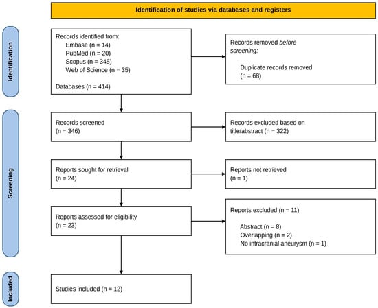
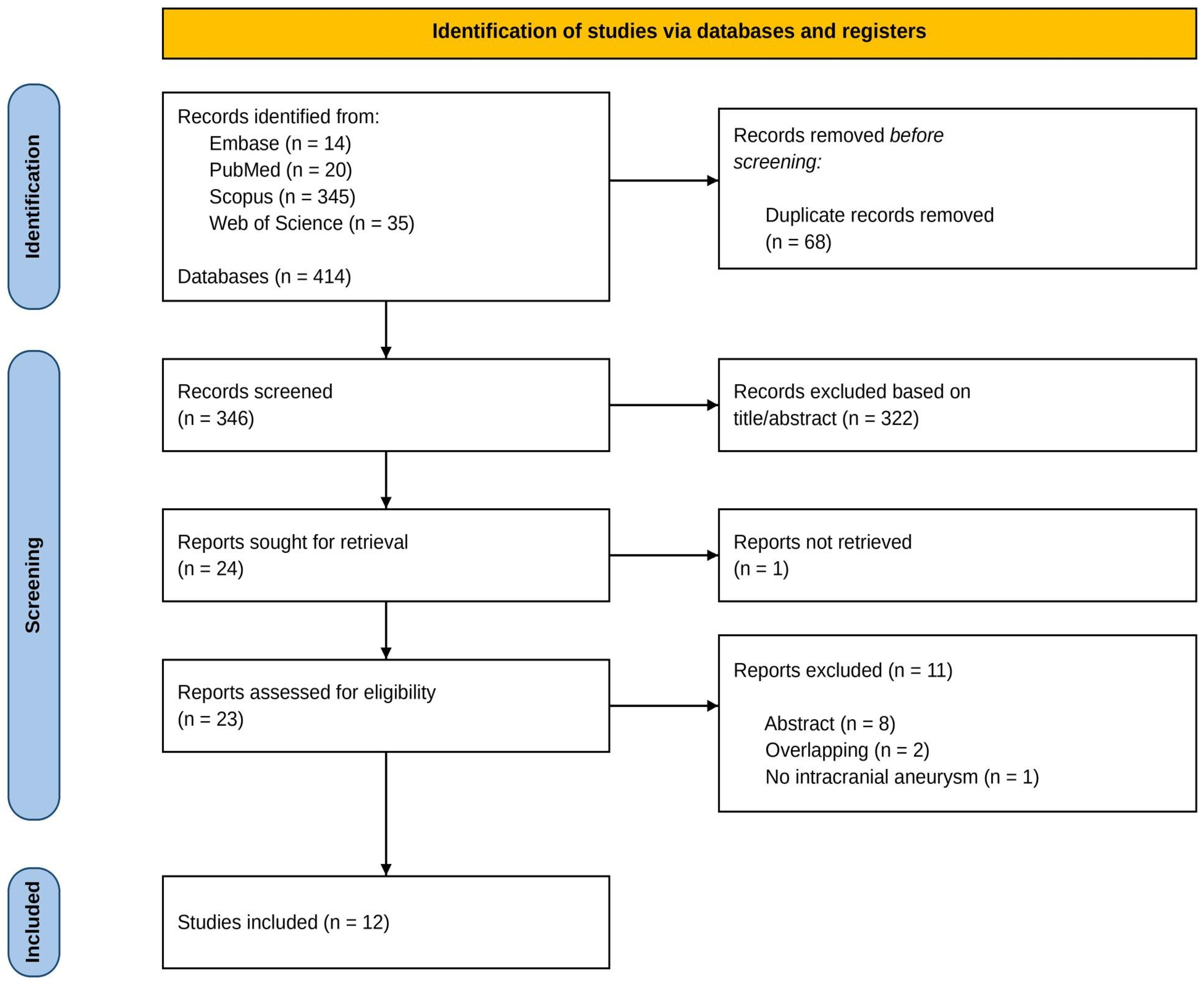

Disclaimer/Publisher’s Note: The statements, opinions and data contained in all publications are solely those of the individual author(s) and contributor(s) and not of MDPI and/or the editor(s). MDPI and/or the editor(s) disclaim responsibility for any injury to people or property resulting from any ideas, methods, instructions or products referred to in the content. |
© 2026 by the authors. Licensee MDPI, Basel, Switzerland. This article is an open access article distributed under the terms and conditions of the Creative Commons Attribution (CC BY) license.