


Abstract
Background: Previous studies have examined bioactive constituents of Prunus dulcis oil, Pinus sylvestris essential oil, and related phytochemical matrices separately; however, their combined chemical profile, comparator-context molecular-docking landscape, and comparative cellular response in melanoma and non-malignant fibroblast models remain insufficiently characterized. Methods: The analyzed study samples were profiled by GC/MS and GC/FID-based fatty-acid methyl ester analysis. An archived molecular-docking dataset was used to summarize structurally plausible interactions of six formulation-associated markers with eight melanoma-relevant or melanoma-adjacent targets and to present representative three-dimensional complexes. Because complete protocol metadata and co-crystallized-ligand redocking records were unavailable, the computational findings were interpreted descriptively. Neutral-red uptake assays assessed the 24 h viability of B16F10 melanoma cells and MRC-5 fibroblasts after exposure to each oil, the dual-oil formulation, or cisplatin. Four-parameter logistic models provided estimated IC50 values, and exact two-sided permutation tests were used for Spearman rank analyses. Results: The dual-oil formulation yielded estimated IC50 values of 1.42% v/v in B16F10 cells and 4.85% v/v in MRC-5 cells, corresponding to an apparent selectivity index of 3.41. The complete eight-concentration series was non-monotonic and showed no significant rank association (B16F10: ρs = −0.4286, exact p = 0.2992; MRC-5: ρs = −0.3810, exact p = 0.3599). In a post hoc analysis of the descending branch (≥0.37% v/v), the association was perfect in B16F10 cells (ρs = −1.0000, exact p = 0.0167) but did not reach significance in MRC-5 cells (ρs = −0.9000, exact p = 0.0833). The docking matrix identified target-dependent numerical prioritization patterns; however, small score differences were not interpreted as evidence of stronger binding or cellular target engagement. Conclusions: The findings define an exploratory chemical–computational–cellular framework and a preliminary formulation-level viability phenotype. They do not establish synergy, apoptosis, pathway modulation, therapeutic efficacy, or clinical safety. Independent multi-batch chemical confirmation, a fully documented and redocking-validated computational protocol, expanded melanoma and normal-skin cell panels, orthogonal functional assays, longer exposure periods, mechanistic validation, and subsequent in vivo studies are required.
1. Introduction
Cutaneous malignant melanoma is characterized by metastatic potential, marked molecular heterogeneity, phenotypic plasticity, and frequent resistance to targeted and immune-based treatment. The RAS–RAF–MEK–ERK axis and several parallel survival, stress-response, inflammatory, and lineage-associated programs contribute to disease progression and therapeutic adaptation [1,2,3,4,5,6,7,8,9,10,11,12,13,14,15,16,17,18,19,20,21,22,23,24]. These biological complexities also require careful separation of preliminary in vitro observations from claims of target engagement, antitumor mechanism, or clinical efficacy.
Natural products, essential oils, monoterpenes, polyphenols, and complex phytochemical mixtures have been investigated in melanoma and other tumor models because they can produce measurable effects on cellular viability, redox balance, mitochondrial function, apoptosis, proliferation, migration, and inflammatory signaling [8,9,13,14,15,16,17,18,19,20,21,22,23,24,25,26,27,28,29,30,31,32,33,34,35,36,37,38,39]. Nevertheless, natural origin does not imply selectivity, safety, reproducibility, or therapeutic value. Chemical composition, botanical provenance, extraction method, storage, vehicle behavior, concentration, exposure duration, assay interference, and the selected cellular model are critical determinants of interpretation [9,21,30,31,32,33,34,35,36,38,39].
α-Pinene and other monoterpenes found in Pinus-derived essential oils have shown biological activity in melanoma-relevant and other tumor systems [25,26,27,28,36,37,38,40,41,42,43,44,45,46,47,48]. Prunus dulcis oil is chemically distinct and is typically enriched in oleic, linoleic, and palmitic acids; Prunus-derived lipidic, phenolic, and terpenoid fractions have also been investigated in cancer models [49,50,51,52,53,54]. Recent analytical studies confirm that almond oils are generally oleic/linoleic-rich and that Pinus sylvestris needle essential oils commonly contain α-pinene, β-pinene, and carene-type monoterpenes, although substantial geographic and processing variability exists [52,53,54,55,56].
Within the literature surveyed for the present study, we did not identify a previous report that integrated study-sample GC/MS and FAME characterization of cold-pressed Prunus dulcis oil and Pinus sylvestris needle essential oil, comparator-context molecular docking of the principal identified markers, representative three-dimensional ligand–protein complexes, and side-by-side 24 h testing of each oil and their combination in B16F10 melanoma cells and MRC-5 fibroblasts. The principal novelty is therefore the integration of chemical characterization, computational prioritization, and comparative formulation-level viability assessment rather than demonstration of a molecular mechanism. This study aimed to characterize the two oil samples, contextualize structurally plausible ligand–target interactions, quantify the 24 h NRU viability phenotype of the individual and combined preparations, estimate IC50 and apparent selectivity indices, and evaluate concentration–viability ordering using exact rank-based statistics.
2. Materials and Methods
2.1. Study Design
This exploratory in vitro study combined chemical profiling of two oil samples with 24 h NRU viability testing in B16F10 melanoma cells and MRC-5 fibroblasts. Each oil was tested individually and in combination; cisplatin served as a reference cytotoxic comparator. This study was designed to characterize a formulation-level cellular phenotype and did not include mechanistic, pathway-specific, animal, or clinical endpoints.
2.2. Oil Samples, Formulation, and Provenance Limitations
The tested formulation contained cold-pressed Prunus dulcis oil and Pinus sylvestris needle essential oil. Single-oil preparations and the combined formulation were evaluated. Chemical identity and composition of the analyzed study samples were assessed by the chromatographic procedures described below. The available source records did not provide complete manufacturer/supplier, catalog, lot, geographic origin, production date, voucher specimen, certificate of analysis, storage history, oxidation index, or independently manufactured batch information. Accordingly, this study does not claim formal botanical authentication or batch-to-batch reproducibility.
2.3. GC/MS and GC/FID-Based FAME Analysis
Gas chromatography/mass spectrometry (GC/MS) analyses were performed using an Agilent Technologies (Stevens Creek Blvd, Santa Clara, CA, USA) 7890B gas chromatograph coupled to an Agilent Technologies (Stevens Creek Blvd, Santa Clara, CA, USA) inert selective 5977A mass selective detector from the same manufacturer. Separations were performed on a nonpolar HP-5MS silica capillary column (5% diphenyl- and 95% dimethyl-polysiloxane; 30 m × 0.25 mm i.d., 0.25 μm film thickness; Agilent Technologies, Santa Clara, CA, USA). Helium was used as the carrier gas at a constant flow rate of 1 mL/min. The MSD transfer line, ion source and quadrupole temperatures were set at 300 °C, 230 °C and 150 °C, respectively, and electron ionization was performed at 70 eV.
Prior to GC/MS and GC/FID analyses, fatty acids from almond oil were converted into the corresponding fatty acid methyl esters (FAMEs) by esterification/transesterification. The obtained FAMEs were dissolved in dichloromethane, and 1 μL of the prepared solution was injected through a split/splitless inlet maintained at 220 °C, operating in split mode at a split ratio of 100:1. The oven temperature was initially held at 150 °C for 5 min, increased to 250 °C at 10 °C/min and held for 10 min, then increased to 300 °C at 30 °C/min and finally held at 300 °C for 3 min. Mass spectra were recorded in scan mode over the range of m/z 10–600.
For essential oil analysis, the sample was dissolved in diethyl ether, and 1 μL of the prepared solution was injected at 220 °C in split mode at a split ratio of 40:1. The oven temperature was programmed from 60 °C to 246 °C at a rate of 3 °C/min. Mass spectra were recorded over the range of m/z 41–415.
Data processing was performed using MSD ChemStation Data Analysis software, revision F.01.00.1903, in combination with AMDIS, version 2.70, and NIST MS Search, version 2.0 g. FAMEs were identified by comparison of their mass spectra with those available in the Wiley, NIST and RTLPEST mass spectral libraries. Essential oil constituents were identified by comparison of their experimentally determined retention indices (RIexp), calculated using a homologous series of n-alkanes C8–C20, with literature values (RIlit) and by comparison of their mass spectra with those from Wiley 6, NIST2011 and RTLPEST3 libraries. For selected constituents, identification was additionally confirmed by co-injection with authentic standards.
GC/FID analyses were carried out under the same chromatographic conditions as the corresponding GC/MS analyses. The flow rates of the carrier gas (He), make-up gas (N2), fuel gas (H2) and oxidizing gas (air) were 1, 25, 30 and 400 mL/min, respectively, and the FID temperature was set at 300 °C.
Quantitative analysis of FAMEs was performed by GC/FID using calibration curves prepared from a certified reference mixture of fatty acid methyl esters (F.A.M.E. Mix RM-6, Sigma-Aldrich, St. Louis, MO, USA). A series of standard solutions was prepared in dichloromethane over the concentration range of 1.1–17.4 mg/mL. Calibration curves were constructed by plotting GC/FID peak areas against the concentrations of the FAME standard solutions. The quantitative composition of FAMEs was expressed as mg/mL.
Semi-quantitative analysis of FAMEs and essential oil constituents, expressed as relative content (%), was performed using the GC/FID peak area normalization method based on automatic integration of GC/FID signals without correction factors.
The available analytical record did not retain the exact esterification/transesterification reagent composition, reagent-to-sample ratio, reaction temperature and duration, number of independent sample preparations, complete blank/QC injection sequence, calibration equations, or prespecified library-match and retention-index acceptance thresholds. These missing operational details limit independent reproduction and are explicitly acknowledged rather than retrospectively reconstructed.
2.4. Molecular Docking and Three-Dimensional Comparator-Context Visualization
The archived computational dataset included six formulation-associated markers (α-pinene, β-pinene, δ-3-carene, oleic acid, linoleic acid, and palmitic acid) and eight targets (BRAF V600E, MEK1, ERK2, KIT, CDK4/6, PARP1, COX-2, and tyrosinase). The retained outputs comprised docking scores, within-target ranking, pose-level descriptors, reference-drug comparisons, and representative three-dimensional ligand–protein complexes. Target-specific reference scores were harmonized with the reference-drug matrix, and all ΔScore values were recalculated as ligand score minus the corresponding comparator score.
The available archive did not retain sufficient information to reconstruct the full docking protocol, including the complete receptor structure identifiers, receptor-preparation sequence, grid centers and dimensions, exhaustiveness, number of generated poses, ligand protonation and tautomeric states, treatment of crystallographic water molecules, software build and random seed, or co-crystallized-ligand redocking validation. No missing parameters were reconstructed retrospectively. The docking outputs are therefore reported as qualitative comparator-context evidence and not as a fully validated quantitative binding study. Numerical differences below approximately 1 kcal/mol were not interpreted as evidence of stronger binding.
2.5. Cell Lines and Neutral-Red Uptake Assay
Cytotoxicity was evaluated in MRC-5 fibroblasts and B16F10 melanoma cells using the neutral-red uptake assay. The Prunus dulcis and Pinus sylvestris oils were tested separately and in combination; cisplatin was used as a reference compound.
MRC-5 and B16F10 cells were seeded in 96-well plates at 1 × 10⁴ cells/well in 200 μL of high-glucose Dulbecco’s Modified Eagle Medium supplemented with 10% heat-inactivated fetal bovine serum, 2 mM glutamine, 100 U/mL penicillin, and 100 μg/mL streptomycin (Sigma-Aldrich, Darmstadt, Germany). Cells were incubated for 24 h at 37 °C in a humidified 5% CO2 atmosphere. The medium was then replaced with preparations containing the oils at 0.045%, 0.09%, 0.18%, 0.37%, 1%, 1.25%, 3%, or 5% v/v or cisplatin at 0.18, 0.5, 1, 2, 3, 4, 5, or 20 mM. Exposure continued for 24 h. Two independent experiments were performed, with triplicate technical measurements in each experiment.
One day before the assay, neutral-red working solution (Sigma-Aldrich, St. Louis, MO, USA) was prepared in DMEM at a 1:65 dilution, supplemented with 20 mM HEPES, and equilibrated overnight at 37 °C in 5% CO2. On the assay day, the solution was passed through a 0.2 μm sterile filter, and 150 μL was added per well.
After treatment, the medium was removed, and cells were washed twice with pre-warmed phosphate-buffered saline. Cells were incubated with neutral-red solution for 3 h at 37 °C in 5% CO2 and washed twice, and lysosomal dye was extracted with 150 μL of destaining solution (50% ethanol, 49% distilled water, and 1% glacial acetic acid; v/v/v) for 10 min at 300 rpm. Absorbance was measured at 540 nm with a 630 nm reference wavelength using an Agilent BioTek Epoch microplate spectrophotometer (Santa Clara, CA, USA). Viability was expressed relative to untreated controls.
2.6. Dose–Response Modeling and Statistical Analysis
Reported IC50 values were obtained by nonlinear four-parameter logistic modeling: Y = Bottom + (Top − Bottom)/[1 + 10^((logIC50 − X) × HillSlope)], where X is the logarithm of concentration. The apparent selectivity index was calculated as SI = IC50(MRC-5)/IC50(B16F10). Because complete replicate-level raw data and all model diagnostics were not available, IC50, SI, and reported between-cell-line p values were interpreted descriptively.
Spearman’s rank-order coefficient was calculated between concentration and mean NRU viability. Exact two-sided p values were obtained by enumerating all possible rank permutations (8! permutations for the complete series and 5! permutations for the post hoc descending branch). The complete eight-concentration analysis was primary. The ≥0.37% v/v branch was selected after visual inspection and was therefore labeled post hoc and exploratory. A nominal p < 0.05 threshold was used without treating the branch analysis as confirmatory.
3. Results
3.1. Study-Sample GC/MS and FAME Characterization
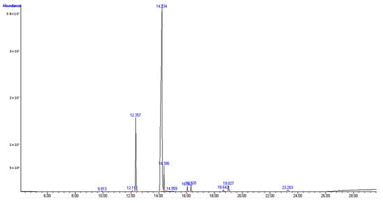
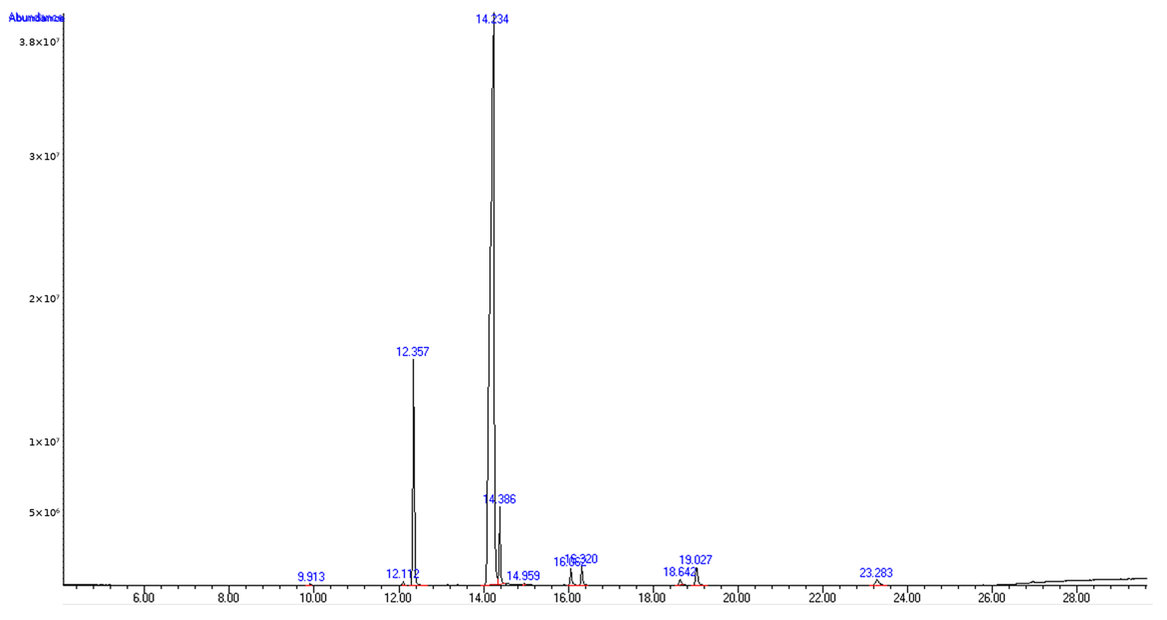
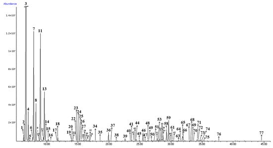
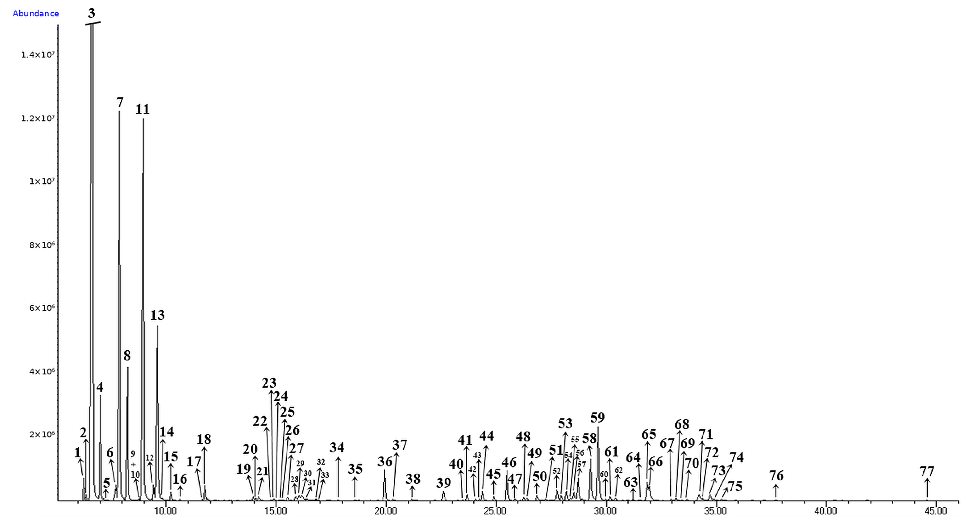
The cold-pressed Prunus dulcis oil sample was dominated by oleic acid (52.7%), linoleic acid (27.9%), palmitic acid (11.0%), and stearic acid (3.2%) (Table 1 and Table 2; Figure 1). The Pinus sylvestris needle essential-oil sample contained 86.5% monoterpene hydrocarbons and was dominated by α-pinene (41.6%), δ-3-carene (14.8%), β-pinene (14.2%), limonene (7.0%), and myrcene (4.0%) (Table 3 and Table 4; Figure 2). These data establish the composition of the analyzed study samples; they do not, by themselves, demonstrate botanical authentication or batch-to-batch reproducibility.
Table 1.
Quantitative and relative composition of the principal fatty acids in the analyzed Prunus dulcis oil sample, determined as fatty acid methyl esters.
Table 2.
Complete fatty-acid profile of the analyzed Prunus dulcis oil sample determined by GC/MS analysis of fatty acid methyl esters.
Figure 1.
Representative GC/MS total-ion chromatogram of fatty acid methyl esters obtained from the analyzed cold-pressed Prunus dulcis oil sample. The black trace represents total-ion abundance, and blue annotations identify retention times (min). Closely spaced annotations correspond to adjacent chromatographic peaks; compound identities are reported in Table 2.
Table 3.
Major constituents and grouped chemical composition of the analyzed Pinus sylvestris needle essential-oil sample determined by GC/MS.
Table 4.
Complete chemical composition of the analyzed Pinus sylvestris needle essential-oil sample determined by GC/MS.
Figure 2.
Representative GC/MS total-ion chromatogram of the analyzed Pinus sylvestris needle essential-oil sample. The black trace represents total-ion abundance. Peak numbers correspond directly to the compounds listed in Table 4; the blue axis labels denote instrument-generated chromatographic annotations.
The GC/MS and FAME profiles of the two analyzed oil samples were compared with recent analytical reports to establish compositional context and expected biological variability (Table 5).
Table 5.
Recent literature comparators used to contextualize the GC/MS and FAME profiles of the analyzed Prunus dulcis and Pinus sylvestris oil samples.
3.2. Comparator-Context Molecular Docking Analysis
The docking dataset comprised six chemically identified formulation-associated markers—α-pinene, β-pinene, δ-3-carene, oleic acid, linoleic acid, and palmitic acid—evaluated across BRAF V600E, MEK1, ERK2, KIT, CDK4/6, PARP1, COX-2, and tyrosinase. The analysis was retained as a comparator-context layer linking the GC/MS/FAME profile to melanoma-relevant or melanoma-adjacent molecular nodes. Docking scores were used for within-target prioritization only and were not interpreted as biochemical inhibition, pharmacological potency, cellular target engagement, pathway modulation, or therapeutic efficacy. The complete target-by-ligand score matrix is summarized in Table 6.
Table 6.
GC-MS/FAME-guided docking scores (kcal/mol) of representative formulation-associated markers across the comparator-context target panel. Target-specific reference values were harmonized with the archived target-specific reference-drug matrix.
Within each target, more negative scores were used to rank candidate ligands relative to the corresponding reference comparator. Because docking scoring functions have intrinsic uncertainty, numerical separations below approximately 1 kcal/mol were treated as indistinguishable for biological interpretation and were not described as evidence of superior binding. Table 7 therefore functions as a transparent prioritization matrix rather than as a potency ranking.
Table 7.
Corrected within-target ligand ranking and ΔScore values relative to the harmonized target-specific reference comparator. ΔScore was calculated as ligand score minus comparator score; negative values indicate a numerically more favorable docking score, without establishing stronger binding.
Pose-level descriptors were retained to document the internal structural annotations associated with the highest-ranked marker for each target. Hydrogen-bond counts, hydrophobic contacts, aromatic interactions, replicate-pose RMSD, interaction-fingerprint similarity, and pocket-position annotations are descriptive outputs and do not independently validate binding. These descriptors are summarized in Table 8.
Table 8.
Pose-level internal descriptors for top-ranked GC-MS/FAME-aligned phytochemical markers.
The selected panel spans oncogenic signaling, cell-cycle control, DNA-damage response, inflammatory signaling, and melanocytic-lineage biology. This breadth is intended to provide a pathway-oriented hypothesis framework rather than to imply that the formulation modulates every target. The target ontology and inclusion rationale are summarized in Table 9.
Table 9.
Melanoma-relevant target ontology and rationale for comparator-context inclusion.
Reference compounds were selected as recognizable pharmacological or mechanistic anchors for the corresponding target pockets. Their inclusion does not imply equivalence between the formulation-associated markers and clinically established inhibitors. The corresponding pharmacological anchors and binding-context assumptions are listed in Table 10.
Table 10.
Reference inhibitors and binding-context assumptions used in the docking framework.
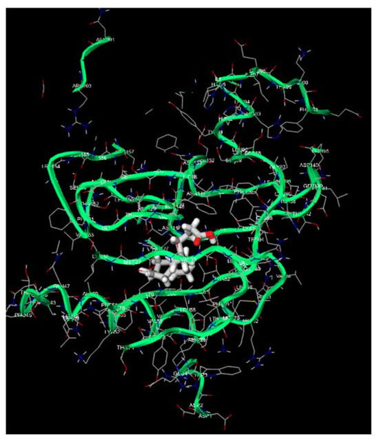
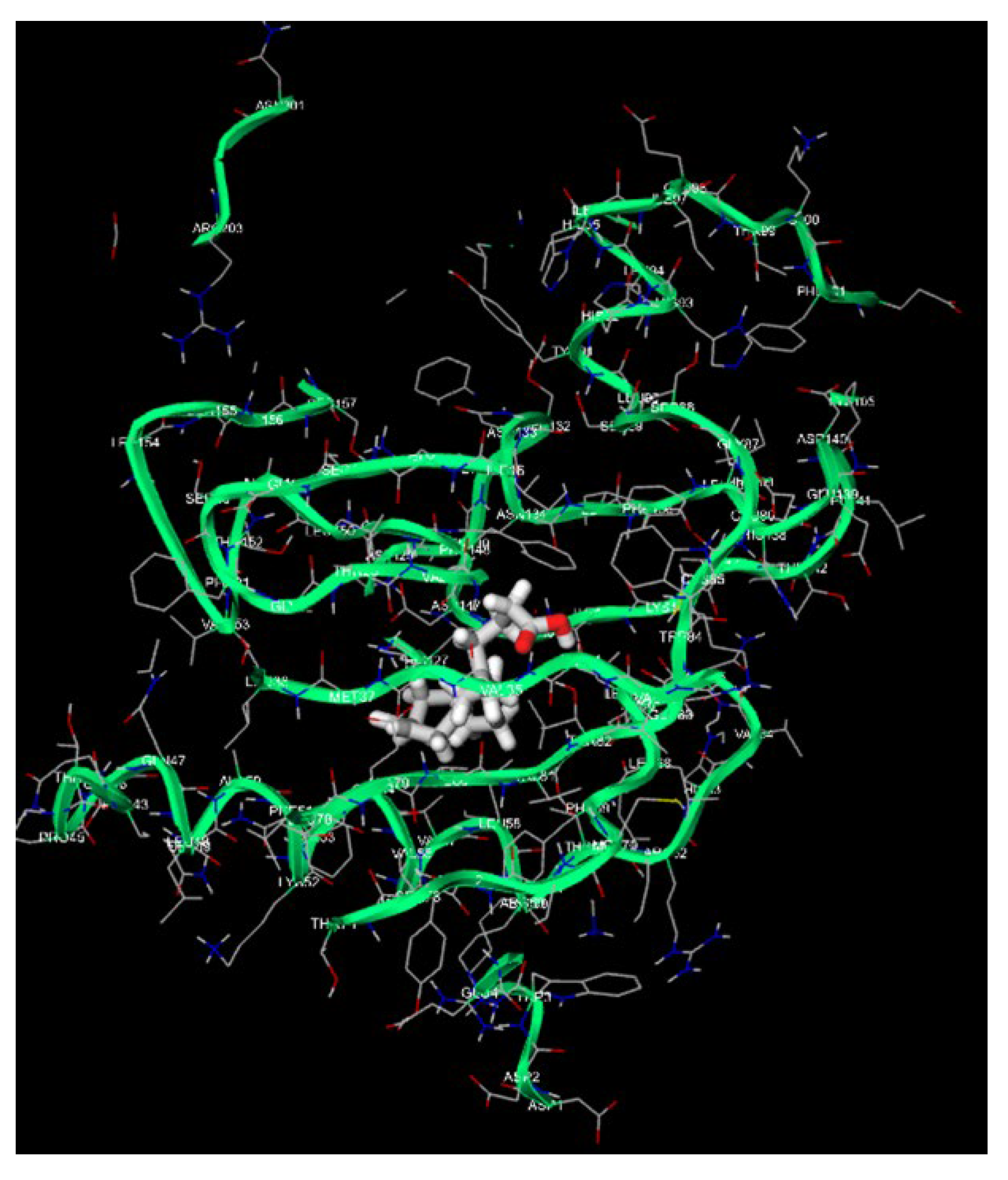
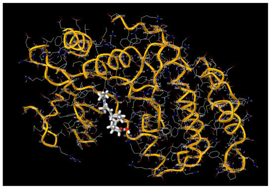
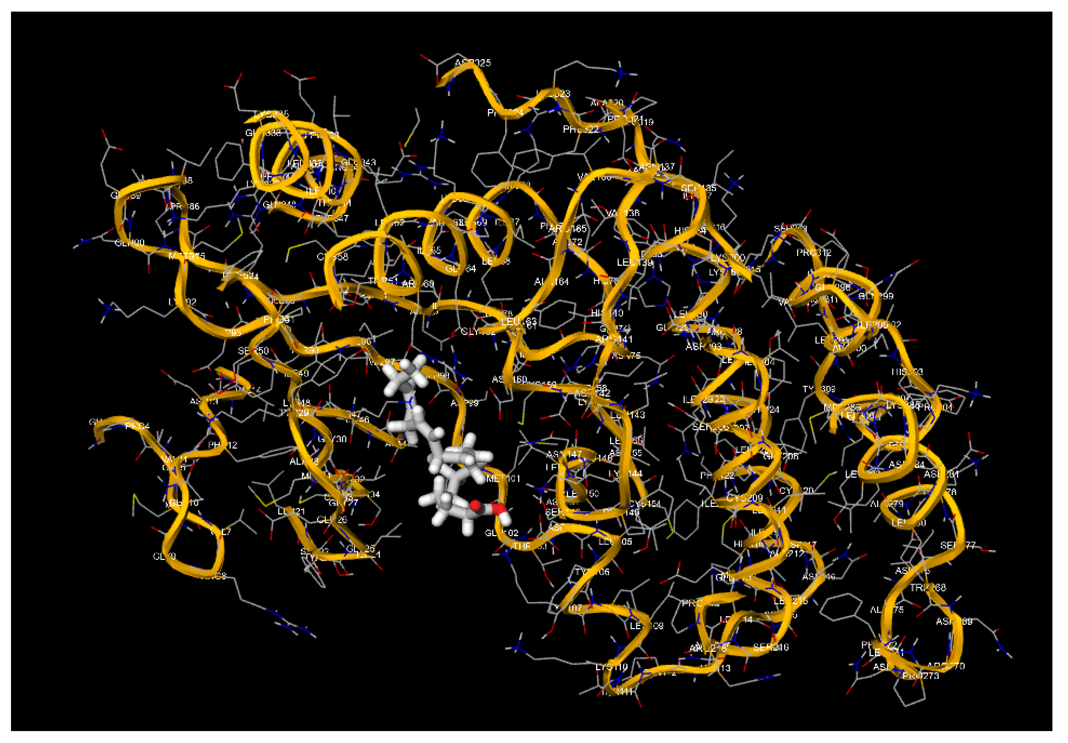
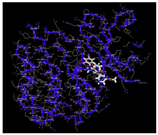
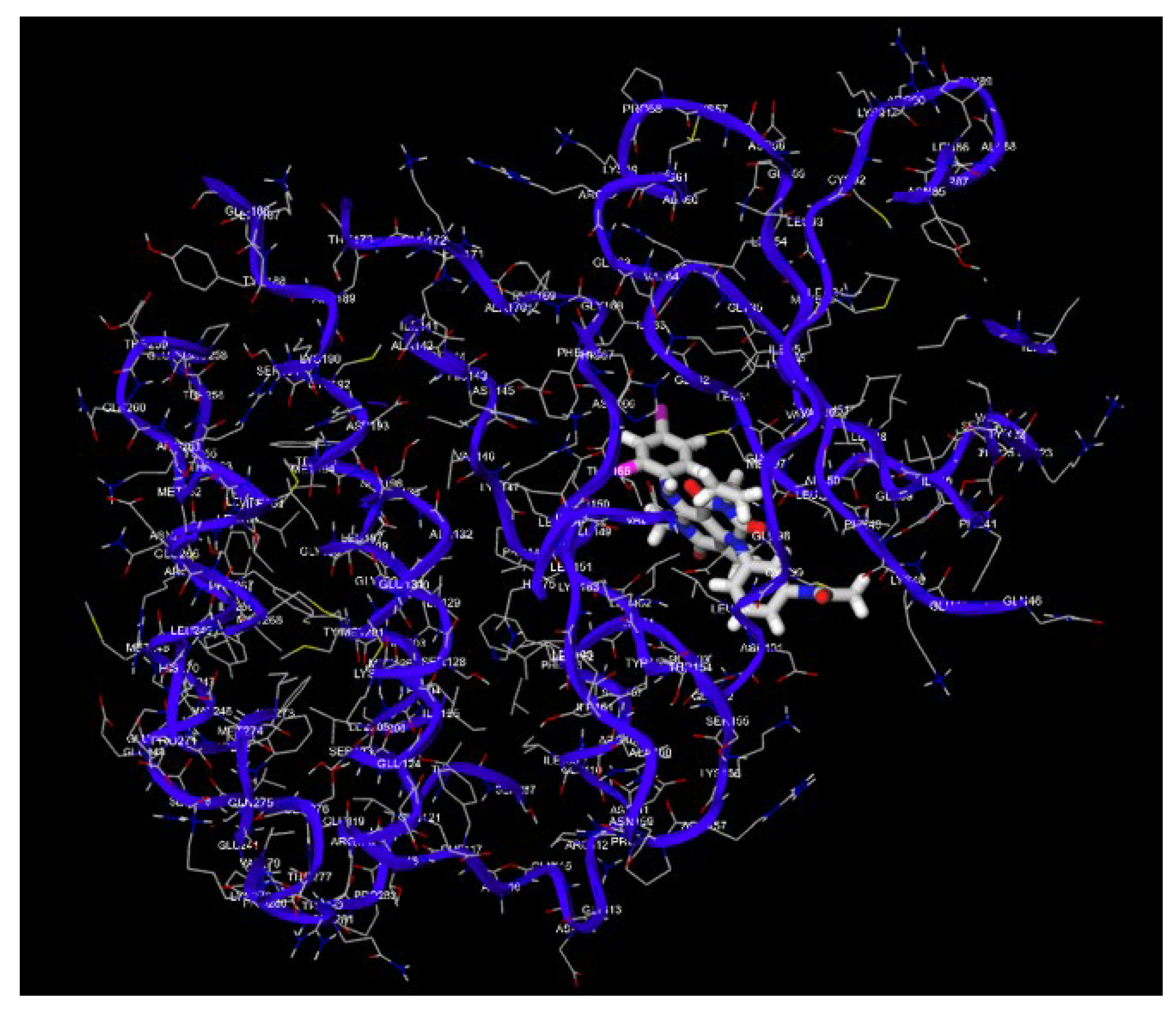
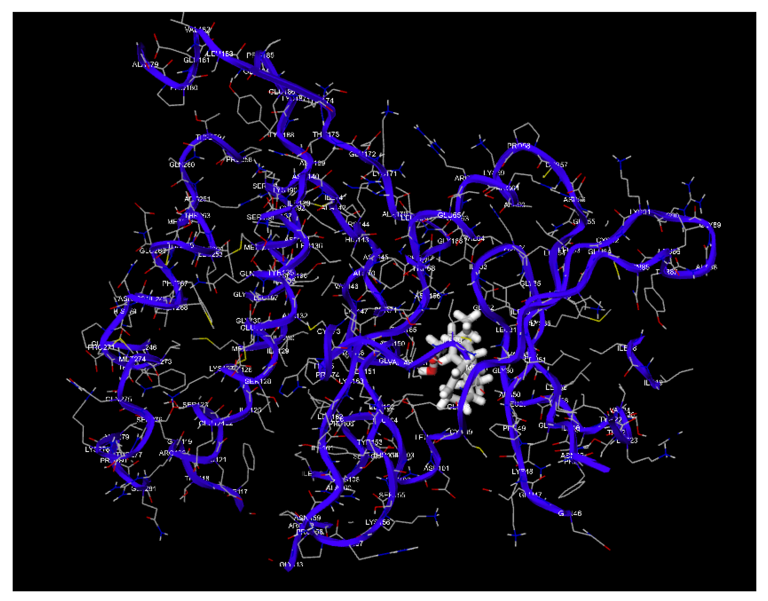
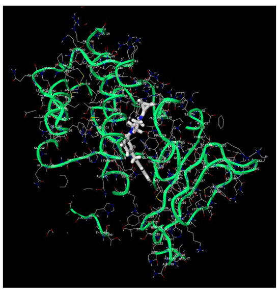
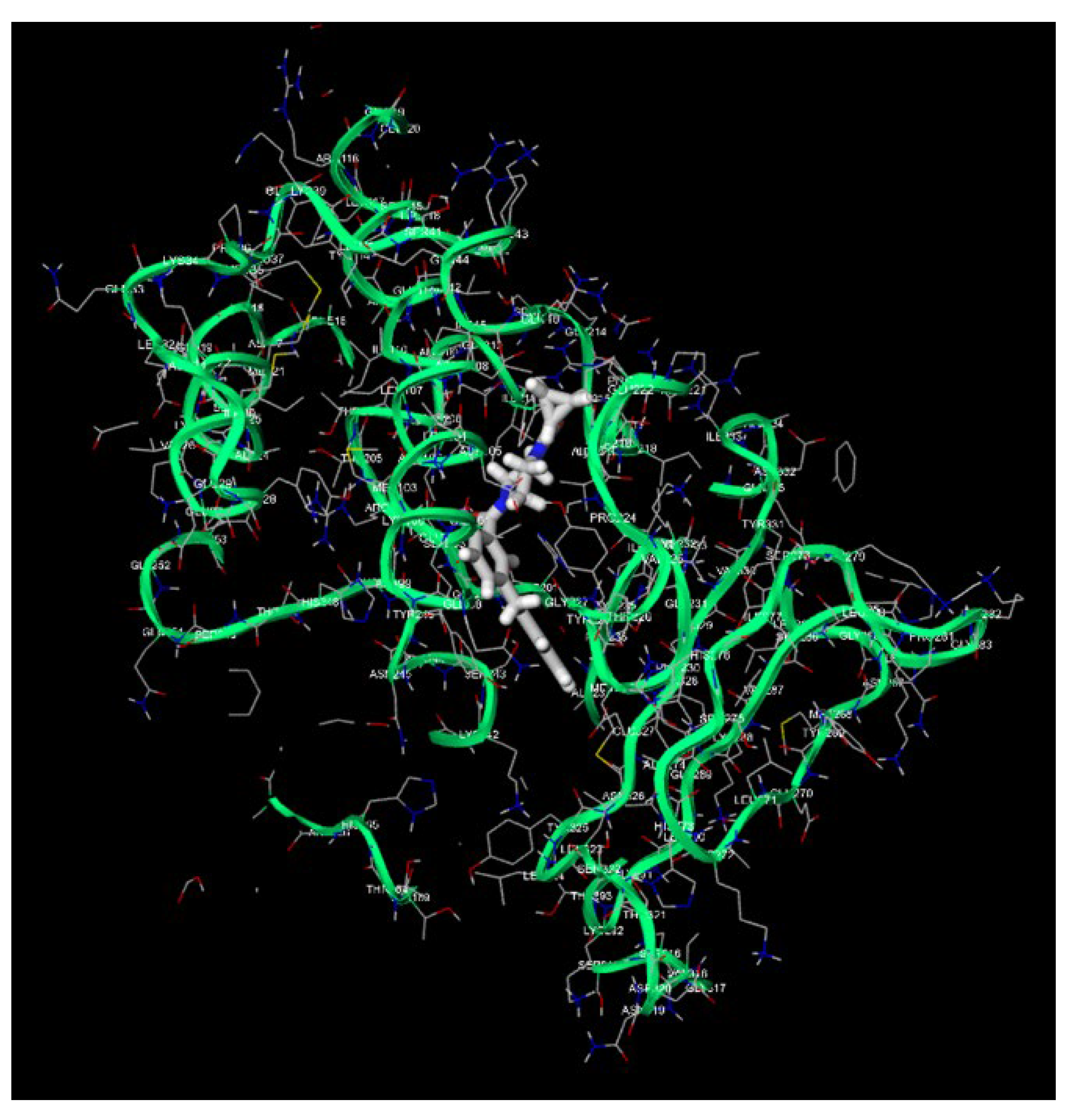
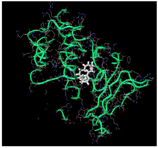
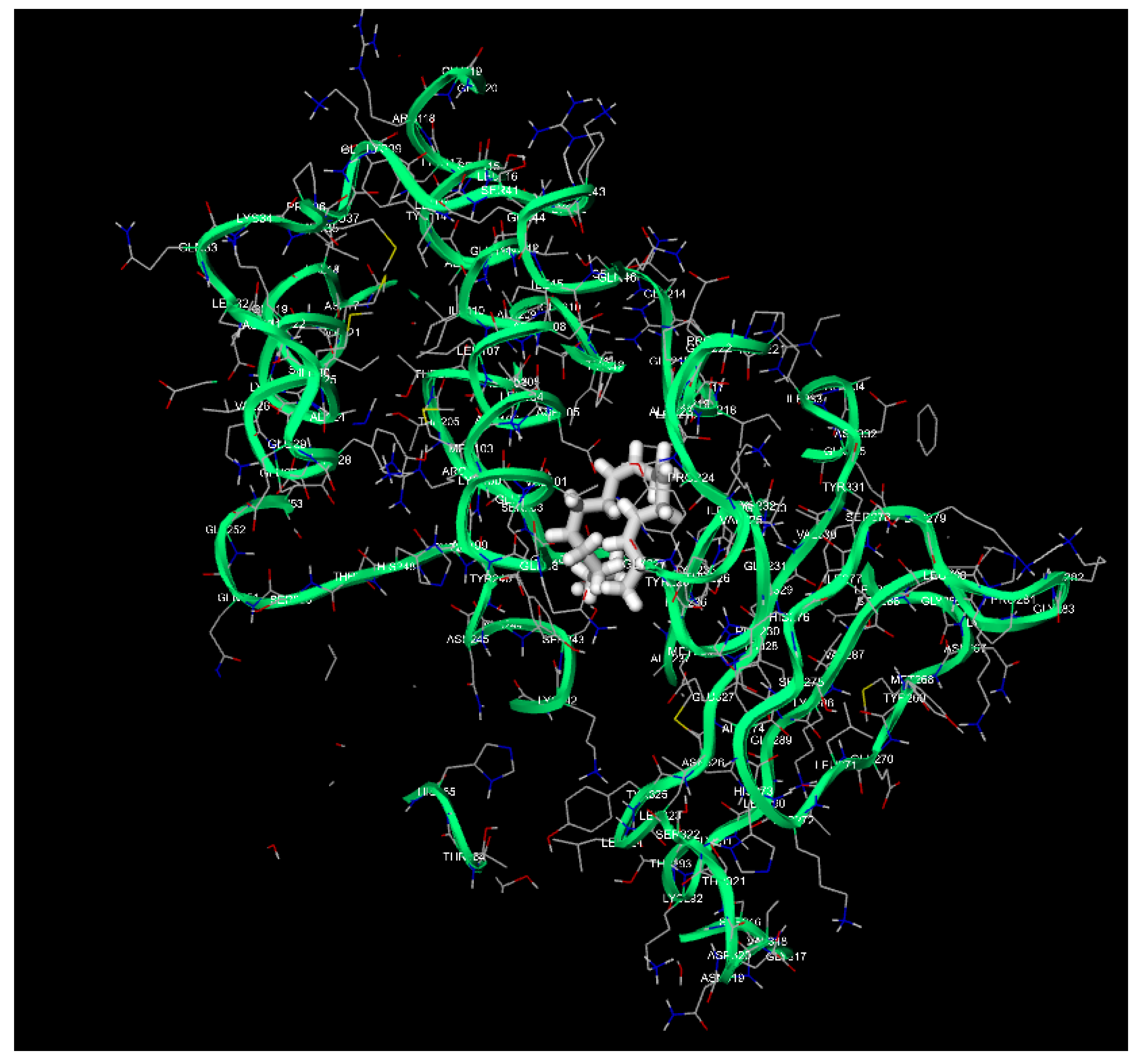
The α-pinene row was harmonized with Table 6 to remove the internal score discrepancy present in the earlier version. Table 11 places α-pinene, the predominant volatile constituent of the analyzed Pinus sylvestris sample, within the same target-by-target matrix as the reference compounds. Representative three-dimensional docking visualizations from the comparator-context analysis are presented in Figure 3, Figure 4, Figure 5, Figure 6, Figure 7 and Figure 8.
Table 11.
Harmonized α-pinene and reference-drug docking scores across the comparator-context target panel.
Figure 3.
Three-dimensional docking pose and interaction environment of kojic acid in tyrosinase. The image contextualizes melanogenesis-related target geometry and does not demonstrate enzymatic inhibition by the formulation.
Figure 4.
Representative three-dimensional comparator–ligand complex from the molecular-docking panel.
Figure 5.
Representative three-dimensional complex of a prioritized formulation-associated ligand within a melanoma-relevant target pocket.
Figure 6.
Representative three-dimensional comparator complex retained from the in silico target panel.
Figure 7.
Additional three-dimensional docking visualization supporting qualitative structural-context interpretation.
Figure 8.
Additional three-dimensional docking visualization from the archived comparator-context analysis.
Representative three-dimensional complexes from the archived computational analysis are shown below. These images are presented as qualitative structural-context visualizations and are not evidence of target inhibition or intracellular engagement.
3.3. Twenty-Four-Hour NRU Viability Profiles
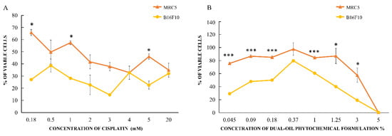
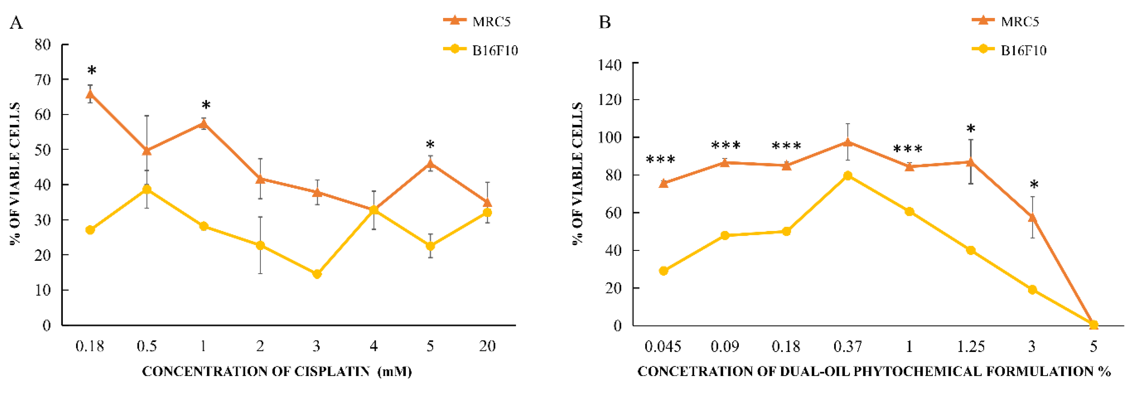
The combined formulation produced a non-monotonic concentration–viability profile in both cell lines (Figure 9; Table 7). Intermediate concentrations were associated with increased viability relative to adjacent concentrations, followed by a pronounced decline at higher exposures. At 3% v/v, mean viability was 18.96% in B16F10 cells and 57.41% in MRC-5 fibroblasts; at 5% v/v, viability approached zero in both cell lines. Accordingly, the fitted IC50 estimates should be interpreted as exploratory model-based summaries rather than as evidence of a uniformly monotonic dose response.
Figure 9.
Twenty-four-hour NRU concentration–response profiles for (A) cisplatin and (B) the dual-oil formulation in B16F10 melanoma cells and MRC-5 fibroblasts. Values are presented as mean ± SE from two independent experiments with triplicate technical measurements. Reported between-cell-line comparisons are descriptive because complete replicate-level model diagnostics were not available. * p < 0.05; *** p < 0.001.
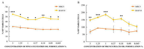
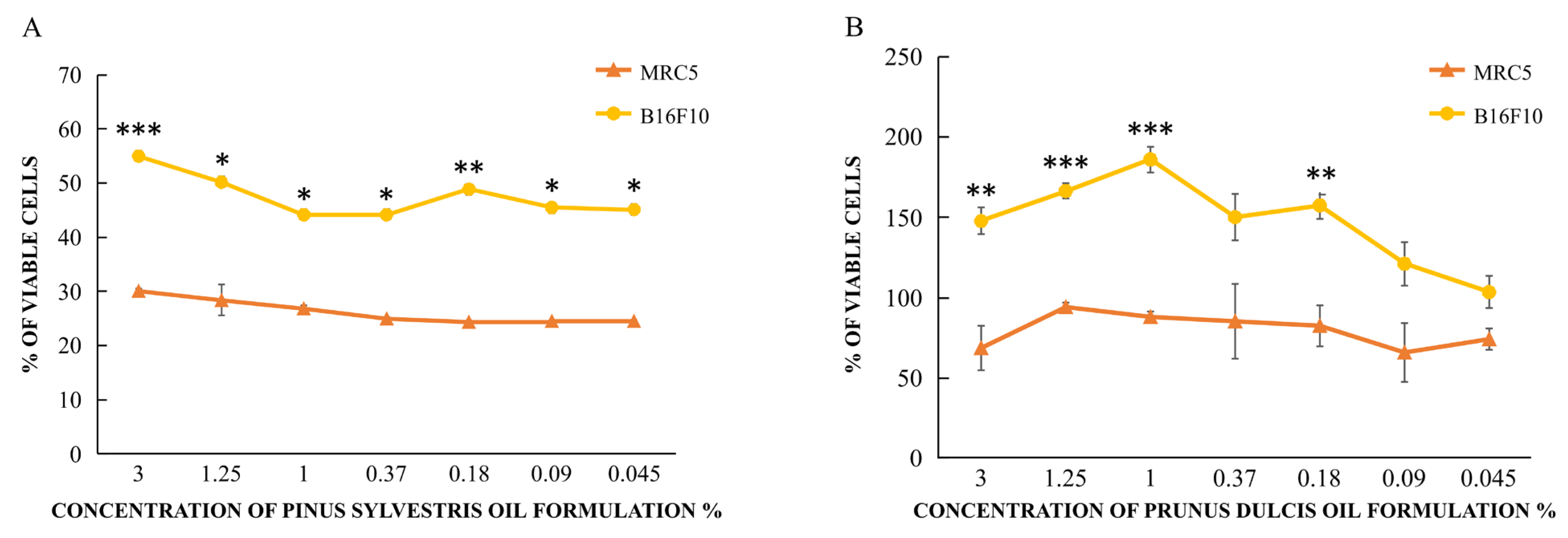
Testing of the individual oils showed that Pinus sylvestris essential oil reduced viability in both cell lines, with reported IC50 values of 1.34% v/v in B16F10 cells and 0.79% v/v in MRC-5 fibroblasts (SI = 0.59), indicating greater sensitivity of the fibroblast model and no tumor-selective pattern. Prunus dulcis oil did not reach an IC50 within the tested concentration range and was associated with increased NRU signal at several concentrations. Thus, neither single oil reproduced the apparent differential response estimated for the combined formulation (Figure 10). These data are insufficient to infer synergy because a factorial dose matrix and a formal interaction model were not used.
Figure 10.
Twenty-four-hour NRU concentration–response profiles for the individual Pinus sylvestris (A) and Prunus dulcis (B) oil samples in B16F10 melanoma cells and MRC-5 fibroblasts. Values are presented as mean ± SE from two independent experiments with triplicate technical measurements. The statistic elaboration was based on these expressions of significance: * p < 0.05, ** p < 0.01, *** p < 0.001.
Cisplatin reduced viability in both B16F10 melanoma cells and MRC-5 fibroblasts, with B16F10 cells generally showing greater sensitivity at low-to-intermediate concentrations. The response was not strictly monotonic, and the difference between the two cell lines diminished at several higher concentrations. The reported descriptive p values indicate statistically distinguishable responses at selected concentrations, whereas other exposure levels showed comparable viability between the two cell lines (Table 12). The dual-oil formulation produced a markedly different viability profile between B16F10 melanoma cells and MRC-5 fibroblasts across most tested concentrations. B16F10 viability was generally lower than MRC-5 viability, indicating greater melanoma-cell sensitivity, although the concentration–response pattern was non-monotonic. At the highest concentration (5% v/v), viability was almost completely suppressed in both cell lines. The reported descriptive p values further indicate concentration-dependent differences between the two cellular models (Table 13). The reported 4PL estimates were 1.42% v/v for B16F10 cells and 4.85% v/v for MRC-5 fibroblasts, yielding an apparent SI of 3.41 (Table 14). This value indicates differential sensitivity in the two tested cell lines under the specified 24 h NRU conditions, but it does not establish general tumor selectivity because only one non-malignant fibroblast line was examined. Cisplatin yielded reported IC50 values of 0.74 mM and 1.21 mM in B16F10 and MRC-5 cells, respectively (SI = 1.63); direct potency comparisons between cisplatin and the oils remain limited by different units, mechanisms, and concentration ranges.
Table 12.
NRU viability after 24 h exposure to cisplatin. Data are presented as mean ± SE; reported p values are descriptive.
Table 13.
NRU viability after 24 h exposure to the dual-oil formulation. Data are presented as mean ± SE; reported p values are descriptive.
Table 14.
Reported four-parameter logistic IC50 estimates and apparent selectivity indices.
3.4. Exact Spearman Rank Analysis
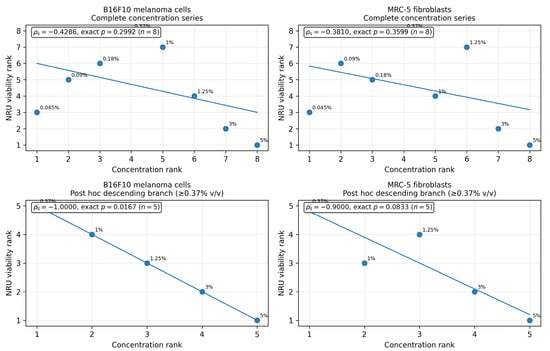
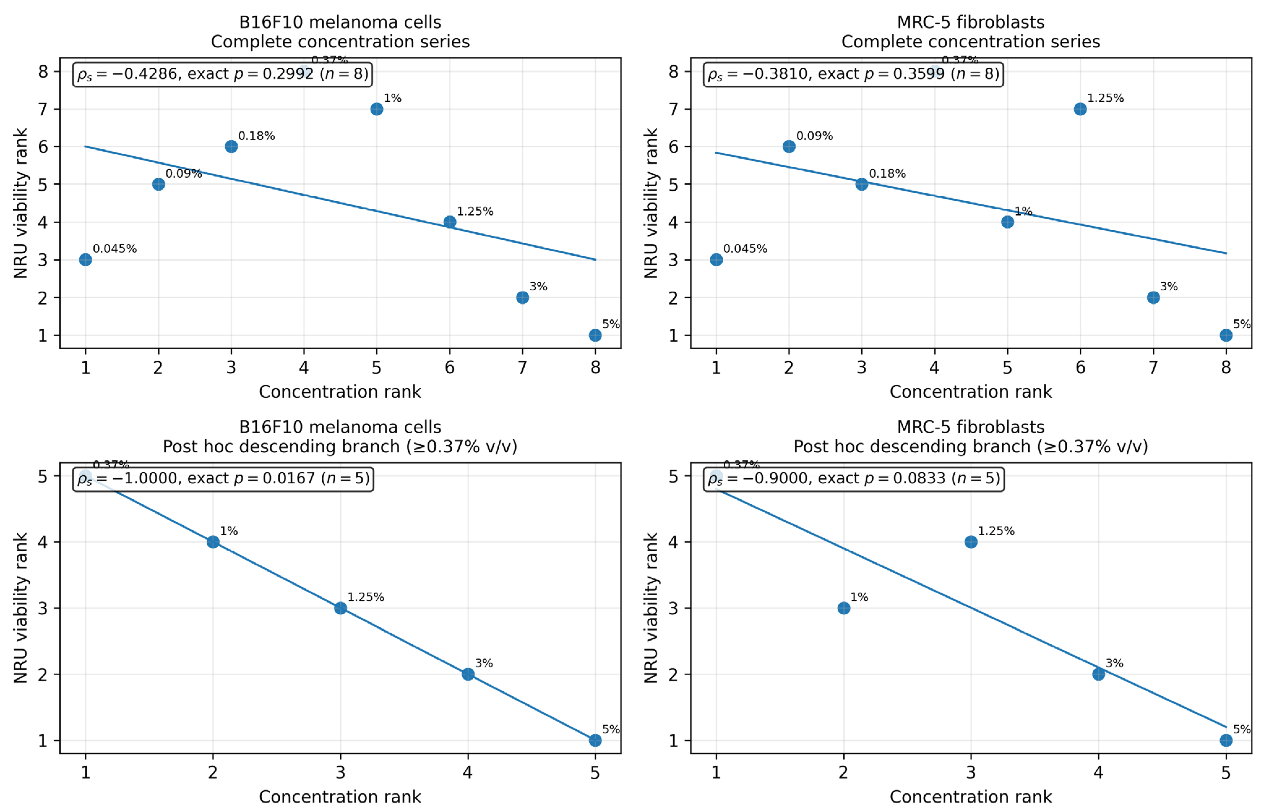
Across all eight dual-oil concentrations, concentration and mean NRU viability were inversely but non-significantly ranked in B16F10 cells (ρs = −0.4286, exact two-sided p = 0.2992) and MRC-5 fibroblasts (ρs = −0.3810, exact two-sided p = 0.3599). Because visual inspection showed a descending response from 0.37% to 5% v/v, a post hoc sensitivity analysis was performed for this five-point branch. The branch showed ρs = −1.0000 with exact p = 0.0167 in B16F10 cells and ρs = −0.9000 with exact p = 0.0833 in MRC-5 cells (Figure 11). The B16F10 result met the nominal 0.05 threshold, whereas the MRC-5 result did not. Both branch analyses remain exploratory because the interval was selected after inspection and was not an independently replicated primary endpoint.
Figure 11.
Exact Spearman rank analysis of dual-oil concentration and 24 h NRU viability. Upper panels show the complete eight-point series; lower panels show the post hoc descending branch (≥0.37% v/v; n = 5). The blue line is a least-squares visual trend across ranked observations and is included only as a visual guide; inferential interpretation is based on Spearman ρs and exact two-sided permutation p values obtained by enumerating all rank permutations.
4. Discussion
4.1. Principal Findings and Study Novelty
This study integrates chemical characterization of the analyzed oils, comparator-context molecular docking of the principal identified markers, representative three-dimensional ligand–protein visualizations, and side-by-side 24 h NRU testing of the individual and combined preparations. The dual-oil formulation produced a lower estimated IC50 in B16F10 melanoma cells than in MRC-5 fibroblasts, whereas Pinus sylvestris oil alone was more toxic to fibroblasts and Prunus dulcis oil did not reach an IC50. Within the literature surveyed, this chemical–computational–cellular integration constitutes the main novelty. The novelty is methodological and hypothesis-generating; it does not demonstrate a new antitumor mechanism.
4.2. Chemical Context and Comparison with Previous Reports
The fatty-acid profile of the Prunus dulcis sample was consistent with recent analyses of almond cultivars and genotypes, which report oleic and linoleic acids as dominant components, with palmitic and stearic acids at lower abundance [49,50,51,52,53,54]. The Pinus sylvestris sample was similarly consistent with monoterpene-rich Scots pine and related Pinus essential oils, in which α-pinene, β-pinene, 3-carene/carenes, limonene, and other monoterpenes frequently predominate [36,38,45,46,47,48,55,56]. This concordance supports the chemical plausibility of the analyzed samples but does not replace formal botanical authentication, oxidation testing, or independent batch replication.
4.3. Biological Interpretation in Relation to Melanoma Literature
α-Pinene and other monoterpenes have been associated with apoptosis, mitochondrial perturbation, oxidative stress, and reduced tumor growth in melanoma-relevant or other cancer models [40,41,42,43,44]. Pinus-derived fractions and essential oils have also shown cytotoxic or pro-apoptotic effects in B16F10 and other tumor systems [36,37,38,45,46,47,48]. In parallel, Prunus dulcis oil and other Prunus-derived fractions have shown composition-dependent biological activity in non-melanoma cancer models [49,50,51]. Essential-oil phytocomplexes such as frankincense, oregano, and Melaleuca alternifolia preparations have been examined in melanoma models using apoptosis, oxidative stress, DNA damage, and in vivo endpoints [25,26,27,28,29]. These studies provide biological context but cannot be used to assign the same mechanisms to the present formulation.
The current experiment measured lysosomal neutral-red accumulation after 24 h. Reduced NRU signal may reflect loss of viable cell number, lysosomal dysfunction, metabolic stress, membrane effects, or assay–matrix interactions; it does not by itself distinguish apoptosis, necrosis, cytostasis, altered proliferation, or impaired clonogenic survival. Likewise, no measurements of MAPK, PARP1, COX-2, CDK4/6, KIT, tyrosinase, inflammatory cytokines, oxidative stress, migration, or invasion were performed. Any pathway-level interpretation must therefore remain a hypothesis for future testing [13,23,37,57,58,59,60,61,62,63,64].
4.4. Computational Interpretation and Target-Validation Boundaries
The docking results provide a structured means of prioritizing ligand–target pairs for subsequent biochemical testing. Target-dependent ranking was observed, with α-pinene, β-pinene, δ-3-carene, linoleic acid, oleic acid, and palmitic acid occupying different numerical positions across BRAF V600E, MEK1, ERK2, KIT, CDK4/6, PARP1, COX-2, and tyrosinase. These differences are compatible with variation in pocket geometry and ligand physicochemical properties, but they cannot establish affinity, selectivity, inhibition, or pathway modulation. In particular, score separations below approximately 1 kcal/mol were treated as being within the practical uncertainty of the scoring function [62,63,64].
The archived docking outputs did not preserve the complete information required for independent reproduction and formal validation, including all receptor identifiers, grid centers and dimensions, exhaustiveness, generated-pose counts, protonation and tautomeric states, crystallographic-water handling, random seeds, and co-crystallized-ligand redocking results. Consequently, the computational layer is presented as descriptive structural context. Experimental assessment by enzyme assays, Western blotting, qPCR, phosphorylation analysis, or orthogonal target-engagement methods is required before any pathway-level inference can be made.
4.5. Selectivity, Interaction Claims, and Principal Limitations
The apparent SI of 3.41 is based on one murine melanoma line and one human fetal-lung fibroblast line. It should not be generalized to normal skin because primary melanocytes, keratinocytes, dermal fibroblasts, immune cells, and reconstructed skin models were not tested. The MRC-5 comparison provides an initial non-malignant reference only. Furthermore, formal synergy cannot be inferred from the present single-concentration-series design. Bliss, Loewe, Highest Single Agent, ZIP, or Chou–Talalay analysis would require a factorial matrix of independently varied component concentrations.
Additional limitations include the use of a single viability assay, a single 24 h time point, two independent experiments, incomplete replicate-level model diagnostics, non-monotonic concentration–response behavior, no human melanoma line, no clonogenic or migration/invasion endpoint, no pathway validation, and no in vivo efficacy or safety assessment. Essential oils may also cause irritation, oxidative stress, or inflammatory responses that are not captured by fibroblast viability alone. The available source records did not fully document producer, lot, geographic origin, voucher authentication, production date, oxidation indices, or the number of independently manufactured oil batches. Consequently, the reported chemistry applies to the analyzed study samples and does not establish batch-to-batch consistency.
4.6. Future Validation Priorities
Future work should first establish provenance and chemical reproducibility across independently sourced batches, including replicated GC/MS/FAME profiling, oxidation and stability indices, and prespecified analytical quality-control criteria. Cellular studies should then test the individual major constituents and reconstituted mixtures in murine and human melanoma lines together with primary melanocytes, keratinocytes, and dermal fibroblasts. Orthogonal endpoints should include ATP- or protein-based viability assays, live/dead imaging, Annexin V/propidium iodide analysis, caspase activation, mitochondrial membrane potential, reactive oxygen species, cell-cycle distribution, clonogenic survival, and migration/invasion assays at 24, 48, and 72 h.
Only after reproducible functional effects are demonstrated should mechanistic studies evaluate candidate pathways by Western blotting, qPCR, enzyme assays, or phosphoprotein profiling. Any future computational prioritization should use a fully documented and validated protocol, including receptor identifiers, ligand and receptor preparation, grid definitions, search parameters, co-crystallized-ligand redocking, and pose-reproduction criteria [62,63,64]. Controlled skin-compatibility and inflammatory-response testing should precede animal studies. In vivo melanoma efficacy, pharmacokinetics, local tolerability, and systemic safety would be required before any clinical application could be considered.
5. Conclusions
The analyzed Prunus dulcis and Pinus sylvestris oil samples showed the expected fatty-acid-rich and monoterpene-rich chemical profiles, respectively. The comparator-context docking dataset provided target-dependent numerical prioritization and representative three-dimensional structural models, while the combined formulation produced an exploratory 24 h NRU phenotype with a lower estimated IC50 in B16F10 melanoma cells than in MRC-5 fibroblasts and an apparent SI of 3.41. The complete concentration series was non-monotonic and not significant by exact Spearman testing. The post hoc descending branch was significant in B16F10 cells but not in MRC-5 fibroblasts. These findings support further controlled investigation but do not establish stronger molecular binding, cellular target engagement, synergy, apoptosis, pathway modulation, broad tumor selectivity, therapeutic efficacy, or clinical safety.
Future studies should prioritize independent multi-batch chemical validation, complete provenance and stability documentation, a fully specified molecular-docking workflow with co-crystallized-ligand redocking and predefined pose-reproduction criteria, purified-marker and reconstituted-mixture testing, expanded human melanoma and normal-skin cell panels, orthogonal functional assays, 48–72 h and clonogenic endpoints, formal interaction modeling, mechanistic pathway measurements, and skin-compatibility testing. In vivo melanoma efficacy and safety studies should be considered only after chemical, computational, and cellular reproducibility have been established.
Supplementary Materials
The following supporting information can be downloaded at: https://www.mdpi.com/article/10.3390/life16081314/s1, Table S1. GC-MS-guided comparator-based docking scores (kcal/mol) of representative formulation-associated phytochemical markers across the melanoma-relevant target panel. Table S2. GC-MS-guided within-target ligand ranking and score differences relative to the corresponding comparator used in the screening workflow. Numerical coefficients are retained from the docking matrix, while ligand labels are aligned to the chemically identified formulation markers. Table S3. Pose-level internal descriptors for top-ranked GC-MS/FAME-aligned phytochemical markers. Table S4. Melanoma target ontology and rationale. Table S5. Reference inhibitors and binding context used for comparator-based docking in the transferred in silico framework. Table S6. α-Pinene and reference-drug docking scores across the melanoma target panel. Table S7. Viability of B16F10 melanoma cells and MRC-5 fibroblasts following treatment with different concentrations of cisplatin, as determined by the NRU assay. Data are presented as mean ± SE with p values. Table S8. Viability of B16F10 melanoma cells and MRC-5 fibroblasts following treatment with different concentrations of dual-oil phytochemical formulation “Nevus Recovery”: P. dulcis + P. sylvestris, as determined by the NRU assay. Data are presented as mean ± SE with p values. Table S9. IC50 and SI values of investigated treatments. Table S10. Summary of maximal inhibiting concentration annotations for the tested preparations. The table is reproduced as a descriptive record of the provided experimental interpretation and does not replace fitted IC50 estimation or formal dose-response modeling. Table S11. Quantitative and relative composition of the main fatty acids in almond oil, determined as fatty acid methyl esters. Table S12. Complete fatty acid profile of almond oil determined by GC/MS analysis of fatty acid methyl esters. Table S13. Major constituents and grouped chemical composition of pine needle essential oil determined by GC/MS. Table S14. Complete chemical composition of pine needle essential oil determined by GC/MS analysis. Table S15. Recent 2025 literature comparators supporting the GC-MS/FAME interpretation of the Prunus dulcis and Pinus sylvestris oil profiles used in this work.
Author Contributions
Conceptualization, S.T. and M.D. (Momir Dunjic); methodology, S.T., M.D. (Momir Dunjic), M.M.S., S.S.K., J.S.S., M.D.K. and B.L.; software, S.T.; validation, B.L., M.G.J., M.M.K. and N.K.; formal analysis, S.T., K.D., M.M.S., S.S.K., J.S.S., M.D.K. and M.D. (Marija Dunjic); investigation, K.D., M.D. (Momir Dunjic), M.D. (Marija Dunjic), B.L., M.G.J., M.M.S., S.S.K., J.S.S., M.D.K. and N.K.; resources, B.L., M.G.J., M.M.K., T.N., M.F., T.F. and Z.J.; data curation, S.T., K.D., and N.K.; writing—original draft preparation, S.T. and K.D.; writing—review and editing, all authors; visualization, S.T.; supervision, S.T., M.D. (Momir Dunjic) and B.L.; project administration, M.D. (Momir Dunjic) and S.T.; funding acquisition: Not applicable. All authors have read and agreed to the published version of the manuscript.
Funding
This research received no external funding.
Institutional Review Board Statement
Not applicable. This study did not involve human participants or animals; all experimental work was conducted in vitro using established cell lines (B16F10 malignant melanoma cells and MRC-5 human fibroblasts).
Informed Consent Statement
Not applicable. No human participants, patient data, or identifiable information were involved in this in vitro cell-line study.
Data Availability Statement
The data supporting the findings of this study are contained within the article and its Supplementary Evaluative Materials, where applicable. Additional raw data may be made available by the corresponding author upon reasonable request.
Acknowledgments
The authors gratefully acknowledge the research group led by Biljana Ljujic at the Faculty of Medical Sciences, University of Kragujevac, Republic of Serbia, as well as the laboratory staff who contributed to the experimental work underlying this study.
Conflicts of Interest
The authors declare no conflicts of interest.
Abbreviations
| 4PL | Four-parameter logistic model |
| B16F10 | Murine malignant melanoma cell line |
| BRAF V600E | BRAF valine-to-glutamate substitution at codon 600 |
| CDK4/6 | Cyclin-dependent kinases 4 and 6 |
| COX-2 | Cyclooxygenase-2 |
| ERK2 | Extracellular signal-regulated kinase 2 |
| IFP | Interaction fingerprint |
| KIT | KIT proto-oncogene receptor tyrosine kinase |
| MAPK | Mitogen-activated protein kinase pathway |
| MEK1 | Mitogen-activated protein kinase 1 |
| PARP1 | Poly(ADP-ribose) polymerase 1 |
| RMSD | Root-mean-square deviation |
| TYR | Tyrosinase |
| FAME | Fatty acid methyl ester |
| FID | Flame ionization detector |
| GC/MS | Gas chromatography/mass spectrometry |
| IC50 | Half-maximal inhibitory concentration |
| MRC-5 | Human fibroblast cell line |
| NRU | Neutral-red uptake |
| ROS | Reactive oxygen species |
| SI | Selectivity index |
| v/v | Volume/volume |
References
- Wellbrock, C.; Arozarena, I. The Complexity of the ERK/MAP-Kinase Pathway and the Treatment of Melanoma Skin Cancer. Front. Cell Dev. Biol. 2016, 4, 33. [Google Scholar] [CrossRef] [Scilit] [PubMed]
- Savoia, P.; Fava, P.; Casoni, F.; Cremona, O. Targeting the ERK Signaling Pathway in Melanoma. Int. J. Mol. Sci. 2019, 20, 1483. [Google Scholar] [CrossRef] [Scilit] [PubMed]
- Patel, M.; Eckburg, A.; Gantiwala, S.; Hart, Z.; Dein, J.; Lam, K.; Puri, N. Resistance to Molecularly Targeted Therapies in Melanoma. Cancers 2021, 13, 1115. [Google Scholar] [CrossRef] [Scilit] [PubMed]
- Fu, Y.; Liu, J.; Mo, Z.; Wang, B.; Deng, Y.; Jiang, Y. Melanoma: Pathogenesis and Targeted Therapy. MedComm 2026, 7, e70566. [Google Scholar] [CrossRef] [Scilit] [PubMed]
- Hamis, S.J.; Kapelyukh, Y.; McLaren, A.; Henderson, C.J.; Wolf, C.R.; Chaplain, M.A.J. Quantifying ERK activity in response to inhibition of the BRAFV600E-MEK-ERK cascade using mathematical modelling. Br. J. Cancer 2021, 125, 1552–1560. [Google Scholar] [CrossRef] [Scilit] [PubMed]
- Kosnopfel, C.; Sinnberg, T.; Mane, S.; Dongo, M.; Garbe, C.; Niessner, H. Combinatorial ERK Inhibition Enhances MAPK Pathway Suppression in BRAF-Mutant Melanoma. Int. J. Mol. Sci. 2025, 26, 9794. [Google Scholar] [CrossRef] [Scilit] [PubMed]
- Phillipps, J.; Nassief, G.; Morecroft, R.; Adeyelu, T.; Elliott, A.; Abdulla, F.; Vanderwalde, A.; Park, S.; Butt, O.; Zhou, A.; et al. Efficacy of PARP inhibitor therapy after targeted BRAF/MEK failure in advanced melanoma. npj Precis Oncol. 2024, 8, 187. [Google Scholar] [CrossRef] [Scilit] [PubMed]
- Kim, H.-D.; Choi, H.; Abekura, F.; Park, J.-Y.; Yang, W.-S.; Yang, S.-H.; Kim, C.-H. Naturally-Occurring Tyrosinase Inhibitors Classified by Enzyme Kinetics and Copper Chelation. Int. J. Mol. Sci. 2023, 24, 8226. [Google Scholar] [CrossRef] [Scilit] [PubMed]
- Baber, M.A.; Crist, C.M.; Devolve, N.L.; Patrone, J.D. Tyrosinase Inhibitors: A Perspective. Molecules 2023, 28, 5762. [Google Scholar] [CrossRef] [Scilit] [PubMed]
- Cardoso, R.; Valente, R.; Souza da Costa, C.H.; Vianez, J.L.d.S.G.; da Costa, K.S.; de Molfetta, F.A.; Alves, C.N. Analysis of Kojic Acid Derivatives as Competitive Inhibitors of Tyrosinase: A Molecular Modeling Approach. Molecules 2021, 26, 2875. [Google Scholar] [CrossRef] [Scilit] [PubMed]
- Nazir, Y.; Rafique, H.; Roshan, S.; Shamas, S.; Ashraf, Z.; Rafiq, M.; Tahir, T.; Qureshi, Z.-U.; Aslam, A.; Bin Asad, M.H.H. Molecular Docking, Synthesis, and Tyrosinase Inhibition Activity of Acetophenone Amide: Potential Inhibitor of Melanogenesis. BioMed Res. Int. 2022, 2022, 1040693. [Google Scholar] [CrossRef] [Scilit] [PubMed]
- Namiecińska, E.; Jaszczak, J.; Hikisz, P.; Daśko, M.; Woźniczka, M.; Budzisz, E. Evaluation of Tyrosinase Inhibitory Activity of Carbothioamidopyrazoles and Their Potential Application in Cosmetic Products and Melanoma Treatment. Int. J. Mol. Sci. 2025, 26, 3882. [Google Scholar] [CrossRef] [Scilit] [PubMed]
- Kusuhara, M.; Maruyama, K.; Ishii, H.; Masuda, Y.; Sakurai, K.; Tamai, E.; Urakami, K. A Fragrant Environment Containing alpha-Pinene Suppresses Tumor Growth in Mice by Modulating the Hypothalamus/Sympathetic Nerve/Leptin Axis and Immune System. Integr. Cancer Ther. 2019, 18, 1534735419845139. [Google Scholar] [CrossRef] [Scilit] [PubMed]
- Matsuo, A.L.; Figueiredo, C.R.; Arruda, D.C.; Pereira, F.V.; Scutti, J.A.B.; Massaoka, M.H.; Travassos, L.R.; Sartorelli, P.; Lago, J.H. Alpha-Pinene isolated from Schinus terebinthifolius Raddi induces apoptosis and confers antimetastatic protection in a melanoma model. Biochem. Biophys. Res. Commun. 2011, 411, 449–454. [Google Scholar] [CrossRef] [Scilit] [PubMed]
- Han, E.-J.; Choi, E.-Y.; Jeon, S.-J.; Moon, J.-M.; Lee, S.-W.; Lee, J.-H.; Jung, G.-H.; Han, S.-H.; Jung, S.-H.; Yang, M.-S.; et al. Anticancer Effects of alpha-Pinene in AGS Gastric Cancer Cells. J. Med. Food 2024, 27, 330–338. [Google Scholar] [CrossRef] [Scilit] [PubMed]
- Di Martile, M.; Garzoli, S.; Sabatino, M.; Valentini, E.; D’aguanno, S.; Ragno, R.; Del Bufalo, D. Antitumor effect of Melaleuca alternifolia essential oil and its main component terpinen-4-ol in combination with target therapy in melanoma models. Cell Death Discov. 2021, 7, 127. [Google Scholar] [CrossRef] [Scilit] [PubMed]
- Liu, W.; Xiao, Y.; Zan, X.; Qi, J.; Ma, T.; Long, Q.; Wu, B.; Cao, Y. Alpha-Terpineol induces apoptosis in melanoma cells and its underlying mechanism. Sci. Rep. 2026, 16, 4278. [Google Scholar] [CrossRef] [Scilit] [PubMed]
- Chu, M.-H.; Hsiao, S.-W.; Kao, Y.-C.; Yin, H.-W.; Kuo, Y.-H.; Lee, C.-K. Cytotoxicity Effect of Constituents of Pinus taiwanensis Hayata Twigs on B16-F10 Melanoma Cells. Molecules 2022, 27, 2731. [Google Scholar] [CrossRef] [Scilit] [PubMed]
- Nguyen, T.H.; Ho, V.D.; Do, T.T.; Orav, A.; Raal, A. Selectivity of Pinus sylvestris extract and essential oil to estrogen-insensitive breast cancer cells. Pharmacogn. Mag. 2015, 11, S290–S295. [Google Scholar] [CrossRef] [Scilit] [PubMed]
- Jo, J.-R.; Park, J.S.; Park, Y.-K.; Chae, Y.Z.; Lee, G.-H.; Park, G.-Y.; Jang, B.-C. Pinus densiflora leaf essential oil induces apoptosis via ROS generation and activation of caspases in YD-8 human oral cancer cells. Int. J. Oncol. 2012, 40, 1238–1245. [Google Scholar] [CrossRef] [Scilit] [PubMed]
- Sarvmeili, N.; Jafarian-Dehkordi, A.; Zolfaghari, B. Cytotoxic effects of Pinus eldarica essential oil and extracts on HeLa and MCF-7 cell lines. Res. Pharm. Sci. 2016, 11, 476–483. [Google Scholar] [CrossRef] [Scilit] [PubMed]
- Mericli, F.; Becer, E.; Kabadayı, H.; Hanoglu, A.; Hanoglu, D.Y.; Yavuz, D.O.; Ozek, T.; Vatansever, S. Fatty acid composition and anticancer activity in colon carcinoma cell lines of Prunus dulcis seed oil. Pharm. Biol. 2017, 55, 1239–1248. [Google Scholar] [CrossRef] [Scilit] [PubMed]
- Mahmood, M.A.; Abd, A.H.; Kadhim, E.J. Assessing the cytotoxicity of phenolic and terpene fractions extracted from Iraqi Prunus arabica against AMJ13 and SK-GT-4 human cancer cell lines. F1000Research 2024, 12, 433. [Google Scholar] [CrossRef] [Scilit] [PubMed]
- Ben Khadher, T.; Sassi-Aydi, S.; Aydi, S.; Mars, M.; Bouajila, J. Phytochemical Profiling and Biological Potential of Prunus dulcis Shell Extracts. Plants 2023, 12, 2733. [Google Scholar] [CrossRef] [Scilit] [PubMed]
- Hakkim, F.L.; Bakshi, H.A.; Khan, S.; Nasef, M.; Farzand, R.; Sam, S.; Rashan, L.; Al-Baloshi, M.S.; Hasson, S.S.A.A.; Al Jabri, A.; et al. Frankincense essential oil suppresses melanoma cancer through down regulation of Bcl-2/Bax cascade signaling and ameliorates hepatotoxicity via phase I and II drug metabolizing enzymes. Oncotarget 2019, 10, 3472–3490. [Google Scholar] [CrossRef] [Scilit] [PubMed]
- Nanni, V.; Di Marco, G.; Sacchetti, G.; Canini, A.; Gismondi, A. Oregano Phytocomplex Induces Programmed Cell Death in Melanoma Lines via Mitochondria and DNA Damage. Foods 2020, 9, 1486. [Google Scholar] [CrossRef] [Scilit] [PubMed]
- Zhang, W.; Lan, Y.; Huang, Q.; Hua, Z. Galangin induces B16F10 melanoma cell apoptosis via mitochondrial pathway and sustained activation of p38 MAPK. Cytotechnology 2013, 65, 447–455. [Google Scholar] [CrossRef] [Scilit] [PubMed]
- Massaoka, M.H.; Matsuo, A.L.; Figueiredo, C.R.; Farias, C.F.; Girola, N.; Arruda, D.C.; Scutti, J.A.B.; Romoff, P.; Favero, O.A.; Ferreira, M.J.P.; et al. Jacaranone Induces Apoptosis in Melanoma Cells via ROS-Mediated Downregulation of Akt and p38 MAPK Activation and Displays Antitumor Activity In Vivo. PLoS ONE 2012, 7, e38698. [Google Scholar] [CrossRef] [Scilit] [PubMed]
- Zhao, W.; Chen, Z.; Fu, W.; Ye, C.; Fu, H.; Xu, T.; Wu, B.; Chen, L.; Shan, S.-J. Induction of apoptosis and hypoxic stress in malignant melanoma cells via graphene-mediated far-infrared radiation. BMC Cancer 2025, 25, 620. [Google Scholar] [CrossRef] [Scilit] [PubMed]
- Ombra, M.N.; Paliogiannis, P.; Stucci, L.S.; Colombino, M.; Casula, M.; Sini, M.C.; Manca, A.; Palomba, G.; Stanganelli, I.; Mandalà, M.; et al. Dietary compounds and cutaneous malignant melanoma: Recent advances from a biological perspective. Nutr. Metab. 2019, 16, 33. [Google Scholar] [CrossRef] [Scilit] [PubMed]
- Bayala, B.; Bassole, I.H.; Scifo, R.; Gnoula, C.; Morel, L.; A Lobaccaro, J.-M.; Simpore, J. Anticancer activity of essential oils and their chemical components—A review. Am. J. Cancer Res. 2014, 4, 591–607. [Google Scholar] [CrossRef] [Scilit]
- Sobral, M.V.; Xavier, A.L.; Lima, T.C.; de Sousa, D.P. Antitumor Activity of Monoterpenes Found in Essential Oils. Sci. World J. 2014, 2014, 953451. [Google Scholar] [CrossRef] [Scilit] [PubMed]
- Mohamed Abdoul-Latif, F.; Ainane, A.; Houmed Aboubaker, I.; Mohamed, J.; Ainane, T. Exploring the Potent Anticancer Activity of Essential Oils and Their Bioactive Compounds. Pharmaceuticals 2023, 16, 1086. [Google Scholar] [CrossRef] [Scilit] [PubMed]
- Carvalho, A.A.; Andrade, L.N.; Vieira de Sousa, E.B.; de Sousa, D.P. Antitumor Phenylpropanoids Found in Essential Oils. BioMed Res. Int. 2015, 2015, 392674. [Google Scholar] [CrossRef] [Scilit] [PubMed]
- Dumitras, D.-A.; Andrei, S. Recent Advances in the Antiproliferative and Proapoptotic Activity of Various Plant Extracts and Constituents against Murine Malignant Melanoma. Molecules 2022, 27, 2585. [Google Scholar] [CrossRef] [Scilit] [PubMed]
- Tabolacci, C.; De Vita, D.; Facchiano, A.; Bozzuto, G.; Beninati, S.; Failla, C.M.; Di Martile, M.; Lintas, C.; Mischiati, C.; Stringaro, A.; et al. Phytochemicals as Immunomodulatory Agents in Melanoma. Int. J. Mol. Sci. 2023, 24, 2657. [Google Scholar] [CrossRef] [Scilit] [PubMed]
- Kim, H.J.; Hong, J.H. Multiplicative Effects of Essential Oils and Other Active Components on Skin Tissue and Skin Cancers. Int. J. Mol. Sci. 2024, 25, 5397. [Google Scholar] [CrossRef] [Scilit] [PubMed]
- Jones, V.; Katiyar, S.K. Emerging phytochemicals for prevention of melanoma invasion. Cancer Lett. 2013, 335, 251–258. [Google Scholar] [CrossRef] [Scilit] [PubMed]
- Chhabra, G.; Ndiaye, M.A.; Garcia-Peterson, L.M.; Ahmad, N. Melanoma Chemoprevention: Current Status and Future Prospects. Photochem. Photobiol. 2017, 93, 975–989. [Google Scholar] [CrossRef] [Scilit] [PubMed]
- AlQathama, A.; Prieto, J.M. Natural products with therapeutic potential in melanoma metastasis. Nat. Prod. Rep. 2015, 32, 1170–1182. [Google Scholar] [CrossRef] [Scilit] [PubMed]
- Isacescu, E.; Chiroi, P.; Zanoaga, O.; Nutu, A.; Budisan, L.; Pirlog, R.; Atanasov, A.G.; Berindan-Neagoe, I. Melanoma Cellular Signaling Transduction Pathways Targeted by Polyphenols Action Mechanisms. Antioxidants 2023, 12, 407. [Google Scholar] [CrossRef] [Scilit] [PubMed]
- Benedusi, M.; Lee, H.; Lim, Y.; Valacchi, G. Oxidative State in Cutaneous Melanoma Progression: A Question of Balance. Antioxidants 2024, 13, 1058. [Google Scholar] [CrossRef] [Scilit] [PubMed]
- Ruzzolini, J.; Peppicelli, S.; Andreucci, E.; Bianchini, F.; Scardigli, A.; Romani, A.; La Marca, G.; Nediani, C.; Calorini, L. Oleuropein has an anti-cancer effect on human BRAF melanoma cells and potentiates cytotoxicity of current chemotherapies. Nutrients 2018, 10, 1950. [Google Scholar] [CrossRef] [Scilit] [PubMed]
- Yanarojana, M.; Tancharoen, S.; Nararatwanchai, T.; Yanarojana, S. Phytochemicals From Houttuynia cordata Thunb as Potential Inhibitors of BRAF, MEK, and ERK: Insights From Molecular Docking. J. Skin. Cancer 2025, 2025, 2565084. [Google Scholar] [CrossRef] [Scilit] [PubMed]
- Shuvo, S.P.; Niaz, S.M.R.; Jannat, S.; Hossain, A.; Mashhur, N.; Ahmed, N.; Sohel, M.; Hasan, I.; Ansari, S.A.; Humayoo, M.; et al. Computational and pharmacophore-based study of Camellia sinensis phytochemicals targeting BRAF in melanoma. Sci. Rep. 2025, 15, 38339. [Google Scholar] [CrossRef] [Scilit] [PubMed]
- Furlan, V.; Ravnik, V.; Bren, U.; Jukic, M. Insights into Molecular Mechanisms of Polyphenolic Compounds from Helichrysum italicum by Inverse Molecular Docking Fingerprint Approach. Pharmaceuticals 2026, 19, 647. [Google Scholar] [CrossRef] [Scilit] [PubMed]
- Strickland, L.R.; Pal, H.C.; Elmets, C.A.; Afaq, F. Targeting Drivers of Melanoma with Synthetic Small Molecules and Phytochemicals. Cancer Lett. 2015, 359, 20–35. [Google Scholar] [CrossRef] [Scilit] [PubMed]
- Pal, H.C.; Hunt, K.M.; Diamond, A.; Elmets, C.A.; Afaq, F. Phytochemicals for the Management of Melanoma. Mini Rev. Med. Chem. 2016, 16, 953–979. [Google Scholar] [CrossRef] [Scilit] [PubMed]
- Juszczak, A.M.; Woelfle, U.; Koncic, M.Z.; Tomczyk, M. Skin cancer, related pathways and therapy and the role of luteolin derivatives as potential therapeutics. Med. Res. Rev. 2022, 42, 1423–1462. [Google Scholar] [CrossRef] [Scilit] [PubMed]
- Singaravelan, N.; Tollefsbol, T.O. Polyphenol-Based Prevention and Treatment of Cancer Through Epigenetic and Combinatorial Mechanisms. Nutrients 2025, 17, 616. [Google Scholar] [CrossRef] [Scilit] [PubMed]
- Tomko, A.M.; Whynot, E.G.; Ellis, L.D.; Dupré, D.J. Anti-Cancer Potential of Cannabinoids, Terpenes, and Flavonoids Present in Cannabis. Cancers 2020, 12, 1985. [Google Scholar] [CrossRef] [Scilit] [PubMed]
- Hundsberger, H.; Stierschneider, A.; Sarne, V.; Ripper, D.; Schimon, J.; Weitzenböck, H.P.; Schild, D.; Jacobi, N.; Eger, A.; Atzler, J.; et al. Concentration-Dependent Pro- and Antitumor Activities of Quercetin in Human Melanoma Spheroids. Molecules 2021, 26, 717. [Google Scholar] [CrossRef] [Scilit] [PubMed]
- Cho, M.-K.; Lee, Y.; Kim, K.D.; Choi, M.H.; Lee, S.-H.; Choi, D.; Nam, H.-S.; Lee, Y.-J. Dietary Polyphenols Curcumin and Resveratrol Exert Selective Anticancer Effects in Melanoma Cells. Nutrients 2026, 18, 548. [Google Scholar] [CrossRef] [Scilit] [PubMed]
- Hamzaoui, E.; Zallez, O.B.Y.; Buñay, J.; Leremboure, M.; Argui, H.; Baron, S.; Said, H.; Lobaccaro, J.-M.A.; Akriche, S. Comparative Study of Essential Oils from Tunisian Pinus halepensis by Hydrodistillation and Microwave-Assisted Processes. ACS Omega 2024, 9, 34128–34139. [Google Scholar] [CrossRef] [Scilit] [PubMed]
- Lü, S.; Shang, B.; Liu, G.; Sun, L.; Wu, Q.; Geng, Y. Effects of pine needle essential oil on melanin synthesis in B16F10 cells and its mechanism. BioMed Rep. 2025, 23, 133. [Google Scholar] [CrossRef] [Scilit] [PubMed]
- Thalappil, M.A.; Butturini, E.; de Prati, A.C.; Bettin, I.; Antonini, L.; Sapienza, F.U.; Garzoli, S.; Ragno, R.; Mariotto, S. Pinus mugo Essential Oil Impairs STAT3 Activation through Oxidative Stress and Induces Apoptosis in Prostate Cancer Cells. Molecules 2022, 27, 4834. [Google Scholar] [CrossRef] [Scilit] [PubMed]
- Shi, A.; Liu, L.; Li, S.; Qi, B. Natural products targeting the MAPK-signaling pathway in cancer: Overview. J. Cancer Res. Clin. Oncol. 2024, 150, 6. [Google Scholar] [CrossRef] [Scilit] [PubMed]
- Maphutha, J.; Twilley, D.; Dawood, M.; Efferth, T.; Lall, N. The Potential of Phytochemicals to Overcome Multidrug Resistance in Metastatic Melanoma. Chem. Biodivers. 2025, 23, e02536. [Google Scholar] [CrossRef] [Scilit] [PubMed]
- Kowalski, S.; Karska, J.; Tota, M.; Skinderowicz, K.; Kulbacka, J.; Drag-Zalesinska, M. Natural Compounds in Non-Melanoma Skin Cancer: Prevention and Treatment. Molecules 2024, 29, 728. [Google Scholar] [CrossRef] [Scilit] [PubMed]
- Babashpour-Asl, M.; Yousefpour-Dokhanieh, A.; Farajzadeh-Memari-Tabrizi, E. Evaluation of nutritional and antioxidant properties of almond cultivars: A comprehensive analysis for health and industry applications. BMC Plant Biol. 2025, 25, 1675. [Google Scholar] [CrossRef] [Scilit] [PubMed]
- Genç, B.; Güney, M. Evaluation of pomological traits and fatty acid profile of almond (Prunus dulcis L. [Mill.] D.A. Webb) genotypes grown in Yozgat province. Eur. Food Res. Technol. 2025, 251, 969–979. [Google Scholar] [CrossRef] [Scilit]
- Dias, F.F.G.; Teixeira, B.F.; Taha, A.Y.; Bell, J.M.L.N.M. Integrated impact of environmentally friendly extraction and recovery methods on almond oil quality: Insights from a lipidomic perspective. J. Am. Oil Chem. Soc. 2025, 102, 995–1004. [Google Scholar] [CrossRef] [Scilit]
- Tuleshova, K.; Ishmuratova, M.; Aynabayev, A.; Sadyrbekov, D.; Kali, A. Comparative phytochemical analysis of hydrodistilled essential oils of Scots pine (Pinus sylvestris L.) needles from Central Kazakhstan. OnLine J. Biol. Sci. 2025, 25, 852–859. [Google Scholar] [CrossRef] [Scilit]
- Mirković, S.; Martinović, M.; Tadić, V.M.; Nešić, I.; Jovanović, A.S.; Žugić, A. Antimicrobial and antioxidant activity of essential oils from selected Pinus species from Bosnia and Herzegovina. Antibiotics 2025, 14, 677. [Google Scholar] [CrossRef] [Scilit] [PubMed]

Disclaimer/Publisher’s Note: The statements, opinions and data contained in all publications are solely those of the individual author(s) and contributor(s) and not of MDPI and/or the editor(s). MDPI and/or the editor(s) disclaim responsibility for any injury to people or property resulting from any ideas, methods, instructions or products referred to in the content. |
© 2026 by the authors. Licensee MDPI, Basel, Switzerland. This article is an open access article distributed under the terms and conditions of the Creative Commons Attribution (CC BY) license.