


Abstract
Platelets are anucleated cell fragments representing the second most abundant blood element in circulation, with approximately 100 billion new platelets released from the bone marrow daily in a healthy individual. For over a century, their hemostatic role was considered their primary function; however, the past few decades of research have revealed significant immunological functions in both innate and adaptive immunity. Through the release of mediators stored in platelet granules and the expression and realignment of a diverse array of surface receptors, platelets influence immune cells while simultaneously recognizing, reacting to, and phagocytosing offending pathogens such as viruses and bacteria. Their activation initiates signaling cascades that either amplify platelet responses or drive the activation, recruitment, migration, and maturation of immune cells to sites of infection. This narrative review synthesizes platelet biology, receptor signaling, granule physiology, and platelet interactions with innate and adaptive immune cells to provide an integrated perspective on platelet immunologic function. We examine platelets as frontline immune sentinels and evaluate their capacity to regulate both innate and adaptive immune responses, detailing specific receptors and granule contents as the mechanistic foundation for their immunologic roles. We further explore the molecular mechanisms by which platelets interact with pathogens and immune cells to drive pathogen clearance, inflammation modulation, and the interplay between thrombosis and immunity, including their contributions to chronic inflammatory disease and tumorigenesis.
1. Introduction
The description of platelets one hundred and fifty years ago by Dr. Giulio Bizzozero as unique peripheral blood elements represents one of the most important discoveries in medicine [1]. Their origin as vascular elements derived from megakaryocytes in the bone marrow was not established until the early 1900s [2], and their involvement in thrombosis and hemostasis would not become apparent until the 1960s [3], a reminder that nearly a century was needed to elucidate the basic platelet biology we now take for granted. Circulating mature platelets are small (2–5 µm) anucleated cellular elements that survive in circulation for up to 10 days and are maintained at a minimum count of approximately 150 × 109/liter of peripheral blood [4,5]. Notably, megakaryocytes residing in extramedullary sites, particularly the lungs, contribute substantially to circulating platelet production, accounting for up to half of total output in murine models, with evidence of analogous hematopoietic progenitor activity in the adult human lung as well [6,7,8,9].
For decades, platelet biology was framed almost exclusively around hemostasis. Upon tissue injury, platelets activate and adhere to exposed subendothelial matrix via receptors binding collagen, von Willebrand factor (vWF), laminin, fibronectin, and thrombospondin, triggering signaling cascades that stabilize adherence and drive aggregation [10,11,12,13]. Integrin αIIbβ3-mediated platelet–platelet interactions then consolidate a fibrin-rich hemostatic plug further reinforced by FcγRIIa-coupled pathways and coordinated inside-out and outside-in integrin signaling that promotes granule release and morphological remodeling. Disruptions to this system, whether inherited, as in Bernard–Soulier syndrome (defects in GPIb-IX-V receptor complex) or Glanzmann thrombasthenia (intact coagulation cascade but impaired αIIbβ3 integrin function) [14], or acquired through medications or organ disease, compromise hemostasis and broader platelet physiology.
However, it is now well established that hemostasis is neither the only nor perhaps the primary function of platelets. Platelets mediate leukocyte and progenitor cell recruitment to injury sites, release both pro- and anti-inflammatory mediators that are characteristic of immune-competent cells, produce angiogenic factors and microparticles, phagocytose microorganisms, and directly activate immune effector cells [15,16,17,18,19,20]. The capacity to directly activate immune effector cells perhaps represents the strongest evidence that platelets are bona fide immune and inflammatory cells. This is further supported by sepsis models in which platelet activation, consumption, and immune cell interactions are consistently observed [21,22,23,24]. Platelets recognize pathogens through toll-like receptors (TLRs), driving auto-activation and enhancing neutrophil and monocyte functions at infection sites [25,26]. They also regulate adaptive immunity through T-cell modulation and endothelial cell activation, thereby bridging innate and adaptive immune responses [17]. These expanding non-hemostatic roles have motivated clinical translation efforts and early-phase trials targeting platelet–immune pathways [27,28,29].
This narrative review was conducted by systematically searching PubMed using terms encompassing platelet biology, immune receptors, granule physiology, innate immunity, adaptive immunity, thromboinflammation, and related pathologic conditions. Studies were selected and synthesized thematically based on their relevance to platelet immunologic function, from the authors’ integrative perspective. The review is organized to first establish the biological foundation of platelet structure and receptor biology, before examining in depth the mechanisms by which platelets modulate innate and adaptive immune responses, and their contributions to inflammatory disease and tumorigenesis.
2. Platelet Biology: From Biogenesis to Structure
Platelets originate in the bone marrow where megakaryocytes complete hematopoietic development through endomitosis [30], becoming large, polypoid cells with a single multilobulated nucleus positioned in extravascular spaces in close proximity to sinusoidal endothelial surfaces [30,31]. From there, megakaryocytes project cytoplasmic extensions rich in organelles into the sinusoidal lumen through a highly elaborated demarcation membrane system (DMS), which ultimately becomes the plasma membrane of nascent platelets [32,33]. The DMS fuses with trans-Golgi network (TGN)-derived vesicles interconnected with the endoplasmic reticulum, both enriched with phosphoinositides—signaling lipids essential to membrane trafficking, cytoskeletal organization, and cell signaling—that partition the megakaryocyte cytoplasm into individual platelet compartments [33,34]. Simultaneously, DMS-derived proplatelet (PPT) processes, guided by microtubules, transport granules and organelles into these compartments [35]. Each megakaryocyte can form up to eight PPTs, each up to 120 µm in length, collectively yielding approximately 1000–3000 platelets [36]. Proplatelet-like barbell structures then undergo fission to generate additional progeny, passing through a circulating “pre-platelet” intermediate (3–10 µm) that transitions into barbell forms before completing fission into mature platelets smaller than 3 µm [37,38]. Throughout this process, megakaryocytes continuously monitor the circulation and receive feedback signals regulating the pace of platelet production [39].
Mature platelets are discoid-shaped, anucleated elements approximately 2–5 µm in diameter with a lifespan of 7–10 days, that despite lacking a nucleus they contain virtually all structural attributes of immune-competent cells (Figure 1) [40,41,42]. Transmission electron microscopy reveals a densely packed cytoplasm containing mitochondria, microtubules, glycogen, peroxisomes, and three distinct granule subtypes: α-granules (most numerous), dense (δ-) granules, and lysosomes [43,44]. The platelet surface comprises an outer plasma membrane, open canalicular systems (OCS) serving as conduits for rapid granule release into the extracellular space, and a marginal band of microtubules underlying the plasma membrane [45]. Upon activation, platelets rapidly transition from their resting discoid shape to a spherical form with pseudopodia and filopodia projections driven by actin cytoskeletal remodeling, while simultaneously expressing or realigning surface receptors and releasing granular contents [42,46,47]. The OCS plays a critical role in this process, dramatically increasing the effective surface area available for receptor engagement and secretion [45]. Platelets vary in size according to their maturation state. Larger “reticulated” or “immature” platelets newly released from the bone marrow, have high RNA content and are metabolically more active. They circulate for only 24–36 h before progressive RNA degradation, and volume reduction yield the smaller ‘mature’ platelet form [48].
Figure 1.
Structural organization of a resting platelet and its granule contents. At rest, platelets adopt a discoid morphology maintained by a marginal band of microtubules. Despite being anucleated, platelets contain a full complement of cellular organelles within a densely packed cytoplasm, including mitochondria, glycogen stores, peroxisomes, and an open canalicular system (OCS) that serves as a conduit for rapid granule content release upon activation. Three distinct granule subtypes are present: α-granules, the most abundant, storing adhesion proteins (fibrinogen, vWF, thrombospondin/TSP, GPIIb/IIIa), chemokines (PF4/CXCL-4, β-thromboglobulin/β-TG, SDF-1α), growth factors (VEGF, IGF, PDGF), and cytokines (TGF-β, CD40L); dense (δ-) granules, storing small bioactive molecules including serotonin (5-hydroxytryptamine/5-HT), ADP, ATP, Ca2+, polyphosphate, and histamine; and lysosomes (λ-granules), containing acid hydrolases (cathepsins, β-glucuronidase, hexosaminidase) and other proteases involved in receptor cleavage, matrix remodeling, and innate immune signaling. Together, these granule compartments constitute the primary secretory arsenal through which platelets exert hemostatic and immunomodulatory functions.
In summary, the megakaryocyte-to-platelet developmental sequence is a tightly regulated process that endows platelets with a rich internal architecture—a dense granule payload and a dynamically remodeled surface receptor repertoire—precisely suited for their dual roles in hemostasis and, as the following sections detail, immune modulation.
3. Immune-Sensing Platelet Receptors
Platelets occupy a dual and at times contradictory position as both host defense effectors and potential initiators of immunopathology [49]. This functional duality is largely mediated through two major systems: the complement system and a broad repertoire of pattern recognition receptors (PRRs) [50]. Together these systems allow platelets to sense an extraordinarily wide range of threats from bacterial cell wall components and viral nucleic acids to host-derived damage signals, and to respond without delay given their lack of a nucleus [51,52].
Platelets express an array of immune-sensing and adhesion receptors on their surface and within intracellular compartments (Figure 2). These include leucine-rich repeat receptors (GP1b-IX-V complex) [53], TLR1–10 [54,55], immunoglobulin superfamily receptors bearing either immune-receptor tyrosine-based activation motif (ITAM) or inhibitory motif (ITIM) signaling domains (GPVI, FcγRIIa/CD32a, FcεRI, PECAM-1/CD31, TLT-1), integrins (αIIbβ3, α2β1, αvβ3) [56], selectins (P-selectin/CD62P), tetraspanins (CD9, CD63, CD151, Tspan9) [57], G protein-coupled receptors (P2Y1, P2Y12, PAR1, PAR4), and co-stimulatory molecules (CD40, CD40L/CD154) [58,59,60]. Engagement of these receptors initiates signaling cascades that drive granule release, receptor realignment, extracellular vesicle production, and intercellular communication with virtually every arm of the immune system [41].
Figure 2.
Diagram of select immune-responsive platelet surface and intracellular receptors categorized by their primary functional roles. Hemostasis-only receptors (surface): protease-activated receptors PAR1 and PAR4, which respond to thrombin cleavage, and the purinergic ADP receptor P2Y12, which amplifies platelet aggregation. Pathogen recognition receptors (surface): FcγRIIa (CD32a), which detects IgG immune complexes; and C-type lectin-like receptor 2 (CLEC-2), which recognizes pathogen-associated carbohydrates and lipids including dengue virus and HIV. Dual hemostatic and pathogen-sensing receptors (surface): the GP1b-IX-V complex and GPVI, which mediate both subendothelial matrix adhesion and immune cell interactions; integrin receptors αIIbβ3 and α2β1, which support platelet–leukocyte aggregates and NETosis; and the serotonin 2A receptor (5-HT2A), which links dense granule serotonin release to immune cell modulation. Immune modulatory receptors (surface): Toll-like receptors (TLRs 1, 2, 4, 5, 6, and 10), which sense bacterial and fungal PAMPs; CD40L (CD154), which co-stimulates B cells, dendritic cells, and T cells; and P-selectin (CD62P), which mediates platelet–leukocyte tethering and leukocyte recruitment via PSGL-1 binding. Intracellular receptors (endosomal/lysosomal compartments): TLR3, TLR7, TLR8, and TLR9, which sense viral and bacterial nucleic acids in the acidic endo-lysosomal environment; and NOD2, which recognizes bacterial peptidoglycans to promote platelet aggregation and innate immune signaling.
Beyond direct receptor-mediated signaling, platelets release extracellular vesicles, including ectosomes (microvesicles) shed from the plasma membrane and exosomes derived from the endosomal system, that transport nucleic acids, proteins, and lipid mediators to recipient immune cells, extending platelet influence beyond sites of direct contact communication [52,61]. Critically, over 300 bioactive proteins have been identified in platelet granules alone, spanning functions in immunity, inflammation, angiogenesis, and cell growth [6,15], underscoring the breadth of the platelet–immune-sensing apparatus.
The downstream consequences of receptor engagement follow a coordinated two-pronged strategy. First, activated platelets generate a physical containment barrier that limits pathogen dissemination [54]. Simultaneously, they release targeted activators of neutrophils, monocytes, lymphocytes, and dendritic cells (DCs) calibrated to the nature of the pathogenic threat [62]. After phagocytosing infectious agents, platelets can also then either directly present antigens or activate professional antigen-presenting cells (APCs), bridging innate and adaptive immune responses [16,63]. NOD-like receptors (NOD1, NOD2, NLRPs) sensing bacterial peptidoglycans promote platelet aggregation and clot formation [64], while C-type lectin receptors such as CLEC-2, recognizing carbohydrates, proteins, and lipids on pathogens, further mediate aggregation responses [65,66,67]. Pathogen-associated molecular patterns (PAMPs) and damage-associated molecular patterns (DAMPs) from damaged host cells are recognized by TLRs to initiate both immune and inflammatory responses [68,69].
In summary, the platelet–immune-sensing receptor repertoire is remarkable in both breadth and functional consequence, encompassing surface PRRs, intracellular nucleic acid sensors, Fc receptors, adhesion molecules, and co-stimulatory ligands that collectively position platelets as versatile first-responders capable of detecting, containing, and coordinating responses to a wide variety of immunologic threats. The following subsections address each receptor class in detail.
3.1. Leucine-Rich Repeat Receptors (LRR) and Toll-Like Receptors (TLRs)
LRR-containing receptors are found on the cell surface, intracellularly, or as secreted proteins. The LRR domain—characterized by recurring three-dimensional arrangements of 20–30 amino acid sequences rich in leucine, with each motif expressing both a β-strand and α-helix linked by loops—enables protein–protein interactions through which surface LRR proteins recognize pathogens and mediate cell-to-cell communication among immune cells [70,71]. On platelets, the most recognizable and abundant LRR-containing receptor is the GP1b-IX-V complex, which is essential to hemostasis and thrombosis, and shares the LRR architecture found in TLR extracellular domains, structurally linking these two receptor families [59,72,73].
In the human innate immune system, TLR1–10 are PRRs expressed on DCs, B cells, macrophages, platelets, fibroblasts, and epithelial cells [55]. Platelets express TLRs both on their surface (TLR1, TLR2, TLR4, TLR5, TLR6, and TLR10) and within endosomal compartments (TLR3, TLR7, TLR8, and TLR9), enabling detection of a broad range of PAMPs and DAMPs [54,55]. All TLRs share a LRR ligand-binding ectodomain, a single transmembrane domain, and a cytoplasmic Toll/IL-1 receptor (TIR) domain that mediates downstream signaling. Upon ligand binding, TLRs form an M-shaped homodimer, with the exception of TLR2, which signals only as a heterodimer with either TLR1 or TLR6 [74,75], a pairing that determines the specific microbial PAMPs recognized [76] and is particularly important for the phagocytic capabilities of platelets [77].
At the platelet surface, TLR4 activation by LPS, an outer membrane component of Gram-negative bacteria, triggers neutrophil activation and neutrophil extracellular trap release (NETosis) through a mechanism distinct from classical platelet aggregation [78], while TLR2 stimulation promotes platelet aggregation via phosphoinositide 3-kinase (PI3K) signaling, facilitating bacterial detection [77]. Within endosomal compartments, TLRs specialize in nucleic acid sensing: TLR3 detects double-stranded RNA [79,80], TLR7 recognizes single-stranded RNA [81,82], and TLR9 binds unmethylated CpG DNA motifs, with platelet-specific T granules serving as organizational platforms for TLR9 signaling [83,84]. For example, the TLR7-dependent platelet response to influenza virus, in which platelet activation induces C3 release that subsequently drives neutrophil-mediated NETosis, is a pathway implicated as a central driver of acute lung injury and thrombotic sequelae frequently observed in severe influenza [85,86].
A particularly significant downstream consequence of platelet TLR activation is assembly of the NLRP3 inflammasome, a multiprotein complex that enables platelets to synthesize and process IL-1β and IL-18 de novo despite being anucleate [87]. TLR4 activation by LPS provides the priming signal, inducing IL-1β pre-mRNA splicing and protein synthesis via MyD88–IRAK1–TRAF6 signaling, while a second signal—such as ATP acting on P2X7 receptors or dengue virus engagement via DC-SIGN—triggers inflammasome assembly, caspase-1 activation, and IL-1β processing [87,88,89,90]. Critically, platelets not only produce IL-1β themselves but amplify inflammasome activation in neighboring innate immune cells, enhancing NLRP3 capacity in macrophages and neutrophils and licensing IL-1β production in monocytes through a contact-independent mechanism [91]. The platelet NLRP3 inflammasome has been implicated in sickle cell disease [92], acute coronary syndrome [93], sepsis [94] and viral hemorrhagic fever [87], establishing it as a key amplifier through which platelet TLR signaling propagates inflammatory responses.
Consequently, the LRR–TLR axis equips platelets with a sophisticated, dual-compartment pathogen-sensing system capable of detecting extracellular bacterial components and intracellular viral nucleic acids alike, and of translating these signals into coordinated innate immune responses ranging from NETosis and complement activation to inflammasome-driven cytokine amplification.
3.2. Cell Adhesion Molecules (CAMs)
CAMs are critical mediators of immune function, operating through homophilic or heterophilic receptor configurations that determine their roles in immune recognition, cell adhesion, and intercellular signaling [95]. They mediate cell-to-cell interactions and binding to the extracellular matrix, and are subdivided into five families: selectins, integrins, immunoglobulin superfamily (IgSF) proteins, cadherins, and mucins. On platelets, CAMs encompass a functionally diverse set of proteins that collectively mediate leukocyte recruitment, pathogen containment, and bidirectional communication between innate and adaptive immunity.
3.2.1. Immunoglobulin Superfamily (IgSF) Receptors
IgSF receptors are a group of membrane and soluble proteins characterized by multiple immunoglobulin-like domains that mediate cell-to-cell contact necessary for development, immune recognition, and cell-fate determination [96,97]. Platelet IgSF receptors include GPVI, FcγRIIa, FcεRI, PECAM-1 (CD31), and TLT-1, each carrying either ITAM or ITIM signaling motifs that, respectively, drive or dampen platelet activation [98,99]. Junctional adhesion molecules (JAMs), present at intercellular junctions of endothelial and epithelial cells as well as on leukocyte and platelet surfaces, also belong to this family; JAM-A on platelets are particularly important to early vascular injury repair and atherosclerotic vascular changes [100].
GPVI
GPVI is a 62 kDa transmembrane receptor expressed exclusively on platelets and megakaryocytes that serves as the principal signaling receptor for extracellular matrix components including collagen, laminin, and nidogen [101]. It forms a complex with the Fc receptor γ-chain (FcRγ) via a disulfide bond, whose ITAMs undergo phosphorylation upon receptor engagement to initiate platelet activation. Exposure to collagen, fibrinogen, and fibrin polymers triggers GPVI-mediated ITAM phosphorylation by Src family kinases (Lyn and Fyn), enabling Syk recruitment and activation, followed by assembly of the LAT–SLP-76 signalosome that orchestrates PLCγ2, PI3K, and Btk activation, culminating in calcium flux, integrin activation, granule secretion, and procoagulant surface formation [102,103,104]. Beyond hemostasis, GPVI activation by fibrin, fibrinogen, and laminin during immune responses supports platelet adhesion and signaling independently of classical thrombus formation, enabling vessel repair in inflamed tissues [105]. In pneumonia-associated sepsis, GPVI enhances leukocyte-mediated bacterial clearance [106], while conversely, in acute lung injury, GPVI signaling promotes neutrophil recruitment, platelet–neutrophil aggregation, and NET release, driving thrombo-inflammatory injury [107], illustrating the context-dependent duality of GPVI immune function.
FcγRIIa
FcγRIIa (CD32a) is a platelet ITAM-bearing low-affinity IgG receptor expressed on human but not murine cells, composed of two extracellular immunoglobulin-like domains, a single transmembrane segment, and a cytoplasmic ITAM tail containing two YXXL motifs [108]. Although it binds monomeric IgG with low affinity, FcγRIIa efficiently recognizes Fc region of antibodies within immune complexes, initiating a robust ITAM-dependent signaling cascade comparable to GPVI, with Syk recruitment as a central event [98,109]. This pathway drives platelet activation but also engages negative feedback through ADAM family metalloproteinases and the Ca2+-dependent protease calpain, resulting in GPVI shedding and proteolytic cleavage of FcγRIIa’s cytoplasmic ITAM domain, attenuating further signaling [99]. Through its capacity to sense immune complexes, FcγRIIa positions platelets as active participants in humoral immune surveillance, discussed further in Section 5.
FcεRI
Platelets express the high-affinity IgE receptor FcεRI—primarily known on mast cells, basophils, and APCs—where it drives IgE-mediated platelet activation, aggregation, and release of inflammatory mediators [110,111]. Platelets additionally express the low-affinity FcεRII (CD23), which downregulates FcεRI activity and contributes to IgE storage [112]. ITAM signaling downstream of FcεRI mediates serotonin and RANTES release from activated platelets, amplifying allergic and inflammatory responses [113].
PECAM-1
PECAM-1 (CD31) is expressed on platelets, granulocytes, T cells, and monocytes, and mediates platelet–endothelium adhesion, endothelial junction integrity, and leukocyte transmigration to sites of infection and antigenic exposure [114,115]. Structurally, its cytoplasmic tail contains ITIMs whose phosphorylation creates docking sites for SHP-1 and SHP-2 phosphatases, which inhibit ITAM-driven platelet activation pathways—making PECAM-1 an important counterbalance to activated IgSF receptors [104,116,117,118].
TLT-1
TLT-1 is found exclusively in platelet α-granules and megakaryocytes, with an extracellular domain homologous to the triggering receptors expressed on myeloid cells (TREM) family [119,120]. Upon platelet activation, TLT-1 is surface-expressed where it serves dual immunomodulatory roles: soluble TLT-1 dampens leukocyte activation during sepsis through competitive inhibition of TREM-1 ligand binding [121], while membrane-bound TLT-1 facilitates controlled leukocyte transmigration and fibrinogen deposition at sites of inflammation in an acute lung injury mouse model [122] and drives platelet–monocyte aggregation to promote IL-10-producing B cells in tuberculosis [123].
3.2.2. Integrins
Integrins are non-covalently bound transmembrane heterodimers composed of α- and β-subunits that link extracellular ligands to intracellular signaling pathways, allowing platelets to recruit leukocytes coupling innate and adaptive immune responses [56,124,125]. Among platelet integrins, αIIbβ3 (GPIIb/IIIa) is the most abundant, undergoing inside-out activation to bind fibrinogen, vWF, and matrix proteins [126]. Beyond mediating aggregation, αIIbβ3 supports platelet–neutrophil aggregate formation via fibrinogen bridging to Mac-1 [127], promotes NETosis [128], and facilitates platelet delivery of CD40L to DCs and B cells, enhancing antigen presentation, class-switch recombination, and CD8+ T cell responses [129]. Additional integrins—α2β1, α5β1, α6β1, and αvβ3—contribute to platelet adhesion to collagen, fibronectin, laminin, and vitronectin within inflamed tissues, expanding the integrin-mediated immune footprint beyond αIIbβ3.
3.2.3. Selectins
Selectins are adhesion receptors distributed across platelets (P-selectin/CD62P), endothelial cells (E-selectin), and lymphocytes (L-selectin) [130]. P-selectin is stored within α-granules at rest and rapidly translocates to the platelet surface upon activation, where it mediates platelet–leukocyte interactions and leukocyte recruitment to injury sites through binding PSGL-1 on leukocytes [131]. E-selectin and L-selectin coordinate leukocyte rolling and trafficking, supporting broader vascular immune responses [132,133].
3.3. Tetraspanins
Tetraspanins are four-pass transmembrane proteins that organize membrane microdomains and regulate the trafficking, clustering, and signaling function of associated receptors [57]. The platelet tetraspanin family includes CD63, CD9, CD151, and Tspan9 [134]. CD63, stored in α-granules at rest, migrates to the platelet surface upon activation where it serves as an essential cofactor for P-selectin-mediated leukocyte rolling and recruitment in a peritonitis mouse model [135]. Notably, platelet-derived αIIbβ3/CD9-enriched membrane tethers exacerbate severe inflammation by redistributing the αIIbβ3/CD9 pool from platelets to the leukocyte–endothelium interface, driving neutrophil activation and infiltration—a mechanism that may explain the paradoxical coexistence of thrombo-inflammation and bleeding in critical illness [136].
3.4. G-Protein-Coupled Receptors (GPCRs)
GPCRs are seven-pass transmembrane proteins that transmit extracellular signals through heterotrimeric G proteins [137]. On platelets, the principal GPCRs are the purinergic ADP receptors P2Y1 and P2Y12, and the thrombin-responsive protease-activated receptors PAR1 and PAR4 [138]. Signal initiation involves ligand binding—or in the case of PARs, proteolytic cleavage by thrombin exposing a tethered ligand—activating Gα subunits (Gα13, Gαq, or Gαi) and dissociation of the Gβγ dimer [139]. Gα13 activates the RhoA–ERK1/2 pathway driving shape change [140], Gαq targets PLCβ to generate IP3 and DAG triggering calcium flux and granule secretion [141], while Gαi and Ca2+ reinforce platelet activation through the SFK–PI3K–AKT axis [142].
Beyond their hemostatic roles, platelet GPCRs actively shape immune responses. ADP promotes monocyte activation in a platelet-dependent manner, an effect blocked by P2Y1 or P2Y12 antagonists [143]; and in chronic hepatitis B, combined P2Y12 inhibition with aspirin reduces intrahepatic HBV-specific CD8+ T cell infiltrates and mitigates immune-mediated hepatocarcinogenesis [144]. Thrombin–PAR1 signaling enhances IFN-β expression during ssRNA viral infection, contributing to innate antiviral defense [145], while PAR4 promotes platelet–leukocyte interactions and granule release, and its inhibition augments Treg activation, underscoring its pro-inflammatory role [146,147].
In summary, platelet CAMs, tetraspanins, and GPCRs collectively constitute a sophisticated surface interface through which platelets physically engage, recruit, and modulate virtually every category of immune effector cell. The ITAM/ITIM balance within IgSF receptors, the bridging functions of integrins and selectins, the membrane-organizing roles of tetraspanins, and the immune-modulatory outputs of GPCR signaling together enable platelets to fine-tune inflammatory responses across a wide range of pathologic contexts.
4. Platelet Granules and Their Mediators
Platelets store the vast majority of their bioactive cargo as pre-packaged proteins within three types of intracellular secretory granules: α-granules, dense (δ-) granules, and lysosomal (λ-) granules (Table 1) [16]. Upon activation, granules translocate to the platelet surface and release their contents through the OCS into the extracellular space, generating cascades of immune and hemostatic signaling events [148]. Platelets additionally release bioactive molecules through direct membrane protein shedding—a process termed the physiologic “sheddome”—further expanding the range of secreted mediators [54,149]. Both α- and δ-granules initiate biogenesis in the trans-Golgi network (TGN) and endosomal compartments of megakaryocytes, progressing through maturing multivesicular bodies (MVBs) transported into proplatelet extensions before being packaged into nascent platelets [35,148]. At the TGN, clathrin- and AP1-associated vesicles fuse with early endosomes [150], while plasma membrane-derived vesicles form through AP2-mediated endocytosis converging on the same compartment [151]. α-granule maturation proceeds within MVBs and requires the BEACH-domain proteins VPS33B [152], VPS16B [153], and NBEAL2 [154], with mutations in any of these resulting in α-granule deficiency syndromes. δ-granules are lysosome-related organelles derived from the endosomal system via BLOC-1/2 complexes and an AP3-dependent trafficking pathway [155], and similarly undergo final maturation within MVB intermediates [156].
Table 1.
Select platelet-derived inflammatory mediators and immune modulators.
4.1. Alpha (α-) Granules
α-granules are the most abundant secretory granule in platelets, numbering approximately 50–80 per platelet [211,212], and store a remarkably diverse arsenal of mediators spanning adhesion, inflammation, immune defense, and tissue repair [63,213]. Their cargo includes adhesion proteins (fibrinogen, vWF, P-selectin, GPIIb/IIIa), chemokines (PF4/CXCL-4, NAP-2/CXCL-7, IL-8/CXCL-8, MIP-1α/CCL-3, RANTES/CCL-5), cytokines (TGF-β, soluble CD40L), growth factors (PDGF, VEGF), and microbicidal proteins (kinocidins, thrombocidins 1 and 2, α-defensin 1). Upon activation, vWF supports leukocyte tethering and rolling by promoting adhesion of leukocytes to platelets and endothelial cells [214], while P-selectin attracts PMNs and initiates rolling adhesion through PSGL-1 binding [215], and GPIIb/IIIa promotes platelet–leukocyte and platelet–endothelial interactions via fibrinogen and vWF bridging [216]. The full scope of chemokine and cytokine targets released from α-granules—spanning PMNs, monocytes, lymphocytes, and endothelial cells—is summarized in Table 1.
4.2. Dense (δ-) Granules
Dense granules are small electron-dense organelles, typically 3–6 per platelet [217,218], whose contents are subdivided into bioactive amines (serotonin, histamine), bioactive ions (Ca2+, PO3−), nucleotides (ADP, ATP, GTP), and inorganic polyphosphates [219]. Histamine acts as a pro-inflammatory modulator of endothelial cells, leukocytes, and lymphocytes [200,220,221], while serotonin modulates monocyte and T cell activation [201,202,203]. Ca2+ and phosphate ions support numerous systemic functions including cell adhesion and the coagulation cascade, and ADP, ATP, and GTP activate purinergic receptors on immune cells to amplify inflammatory responses [16,63]. Notably, ATP released from δ-granules also provides the second signal for NLRP3 inflammasome assembly via P2X7 receptor engagement, linking δ-granule secretion directly to the inflammasome pathway described in Section 3.1. Deficiencies in δ-granules result in “delta-storage pool diseases,” exemplified by Hermansky–Pudlak syndrome, with associated bleeding and immune dysregulation [222].
4.3. Lysosomal (λ-) Granules
Lysosomal granules are membrane-bound organelles, typically 1–3 per mature platelet [148], enriched with acid hydrolases—including cathepsins, arylsulfatase, hexosaminidase, β-glucuronidase, β-galactosidase, and acid phosphatase—and bearing surface markers CD63 and LAMP-1/2 shared with δ-granules [223]. Unlike α- and δ-granules, which are released in the first wave of platelet exocytosis, lysosomes require stronger agonist stimulation and release their contents in a delayed second wave [224], contributing to receptor cleavage, fibrin degradation, extracellular matrix remodeling, and vascular repair [225]. Their degradative and phagocytic properties, while less well characterized than those of other granule types, share enzymatic composition with lysosomes in nucleated immune cells and involve unconventional secretory pathways [224,226].
Platelet lysosomes are tightly linked to a constitutively active autophagy pathway upregulated under nutrient deprivation or mTOR inhibition via a PI3K-dependent mechanism [227,228]. Core autophagy proteins—ATG5, ATG7, LC3, Beclin 1, and PIK3C3—are expressed in platelets, and their disruption has marked functional consequences: heterozygous Beclin 1 loss impairs platelet aggregation and adhesion [228], while ATG7 deletion disrupts megakaryocyte and platelet granule packaging, causing severe bleeding phenotypes [227]. Autophagy induction has also been shown to be protective in immune thrombocytopenia [229].
Platelet lysosomes intersect with innate immunity through endosomal TLR signaling [230]. The acidic endo-lysosomal environment is required to activate TLR7 and TLR9 following internalization of pathogen-derived nucleic acids [50], and this endosomal TLR activation coordinates downstream systemic immune responses, including platelet–neutrophil interactions, NETosis, and complement cascade amplification through C3 surface expression [78,86].
To conclude, the three granule types of platelets constitute a tiered and highly coordinated secretory arsenal: α-granules deliver the broadest immunomodulatory payload targeting virtually every arm of the immune system; δ-granules provide rapid-release small molecules that amplify local inflammatory and inflammasome responses; and lysosomal granules contribute degradative enzymes and serve as the intracellular platform through which platelets sense and respond to internalized pathogens via endosomal TLR signaling. Together, these compartments translate platelet activation into a sustained, multifaceted immune response.
5. Platelets as Immune Modulators
Platelet activation initiates a series of complex intracellular signaling cascades that prime platelets to respond to the specific nature of the detected insult or threat [104]. These signals drive rapid morphological transformation—from a resting discoid form to a spherical shape with filopodia and lamellipodia projections—that expand the platelet surface area and enable dynamic sampling of the immediate microenvironment [63]. This morphological change, driven by actin cytoskeletal reorganization and polymerization, is not merely structural: it physically positions platelets to engage immune effector cells, release granule contents in a directional manner, and establish direct intercellular contacts that bridge innate and adaptive immunity. The following subsections examine the mechanisms through which activated platelets modulate innate and adaptive immune responses.
5.1. Innate Immune Modulation
5.1.1. Platelet–Neutrophil Interactions
Platelets interact with neutrophils through multiple complementary mechanisms to coordinate recruitment, activation, and effector function at sites of injury and infection [231,232]. At the earliest stage, platelets adhere to neutrophils and guide their extravasation to sites of inflammation while simultaneously preventing premature neutrophil cell death and dynamically regulating their immunometabolic state [233,234]. Once in contact, platelets trigger neutrophil oxidative bursts and prime NET release, which further amplifies the local procoagulant environment by expanding the cell surface available for coagulation factor binding [235,236].
The molecular basis of platelet–neutrophil engagement begins with P-selectin binding to PSGL-1 on neutrophils, enabling tethering, rolling, and activation at sites of inflammation [237,238]. This initial contact is reinforced and extended by platelet-derived serotonin metabolite 5-hydroxyindoleacetic acid (5-HIAA), which acts as a ligand for GPR35 on neutrophils to enhance their recruitment [239]. Similarly, platelet TLR4 activation promotes platelet–neutrophil aggregate formation, augmenting neutrophil adhesion to the endothelium and transmigration [78,240], while activated platelet-derived nanovesicles independently recruit neutrophils as anti-tumor effectors upon detection of malignant transformation [241]. Not all platelet–neutrophil interactions, however, are amplificatory: in acute toxic liver injury, platelet-initiated CLEC-2 signaling paradoxically worsens outcome by reducing TNF-α production and limiting reparative neutrophil recruitment to the liver, illustrating the context-dependence of these interactions [242].
Past initial recruitment, platelets directly regulate neutrophil activation through formation of platelet–neutrophil complexes (PNCs), a process representing a positive bidirectional feedback loop in which platelets release neutrophil activators while activated neutrophils in turn release factors that further enhance platelet activation [243]. Within PNCs, neutrophil Mac-1 binds platelet GPIbα, JAM-3, and fibrinogen bridged through αIIbβ3, while neutrophil LFA-1 engages platelet ICAM-2. Additionally, neutrophil SLC44A2 binds activated platelet αIIbβ3 under shear conditions, promoting NETosis [41,127,128,244]. Platelet-derived chemokines PF4/CXCL4 and NAP-2/CXCL7, together with neutrophil-driven myeloperoxidase (MPO), mediate key activating signals within these complexes [105], while platelet-derived arachidonic acid generates leukotrienes LTB4 and LTA4 that feed back to amplify neutrophil activation [245]. MPO and platelet-derived serotonin additionally promote neutrophil degranulation, upregulate the adhesion molecule CD11b, and induce further MPO and hydrogen peroxide release, sustaining the inflammatory loop [246]. Counterbalancing these amplificatory mechanisms, platelet ACKR3/CXCR7 activation dampens platelet-neutrophil aggregate formation and thromboinflammatory secretion through generation of antithrombotic lipids and cAMP/PKA-mediated platelet inhibition, representing a protective checkpoint against excessive platelet–PMN-driven tissue damage [247,248,249].
5.1.2. Platelet–Monocyte/Macrophage Interactions and Inflammatory Resolution
Beyond neutrophils, platelets modulate monocytes and macrophages that shape the quality and duration of innate immune responses. As described in Section 3.4, ADP released from activated platelets promotes monocyte activation in a P2Y12-dependent manner [143], while platelet-derived serotonin modulates monocyte cytokine and chemokine production through engagement of multiple 5-HT receptor subtypes [201,202,203]. Platelet-derived PDGF drives monocyte and macrophage differentiation [165,166], and TGF-β released from α-granules promotes macrophage phenotypic switching toward anti-inflammatory M2-like state [188]. Notably, platelets orchestrate resolution of pulmonary inflammation through Treg cell repositioning and macrophage education [250], underscoring their active role not merely in initiating but in resolving innate immune responses. TLT-1-mediated platelet–monocyte aggregate formation further extends this regulatory capacity by promoting IL-10-producing B cells in tuberculosis [123], establishing a mechanistic link between platelet–monocyte interactions and downstream humoral immunity.
Thus, platelet innate immune modulation is executed through an organized, receptor-driven program encompassing direct cellular contacts, granule-mediated paracrine signaling, extracellular vesicle communication, and NET regulation. The platelet–neutrophil axis dominates acute inflammatory amplification, while platelet–monocyte/macrophage interactions contribute to inflammatory resolution and transition toward adaptive immunity—themes that are explored in the following section.
5.2. Adaptive Immune Modulation
Adaptive immunity, mediated by antigen-specific memory T and B cells, follows the initial innate immune response to ensure a robust and long-lasting defense against pathogens. Platelets facilitate lymphocyte trafficking to lymphoid organs, enabling antigen presentation while simultaneously influencing lymphocyte maturation and polarization [51,251].
5.2.1. Platelets in T Cell Function and Differentiation
Lymph nodes serve as hubs for antigen presentation and lymphocyte activation, with APCs such as lung-derived DCs migrating to the spleen for T cell priming during influenza infection [252]. Platelet CLEC-2 is critical for lymphatic vascular development [253] and maintenance of lymph node architecture—its deletion leads to lymph node fibrosis and blood backfilling, impairing immune cell recirculation and secondary immune responses [254,255]. Similarly, platelets actively guide immune cell recruitment to sites of antigen exposure [60]. Once activated, platelets translocate P-selectin to promote T cell rolling into high endothelial venules (HEVs), enabling lymphocyte entry into secondary lymphoid organs [256,257]. This is demonstrated in infectious models showing that activated platelets are required for intrahepatic accumulation of virus-specific cytotoxic T cells in acute viral hepatitis [258], that PF4–CXCR3 signaling is essential for T cell recruitment to the brain in malaria [168], and that platelet depletion reduces antigen-specific CD4+ T cells and improves outcomes in murine experimental autoimmune encephalomyelitis [259].
Platelets influence antigen presentation both directly and indirectly. They promote DC differentiation from monocytes via P-selectin/PSGL-1 signaling [260] and enhance DC maturation and type I interferon production through CD40L [261]. Platelets themselves directly present antigen via MHC class I, with upregulation of platelet MHC class I enabling internalization, processing, and presentation of antigens to CD8+ T cells—a mechanism that suppresses T cell proliferation and limits functional responses during sepsis [262,263]. Additionally, platelets shuttle complement 3-opsonized Listeria monocytogenes to splenic CD8α+ DCs via GPIb and complement-dependent mechanisms to trigger adaptive immune responses [264], with analogous trafficking observed with adenovirus [265]. Platelets also express co-stimulatory molecules CD80 and CD86, which bind CD28 on T cells to enhance activation, proliferation, and cytokine production [262].
Platelets are active orchestrators of T cell-dependent immunity through CD40L. Platelet-derived CD40L restores CD8+ T cell activity in CD40L-deficient mice and its depletion reduces cytotoxic T cell responses [266,267]. Remarkably, platelet CD40L alone can drive T cell priming and induce B cell isotype switching during viral infection even under low antigen exposure [129,268]. In the liver, platelets serve as intravascular docking sites via CD44 on sinusoidal hyaluronan, enabling CD8+ T cells to crawl along sinusoids, extend protrusions, and survey hepatocytes for antigens without exiting the bloodstream—a mechanism through which antiplatelet therapy mitigates inflammation and prevents hepatocellular carcinoma development [144,269].
Platelets also shape T cell polarization by modulating the cytokine milieu. Naïve CD4+ T helper cells differentiate into Th1, Th2, Th17, and Tregs based on the prevailing cytokine environment [270], and platelets—as major sources of plasma cytokines and chemokines—actively sculpt this environment. Platelet exposure to Candida albicans via GPIb triggers Dickkopf-1 (Dkk1) release, promoting Th2 and Th17 polarization and coordinating a protective allergic immune response against fungal airway infection [271]. As the primary circulating reservoir of TGF-β, platelets drive Treg polarization through its release [272], consistent with the reduced Treg counts observed in thrombocytopenia [273]. Platelet-derived microparticles further stabilize Tregs by inhibiting IL-17 and IFN-γ production through P-selectin–PSGL-1 signaling [274], while direct physical platelet–Treg interactions modulate the Treg transcriptome to promote IL-10 and TGF-β secretion [250]. Collectively, platelets regulate T cell biology at every level—guiding migration, enabling activation, and fine-tuning functional fate.
5.2.2. Platelet–B Cell and Humoral Immune Interactions
Platelets actively shape humoral immunity through two principal mechanisms: FcγRIIa-mediated immune complex sensing and CD40L/PF4-dependent B cell co-stimulation [275].
As the largest pool of FcγRIIa in humans, platelets bind circulating IgG-containing immune complexes, facilitating their clearance [251]. Cross-linking of FcγRIIa by IgG-opsonized pathogens or pathological autoantibodies triggers ITAM phosphorylation via Src family kinases, followed by Syk recruitment and assembly of the LAT–SLP-76 signalosome, activating PLCγ2, PI3K, and Btk-dependent amplification to drive calcium mobilization, integrin activation, degranulation, and procoagulant surface formation [104,276]. This enables formation of large platelet–leukocyte aggregates that immobilize pathogens and facilitate phagocytic clearance [277], and directly enables platelets to recognize and kill IgG-opsonized bacteria [278,279]. Pharmacologic Syk inhibition blocks this pathway [280]. Pathologically, FcγRIIa cross-linking drives systemic anaphylaxis [281] and underlies heparin-induced thrombocytopenia (HIT), where PF4–heparin complexes elicit pathogenic antibodies that activate platelets via FcγRIIa, causing thrombosis and thrombocytopenia [282]. The FcγRIIa H131R polymorphism influences IgG subclass binding and modifies thrombotic risk in HIT [283,284], and analogous FcγRIIa-dependent mechanisms are implicated in vaccine-induced immune thrombotic thrombocytopenia, though initiating antigens and risk factors remain incompletely defined [285].
Platelet CD40L provides direct co-stimulatory signals to B cells, enhancing their activation and antibody production [63,286], while PF4 promotes early B-lineage differentiation via STAT5 phosphorylation [287]. Platelets additionally store pathogen-specific IgG in α-granules and release it upon re-encountering the relevant pathogen—transfusion of influenza-experienced platelets into naïve mice conferred antibody-mediated immunity comparable to that of seropositive animals, demonstrating a form of platelet-mediated serological memory [288]. Notably, PF4 serves as an amplifier of these humoral interactions: released from α-granules, PF4 binds negatively charged polyanions on bacterial surfaces, opsonizing them, altering their conformation, and exposing neoepitopes that trigger anti-PF4/polyanion antibody formation [279]. The resulting immune complexes further activate platelets via FcγRIIa, enhancing bacterial clearance in a self-amplifying loop [279].
Therefore, platelets contribute to adaptive immunity across a remarkable functional breadth—structurally supporting lymphoid organ architecture, directing lymphocyte trafficking, presenting antigens, co-stimulating T and B cells, shaping T cell polarization, and mediating humoral immune surveillance through Fc receptor signaling and CD40L/PF4-dependent pathways. Together, these mechanisms establish platelets as active and indispensable participants in adaptive immune responses rather than passive bystanders, and set the stage for understanding how dysregulation of these same pathways contributes to inflammatory disease and tumorigenesis examined in the following section.
The receptor–ligand interactions described throughout Section 3, Section 4, Section 5.1 and Section 5.2, collectively define the mechanistic foundation of platelet immunologic function. Table 2 summarizes these key pairings alongside their immune partner cells and functional outcomes to provide an integrated reference before examining how dysregulation of these same pathways drives disease pathogenesis.
Table 2.
Key platelet receptor-ligand interactions and their immunologic roles.
5.3. Platelet Contribution to Pathogenesis
5.3.1. Chronic Inflammatory and Systemic Disease
The same immune capabilities that make platelets effective host defenders render them potent drivers of pathology when dysregulated. Platelets are uniquely positioned to amplify chronic inflammation by virtue of three characteristics: their abundance in circulation, their broad receptor repertoire capable of activating multiple immune pathways, and their capacity for rapid, sustained granule release [18]. When platelet activation is prolonged or insufficiently regulated, these properties drive immune-mediated inflammatory diseases (IMIDs) including rheumatoid arthritis, systemic lupus erythematosus, Sjögren’s syndrome, and systemic sclerosis [309,310,311]—conditions in which elevated circulating platelet–leukocyte complexes, platelet microparticles in lymphatics, and enhanced immunomodulatory receptor expression are consistently observed. Outside IMIDs, dysregulated platelet activation contributes to atherosclerosis, transplant rejection, and the chronic complications of malaria [168,312].
The mechanistic basis for platelet-driven chronicity centers on a self-reinforcing loop between granule release and sustained immune activation [17]. Prolonged TGF-β release from α-granules drives pro-fibrotic tissue remodeling—a mechanism particularly relevant in systemic sclerosis [313]— and vascular remodeling in sickle cell disease [314], while the pro-inflammatory milieu generated by sustained platelet activation may eventually overwhelm the regulatory capacity of TGF-β-induced immune checkpoints. In the acute phase of infection, vascular injury, or immune activation, platelets initiate protective immunothrombosis—a pathogen-restricting mechanism coordinated through IL-6 and IL-8 signaling from immune cells [107,315]. However, with continued platelet activation, this initially protective program becomes maladaptive: immunothrombosis transitions to systemic thrombo-inflammation, IL-6 and IL-8 drive progressive endothelial injury, and platelet consumption contributes to coagulopathy [316]. Targeting IL-6 and IL-8 signaling, or selectively dampening platelet immunoactivation at this inflection point, represents a rational but as yet incompletely realized therapeutic strategy [317].
5.3.2. Immune Evasion and Tumorigenesis
Platelet–immune duality extends into the cancer context. In early or pre-malignant settings, platelets exert protective anti-tumorigenic effects through thrombospondin-1, an endogenous angiogenesis inhibitor stored in α-granules [318], and through P2Y12-dependent CD40L release that limits liver tumor growth in the context of NAFLD-associated hepatocellular carcinoma [319]. However, as tumors evolve, they exploit platelet–immune functions to promote immune evasion and metastasis, and these pro-tumorigenic roles are considerably better characterized.
Tumors actively reprogram circulating platelets into tumor-educated platelets (TEPs) through bidirectional cargo exchange, extracellular vesicle transfer, and receptor remodeling [320]. Upon activation, TEPs aggregate around circulating tumor cells to form microtumor thrombi that shield malignant cells from shear stress and NK cell cytolysis, while platelet-derived TGF-β suppresses NK cell reactivity by downregulating NKG2D and inhibiting the CD226/CD96–CD112/CD155 co-stimulatory axis [321]. Likewise, platelets impair adaptive anti-tumor responses by upregulating PD-L1 on cancer cells in an EGFR-dependent manner [322], and TEPs directly acquire PD-L1 via a fibronectin-1/GPIbα/integrin α5β1-dependent transfer mechanism—suppressing T cell immunity even in PD-L1-negative tumors, which may explain checkpoint inhibitor efficacy in patients whose tumors lack canonical PD-L1 expression [323,324]. In non-small cell lung cancer (NSCLC), tumor-associated platelets release TLT-1, which binds CD3ε and activates NF-κB to directly suppress CD8+ T cell function, with TLT-1 levels correlating inversely with patient survival [325]. Within the tumor microvasculature, tumor-released nucleic acids induce procoagulant platelet activation, generating activated platelets that deliver immune checkpoint molecules to suppress anti-tumorigenic lymphocytes while activating pro-tumorigenic myeloid leukocytes—disrupting these platelet–leukocyte interactions reduces tumor progression with efficacy comparable to systemic immunotherapy in orthotopic triple-negative breast cancer models [326].
Clinically, pretreatment thrombocytosis and elevated platelet counts consistently predict inferior survival across multiple malignancies [327,328], underscoring the translational relevance of these mechanisms. Platelet-derived indices refine this prognostic signal further: an elevated platelet distribution width-to-platelet ratio and red cell distribution width-to-platelet ratio are independent predictors of poor disease-free survival in breast cancer [329,330], while a higher preoperative platelet-to-albumin ratio independently may predict poor overall survival in NSCLC [331]. These findings converge with clinical evidence that concomitant aspirin use has been associated with extended survival in patients receiving immune checkpoint inhibitors [332], that adjuvant low-dose aspirin significantly reduces recurrence in resected colorectal cancer harboring PI3K pathway alterations—a finding now incorporated into NCCN guidelines recommending aspirin 100–162 mg daily for 3 years in stage II–III colorectal cancer with somatic PI3K alterations [333]—and that long-term aspirin use enhances immunotherapy efficacy in advanced NSCLC [334].
Taken together, platelets appear to support cellular immunity and suppress tumor growth in pre-malignant or early-cancer stages, while undergoing a functional pivot toward immunosuppressive and pro-tumorigenic roles in established advanced disease. This stage-dependent duality underscores the need to better characterize the molecular triggers of this transition and to develop targeted interventions capable of exploiting the anti-tumorigenic functions of platelets while neutralizing their pro-tumorigenic ones.
6. Conclusions and Future Directions
Platelets emerge from this review as dynamic immune sentinels whose canonical hemostatic machinery is repurposed to sense danger signals, shape inflammation, and coordinate host defense. Their rich repertoire of surface and intracellular receptors—including TLRs, integrins, selectins, GPVI, FcγRIIa, and CD40L—enables rapid detection of PAMPs, DAMPs, immune complexes, and vascular injury cues. Preformed mediators stored in α-, δ-, and λ-granules, together with plasma membrane-derived extracellular vesicles, allow for the temporally controlled release of chemokines, cytokines, growth factors, adhesion proteins, and microbicidal peptides. Through these mechanisms, platelets recruit and activate neutrophils, monocytes, macrophages, DCs, NK cells, and lymphocytes, bridging innate and adaptive immunity through antigen presentation, co-stimulatory signaling, and Fc receptor-mediated humoral surveillance. Conversely, unchecked platelet activation drives pathogenesis in sepsis, thrombo-inflammatory disorders, autoimmune diseases, and cancer—underscoring their remarkable functional breadth covered in this review.
The central challenge is that platelet–immune functions are bidirectional, context-dependent, pathogen-specific, and stage-dependent, making selective therapeutic targeting difficult. Current antiplatelet agents are blunt instruments that cannot discriminate between harmful and beneficial platelet–immune interactions. Several specific gaps warrant priority attention. First, in sepsis and critical illness, the precise temporal and molecular checkpoint at which protective immunothrombosis converts to pathologic thrombo-inflammation and DIC remains undefined—identifying this inflection point is a prerequisite to timed, context-specific intervention. Second, receptor-specific antagonists targeting GPVI for thrombo-inflammatory lung injury, FcγRIIa for HIT and immune complex-driven pathology, or discrete TLR isoforms for pathogen-specific modulation are needed to uncouple immune from hemostatic platelet function. Third, TEP RNA signatures require prospective validation as liquid biopsy biomarkers for cancer detection and immunotherapy response monitoring, and platelet-derived extracellular vesicles warrant systematic evaluation as engineered drug delivery platforms for immunomodulatory cargo in tumor microenvironments. Finally, integrating soluble platelet activation biomarkers—CD40L, PF4, P-selectin, and microparticle profiles—into clinical trial design will enable patient stratification and proof-of-concept testing of immune-focused antiplatelet strategies that preserve primary hemostasis. Until such precision tools are developed, antiplatelet therapy for immunomodulation will remain empiric rather than mechanism-guided.
Author Contributions
E.L.W. performed literature search and co-wrote the manuscript. W.K.L.D. and R.W.M. conceptualized the topic, performed literature search, co-wrote the manuscript, and edited the text to its final version. All authors have read and agreed to the published version of the manuscript.
Funding
This research received no external funding.
Institutional Review Board Statement
Not applicable.
Informed Consent Statement
Not applicable.
Data Availability Statement
All relevant data is included in the manuscript.
Acknowledgments
The authors have reviewed and edited the output and take full responsibility for the content of this publication.
Conflicts of Interest
The authors declare no conflicts of interest.
References
- Ribatti, D.; Crivellato, E. Giulio Bizzozero and the discovery of platelets. Leuk. Res. 2007, 31, 1339–1341. [Google Scholar] [CrossRef] [Scilit] [PubMed]
- Hvas, A.M. Crucial Stepping Stones in Platelet History. Semin. Thromb. Hemost. 2023, 49, 272–278. [Google Scholar] [CrossRef] [Scilit] [PubMed]
- de Gaetano, G. Historical overview of the role of platelets in hemostasis and thrombosis. Haematologica 2001, 86, 349–356. [Google Scholar] [PubMed]
- Hong, H.; Xiao, W.; Maitta, R.W. Steady increment of immature platelet fraction is suppressed by irradiation in single-donor platelet components during storage. PLoS ONE 2014, 9, e85465. [Google Scholar] [CrossRef] [Scilit] [PubMed]
- Tavassoli, M.; Aoki, M. Localization of megakaryocytes in the bone marrow. Blood Cells 1989, 15, 3–14. [Google Scholar] [PubMed]
- Repsold, L.; Joubert, A.M. Platelet function, role in thrombosis, inflammation, and consequences in chronic myeloproliferative disorders. Cells 2021, 10, 3034. [Google Scholar] [CrossRef] [Scilit] [PubMed]
- Lefrancais, E.; Ortiz-Munoz, G.; Caudrillier, A.; Mallavia, B.; Liu, F.; Sayah, D.M.; Thornton, E.E.; Headley, M.B.; David, T.; Coughlin, S.R.; et al. The lung is a site of platelet biogenesis and a reservoir for haematopoietic progenitors. Nature 2017, 544, 105–109. [Google Scholar] [CrossRef] [Scilit] [PubMed]
- Livada, A.C.; McGrath, K.E.; Malloy, M.W.; Li, C.; Ture, S.K.; Kingsley, P.D.; Koniski, A.D.; Vit, L.A.; Nolan, K.E.; Mickelsen, D. Long-lived lung megakaryocytes contribute to platelet recovery in thrombocytopenia models. J. Clin. Investig. 2024, 134, e181111. [Google Scholar] [CrossRef] [Scilit] [PubMed]
- Conrad, C.; Magnen, M.; Tsui, J.; Wismer, H.; Naser, M.; Venkataramani, U.; Samad, B.; Cleary, S.J.; Qiu, L.; Tian, J.J. Decoding functional hematopoietic progenitor cells in the adult human lung. Blood 2025, 145, 1975–1986. [Google Scholar] [CrossRef] [Scilit] [PubMed]
- Holinstat, M. Normal platelet function. Cancer Metastasis Rev. 2017, 36, 195–198. [Google Scholar] [CrossRef] [Scilit] [PubMed]
- Scridon, A. Platelets and Their Role in Hemostasis and Thrombosis—From Physiology to Pathophysiology and Therapeutic Implications. Int. J. Mol. Sci. 2022, 23, 12772. [Google Scholar] [CrossRef] [Scilit] [PubMed]
- Broos, K.; Feys, H.B.; De Meyer, S.F.; Vanhoorelbeke, K.; Deckmyn, H. Platelets at work in primary hemostasis. Blood Rev. 2011, 25, 155–167. [Google Scholar] [CrossRef] [Scilit] [PubMed]
- de Groot, P.G.; Urbanus, R.T.; Roest, M. Platelet interaction with the vessel wall. In Antiplatelet Agents; Handbook of Experimental Pharmacology; Springer: Berlin/Heidelberg, Germany, 2012; pp. 87–110. [Google Scholar] [CrossRef] [Scilit] [PubMed]
- Nurden, A.; Nurden, P. Advances in our understanding of the molecular basis of disorders of platelet function. J. Thromb. Haemost. 2011, 9, 76–91. [Google Scholar] [CrossRef] [Scilit] [PubMed]
- Smyth, S.S.; McEver, R.P.; Weyrich, A.S.; Morrell, C.N.; Hoffman, M.R.; Arepally, G.M.; French, P.A.; Dauerman, H.L.; Becker, R.C.; The 2009 Platelet Colloquium Participants. Platelet functions beyond hemostasis. J. Thromb. Haemost. 2009, 7, 1759–1766. [Google Scholar] [CrossRef] [Scilit] [PubMed]
- Ali, R.A.; Wuescher, L.M.; Worth, R.G. Platelets: Essential components of the immune system. Curr. Trends Immunol. 2015, 16, 65–78. [Google Scholar] [PubMed]
- Scherlinger, M.; Richez, C.; Tsokos, G.C.; Boilard, E.; Blanco, P. The role of platelets in immune-mediated inflammatory diseases. Nat. Rev. Immunol. 2023, 23, 495–510. [Google Scholar] [CrossRef] [Scilit] [PubMed]
- Morrell, C.N.; Aggrey, A.A.; Chapman, L.M.; Modjeski, K.L. Emerging roles for platelets as immune and inflammatory cells. Blood 2014, 123, 2759–2767. [Google Scholar] [CrossRef] [Scilit] [PubMed]
- Burnouf, T.; Walker, T.L. The multifaceted role of platelets in mediating brain function. Blood 2022, 140, 815–827. [Google Scholar] [CrossRef] [Scilit] [PubMed]
- Portier, I.; Campbell, R.A. Role of Platelets in Detection and Regulation of Infection. Arterioscler. Thromb. Vasc. Biol. 2021, 41, 70–78. [Google Scholar] [CrossRef] [Scilit] [PubMed]
- Wen, H. Sepsis induced by cecal ligation and puncture. In Mouse Models of Innate Immunity: Methods and Protocols; Methods in Molecular Biology; Humana Press: Totowa, NJ, USA, 2013; Volume 1031, pp. 117–124. [Google Scholar] [CrossRef] [Scilit] [PubMed]
- Bojalil, R.; Ruíz-Hernández, A.; Villanueva-Arias, A.; Amezcua-Guerra, L.M.; Cásarez-Alvarado, S.; Hernández-Dueñas, A.M.; Rodríguez-Galicia, V.; Pavón, L.; Marquina, B.; Becerril-Villanueva, E.; et al. Two murine models of sepsis: Immunopathological differences between the sexes—Possible role of TGFβ1 in female resistance to endotoxemia. Biol. Res. 2023, 56, 54. [Google Scholar] [CrossRef] [Scilit] [PubMed]
- Corral, J.; Yélamos, J.; Hernández-Espinosa, D.; Monreal, Y.; Mota, R.; Arcas, I.; Miñano, A.; Parrilla, P.; Vicente, V. Role of Lipopolysaccharide and Cecal Ligation and Puncture on Blood Coagulation and Inflammation in Sensitive and Resistant Mice Models. Am. J. Pathol. 2005, 166, 1089–1098. [Google Scholar] [CrossRef] [Scilit] [PubMed]
- Liverani, E.; Mondrinos, M.J.; Sun, S.; Kunapuli, S.P.; Kilpatrick, L.E. Role of Protein Kinase C-delta in regulating platelet activation and platelet-leukocyte interaction during sepsis. PLoS ONE 2018, 13, e0195379. [Google Scholar] [CrossRef] [Scilit] [PubMed]
- Carestia, A.; Godin, L.C.; Jenne, C.N. Step up to the platelet: Role of platelets in inflammation and infection. Thromb. Res. 2023, 231, 182–194. [Google Scholar] [CrossRef] [Scilit] [PubMed]
- Carestia, A.; Kaufman, T.; Rivadeneyra, L.; Landoni, V.I.; Pozner, R.G.; Negrotto, S.; D’Atri, L.P.; Gomez, R.M.; Schattner, M. Mediators and molecular pathways involved in the regulation of neutrophil extracellular trap formation mediated by activated platelets. J. Leukoc. Biol. 2016, 99, 153–162. [Google Scholar] [CrossRef] [Scilit] [PubMed]
- Cao, Y.; Ma, W.; Liu, Z.; Pei, Y.; Zhu, Y.; Chen, F.; Zou, L.; Jiang, Y.; Liu, X.; Huang, J.; et al. Early predictive value of platelet function for clinical outcome in sepsis. J. Infect. 2022, 84, 628–636. [Google Scholar] [CrossRef] [Scilit] [PubMed]
- Li, M.-F.; Li, X.-L.; Fan, K.-L.; Yu, Y.-Y.; Gong, J.; Geng, S.-Y.; Liang, Y.-F.; Huang, L.; Qiu, J.-H.; Tian, X.-H.; et al. Platelet desialylation is a novel mechanism and a therapeutic target in thrombocytopenia during sepsis: An open-label, multicenter, randomized controlled trial. J. Hematol. Oncol. 2017, 10, 104. [Google Scholar] [CrossRef] [Scilit] [PubMed]
- Liu, X.; Yin, W.; Li, Y.; Qin, Y.; Zou, T. Association between minimal decrease in platelet counts and outcomes in septic patients: A retrospective observational study. BMJ Open 2023, 13, e069027. [Google Scholar] [CrossRef] [Scilit] [PubMed]
- Chen, C.; Wang, N.; Zhang, X.; Fu, Y.; Zhong, Z.; Wu, H.; Wei, Y.; Duan, Y. Highly efficient generation of mature megakaryocytes and functional platelets from human embryonic stem cells. Stem Cell Res. Ther. 2024, 15, 454. [Google Scholar] [CrossRef] [Scilit] [PubMed]
- Italiano, J.E., Jr.; Machlus, K.R. Evidence for a cytoplasmic proplatelet promoting factor that triggers platelet production. Haematologica 2024, 109, 2341–2345. [Google Scholar] [CrossRef] [Scilit] [PubMed]
- Wang, Y.; Lv, Y.; Jiang, X.; Yu, X.; Wang, D.; Liu, D.; Liu, X.; Sun, Y. Long non-coding RNA NORAD regulates megakaryocyte differentiation and proplatelet formation via the DUSP6/ERK signaling pathway. Biochem. Biophys. Res. Commun. 2024, 715, 150004. [Google Scholar] [CrossRef] [Scilit] [PubMed]
- Eckly, A.; Heijnen, H.; Pertuy, F.; Geerts, W.; Proamer, F.; Rinckel, J.Y.; Leon, C.; Lanza, F.; Gachet, C. Biogenesis of the demarcation membrane system (DMS) in megakaryocytes. Blood 2014, 123, 921–930. [Google Scholar] [CrossRef] [Scilit] [PubMed]
- Posor, Y.; Jang, W.; Haucke, V. Phosphoinositides as membrane organizers. Nat. Rev. Mol. Cell Biol. 2022, 23, 797–816. [Google Scholar] [CrossRef] [Scilit] [PubMed]
- Geddis, A.E. The regulation of proplatelet production. Haematologica 2009, 94, 756–759. [Google Scholar] [CrossRef] [Scilit] [PubMed]
- Patel, S.R.; Hartwig, J.H.; Italiano, J.E., Jr. The biogenesis of platelets from megakaryocyte proplatelets. J. Clin. Investig. 2005, 115, 3348–3354. [Google Scholar] [CrossRef] [Scilit] [PubMed]
- Thon, J.N.; Montalvo, A.; Patel-Hett, S.; Devine, M.T.; Richardson, J.L.; Ehrlicher, A.; Larson, M.K.; Hoffmeister, K.; Hartwig, J.H.; Italiano, J.E., Jr. Cytoskeletal mechanics of proplatelet maturation and platelet release. J. Cell Biol. 2010, 191, 861–874. [Google Scholar] [CrossRef] [Scilit] [PubMed]
- Schwertz, H.; Köster, S.; Kahr, W.H.; Michetti, N.; Kraemer, B.F.; Weitz, D.A.; Blaylock, R.C.; Kraiss, L.W.; Greinacher, A.; Zimmerman, G.A. Anucleate platelets generate progeny. Blood 2010, 115, 3801–3809. [Google Scholar] [CrossRef] [Scilit] [PubMed]
- Xu, B.; Ye, X.; Wen, Z.; Chen, S.; Wang, J. Epigenetic regulation of megakaryopoiesis and platelet formation. Haematologica 2024, 109, 3125–3137. [Google Scholar] [CrossRef] [Scilit] [PubMed]
- Machlus, K.R.; Thon, J.N.; Italiano, J.E., Jr. Interpreting the developmental dance of the megakaryocyte: A review of the cellular and molecular processes mediating platelet formation. Br. J. Haematol. 2014, 165, 227–236. [Google Scholar] [CrossRef] [Scilit] [PubMed]
- Yan, C.; Wu, H.; Fang, X.; He, J.; Zhu, F. Platelet, a key regulator of innate and adaptive immunity. Front. Med. 2023, 10, 1074878. [Google Scholar] [CrossRef] [Scilit] [PubMed]
- Means, R.T., Jr. Wintrobe’s Clinical Hematology, 15th ed.; Lippincott Williams & Wilkins (LWW): Philadelphia, PA, USA, 2024. [Google Scholar]
- Chen, Y.; Yuan, Y.; Li, W. Sorting machineries: How platelet-dense granules differ from α-granules. Biosci. Rep. 2018, 38, BSR20180458. [Google Scholar] [CrossRef] [Scilit] [PubMed]
- Boscher, J.; Guinard, I.; Eckly, A.; Lanza, F.; Leon, C. Blood platelet formation at a glance. J. Cell Sci. 2020, 133, jcs244731. [Google Scholar] [CrossRef] [Scilit] [PubMed]
- Selvadurai, M.V.; Hamilton, J.R. Structure and function of the open canalicular system—The platelet’s specialized internal membrane network. Platelets 2018, 29, 319–325. [Google Scholar] [CrossRef] [Scilit] [PubMed]
- Diagouraga, B.; Grichine, A.; Fertin, A.; Wang, J.; Khochbin, S.; Sadoul, K. Motor-driven marginal band coiling promotes cell shape change during platelet activation. J. Cell Biol. 2014, 204, 177–185. [Google Scholar] [CrossRef] [Scilit] [PubMed]
- Sorrentino, S.; Conesa, J.J.; Cuervo, A.; Melero, R.; Martins, B.; Fernandez-Gimenez, E.; de Isidro-Gomez, F.P.; de la Morena, J.; Studt, J.D.; Sorzano, C.O.S.; et al. Structural analysis of receptors and actin polarity in platelet protrusions. Proc. Natl. Acad. Sci. USA 2021, 118, e2105004118. [Google Scholar] [CrossRef] [Scilit] [PubMed]
- Zhang, Y.; Wang, Z.; Zhou, P.; Zhang, H. From reticulated platelets to immature platelet fraction: Structure, function, and clinical applications. Platelets 2025, 36, 2467383. [Google Scholar] [CrossRef] [Scilit] [PubMed]
- Rayes, J.; Bourne, J.H.; Brill, A.; Watson, S.P. The dual role of platelet-innate immune cell interactions in thrombo-inflammation. Res. Pract. Thromb. Haemost. 2020, 4, 23–35. [Google Scholar] [CrossRef] [Scilit] [PubMed]
- Koupenova, M.; Livada, A.C.; Morrell, C.N. Platelet and Megakaryocyte Roles in innate and adaptive immunity. Circ. Res. 2022, 130, 288–308. [Google Scholar] [CrossRef] [Scilit] [PubMed]
- Nicolai, L.; Pekayvaz, K.; Massberg, S. Platelets: Orchestrators of immunity in host defense and beyond. Immunity 2024, 57, 957–972. [Google Scholar] [CrossRef] [Scilit] [PubMed]
- Koupenova, M.; Clancy, L.; Corkrey, H.A.; Freedman, J.E. Circulating platelets as mediators of immunity, inflammation, and thrombosis. Circ. Res. 2018, 122, 337–351. [Google Scholar] [CrossRef] [Scilit] [PubMed]
- Ware, J.; Russell, S.R.; Marchese, P.; Murata, M.; Mazzucato, M.; De Marco, L.; Ruggeri, Z.M. Point mutation in a leucine-rich repeat of platelet glycoprotein Ib alpha resulting in the Bernard-Soulier syndrome. J. Clin. Investig. 1993, 92, 1213–1220. [Google Scholar] [CrossRef] [Scilit] [PubMed]
- Ebermeyer, T.; Cognasse, F.; Berthelot, P.; Mismetti, P.; Garraud, O.; Hamzeh-Cognasse, H. Platelet innate immune receptors and TLRs: A double-edged sword. Int. J. Mol. Sci. 2021, 22, 7894. [Google Scholar] [CrossRef] [Scilit] [PubMed]
- Kawasaki, T.; Kawai, T. Toll-like receptor signaling pathways. Front. Immunol. 2014, 5, 461. [Google Scholar] [CrossRef] [Scilit] [PubMed]
- Bennett, J.S.; Berger, B.W.; Billings, P.C. The structure and function of platelet integrins. J. Thromb. Haemost. 2009, 7, 200–205. [Google Scholar] [CrossRef] [Scilit] [PubMed]
- Zhang, F.; Kotha, J.; Jennings, L.K.; Zhang, X.A. Tetraspanins and vascular functions. Cardiovasc. Res. 2009, 83, 7–15. [Google Scholar] [CrossRef] [Scilit] [PubMed]
- Rivera, J.; Lozano, M.L.; Navarro-Nunez, L.; Vicente, V. Platelet receptors and signaling in the dynamics of thrombus formation. Haematologica 2009, 94, 700–711. [Google Scholar] [CrossRef] [Scilit] [PubMed]
- Saboor, M.; Ayub, Q.; Ilyas, S.; Moinuddin. Platelet receptors: An instrumental of platelet physiology. Pak. J. Med. Sci. 2013, 29, 891–896. [Google Scholar] [CrossRef] [Scilit] [PubMed]
- Ribeiro, L.S.; Migliari Branco, L.; Franklin, B.S. Regulation of Innate Immune Responses by Platelets. Front. Immunol. 2019, 10, 1320. [Google Scholar] [CrossRef] [Scilit] [PubMed]
- Boilard, E.; Burger, D.; Buzas, E.; Gresele, P.; Machlus, K.R.; Mackman, N.; Siljander, P.; Nieuwland, R. Deciphering platelets: Are they cells or an evolved form of extracellular vesicles? Circ. Res. 2025, 136, 442–452. [Google Scholar] [PubMed]
- Yeaman, M.R. Platelets: At the nexus of antimicrobial defence. Nat. Rev. Microbiol. 2014, 12, 426–437. [Google Scholar] [CrossRef] [Scilit] [PubMed]
- Yan, M.; Wang, Z.; Qiu, Z.; Cui, Y.; Xiang, Q. Platelet signaling in immune landscape: Comprehensive mechanism and clinical therapy. Biomark. Res. 2024, 12, 164. [Google Scholar] [CrossRef] [Scilit] [PubMed]
- Zhang, S.; Zhang, S.; Hu, L.; Zhai, L.; Xue, R.; Ye, J.; Chen, L.; Cheng, G.; Mruk, J.; Kunapuli, S.P.; et al. Nucleotide-binding oligomerization domain 2 receptor is expressed in platelets and enhances platelet activation and thrombosis. Circulation 2015, 131, 1160–1170. [Google Scholar] [CrossRef] [Scilit] [PubMed]
- Sun, L.; Wang, Z.; Liu, Z.; Mu, G.; Cui, Y.; Xiang, Q. C-type lectin-like receptor 2: Roles and drug target. Thromb. J. 2024, 22, 27. [Google Scholar] [CrossRef] [Scilit] [PubMed]
- Sung, P.S.; Huang, T.F.; Hsieh, S.L. Extracellular vesicles from CLEC2-activated platelets enhance dengue virus-induced lethality via CLEC5A/TLR2. Nat. Commun. 2019, 10, 2402. [Google Scholar] [CrossRef] [Scilit] [PubMed]
- Chaipan, C.; Soilleux, E.J.; Simpson, P.; Hofmann, H.; Gramberg, T.; Marzi, A.; Geier, M.; Stewart, E.A.; Eisemann, J.; Steinkasserer, A.; et al. DC-SIGN and CLEC-2 mediate human immunodeficiency virus type 1 capture by platelets. J. Virol. 2006, 80, 8951–8960. [Google Scholar] [CrossRef] [Scilit] [PubMed]
- Roh, J.S.; Sohn, D.H. Damage-Associated Molecular Patterns in Inflammatory Diseases. Immune Netw. 2018, 18, e27. [Google Scholar] [CrossRef] [Scilit] [PubMed]
- Cognasse, F.; Nguyen, K.A.; Damien, P.; McNicol, A.; Pozzetto, B.; Hamzeh-Cognasse, H.; Garraud, O. The inflammatory role of platelets via their TLRs and siglec receptors. Front. Immunol. 2015, 6, 83. [Google Scholar] [CrossRef] [Scilit] [PubMed]
- Ozkan, E.; Carrillo, R.A.; Eastman, C.L.; Weiszmann, R.; Waghray, D.; Johnson, K.G.; Zinn, K.; Celniker, S.E.; Garcia, K.C. An extracellular interactome of immunoglobulin and LRR proteins reveals receptor-ligand networks. Cell 2013, 154, 228–239. [Google Scholar] [CrossRef] [Scilit] [PubMed]
- Bella, J.; Hindle, K.L.; McEwan, P.A.; Lovell, S.C. The leucine-rich repeat structure. Cell. Mol. Life Sci. 2008, 65, 2307–2333. [Google Scholar] [CrossRef] [Scilit] [PubMed]
- Poulter, N.S.; Pollitt, A.Y.; Owen, D.M.; Gardiner, E.E.; Andrews, R.K.; Shimizu, H.; Ishikawa, D.; Bihan, D.; Farndale, R.W.; Moroi, M.; et al. Clustering of glycoprotein VI (GPVI) dimers upon adhesion to collagen as a mechanism to regulate GPVI signaling in platelets. J. Thromb. Haemost. 2017, 15, 549–564. [Google Scholar] [CrossRef] [Scilit] [PubMed]
- Botos, I.; Segal, D.M.; Davies, D.R. The structural biology of Toll-like receptors. Structure 2011, 19, 447–459. [Google Scholar] [CrossRef] [Scilit] [PubMed]
- Jin, M.S.; Kim, S.E.; Heo, J.Y.; Lee, M.E.; Kim, H.M.; Paik, S.G.; Lee, H.; Lee, J.O. Crystal structure of the TLR1-TLR2 heterodimer induced by binding of a tri-acylated lipopeptide. Cell 2007, 130, 1071–1082. [Google Scholar] [CrossRef] [Scilit] [PubMed]
- Kang, J.Y.; Nan, X.; Jin, M.S.; Youn, S.J.; Ryu, Y.H.; Mah, S.; Han, S.H.; Lee, H.; Paik, S.G.; Lee, J.O. Recognition of lipopeptide patterns by Toll-like receptor 2-Toll-like receptor 6 heterodimer. Immunity 2009, 31, 873–884. [Google Scholar] [CrossRef] [Scilit] [PubMed]
- Oliveira-Nascimento, L.; Massari, P.; Wetzler, L.M. The Role of TLR2 in Infection and Immunity. Front. Immunol. 2012, 3, 79. [Google Scholar] [CrossRef] [Scilit] [PubMed]
- Blair, P.; Rex, S.; Vitseva, O.; Beaulieu, L.; Tanriverdi, K.; Chakrabarti, S.; Hayashi, C.; Genco, C.A.; Iafrati, M.; Freedman, J.E. Stimulation of Toll-like receptor 2 in human platelets induces a thromboinflammatory response through activation of phosphoinositide 3-kinase. Circ. Res. 2009, 104, 346–354. [Google Scholar] [CrossRef] [Scilit] [PubMed]
- Clark, S.R.; Ma, A.C.; Tavener, S.A.; McDonald, B.; Goodarzi, Z.; Kelly, M.M.; Patel, K.D.; Chakrabarti, S.; McAvoy, E.; Sinclair, G.D. Platelet TLR4 activates neutrophil extracellular traps to ensnare bacteria in septic blood. Nat. Med. 2007, 13, 463–469. [Google Scholar] [CrossRef] [Scilit] [PubMed]
- Alexopoulou, L.; Holt, A.C.; Medzhitov, R.; Flavell, R.A. Recognition of double-stranded RNA and activation of NF-kappaB by Toll-like receptor 3. Nature 2001, 413, 732–738. [Google Scholar] [CrossRef] [Scilit] [PubMed]
- D’Atri, L.P.; Etulain, J.; Rivadeneyra, L.; Lapponi, M.J.; Centurion, M.; Cheng, K.; Yin, H.; Schattner, M. Expression and functionality of Toll-like receptor 3 in the megakaryocytic lineage. J. Thromb. Haemost. 2015, 13, 839–850. [Google Scholar] [CrossRef] [Scilit] [PubMed]
- Diebold, S.S.; Kaisho, T.; Hemmi, H.; Akira, S.; Sousa, C.R.E. Innate antiviral responses by means of TLR7-mediated recognition of single-stranded RNA. Science 2004, 303, 1529–1531. [Google Scholar] [CrossRef] [Scilit] [PubMed]
- Koupenova, M.; Vitseva, O.; MacKay, C.R.; Beaulieu, L.M.; Benjamin, E.J.; Mick, E.; Kurt-Jones, E.A.; Ravid, K.; Freedman, J.E. Platelet-TLR7 mediates host survival and platelet count during viral infection in the absence of platelet-dependent thrombosis. Blood 2014, 124, 791–802. [Google Scholar] [CrossRef] [Scilit] [PubMed]
- Latz, E.; Schoenemeyer, A.; Visintin, A.; Fitzgerald, K.A.; Monks, B.G.; Knetter, C.F.; Lien, E.; Nilsen, N.J.; Espevik, T.; Golenbock, D.T. TLR9 signals after translocating from the ER to CpG DNA in the lysosome. Nat. Immunol. 2004, 5, 190–198. [Google Scholar] [CrossRef] [Scilit] [PubMed]
- Thon, J.N.; Peters, C.G.; Machlus, K.R.; Aslam, R.; Rowley, J.; Macleod, H.; Devine, M.T.; Fuchs, T.A.; Weyrich, A.S.; Semple, J.W.; et al. T granules in human platelets function in TLR9 organization and signaling. J. Cell Biol. 2012, 198, 561–574. [Google Scholar] [CrossRef] [Scilit] [PubMed]
- Narasaraju, T.; Yang, E.; Samy, R.P.; Ng, H.H.; Poh, W.P.; Liew, A.-A.; Phoon, M.C.; van Rooijen, N.; Chow, V.T. Excessive neutrophils and neutrophil extracellular traps contribute to acute lung injury of influenza pneumonitis. Am. J. Pathol. 2011, 179, 199–210. [Google Scholar] [CrossRef] [Scilit] [PubMed]
- Koupenova, M.; Corkrey, H.A.; Vitseva, O.; Manni, G.; Pang, C.J.; Clancy, L.; Yao, C.; Rade, J.; Levy, D.; Wang, J.P.; et al. The role of platelets in mediating a response to human influenza infection. Nat. Commun. 2019, 10, 1780. [Google Scholar] [CrossRef] [Scilit] [PubMed]
- Hottz, E.D.; Lopes, J.F.; Freitas, C.; Valls-de-Souza, R.; Oliveira, M.F.; Bozza, M.T.; Da Poian, A.T.; Weyrich, A.S.; Zimmerman, G.A.; Bozza, F.A. Platelets mediate increased endothelium permeability in dengue through NLRP3-inflammasome activation. Blood 2013, 122, 3405–3414. [Google Scholar] [CrossRef] [Scilit] [PubMed]
- Xing, Y.; Yao, X.; Li, H.; Xue, G.; Guo, Q.; Yang, G.; An, L.; Zhang, Y.; Meng, G. Cutting edge: TRAF6 mediates TLR/IL-1R signaling-induced nontranscriptional priming of the NLRP3 inflammasome. J. Immunol. 2017, 199, 1561–1566. [Google Scholar] [CrossRef] [Scilit] [PubMed]
- Juliana, C.; Fernandes-Alnemri, T.; Kang, S.; Farias, A.; Qin, F.; Alnemri, E.S. Non-transcriptional priming and deubiquitination regulate NLRP3 inflammasome activation. J. Biol. Chem. 2012, 287, 36617–36622. [Google Scholar] [CrossRef] [Scilit] [PubMed]
- Hottz, E.D.; Oliveira, M.F.; Nunes, P.C.; Nogueira, R.M.; Valls-de-Souza, R.; Da Poian, A.T.; Weyrich, A.S.; Zimmerman, G.A.; Bozza, P.T.; Bozza, F.A. Dengue induces platelet activation, mitochondrial dysfunction and cell death through mechanisms that involve DC-SIGN and caspases. J. Thromb. Haemost. 2013, 11, 951–962. [Google Scholar] [CrossRef] [Scilit] [PubMed]
- Rolfes, V.; Ribeiro, L.S.; Hawwari, I.; Böttcher, L.; Rosero, N.; Maasewerd, S.; Santos, M.L.S.; Prochnicki, T.; de Souza Silva, C.M.; de Souza Wanderley, C.W. Platelets fuel the inflammasome activation of innate immune cells. Cell Rep. 2020, 31, 107615. [Google Scholar] [CrossRef] [Scilit] [PubMed]
- Vogel, S.; Kamimura, S.; Arora, T.; Smith, M.L.; Almeida, L.E.F.; Combs, C.A.; Thein, S.L.; Quezado, Z.M.N. NLRP3 inflammasome and bruton tyrosine kinase inhibition interferes with upregulated platelet aggregation and in vitro thrombus formation in sickle cell mice. Biochem. Biophys. Res. Commun. 2021, 555, 196–201. [Google Scholar] [CrossRef] [Scilit] [PubMed]
- Qiao, J.; Wu, X.; Luo, Q.; Wei, G.; Xu, M.; Wu, Y.; Liu, Y.; Li, X.; Zi, J.; Ju, W.; et al. NLRP3 regulates platelet integrin αIIbβ3 outside-in signaling, hemostasis and arterial thrombosis. Haematologica 2018, 103, 1568–1576. [Google Scholar] [CrossRef] [Scilit] [PubMed]
- Cornelius, D.C.; Travis, O.K.; Tramel, R.W.; Borges-Rodriguez, M.; Baik, C.H.; Greer, M.; Giachelli, C.A.; Tardo, G.A.; Williams, J.M. NLRP3 inflammasome inhibition attenuates sepsis-induced platelet activation and prevents multi-organ injury in cecal-ligation puncture. PLoS ONE 2020, 15, e0234039. [Google Scholar] [CrossRef] [Scilit] [PubMed]
- Guerra-Espinosa, C.; Jimenez-Fernandez, M.; Sanchez-Madrid, F.; Serrador, J.M. ICAMs in immunity, intercellular adhesion and communication. Cells 2024, 13, 339. [Google Scholar] [CrossRef] [Scilit] [PubMed]
- Li, H.; Zhang, Y.; Zhu, Y.; Zhao, Q.; Xu, J.; Li, X.; Zhao, L.; Li, H.; Liu, M.; Qian, Y.; et al. Functional insights into immunoglobulin superfamily proteins in invertebrate neurobiology and immunity. Front. Immunol. 2025, 16, 1552151. [Google Scholar] [CrossRef] [Scilit] [PubMed]
- Verschueren, E.; Husain, B.; Yuen, K.; Sun, Y.; Paduchuri, S.; Senbabaoglu, Y.; Lehoux, I.; Arena, T.A.; Wilson, B.; Lianoglou, S.; et al. The immunoglobulin superfamily receptome defines cancer-relevant networks associated with clinical outcome. Cell 2020, 182, 329–344 e319. [Google Scholar] [CrossRef] [Scilit] [PubMed]
- Huang, Z.Y.; Chien, P.; Indik, Z.K.; Schreiber, A.D. Human platelet FcγRIIA and phagocytes in immune-complex clearance. Mol. Immunol. 2011, 48, 691–696. [Google Scholar] [CrossRef] [Scilit] [PubMed]
- Gardiner, E.E.; Karunakaran, D.; Arthur, J.F.; Mu, F.T.; Powell, M.S.; Baker, R.I.; Hogarth, P.M.; Kahn, M.L.; Andrews, R.K.; Berndt, M.C. Dual ITAM-mediated proteolytic pathways for irreversible inactivation of platelet receptors: De-ITAM-izing FcγRIIa. Blood 2008, 111, 165–174. [Google Scholar] [CrossRef] [Scilit] [PubMed]
- Zhao, Z.; Vajen, T.; Karshovska, E.; Dickhout, A.; Schmitt, M.M.; Megens, R.T.A.; von Hundelshausen, P.; Koeppel, T.A.; Hackeng, T.M.; Weber, C.; et al. Deletion of junctional adhesion molecule A from platelets increases early-stage neointima formation after wire injury in hyperlipidemic mice. J. Cell. Mol. Med. 2017, 21, 1523–1531. [Google Scholar] [CrossRef] [Scilit] [PubMed]
- Moroi, M.; Jung, S.M. Platelet glycoprotein VI: Its structure and function. Thromb. Res. 2004, 114, 221–233. [Google Scholar] [CrossRef] [Scilit] [PubMed]
- Stalker, T.J.; Newman, D.K.; Ma, P.; Wannemacher, K.M.; Brass, L.F. Platelet signaling. In Antiplatelet Agents; Handbook of Experimental Pharmacology; Springer: Berlin/Heidelberg, Germany, 2012; pp. 59–85. [Google Scholar] [CrossRef] [Scilit] [PubMed]
- Judd, B.A.; Myung, P.S.; Obergfell, A.; Myers, E.E.; Cheng, A.M.; Watson, S.P.; Pear, W.S.; Allman, D.; Shattil, S.J.; Koretzky, G.A. Differential requirement for LAT and SLP-76 in GPVI versus T cell receptor signaling. J. Exp. Med. 2002, 195, 705–717. [Google Scholar] [CrossRef] [Scilit] [PubMed]
- Bye, A.P.; Unsworth, A.J.; Gibbins, J.M. Platelet signaling: A complex interplay between inhibitory and activatory networks. J. Thromb. Haemost. 2016, 14, 918–930. [Google Scholar] [CrossRef] [Scilit] [PubMed]
- Mandel, J.; Casari, M.; Stepanyan, M.; Martyanov, A.; Deppermann, C. Beyond hemostasis: Platelet innate immune interactions and thromboinflammation. Int. J. Mol. Sci. 2022, 23, 3868. [Google Scholar] [CrossRef] [Scilit] [PubMed]
- Claushuis, T.A.M.; de Vos, A.F.; Nieswandt, B.; Boon, L.; Roelofs, J.; de Boer, O.J.; van’t Veer, C.; van der Poll, T. Platelet glycoprotein VI aids in local immunity during pneumonia-derived sepsis caused by gram-negative bacteria. Blood 2018, 131, 864–876. [Google Scholar] [CrossRef] [Scilit] [PubMed]
- Burkard, P.; Schonhart, C.; Vögtle, T.; Köhler, D.; Tang, L.; Johnson, D.; Hemmen, K.; Heinze, K.G.; Zarbock, A.; Hermanns, H.M.; et al. A key role for platelet GPVI in neutrophil recruitment, migration, and NETosis in the early stages of acute lung injury. Blood 2023, 142, 1463–1477. [Google Scholar] [CrossRef] [Scilit] [PubMed]
- Hibbs, M.L.; Bonadonna, L.; Scott, B.M.; McKenzie, I.F.; Hogarth, P.M. Molecular cloning of a human immunoglobulin G Fc receptor. Proc. Natl. Acad. Sci. USA 1988, 85, 2240–2244. [Google Scholar] [CrossRef] [Scilit] [PubMed]
- Boulaftali, Y.; Hess, P.R.; Kahn, M.L.; Bergmeier, W. Platelet immunoreceptor tyrosine-based activation motif (ITAM) signaling and vascular integrity. Circ. Res. 2014, 114, 1174–1184. [Google Scholar] [CrossRef] [Scilit] [PubMed]
- Sun, Y.; Vandenbriele, C.; Kauskot, A.; Verhamme, P.; Hoylaerts, M.F.; Wright, G.J. A human platelet receptor protein microarray identifies the high affinity immunoglobulin E receptor subunit α (FcεR1α) as an activating platelet endothelium aggregation receptor 1 (PEAR1) ligand. Mol. Cell Proteom. 2015, 14, 1265–1274. [Google Scholar] [CrossRef] [Scilit] [PubMed]
- Joseph, M.; Gounni, A.S.; Kusnierz, J.P.; Vorng, H.; Sarfati, M.; Kinet, J.P.; Tonnel, A.B.; Capron, A.; Capron, M. Expression and functions of the high-affinity IgE receptor on human platelets and megakaryocyte precursors. Eur. J. Immunol. 1997, 27, 2212–2218. [Google Scholar] [CrossRef] [Scilit] [PubMed]
- Klouche, M.; Klinger, M.H.; Kuhnel, W.; Wilhelm, D. Endocytosis, storage, and release of IgE by human platelets: Differences in patients with type I allergy and nonatopic subjects. J. Allergy Clin. Immunol. 1997, 100, 235–241. [Google Scholar] [CrossRef] [Scilit] [PubMed]
- Lee, R.H.; Bergmeier, W. Platelet immunoreceptor tyrosine-based activation motif (ITAM) and hemITAM signaling and vascular integrity in inflammation and development. J. Thromb. Haemost. 2016, 14, 645–654. [Google Scholar] [CrossRef] [Scilit] [PubMed]
- Newman, P.J.; Berndt, M.C.; Gorski, J.; White, G.C., 2nd; Lyman, S.; Paddock, C.; Muller, W.A. PECAM-1 (CD31) cloning and relation to adhesion molecules of the immunoglobulin gene superfamily. Science 1990, 247, 1219–1222. [Google Scholar] [CrossRef] [Scilit] [PubMed]
- Lertkiatmongkol, P.; Liao, D.; Mei, H.; Hu, Y.; Newman, P.J. Endothelial functions of platelet/endothelial cell adhesion molecule-1 (CD31). Curr. Opin. Hematol. 2016, 23, 253–259. [Google Scholar] [CrossRef] [Scilit] [PubMed]
- O’Brien, C.D.; Cao, G.; Makrigiannakis, A.; DeLisser, H.M. Role of immunoreceptor tyrosine-based inhibitory motifs of PECAM-1 in PECAM-1-dependent cell migration. Am. J. Physiol. Cell Physiol. 2004, 287, C1103–C1113. [Google Scholar] [CrossRef] [Scilit] [PubMed]
- Lorenz, U. SHP-1 and SHP-2 in T cells: Two phosphatases functioning at many levels. Immunol. Rev. 2009, 228, 342–359. [Google Scholar] [CrossRef] [Scilit] [PubMed]
- Xu, X.; Masubuchi, T.; Cai, Q.; Zhao, Y.; Hui, E. Molecular features underlying differential SHP1/SHP2 binding of immune checkpoint receptors. eLife 2021, 10, e74276. [Google Scholar] [CrossRef] [Scilit] [PubMed]
- Washington, A.V.; Gibot, S.; Acevedo, I.; Gattis, J.; Quigley, L.; Feltz, R.; De La Mota, A.; Schubert, R.L.; Gomez-Rodriguez, J.; Cheng, J.; et al. TREM-like transcript-1 protects against inflammation-associated hemorrhage by facilitating platelet aggregation in mice and humans. J. Clin. Investig. 2009, 119, 1489–1501. [Google Scholar] [CrossRef] [Scilit] [PubMed]
- Washington, A.V.; Schubert, R.L.; Quigley, L.; Disipio, T.; Feltz, R.; Cho, E.H.; McVicar, D.W. A TREM family member, TLT-1, is found exclusively in the α-granules of megakaryocytes and platelets. Blood 2004, 104, 1042–1047. [Google Scholar] [CrossRef] [Scilit] [PubMed]
- Derive, M.; Bouazza, Y.; Sennoun, N.; Marchionni, S.; Quigley, L.; Washington, V.; Massin, F.; Max, J.P.; Ford, J.; Alauzet, C.; et al. Soluble TREM-like transcript-1 regulates leukocyte activation and controls microbial sepsis. J. Immunol. 2012, 188, 5585–5592. [Google Scholar] [CrossRef] [Scilit] [PubMed]
- Morales-Ortíz, J.; Deal, V.; Reyes, F.; Maldonado-Martínez, G.; Ledesma, N.; Staback, F.; Croft, C.; Pacheco, A.; Ortiz-Zuazaga, H.; Yost, C.C.; et al. Platelet-derived TLT-1 is a prognostic indicator in ALI/ARDS and prevents tissue damage in the lungs in a mouse model. Blood 2018, 132, 2495–2505. [Google Scholar] [CrossRef] [Scilit] [PubMed]
- Wang, M.; Li, X.; Wang, Q.; Zhang, M.; He, J.; Ming, S.; Wang, Z.; Cao, C.; Zhang, S.; Geng, L.; et al. TLT-1 promotes platelet-monocyte aggregate formation to induce IL-10-producing B cells in tuberculosis. J. Immunol. 2022, 208, 1642–1651. [Google Scholar] [CrossRef] [Scilit] [PubMed]
- Campbell, I.D.; Humphries, M.J. Integrin structure, activation, and interactions. Cold Spring Harb. Perspect. Biol. 2011, 3, a004994. [Google Scholar] [CrossRef] [Scilit] [PubMed]
- von Hundelshausen, P.; Weber, C. Platelets as immune cells: Bridging inflammation and cardiovascular disease. Circ. Res. 2007, 100, 27–40. [Google Scholar] [CrossRef] [Scilit] [PubMed]
- Shattil, S.J.; Newman, P.J. Integrins: Dynamic scaffolds for adhesion and signaling in platelets. Blood 2004, 104, 1606–1615. [Google Scholar] [CrossRef] [Scilit] [PubMed]
- Weber, C.; Springer, T.A. Neutrophil accumulation on activated, surface-adherent platelets in flow is mediated by interaction of Mac-1 with fibrinogen bound to alphaIIbbeta3 and stimulated by platelet-activating factor. J. Clin. Investig. 1997, 100, 2085–2093. [Google Scholar] [CrossRef] [Scilit] [PubMed]
- Constantinescu-Bercu, A.; Grassi, L.; Frontini, M.; Salles, C., II; Woollard, K.; Crawley, J.T. Activated αIIbβ3 on platelets mediates flow-dependent NETosis via SLC44A2. eLife 2020, 9, e53353. [Google Scholar] [CrossRef] [Scilit] [PubMed]
- Elzey, B.D.; Tian, J.; Jensen, R.J.; Swanson, A.K.; Lees, J.R.; Lentz, S.R.; Stein, C.S.; Nieswandt, B.; Wang, Y.; Davidson, B.L. Platelet-mediated modulation of adaptive immunity: A communication link between innate and adaptive immune compartments. Immunity 2003, 19, 9–19. [Google Scholar] [CrossRef] [Scilit] [PubMed]
- Tvaroska, I.; Selvaraj, C.; Koca, J. Selectins-the two Dr. Jekyll and Mr. Hyde faces of adhesion molecules—A review. Molecules 2020, 25, 2835. [Google Scholar] [CrossRef] [Scilit] [PubMed]
- Merten, M.; Thiagarajan, P. P-selectin expression on platelets determines size and stability of platelet aggregates. Circulation 2000, 102, 1931–1936. [Google Scholar] [CrossRef] [Scilit] [PubMed]
- Bevilacqua, M.P.; Stengelin, S.; Gimbrone, M.A., Jr.; Seed, B. Endothelial leukocyte adhesion molecule 1: An inducible receptor for neutrophils related to complement regulatory proteins and lectins. Science 1989, 243, 1160–1165. [Google Scholar] [CrossRef] [Scilit] [PubMed]
- Ley, K.; Gaehtgens, P.; Fennie, C.; Singer, M.S.; Lasky, L.A.; Rosen, S.D. Lectin-like cell adhesion molecule 1 mediates leukocyte rolling in mesenteric venules in vivo. Blood 1991, 77, 2553–2555. [Google Scholar] [CrossRef] [Scilit] [PubMed]
- Haining, E.J.; Matthews, A.L.; Noy, P.J.; Romanska, H.M.; Harris, H.J.; Pike, J.; Morowski, M.; Gavin, R.L.; Yang, J.; Milhiet, P.E.; et al. Tetraspanin Tspan9 regulates platelet collagen receptor GPVI lateral diffusion and activation. Platelets 2017, 28, 629–642. [Google Scholar] [CrossRef] [Scilit] [PubMed]
- Doyle, E.L.; Ridger, V.; Ferraro, F.; Turmaine, M.; Saftig, P.; Cutler, D.F. CD63 is an essential cofactor to leukocyte recruitment by endothelial P-selectin. Blood 2011, 118, 4265–4273. [Google Scholar] [CrossRef] [Scilit] [PubMed]
- Kusch, C.; Stegner, D.; Weiss, L.J.; Nurden, P.; Burkard, P.; Johnson, D.; Bergmeier, W.; Onursal, C.; Navarro, S.; Hackenbroch, C.; et al. Platelet-derived integrin- and tetraspanin-enriched tethers exacerbate severe inflammation. Science 2026, 391, eadu2825. [Google Scholar] [CrossRef] [Scilit] [PubMed]
- Heuberger, D.M.; Schuepbach, R.A. Protease-activated receptors (PARs): Mechanisms of action and potential therapeutic modulators in PAR-driven inflammatory diseases. Thromb. J. 2019, 17, 4. [Google Scholar] [CrossRef] [Scilit] [PubMed]
- Offermanns, S. Activation of platelet function through G protein—Coupled receptors. Circ. Res. 2006, 99, 1293–1304. [Google Scholar] [CrossRef] [Scilit] [PubMed]
- Syrovatkina, V.; Alegre, K.O.; Dey, R.; Huang, X.Y. Regulation, signaling, and physiological functions of G-proteins. J. Mol. Biol. 2016, 428, 3850–3868. [Google Scholar] [CrossRef] [Scilit] [PubMed]
- Ngo, A.T.P.; Parra-Izquierdo, I.; Aslan, J.E.; McCarty, O.J.T. Rho GTPase regulation of reactive oxygen species generation and signalling in platelet function and disease. Small GTPases 2021, 12, 440–457. [Google Scholar] [CrossRef] [Scilit] [PubMed]
- Stegner, D.; Nieswandt, B. Platelet receptor signaling in thrombus formation. J. Mol. Med. 2011, 89, 109–121. [Google Scholar] [CrossRef] [Scilit] [PubMed]
- Estevez, B.; Du, X. New Concepts and Mechanisms of platelet activation signaling. Physiology 2017, 32, 162–177. [Google Scholar] [CrossRef] [Scilit] [PubMed]
- Siegel, P.M.; Sander, L.; Fricke, A.; Stamm, J.; Wang, X.; Sharma, P.; Bassler, N.; Ying, Y.L.; Olivier, C.B.; Eisenhardt, S.U.; et al. P2Y12 receptor blockers are anti-inflammatory drugs inhibiting both circulating monocytes and macrophages including THP-1 cells. Sci. Rep. 2021, 11, 17459. [Google Scholar] [CrossRef] [Scilit] [PubMed]
- Sitia, G.; Aiolfi, R.; Di Lucia, P.; Mainetti, M.; Fiocchi, A.; Mingozzi, F.; Esposito, A.; Ruggeri, Z.M.; Chisari, F.V.; Iannacone, M.; et al. Antiplatelet therapy prevents hepatocellular carcinoma and improves survival in a mouse model of chronic hepatitis B. Proc. Natl. Acad. Sci. USA 2012, 109, E2165–E2172. [Google Scholar] [CrossRef] [Scilit] [PubMed]
- Antoniak, S.; Owens, A.P., 3rd; Baunacke, M.; Williams, J.C.; Lee, R.D.; Weithäuser, A.; Sheridan, P.A.; Malz, R.; Luyendyk, J.P.; Esserman, D.A.; et al. PAR-1 contributes to the innate immune response during viral infection. J. Clin. Investig. 2013, 123, 1310–1322. [Google Scholar] [CrossRef] [Scilit] [PubMed]
- Rigg, R.A.; Healy, L.D.; Chu, T.T.; Ngo, A.T.P.; Mitrugno, A.; Zilberman-Rudenko, J.; Aslan, J.E.; Hinds, M.T.; Vecchiarelli, L.D.; Morgan, T.K.; et al. Protease-activated receptor 4 activity promotes platelet granule release and platelet-leukocyte interactions. Platelets 2019, 30, 126–135. [Google Scholar] [CrossRef] [Scilit] [PubMed]
- Bock, M.; Bergmann, C.B.; Jung, S.; Biberthaler, P.; Heimann, L.; Hanschen, M. Platelets differentially modulate CD4(+) Treg activation via GPIIa/IIIb-, fibrinogen-, and PAR4-dependent pathways. Immunol. Res. 2022, 70, 185–196. [Google Scholar] [CrossRef] [Scilit] [PubMed]
- Sharda, A.; Flaumenhaft, R. The life cycle of platelet granules. F1000Research 2018, 7, 236. [Google Scholar] [CrossRef] [Scilit] [PubMed]
- Fong, K.P.; Barry, C.; Tran, A.N.; Traxler, E.A.; Wannemacher, K.M.; Tang, H.Y.; Speicher, K.D.; Blair, I.A.; Speicher, D.W.; Grosser, T.; et al. Deciphering the human platelet sheddome. Blood 2011, 117, e15–e26. [Google Scholar] [CrossRef] [Scilit] [PubMed]
- Burgess, J.; Jauregui, M.; Tan, J.; Rollins, J.; Lallet, S.; Leventis, P.A.; Boulianne, G.L.; Chang, H.C.; Le Borgne, R.; Krämer, H. AP-1 and clathrin are essential for secretory granule biogenesis in Drosophila. Mol. Biol. Cell 2011, 22, 2094–2105. [Google Scholar] [CrossRef] [Scilit] [PubMed]
- Motley, A.; Bright, N.A.; Seaman, M.N.; Robinson, M.S. Clathrin-mediated endocytosis in AP-2–depleted cells. J. Cell Biol. 2003, 162, 909–918. [Google Scholar] [CrossRef] [Scilit] [PubMed]
- Lo, B.; Li, L.; Gissen, P.; Christensen, H.; McKiernan, P.J.; Ye, C.; Abdelhaleem, M.; Hayes, J.A.; Williams, M.D.; Chitayat, D. Requirement of VPS33B, a member of the Sec1/Munc18 protein family, in megakaryocyte and platelet α-granule biogenesis. Blood 2005, 106, 4159–4166. [Google Scholar] [CrossRef] [Scilit] [PubMed]
- Urban, D.; Li, L.; Christensen, H.; Pluthero, F.G.; Chen, S.Z.; Puhacz, M.; Garg, P.M.; Lanka, K.K.; Cummings, J.J.; Kramer, H. The VPS33B-binding protein VPS16B is required in megakaryocyte and platelet α-granule biogenesis. Blood 2012, 120, 5032–5040. [Google Scholar] [CrossRef] [Scilit] [PubMed]
- Gunay-Aygun, M.; Falik-Zaccai, T.C.; Vilboux, T.; Zivony-Elboum, Y.; Gumruk, F.; Cetin, M.; Khayat, M.; Boerkoel, C.F.; Kfir, N.; Huang, Y. NBEAL2 is mutated in gray platelet syndrome and is required for biogenesis of platelet α-granules. Nat. Genet. 2011, 43, 732–734. [Google Scholar] [CrossRef] [Scilit] [PubMed]
- Di Pietro, S.M.; Falcón-Pérez, J.M.; Tenza, D.; Setty, S.R.; Marks, M.S.; Raposo, G.; Dell’Angelica, E.C. BLOC-1 interacts with BLOC-2 and the AP-3 complex to facilitate protein trafficking on endosomes. Mol. Biol. Cell 2006, 17, 4027–4038. [Google Scholar] [CrossRef] [Scilit] [PubMed]
- Youssefian, T.; Cramer, E.M. Megakaryocyte dense granule components are sorted in multivesicular bodies. Blood 2000, 95, 4004–4007. [Google Scholar] [CrossRef] [Scilit] [PubMed]
- Czapiga, M.; Kirk, A.D.; Lekstrom-Himes, J. Platelets deliver costimulatory signals to antigen-presenting cells: A potential bridge between injury and immune activation. Exp. Hematol. 2004, 32, 135–139. [Google Scholar] [CrossRef] [Scilit] [PubMed]
- Xu, H.; Zhang, X.; Mannon, R.B.; Kirk, A.D. Platelet-derived or soluble CD154 induces vascularized allograft rejection independent of cell-bound CD154. J. Clin. Investig. 2006, 116, 769–774. [Google Scholar] [CrossRef] [Scilit] [PubMed]
- Xu, H.; Arnaud, F.; Tadaki, D.K.; Burkly, L.C.; Harlan, D.M.; Kirk, A.D. Human platelets activate porcine endothelial cells through a CD154-dependent pathway12. Transplantation 2001, 72, 1858–1861. [Google Scholar] [CrossRef] [Scilit] [PubMed]
- Elvers, M.; Herrmann, A.; Seizer, P.; Münzer, P.; Beck, S.; Schönberger, T.; Borst, O.; Martin-Romero, F.J.; Lang, F.; May, A.E. Intracellular cyclophilin A is an important Ca2+ regulator in platelets and critically involved in arterial thrombus formation. Blood 2012, 120, 1317–1326. [Google Scholar] [CrossRef] [Scilit] [PubMed]
- Wang, L.; Soe, N.N.; Sowden, M.; Xu, Y.; Modjeski, K.; Baskaran, P.; Kim, Y.; Smolock, E.M.; Morrell, C.N.; Berk, B.C. Cyclophilin A is an important mediator of platelet function by regulating integrin αIIbβ3 bidirectional signalling. Thromb. Haemost. 2014, 112, 873–882. [Google Scholar] [CrossRef] [Scilit] [PubMed]
- Chung, A.W.; Radomski, A.; Alonso-Escolano, D.; Jurasz, P.; Stewart, M.W.; Malinski, T.; Radomski, M.W. Platelet–leukocyte aggregation induced by PAR agonists: Regulation by nitric oxide and matrix metalloproteinases. Br. J. Pharmacol. 2004, 143, 845–855. [Google Scholar] [CrossRef] [Scilit] [PubMed]
- Kimata, H.; Yoshida, A.; Ishioka, C.; Fujimoto, M.; Lindley, I.; Furusho, K. RANTES and macrophage inflammatory protein 1 alpha selectively enhance immunoglobulin (IgE) and IgG4 production by human B cells. J. Exp. Med. 1996, 183, 2397–2402. [Google Scholar] [CrossRef] [Scilit] [PubMed]
- Reichel, C.A.; Rehberg, M.; Lerchenberger, M.; Berberich, N.; Bihari, P.; Khandoga, A.G.; Zahler, S.; Krombach, F. Ccl2 and Ccl3 mediate neutrophil recruitment via induction of protein synthesis and generation of lipid mediators. Arterioscler. Thromb. Vasc. Biol. 2009, 29, 1787–1793. [Google Scholar] [CrossRef] [Scilit] [PubMed]
- Bezuidenhout, L.; Bracher, M.; Davison, G.; Zilla, P.; Davies, N. Ang-2 and PDGF-BB cooperatively stimulate human peripheral blood monocyte fibrinolysis. J. Leukoc. Biol. 2007, 81, 1496–1503. [Google Scholar] [CrossRef] [Scilit] [PubMed]
- Krettek, A.; Ostergren-Lundén, G.; Fager, G.; Rosmond, C.; Bondjers, G.; Lustig, F. Expression of PDGF receptors and ligand-induced migration of partially differentiated human monocyte-derived macrophages: Influence of IFN-γ and TGF-β. Atherosclerosis 2001, 156, 267–275. [Google Scholar] [CrossRef] [Scilit] [PubMed]
- Aziz, K.A.; Cawley, J.C.; Zdzel, M. Platelets prime PMN via released PF4: Mechanism of priming and synergy with GM-CSF. Br. J. Haematol. 1995, 91, 846–853. [Google Scholar] [CrossRef] [Scilit] [PubMed]
- Srivastava, K.; Cockburn, I.A.; Swaim, A.; Thompson, L.E.; Tripathi, A.; Fletcher, C.A.; Shirk, E.M.; Sun, H.; Kowalska, M.A.; Fox-Talbot, K.; et al. Platelet factor 4 mediates inflammation in experimental cerebral malaria. Cell Host Microbe 2008, 4, 179–187. [Google Scholar] [CrossRef] [Scilit] [PubMed]
- Shi, G.; Field, D.J.; Ko, K.A.; Ture, S.; Srivastava, K.; Levy, S.; Kowalska, M.A.; Poncz, M.; Fowell, D.J.; Morrell, C.N. Platelet factor 4 limits Th17 differentiation and cardiac allograft rejection. J. Clin. Investig. 2014, 124, 543–552. [Google Scholar] [CrossRef] [Scilit] [PubMed]
- Min, Y.; Hao, L.; Liu, X.; Tan, S.; Song, H.; Ni, H.; Sheng, Z.; Jooss, N.; Liu, X.; Malmström, R.E.; et al. Platelets fine-tune effector responses of naïve CD4+ T cells via platelet factor 4-regulated transforming growth factor β signaling. Cell. Mol. Life Sci. 2022, 79, 247. [Google Scholar] [CrossRef] [Scilit] [PubMed]
- González-Cortés, C.; Diez-Tascón, C.; Guerra-Laso, J.M.; González-Cocaño, M.C.; Rivero-Lezcano, O.M. Non-chemotactic influence of CXCL7 on human phagocytes. Modulation of antimicrobial activity against L. pneumophila. Immunobiology 2012, 217, 394–401. [Google Scholar] [CrossRef] [Scilit] [PubMed]
- Walz, A.; Baggiolini, M. Generation of the neutrophil-activating peptide NAP-2 from platelet basic protein or connective tissue-activating peptide III through monocyte proteases. J. Exp. Med. 1990, 171, 449–454. [Google Scholar] [CrossRef] [Scilit] [PubMed]
- Walz, A.; Dewald, B.; von Tscharner, V.; Baggiolini, M. Effects of the neutrophil-activating peptide NAP-2, platelet basic protein, connective tissue-activating peptide III and platelet factor 4 on human neutrophils. J. Exp. Med. 1989, 170, 1745–1750. [Google Scholar] [CrossRef] [Scilit] [PubMed]
- Hwaiz, R.; Rahman, M.; Syk, I.; Zhang, E.; Thorlacius, H. Rac1-dependent secretion of platelet-derived CCL5 regulates neutrophil recruitment via activation of alveolar macrophages in septic lung injury. J. Leukoc. Biol. 2015, 97, 975–984. [Google Scholar] [CrossRef] [Scilit] [PubMed]
- Yu, C.; Zhang, S.; Wang, Y.; Zhang, S.; Luo, L.; Thorlacius, H. Platelet-derived CCL5 regulates CXC chemokine formation and neutrophil recruitment in acute experimental colitis. J. Cell. Physiol. 2016, 231, 370–376. [Google Scholar] [CrossRef] [Scilit] [PubMed]
- Von Hundelshausen, P.; Weber, K.S.; Huo, Y.; Proudfoot, A.E.; Nelson, P.J.; Ley, K.; Weber, C. RANTES deposition by platelets triggers monocyte arrest on inflamed and atherosclerotic endothelium. Circulation 2001, 103, 1772–1777. [Google Scholar] [CrossRef] [Scilit] [PubMed]
- Danese, S.; de la Motte, C.; Reyes, B.M.R.; Sans, M.; Levine, A.D.; Fiocchi, C. Cutting edge: T cells trigger CD40-dependent platelet activation and granular RANTES release: A novel pathway for immune response amplification. J. Immunol. 2004, 172, 2011–2015. [Google Scholar] [CrossRef] [Scilit] [PubMed]
- Mayadas, T.N.; Johnson, R.C.; Rayburn, H.; Hynes, R.O.; Wagner, D.D. Leukocyte rolling and extravasation are severely compromised in P selectin-deficient mice. Cell 1993, 74, 541–554. [Google Scholar] [CrossRef] [Scilit] [PubMed]
- Larsen, E.; Palabrica, T.; Sajer, S.; Gilbert, G.E.; Wagner, D.D.; Furie, B.C.; Furie, B. PADGEM-dependent adhesion of platelets to monocytes and neutrophils is mediated by a lineage-specific carbohydrate, LNF III (CD15). Cell 1990, 63, 467–474. [Google Scholar] [CrossRef] [Scilit] [PubMed]
- Del Conde, I.; Crúz, M.A.; Zhang, H.; López, J.A.; Afshar-Kharghan, V. Platelet activation leads to activation and propagation of the complement system. J. Exp. Med. 2005, 201, 871–879. [Google Scholar] [CrossRef] [Scilit] [PubMed]
- Jiménez, B.; Volpert, O.V.; Crawford, S.E.; Febbraio, M.; Silverstein, R.L.; Bouck, N. Signals leading to apoptosis-dependent inhibition of neovascularization by thrombospondin-1. Nat. Med. 2000, 6, 41–48. [Google Scholar] [CrossRef] [Scilit] [PubMed]
- Narizhneva, N.V.; Razorenova, O.V.; Podrez, E.A.; Chen, J.; Chandrasekharan, U.M.; DiCorleto, P.E.; Plow, E.F.; Topol, E.J.; Byzova, T.V. Thrombospondin-1 up-regulates expression of cell adhesion molecules and promotes monocyte binding to endothelium. FASEB J. 2005, 19, 1158–1160. [Google Scholar] [CrossRef] [Scilit] [PubMed]
- Silverstein, R.L.; Nachman, R.L. Thrombospondin binds to monocytes-macrophages and mediates platelet-monocyte adhesion. J. Clin. Investig. 1987, 79, 867–874. [Google Scholar] [CrossRef] [Scilit] [PubMed]
- Takimoto, T.; Wakabayashi, Y.; Sekiya, T.; Inoue, N.; Morita, R.; Ichiyama, K.; Takahashi, R.; Asakawa, M.; Muto, G.; Mori, T.; et al. Smad2 and Smad3 are redundantly essential for the TGF-β-mediated regulation of regulatory T plasticity and Th1 development. J. Immunol. 2010, 185, 842–855. [Google Scholar] [CrossRef] [Scilit] [PubMed]
- Santarlasci, V.; Maggi, L.; Capone, M.; Frosali, F.; Querci, V.; De Palma, R.; Liotta, F.; Cosmi, L.; Maggi, E.; Romagnani, S.; et al. TGF-β indirectly favors the development of human Th17 cells by inhibiting Th1 cells. Eur. J. Immunol. 2009, 39, 207–215. [Google Scholar] [CrossRef] [Scilit] [PubMed]
- Ehrhardt, R.O.; Strober, W.; Harriman, G.R. Effect of transforming growth factor (TGF)-beta 1 on IgA isotype expression. TGF-beta 1 induces a small increase in sIgA+ B cells regardless of the method of B cell activation. J. Immunol. 1992, 148, 3830–3836. [Google Scholar] [CrossRef] [Scilit] [PubMed]
- Chong, D.L.W.; Trinder, S.; Labelle, M.; Rodriguez-Justo, M.; Hughes, S.; Holmes, A.M.; Scotton, C.J.; Porter, J.C. Platelet-derived transforming growth factor-β1 promotes keratinocyte proliferation in cutaneous wound healing. J. Tissue Eng. Regen. Med. 2020, 14, 645–649. [Google Scholar] [CrossRef] [Scilit] [PubMed]
- Zhang, F.; Wang, H.; Wang, X.; Jiang, G.; Liu, H.; Zhang, G.; Wang, H.; Fang, R.; Bu, X.; Cai, S.; et al. TGF-β induces M2-like macrophage polarization via SNAIL-mediated suppression of a pro-inflammatory phenotype. Oncotarget 2016, 7, 52294–52306. [Google Scholar] [CrossRef] [Scilit] [PubMed]
- Cross, D.; Cambier, J.C. Transforming growth factor beta 1 has differential effects on B cell proliferation and activation antigen expression. J. Immunol. 1990, 144, 432–439. [Google Scholar] [CrossRef] [Scilit] [PubMed]
- Lee, C.G.; Link, H.; Baluk, P.; Homer, R.J.; Chapoval, S.; Bhandari, V.; Kang, M.J.; Cohn, L.; Kim, Y.K.; McDonald, D.M.; et al. Vascular endothelial growth factor (VEGF) induces remodeling and enhances TH2-mediated sensitization and inflammation in the lung. Nat. Med. 2004, 10, 1095–1103. [Google Scholar] [CrossRef] [Scilit] [PubMed]
- Waldner, M.J.; Wirtz, S.; Jefremow, A.; Warntjen, M.; Neufert, C.; Atreya, R.; Becker, C.; Weigmann, B.; Vieth, M.; Rose-John, S.; et al. VEGF receptor signaling links inflammation and tumorigenesis in colitis-associated cancer. J. Exp. Med. 2010, 207, 2855–2868. [Google Scholar] [CrossRef] [Scilit] [PubMed]
- Azimi-Nezhad, M.; Stathopoulou, M.G.; Bonnefond, A.; Rancier, M.; Saleh, A.; Lamont, J.; Fitzgerald, P.; Ndiaye, N.C.; Visvikis-Siest, S. Associations of vascular endothelial growth factor (VEGF) with adhesion and inflammation molecules in a healthy population. Cytokine 2013, 61, 602–607. [Google Scholar] [CrossRef] [Scilit] [PubMed]
- Chauhan, A.K.; Kisucka, J.; Brill, A.; Walsh, M.T.; Scheiflinger, F.; Wagner, D.D. ADAMTS13: A new link between thrombosis and inflammation. J. Exp. Med. 2008, 205, 2065–2074. [Google Scholar] [CrossRef] [Scilit] [PubMed]
- Zhao, B.Q.; Chauhan, A.K.; Canault, M.; Patten, I.S.; Yang, J.J.; Dockal, M.; Scheiflinger, F.; Wagner, D.D. Von Willebrand factor-cleaving protease ADAMTS13 reduces ischemic brain injury in experimental stroke. Blood 2009, 114, 3329–3334. [Google Scholar] [CrossRef] [Scilit] [PubMed]
- Evangelista, V.; Manarini, S.; Dell’Elba, G.; Martelli, N.; Napoleone, E.; Di Santo, A.; Lorenzet, P.S. Clopidogrel inhibits platelet-leukocyte adhesion and platelet-dependent leukocyte activation. Thromb. Haemost. 2005, 94, 568–577. [Google Scholar] [CrossRef] [Scilit] [PubMed]
- Kronlage, M.; Song, J.; Sorokin, L.; Isfort, K.; Schwerdtle, T.; Leipziger, J.; Robaye, B.; Conley, P.B.; Kim, H.C.; Sargin, S.; et al. Autocrine purinergic receptor signaling is essential for macrophage chemotaxis. Sci. Signal 2010, 3, ra55. [Google Scholar] [CrossRef] [Scilit] [PubMed]
- Brown, T.P.; Forouzan, O.; Shevkoplyas, S.S.; Khismatullin, D.B. Histamine reduces GPIbα-mediated adhesion of platelets to TNF-α-activated vascular endothelium. Thromb. Res. 2013, 131, 150–157. [Google Scholar] [CrossRef] [Scilit] [PubMed]
- Masini, E.; Di Bello, M.G.; Raspanti, S.; Ndisang, J.F.; Baronti, R.; Cappugi, P.; Mannaioni, P.F. The role of histamine in platelet aggregation by physiological and immunological stimuli. Inflamm. Res. 1998, 47, 211–220. [Google Scholar] [CrossRef] [Scilit] [PubMed]
- Karhausen, J.; Choi, H.W.; Maddipati, K.R.; Mathew, J.P.; Ma, Q.; Boulaftali, Y.; Lee, R.H.; Bergmeier, W.; Abraham, S.N. Platelets trigger perivascular mast cell degranulation to cause inflammatory responses and tissue injury. Sci. Adv. 2020, 6, eaay6314. [Google Scholar] [CrossRef] [Scilit] [PubMed]
- Sun, W.Y.; Abeynaike, L.D.; Escarbe, S.; Smith, C.D.; Pitson, S.M.; Hickey, M.J.; Bonder, C.S. Rapid histamine-induced neutrophil recruitment is sphingosine kinase-1 dependent. Am. J. Pathol. 2012, 180, 1740–1750. [Google Scholar] [CrossRef] [Scilit] [PubMed]
- O’Connell, P.J.; Wang, X.; Leon-Ponte, M.; Griffiths, C.; Pingle, S.C.; Ahern, G.P. A novel form of immune signaling revealed by transmission of the inflammatory mediator serotonin between dendritic cells and T cells. Blood 2006, 107, 1010–1017. [Google Scholar] [CrossRef] [Scilit] [PubMed]
- León-Ponte, M.; Ahern, G.P.; O’Connell, P.J. Serotonin provides an accessory signal to enhance T-cell activation by signaling through the 5-HT7 receptor. Blood 2007, 109, 3139–3146. [Google Scholar] [CrossRef] [Scilit] [PubMed]
- Dürk, T.; Panther, E.; Müller, T.; Sorichter, S.; Ferrari, D.; Pizzirani, C.; Di Virgilio, F.; Myrtek, D.; Norgauer, J.; Idzko, M. 5-Hydroxytryptamine modulates cytokine and chemokine production in LPS-primed human monocytes via stimulation of different 5-HTR subtypes. Int. Immunol. 2005, 17, 599–606. [Google Scholar] [CrossRef] [Scilit] [PubMed]
- Swaim, A.F.; Field, D.J.; Fox-Talbot, K.; Baldwin, W.M., 3rd; Morrell, C.N. Platelets contribute to allograft rejection through glutamate receptor signaling. J. Immunol. 2010, 185, 6999–7006. [Google Scholar] [CrossRef] [Scilit] [PubMed]
- Sarchielli, P.; Di Filippo, M.; Candeliere, A.; Chiasserini, D.; Mattioni, A.; Tenaglia, S.; Bonucci, M.; Calabresi, P. Expression of ionotropic glutamate receptor GLUR3 and effects of glutamate on MBP- and MOG-specific lymphocyte activation and chemotactic migration in multiple sclerosis patients. J. Neuroimmunol. 2007, 188, 146–158. [Google Scholar] [CrossRef] [Scilit] [PubMed]
- Dinarvand, P.; Hassanian, S.M.; Qureshi, S.H.; Manithody, C.; Eissenberg, J.C.; Yang, L.; Rezaie, A.R. Polyphosphate amplifies proinflammatory responses of nuclear proteins through interaction with receptor for advanced glycation end products and P2Y1 purinergic receptor. Blood 2014, 123, 935–945. [Google Scholar] [CrossRef] [Scilit] [PubMed]
- Aggrey, A.A.; Srivastava, K.; Ture, S.; Field, D.J.; Morrell, C.N. Platelet induction of the acute-phase response is protective in murine experimental cerebral malaria. J. Immunol. 2013, 190, 4685–4691. [Google Scholar] [CrossRef] [Scilit] [PubMed]
- Lindemann, S.; Tolley, N.D.; Dixon, D.A.; McIntyre, T.M.; Prescott, S.M.; Zimmerman, G.A.; Weyrich, A.S. Activated platelets mediate inflammatory signaling by regulated interleukin 1β synthesis. J. Cell Biol. 2001, 154, 485–490. [Google Scholar] [CrossRef] [Scilit] [PubMed]
- Simon, D.I.; Chen, Z.; Xu, H.; Li, C.Q.; Dong, J.; McIntire, L.V.; Ballantyne, C.M.; Zhang, L.; Furman, M.I.; Berndt, M.C.; et al. Platelet glycoprotein Ibα is a counterreceptor for the leukocyte integrin Mac-1 (CD11b/CD18). J. Exp. Med. 2000, 192, 193–204. [Google Scholar] [CrossRef] [Scilit] [PubMed]
- Morrell, C.N.; Matsushita, K.; Chiles, K.; Scharpf, R.B.; Yamakuchi, M.; Mason, R.J.; Bergmeier, W.; Mankowski, J.L.; Baldwin, W.M., 3rd; Faraday, N.; et al. Regulation of platelet granule exocytosis by S-nitrosylation. Proc. Natl. Acad. Sci. USA 2005, 102, 3782–3787. [Google Scholar] [CrossRef] [Scilit] [PubMed]
- Chen, C.H.; Lo, R.W.; Urban, D.; Pluthero, F.G.; Kahr, W.H. α-granule biogenesis: From disease to discovery. Platelets 2017, 28, 147–154. [Google Scholar] [CrossRef] [Scilit] [PubMed]
- Frojmovic, M.M.; Milton, J.G. Human platelet size, shape, and related functions in health and disease. Physiol. Rev. 1982, 62, 185–261. [Google Scholar] [CrossRef] [Scilit] [PubMed]
- Blair, P.; Flaumenhaft, R. Platelet α-granules: Basic biology and clinical correlates. Blood Rev. 2009, 23, 177–189. [Google Scholar] [CrossRef] [Scilit] [PubMed]
- Bernardo, A.; Ball, C.; Nolasco, L.; Choi, H.; Moake, J.L.; Dong, J.F. Platelets adhered to endothelial cell-bound ultra-large von Willebrand factor strings support leukocyte tethering and rolling under high shear stress. J. Thromb. Haemost. 2005, 3, 562–570. [Google Scholar] [CrossRef] [Scilit] [PubMed]
- Lorant, D.E.; McEver, R.P.; McIntyre, T.M.; Moore, K.L.; Prescott, S.M.; Zimmerman, G.A. Activation of polymorphonuclear leukocytes reduces their adhesion to P-selectin and causes redistribution of ligands for P-selectin on their surfaces. J. Clin. Investig. 1995, 96, 171–182. [Google Scholar] [CrossRef] [Scilit] [PubMed]
- Bombeli, T.; Schwartz, B.R.; Harlan, J.M. Adhesion of activated platelets to endothelial cells: Evidence for a GPIIbIIIa-dependent bridging mechanism and novel roles for endothelial intercellular adhesion molecule 1 (ICAM-1), αvβ3 integrin, and GPIbα. J. Exp. Med. 1998, 187, 329–339. [Google Scholar] [CrossRef] [Scilit] [PubMed]
- Shim, J.-R.; Kang, M.; Woo, K.-S.; Rha, S.-H.; Han, J.-Y. Whole-mount electron microscopy to quantitate platelet dense granules: Reference intervals for healthy controls in Korea. Ann. Lab. Med. 2023, 43, 208–210. [Google Scholar] [CrossRef] [Scilit] [PubMed]
- McKenzie, M.; Gunning, W.; Smith, M.; Lachant, N.; Turner, J. Analysis of platelet dense granules in storage pool deficient patients. Microsc. Microanal. 1997, 3, 73–74. [Google Scholar] [CrossRef] [Scilit]
- Dupuis, A.; Bordet, J.C.; Eckly, A.; Gachet, C. Platelet δ-storage pool disease: An update. J. Clin. Med. 2020, 9, 2508. [Google Scholar] [CrossRef] [Scilit] [PubMed]
- Jutel, M.; Watanabe, T.; Klunker, S.; Akdis, M.; Thomet, O.A.; Malolepszy, J.; Zak-Nejmark, T.; Koga, R.; Kobayashi, T.; Blaser, K.; et al. Histamine regulates T-cell and antibody responses by differential expression of H1 and H2 receptors. Nature 2001, 413, 420–425. [Google Scholar] [CrossRef] [Scilit] [PubMed]
- Talreja, J.; Kabir, M.H.; Filla, M.B.; Stechschulte, D.J.; Dileepan, K.N. Histamine induces Toll-like receptor 2 and 4 expression in endothelial cells and enhances sensitivity to Gram-positive and Gram-negative bacterial cell wall components. Immunology 2004, 113, 224–233. [Google Scholar] [CrossRef] [Scilit] [PubMed]
- Hermansky, F.; Pudlak, P. Albinism associated with hemorrhagic diathesis and unusual pigmented reticular cells in the bone marrow: Report of two cases with histochemical studies. Blood 1959, 14, 162–169. [Google Scholar] [CrossRef] [Scilit] [PubMed]
- Heijnen, H.; van der Sluijs, P. Platelet secretory behaviour: As diverse as the granules… or not? J. Thromb. Haemost. 2015, 13, 2141–2151. [Google Scholar] [CrossRef] [Scilit] [PubMed]
- Södergren, A.L.; Svensson Holm, A.C.; Ramström, S.; Lindström, E.G.; Grenegård, M.; Öllinger, K. Thrombin-induced lysosomal exocytosis in human platelets is dependent on secondary activation by ADP and regulated by endothelial-derived substances. Platelets 2016, 27, 86–92. [Google Scholar] [CrossRef] [Scilit] [PubMed]
- Ciferri, S.; Emiliani, C.; Guglielmini, G.; Orlacchio, A.; Nenci, G.G.; Gresele, P. Platelets release their lysosomal content in vivo in humans upon activation. Thromb. Haemost. 2000, 83, 157–164. [Google Scholar] [CrossRef] [Scilit] [PubMed]
- Chen, D.; Lemons, P.P.; Schraw, T.; Whiteheart, S.W. Molecular mechanisms of platelet exocytosis: Role of SNAP-23 and syntaxin 2 and 4 in lysosome release. Blood 2000, 96, 1782–1788. [Google Scholar] [CrossRef] [Scilit] [PubMed]
- Ouseph, M.M.; Huang, Y.; Banerjee, M.; Joshi, S.; MacDonald, L.; Zhong, Y.; Liu, H.; Li, X.; Xiang, B.; Zhang, G.; et al. Autophagy is induced upon platelet activation and is essential for hemostasis and thrombosis. Blood 2015, 126, 1224–1233. [Google Scholar] [CrossRef] [Scilit] [PubMed]
- Feng, W.; Chang, C.; Luo, D.; Su, H.; Yu, S.; Hua, W.; Chen, Z.; Hu, H.; Liu, W. Dissection of autophagy in human platelets. Autophagy 2014, 10, 642–651. [Google Scholar] [CrossRef] [Scilit] [PubMed]
- Wang, C.Y.; Ma, S.; Bi, S.J.; Su, L.; Huang, S.Y.; Miao, J.Y.; Ma, C.H.; Gao, C.J.; Hou, M.; Peng, J. Enhancing autophagy protects platelets in immune thrombocytopenia patients. Ann. Transl. Med. 2019, 7, 134. [Google Scholar] [CrossRef] [Scilit] [PubMed]
- Hu, Y.; Dai, S.; Qiao, C.; Ye, Y.; Ren, J.; Wang, K.; Li, L.; Liu, Z. Platelets in infection: Intrinsic roles and functional outcomes. Front. Immunol. 2025, 16, 1616783. [Google Scholar] [CrossRef] [Scilit] [PubMed]
- Sonmez, O.; Sonmez, M. Role of platelets in immune system and inflammation. Porto Biomed. J. 2017, 2, 311–314. [Google Scholar] [CrossRef] [Scilit] [PubMed]
- Kral, J.B.; Schrottmaier, W.C.; Salzmann, M.; Assinger, A. Platelet interaction with innate immune cells. Transfus. Med. Hemother. 2016, 43, 78–88. [Google Scholar] [CrossRef] [Scilit] [PubMed]
- van Grinsven, E.; Udalova, I.A. You reap what you sow: Neutrophils “plucking” platelets harvest prothrombotic effects. Immunity 2022, 55, 2217–2219. [Google Scholar] [CrossRef] [Scilit] [PubMed]
- Mosso-Pani, M.A.; Barreda, D.; Salazar, M.I. Dynamic regulation of neutrophil immunometabolism by platelet-derived metabolites. Front. Immunol. 2025, 16, 1542438. [Google Scholar] [CrossRef] [Scilit] [PubMed]
- Kim, S.J.; Jenne, C.N. Role of platelets in neutrophil extracellular trap (NET) production and tissue injury. Semin. Immunol. 2016, 28, 546–554. [Google Scholar] [CrossRef] [Scilit] [PubMed]
- Wienkamp, A.K.; Erpenbeck, L.; Rossaint, J. Platelets in the NETworks interweaving inflammation and thrombosis. Front. Immunol. 2022, 13, 953129. [Google Scholar] [CrossRef] [Scilit] [PubMed]
- Furie, B.; Furie, B.C. Role of platelet P-selectin and microparticle PSGL-1 in thrombus formation. Trends Mol. Med. 2004, 10, 171–178. [Google Scholar] [CrossRef] [Scilit] [PubMed]
- Zuchtriegel, G.; Uhl, B.; Puhr-Westerheide, D.; Pörnbacher, M.; Lauber, K.; Krombach, F.; Reichel, C.A. Platelets Guide Leukocytes to Their Sites of Extravasation. PLoS Biol. 2016, 14, e1002459. [Google Scholar] [CrossRef] [Scilit] [PubMed]
- De Giovanni, M.; Tam, H.; Valet, C.; Xu, Y.; Looney, M.R.; Cyster, J.G. GPR35 promotes neutrophil recruitment in response to serotonin metabolite 5-HIAA. Cell 2022, 185, 815–830.e19. [Google Scholar] [CrossRef] [Scilit] [PubMed]
- Andonegui, G.; Kerfoot, S.M.; McNagny, K.; Ebbert, K.V.; Patel, K.D.; Kubes, P. Platelets express functional Toll-like receptor-4. Blood 2005, 106, 2417–2423. [Google Scholar] [CrossRef] [Scilit] [PubMed]
- Shang, Y.; Sun, J.; Wu, X.; Wang, Q. Activated platelet membrane nanovesicles recruit neutrophils to exert the antitumor efficiency. Front. Chem. 2022, 10, 955995. [Google Scholar] [CrossRef] [Scilit] [PubMed]
- Chauhan, A.; Sheriff, L.; Hussain, M.T.; Webb, G.J.; Patten, D.A.; Shepherd, E.L.; Shaw, R.; Weston, C.J.; Haldar, D.; Bourke, S.; et al. The platelet receptor CLEC-2 blocks neutrophil mediated hepatic recovery in acetaminophen induced acute liver failure. Nat. Commun. 2020, 11, 1939. [Google Scholar] [CrossRef] [Scilit] [PubMed]
- Xu, P.; Xin, L.; Xiao, X.; Huang, Y.; Lin, C.; Liu, X.; Wei, H.; Xu, R.; Chen, Y. Neutrophils: As a Key Bridge between Inflammation and Thrombosis. Evid.-Based Complement. Altern. Med. 2022, 2022, 1151910. [Google Scholar] [CrossRef] [Scilit] [PubMed]
- Santoso, S.; Sachs, U.J.; Kroll, H.; Linder, M.; Ruf, A.; Preissner, K.T.; Chavakis, T. The junctional adhesion molecule 3 (JAM-3) on human platelets is a counterreceptor for the leukocyte integrin Mac-1. J. Exp. Med. 2002, 196, 679–691. [Google Scholar] [CrossRef] [Scilit] [PubMed]
- Hackenbroch, C.; Cimen, T.; Gross, C.; Rubenzucker, S.; Burkard, P.; Gross, N.; Ahrends, R.; Girbl, T.; Stegner, D. Neutrophil recruitment depends on platelet-derived leukotriene B4. Blood Adv. 2025, 9, 2226–2230. [Google Scholar] [CrossRef] [Scilit] [PubMed]
- Lau, D.; Mollnau, H.; Eiserich, J.P.; Freeman, B.A.; Daiber, A.; Gehling, U.M.; Brummer, J.; Rudolph, V.; Munzel, T.; Heitzer, T.; et al. Myeloperoxidase mediates neutrophil activation by association with CD11b/CD18 integrins. Proc. Natl. Acad. Sci. USA 2005, 102, 431–436. [Google Scholar] [CrossRef] [Scilit] [PubMed]
- Cebo, M.; Dittrich, K.; Fu, X.; Manke, M.C.; Emschermann, F.; Rheinlaender, J.; von Eysmondt, H.; Ferreirós, N.; Sudman, J.; Witte, A. Platelet ACKR3/CXCR7 favors antiplatelet lipids over an atherothrombotic lipidome and regulates thromboinflammation. Blood 2022, 139, 1722–1742. [Google Scholar] [CrossRef] [Scilit] [PubMed]
- Rohlfing, A.-K.; Kolb, K.; Sigle, M.; Ziegler, M.; Bild, A.; Münzer, P.; Sudmann, J.; Dicenta, V.; Harm, T.; Manke, M.-C. ACKR3 regulates platelet activation and ischemia-reperfusion tissue injury. Nat. Commun. 2022, 13, 1823. [Google Scholar] [CrossRef] [Scilit] [PubMed]
- Fu, X.; Brun, A.; Dittrich, K.; Cebo, M.; Lackner, M.; Menig-Benzig, L.-S.; Bouzabia, B.; Rheinlaender, J.; Banks, H.; von Eysmondt, H. Targeting ACKR3/CXCR7 enhances platelet anticoagulant acylcarnitines and modulates procoagulant function. Blood 2026. [Google Scholar] [CrossRef] [Scilit] [PubMed]
- Rossaint, J.; Thomas, K.; Mersmann, S.; Skupski, J.; Margraf, A.; Tekath, T.; Jouvene, C.C.; Dalli, J.; Hidalgo, A.; Meuth, S.G.; et al. Platelets orchestrate the resolution of pulmonary inflammation in mice by T reg cell repositioning and macrophage education. J. Exp. Med. 2021, 218, e20201353. [Google Scholar] [CrossRef] [Scilit] [PubMed]
- Maouia, A.; Rebetz, J.; Kapur, R.; Semple, J.W. The immune nature of platelets revisited. Transfus. Med. Rev. 2020, 34, 209–220. [Google Scholar] [CrossRef] [Scilit] [PubMed]
- Jenkins, M.M.; Bachus, H.; Botta, D.; Schultz, M.D.; Rosenberg, A.F.; Leon, B.; Ballesteros-Tato, A. Lung dendritic cells migrate to the spleen to prime long-lived TCF1hi memory CD8+ T cell precursors after influenza infection. Sci. Immunol. 2021, 6, eabg6895. [Google Scholar] [CrossRef] [Scilit] [PubMed]
- Bertozzi, C.C.; Schmaier, A.A.; Mericko, P.; Hess, P.R.; Zou, Z.; Chen, M.; Chen, C.-Y.; Xu, B.; Lu, M.-M.; Zhou, D. Platelets regulate lymphatic vascular development through CLEC-2–SLP-76 signaling. Blood 2010, 116, 661–670. [Google Scholar] [CrossRef] [Scilit] [PubMed]
- Benezech, C.; Nayar, S.; Finney, B.A.; Withers, D.R.; Lowe, K.; Desanti, G.E.; Marriott, C.L.; Watson, S.P.; Caamano, J.H.; Buckley, C.D.; et al. CLEC-2 is required for development and maintenance of lymph nodes. Blood 2014, 123, 3200–3207. [Google Scholar] [CrossRef] [Scilit] [PubMed]
- Hess, P.R.; Rawnsley, D.R.; Jakus, Z.; Yang, Y.; Sweet, D.T.; Fu, J.; Herzog, B.; Lu, M.; Nieswandt, B.; Oliver, G.; et al. Platelets mediate lymphovenous hemostasis to maintain blood-lymphatic separation throughout life. J. Clin. Investig. 2014, 124, 273–284. [Google Scholar] [CrossRef] [Scilit] [PubMed]
- Diacovo, T.G.; Puri, K.D.; Warnock, R.A.; Springer, T.A.; von Andrian, U.H. Platelet-mediated lymphocyte delivery to high endothelial venules. Science 1996, 273, 252–255. [Google Scholar] [CrossRef] [Scilit] [PubMed]
- Hussain, B.; Kasinath, V.; Ashton-Rickardt, G.P.; Clancy, T.; Uchimura, K.; Tsokos, G.; Abdi, R. High endothelial venules as potential gateways for therapeutics. Trends Immunol. 2022, 43, 728–740. [Google Scholar] [CrossRef] [Scilit] [PubMed]
- Iannacone, M.; Sitia, G.; Isogawa, M.; Marchese, P.; Castro, M.G.; Lowenstein, P.R.; Chisari, F.V.; Ruggeri, Z.M.; Guidotti, L.G. Platelets mediate cytotoxic T lymphocyte-induced liver damage. Nat. Med. 2005, 11, 1167–1169. [Google Scholar] [CrossRef] [Scilit] [PubMed]
- Kocovski, P.; Jiang, X.; D’Souza, C.S.; Li, Z.; Dang, P.T.; Wang, X.; Chen, W.; Peter, K.; Hale, M.W.; Orian, J.M. Platelet depletion is effective in ameliorating anxiety-like behavior and reducing the pro-inflammatory environment in the hippocampus in murine experimental autoimmune encephalomyelitis. J. Clin. Med. 2019, 8, 162. [Google Scholar] [CrossRef] [Scilit] [PubMed]
- Han, P.; Hanlon, D.; Arshad, N.; Lee, J.S.; Tatsuno, K.; Robinson, E.; Filler, R.; Sobolev, O.; Cote, C.; Rivera-Molina, F.; et al. Platelet P-selectin initiates cross-presentation and dendritic cell differentiation in blood monocytes. Sci. Adv. 2020, 6, eaaz1580. [Google Scholar] [CrossRef] [Scilit] [PubMed]
- Duffau, P.; Seneschal, J.; Nicco, C.; Richez, C.; Lazaro, E.; Douchet, I.; Bordes, C.; Viallard, J.F.; Goulvestre, C.; Pellegrin, J.L.; et al. Platelet CD154 potentiates interferon-α secretion by plasmacytoid dendritic cells in systemic lupus erythematosus. Sci. Transl. Med. 2010, 2, 47ra63. [Google Scholar] [CrossRef] [Scilit] [PubMed]
- Chapman, L.M.; Aggrey, A.A.; Field, D.J.; Srivastava, K.; Ture, S.; Yui, K.; Topham, D.J.; Baldwin, W.M., 3rd; Morrell, C.N. Platelets present antigen in the context of MHC class I. J. Immunol. 2012, 189, 916–923. [Google Scholar] [CrossRef] [Scilit] [PubMed]
- Guo, L.; Shen, S.; Rowley, J.W.; Tolley, N.D.; Jia, W.; Manne, B.K.; McComas, K.N.; Bolingbroke, B.; Kosaka, Y.; Krauel, K.; et al. Platelet MHC class I mediates CD8+ T-cell suppression during sepsis. Blood 2021, 138, 401–416. [Google Scholar] [CrossRef] [Scilit] [PubMed]
- Verschoor, A.; Neuenhahn, M.; Navarini, A.A.; Graef, P.; Plaumann, A.; Seidlmeier, A.; Nieswandt, B.; Massberg, S.; Zinkernagel, R.M.; Hengartner, H.; et al. A platelet-mediated system for shuttling blood-borne bacteria to CD8α+ dendritic cells depends on glycoprotein GPIb and complement C3. Nat. Immunol. 2011, 12, 1194–1201. [Google Scholar] [CrossRef] [Scilit] [PubMed]
- Nicolai, L.; Leunig, A.; Pekayvaz, K.; Esefeld, M.; Anjum, A.; Rath, J.; Riedlinger, E.; Ehreiser, V.; Mader, M.; Eivers, L.; et al. Thrombocytopenia and splenic platelet-directed immune responses after IV ChAdOx1 nCov-19 administration. Blood 2022, 140, 478–490. [Google Scholar] [CrossRef] [Scilit] [PubMed]
- Henn, V.; Slupsky, J.R.; Gräfe, M.; Anagnostopoulos, I.; Förster, R.; Müller-Berghaus, G.; Kroczek, R.A. CD40 ligand on activated platelets triggers an inflammatory reaction of endothelial cells. Nature 1998, 391, 591–594. [Google Scholar] [CrossRef] [Scilit] [PubMed]
- Elzey, B.D.; Grant, J.F.; Sinn, H.W.; Nieswandt, B.; Waldschmidt, T.J.; Ratliff, T.L. Cooperation between platelet-derived CD154 and CD4+ T cells for enhanced germinal center formation. J. Leukoc. Biol. 2005, 78, 80–84. [Google Scholar] [CrossRef] [Scilit] [PubMed]
- Elzey, B.D.; Schmidt, N.W.; Crist, S.A.; Kresowik, T.P.; Harty, J.T.; Nieswandt, B.; Ratliff, T.L. Platelet-derived CD154 enables T-cell priming and protection against Listeria monocytogenes challenge. Blood 2008, 111, 3684–3691. [Google Scholar] [CrossRef] [Scilit] [PubMed]
- Guidotti, L.G.; Inverso, D.; Sironi, L.; Di Lucia, P.; Fioravanti, J.; Ganzer, L.; Fiocchi, A.; Vacca, M.; Aiolfi, R.; Sammicheli, S.; et al. Immunosurveillance of the liver by intravascular effector CD8+ T cells. Cell 2015, 161, 486–500. [Google Scholar] [CrossRef] [Scilit] [PubMed]
- Luckheeram, R.V.; Zhou, R.; Verma, A.D.; Xia, B. CD4+T cells: Differentiation and functions. Clin. Dev. Immunol. 2012, 2012, 925135. [Google Scholar] [CrossRef] [Scilit] [PubMed]
- Wu, Y.; Zeng, Z.; Guo, Y.; Song, L.; Weatherhead, J.E.; Huang, X.; Zeng, Y.; Bimler, L.; Chang, C.Y.; Knight, J.M.; et al. Candida albicans elicits protective allergic responses via platelet mediated T helper 2 and T helper 17 cell polarization. Immunity 2021, 54, 2595–2610.e7. [Google Scholar] [CrossRef] [Scilit] [PubMed]
- Rachidi, S.; Metelli, A.; Riesenberg, B.; Wu, B.X.; Nelson, M.H.; Wallace, C.; Paulos, C.M.; Rubinstein, M.P.; Garrett-Mayer, E.; Hennig, M.; et al. Platelets subvert T cell immunity against cancer via GARP-TGFβ axis. Sci. Immunol. 2017, 2, eaai7911. [Google Scholar] [CrossRef] [Scilit] [PubMed]
- Yu, J.; Heck, S.; Patel, V.; Levan, J.; Yu, Y.; Bussel, J.B.; Yazdanbakhsh, K. Defective circulating CD25 regulatory T cells in patients with chronic immune thrombocytopenic purpura. Blood 2008, 112, 1325–1328. [Google Scholar] [CrossRef] [Scilit] [PubMed]
- Dinkla, S.; van Cranenbroek, B.; van der Heijden, W.A.; He, X.; Wallbrecher, R.; Dumitriu, I.E.; van der Ven, A.J.; Bosman, G.J.; Koenen, H.J.; Joosten, I. Platelet microparticles inhibit IL-17 production by regulatory T cells through P-selectin. Blood 2016, 127, 1976–1986. [Google Scholar] [CrossRef] [Scilit] [PubMed]
- Elzey, B.D.; Ratliff, T.L.; Sowa, J.M.; Crist, S.A. Platelet CD40L at the interface of adaptive immunity. Thromb. Res. 2011, 127, 180–183. [Google Scholar] [CrossRef] [Scilit] [PubMed]
- Zhi, H.; Dai, J.; Liu, J.; Zhu, J.; Newman, D.K.; Gao, C.; Newman, P.J. Platelet activation and thrombus formation over IgG immune complexes requires integrin αIIbβ3 and Lyn kinase. PLoS ONE 2015, 10, e0135738. [Google Scholar] [CrossRef] [Scilit] [PubMed]
- Hamzeh-Cognasse, H.; Damien, P.; Chabert, A.; Pozzetto, B.; Cognasse, F.; Garraud, O. Platelets and infections—Complex interactions with bacteria. Front. Immunol. 2015, 6, 82. [Google Scholar] [CrossRef] [Scilit] [PubMed]
- Riaz, A.H.; Tasma, B.E.; Woodman, M.E.; Wooten, R.M.; Worth, R.G. Human platelets efficiently kill IgG-opsonized E. coli. FEMS Immunol. Med. Microbiol. 2012, 65, 78–83. [Google Scholar] [CrossRef] [Scilit] [PubMed]
- Palankar, R.; Kohler, T.P.; Krauel, K.; Wesche, J.; Hammerschmidt, S.; Greinacher, A. Platelets kill bacteria by bridging innate and adaptive immunity via platelet factor 4 and FcγRIIA. J. Thromb. Haemost. 2018, 16, 1187–1197. [Google Scholar] [CrossRef] [Scilit] [PubMed]
- Reilly, M.P.; Sinha, U.; André, P.; Taylor, S.M.; Pak, Y.; Deguzman, F.R.; Nanda, N.; Pandey, A.; Stolla, M.; Bergmeier, W.; et al. PRT-060318, a novel Syk inhibitor, prevents heparin-induced thrombocytopenia and thrombosis in a transgenic mouse model. Blood 2011, 117, 2241–2246. [Google Scholar] [CrossRef] [Scilit] [PubMed]
- Beutier, H.; Hechler, B.; Godon, O.; Wang, Y.; Gillis, C.M.; de Chaisemartin, L.; Gouel-Cheron, A.; Magnenat, S.; Macdonald, L.E.; Murphy, A.J.; et al. Platelets expressing IgG receptor FcγRIIA/CD32A determine the severity of experimental anaphylaxis. Sci. Immunol. 2018, 3, eaan5997. [Google Scholar] [CrossRef] [Scilit] [PubMed]
- Reilly, M.P.; Taylor, S.M.; Hartman, N.K.; Arepally, G.M.; Sachais, B.S.; Cines, D.B.; Poncz, M.; McKenzie, S.E. Heparin-induced thrombocytopenia/thrombosis in a transgenic mouse model requires human platelet factor 4 and platelet activation through FcγRIIA. Blood 2001, 98, 2442–2447. [Google Scholar] [CrossRef] [Scilit] [PubMed]
- Carlsson, L.E.; Santoso, S.; Baurichter, G.; Kroll, H.; Papenberg, S.; Eichler, P.; Westerdaal, N.A.; Kiefel, V.; van de Winkel, J.G.; Greinacher, A. Heparin-induced thrombocytopenia: New insights into the impact of the FcgammaRIIa-R-H131 polymorphism. Blood 1998, 92, 1526–1531. [Google Scholar] [CrossRef] [Scilit] [PubMed]
- Rollin, J.; Pouplard, C.; Sung, H.C.; Leroux, D.; Saada, A.; Gouilleux-Gruart, V.; Thibault, G.; Gruel, Y. Increased risk of thrombosis in FcγRIIA 131RR patients with HIT due to defective control of platelet activation by plasma IgG2. Blood 2015, 125, 2397–2404. [Google Scholar] [CrossRef] [Scilit] [PubMed]
- Abdelouahed, M.; Yateem, D.; Fredericks, S. FcγRIIa—Dependent platelet activation identified in COVID-19 vaccine-induced immune thrombotic thrombocytopenia-, heparin-induced thrombocytopenia, streptokinase- and anisoylated plasminogen-streptokinase activator complex-induced platelet activation. Front. Cardiovasc. Med. 2023, 10, 1282637. [Google Scholar] [CrossRef] [Scilit] [PubMed]
- Cognasse, F.; Hamzeh-Cognasse, H.; Lafarge, S.; Chavarin, P.; Cogné, M.; Richard, Y.; Garraud, O. Human platelets can activate peripheral blood B cells and increase production of immunoglobulins. Exp. Hematol. 2007, 35, 1376–1387. [Google Scholar] [CrossRef] [Scilit] [PubMed]
- Field, D.J.; Aggrey-Amable, A.A.; Blick, S.K.; Ture, S.K.; Johanson, A.; Cameron, S.J.; Roy, S.; Morrell, C.N. Platelet factor 4 increases bone marrow B cell development and differentiation. Immunol. Res. 2017, 65, 1089–1094. [Google Scholar] [CrossRef] [Scilit] [PubMed]
- Schrottmaier, W.C.; Salzmann, M.; Badrnya, S.; Mussbacher, M.; Kral-Pointner, J.B.; Morava, S.; Pirabe, A.; Brunnthaler, L.; Yaiw, K.C.; Heber, U.M.; et al. Platelets mediate serological memory to neutralize viruses in vitro and in vivo. Blood Adv. 2020, 4, 3971–3976. [Google Scholar] [CrossRef] [Scilit] [PubMed]
- Petri, B.; Broermann, A.; Li, H.; Khandoga, A.G.; Zarbock, A.; Krombach, F.; Goerge, T.; Schneider, S.W.; Jones, C.; Nieswandt, B.; et al. Von Willebrand factor promotes leukocyte extravasation. Blood 2010, 116, 4712–4719. [Google Scholar] [CrossRef] [Scilit] [PubMed]
- Matsumoto, K.; Yasuoka, H.; Yoshimoto, K.; Suzuki, K.; Takeuchi, T. Platelet CXCL4 mediates neutrophil extracellular traps formation in ANCA-associated vasculitis. Sci. Rep. 2021, 11, 222. [Google Scholar] [CrossRef] [Scilit] [PubMed]
- Herzog, B.H.; Fu, J.; Wilson, S.J.; Hess, P.R.; Sen, A.; McDaniel, J.M.; Pan, Y.; Sheng, M.; Yago, T.; Silasi-Mansat, R.; et al. Podoplanin maintains high endothelial venule integrity by interacting with platelet CLEC-2. Nature 2013, 502, 105–109. [Google Scholar] [CrossRef] [Scilit] [PubMed]
- Gros, A.; Syvannarath, V.; Lamrani, L.; Ollivier, V.; Loyau, S.; Goerge, T.; Nieswandt, B.; Jandrot-Perrus, M.; Ho-Tin-Noé, B. Single platelets seal neutrophil-induced vascular breaches via GPVI during immune-complex-mediated inflammation in mice. Blood 2015, 126, 1017–1026. [Google Scholar] [CrossRef] [Scilit] [PubMed]
- Rayes, J.; Watson, S.P.; Nieswandt, B. Functional significance of the platelet immune receptors GPVI and CLEC-2. J. Clin. Investig. 2019, 129, 12–23. [Google Scholar] [CrossRef] [Scilit] [PubMed]
- Arman, M.; Krauel, K.; Tilley, D.O.; Weber, C.; Cox, D.; Greinacher, A.; Kerrigan, S.W.; Watson, S.P. Amplification of bacteria-induced platelet activation is triggered by FcγRIIA, integrin αIIbβ3, and platelet factor 4. Blood 2014, 123, 3166–3174. [Google Scholar] [CrossRef] [Scilit] [PubMed]
- Bellio, M.; Allaeys, I.; Doré, E.; Vaillancourt, M.; Lévesque, T.; Monteil, M.; Vallières, N.; Desaulniers, P.; Bertrand, N.; Washington, V.A.; et al. Immobilized IgG-containing immune complexes require platelets to recruit neutrophils during inflammation. J. Clin. Investig. 2026, 136, e195987. [Google Scholar] [CrossRef] [Scilit] [PubMed]
- Treverton, J.; Arnold, D.M.; Ivanov, D.G.; Ivetic, N.; Zhang, Y.; Ali, H.A.; Clare, R.; Kaltashov, I.A.; Kelton, J.G.; Nazy, I. Monoclonal antibodies in the pathogenesis of heparin-induced thrombocytopenia. N. Engl. J. Med. 2025, 393, 879–886. [Google Scholar] [CrossRef] [Scilit] [PubMed]
- Nishida, A.; Nagai, J.; Hastings, M.; Zaleski, K.; Sasaki, M.; Samir, O.; Lai, J.; Marshall, S.A.; Hayashi, H.; Majumdar, S.; et al. Platelets engage mast cells in a bilateral IL-33-driven feed-forward loop. Proc. Natl. Acad. Sci. USA 2025, 122, e2512193122. [Google Scholar] [CrossRef] [Scilit] [PubMed]
- Hasegawa, S.; Tashiro, N.; Matsubara, T.; Furukawa, S.; Ra, C. A comparison of FcεRI-mediated RANTES release from human platelets between allergic patients and healthy individuals. Int. Arch. Allergy Immunol. 2001, 125, 42–47. [Google Scholar] [CrossRef] [Scilit] [PubMed]
- Matsuda, H.; Ushio, H.; Geba, G.P.; Askenase, P.W. Human platelets can initiate T cell-dependent contact sensitivity through local serotonin release mediated by IgE antibodies. J. Immunol. 1997, 158, 2891–2897. [Google Scholar] [CrossRef] [Scilit] [PubMed]
- Muller, W.A.; Weigl, S.A.; Deng, X.; Phillips, D.M. PECAM-1 is required for transendothelial migration of leukocytes. J. Exp. Med. 1993, 178, 449–460. [Google Scholar] [CrossRef] [Scilit] [PubMed]
- Moraes, L.A.; Barrett, N.E.; Jones, C.I.; Holbrook, L.M.; Spyridon, M.; Sage, T.; Newman, D.K.; Gibbins, J.M. Platelet endothelial cell adhesion molecule-1 regulates collagen-stimulated platelet function by modulating the association of phosphatidylinositol 3-kinase with Grb-2-associated binding protein-1 and linker for activation of T cells. J. Thromb. Haemost. 2010, 8, 2530–2541. [Google Scholar] [CrossRef] [Scilit] [PubMed]
- Norman, K.E.; Katopodis, A.G.; Thoma, G.; Kolbinger, F.; Hicks, A.E.; Cotter, M.J.; Pockley, A.G.; Hellewell, P.G. P-selectin glycoprotein ligand-1 supports rolling on E- and P-selectin in vivo. Blood 2000, 96, 3585–3591. [Google Scholar] [CrossRef] [Scilit] [PubMed]
- Etulain, J.; Martinod, K.; Wong, S.L.; Cifuni, S.M.; Schattner, M.; Wagner, D.D. P-selectin promotes neutrophil extracellular trap formation in mice. Blood 2015, 126, 242–246. [Google Scholar] [CrossRef] [Scilit] [PubMed]
- Kuijper, P.H.; Gallardo Tores, H.I.; Lammers, J.W.; Sixma, J.J.; Koenderman, L.; Zwaginga, J.J. Platelet associated fibrinogen and ICAM-2 induce firm adhesion of neutrophils under flow conditions. Thromb. Haemost. 1998, 80, 443–448. [Google Scholar] [CrossRef] [Scilit] [PubMed]
- Peng, Q.; Ratnasothy, K.; Boardman, D.A.; Jacob, J.; Tung, S.L.; McCluskey, D.; Smyth, L.A.; Lechler, R.I.; Dorling, A.; Lombardi, G. Protease activated receptor 4 as a novel modulator of regulatory T cell function. Front. Immunol. 2019, 10, 1311. [Google Scholar] [CrossRef] [Scilit] [PubMed]
- Mueller, A.; Meiser, A.; McDonagh, E.M.; Fox, J.M.; Petit, S.J.; Xanthou, G.; Williams, T.J.; Pease, J.E. CXCL4-induced migration of activated T lymphocytes is mediated by the chemokine receptor CXCR3. J. Leukoc. Biol. 2008, 83, 875–882. [Google Scholar] [CrossRef] [Scilit] [PubMed]
- Krauel, K.; Pötschke, C.; Weber, C.; Kessler, W.; Fürll, B.; Ittermann, T.; Maier, S.; Hammerschmidt, S.; Bröker, B.M.; Greinacher, A. Platelet factor 4 binds to bacteria, inducing antibodies cross-reacting with the major antigen in heparin-induced thrombocytopenia. Blood 2011, 117, 1370–1378. [Google Scholar] [CrossRef] [Scilit] [PubMed]
- Nguyen, T.H.; Medvedev, N.; Delcea, M.; Greinacher, A. Anti-platelet factor 4/polyanion antibodies mediate a new mechanism of autoimmunity. Nat. Commun. 2017, 8, 14945. [Google Scholar] [CrossRef] [Scilit] [PubMed]
- Tessandier, N.; Melki, I.; Cloutier, N.; Allaeys, I.; Miszta, A.; Tan, S.; Milasan, A.; Michel, S.; Benmoussa, A.; Levesque, T.; et al. Platelets disseminate extracellular vesicles in lymph in rheumatoid arthritis. Arterioscler. Thromb. Vasc. Biol. 2020, 40, 929–942. [Google Scholar] [CrossRef] [Scilit] [PubMed]
- Leleu, D.; Levionnois, E.; Laurent, P.; Lazaro, E.; Richez, C.; Duffau, P.; Blanco, P.; Sisirak, V.; Contin-Bordes, C.; Truchetet, M.E. Elevated circulatory levels of microparticles are associated to lung fibrosis and vasculopathy during systemic sclerosis. Front. Immunol. 2020, 11, 532177. [Google Scholar] [CrossRef] [Scilit] [PubMed]
- Joseph, J.E.; Harrison, P.; Mackie, I.J.; Isenberg, D.A.; Machin, S.J. Increased circulating platelet-leucocyte complexes and platelet activation in patients with antiphospholipid syndrome, systemic lupus erythematosus and rheumatoid arthritis. Br. J. Haematol. 2001, 115, 451–459. [Google Scholar] [CrossRef] [Scilit] [PubMed]
- Nording, H.M.; Seizer, P.; Langer, H.F. Platelets in inflammation and atherogenesis. Front. Immunol. 2015, 6, 98. [Google Scholar] [CrossRef] [Scilit] [PubMed]
- Liu, Q.; Zhu, W.; Wen, X.; Da, Y. The role of platelet–neutrophil interactions in driving autoimmune diseases. Immunology 2025, 175, 1–15. [Google Scholar] [CrossRef] [Scilit] [PubMed]
- Santiago, R.P.; Carvalho, M.O.; Figueiredo, C.V.; Fiuza, L.M.; Oliveira, R.M.; Yahouédéhou, S.C.; Nascimento, V.M.; Lyra, I.M.; Araujo-Santos, T.; Luz, N.F. Associations between TGF-β1 levels and markers of hemolysis, inflammation, and tissue remodeling in pediatric sickle cell patients. Mediat. Inflamm. 2021, 2021, 4651891. [Google Scholar] [CrossRef] [Scilit] [PubMed]
- Bernardo, A.; Ball, C.; Nolasco, L.; Moake, J.F.; Dong, J.F. Effects of inflammatory cytokines on the release and cleavage of the endothelial cell-derived ultralarge von Willebrand factor multimers under flow. Blood 2004, 104, 100–106. [Google Scholar] [CrossRef] [Scilit] [PubMed]
- Scopelliti, F.; Cattani, C.; Dimartino, V.; Mirisola, C.; Cavani, A. Platelet derivatives and the immunomodulation of wound healing. Int. J. Mol. Sci. 2022, 23, 8370. [Google Scholar] [CrossRef] [Scilit] [PubMed]
- Ministrini, S.; Liberale, L.; Puspitasari, Y.M.; Han, J.; Kirmes, K.; Unkelbach, L.P.; Tirandi, A.; Niederberger, R.; Bengs, S.; Beer, J.H.; et al. Direct interleukin-6 inhibition blunts arterial thrombosis by reducing collagen-mediated platelet activation. Arterioscler. Thromb. Vasc. Biol. 2025, 45, 1432–1439. [Google Scholar] [CrossRef] [Scilit] [PubMed]
- Zaslavsky, A.; Baek, K.-H.; Lynch, R.C.; Short, S.; Grillo, J.; Folkman, J.; Italiano, J.E., Jr.; Ryeom, S. Platelet-derived thrombospondin-1 is a critical negative regulator and potential biomarker of angiogenesis. Blood 2010, 115, 4605–4613. [Google Scholar] [CrossRef] [Scilit] [PubMed]
- Ma, C.; Fu, Q.; Diggs, L.P.; McVey, J.C.; McCallen, J.; Wabitsch, S.; Ruf, B.; Brown, Z.; Heinrich, B.; Zhang, Q. Platelets control liver tumor growth through P2Y12-dependent CD40L release in NAFLD. Cancer Cell 2022, 40, 986–998.e5. [Google Scholar] [CrossRef] [Scilit] [PubMed]
- Li, Z.; Liu, J.; Yan, M.; Deng, Z. Tumor-educated platelets: From molecular mechanisms to liquid biopsy and therapeutic applications. Int. J. Pharm. 2026, 694, 126708. [Google Scholar] [CrossRef] [Scilit] [PubMed]
- Cluxton, C.D.; Spillane, C.; O’Toole, S.A.; Sheils, O.; Gardiner, C.M.; O’Leary, J.J. Suppression of Natural Killer cell NKG2D and CD226 anti-tumour cascades by platelet cloaked cancer cells: Implications for the metastatic cascade. PLoS ONE 2019, 14, e0211538. [Google Scholar] [CrossRef] [Scilit] [PubMed]
- Guo, Q.; Malloy, M.W.; Roweth, H.G.; McAllister, S.S.; Italiano, J.E., Jr.; Battinelli, E.M. Platelets upregulate tumor cell programmed death ligand 1 in an epidermal growth factor receptor-dependent manner in vitro. Blood Adv. 2022, 6, 5668–5675. [Google Scholar] [CrossRef] [Scilit] [PubMed]
- Hinterleitner, C.; Straehle, J.; Malenke, E.; Hinterleitner, M.; Henning, M.; Seehawer, M.; Bilich, T.; Heitmann, J.; Lutz, M.; Mattern, S. Platelet PD-L1 reflects collective intratumoral PD-L1 expression and predicts immunotherapy response in non-small cell lung cancer. Nat. Commun. 2021, 12, 7005. [Google Scholar] [CrossRef] [Scilit] [PubMed]
- Zaslavsky, A.B.; Adams, M.; Cao, X.; Maj, T.; Choi, J.; Stangl-Kremser, J.; Patel, S.; Putelo, A.; Lee, S.; Nallandhighal, S. Platelet PD-L1 suppresses anti-cancer immune cell activity in PD-L1 negative tumors. Sci. Rep. 2020, 10, 19296. [Google Scholar] [CrossRef] [Scilit] [PubMed]
- Tyagi, T.; Jain, K.; Yarovinsky, T.O.; Chiorazzi, M.; Du, J.; Castro, C.; Griffin, J.; Korde, A.; Martin, K.A.; Takyar, S.S. Platelet-derived TLT-1 promotes tumor progression by suppressing CD8+ T cells. J. Exp. Med. 2022, 220, e20212218. [Google Scholar] [CrossRef] [Scilit] [PubMed]
- Schaubaecher, J.B.; Smiljanov, B.; Haring, F.; Steiger, K.; Wu, Z.; Ugurluoglu, A.; Luft, J.; Ballke, S.; Mahameed, S.; Schneewind, V. Procoagulant platelets promote immune evasion in triple-negative breast cancer. Blood 2024, 144, 216–226. [Google Scholar] [CrossRef] [Scilit] [PubMed]
- Shu, C.; Wang, X.; Li, C.; Huang, J.; Xie, X.; Li, H.; Zhao, J.; Wang, Z.; He, Y.; Zhou, Y. Revisiting the association between pretreatment thrombocytosis and cancer survival outcomes: An umbrella review of meta-analyses. BMC Cancer 2024, 24, 1246. [Google Scholar] [CrossRef] [Scilit] [PubMed]
- Giannakeas, V.; Kotsopoulos, J.; Brooks, J.D.; Cheung, M.C.; Rosella, L.; Lipscombe, L.; Akbari, M.R.; Austin, P.C.; Narod, S.A. Platelet count and survival after cancer. Cancers 2022, 14, 549. [Google Scholar] [CrossRef] [Scilit] [PubMed]
- Takeuchi, H.; Abe, M.; Takumi, Y.; Hashimoto, T.; Kobayashi, R.; Osoegawa, A.; Miyawaki, M.; Okamoto, T.; Sugio, K. The prognostic impact of the platelet distribution width-to-platelet count ratio in patients with breast cancer. PLoS ONE 2017, 12, e0189166. [Google Scholar] [CrossRef] [Scilit] [PubMed]
- Takeuchi, H.; Abe, M.; Takumi, Y.; Hashimoto, T.; Miyawaki, M.; Okamoto, T.; Sugio, K. Elevated red cell distribution width to platelet count ratio predicts poor prognosis in patients with breast cancer. Sci. Rep. 2019, 9, 3033. [Google Scholar] [CrossRef] [Scilit] [PubMed]
- Guo, M.; Sun, T.; Zhao, Z.; Ming, L. Preoperative platelet to albumin ratio predicts outcome of patients with non-small-cell lung cancer. Ann. Thorac. Cardiovasc. Surg. 2021, 27, 84–90. [Google Scholar] [CrossRef] [Scilit] [PubMed]
- Yazgan, S.C.; Kanburoglu, N.; Karaoğlan, B.B.; Bolek, H.; Koksoy, E.B.; Tolunay, P.K.; Yekeduz, E.; Akbulut, H.; Senler, F.C.; Utkan, G.; et al. Aspirin use and outcomes with immune checkpoint inhibitors in metastatic cancer. J. Clin. Oncol. 2026, 44, e14584. [Google Scholar] [CrossRef] [Scilit]
- Martling, A.; Hed Myrberg, I.; Nilbert, M.; Grönberg, H.; Granath, F.; Eklund, M.; Öresland, T.; Iversen, L.H.; Haapamäki, C.; Janson, M. Low-dose aspirin for PI3K-altered localized colorectal cancer. N. Engl. J. Med. 2025, 393, 1051–1064. [Google Scholar] [CrossRef] [Scilit] [PubMed]
- Li, Y.; Jia, X.; Liu, M.; Zhu, Z.; Qiao, H.; Jiang, J.; Li, M.; Xia, W.; Xu, L.; Zhang, E. Impact of NSAID type, initiation timing, duration and dose on clinical outcomes of immunotherapy in NSCLC: A multicenter two-cohort study. J. Immunother. Cancer 2026, 14, e014360. [Google Scholar] [CrossRef] [Scilit] [PubMed]
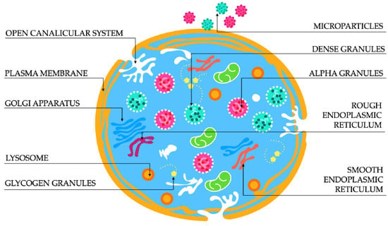
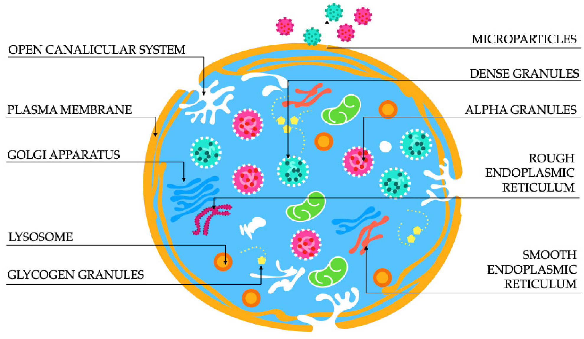
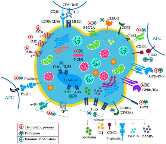
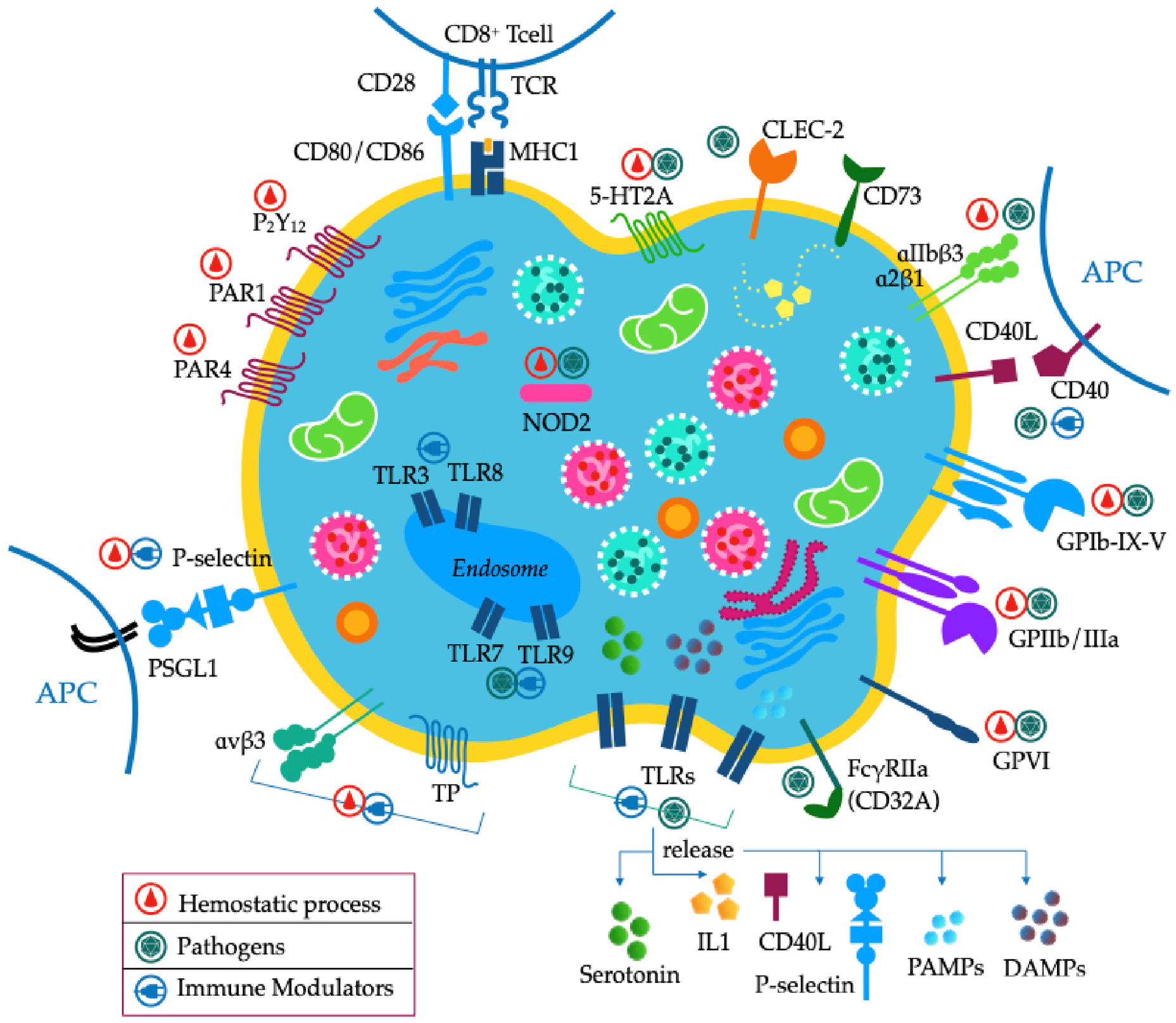

Disclaimer/Publisher’s Note: The statements, opinions and data contained in all publications are solely those of the individual author(s) and contributor(s) and not of MDPI and/or the editor(s). MDPI and/or the editor(s) disclaim responsibility for any injury to people or property resulting from any ideas, methods, instructions or products referred to in the content. |
© 2026 by the authors. Licensee MDPI, Basel, Switzerland. This article is an open access article distributed under the terms and conditions of the Creative Commons Attribution (CC BY) license.