


Abstract
This study characterized six Enterococcus faecium strains isolated from Algerian camel milk to assess their molecular features and antimicrobial potential as food-associated isolates. Identification was performed using phenotypic tests, matrix-assisted laser desorption/ionization time-of-flight mass spectrometry (MALDI-TOF MS), and 16S rRNA gene sequencing. Strain diversity was assessed by repetitive sequence-based polymerase chain reaction (rep-PCR), including (GTG)5-PCR, BOX-PCR, and enterobacterial repetitive intergenic consensus PCR (ERIC-PCR). Polymerase chain reaction (PCR) screening for selected virulence-associated and antimicrobial resistance genes showed that the two selected isolates were negative for the targeted markers, indicating a preliminary molecular profile of interest but not allowing definitive safety conclusions without whole-genome sequencing and phenotypic antimicrobial susceptibility testing. Both strains showed antimicrobial activity against Gram-positive and Gram-negative indicators. Strain 9 showed notable inhibition against Klebsiella pneumoniae subsp. pneumoniae ATCC 13883, with an inhibition zone of 23.4 ± 2.3 mm using native supernatant, which increased after concentration. Trypsin treatment abolished activity, supporting the proteinaceous nature of the inhibitory compounds. The enterocin genes entA and entB were detected in strains 9 and 13, indicating genetic potential only. Further genomic, safety, and functional investigations are required.
1. Introduction
Camel milk is a valuable nutritional resource in arid and semi-arid regions, including Algeria, and harbours a diverse microbiota that may influence the safety, quality, and functional properties of traditional dairy products [1,2]. Its reported health-promoting properties have been associated with bioactive components, antimicrobial proteins, and beneficial microbial populations [3,4]. Among the microbiota associated with camel milk, lactic acid bacteria, particularly Enterococcus faecium (E. faecium), have attracted considerable attention because of their potential contribution to fermentation processes, flavour development, antimicrobial compound production, and food biopreservation. Some E. faecium strains have also been investigated for probiotic-related traits, including tolerance to gastrointestinal stress conditions [5,6,7].
Several technologically relevant lactic acid bacteria have been described in camel milk, including species belonging to Lactiplantibacillus, Limosilactobacillus, Lacticaseibacillus, Lactococcus, Leuconostoc, Pediococcus, and Weissella, highlighting the functional diversity of this ecosystem [8,9]. These microorganisms may contribute to fermentation, preservation, and the production of bioactive metabolites such as organic acids, bacteriocins, and antioxidant compounds [10,11].
Molecular investigations have shown that camel milk-derived E. faecium strains may produce bacteriocins, including enterocins, with inhibitory activity against foodborne pathogens such as Listeria monocytogenes and Clostridium perfringens [12,13]. These antimicrobial peptides are of interest as natural preservatives because they may contribute to food safety, shelf-life extension, and the reduction in undesirable microbial populations in fermented dairy products. In addition, recent studies have emphasized the technological and antimicrobial potential of Enterococcus spp. within the camel milk microbiota, supporting their possible relevance in food biotechnology [14].
However, the use of E. faecium in food-related applications requires careful strain-level evaluation. Although some strains may display useful technological or functional traits, E. faecium also includes hospital-associated lineages, including vancomycin-resistant enterococci, that may harbour antimicrobial resistance determinants, virulence-associated genes, and mobile genetic elements. The ability of enterococci to acquire and transfer genetic determinants through horizontal gene transfer represents an important safety concern, particularly when strains are considered for food biotechnology applications [15,16,17]. Therefore, screening for virulence-associated genes and antimicrobial resistance determinants represents an essential preliminary step in the characterization of food-associated E. faecium strains before considering any technological use.
The selected gene panel was chosen to provide a preliminary assessment of safety-related and functional traits commonly investigated in Enterococcus spp. [12,13,15,16,17]. Virulence-associated targets included genes related to cytolysin production (cylM and cylA), gelatinase activity (gelE), adhesion or aggregation traits (agg and ace), hyaluronidase activity (hyl), and pheromone-related determinants (cob and ccf) [15,16,17]. Antimicrobial resistance targets included genes associated with vancomycin resistance (vanA, vanB, and vanC1) and macrolide resistance (ermA and ermB) [15,16,17]. Enterocin structural genes were also screened to evaluate the genetic potential for bacteriocin production [12,13].
Accordingly, the objective of this study was to characterize camel milk-derived E. faecium isolates using phenotypic, proteomic, and molecular methods and to evaluate their antimicrobial activity against selected indicator pathogens. For this purpose, six E. faecium strains isolated from Algerian camel milk were investigated using MALDI-TOF MS, 16S rRNA gene sequencing, rep-PCR genotyping, PCR-based screening of selected enterocin structural genes, virulence-associated genes and antimicrobial resistance determinants, and in vitro antimicrobial activity testing. This approach aimed to provide a preliminary molecular and functional assessment of camel milk-derived E. faecium strains and to identify isolates that warrant further genomic, safety, and functional investigation.
2. Materials and Methods
2.1. Bacterial Cultures and Phenotypical Identification of the Isolates
Six bacterial isolates previously recovered from four raw camel milk samples collected between 2015 and 2016 in southwestern Algeria, specifically in the regions of Ghardaïa and Béchar, were used in this study. The isolates were coded 9, 10, 11, 12, 13, and 17 for laboratory traceability. These isolates were selected from a previously established collection of lactic acid bacteria isolated and preliminarily characterized as described by Saidi et al. [18]. Briefly, camel milk samples were collected directly from lactating camels, transported to the laboratory at 4 °C in an insulated cooler containing ice packs, and processed immediately upon arrival. Samples were serially diluted in sterile peptone water and plated on MRS and M17 agar under selective growth conditions. Isolated colonies were purified by successive streaking steps before preliminary phenotypic characterization.
The isolates were originally preserved in MRS broth supplemented with 30% (v/v) glycerol and stored at −20 °C. For experimental work, frozen stocks were reactivated by inoculation into fresh MRS broth at approximately 1% (v/v) and incubated at 30 °C for 24 h under aerobic conditions. For long-term preservation, stock cultures were maintained in MRS broth containing 30% glycerol and stored at −80 °C.
Preliminary taxonomic identification followed standard morphological and biochemical assays for lactic acid bacteria, as previously described by Saidi et al. [18]. These assays included Gram staining, catalase reaction, gas production from glucose using the Durham tube method, tolerance to NaCl at 4% and 6.5%, growth at alkaline pH 9.6, and growth at 45 °C. Presumptive identification as Enterococcus spp. was based on Gram-positive coccoid morphology, catalase-negative reaction, growth at 45 °C, tolerance to 6.5% NaCl, and growth at pH 9.6. Only isolates showing traits compatible with the genus Enterococcus were selected for further molecular screening and antimicrobial characterization.
2.2. Identification and Genotyping of Isolates
2.2.1. MALDI-TOF MS Identification
MALDI-TOF MS identification was performed at the Genomics Technology Platform of the Higher School of Biological Sciences of Oran (ESSBO, Oran, Algeria) using the MALDI Biotyper Sirius GP System (Bruker Daltonics GmbH, Bremen, Germany). Isolates were first grown on MRS agar at 30 °C for 24 h under aerobic conditions. For each isolate, a single colony was deposited onto an MBT Biotarget 96 plate (Bruker), overlaid with 1 µL of 70% formic acid, and allowed to dry. Then, 1 µL of α-cyano-4-hydroxycinnamic acid (HCCA) matrix solution was added before analysis, following the manufacturer’s recommendations and the procedure described by [19].
Spectra were acquired with FlexControl software (version 3.4, Bruker) and processed using MBT Compass HT software (version 5.1, Bruker). Before analysis, calibration was performed using the Bruker Bacterial Test Standard. Identification scores were interpreted according to the Bruker criteria: values ≥ 2.0 were considered reliable for species-level identification, whereas scores between 1.70 and 1.99 were considered reliable only at the genus level. For each isolate, the best-matching species identification and the corresponding MALDI-TOF MS log(score) value were recorded individually.
2.2.2. DNA Extraction
Genomic DNA was prepared from the selected isolates using the PureLink Genomic DNA Mini Kit (Thermo Scientific, Waltham, MA, USA), according to the manufacturer’s instructions. A lysozyme pretreatment step (10 mg/mL) was included to improve lysis of Gram-positive cells, as previously described [19]. DNA quality was assessed with a ScanDrop spectrophotometer (Analytik Jena, Jena, Germany). The A260/A280 ratios ranged from 1.8 to 2.0, while A260/A230 ratios were above 2.0, indicating satisfactory purity. DNA integrity was checked by agarose gel electrophoresis, and the extracted DNA was stored at –20 °C until use.
2.2.3. 16S rRNA Gene Amplification, Sequencing, and Phylogenetic Analysis
For molecular confirmation, the 16S rRNA gene was amplified using the universal bacterial primers 27F and 1495R (Table 1), following previously described conditions [20,21]. Primers were synthesized by the Genomics Technology Platform of the Higher School of Biological Sciences of Oran (ESSBO, Oran, Algeria). PCR was performed in a final volume of 50 µL using 2× PCR Master Mix (Thermo Scientific, Waltham, MA, USA) in a SimpliAmp thermal cycler (Applied Biosystems, Foster City, CA, USA).
Table 1.
PCR primers and annealing temperatures used for molecular characterization of Enterococcus spp.
The amplification program included an initial denaturation at 94 °C for 10 min, followed by 35 cycles of 94 °C for 45 s, 54 °C for 45 s, and 72 °C for 90 s. A final extension was carried out at 72 °C for 10 min. The amplified fragments, approximately 1500 bp in size, were separated on a 1% agarose gel prepared in TBE buffer, and fragment size was estimated using a GeneRuler Express DNA Ladder (Thermo Scientific, Waltham, MA, USA). After sequencing, raw reads were quality-trimmed before analysis; consequently, approximately 700 bp of high-quality sequence were retained for BLAST comparison and phylogenetic analysis. Phylogenetic relationships were inferred using the maximum likelihood method with 1000 bootstrap replicates, and Lactiplantibacillus plantarum was used as the outgroup.
2.2.4. rep-PCR Genotyping of Isolates
To evaluate genetic diversity and confirm that the isolates represented distinct strains, repetitive sequence-based PCR (rep-PCR) was performed using three primer systems: GTG5, BOXA1R, and ERIC [21,36,37,38]. Primers were synthesized by the Genomics Technology Platform (ESSBO, Oran, Algeria). This combined fingerprinting approach was used to compare the isolates at the strain level and to support the selection of non-redundant representatives for further analyses [39].
PCR amplifications followed the optimized protocol reported by [21]. Each 40 µL reaction contained approximately 100 ng of genomic DNA, 2× DreamTaq PCR Master Mix (Thermo Scientific, Waltham, MA, USA), DMSO, BSA, nuclease-free water, and the corresponding primers. For (GTG)5-PCR and BOX-PCR, 3 µL of 5 µM primer were used, whereas ERIC-PCRs contained 1.5 µL of each primer. Negative controls were included in each run, and amplification was performed for 35 cycles in a SimpliAmp thermal cycler (Applied Biosystems).
The PCR products were separated on 1.5% agarose gels prepared in 1× TBE buffer at 100 V for 2 h. Gels were stained with SYBR Safe and visualized using a ChemiDoc Imaging System (Bio-Rad, Hercules, CA, USA). GeneRuler Express and 1 kb Plus DNA ladders (Thermo Scientific, Waltham, MA, USA) were used as molecular size markers. To ensure reproducibility, each rep-PCR assay was repeated several times, and only reproducible banding patterns were retained for profile comparison and cluster analysis.
Banding profiles were analysed with GelJ software (version 2.0.0). Similarity matrices were generated using the Dice coefficient, and dendrograms were constructed by the UPGMA clustering method to assess genetic relationships among isolates [40,41].
2.3. PCR-Based Detection of Virulence-Associated, Antimicrobial Resistance, and Enterocin-Encoding Genes
To provide a preliminary molecular assessment of the safety-related and functional traits of the isolates, PCR-based screening was performed to detect selected virulence-associated genes, antibiotic resistance genes, and enterocin structural genes.
2.3.1. Detection of Virulence-Associated and Antimicrobial Resistance Markers
All six isolates were first tested for five virulence-associated determinants: cylM, cylA, cob, ccf, and gelE. Isolates that were negative in this initial screening were then subjected to a second PCR screening targeting the ddl molecular marker, additional virulence-associated genes (agg, hyl, and ace) and selected antimicrobial resistance genes (vanA, vanB, vanC1, ermA, and ermB). Primer sequences, expected amplicon sizes, and annealing temperatures are provided in Table 1. As outlined in the Introduction, this targeted gene panel was selected to provide a preliminary PCR-based assessment of safety-related and functional traits in Enterococcus spp.
PCRs were carried out in a final volume of 25 µL containing 12.5 µL of 2× PCR Master Mix (Thermo Scientific, Waltham, MA, USA), 1 µL of each primer, 5 µL of genomic DNA corresponding to approximately 150 ng, and nuclease-free water. The cycling program started with denaturation at 94 °C for 5 min, followed by 35 cycles of 94 °C for 1 min, primer-specific annealing at the temperature indicated in Table 1 for 1 min, and extension at 72 °C for 1 min 45 s. A final extension was performed at 72 °C for 5 min. Amplicons were separated on 1.2% agarose gels. A GeneRuler 1 kb DNA Ladder (Thermo Scientific, Waltham, MA, USA) was used for size estimation. Gels were stained with SYBR Safe DNA Gel Stain (Thermo Scientific, Waltham, MA, USA) and visualized using a ChemiDoc Imaging System (Bio-Rad, Hercules, CA, USA). For the secondary screening of the two selected isolates, coded 9 and 13, 16S rRNA gene amplification was used as a DNA amplifiability control.
2.3.2. Detection of Enterocin-Encoding Genes
Screening for enterocin structural genes (entA, entB, entL50A, entL50B, ent31, entP, entAS-48, and entCRL35) was carried out with the gene-specific primers listed in Table 1. The reaction mixture and electrophoretic analysis were the same as those described for virulence and antimicrobial resistance gene screening.
For enterocin genes, amplification was performed using a specific thermal program: 95 °C for 30 s, followed by 35 cycles of 95 °C for 30 s, primer-specific annealing at the temperature indicated in Table 1 for 30 s, and extension at 72 °C for 1 min 40 s. A final extension step was then performed at 72 °C for 7 min. For the secondary enterocin gene screening, amplification of the housekeeping gene adk was used as a DNA amplifiability control.
2.4. Antimicrobial Activity Assay
The antimicrobial activity of the two selected isolates, coded 9 and 13, which were negative for the targeted virulence-associated and antimicrobial resistance genes, was tested against nine ATCC indicator strains listed in Table 2. Antagonistic activity was assessed using an agar well diffusion assay adapted from [21].
Table 2.
Indicator strains used for antimicrobial activity evaluation.
The tested isolates were grown in MRS broth at 37 °C for 24 h. Indicator strains were cultured in Luria–Bertani broth (LB) (Merck, Darmstadt, Germany) under the same incubation conditions and adjusted to 0.5 McFarland turbidity, corresponding to approximately 1.5 × 10⁸ CFU/mL. These standardized suspensions were spread onto LB soft agar containing 0.75% agar.
Cell-free supernatants (CFS) were prepared from cultures of the two selected isolates, coded 9 and 13, by centrifugation at 10,000× g for 15 min at 4 °C. To limit inhibition related to acidification, particularly lactic acid produced during growth in MRS broth, the supernatants were adjusted to pH 7.0 with sterile 1 M NaOH. They were then sterilized by filtration through 0.22 µm syringe filters (Merck Millipore, Darmstadt, Germany).
Native supernatants (NS) corresponded to neutralized and filter-sterilized CFS used without further concentration. Concentrated supernatants (CS) were prepared by freeze-drying approximately 50 mL of neutralized and filter-sterilized CFS. The lyophilized material was then reconstituted in 1 mL of sterile MRS broth, resulting in an approximately fifty-fold concentration.
For the agar well diffusion assay, wells of 6 mm in diameter were filled with 100 µL of either NS or CS. Plates were kept at 4 °C for 2 h to allow diffusion, then incubated at 37 °C for 24 h. Inhibition was evaluated by measuring the diameter of the clear zones around the wells, including the 6 mm well diameter, and results were expressed in millimetres (mm). Sterile uninoculated MRS broth was used as the negative control.
To assess whether the inhibitory activity was associated with proteinaceous compounds, CFS were treated with trypsin at a final concentration of 1 mg/mL of cell-free supernatant (Sigma-Aldrich, Saint Louis, MO, USA). Treated samples were incubated at 37 °C for 2 h, after which trypsin was inactivated by heating at 100 °C for 3 min. Trypsin-treated and untreated samples were then tested using the same agar well diffusion assay against the same indicator strains [21].
2.5. Statistical Analysis
Experiments were carried out in triplicate, and data are presented as mean ± standard deviation (SD). Statistical comparisons were performed using one-way analysis of variance (ANOVA), followed by Tukey’s post hoc multiple comparison test. For antimicrobial activity assays, statistical comparisons were performed separately for each producing isolate and each indicator organism to evaluate the effect of supernatant concentration on inhibition zone diameters. Analyses were conducted with GraphPad Prism software (version 9.0.0; GraphPad Software, San Diego, CA, USA). Differences were considered statistically significant at p ≤ 0.05.
3. Results
3.1. Presumptive Identification of Enterococcal Isolates
The six camel milk isolates were first examined using basic phenotypic tests. All isolates were Gram-positive, catalase-negative cocci, in agreement with the general profile of lactic acid bacteria. Microscopic observation confirmed their coccoid morphology. The isolates grew in the presence of 4% and 6.5% NaCl, tolerated pH 9.6, and were able to grow at 45 °C. Together, these characteristics supported their presumptive assignment to the genus Enterococcus before molecular identification.
3.2. Confirmation of the Isolates as E. faecium
A combined proteomic and molecular identification strategy was applied to confirm the taxonomic status of the isolates.
3.2.1. MALDI-TOF MS Identification
MALDI-TOF MS analysis identified all six isolates as E. faecium, with log(score) values ranging from 2.20 to 2.34. The individual log(score) values were 2.31, 2.26, 2.34, 2.21, 2.20, and 2.32 for the isolates coded 9, 10, 11, 12, 13, and 17, respectively. Since all scores were above the 2.0 threshold, the identifications were considered reliable at the species level. The Bacterial Test Standard (BTS) consistently produced scores above 2.0, confirming that the system was properly calibrated during the analyses.
3.2.2. 16S rRNA-Based Confirmation
Partial 16S rRNA gene sequencing was used to support the MALDI-TOF MS identification. Sequence comparison confirmed that the six isolates belonged to E. faecium, in agreement with both the proteomic identification and the phenotypic profile. BLAST 2.17.0 analysis against the NCBI GenBank database showed 98–99% similarity with reference E. faecium sequences, based on approximately 700 bp of high-quality trimmed sequences.
3.3. Phylogenetic Relationships Among the Isolates
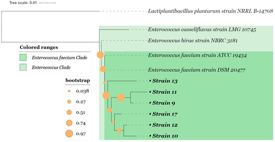
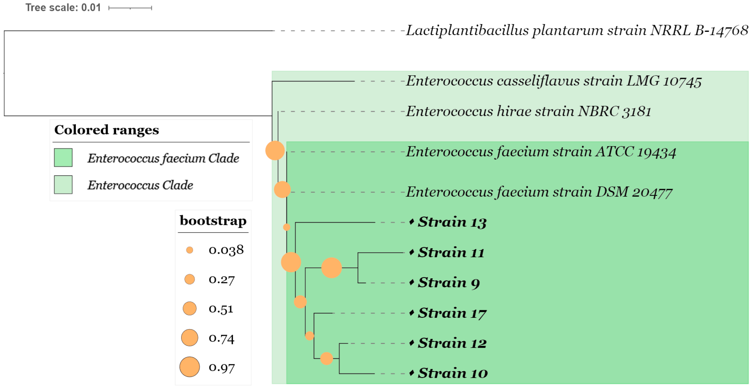
Phylogenetic relationships among the six Enterococcus isolates recovered from Algerian camel milk were analysed using the Maximum Likelihood method based on partial 16S rRNA gene sequences. Lactiplantibacillus plantarum was used as the outgroup. The resulting tree separated the isolates within the Enterococcus genus and supported their taxonomic affiliation with E. faecium (Figure 1).
Figure 1.
Phylogenetic tree of six Enterococcus isolates based on 16S rRNA sequences using the maximum likelihood method with 1000 bootstrap replicates, with Lactiplantibacillus plantarum as the outgroup.
All six isolates, coded 9, 10, 11, 12, 13, and 17, clustered within the E. faecium group together with the reference strains E. faecium ATCC 19434 and DSM 20477. The short branch lengths observed among the isolates indicated close genetic relatedness, which is consistent with their classification within the same species. Slight differences in branch length may reflect limited intraspecific variation among the camel milk isolates.
The tree also distinguished E. faecium from the closely related species Enterococcus casseliflavus LMG 10745 and Enterococcus hirae NBRC 3181. These species formed separate neighbouring branches, while the outgroup, Lactiplantibacillus plantarum, was clearly positioned outside the Enterococcus cluster.
Overall, the phylogenetic analysis supported the identification of the camel milk isolates as E. faecium and complemented the MALDI-TOF MS and 16S rRNA sequence similarity results.
3.4. Genetic Diversity Revealed by rep-PCR
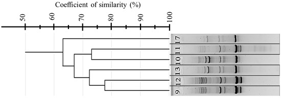
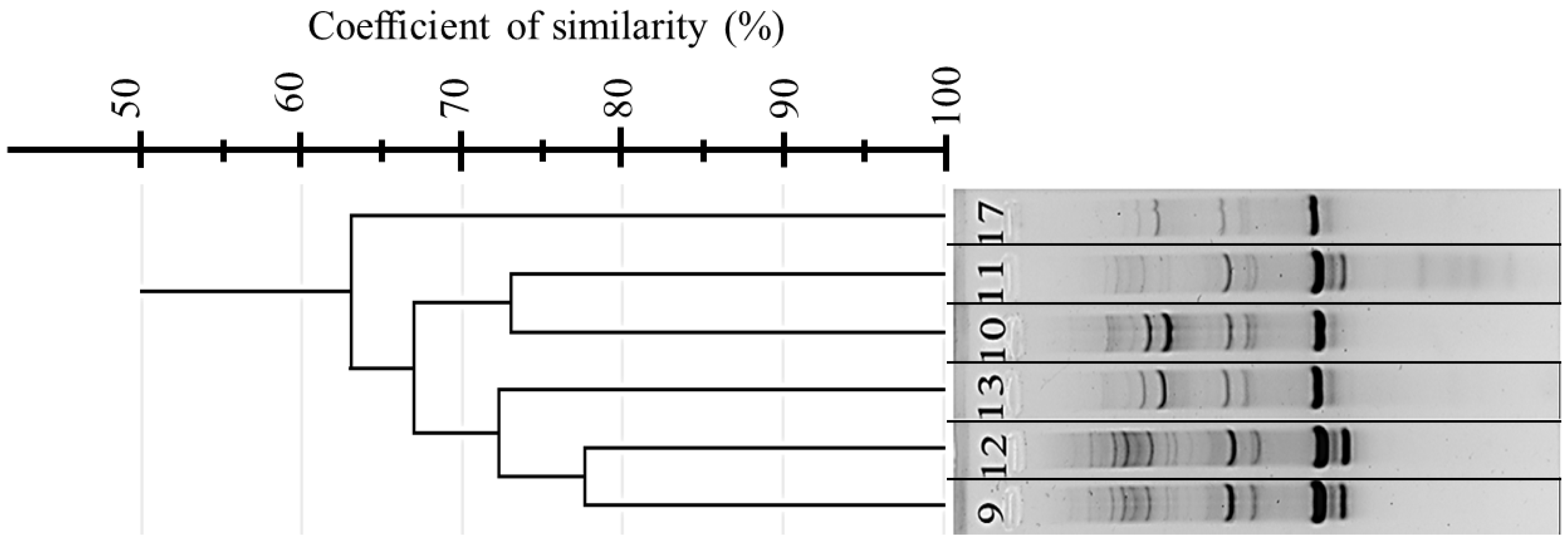
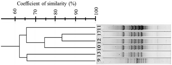
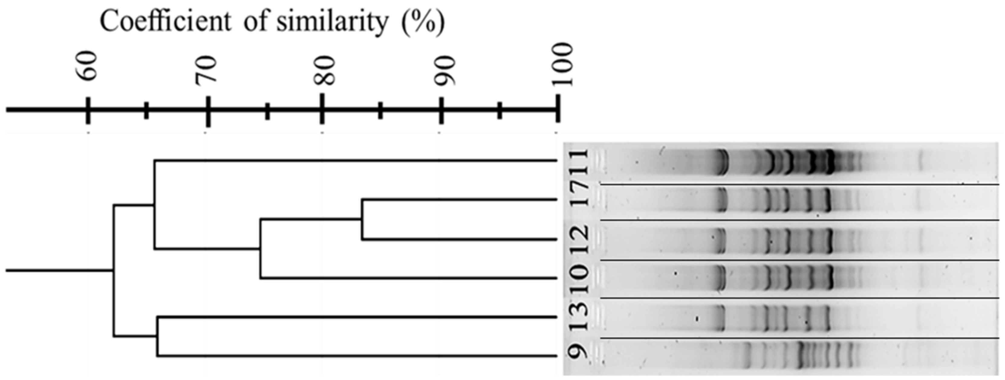
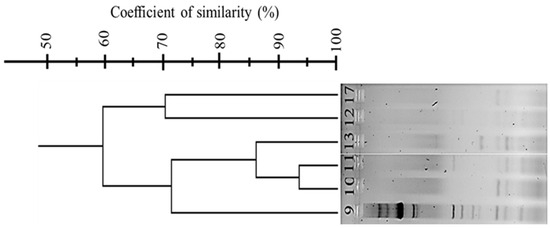
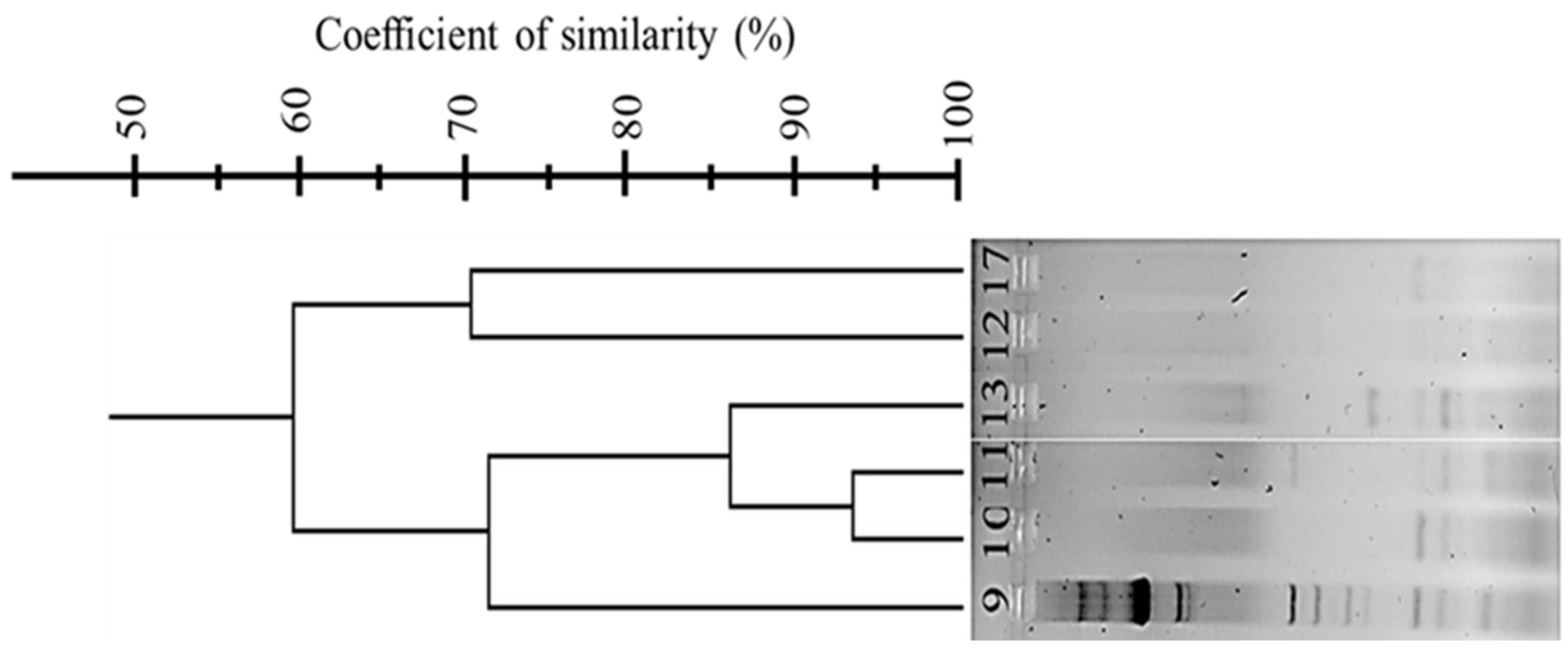
Genetic diversity among the six E. faecium isolates was investigated using three rep-PCR fingerprinting approaches: (GTG)5-PCR, BOX-PCR, and ERIC-PCR. The banding patterns were compared using the Dice similarity coefficient and clustered by the UPGMA method. The dendrograms obtained from the three approaches are shown in Figure 2, Figure 3 and Figure 4.
Figure 2.
Dendrogram showing the genetic relatedness among six E. faecium isolates based on (GTG)5-PCR fingerprinting using the (GTG)5 primer. Clustering was performed using UPGMA and Dice similarity coefficients. Three distinct clusters were observed. Non-adjacent lanes from the same gel were spliced and reordered to match the dendrogram; splicing is indicated by visible separating lines.
Figure 3.
Dendrogram showing the genetic relatedness among six E. faecium isolates based on BOX-PCR fingerprinting using the BOXA1R primer. Clustering was performed with the UPGMA algorithm and Dice similarity coefficients. Resolution of three genetically distinct clusters was achieved.
Figure 4.
Dendrogram showing the genetic relatedness among six E. faecium isolates based on ERIC-PCR fingerprinting using primers ERIC1R and ERIC2. Clustering was performed using UPGMA and Dice similarity coefficients. Two major clusters were identified, indicating reduced discriminatory capacity compared to BOX-PCR and (GTG)5-PCR.
The (GTG)5-PCR profiles showed several distinct bands and allowed separation of the isolates into three clusters (Figure 2). Cluster A included isolates 9, 12, and 13, whereas isolates 10 and 11 were grouped in Cluster B. Isolate 17 formed a separate cluster, indicating a more distinct fingerprinting profile compared with the other isolates.
BOX-PCR produced more complex banding profiles and showed a higher discriminatory capacity than (GTG)5-PCR (Figure 3). Three clusters were obtained. Isolate 11 formed an independent cluster, while isolates 17, 12, and 10 clustered together. Isolates 13 and 9 formed a third cluster.
ERIC-PCR generated fewer bands than the two other rep-PCR methods and therefore showed lower resolution (Figure 4). This method separated the isolates into two clusters: one including isolates 9, 10, 11, and 13, and another including isolates 12 and 17. Despite its lower discriminatory power, ERIC-PCR showed partial agreement with the clustering patterns obtained using (GTG)5-PCR and BOX-PCR. Non-adjacent lanes from the same gel were spliced and reordered to match the dendrogram; splicing is indicated by visible separating lines.
3.5. Virulence-Associated and Antimicrobial Resistance Gene Profiles
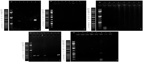
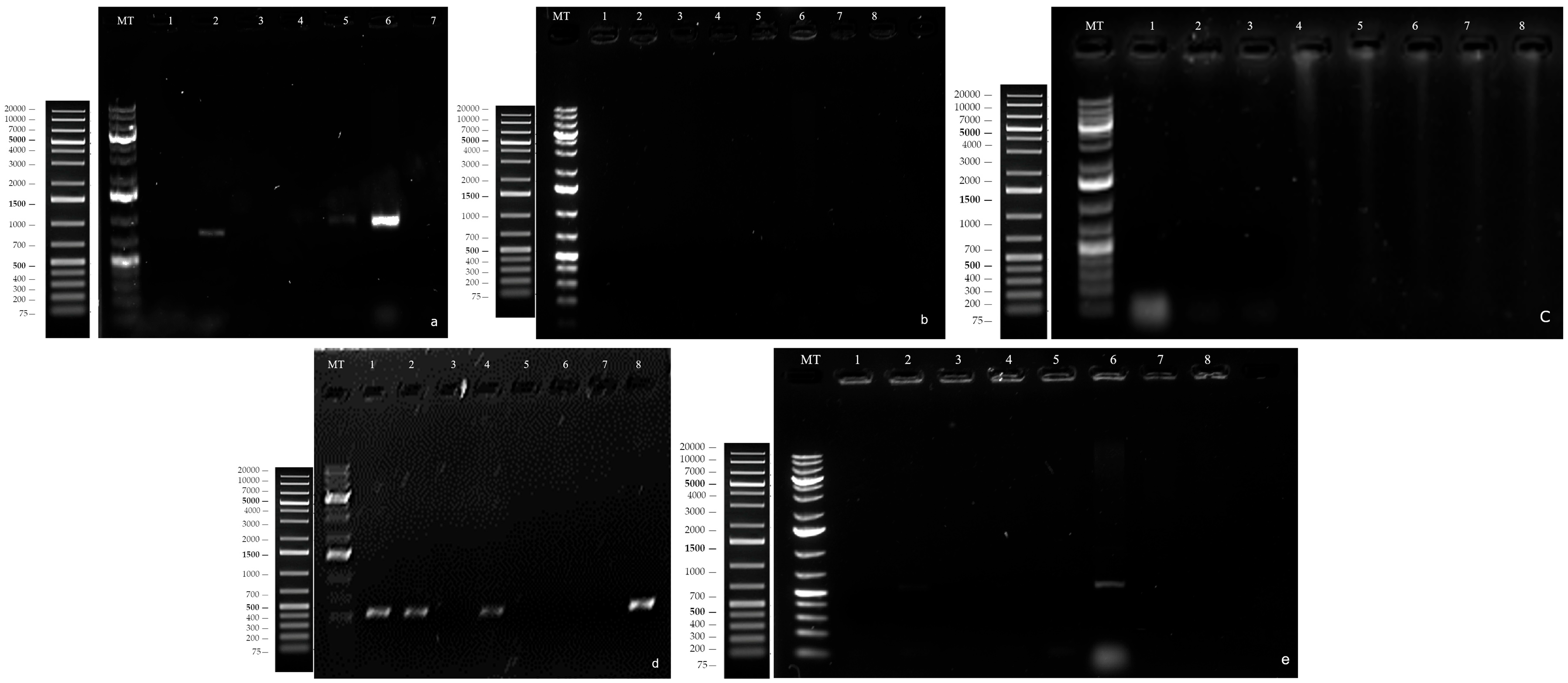
PCR screening was used to evaluate selected safety-related molecular markers in the six E. faecium isolates. The first screening targeted five virulence-associated genes: cylM, cylA, cob, ccf, and gelE (Figure 5). The two isolates coded 9 and 13 were negative for all five targets. In contrast, the other isolates carried at least one of these genes: the isolate coded 10 was positive for cob and ccf, the isolate coded 11 for gelE, the isolate coded 12 for cob and gelE, and the isolate coded 17 for ccf.
Figure 5.
PCR screening of selected virulence-associated genes in E. faecium isolates. PCR products were resolved on 1.2% agarose gels. Panels: (a) gelE; (b) cylM; (c) cylA; (d) ccf; (e) cob. MT: DNA size marker, GeneRuler 1 kb DNA Ladder. Lane assignments: lane 1 and 2, controls; lane 3, isolate coded 9; lane 4, isolate coded 10; lane 5, isolate coded 11; lane 6, isolate coded 12; lane 7, isolate coded 13; lane 8, isolate coded 17. The isolates coded 9 and 13 were negative for all targeted virulence-associated genes.
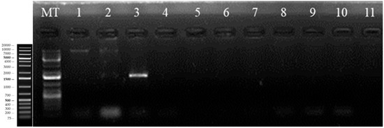
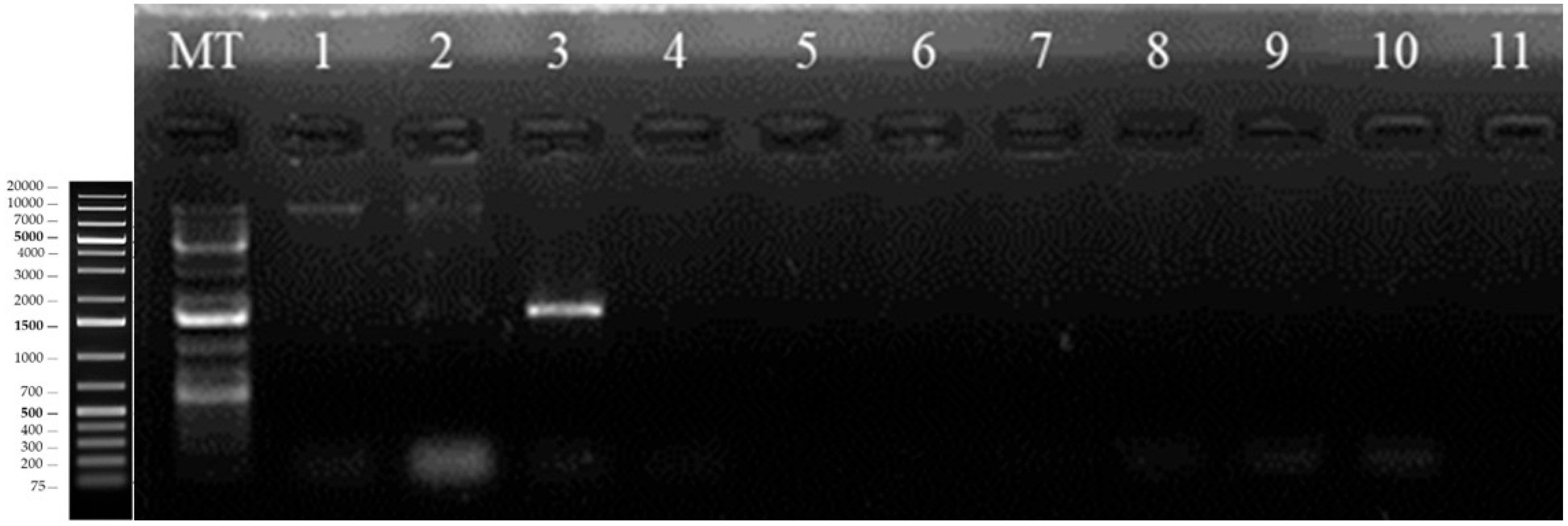
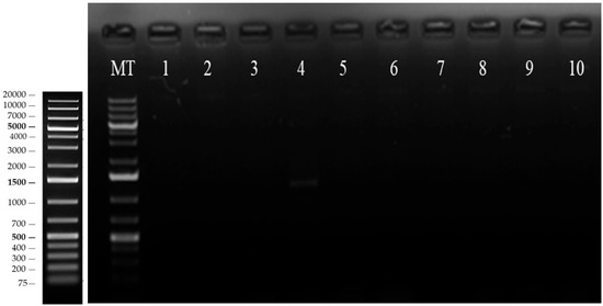
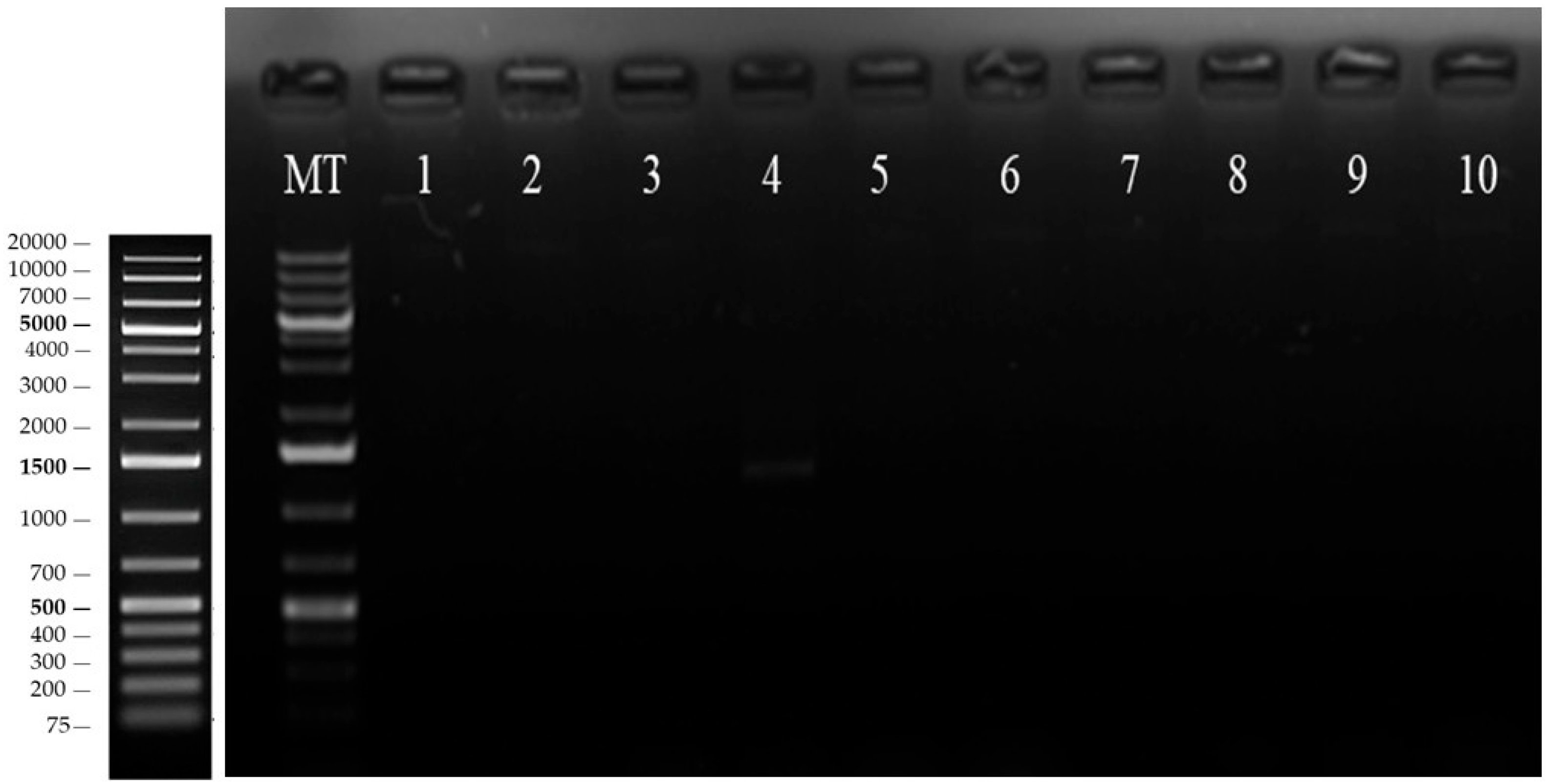
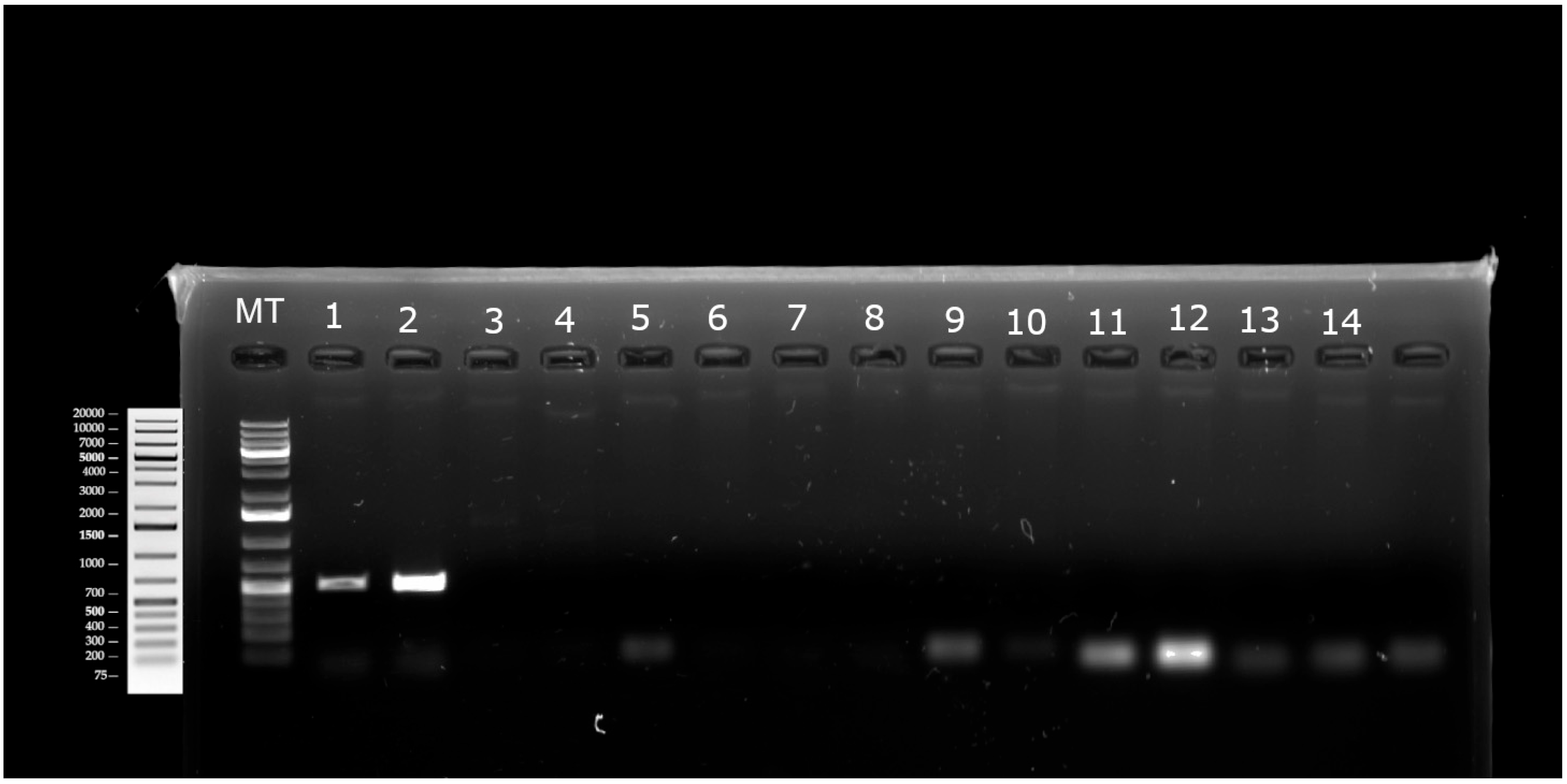
Because the isolates coded 9 and 13 lacked the initially screened virulence-associated genes, they were further analysed using an extended PCR panel. This second screening included the ddl molecular marker, additional selected virulence-associated genes (agg, hyl, and ace) (Figure 6), as well as selected antimicrobial resistance genes (vanA, vanB, vanC1, ermA, and ermB) (Figure 7). The 16S rRNA amplification control yielded the expected PCR product during this secondary screening. No PCR product was observed for the ddl molecular marker, the additional selected virulence-associated genes, or the selected antimicrobial resistance genes in either selected isolate. This PCR profile was therefore considered a preliminary molecular profile of interest and supported the selection of these two isolates for subsequent antimicrobial activity assays.
Figure 6.
Secondary PCR screening of the ddl molecular marker and selected virulence-associated genes in the two selected isolates, coded 9 and 13. PCR products corresponding to agg, ddl, hyl, and ace were resolved on a 1.2% agarose gel. MT: DNA size marker, GeneRuler 1 kb DNA Ladder. Lane assignments: lane 1, genomic DNA from isolate coded 9; lane 2, genomic DNA from isolate coded 13; lane 3, 16S rRNA amplification control; lanes 4–5, agg screening for isolates coded 9 and 13; lanes 6–7, ddl screening for isolates coded 9 and 13; lanes 8–9, hyl screening for isolates coded 9 and 13; lanes 10–11, ace screening for isolates coded 9 and 13.
Figure 7.
PCR screening of selected antimicrobial resistance genes in the two selected isolates, coded 9 and 13. PCR products corresponding to vanA, vanB, vanC1, ermA, and ermB were resolved on a 1.2% agarose gel. MT: DNA size marker, GeneRuler 1 kb DNA Ladder. Lane assignments: lanes 1–2, vanA screening for isolates coded 9 and 13; lanes 3–4, vanB screening for isolates coded 9 and 13; lanes 5–6, vanC1 screening for isolates coded 9 and 13; lanes 7–8, ermA screening for isolates coded 9 and 13; lanes 9–10, ermB screening for isolates coded 9 and 13.
3.6. Enterocin Gene Profiles of the Selected Isolates
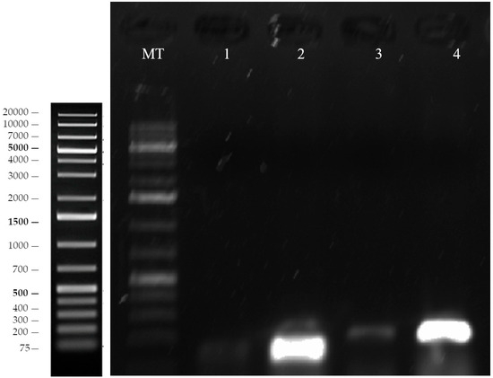
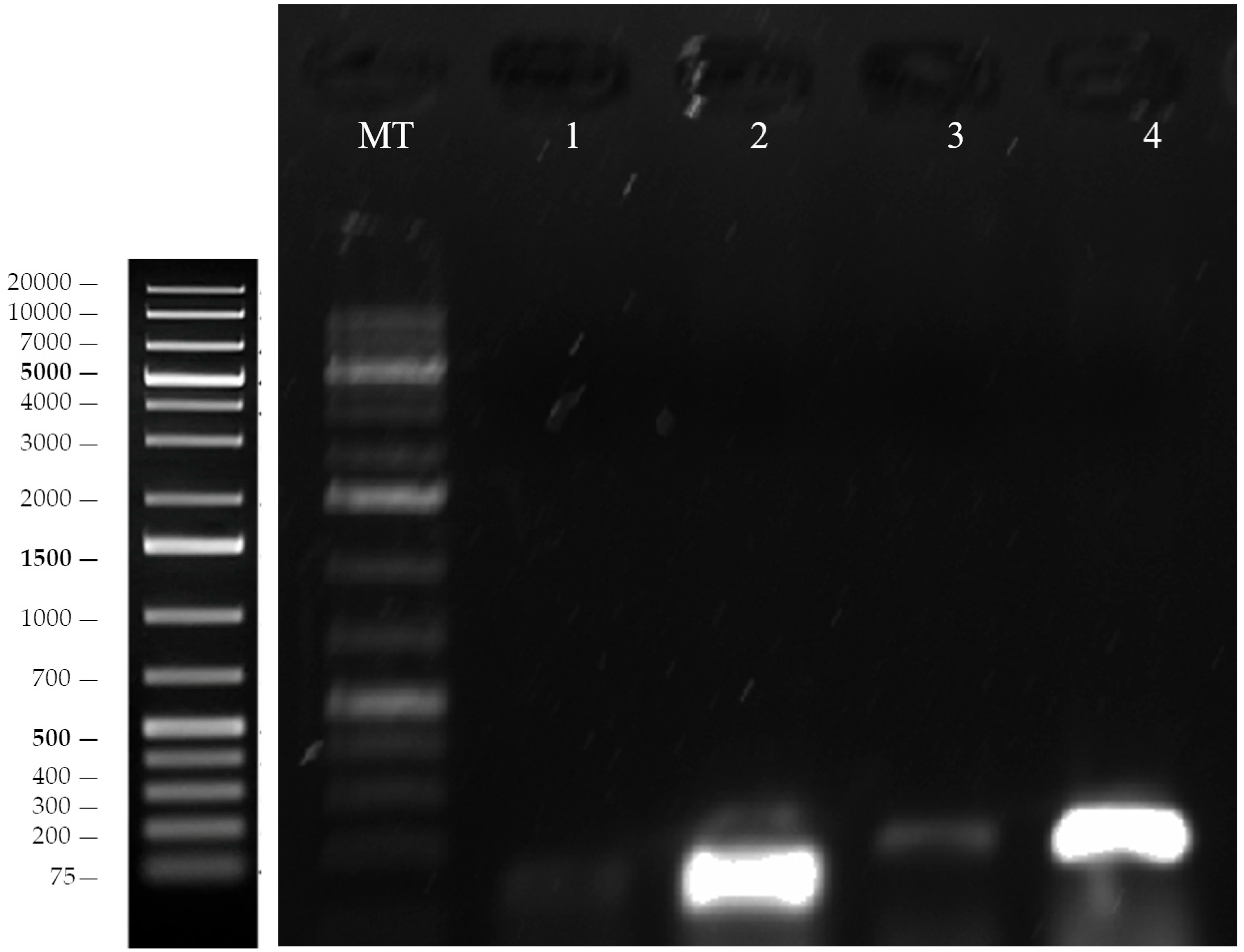
PCR screening was performed to detect enterocin structural genes in the tested isolates, including entA, entB, entL50A, entL50B, ent31, entP, entAS-48, and entCRL35. Among the screened isolates, only the two selected isolates coded 9 and 13 showed amplification for entA and entB, corresponding to enterocin A and enterocin B, respectively (Figure 8). No PCR product was observed for the other enterocin genes tested. The adk amplification control yielded the expected PCR product during the secondary enterocin gene screening (Figure 9). These results indicate that the two selected isolates carry genetic determinants associated with enterocin A and enterocin B.
Figure 8.
PCR screening of enterocin structural genes entA and entB in the two selected isolates, coded 9 and 13. PCR products were resolved on a 1.2% agarose gel. MT: DNA size marker, GeneRuler 1 kb DNA Ladder. Lane assignments: lanes 1–2, entA screening for isolates coded 9 and 13; lanes 3–4, entB screening for isolates coded 9 and 13.
Figure 9.
Secondary PCR screening of enterocin structural genes in the two selected isolates, coded 9 and 13. PCR products corresponding to entL50A, entL50B, ent31, entP, entAS-48, and entCRL35 were resolved on a 1.2% agarose gel. MT: DNA size marker, GeneRuler 1 kb DNA Ladder. Lane assignments: lanes 1–2, adk amplification control for isolates coded 9 and 13; lanes 3–4, entL50A screening for isolates coded 9 and 13; lanes 5–6, entL50B screening for isolates coded 9 and 13; lanes 7–8, ent31 screening for isolates coded 9 and 13; lanes 9–10, entP screening for isolates coded 9 and 13; lanes 11–12, entAS-48 screening for isolates coded 9 and 13; lanes 13–14, entCRL35 screening for isolates coded 9 and 13.
3.7. Antimicrobial Activity of Native and Concentrated Supernatants
The two isolates coded 9 and 13 were retained for antimicrobial activity testing because they were negative for the targeted virulence-associated and antimicrobial resistance genes. Their inhibitory activity was evaluated against nine ATCC indicator strains using the agar well diffusion assay (Figure 10, Figure 11 and Figure 12). In this assay, indicator strains showing clear inhibition zones were considered susceptible to the tested cell-free supernatants, whereas the absence of inhibition was interpreted as no detectable susceptibility under the assay conditions.
Figure 10.
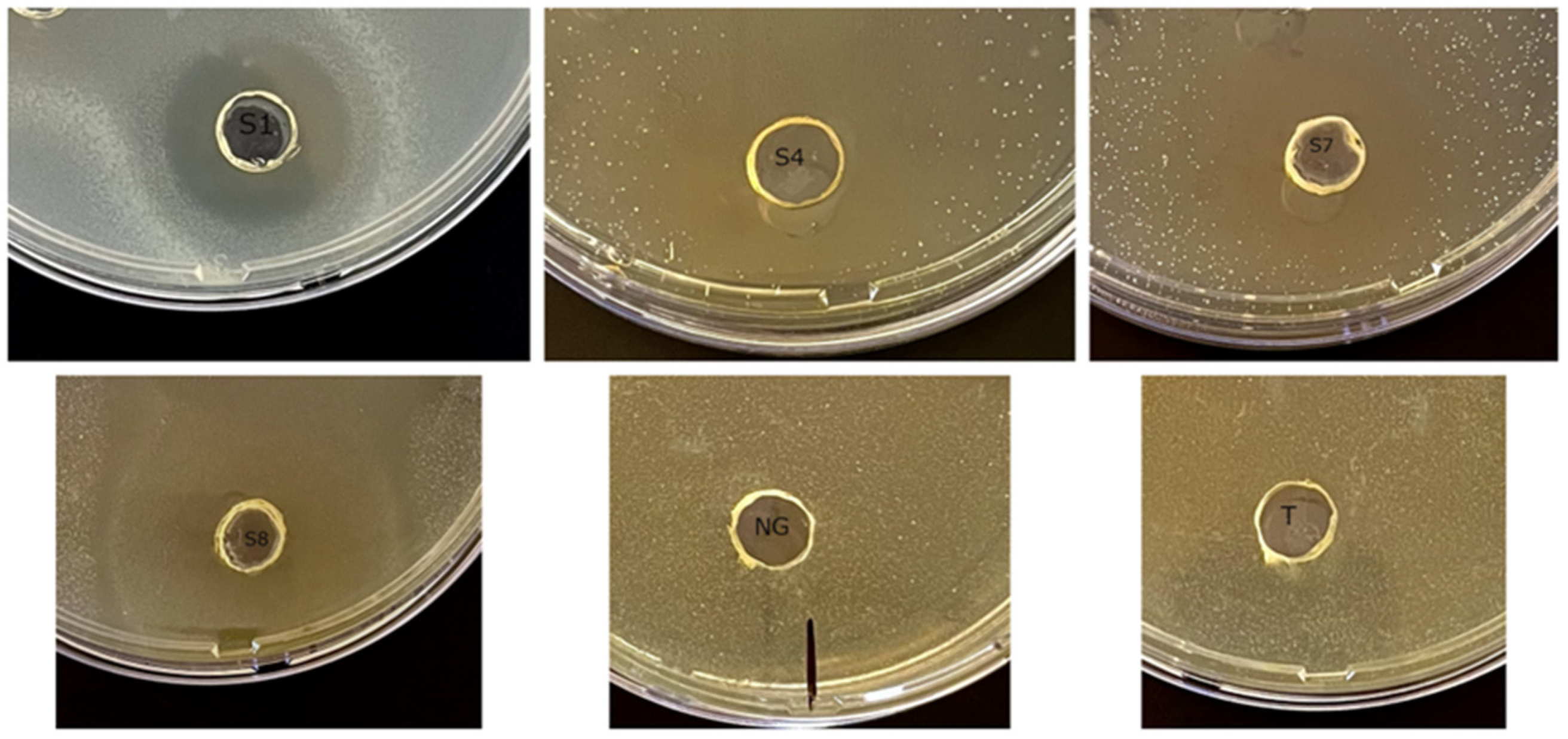
Antimicrobial activity of concentrated cell-free supernatant from the selected E. faecium isolate coded 9 against ATCC indicator strains. S1: methicillin-sensitive Staphylococcus aureus ATCC 29213; S4: Selenomonas sputigena ATCC 33150; S7: Staphylococcus aureus subsp. aureus ATCC 25923; S8: Klebsiella pneumoniae subsp. pneumoniae ATCC 13883. CS: concentrated supernatant; NG: negative control; T: trypsin-treated supernatant.
Figure 11.
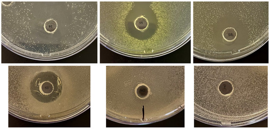
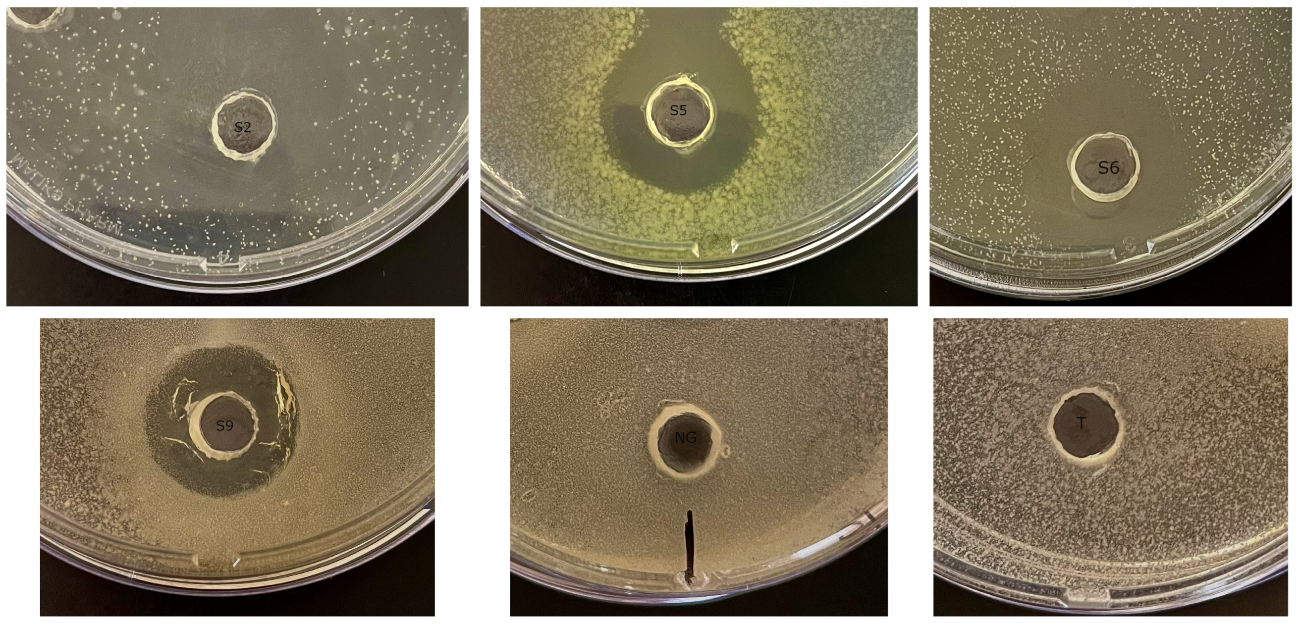
Antimicrobial activity of concentrated cell-free supernatant from the selected E. faecium isolate coded 13 against ATCC indicator strains. S2: Salmonella enterica subsp. enterica serovar Typhimurium ATCC 14028; S5: Pseudomonas aeruginosa ATCC 27853; S6: Escherichia coli ATCC 25922; S9: Bacillus cereus ATCC 11778. CS: concentrated supernatant; NG: negative control; T: trypsin-treated supernatant.
Figure 12.
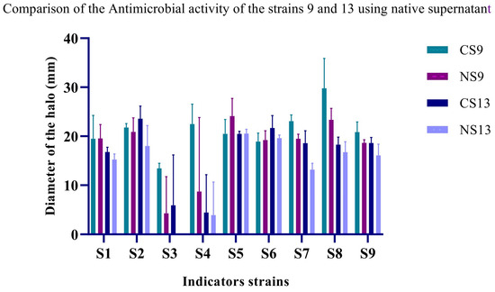
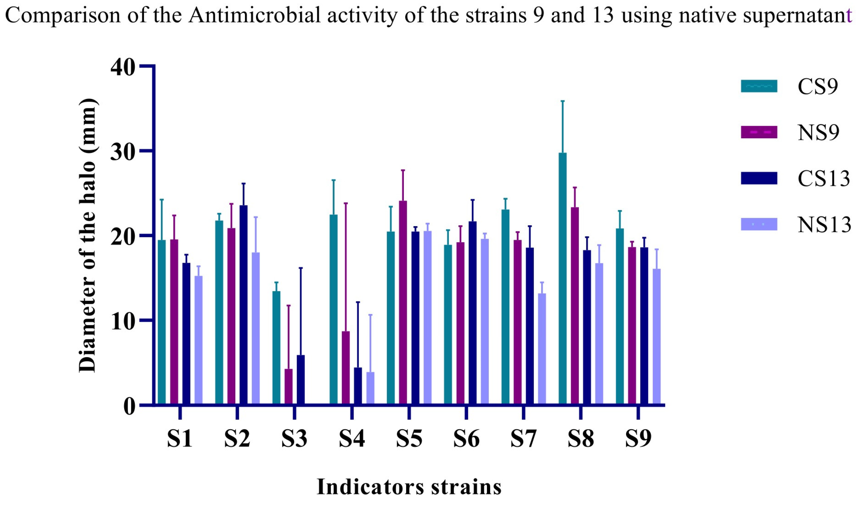
Comparative antimicrobial activity of native and concentrated cell-free supernatants from the two selected E. faecium isolates, coded 9 and 13, against ATCC indicator strains. NS: native supernatant; CS: concentrated supernatant; S1: methicillin-sensitive Staphylococcus aureus ATCC 29213; S2: Salmonella enterica subsp. enterica serovar Typhimurium ATCC 14028; S3: methicillin-resistant Staphylococcus aureus ATCC 43300; S4: Selenomonas sputigena ATCC 33150; S5: Pseudomonas aeruginosa ATCC 27853; S6: Escherichia coli ATCC 25922; S7: Staphylococcus aureus subsp. aureus ATCC 25923; S8: Klebsiella pneumoniae subsp. pneumoniae ATCC 13883; S9: Bacillus cereus ATCC 11778. Data represent mean inhibition zone diameters from triplicate assays.
For the isolate coded 9, both native and concentrated cell-free supernatants inhibited the tested Gram-positive and Gram-negative indicator strains. Concentrated supernatants generally produced larger inhibition zones than native supernatants, with increases varying according to the indicator strain (Figure 10).
Among Gram-positive indicators, methicillin-resistant Staphylococcus aureus (MRSA, S3) showed weak and variable inhibition when tested with native supernatant. After concentration, inhibition increased markedly to 13.5 ± 1.0 mm. Increased inhibition after concentration was also observed for Selenomonas sputigena (S4).
Klebsiella pneumoniae subsp. pneumoniae (S8) was the most susceptible indicator strain, with inhibition zones of 23.4 ± 2.3 mm for native supernatant and 29.8 ± 6.1 mm for concentrated supernatant. The Staphylococcus aureus strains S1 and S7 were also clearly inhibited by the supernatants of the isolate coded 9.
Gram-negative indicators, including Salmonella enterica, Escherichia coli, and Pseudomonas aeruginosa, were inhibited by both native and concentrated supernatants, with larger zones generally observed after concentration. The isolate coded 13 also inhibited the tested indicator strains, but the inhibition zones were generally smaller and more variable than those observed with the isolate coded 9 (Figure 11). Concentration particularly enhanced the activity of the isolate coded 13 against Salmonella enterica (S2) and S. aureus (S7), whereas MRSA (S3) and Selenomonas sputigena (S4) showed weak or no detectable susceptibility to the native supernatant.
Trypsin treatment completely abolished the antimicrobial activity of both selected isolates, supporting the proteinaceous nature of the inhibitory compounds and indicating the possible involvement of bacteriocin-like substances. Statistical analysis using one-way ANOVA followed by Tukey’s post hoc test showed that concentration significantly increased the antimicrobial activity of the isolate coded 9 against MRSA (S3) (F(1,4) = 12.9, p = 0.022), S. aureus (S7) (F(1,4) = 21.3, p = 0.009), and K. pneumoniae (S8) (F(1,4) = 12.4, p = 0.023). For the isolate coded 13, a significant increase was observed only against S. aureus (S7) (F(1,4) = 7.8, p = 0.043), whereas the differences observed for the other indicator strains were not statistically significant.
Overall, the isolate coded 9 showed stronger and more consistent antimicrobial activity than the isolate coded 13, particularly after concentration of the supernatant. These findings support further investigation of both selected isolates as potential sources of proteinaceous antimicrobial compounds.
4. Discussion
This study provides a taxonomic and phylogenetic characterization of E. faecium isolates recovered from Algerian camel milk, a traditional dairy product known for its nutritional value and microbial diversity [42,43]. The isolates showed phenotypic traits compatible with the genus Enterococcus, including Gram-positive, catalase-negative coccoid morphology, tolerance to elevated NaCl concentrations, growth at alkaline pH, and ability to grow at 45 °C. These physiological characteristics are frequently reported among food-associated enterococci and may contribute to their adaptation and technological behaviour in dairy environments [44,45,46].
Species identification was supported by the combined use of MALDI-TOF MS and 16S rRNA gene sequencing. All isolates showed MALDI log(score) values ≥ 2.0, allowing species-level identification. Although partial 16S rRNA sequencing (~700 bp) has limited discriminatory power at the strain level because of the conserved nature of this gene, its agreement with the MALDI-TOF MS results supports the assignment of the isolates to E. faecium [18,47]. The minor sequence differences observed among isolates, together with the rep-PCR fingerprinting profiles described below, are consistent with intra-species diversity among the camel milk-derived E. faecium isolates [48,49]. Thus, the combination of proteomic and molecular identification provided complementary evidence for the taxonomic classification of the isolates.
Phylogenetic analysis based on the Maximum Likelihood method placed all camel milk-derived isolates within the E. faecium clade, in close association with the reference strains ATCC 19434 and DSM 20477. The short internal branch lengths suggest close genetic relatedness among the isolates, which is commonly observed among food-associated E. faecium populations recovered from dairy environments [50,51]. Their separation from neighbouring species, including E. hirae and E. casseliflavus, further supports the species-level assignment and is consistent with the taxonomic distinction observed within the genus Enterococcus [52,53].
Comparative genomic studies have shown that E. faecium includes distinct ecological lineages, particularly hospital-adapted populations often associated with antimicrobial resistance and virulence traits, and food-associated lineages generally considered to have lower pathogenic potential [54,55]. The clustering of the camel milk isolates is compatible with their origin from a food-associated environment. However, PCR-based screening remains a preliminary approach and cannot replace whole-genome sequencing (WGS). WGS would be required to define their genomic background more precisely and to assess the possible presence of mobile genetic elements or horizontally transferable resistance determinants, especially because E. faecium is known for its genomic plasticity [54]. Taken together, the taxonomic, phylogenetic, and molecular screening data support a preliminary classification of these isolates as food-associated E. faecium strains, while highlighting the need for genomic validation before any technological application.
Genotyping based on (GTG)5-PCR, BOX-PCR, and ERIC-PCR showed that the three fingerprinting methods differed in their discriminatory capacity. Both (GTG)5-PCR and BOX-PCR separated the isolates into three genetic clusters, with BOX-PCR providing the clearest resolution. This observation is in line with previous studies reporting the usefulness of BOX-PCR for distinguishing closely related food-derived E. faecium strains [56,57]. By contrast, ERIC-PCR produced fewer and less intense bands and separated the isolates into only two clusters. This lower resolution may be related to the distribution of ERIC sequences in the genome or to methodological sensitivity, as previously reported for enterococci [19,56]. The variability observed among the camel milk isolates is consistent with the intra-species heterogeneity described in both food and clinical E. faecium populations [58,59,60]. The use of three independent rep-PCR systems therefore provided complementary information and supported the presence of strain-level diversity within this dairy-associated group.
Virulence-associated determinants contribute to traits such as adhesion, biofilm formation, and host colonization, and are therefore important markers to consider when assessing the safety of food-associated Enterococcus strains [23,61,62]. In the present study, the two selected isolates, coded 9 and 13, were the only isolates negative for all selected virulence-associated genes (cylM, cylA, agg, gelE, ace, hyl, cob, and ccf), suggesting a lower burden of the investigated virulence-associated markers. These two selected isolates were also negative for the selected antimicrobial resistance genes (vanA, vanB, vanC1, ermA, and ermB), supporting a preliminary molecular profile of interest among the tested isolates.
This observation agrees with previous reports suggesting that food-associated E. faecium strains may present more favourable safety profiles than hospital-adapted lineages [19,63,64]. However, targeted PCR screening only covers a limited set of genes and cannot rule out the presence of additional resistance determinants, uncharacterized virulence factors, or mobile genetic elements. Therefore, whole-genome sequencing and phenotypic antimicrobial susceptibility testing would be necessary to complete the safety assessment of these isolates. The ability to produce biogenic amines is another relevant safety aspect for enterococci intended for food-related applications [65,66,67]. This parameter was not evaluated in the present work and should therefore be included in future investigations to provide a more complete safety profile.
PCR screening showed that the two selected isolates, coded 9 and 13, carried the enterocin structural genes entA and entB. Although amplification intensity differed between the two isolates, both showed detectable bands, indicating the presence of these bacteriocin-related genes. The detection of entA and entB suggests that these two selected isolates have the genetic potential to produce class II enterocins. Enterocins produced by E. faecium, including enterocins A and B, have been reported to act mainly against Gram-positive bacteria, particularly Listeria spp., Enterococcus spp., Lactococcus spp., and other lactic acid bacteria, although the activity spectrum may vary according to the producer strain and the target organism [68,69,70]. Nevertheless, the presence of enterocin genes alone does not demonstrate their expression or activity, since bacteriocin production may depend on regulation, culture conditions, and post-translational processing.
Both selected isolates inhibited Gram-positive and selected Gram-negative indicator organisms, with the isolate coded 9 showing the broadest and most consistent antimicrobial activity. Concentration of the cell-free supernatants increased the inhibitory effect, particularly against MRSA and K. pneumoniae. Since the supernatants were neutralized to pH 7.0 before testing, the inhibition observed is unlikely to be explained only by organic acid-mediated acidification. In addition, the complete loss of activity after trypsin treatment strongly supports the proteinaceous nature of the inhibitory compounds. Together with the clear inhibition zones observed against several indicator bacteria, these findings are consistent with the presence of bacteriocin-like antimicrobial substances in the active supernatants. The inhibition observed against Gram-negative bacteria is consistent with previous reports indicating that some enterocin-producing E. faecium strains may inhibit Gram-negative bacteria despite the outer membrane barrier. This activity may depend on bacteriocin concentration, the producer isolate, and the experimental conditions used [19].
The association of a preliminary molecular profile of interest with detectable antimicrobial activity supports the relevance of the two isolates, coded 9 and 13, for further investigation. Overall, the phenotypic, proteomic, molecular, genotyping, and antimicrobial data provide a coherent preliminary characterization of camel milk-derived E. faecium isolates. However, the study included a limited number of isolates, and the functional evaluation mainly focused on two selected isolates. Therefore, these findings should be interpreted with caution and should not be generalized to all camel milk-derived E. faecium populations.
The safety assessment was also based on targeted PCR screening of selected virulence-associated and antimicrobial resistance genes. Although the two selected isolates, coded 9 and 13, were negative for the markers investigated in this study, this does not exclude the presence of other resistance determinants, latent virulence factors, or mobile genetic elements not targeted by the PCR assays. Phenotypic antimicrobial susceptibility testing was not performed and should be included in future evaluations.
Further investigations are therefore required to better assess the biotechnological relevance and safety of these isolates. Whole-genome sequencing would provide a more complete view of their genomic background and would help evaluate genes or mobile elements associated with horizontal gene transfer, antimicrobial resistance, or virulence. Assessment of biogenic amine production should also be included, given its importance for food safety. From a functional point of view, purification and structural characterization of the antimicrobial compounds would help confirm their bacteriocin nature and clarify their activity spectrum. Additional mechanistic studies and validation in relevant food model systems would also be necessary before considering any practical food-related application.
Based on the current dataset, the two selected isolates, coded 9 and 13, should be considered camel milk-derived E. faecium isolates with antimicrobial potential that requires further genomic, safety, and functional investigation. Collectively, these results support the interest of Algerian camel milk as a source of E. faecium isolates with potential technological relevance and encourage further exploration of its microbial diversity for food biotechnology applications.
5. Conclusions
This study provides a preliminary integrated molecular and phenotypic characterization of camel milk-derived E. faecium isolates and highlights their antimicrobial potential. Among the six isolates investigated, the two selected isolates, coded 9 and 13, showed a preliminary molecular profile of interest, based on the absence of the selected virulence-associated and antimicrobial resistance markers investigated, together with detectable proteinaceous antimicrobial activity. Notably, the isolate coded 9 displayed a broader and more consistent antimicrobial activity than the isolate coded 13, despite carrying the same enterocin structural genes (entA and entB), which may reflect differences in gene expression, regulation, antimicrobial compound production, or other isolate-dependent factors. This finding emphasizes the importance of functional characterization in addition to gene detection.
Future work should focus on whole-genome sequencing, extended phenotypic safety assessment, assessment of biogenic amine production, characterization of the antimicrobial compounds, and validation in relevant food models to further support the technological evaluation of these isolates. Overall, these findings identify Algerian camel milk as a valuable source of E. faecium isolates with antimicrobial potential and support further investigation of their possible relevance for food biotechnology.
Author Contributions
Conceptualization, Y.S. and M.M.; methodology, C.N.B., K.B., S.B. and M.A.; investigation, Y.S., C.N.B., K.B., S.B., M.A. and H.M.; data curation, Y.S. and H.B.; formal analysis, Y.S., Y.Z.Z. and A.I.H.; resources, M.M. and D.S.; supervision, M.M., D.S. and S.D.T.; writing—original draft preparation, Y.S.; writing—review and editing, all authors. All authors have read and agreed to the published version of the manuscript.
Funding
This work was financially supported by the Directorate General for Scientific Research and Technological Development (DGRSDT), under the auspices of the Ministry of Higher Education and Scientific Research (MESRS), Algeria.
Institutional Review Board Statement
Not applicable.
Informed Consent Statement
Not applicable.
Data Availability Statement
The datasets generated and/or analyzed during the current study, including the partial 16S rRNA gene sequences, are available from the correspondence author upon reasonable request.
Acknowledgments
The authors express their sincere gratitude to the Genomics Technology Platform of the Higher School of Biological Sciences of Oran (ESSBO, Oran, Algeria) for providing access to laboratory facilities and technical resources. All MALDI-TOF MS analyses, molecular assays, and sequencing procedures were conducted at the Platform, whose technical staff are gratefully acknowledged for their support.
Conflicts of Interest
The authors declare no conflicts of interest.
References
- El-Agamy, E.I. Camel Milk. In Handbook of Milk of Non-Bovine Mammals; Park, Y.W., Haenlein, G.F.W., Eds.; Blackwell Publishing: Ames, IA, USA, 2006; pp. 297–344. [Google Scholar]
- Konuspayeva, G.; Faye, B.; Loiseau, G. The Composition of Camel Milk: A Meta-Analysis of the Literature Data. J. Food Compos. Anal. 2009, 22, 95–101. [Google Scholar] [CrossRef] [Scilit]
- Al Haj, O.A.; Al Kanhal, H.A. Compositional, Technological and Nutritional Aspects of Dromedary Camel Milk. Int. Dairy J. 2010, 20, 811–821. [Google Scholar] [CrossRef] [Scilit]
- Swelum, A.A.; El-Saadony, M.T.; Abdo, M.; Ombarak, R.A.; Hussein, E.O.S.; Suliman, G.; Alhimaidi, A.R.; Ammari, A.A.; Ba-Awadh, H.; Taha, A.E.; et al. Nutritional, Antimicrobial and Medicinal Properties of Camel’s Milk: A Review. Saudi J. Biol. Sci. 2021, 28, 3126–3136. [Google Scholar] [CrossRef] [Scilit] [PubMed]
- Moreno, M.F.; Sarantinopoulos, P.; Tsakalidou, E.; De Vuyst, L. The Role and Application of Enterococci in Food and Health. Int. J. Food Microbiol. 2006, 106, 1–24. [Google Scholar] [CrossRef] [Scilit] [PubMed]
- Sheikh, A.; Ibrahim, H.I.M.; Almathen, F.; Alfattah, M.; Khalifa, A. Beneficial Probiotic Bacteria Prevalence in Different Lactating Dromedary Camel Milk of Saudi Arabia. Saudi J. Biol. Sci. 2024, 31, 103879. [Google Scholar] [CrossRef] [Scilit] [PubMed]
- Davati, N.; Yazdi, F.T.; Zibaee, S.; Shahidi, F.; Edalatian, M.R. Study of Lactic Acid Bacteria Community from Raw Milk of Iranian One Humped Camel and Evaluation of Their Probiotic Properties. Jundishapur J. Microbiol. 2015, 8, e16750. [Google Scholar] [CrossRef] [Scilit] [PubMed]
- Zhumabay, A.; Serikbayeva, A.; Kozykan, S.; Sarimbekova, S.; Kossaliyeva, G.; Alimov, A. The Importance of Camel Milk and Its Dairy Products-a Review. Slovak J. Food Sci. 2024, 18, 77–96. [Google Scholar] [CrossRef] [Scilit] [PubMed]
- Abushelaibi, A.; Al-Mahadin, S.; El-Tarabily, K.; Shah, N.P.; Ayyash, M. Characterization of Potential Probiotic Lactic Acid Bacteria Isolated from Camel Milk. LWT-Food Sci. Technol. 2017, 79, 316–325. [Google Scholar] [CrossRef] [Scilit]
- Hamed, N.S.; Mbye, M.; Ayyash, M.; Ulusoy, B.H.; Kamal-Eldin, A. Camel Milk: Antimicrobial Agents, Fermented Products, and Shelf Life. Foods 2024, 13, 381. [Google Scholar] [CrossRef] [Scilit] [PubMed]
- Ouwehand, A.C.; Salminen, S.; Isolauri, E. Probiotics: An Overview of Beneficial Effects. Antonie Leeuwenhoek 2002, 82, 279–289. [Google Scholar] [CrossRef] [Scilit]
- Vimont, A.; Fernandez, B.; Hammami, R.; Ababsa, A.; Daba, H.; Fliss, I. Bacteriocin-Producing Enterococcus faecium LCW 44: A High Potential Probiotic Candidate from Raw Camel Milk. Front. Microbiol. 2017, 8, 865. [Google Scholar] [CrossRef] [Scilit] [PubMed]
- Martín Tornero, I.; Rodríguez Jiménez, A.; Alía Muñoz, A.; Martínez Pérez, R.; Córdoba, J.J. Selection and Characterization of Lactic Acid Bacteria with Activity against Listeria monocytogenes from Traditional RTE Ripened Foods. LWT-Food Sci. Technol. 2022, 163, 113579. [Google Scholar] [CrossRef] [Scilit]
- Belkheir, K.; Laref, N. Autochthonous Lactic Acid Bacteria from Camelus Dromedarius Milk. An Updated Systematic Review. Food Rev. Int. 2025, 41, 1576–1591. [Google Scholar] [CrossRef] [Scilit]
- Arias, C.A.; Murray, B.E. The Rise of the Enterococcus: Beyond Vancomycin Resistance. Nat. Rev. Microbiol. 2012, 10, 266–278. [Google Scholar] [CrossRef] [Scilit] [PubMed]
- Hollenbeck, B.L.; Rice, L.B. Intrinsic and Acquired Resistance Mechanisms in Enterococcus. Virulence 2012, 3, 421–569. [Google Scholar] [CrossRef] [Scilit] [PubMed]
- El-Gendy, A.O.; Brede, D.A.; Essam, T.M.; Amin, M.A.; Ahmed, S.H.; Holo, H.; Nes, I.F.; Shamikh, Y.I. Purification and Characterization of Bacteriocins-like Inhibitory Substances from Food Isolated Enterococcus faecalis OS13 with Activity against Nosocomial Enterococci. Sci. Rep. 2021, 11, 3795. [Google Scholar] [CrossRef] [Scilit] [PubMed]
- Saidi, Y.; Del Rio, B.; Senouci, D.E.; Redruello, B.; Martinez, B.; Ladero, V.; Kihal, M.; Alvarez, M.A. Polyphasic Characterisation of Non-Starter Lactic Acid Bacteria from Algerian Raw Camel’s Milk and Their Technological Aptitudes. Food Technol. Biotechnol. 2020, 58, 260. [Google Scholar] [CrossRef] [Scilit] [PubMed]
- Merzoug, M.; Bendida, K.; Aireche, M.; Zater, Z.Y.; Brakna, C.N.; Hammadi, A.I.; Saidi, Y.; Todorov, S.D.; Saidi, D. Isolation and Characterization of Enterocin-Producing Enterococcus faecium Strains from Algerian Traditional Food “Dried Figs Marinated in Olive Oil”: Functional and Safety Evaluations. Foods 2025, 14, 766. [Google Scholar] [CrossRef] [Scilit] [PubMed]
- Zater, Z.Y.; Merzoug, M.; Baltaci, M.O.; Todorov, S.D.; Adiguzel, A.; Roudj, S. Impact of a Novel Caseinolytic Protease Single Mutation on Lactiplantibacillus pentosus Growth Performance. Process Biochem. 2024, 145, 145–152. [Google Scholar] [CrossRef] [Scilit]
- Aireche, M.; Merzoug, M.; Hammadi, A.I.; Zater, Z.Y.; Bendida, K.; Brakna, C.N.; Berrazeg, M.; Aireche, A.Y.; Saidi, Y.; Todorov, S.D. Taxonomic Diversity and Antimicrobial Potential of Thermophilic Bacteria from Two Extreme Algerian Hot Springs. Microorganisms 2025, 13, 1425. [Google Scholar] [CrossRef] [Scilit] [PubMed]
- Bogut, A.; Mrozik, P.; Czaja, G.; Stawecka-Hamerla, M. Analysis of the Phenotypic and Genotypic Antimicrobial Resistance Profiles of Clinically Significant Enterococci Isolated in the Provincial Specialist Hospital in Lublin, Poland. Curr. Issues Pharm. Med. Sci. 2021, 34, 174–180. [Google Scholar] [CrossRef] [Scilit]
- Eaton, T.J.; Gasson, M.J. Molecular Screening of Enterococcus Virulence Determinants and Potential for Genetic Exchange between Food and Medical Isolates. Appl. Environ. Microbiol. 2001, 67, 1628–1635. [Google Scholar] [CrossRef] [Scilit] [PubMed]
- Al-shawi, D.A.N.; Al-Quraishi, G. Distribution of Some Virulence Genes among Enterococcus faecalis Isolates from Urine Samples. Egypt. J. Hosp. Med. 2023, 90, 166–171. [Google Scholar] [CrossRef] [Scilit]
- Lopes, M.d.F.S.; Simões, A.P.; Tenreiro, R.; Marques, J.J.F.; Crespo, M.T.B. Activity and Expression of a Virulence Factor, Gelatinase, in Dairy Enterococci. Int. J. Food Microbiol. 2006, 112, 208–214. [Google Scholar] [CrossRef] [Scilit] [PubMed]
- Comerlato, C.B.; Resende, M.C.C.d.; Caierão, J.; d’Azevedo, P.A. Presence of Virulence Factors in Enterococcus faecalis and Enterococcus faecium Susceptible and Resistant to Vancomycin. Mem. Inst. Oswaldo Cruz 2013, 108, 590–595. [Google Scholar] [CrossRef] [Scilit] [PubMed]
- Nami, Y.; Vaseghi Bakhshayesh, R.; Mohammadzadeh Jalaly, H.; Lotfi, H.; Eslami, S.; Hejazi, M.A. Probiotic Properties of Enterococcus Isolated from Artisanal Dairy Products. Front. Microbiol. 2019, 10, 300. [Google Scholar] [CrossRef] [Scilit] [PubMed]
- Ullah, M.A.; Islam, M.S.; Rana, M.L.; Ferdous, F.B.; Neloy, F.H.; Firdous, Z.; Hassan, J.; Rahman, M.T. Resistance Profiles and Virulence Determinants in Biofilm-Forming Enterococcus faecium Isolated from Raw Seafood in Bangladesh. Pathogens 2023, 12, 1101. [Google Scholar] [CrossRef] [Scilit] [PubMed]
- Reviriego, C.; Eaton, T.; Martín, R.; Jiménez, E.; Fernández, L.; Gasson, M.J.; Rodríguez, J.M. Screening of Virulence Determinants in Enterococcus faecium Strains Isolated from Breast Milk. J. Hum. Lact. 2005, 21, 131–137. [Google Scholar] [CrossRef] [Scilit] [PubMed]
- Kariyama, R.; Mitsuhata, R.; Chow, J.W.; Clewell, D.B.; Kumon, H. Simple and Reliable Multiplex PCR Assay for Surveillance Isolates of Vancomycin-Resistant Enterococci. J. Clin. Microbiol. 2000, 38, 3092–3095. [Google Scholar] [CrossRef] [Scilit] [PubMed]
- Sutcliffe, J.; Grebe, T.; Tait-Kamradt, A.; Wondrack, L. Detection of Erythromycin-Resistant Determinants by PCR. Antimicrob. Agents Chemother. 1996, 40, 2562–2566. [Google Scholar] [CrossRef] [Scilit] [PubMed]
- Brenciani, A.; Bacciaglia, A.; Vecchi, M.; Vitali, L.A.; Varaldo, P.E.; Giovanetti, E. Genetic Elements Carrying erm(B) in Streptococcus pyogenes and Association with tet(M) Tetracycline Resistance Gene. Antimicrob. Agents Chemother. 2007, 51, 1209–1216. [Google Scholar] [CrossRef] [Scilit] [PubMed]
- Özdemir, G.B.; Oryaşın, E.; Bıyık, H.H.; Özteber, M.; Bozdoğan, B. Phenotypic and Genotypic Characterization of Bacteriocins in Enterococcal Isolates of Different Sources. Indian J. Microbiol. 2011, 51, 182–187. [Google Scholar] [CrossRef] [Scilit] [PubMed]
- De Vuyst, L.; Moreno, M.F.; Revets, H. Screening for Enterocins and Detection of Hemolysin and Vancomycin Resistance in Enterococci of Different Origins. Int. J. Food Microbiol. 2003, 84, 299–318. [Google Scholar] [CrossRef] [Scilit] [PubMed]
- Toit, M.D.; Franz, C.M.; Dicks, L.; Holzapfel, W. Preliminary Characterization of Bacteriocins Produced by Enterococcus faecium and Enterococcus faecalis Isolated from Pig Faeces. J. Appl. Microbiol. 2000, 88, 482–494. [Google Scholar] [CrossRef] [Scilit] [PubMed]
- Versalovic, J.; Koeuth, T.; Lupski, R. Distribution of Repetitive DNA Sequences in Eubacteria and Application to Fingerprinting of Bacterial Genomes. Nucleic Acids Res. 1991, 19, 6823–6831. [Google Scholar] [CrossRef] [Scilit] [PubMed]
- Versalovic, J. Genomic Fingerprinting of Bacteria Using Repetitive Sequence-Based Polymerase Chain Reaction. Methods Mol. Cell. Biol. 1994, 5, 25–40. [Google Scholar]
- Bilung, L.M.; Pui, C.F.; Su’ut, L.; Apun, K. Evaluation of BOX-PCR and ERIC-PCR as Molecular Typing Tools for Pathogenic Leptospira. Dis. Markers 2018, 2018, 1351634. [Google Scholar] [CrossRef] [Scilit] [PubMed]
- Ahmadi, F.; Siasi, T.E.; Amini, K. A Comparative Study of ERIC-PCR and BOX-PCR Methods for Evaluation of Genomic Polymorphism among Multidrug-Resistant Enterococcus faecium Clinical Isolates. Mljgoums 2023, 17, 13–19. [Google Scholar] [CrossRef] [Scilit]
- Hassan, W.M.; Wang, S.Y.; Ellender, R.D. Methods to Increase Fidelity of Repetitive Extragenic Palindromic PCR Fingerprint-Based Bacterial Source Tracking Efforts. Appl. Environ. Microbiol. 2005, 71, 512–518. [Google Scholar] [CrossRef] [Scilit] [PubMed]
- Heras, J.; Domínguez, C.; Mata, E.; Pascual, V.; Lozano, C.; Torres, C.; Zarazaga, M. A Survey of Tools for Analysing DNA Fingerprints. Brief. Bioinform. 2016, 17, 903–911. [Google Scholar] [CrossRef] [Scilit] [PubMed]
- Bouhaddaoui, S.; Chabir, R.; Errachidi, F.; El Ghadraoui, L.; El Khalfi, B.; Benjelloun, M.; Soukri, A. Study of the Biochemical Biodiversity of Camel Milk. Sci. World J. 2019, 2019, 2517293. [Google Scholar] [CrossRef] [Scilit] [PubMed]
- Abdelazez, A.; Abd-elmotaal, H.; Abady, G. Exploring the Potential of Camel Milk as a Functional Food: Physicochemical Characteristics, Bioactive Components, Innovative Therapeutic Applications, and Development Opportunities Analysis. Food Mater. Res. 2024, 4, e031. [Google Scholar] [CrossRef] [Scilit]
- Banwo, K.; Sanni, A.; Tan, H. Technological Properties and Probiotic Potential of Enterococcus faecium Strains Isolated from Cow Milk. J. Appl. Microbiol. 2013, 114, 229–241. [Google Scholar] [CrossRef] [Scilit] [PubMed]
- Dikbaş, N.; Orman, Y.C.; Alım, Ş.; Uçar, S.; Tülek, A. Evaluating Enterococcus faecium 9 N-2 as a Probiotic Candidate from Traditional Village White Cheese. Food Sci. Nutr. 2024, 12, 1847–1856. [Google Scholar] [CrossRef] [Scilit] [PubMed]
- Gaca, A.O.; Lemos, J.A. Adaptation to Adversity: The Intermingling of Stress Tolerance and Pathogenesis in Enterococci. Microbiol. Mol. Biol. Rev. 2019, 83, 10–1128. [Google Scholar] [CrossRef] [Scilit] [PubMed]
- Poyart, C.; Quesnes, G.; Trieu-Cuot, P. Sequencing the Gene Encoding Manganese-Dependent Superoxide Dismutase for Rapid Species Identification of Enterococci. J. Clin. Microbiol. 2000, 38, 415–418. [Google Scholar] [CrossRef] [Scilit] [PubMed]
- Aydın, F.; Kahve, H.İ.; Ardıç, M.; Çakır, İ. Identification of enterococci by MALDI-TOF-MS & 16S rRNA Sequencing Isolated from Squeezed Cheeses and Evaluation of Antibiotic Susceptibility and Antibacterial Activity. Turk. Bull. Hyg. Exp. Biol. 2020, 77, 399–412. [Google Scholar] [CrossRef] [Scilit]
- Schwartzman, J.A.; Lebreton, F.; Salamzade, R.; Shea, T.; Martin, M.J.; Schaufler, K.; Urhan, A.; Abeel, T.; Camargo, I.L.; Sgardioli, B.F. Global Diversity of Enterococci and Description of 18 Previously Unknown Species. Proc. Natl. Acad. Sci. USA 2024, 121, e2310852121. [Google Scholar] [CrossRef] [Scilit] [PubMed]
- Dapkevicius, M.d.L.E.; Sgardioli, B.; Câmara, S.P.; Poeta, P.; Malcata, F.X. Current Trends of Enterococci in Dairy Products: A Comprehensive Review of Their Multiple Roles. Foods 2021, 10, 821. [Google Scholar] [CrossRef] [Scilit] [PubMed]
- Apostolakos, I.; Tsigkrimani, M.; Paramithiotis, S.; Mataragas, M. Whole-Genome Sequencing and Comparative Genomic Analysis of Enterococcus spp. Isolated from Dairy Products: Genomic Diversity, Functional Characteristics, and Pathogenic Potential. Appl. Sci. 2022, 12, 9620. [Google Scholar] [CrossRef] [Scilit]
- Zheng, J.; Wittouck, S.; Salvetti, E.; Franz, C.M.; Harris, H.M.; Mattarelli, P.; O’toole, P.W.; Pot, B.; Vandamme, P.; Walter, J. A Taxonomic Note on the Genus Lactobacillus: Description of 23 Novel Genera, Emended Description of the Genus Lactobacillus Beijerinck 1901, and Union of Lactobacillaceae and Leuconostocaceae. Int. J. Syst. Evol. Microbiol. 2020, 70, 2782–2858. [Google Scholar] [CrossRef] [Scilit] [PubMed]
- Choi, D.G.; Baek, J.H.; Han, D.M.; Khan, S.A.; Jeon, C.O. Comparative Pangenome Analysis of Enterococcus faecium and Enterococcus lactis Provides New Insights into the Adaptive Evolution by Horizontal Gene Acquisitions. BMC Genom. 2024, 25, 28. [Google Scholar] [CrossRef] [Scilit] [PubMed]
- Wei, Y.; Palacios Araya, D.; Palmer, K.L. Enterococcus faecium: Evolution, Adaptation, Pathogenesis and Emerging Therapeutics. Nat. Rev. Microbiol. 2024, 22, 705–721. [Google Scholar] [CrossRef] [Scilit] [PubMed]
- van Hal, S.J.; Willems, R.J.; Gouliouris, T.; Ballard, S.A.; Coque, T.M.; Hammerum, A.M.; Hegstad, K.; Westh, H.T.; Howden, B.P.; Malhotra-Kumar, S. The Global Dissemination of Hospital Clones of Enterococcus faecium. Genome Med. 2021, 13, 52. [Google Scholar] [CrossRef] [Scilit] [PubMed]
- Mohapatra, B.R.; Broersma, K.; Mazumder, A. Comparison of Five Rep-PCR Genomic Fingerprinting Methods for Differentiation of Fecal Escherichia coli from Humans, Poultry and Wild Birds. FEMS Microbiol. Lett. 2007, 277, 98–106. [Google Scholar] [CrossRef] [Scilit] [PubMed]
- Bourdon, N.; Lemire, A.; Fines-Guyon, M.; Auzou, M.; Périchon, B.; Courvalin, P.; Cattoir, V.; Leclercq, R. Comparison of Four Methods, Including Semi-Automated Rep-PCR, for the Typing of Vancomycin-Resistant Enterococcus faecium. J. Microbiol. Methods 2011, 84, 74–80. [Google Scholar] [CrossRef] [Scilit] [PubMed]
- Asadian, M.; Sadeghi, J.; Lari, A.R.; Razavi, S.; Bibalan, M.H.; Talebi, M. Antimicrobial Resistance Pattern and Genetic Correlation in Enterococcus faecium Isolated from Healthy Volunteers. Microb. Pathog. 2016, 92, 54–59. [Google Scholar] [CrossRef] [Scilit] [PubMed]
- Choeisoongnern, T.; Sirilun, S.; Waditee-Sirisattha, R.; Pintha, K.; Peerajan, S.; Chaiyasut, C. Potential Probiotic Enterococcus faecium OV3-6 and Its Bioactive Peptide as Alternative Bio-Preservation. Foods 2021, 10, 2264. [Google Scholar] [CrossRef] [Scilit] [PubMed]
- Toplu, M.S.; Tuncer, B.Ö. Evaluation of the Functional Properties and Safety of Enterocin-Producing Enterococcus faecium BT29.11 Isolated from Turkish Beyaz Cheese and Its Inhibitory Activity against Listeria Monocytogenes in UHT Whole Milk. Ital. J. Food Sci. Ital. Sci. Degli Alimenti 2023, 35, 54–70. [Google Scholar] [CrossRef] [Scilit]
- Van Tyne, D.; Martin, M.J.; Gilmore, M.S. Structure, Function, and Biology of the Enterococcus faecalis Cytolysin. Toxins 2013, 5, 895–911. [Google Scholar] [CrossRef] [Scilit] [PubMed]
- Kim, Y.; Choi, S.-I.; Jeong, Y.; Kang, C.-H. Evaluation of Safety and Probiotic Potential of Enterococcus faecalis MG5206 and Enterococcus faecium MG5232 Isolated from Kimchi, a Korean Fermented Cabbage. Microorganisms 2022, 10, 2070. [Google Scholar] [CrossRef] [Scilit] [PubMed]
- Zommiti, M.; Cambronel, M.; Maillot, O.; Barreau, M.; Sebei, K.; Feuilloley, M.; Ferchichi, M.; Connil, N. Evaluation of Probiotic Properties and Safety of Enterococcus faecium Isolated from Artisanal Tunisian Meat “Dried Ossban”. Front. Microbiol. 2018, 9, 1685. [Google Scholar] [CrossRef] [Scilit] [PubMed]
- Kouhi, F.; Mirzaei, H.; Nami, Y.; Khandaghi, J.; Javadi, A. Potential Probiotic and Safety Characterisation of Enterococcus Bacteria Isolated from Indigenous Fermented Motal Cheese. Int. Dairy J. 2022, 126, 105247. [Google Scholar] [CrossRef] [Scilit]
- Ladero, V.; Fernández, M.; Calles-Enríquez, M.; Sánchez-Llana, E.; Cañedo, E.; Martín, M.C.; Alvarez, M.A. Is the Production of the Biogenic Amines Tyramine and Putrescine a Species-Level Trait in Enterococci? Food Microbiol. 2012, 30, 132–138. [Google Scholar] [CrossRef] [Scilit] [PubMed]
- Linares, D.M.; Del Río, B.; Ladero, V.; Martínez, N.; Fernández, M.; Martín, M.C.; Álvarez, M.A. Factors Influencing Biogenic Amines Accumulation in Dairy Products. Front. Microbiol. 2012, 3, 180. [Google Scholar] [CrossRef] [Scilit] [PubMed]
- Houicher, A.; Kuley, E.; Bensid, A.; Yazgan, H.; Özogul, F. In Vitro Study of Biogenic Amine Production and Gastrointestinal Stress Tolerance by Some Enterococci Strains. J. Sci. Food Agric. 2024, 104, 500–507. [Google Scholar] [CrossRef] [Scilit] [PubMed]
- Kim, D.H.; Kim, S.-A.; Jo, N.G.; Bae, J.-H.; Nguyen, M.T.; Jo, Y.M.; Han, N.S. Phenotypic and Genomic Analyses of Bacteriocin-Producing Probiotic Enterococcus faecium EFEL8600 Isolated from Korean Soy-Meju. Front. Microbiol. 2023, 14, 1237442. [Google Scholar] [CrossRef] [Scilit] [PubMed]
- Fujii, A.; Kawada-Matsuo, M.; Le, M.N.-T.; Suzuki, Y.; Nishihama, S.; Shiba, H.; Aikawa, T.; Komatsuzawa, H. Comprehensive Analysis of Bacteriocins Produced by Clinical Enterococcal Isolates and Their Antibacterial Activity against Enterococci Including VRE. Sci. Rep. 2025, 15, 4846. [Google Scholar] [CrossRef] [Scilit] [PubMed]
- Rahmeh, R.; Akbar, A.; Kishk, M.; Almomin, S.; Al-Ateeqi, A.; Shajan, A. Antibacterial Activity of Bacteriocinogenic Enterococcus faecium R9 and the Effect of Bacteriocins on the Microbial Population of Raw Camel Milk. Int. Dairy J. 2025, 171, 106399. [Google Scholar] [CrossRef] [Scilit]

Disclaimer/Publisher’s Note: The statements, opinions and data contained in all publications are solely those of the individual author(s) and contributor(s) and not of MDPI and/or the editor(s). MDPI and/or the editor(s) disclaim responsibility for any injury to people or property resulting from any ideas, methods, instructions or products referred to in the content. |
© 2026 by the authors. Licensee MDPI, Basel, Switzerland. This article is an open access article distributed under the terms and conditions of the Creative Commons Attribution (CC BY) license.