


Abstract
Diabetic kidney disease (DKD) is a leading cause of chronic kidney disease and end-stage kidney disease worldwide, imposing a major clinical and economic burden. Conventional diagnostic markers, including albuminuria and estimated glomerular filtration rate (eGFR), have limited sensitivity and specificity for early disease detection and for accurately predicting progression. Increasing evidence suggests that DKD involves complex glomerular, tubular, inflammatory, fibrotic, and oxidative stress pathways that precede overt clinical manifestations. Consequently, considerable attention has focused on identifying novel noninvasive biomarkers, particularly urinary biomarkers, alongside selected circulating biomarkers and emerging multi-omics signatures. Proteins, peptides, extracellular vesicles, and RNA-based biomarkers have demonstrated promising diagnostic and prognostic potential for detecting early renal injury, improving risk stratification, and monitoring therapeutic response. This review summarizes recent advances in biomarker research for DKD, highlighting emerging molecular and omics-based signatures that may complement conventional markers in improving early detection, prognostic assessment, and disease phenotyping. While numerous biomarkers have shown promising associations with renal outcomes and disease progression, the majority remain investigational. Their translation into routine clinical practice will depend on rigorous external validation, standardized analytical methods, and demonstration of added value beyond established clinical measures.
1. Introduction
Diabetic kidney disease (DKD) is the most frequent microvascular complication of type 2 diabetes mellitus (T2D) and a leading cause of end-stage kidney disease (ESKD) worldwide [1,2,3,4]. Despite advances in cardiometabolic care, kidney injury in diabetes remains underrecognized until substantial nephron loss has already occurred. The clinical paradigm still relies on estimated glomerular filtration rate (eGFR) and urinary albumin excretion for the detection and staging of kidney disease [5,6,7]. However, these metrics are imperfect biomarkers of early disease: albuminuria is biologically variable, can be absent in a sizeable subset with progressive “non-albuminuric” NADKD, and often appears after structural injury is established, while eGFR decline lags behind active pathophysiology. Consequently, there is an urgent need for biomarkers that identify kidney injury at its earliest stages, refine risk stratification, and guide timely, mechanism-based interventions [7].
DKD involves interconnected dysfunction of glomerular, tubular, interstitial, and vascular compartments, driven by chronic hyperglycemia, insulin resistance, hemodynamic stress, lipotoxicity, oxidative stress, and sterile inflammation [8,9]. Early events include endothelial glycocalyx damage, podocyte stress and detachment, mesangial expansion, and proximal tubular dysfunction. These changes may be detectable in blood or urine before the onset of persistent albuminuria or a decline in eGFR has occurred [10]. An effective early-detection biomarker should therefore capture one or more of these initiating pathways, exhibit analytical robustness and biological stability, and demonstrate incremental prognostic value beyond albuminuria and eGFR with clear implications on treatment decisions [2,7,10].
A rapidly expanding biomarker toolkit reflects these mechanistic domains, where candidate biomarkers map: glomerular/podocyte injury, tubular stress and injury, inflammatory activation, fibrosis and maladaptive repair, and endothelial dysfunction. In parallel, multi-omics approaches, such as urinary proteomic classifiers, metabolomic and lipidomic fingerprints, and non-coding RNAs in biofluids and extracellular vesicles, offer integrated, non-invasive readouts that may anticipate albuminuria and eGFR decline several years in advance [7].
This review evaluates the current evidence supporting emerging biomarkers for the early detection of DKD, encompassing targeted indicators of glomerular and tubular injury as well as systems-level insights from multi-omics technologies. Additionally, it provides a balanced assessment of mechanistic relevance, diagnostic and prognostic performance, and potential to provide added value beyond traditional clinical indices [11,12], with the aim of advancing biomarker-driven risk stratification, highlighting challenges in standardization and clinical implementation, and supporting the transition toward precision medicine in DKD.
2. Methodology
This article presents a narrative review, without quantitative pooling of effect estimates. A structured literature search was conducted by PubMed/MEDLINE, Scopus, and Web of Science for articles published between January 1994 and March 2026. The search strategy combined disease-related terms (such as “diabetic kidney disease,” “diabetic nephropathy,” “chronic kidney disease,” and “type 2 diabetes”) with biomarker- or mechanism-related terms (including “albuminuria,” “estimated glomerular filtration rate,” “tubular injury,” “NGAL,” “KIM-1,” “L-FABP,” “nephrin,” “transferrin,” “MCP-1,” “IL-18,” “TNFR1,” “TNFR2,” “suPAR,” “TGF-β,” “CTGF,” “oxidative stress,” “microRNA,” “proteomics,” “CKD273,” “metabolomics,” “extracellular vesicles,” and “multi-omics”) using Boolean operators. Reference lists of key reviews and primary studies were also hand-searched. Eligible publications included peer-reviewed clinical, observational, translational, and experimental studies, as well as systematic reviews, meta-analyses, and clinical practice guidelines addressing the pathophysiology, diagnostic performance, prognostic value, or clinical application of biomarkers in DKD. Preclinical and mechanistic studies were included only for sections addressing underlying biology, while claims regarding diagnostic or prognostic performance were limited to human cohorts. Conference abstracts without full-text availability, non-English-language publications without accessible translation, and studies lacking sufficient methodological detail to assess validity were excluded. Titles and abstracts were screened for relevance, and full texts of potentially eligible articles were reviewed in detail. Article selection was based on methodological quality, sample size, presence of independent or external validation, and relevance to the mechanistic domains covered in this review (glomerular and podocyte injury, tubular injury, inflammation, fibrosis, oxidative stress, and multi-omics signatures). When multiple studies addressed the same biomarker, preference was given to the larger, multicenter, or multicohort validation studies, and to systematic reviews or meta-analyses over single small studies. To contextualize the strength of evidence supporting each biomarker, a hierarchy of evidence was applied in descending order: (1) systematic reviews and meta-analyses; (2) prospective, multicenter, or multicohort validation studies; (3) single-center prospective cohort studies; (4) retrospective or cross-sectional studies; and (5) mechanistic, preclinical, or exploratory studies providing biological plausibility without direct clinical validation. This hierarchy underpins the evidence-level and clinical-readiness classifications presented in Table 1, Table 2 and Table 3 and informs the qualifying language used throughout the manuscript to describe the clinical maturity of individual biomarkers (such as “established,” “emerging,” “investigational,” or “research-use only”) for implementation in the current clinical practice.
3. Pathophysiology of Diabetic Kidney Disease
DKD is increasingly recognized as a heterogeneous, multicompartment disorder involving glomerular, tubular, vascular, and interstitial injury rather than a solely albuminuria-driven condition. Chronic hyperglycemia, insulin resistance, and intrarenal hemodynamic disturbances trigger interconnected metabolic, inflammatory, and oxidative pathways that progressively disrupt renal structure and function [13,14]. Importantly, structural and molecular alterations often precede clinically detectable albuminuria or decline in eGFR, highlighting the limitations of conventional markers for early disease detection.
Endothelial dysfunction, mesangial expansion, podocyte stress, and proximal tubular injury collectively compromise the integrity of the filtration barrier and promote tubulointerstitial remodeling [15,16,17,18,19,20,21,22,23]. The identification of NADKD further demonstrates that tubular and vascular injury can contribute significantly to disease progression independently of overt albuminuria. Persistent inflammatory and profibrotic signaling, along with maladaptive repair responses, ultimately leads to extracellular matrix accumulation and fibrosis, the principal histopathological features of progressive renal decline [24,25,26].
Recent studies implicate epigenetic modifications and persistent metabolic signaling in sustaining inflammatory and profibrotic pathways even as glycemic control improves, contributing to heterogeneity in disease trajectories and clinical phenotypes [27,28]. These pathophysiological mechanisms generate measurable molecular signals in blood and urine that form the basis for biomarker development. Biomarkers reflecting glomerular and podocyte injury, tubular dysfunction, inflammation, fibrosis, oxidative stress, and multi-omics signatures may detect disease activity earlier and more comprehensively than conventional clinical indices [29]. A thorough understanding of these mechanistic domains is essential for interpreting the diagnostic and prognostic value of emerging biomarkers in DKD. Figure 1 provides an overview of the principal pathways and associated biomarker domains.
Figure 1.
Pathophysiological framework and clinical utility of emerging biomarkers in diabetic kidney disease.
Emerging biomarkers in diabetic kidney disease (DKD) reflect distinct but interconnected pathophysiological processes across renal compartments, including glomerular, podocyte, tubular, inflammatory, fibrotic, and metabolic pathways. Representative biomarkers include transferrin and immunoglobulins (glomerular permeability), nephrin and podocyte-associated proteins (podocyte injury), NGAL, KIM-1, and L-FABP (tubular stress and injury), MCP-1 and IL-18 (inflammation), TGF-β1 and CTGF (fibrosis), and oxidative stress markers such as 8-OHdG and advanced glycation end products (AGEs). Multi-omics approaches, including proteomic, transcriptomic, and extracellular vesicle–derived signatures, provide integrated molecular profiles of disease activity.
Temporal changes in biomarker expression illustrate that many of these markers are detectable in early or subclinical stages, preceding the onset of persistent albuminuria and decline in estimated glomerular filtration rate (eGFR). Compared with conventional markers, such as urinary albumin-to-creatinine ratio (UACR) and eGFR, these biomarkers offer improved sensitivity for early kidney injury, better characterization of non-albuminuric NADKD, and enhanced mechanistic insight.
4. Traditional Biomarkers of Diabetic Kidney Disease
Albuminuria as a cornerstone biomarker
Albuminuria, most commonly quantified by the urinary albumin-to-creatinine ratio (UACR), together with eGFR, remains central to the diagnosis, staging, and risk stratification of DKD [30]. Clinically, albuminuria is defined as a UACR ≥ 30 mg/g, corresponding to a urinary albumin excretion of approximately ≥30 mg/day, and is widely recognized as the earliest detectable marker of renal involvement in diabetes. Physiologically, albumin is normally retained within the circulation by the size- and charge-selective properties of the glomerular filtration barrier. In the diabetic milieu, metabolic and hemodynamic perturbations, including insulin dysregulation and glomerular hyperfiltration, disrupt barrier integrity, increasing glomerular permeability and albumin filtration [31].
Although glomerular dysfunction is the primary cause of albuminuria, tubular mechanisms also contribute. Filtered albumin is normally reabsorbed in the proximal tubule via megalin–cubilin-mediated endocytosis, and impaired megalin expression or genetic variants in CUBN increase susceptibility to albuminuria [32,33,34]. Clinically, albuminuria is categorized as A1 (<30 mg/g), A2 (30–300 mg/g), and A3 (>300 mg/g), a staging system that corresponds with disease severity and informs both prognosis and therapeutic decisions [35,36].
This staging system also carries prognostic weight: within the traditional DKD framework, rising albuminuria typically precedes a decline in eGFR and is a strong, independent predictor of progression to ESKD and adverse cardiovascular outcomes [31,37]. Notably, even patients who experience regression of albuminuria continue to exhibit higher residual cardiovascular risk than those who never developed albuminuria [38].
Beyond its role in kidney disease, albuminuria is increasingly acknowledged as a marker of systemic endothelial dysfunction and vascular injury: elevated UACR is independently associated with subclinical atherosclerosis, myocardial infarction, heart failure, fibrosis, and all-cause mortality [33,38,39,40,41]. It is also strongly associated with obesity, metabolic dysfunction-associated steatotic liver disease (MASLD), and insulin resistance, likely reflecting shared mechanisms, including oxidative stress, chronic inflammation, endothelial dysfunction, and intraglomerular hypertension [42,43,44,45,46,47,48,49,50,51,52,53]. Collectively, these associations support the view of albuminuria as an indicator of widespread cardiometabolic stress rather than isolated renal pathology.
This cardiometabolic dimension gives albuminuria direct therapeutic relevance: detecting elevated UACR prompts intensified glycemic and blood pressure management and supports the use of renoprotective and cardioprotective agents, including renin–angiotensin–aldosterone system (RAAS) inhibitors, sodium–glucose cotransporter-2 inhibitors (SGLT2i), nonsteroidal mineralocorticoid receptor antagonists, and glucagon-like peptide-1 receptor agonists (GLP-1 RAs), all of which reduce renal injury and cardiovascular risk [39].
Despite this established clinical utility, albuminuria has important limitations. Its substantial intra-individual variability necessitates repeated measurements, particularly in the A2 range, and its trajectory is not invariably linear: it may regress, remain stable, or progress independently of kidney function decline. Notably, 20–40% of patients with DKD develop NADKD, characterized by impaired eGFR despite normoalbuminuria, likely reflecting predominant tubulointerstitial or vascular injury [31,54,55]. Finally, analytical variability from non-standardized assays may compromise comparability across laboratories, underscoring the need for harmonized reference systems and metrological traceability to enhance the reliability of albuminuria measurement in clinical practice [56].
eGFR
Despite its central role in assessing renal function, eGFR typically declines only after significant structural damage has already occurred, thereby limiting its sensitivity for early disease detection [57]. Collectively, these limitations indicate that conventional markers, including albuminuria and UACR, do not fully capture the complexity and heterogeneity of DKD pathophysiology. Such understanding has prompted increased interest in identifying novel biomarkers that reflect complementary dimensions of kidney injury, including glomerular permeability, tubular dysfunction, inflammation, oxidative stress, and endothelial impairment. Compared with conventional markers such as UACR and eGFR, emerging biomarkers offer improved sensitivity and mechanistic insight. Given the marked variability in DKD progression among patients with similar baseline kidney function, early identification of those at high risk is essential for guiding treatment strategies. Table 1 summarizes the clinical advantages of emerging biomarkers compared with traditional markers in diabetic kidney disease.
Table 1.
Clinical advantages of emerging biomarkers over traditional markers in diabetic kidney disease.
5. Emerging Biomarkers in Diabetic Kidney Disease
5.1. Protein and Membrane Biomarkers of Glomerular Filtration Barrier Integrity
Among emerging candidates, several protein-based biomarkers have shown promise in detecting early glomerular alterations that precede overt albuminuria. These markers may provide additional sensitivity for identifying subclinical renal injury and offer complementary information to traditional measures.
Transferrin
Urinary transferrin (Tf) has emerged as a sensitive biomarker for the early detection and monitoring of DKD. Owing to its relatively low molecular weight (76.5 kDa) and higher isoelectric point, urinary Tf passes earlier than albumin through a compromised glomerular filtration barrier, allowing it to reflect subtle glomerular dysfunction before the onset of overt albuminuria. In patients with T2D, urinary Tf excretion correlates strongly with microalbuminuria but increases to a greater extent, suggesting greater sensitivity to early renal damage [58]. A meta-analysis of six studies reported a pooled sensitivity of 82% (95% CI: 71–89%) and specificity of 88% (95% CI: 84–92%) for urinary Tf in diagnosing early DKD, with a positive likelihood ratio of 7.07, a negative likelihood ratio of 0.20, a diagnostic odds ratio of 34.49, and an AUC of 0.92, indicating excellent discriminative accuracy [59]. As summarized in a review of urinary markers of glomerular injury, urinary transferrin levels are associated with the progression of biopsy-confirmed glomerular lesions and interstitial fibrosis [60], and a separate study reported a progressive rise in urinary transferrin excretion with worsening structural damage [61].
Beyond its diagnostic utility, transferrin may also contribute to disease pathogenesis. Under hyperglycemic conditions, transferrin undergoes non-enzymatic glycation to form advanced glycation end product-modified transferrin (AGE-Tf), which has impaired iron-binding capacity. This directly increases the release of non-transferrin-bound iron, raising oxidative stress through elevated reactive oxygen species (ROS) production. AGE-Tf also induces podocyte injury, apoptosis, and ferroptosis via activation of the receptor for AGEs (RAGE), contributing further to glomerular damage [62].
Overall, these findings support urinary transferrin as a sensitive marker of early glomerular injury with both diagnostic and prognostic value. However, reported diagnostic performance varies considerably across studies, likely reflecting heterogeneity in study populations, diabetes duration, glycemic control, assay methodology, and urine normalization strategies. Urinary transferrin concentrations may also be influenced by hydration status, time of sample collection, recent physical activity, and dietary factors, variables that remain insufficiently standardized across studies. In addition, transferrin excretion is not specific to DKD and may reflect generalized glomerular permeability changes in other kidney diseases [60]. While these limitations persist, ongoing efforts to standardize assays will be essential for broader clinical adoption. These considerations also motivate the search for additional early markers of glomerular injury, such as urinary immunoglobulins, discussed in the following section.
Urinary immunoglobulins
In addition to transferrin, urinary immunoglobulin G (IgG) and immunoglobulin M (IgM) provide further insight into early glomerular injury, with their molecular size reflecting barrier integrity. Urinary IgG concentrations increase in diabetes prior to the onset of microalbuminuria and correlate with the severity of glomerular damage [63]. Since IgG is too large to traverse an intact glomerular barrier in significant amounts, its detection indicates a structural defect, and elevated levels are predictive of both DKD onset and progression. Although a threshold of greater than 2.45 mg/L has been proposed to indicate increased risk, this value is derived from a single retrospective cohort and remains provisional [64].
IgM, which is even larger than IgG, is typically absent from urine; therefore, its presence indicates a more substantial breach of the glomerular barrier and is rarely attributable to causes other than significant structural injury. Elevated urinary IgM independently predicts renal function decline regardless of albuminuria status [65]. Additionally, the IgG2/IgG4 ratio provides further detail by detecting early alterations in glomerular size and charge selectivity [66].
These biomarkers provide prognostic information prior to conventional diagnosis. Elevated IgG, particularly in combination with transferrin and ceruloplasmin, predicts the onset of microalbuminuria in normoalbuminuric individuals [67]. A related study observed concurrent increases in IgG, ceruloplasmin, transferrin, and orosomucoid within the same population [68], indicating coordinated, multi-protein glomerular leakage preceding the development of albuminuria. Notably, improved glycemic control reduces urinary immunoglobulin excretion, suggesting that this form of injury may be at least partially reversible [68].
These immunoglobulin markers show that prognostic signal outpaces clinical readiness. They predict outcomes well, including an adjusted hazard ratio of 3.6 for IgM in one cohort [65], yet rest on small, heterogeneous studies and unvalidated thresholds [64], with results further confounded by hydration and concurrent therapy. For now, they should refine risk stratification alongside albuminuria and eGFR, rather than replace them.
Podocyte-specific biomarkers
Podocyte injury represents a key early event in the pathogenesis of DKD, reflecting the susceptibility of these highly specialized cells that maintain the glomerular filtration barrier. Under physiological conditions, podocytes form an intricate network of interdigitating foot processes, linked by the slit diaphragm, which facilitates selective filtration of plasma while preventing protein loss [69]. In the diabetic milieu, chronic hyperglycemia triggers a cascade of metabolic and hemodynamic disturbances, including oxidative stress, mitochondrial dysfunction, lipid accumulation, and activation of inflammatory and profibrotic signaling pathways, that compromise podocyte integrity [70]. Multiple intracellular pathways, such as PI3K/Akt, mTOR, JAK/STAT, TGF-β/Smad, Wnt/β-catenin, MAPK, and the NLRP3 inflammasome, have been implicated in this process, collectively promoting podocyte apoptosis, impaired autophagy, and cytoskeletal remodeling [70,71,72,73]. These alterations ultimately disrupt podocyte homeostasis, leading to foot process effacement, detachment, and loss, which clinically manifest as proteinuria.
Among podocyte-associated molecules, nephrin is a central structural and functional part of the slit diaphragm and a key mediator of podocyte integrity. This transmembrane glycoprotein not only supports filtration barrier architecture but also regulates actin cytoskeleton activity and cell survival [74]. In DKD, hyperglycemia-induced oxidative stress, advanced glycation end products, and inflammatory cytokines disrupt the expression, phosphorylation, and localization of nephrin [75]. These changes lead to slit diaphragm disintegration and contribute directly to podocyte injury. Importantly, urinary nephrin excretion (nephrinuria) has emerged as a sensitive and non-invasive biomarker of early glomerular damage, often detectable before the onset of overt proteinuria [76]. Its presence in normoalbuminuric diabetic patients underscores its potential to identify subclinical renal injury earlier than conventional markers.
The clinical relevance of nephrinuria is supported by evidence demonstrating superior diagnostic performance compared with traditional biomarkers, such as microalbuminuria [77,78]. Urinary nephrin levels correlate with established indicators of renal dysfunction, such as albuminuria, serum creatinine, and eGFR, suggesting that nephrinuria reflects both structural and functional impairment [79,80]. Furthermore, reduced nephrin expression in diabetic glomeruli indicates that nephrinuria likely results from dysregulated gene and protein expression rather than passive leakage [33,81,82,83]. In addition to its diagnostic role, nephrinuria appears to have prognostic significance, as elevated levels are linked with disease progression even in the absence of overt proteinuria [84]. However, its clinical implementation remains limited by methodological variability, lack of assay standardization, and the absence of universally accepted reference ranges [33].
Beyond nephrin, several additional podocyte-associated biomarkers offer a complementary perspective on the multifaceted nature of podocyte injury. Slit diaphragm-associated proteins such as podocin and CD2-associated protein (CD2AP) are critical for anchoring nephrin to the actin cytoskeleton, and their urinary expression reflects podocyte stress and detachment. Apical membrane proteins, particularly podocalyxin, help maintain the glomerular charge barrier and are released early during podocyte injury [33]. Notably, intrarenal podocalyxin expression has been associated with adverse renal outcomes, and urinary podocin has shown a modest but significant correlation with the rate of renal function decline [85]. It was reported as a non-invasive indicator of disease progression. These data emphasize the importance of distinguishing between intrarenal and urinary biomarkers when interpreting their clinical significance [86]. Cytoskeletal proteins such as synaptopodin and α-actinin-4 reflect structural changes in podocytes and are linked to disease progression and glomerulosclerosis [86,87]. Emerging biomarkers, including podocyte-derived extracellular vesicles and microparticles, are promising for early detection because they carry proteins and signaling molecules released during cellular stress. RNA-based biomarkers, particularly non-coding RNAs like microRNAs (miRNAs) and long non-coding RNAs (lncRNAs), have also been identified as complementary urinary biomarkers that provide mechanistic insight [88]. These molecules regulate podocyte homeostasis, cytoskeletal organization, and stress-response pathways at the post-transcriptional level. Certain urinary miRNAs, especially those involved in TGF-β signaling, oxidative stress, and apoptosis, display altered expression in early DKD, sometimes before structural or functional abnormalities appear [33]. Unlike protein biomarkers, RNA signatures can reflect upstream regulatory changes and epigenetic reprogramming linked to podocyte dysfunction. However, clinical application is limited by issues such as variable stability in urine, reliance on extracellular vesicle encapsulation, and the absence of standardized normalization methods and reference controls [89]. Additionally, the broad activity and limited cell-type specificity of many miRNAs complicate their interpretation in complex renal diseases. Therefore, while RNA-based biomarkers offer valuable insights into podocyte injury, further standardization and validation are needed before they can be routinely utilized in clinical practice [90].
Recent experimental and clinical studies have provided insight into the therapeutic modulation of podocyte injury. In particular, SGLT2i and hypoxia-inducible factor prolyl hydroxylase inhibitors (HIF-PHI) have been shown to exert direct protective effects on podocytes [91]. Under hyperglycemic conditions, these agents partially restore the expression and localization of key podocyte proteins, including nephrin, podocin, podocalyxin, and synaptopodin, thereby enhancing cytoskeletal organization and cell structure [92]. Observations from kidney biopsy samples further support these data, demonstrating reestablishment of normal podocyte marker distribution following SGLT2i therapy [85,93].
Mechanistically, SGLT2 inhibitors exert pleiotropic effects that counteract the pathogenic processes causing podocyte injury [92]. These include inhibition of mTORC1 signaling, activation of AMPK pathways, attenuation of TGF-β/Smad- and NF-κB-mediated fibrosis, and reduction in oxidative stress by suppressing NOX and NLRP3 inflammasome activity. Through these actions, SGLT2 inhibitors not only reduce glomerular hyperfiltration and proteinuria but also directly preserve podocyte structure and function [92,94,95].
Collectively, podocyte-associated biomarkers reveal a field with one validated leader and a long tail of unproven candidates. Nephrin alone has cleared meaningful evidentiary bars: its association with early glomerular injury has been replicated across independent cohorts, and a recent meta-analysis confirmed high sensitivity, though specificity remained comparatively modest [86]. Podocin, podocalyxin, CD2AP, synaptopodin, extracellular vesicles, and urinary RNA signatures have not cleared this bar; their evidence derives largely from small, single-cohort studies without longitudinal validation [80,85,86,87,88,89]. This distinction matters because consistent correlation with albuminuria and eGFR is not the same claim as added predictive value, and at present none of these markers, nephrin included, has demonstrated that it improves risk prediction beyond what albuminuria and eGFR already provide. Part of this uncertainty is methodological rather than biological: assay platforms, urine normalization, and sampling conditions vary across studies, and the effects of glycemic control and nephroprotective therapies such as SGLT2 inhibitors on these markers remain largely unexamined. Until that methodological noise is resolved through standardized, prospective validation in ethnically diverse cohorts, the central question for podocyte biomarkers, whether they add anything to what clinicians can already measure, remains open.
5.2. Tubular Injury Biomarkers: Indicators of Early Tubulointerstitial Damage
Tubulointerstitial injury is increasingly recognized as a central component in the pathogenesis of DKD, often manifesting before clinically evident glomerular dysfunction or measurable decline in renal function. Conventional biomarkers, particularly albuminuria, primarily reflect glomerular damage and may not adequately capture early tubular involvement. This limitation has driven growing interest in biomarkers that more specifically reflect tubular injury, among which neutrophil gelatinase-associated lipocalin (NGAL) has emerged as one of the most extensively investigated candidates [96].
NGAL
NGAL is a low-molecular-weight protein initially identified in neutrophils but subsequently recognized as a stress-responsive molecule markedly upregulated in renal tubular epithelial cells following ischemic, inflammatory, or toxic injury. Its rapid induction and release into both plasma and urine render it a sensitive indicator of early tubular stress, often detectable before alterations in eGFR or the onset of albuminuria. This early responsiveness positions NGAL as a valuable marker for identifying subclinical renal injury in patients with diabetes [97,98,99].
Clinical studies have consistently demonstrated that elevated NGAL levels are associated with key indicators of disease severity, including reduced eGFR, increased albuminuria, and longer duration of diabetes. Notably, differential patterns between circulating and urinary NGAL have been described. Plasma NGAL concentrations may increase in individuals with longstanding diabetes even in the absence of overt kidney disease, whereas urinary NGAL elevations are more closely linked to established renal involvement, particularly in the presence of significant albuminuria [100]. These observations suggest that NGAL reflects both systemic and kidney-specific processes, depending on the biological compartment assessed.
Meta-analytic data further support the diagnostic utility of NGAL, with reported high sensitivity and specificity, particularly in longitudinal cohort studies. However, variability across study designs and patient populations highlights the need for cautious interpretation and standardization of cutoff values [101]. In clinical practice, urinary NGAL is frequently normalized to creatinine (uNGAL-to-creatinine ratio), which has been shown to correlate with histopathological evidence of tubulointerstitial damage, proteinuria, and declining renal function [102]. Elevated values have also demonstrated potential utility in distinguishing DKD from non-diabetic renal pathology and may assist in guiding decisions regarding kidney biopsy [103].
Despite these promising findings, NGAL performance appears to be influenced by disease stage [99]. While consistently elevated in advanced DKD and associated with disease progression, its predictive capacity in early-stage disease remains less consistent. In cohorts with minimal tubular injury, NGAL may lack sufficient sensitivity to independently predict future decline in renal function. This stage-dependent behavior underscores the importance of integrating NGAL within multimarker strategies rather than relying on it as a standalone indicator [99].
Beyond its role as a biomarker, NGAL is also implicated in the pathophysiology of renal injury. It participates in processes such as modulation of tubular cell proliferation, apoptosis, and inflammatory signaling, including pathways involving epidermal growth factor receptor activation and hypoxia-inducible factors. These mechanistic associations further support its relevance as both a marker and mediator of the disease progression [104,105,106].
Prospective studies have demonstrated that elevated baseline urinary NGAL levels are associated with a higher risk of rapid decline in kidney function and rapid progression to ESKD, independent of conventional risk factors. Collectively, these findings highlight the dual diagnostic and prognostic utility of NGAL [99]. Its non-invasive measurement and ability to capture early tubular injury provide meaningful advantages over traditional markers, although variability in assay methodologies and thresholds continues to limit its routine clinical implementation [107].
Among tubular injury biomarkers, NGAL has the most robust evidence base in DKD, with consistent associations with albuminuria, declining eGFR, and adverse renal outcomes across observational studies, prospective cohorts, and meta-analyses [98,99,100,101,103,104,105]. However, this strength is primarily observed in a specific context: NGAL performs optimally as a marker of established or progressive disease, whereas its predictive value in normoalbuminuric or very early DKD remains inconsistent [99,101,105]. Furthermore, the diagnostic utility of NGAL is influenced by the measured biological compartment; urinary NGAL provides greater kidney specificity than plasma NGAL, which is affected by systemic inflammation and comorbidity burden [100,101]. This compartment and stage-dependence represent a central limitation, indicating that NGAL’s clinical value is conditional rather than universal. The substantial variability in reported performance across studies likely reflects this dependence as much as differences in assay methodology. Consequently, NGAL is closer to clinical translation than most tubular biomarkers, but current evidence supports its use as a complementary marker of tubular stress and disease progression rather than as a standalone diagnostic test.
KIM-1
Beyond NGAL, kidney injury molecule-1 (KIM-1) has emerged as a highly informative biomarker of tubular damage in DKD, particularly in the early phases preceding the onset of clinically detectable albuminuria. Under physiological conditions, KIM-1 expression in the kidney is minimal, while it is rapidly induced in proximal tubular epithelial cells following injury. The extracellular domain of KIM-1 is subsequently cleaved and released into the urine, enabling non-invasive assessment of tubular epithelial stress and damage [102,108].
Accumulating evidence indicates that tubular dysfunction represents an early and independent component of DKD pathogenesis. In this context, elevated urinary KIM-1 (uKIM-1) levels have been consistently detected in patients with T2D even in the absence of albuminuria, supporting its role as a marker of subclinical renal injury [109,110]. Associations with metabolic disturbances, including insulin resistance, further suggest that KIM-1 reflects early pathogenic processes not captured by traditional glomerular markers. These findings underscore the limitations of albuminuria as a sole indicator of kidney involvement and reinforce the contribution of tubulointerstitial injury to the disease initiation [111,112].
As DKD progresses, uKIM-1 concentrations show robust associations with established markers of renal impairment. Positive correlations have been reported with proteinuria, serum creatinine, and markers of inflammation and fibrosis, alongside an inverse association with eGFR. Interestingly, some studies suggest that uKIM-1 levels may peak during earlier stages of CKD, potentially reflecting active tubular injury, whereas lower levels in advanced stages may be attributable to nephron loss and reduced cellular mass [109,110,113]. This stage-dependent pattern highlights the dynamic nature of KIM-1 expression across the disease continuum.
From a prognostic perspective, elevated uKIM-1 levels have been linked with adverse renal outcomes and increased mortality in patients with diabetes and CKD, independent of conventional risk factors [114]. However, the strength of these associations varies across studies, with some analyses reporting attenuation after adjustment for clinical covariates, indicating that their predictive value may be influenced by population characteristics and study design [115]. When combined with β2-microglobulin and standard clinical variables such as eGFR and albuminuria, it provided a modest but reproducible improvement in risk prediction across diverse cohorts. Interestingly, larger biomarker panels do not enhance predictive performance beyond this combination, highlighting KIM-1 as a key, nonredundant biomarker for stratifying risk of kidney disease progression [116,117]. These discrepancies emphasize the need for standardized assessment frameworks and validation in larger, diverse cohorts [33].
Beyond its utility as a biomarker, KIM-1 is increasingly identified as an active participant in the disease pathophysiology. In acute injury, KIM-1 contributes to epithelial repair through the phagocytic clearance of cellular debris. In contrast, sustained expression in chronic conditions may promote maladaptive responses, including persistent inflammation, oxidative stress, and fibrogenesis. Mechanistically, KIM-1 has been implicated in mediating the uptake of albumin-bound lipids by proximal tubular cells, thereby amplifying lipotoxicity and downstream inflammatory signaling pathways. These effects may contribute to progressive tubulointerstitial damage and secondary glomerulosclerosis. Notably, experimental inhibition of KIM-1-mediated pathways has been associated with attenuation of renal injury, supporting its potential as a therapeutic target [33,108,118,119].
Current evidence suggests that KIM-1 is supported by strong biological plausibility and an expanding body of observational and prospective evidence linking elevated levels with renal function decline, CKD progression, and mortality in DKD [109,110,111,112,113,114,115,116,117,118,119]. Nevertheless, the strength of these associations is not entirely consistent, with several studies reporting attenuation after adjustment for established clinical predictors, including albuminuria, eGFR, and metabolic risk factors [114,115,116,117]. These discrepancies may partly reflect differences in disease stage, as KIM-1 may be most informative during periods of active tubular epithelial injury, whereas lower concentrations in advanced disease may result from nephron loss and reduced tubular cellular mass [109,110,113]. Variability in assay methodology, study design, follow-up duration, and population characteristics may further contribute to heterogeneous findings. Consequently, KIM-1 currently appears most clinically valuable as an adjunctive biomarker for risk stratification within multimarker approaches rather than as an independent replacement for conventional measures [114,115,116,117].
L-FABP
In addition to KIM-1, liver-type fatty acid-binding protein (L-FABP) provides complementary insight into the metabolic and lipid-mediated stress within proximal tubular cells, capturing a distinct dimension of tubular injury in DKD. L-FABP is a small cytosolic protein predominantly expressed in renal proximal tubular epithelial cells, where it facilitates intracellular transport and metabolism of free fatty acids [120]. Under conditions of metabolic stress, including hyperglycemia and increased lipid exposure, its expression is upregulated and subsequently released into the urine, enabling non-invasive assessment of tubular dysfunction [120,121,122]. Mechanistically, increased urinary L-FABP reflects enhanced tubular exposure to free fatty acids and relative hypoxic stress, both of which contribute to oxidative injury and progressive tubulointerstitial damage.
Clinical evidence indicates that urinary L-FABP (uL-FABP) levels are elevated in patients with diabetes even in the absence of albuminuria, suggesting that tubular injury may occur early in the disease course, prior to detectable glomerular involvement [121]. Furthermore, uL-FABP concentrations increase progressively with advancing disease severity, demonstrating positive associations with albumin excretion and inverse relationships with eGFR, thereby reflecting both structural and functional deterioration [120,121,122].
Beyond its diagnostic role, uL-FABP has also demonstrated a significant prognostic value. Longitudinal studies show that elevated baseline levels are associated with an expanded risk of developing microalbuminuria, progression to advanced nephropathy, accelerated decline in renal function, and progression to ESKD [121,123,124]. In addition, higher uL-FABP concentrations have been linked to an increased cardiovascular risk, underscoring its broader relevance as a marker of systemic metabolic and vascular stress [125].
From a practical perspective, uL-FABP offers several advantages that support its clinical applicability. It can be reliably measured in urine using standardized assays, including enzyme-linked immunosorbent techniques and point-of-care platforms, facilitating its use in both specialized and resource-limited settings [126]. Moreover, dynamic changes in uL-FABP levels have been shown to reflect response to renoprotective and glucose-lowering therapies, suggesting a potential role in monitoring treatment efficacy and guiding clinical decision-making [122,126,127].
Importantly, uL-FABP provides complementary information to traditional biomarkers. While albuminuria predominantly reflects glomerular injury, L-FABP captures tubular-specific processes, allowing a more integrated assessment of renal involvement in diabetes. The combined use of these markers may enhance diagnostic accuracy and improve risk stratification by capturing distinct but interrelated aspects of kidney pathology [126,128].
Compared with NGAL and KIM-1, evidence supporting L-FABP is promising but comparatively less mature. Available longitudinal studies and meta-analytic data support associations between elevated urinary L-FABP and nephropathy progression, accelerated eGFR decline, and cardiovascular risk [121,123,124,125]. Its principal advantage lies in its mechanistic specificity for lipid-mediated tubular stress and hypoxic injury, thereby capturing aspects of DKD pathophysiology that are not adequately reflected by glomerular biomarkers alone [120,121,122]. However, replication in large and ethnically diverse populations remains limited, and the magnitude of its incremental predictive value beyond albuminuria and eGFR has not yet been consistently established [121,124,125]. Therefore, while L-FABP represents a promising component of multimarker strategies, further validation and harmonization of assays are needed before routine clinical implementation can be proposed [128].
Taken together, tubular injury biomarkers provide compelling evidence that DKD extends beyond glomerular dysfunction and involves early tubulointerstitial injury that may precede overt albuminuria. NGAL and KIM-1 currently appear the most clinically mature biomarkers because their associations with renal decline and adverse outcomes have been replicated across multiple observational and prospective studies [101,114,115,116,117]. By contrast, L-FABP offers valuable mechanistic insight into lipid-mediated tubular stress and hypoxic injury but remains comparatively less validated in large external cohorts [121,123,124,125,126]. At present, these biomarkers should be regarded as complementary rather than substitutive tools, adding pathophysiological and prognostic information to established measures such as UACR and eGFR.
Clinical translation, however, remains constrained by several unresolved issues, including assay heterogeneity, lack of universally accepted thresholds, and uncertainty regarding the influence of hydration status, physical activity, diet, and treatment exposure on urinary biomarker concentrations [101,116,121,126]. Moreover, whether biomarker dynamics primarily reflect active disease, irreversible structural injury, or therapeutic response remains incompletely understood. Accordingly, the most realistic near-term application lies in multimarker models that integrate tubular biomarkers with conventional clinical variables to improve risk stratification and earlier identification of progressive DKD.
Beyond these tubular injury markers, a further group of biomarkers reflects the inflammatory and oxidative pathways that drive disease progression in DKD, discussed below.
5.3. Markers of Inflammation and Oxidative Stress
MCP-1/CCL2
A marker of the progression of DKD is represented by the infiltration of monocytes and macrophages in the kidneys. The accumulation of macrophages at that level is influenced by MCP 1, a member of the CC chemokine family. The level of urinary MCP-1 correlates with metabolic and inflammatory stress, suggested by the correlation with hyperglycemia (HbA1c) and inflammatory markers (IL-6, TNF-α), as it was shown in a cross-sectional study performed in 185 patients [129].
Regarding the relationship with DKD, MCP-1 seems to be a marker, and also a mediator of an early diabetic nephropathy. In a case control study including patients of the Randomized Olmesartan And Diabetes Microalbuminuria Prevention (ROADMAP) study and its Observational Follow-up (OFU) cohort, it has been shown that serum and urinary MCP-1 predicted the development of the early biomarker microalbuminuria, suggesting MCP-1 elevation precedes clinical DKD [130]. Regarding the relationship of urinary MCP-1 with albuminuria in DKD patients, in a meta-analysis, five case control studies, comprising 295 patients, have been identified and the correlation coefficient was 0.694 (95% CI: 0.575–0.784, p < 0.001) [131].
Besides being an early marker of DKD, urinary MCP-1 shows also the degree of kidney disease progression, varying according to different albuminuria statuses and eGFR in diabetic patients. Regarding the association with outcomes of DKD, prospective observational studies have shown that urinary MCP-1 was positively associated with the risk of doubling of serum creatinine or death [105,132,133].
The pathological link between the upregulation of MCP-1 and DKD is represented by the fact that the expression of MCP-1 in renal cells is induced by inflammatory cytokines and constitutes the starting point for the development of glomerular and tubular inflammation [134]. The extent of interstitial inflammatory infiltrate correlates with the expression of MCP-1 in the diabetic glomerular and tubular epithelium [22].
There is also a direct effect of the diabetic milieu on MCP-1 production in mesangial cells or tubular epithelial cells, via NF-kB, ROS or other pathways. On the other hand, MCP-1 leads to increased expression of fibronectin and type IV collagen in mesangial cells, suggesting that MCP-1, which is produced by mesangial cells in hyperglycemic conditions, may also play an autocrine role in extracellular matrix deposition, as it has been shown in studies performed with cell cultures [135].
The evidence, although deriving mainly from small case-control studies, indicates that MCP-1 is not merely a marker but likely a mediator in the pathogenesis of DKD. MCP-1 is upregulated in the diabetic kidney, correlates with worse disease, promotes macrophage recruitment and inflammation, and genetic/therapeutic inhibition of the MCP-1 pathway mitigates renal injury in animal models.
IL-18
Besides MCP-1, different inflammatory mediators have an important role in the pathophysiology of DKD, such as different members of the interleukin family. A specific role is played by IL-18, expressed at the level of the renal structures and upregulated by different stimuli, such as hyperglycemia, but also hyperlipemia, oxidative stress or hyperuricemia [136]. The link between metabolic abnormalities and interleukin production is through an activation of different inflammasomes, such as NLRP3 [33]. In order to be activated, inactive IL-18 must be cleaved by caspase-1, afterwards it induces inflammatory mediators through the NF-kB pathway [137]. Activation of IL-18, together with other different cytokines and chemokines (IL-1beta, TNF alpha, IL6, etc.) lead to podocyte damage, mesangial cells accumulation, extracellular matrix synthesis and albuminuria. On the other hand, in an animal model of DKD, the inhibition of NLRP3 and caspase-1 protects the subject from diabetic-mediated renal damage [138].
Both the expression and urinary level of IL-18 correlate with the disease progression. Tubular cells are not only origin, but are also target of IL-18. The pathway represented by NLRP3 and IL-18 is not only responsible for the pyroptotic cell death in AKI, but is involved in CKD, including DKD, by the promotion of fibrosis [139].
Therefore IL-18 could be used as a biomarker of renal involvement in diabetic patients. In a cross-sectional study performed in 250 patients and 65 healthy controls higher IL-18 concentrations in patients with more advanced diabetic kidney disease have been reported [140]. Higher levels of IL-18 could also be predictive of future renal dysfunction in type 2 diabetic patients with normoalbuminuria in an observational study performed in 249 patients followed up for 7 years [141].
Besides being central to T2D pathophysiology, inflammation is also linked to CV disease. The connection between renal inflammation and systemic vascular damage is proven by the fact that urinary IL-18 is associated to increased arterial stiffness in patients with DKD, as it has been shown in 180 patients with and without renal disease [142].
The knowledge of the role of IL18 could indicate a potential therapeutic role of targeting IL-18 or upstream inflammasome pathways (especially NLRP3) in DKD. Recent animal studies suggest that SGLT2 inhibitors modulate the NLRP3 inflammasome and thus reduce pro-inflammatory markers [143]. Thus, the role of IL-18 as a marker of progression of DKD has been proven in large but observational studies.
TNFR1 and TNFR2
Tumor necrosis factor receptors 1 and 2 (TNFR1 and TNFR2) have emerged as robust inflammatory and prognostic biomarkers in DKD, reflecting activation of tumor necrosis factor (TNF)-mediated inflammatory pathways implicated in progressive renal injury [144,145,146]. Elevated circulating TNFR concentrations have been consistently associated with accelerated decline in kidney function, progression to ESKD, and mortality, independent of conventional renal risk markers such as albuminuria and eGFR [144,145,146]. In a landmark prospective study by Niewczas et al., involving patients with T2D followed for 12 years, individuals within the highest quartile of circulating TNFR1 demonstrated a cumulative incidence of ESKD of approximately 54%, compared with only 3% among those in the lower quartiles, highlighting the remarkable prognostic strength of these biomarkers [144]. Importantly, the predictive value of TNFR1 and TNFR2 remained significant after adjustment for baseline renal function and albuminuria, suggesting their ability to identify high-risk individuals before clinically advanced DKD becomes evident [144,145].
Subsequent studies reinforced the role of TNFRs as early indicators of DKD progression. Gohda et al. demonstrated that circulating TNFR1 and TNFR2 concentrations were significantly elevated in patients with type 2 diabetes despite preserved kidney function, with median TNFR1 and TNFR2 concentrations of 1357 pg/mL and 2904 pg/mL, respectively, compared with 992 pg/mL and 1951 pg/mL in healthy controls (p < 0.0001 for both) [147]. Furthermore, TNFR concentrations correlated inversely with cystatin C–based eGFR (TNFR1: r = −0.30; TNFR2: r = −0.28; p < 0.001), supporting their potential utility as biomarkers of early renal dysfunction before overt kidney impairment becomes clinically apparent [147]. Beyond functional decline, elevated TNFR levels have also been associated with structural kidney injury, including mesangial expansion, glomerular endothelial damage, and tubulointerstitial fibrosis, suggesting that these biomarkers may reflect ongoing renal tissue remodeling rather than systemic inflammation alone [148].
The prognostic significance of TNFR1 and TNFR2 has been further substantiated by meta-analytic evidence. A 2019 meta-analysis demonstrated that individuals with the highest circulating TNFR concentrations had a significantly increased risk of DKD progression compared with those with lower levels, with pooled risk ratios of 2.51 (95% CI: 1.92–3.27) for TNFR1 and 3.23 (95% CI: 1.99–5.26) for TNFR2 [149]. These findings reinforce the reproducibility and robustness of TNFRs as prognostic biomarkers across diverse diabetic populations. Importantly, recent therapeutic studies suggest that TNFR1 and TNFR2 may also function as biomarkers of treatment response. In the CANVAS trial, treatment with the SGLT2-i canagliflozin modestly reduced circulating TNFR1 and TNFR2 concentrations compared with placebo, while early reductions in TNFR levels were independently associated with lower subsequent kidney risk [150]. These observations suggest that TNFR1 and TNFR2 may not only identify patients at increased risk of DKD progression but also reflect modulation of inflammatory pathways in response to renoprotective therapies. Nevertheless, despite their strong predictive performance, further studies are required to establish standardized thresholds and define their integration into routine nephrology practice.
suPAR
Soluble urokinase plasminogen activator receptor (suPAR) has emerged as a promising biomarker for renal injury and disease progression in DKD, reflecting the complex interactions among chronic inflammation, innate immune activation, and structural kidney damage [151,152]. Elevated circulating suPAR concentrations are consistently associated with worsening renal outcomes, indicating potential utility beyond conventional markers such as albuminuria and eGFR [151,153].
Clinical studies demonstrate a strong association between suPAR levels and the severity of renal dysfunction in DKD. In a cross-sectional study by Lupușoru et al. involving 75 patients with DKD, median serum suPAR concentrations were 2857.2 pg/mL in the overall cohort and increased significantly as kidney function declined, from 1610.1 pg/mL in early chronic kidney disease (CKD) stages to 3236.1 pg/mL in advanced disease (CKD G3b–G5; p < 0.001) [152]. Serum suPAR levels correlated inversely with eGFR (r = −0.684, p < 0.001) and positively with albuminuria (r = 0.525, p < 0.001) and 24-h proteinuria (r = 0.490, p < 0.001), supporting its association with progressive renal impairment [152].
In addition to functional decline, suPAR is associated with histopathological severity in biopsy-confirmed DKD. Higher suPAR concentrations are observed in advanced Renal Pathology Society (RPS) classes, reaching 3940.7 pg/mL in class III disease compared with 1491 pg/mL in class I nephropathy [152]. Similarly, serum suPAR increases progressively with worsening interstitial fibrosis and tubular atrophy (IFTA), rising from 1738 pg/mL in mild lesions to 3272.7 pg/mL in severe tubulointerstitial injury (p = 0.02). Significant correlations are also reported between suPAR levels and histological class (r = 0.493, p = 0.008), IFTA score (r = 0.506, p = 0.006), and interstitial inflammation (r = 0.412, p = 0.029), indicating that suPAR reflects both glomerular and tubulointerstitial injury [152].
The prognostic value of suPAR extends beyond disease characterization to early risk stratification. Hayek et al. demonstrated that elevated suPAR concentrations independently predict kidney function decline. Individuals in the highest quartile experienced a substantially greater annual reduction in eGFR compared to those in the lowest quartile (−4.2 vs. −0.9 mL/min/1.73 m2/year) and had a 3.13-fold increased risk of progression to CKD stage 3, independent of conventional risk factors [151]. Similarly, Guthoff et al. reported that elevated suPAR levels predicted the onset of microalbuminuria before the development of clinically overt DKD [154].
In addition to its prognostic role, emerging evidence indicates that suPAR may actively contribute to the pathogenesis of DKD rather than solely reflect renal injury. Mechanistically, suPAR promotes activation of αvβ3 integrin signaling in podocytes, leading to cytoskeletal rearrangement, foot process effacement, and disruption of the glomerular filtration barrier, which contributes to proteinuria [155]. Recent studies suggest that suPAR acts synergistically with other nephrotoxic factors, including chronic inflammation, metabolic stress, and APOL1 risk variants, rather than inducing kidney injury independently [156,157]. This perspective positions suPAR within the broader innate immunity and kidney axis and may partly explain interindividual variability in DKD progression [156]. Additionally, suPAR demonstrates relative long-term stability and minimal circadian variability, enhancing its suitability as a biomarker for chronic renal risk assessment [156]. Emerging anti-suPAR therapeutic strategies, including monoclonal antibodies under clinical investigation, further support the potential of suPAR as a biomarker-guided therapeutic target in DKD [156]. However, larger prospective studies are necessary before routine clinical implementation can be recommended [151,156].
5.4. Markers of Fibrosis and Extracellular Matrix (ECM) Remodeling
TGF-β
TGF-β1 is the most frequent of the isoforms of TGF and is synthesized by all types of resident renal cells and infiltrating inflammatory cells. The activation of the latent form of TGF-β1 occurs due to hyperglycemia, mechanical stretch, increased angiotensin II or advanced glycation end products. Active TGF-β1 interacts with its receptors and leads to downstream signaling through Smad-dependent and Smad-independent pathways [158].
The result of TGF-β1 activation is involvement in fibrogenesis, and the mechanism is multifactorial, involving increased production and decreased degradation of extracellular matrix, but also activation of the de-differentiation of proximal tubular and endothelial cells, leading to interstitial fibrosis. In addition, TGF-β1 is exerting its effect on podocytes throughout foot processes effacement with consequent increased glomerular permeability, but also reduction in podocyte migration, resulting in podocyte death and glomerulosclerosis [159].
Further evidence supporting the involvement of TGF-β1 in the pathogenesis of DKD comes from renal biopsy studies, which have demonstrated increased TGF-β1 expression in areas of glomerulosclerosis and interstitial fibrosis [8].
Measuring the level of urinary or serum TGF-β1 could be helpful as marker of DKD, with levels being higher in patients compared to controls as it has been confirmed in different case-control studies with subject numbers ranging from 20 to below 130. It has also been shown that higher blood levels of TGF-β could be associated with early stages of DKD, and that there is a positive correlation between levels in the urine and sera with proteinuria in these patients [160,161].
At present, there are some available data with regard to the therapeutic efficacy of blocking TGF-β. Surprisingly, when studied in diabetic animals, the use of anti-TGF-β antibodies induced a reduction in glomerular hypertrophy, the ECM accumulation and renal fibrosis, without influencing albuminuria [162].
In human clinical trials, it has been observed that baricitinib, an oral, reversible, selective inhibitor of Janus kinase 1 (JAK1) and JAK2, and fenofibrate both lead to a reduction in albuminuria in diabetic patients.
On the other hand, some interventions show that modulating TGF-β, rather than directly blocking TGF-β ligands/receptors, may be a good antifibrosis strategy, because throughout a complete blockade, the beneficial functions of TGF-β are blocked as well [161]. TGF-β functions both as a marker of DKD, as has been proven in observational studies, and as a treatment target, as shown not only in animal studies, but also in small trials.
CTGF
A key downstream mediator of renal fibrosis induced by TGF-β is represented by Connective Tissue Growth Factor (CTGF). Increased CTGF expression in renal cells in DKD is induced by several different factors, including the high glucose environment, mechanical strain, TGF-β, AGEs proteins and ROS [163].
CTGF increases synthesis of fibronectin and plasminogen-activator inhibitor-1 [164]. At the level of the renal tissue CTGF expression is mainly localized in podocytes and parietal glomerular epithelial cells, and less prominent in mesangial cells [165].
The result of CTGF stimulation is represented by extracellular matrix accumulation and fibrosis [166]. It could be considered that CTGF is involved in the progression of DKD, being also a biomarker of the severity of the disease.
In a large cross-sectional study of 318 patients with type 1 diabetes it has been shown that the urinary level of CTGF is correlating with albuminuria and eGFR [167], whereas plasma CTGF showed a correlation with mortality and progression to ESKD in a study performed in 386 patients followed up for 12.8 years [168].
Antagonizing CTGF function leads to a reduction in the progression of glomerulosclerosis, leading to a non-complete blockade; this fact seems to avoid the dangers associated with the blockade of TGF-β. The use of a human monoclonal antibody on CTGF in a phase 1 study of DKD patients was well tolerated and associated with a decrease in albuminuria [169].
Although large-cohort studies and one clinical trial support the role of CTGF (urinary and plasmatic) both as disease marker and as treatment target, the current evidence base remains limited, and additional validation studies are needed before CTGF can be considered a clinically established biomarker.
Collagen IV and fibronectin
As already mentioned above, the hallmark of renal fibrosis is extracellular matrix production and deposition. Of the key ECM proteins, collagen IV and fibronectin are associated with the development and progression of DKD, being involved in the mesangial expansion, thickening of the GBM, glomerulosclerosis and tubulointerstitial fibrosis [170].
The expression of these proteins in DKD kidneys is altered. Both, fibronectin-1 and collagen IV are certainly present in the healthy human kidney. Type IV collagen is an abundant protein of the glomerular ECM and may be observed in the GBM and the mesangium [170].
The composition and arrangement of the glomerular ECM is profoundly altered in patients with diabetes. Immunohistochemical studies reveal that the amount of mesangial fibronectin-1 is abnormal and varies with the DKD progression, while the type IV collagen in the GBM shows reduced staining compared to normal tissue [171].
Both ECM proteins have adopted their role as biomarkers of DKD. Urinary type IV collagen is an indicator of early onset and disease progression in both type-1 and type-2 diabetic patients, and its higher concentration in the serum indicates the progression of kidney disease, as it has been shown in a multicenter study performed in 889 patients [172]. However, these results have not been confirmed in other studies [173]. Urinary and plasma fibronectin was found to be linked with micro- and macrovascular complications such as nephropathy, retinopathy, neuropathy, and cardiovascular incidence among diabetic patients, but due to the fact that fibronectin can be synthesized from different sources from the kidney, its role as a marker of DKD still needs to be established [174].
5.5. Markers of Oxidative and Metabolic Stress
8-OHdG
In the pathogenesis of DKD, besides metabolic control and hemodynamic alterations, there is also involvement of oxidative stress. Here, DNA oxidation is represented by 8-hydroxy-2′-deoxyguanosine (8-OHdG) marker.
The fact that 8-OHdG is found in plasma and is excreted in urine after DNA repair by nuclease activity suggests that it could be used as a biomarker of oxidative DNA damage in diabetic patients [175]. Serum and urine levels of 8-OHdG are increased with the degree of albuminuria, and indirectly correlate with antioxidant markers, such as superoxide dismutase (SOD) [176]. It has been also shown that 8-OHdG plasma levels are associated with an increased risk of kidney disease in individuals with type 1 diabetes as it has been shown in participants from two large cohorts GENEDIAB (n = 348) and GENESIS (n = 571) [177].
The proof of the involvement of oxidative stress in DKD comes also from renal biopsy studies, which show increased 8-OHdG staining in the glomeruli, indicating local oxidative DNA damage in the kidneys [176].
While large cohort studies support an association between 8-OHdG and DKD progression, additional studies are needed to better define the diagnostic and prognostic value of oxidative stress biomarkers in DKD.
Advanced glycation end products
Hyperglycemia-related AGEs formation plays a central role in the pathogenesis of DKD. The accumulation of RAGE triggers oxidative stress and inflammation [177,178].
The severity of the expression in glomerular and tubulointerstitial compartments of AGE and RAGE correlates with DKD. The presence of AGEs alters the metabolism of ECM components leading to a thickening of the glomerular and tubular basement membranes. AGEs are also involved in the podocyte dysfunction, as well as in the macrophage migration and inflammation [179].
It was shown in a study of nondiabetic rats in whom AGEs were administered that proteinuria was obtained, this being proof of the role of AGEs in the pathogenesis of renal damage [180].
In large cohorts of subjects with T2D (Action to Control Cardiovascular Risk in Diabetes -ACCORD, n = 1150 and Veterans Affairs Diabetes Trial-VADT-n = 447), increased levels of AGEs, assessed using composite AGE scores (multiple AGE metabolites), were associated with the progression of DKD, showing the potential role of AGEs as markers of DKD [181].
Targeting RAGE with its ligands mediated oxidative stress and chronic inflammation is considered as an additional intervention strategy for DKD. The adoption of an anti-AGE therapy could be a good choice for possible DKD prevention. The regimen should regulate blood glucose to the normal range, use antioxidants and anti-AGE compounds in the diet to protect from AGE-mediated complications and improve renal function, particularly in diabetics [182]. Table 2 summarizes the clinical utility of emerging biomarkers in diabetic kidney disease, including their role in early detection, risk stratification, and prediction of disease progression and key recommendations for clinicians are presented in Box 1.
Table 2.
Clinical readiness of emerging biomarkers in diabetic kidney disease: diagnostic, prognostic, and translational implications.
Clinical readiness categories were assigned according to the extent of prospective validation, replication across independent cohorts, assay standardization, and evidence of incremental prognostic value beyond established clinical markers such as urinary albumin-to-creatinine ratio (UACR) and estimated glomerular filtration rate (eGFR).
Box 1. Key recommendations for clinicians.
- UACR and eGFR remain the cornerstone biomarkers for the diagnosis, staging, and monitoring of diabetic kidney disease and should continue to guide routine clinical decision-making.
- TNFR1 and TNFR2 currently show the strongest prognostic evidence among emerging biomarkers, consistently predicting kidney function decline, progression to end-stage kidney disease, and mortality independent of conventional risk factors.
- NGAL and KIM-1 are the most clinically mature tubular injury biomarkers and may provide complementary information on tubulointerstitial injury and disease progression.
- L-FABP, nephrin, transferrin, MCP-1, and suPAR are promising adjunctive biomarkers, but require further multicenter validation, assay harmonization, and demonstration of incremental clinical utility before routine implementation.
- IL-18, TGF-β1, and CTGF remain investigational, with current evidence insufficient to support clinical use outside research settings.
- Podocyte-derived proteins, extracellular vesicles, and urinary non-coding RNAs should currently be considered research-use biomarkers, although they may contribute to future precision-nephrology approaches.
- No emerging biomarker currently has sufficient evidence to replace UACR or eGFR. The most realistic near-term application is incorporation into multimarker risk-stratification models integrating glomerular, tubular, inflammatory, and fibrotic pathways.
- Future research should prioritize assay standardization, external validation across diverse populations, demonstration of cost-effectiveness, and evaluation of clinical utility in biomarker-guided therapeutic strategies.
5.6. Non-Albuminuric Diabetic Kidney Disease: Implications for Biomarker-Based Detection and Risk Stratification
The recognition of NADKD has challenged the traditional paradigm that albuminuria represents the earliest clinical manifestation of diabetic renal injury [183,184]. Growing evidence shows that a substantial proportion of patients develop progressive renal dysfunction despite normal urinary albumin excretion, suggesting that tubular injury, inflammation, and impaired reparative mechanisms may precede or occur independently of glomerular permeability changes [36].
Among the biomarkers investigated in NADKD, markers of tubular injury have shown particular promise. NGAL is one of the most extensively studied biomarkers and is frequently elevated in normoalbuminuric diabetic patients [185,186]. Although individual studies have reported high diagnostic accuracy, pooled analyses indicate only moderate sensitivity and specificity, and the absence of standardized cut-off values currently limits widespread clinical implementation [187,188]. Similarly, KIM-1 and L-FABP have been associated with early tubular damage and progressive renal dysfunction, independent of albuminuria [111,183,189,190,191,192,193]. These biomarkers provide complementary information by reflecting different aspects of tubular stress, epithelial injury, and lipid-mediated toxicity, linked to early renal structural damage and subsequent loss of kidney function even in the absence of albuminuria, highlighting their potential for identifying patients at increased risk of disease progression before conventional markers become abnormal. Despite promising evidence, these biomarkers have not yet been incorporated into routine clinical practice and require further validation in large prospective studies.
Cystatin C represents an important filtration marker in NADKD. Unlike creatinine, its concentration is minimally influenced by muscle mass and may detect early reductions in glomerular filtration before overt albuminuria develops. Elevated cystatin C levels have been reported in normoalbuminuric diabetic patients with impaired renal function, supporting its utility for early identification of renal decline and risk stratification in this population [194,195].
Inflammatory and reparative biomarkers further support the concept that NADKD is driven by mechanisms extending beyond glomerular injury. uMCP-1, a key mediator of macrophage recruitment and intrarenal inflammation, has demonstrated strong associations with both renal function decline and disease progression [196]. Likewise, urinary epidermal growth factor (uEGF), a marker of tubular regenerative capacity, and the uEGF/MCP-1 ratio provide insight into the balance between ongoing injury and renal repair [105,197,198,199]. Additional markers, including urinary vitamin D-binding protein (uVDBP) and urinary heat shock protein-72 (uHSP72), have shown encouraging diagnostic performance in normoalbuminuric diabetic patients currently showing the most consistent sensitivity-specificity profiles among individual analytes, implicating disrupted repair-inflammation coupling as a central, and possibly modifiable, driver of nonalbuminuric progression [200,201].
Collectively, these findings suggest that NADKD represents a distinct biological phenotype characterized by predominant tubulointerstitial injury, chronic inflammation, and impaired reparative signaling rather than isolated glomerular damage. While individual biomarkers provide valuable mechanistic information, no single marker fully captures the complexity of the disease. Consequently, composite biomarker approaches, particularly proteomic classifiers such as CKD273, have emerged as promising tools for identifying high-risk normoalbuminuric patients and improving risk prediction beyond albuminuria alone. The growing recognition of NADKD therefore reinforces the need for multimarker strategies that can detect renal injury earlier and enable more precise risk stratification. Table 3 details the clinical readiness of these candidate biomarkers.
Table 3.
Clinical readiness and translational potential of emerging biomarkers in diabetic kidney disease.
Although individual biomarkers and targeted biomarker panels provide valuable insights into specific pathogenic pathways, they capture only a limited subset of the complex molecular landscape of DKD. This constraint has led to growing interest in multi-omics approaches, which integrate data from multiple biological layers to facilitate a more comprehensive characterization of disease processes.
6. From Single Biomarkers to Multi-Omics Signatures in DKD
In addition to studying single biomarkers, there has been a growing trend in recent DKD research towards multi-omics approaches. This direction is sensible, since DKD is not a disease that can be explained by one pathway or one structural compartment of the kidney. It includes glomerular, tubular, vascular, inflammatory, fibrotic, mitochondrial and metabolic changes that may occur at different times and with different severity among patients. Therefore, the complexity of this process cannot be represented by a single urinary or circulating marker.
Liquid biopsy-based multi-omics technologies enable non-invasive assessment of multiple biological layers, including RNA expression, urinary proteins and peptides, extracellular vesicle cargo, lipid remodeling and metabolite signatures [202,203,204]. Urine is of particular interest in this context, because it may contain molecular signals directly derived from tubular cells, podocytes, endothelial cells and inflammatory cells involved in renal injury. Thus, omics approaches based on urine may offer a more direct insight into intrarenal processes than many systemic biomarkers.
Integration of transcriptomic, proteomic, extracellular vesicle and metabolomic data may provide a more integrated view of disease activity than albuminuria and estimated glomerular filtration rate (eGFR) alone [205,206]. However, it is worth noting that this field is still largely in the biomarker discovery and validation phase. Most of these markers are not ready for clinical decision-making for routine purposes. The main limitations are related to differences in sample collection and storage, analytical platforms, normalization strategies, bioinformatic pipelines, costs and limited external validation in different populations [204,206]. Thus, multi-omics approaches at present should be viewed as complementary tools for improved biological phenotyping and risk stratification, not as replacements for albuminuria and eGFR.
6.1. MicroRNAs (miRNAs)
MicroRNAs (miRNAs) are a class of small non-coding RNAs that function at the post-transcriptional level to control gene expression. They are of particular interest in DKD as they may represent early regulatory changes involved in fibrosis, inflammation, oxidative stress, endothelial dysfunction, podocyte injury and tubular stress [12,207]. Unlike albuminuria and eGFR, which are typically markers of established functional or structural damage, miRNA changes could occur earlier in the course of disease. This makes them attractive as potential early biomarkers, but also as markers of active molecular pathways.
Several miRNAs have been linked to DKD, including miR-21, miR-29, miR-192, and miR-377 [207]. However, these candidates should not be interpreted as having the same level of clinical validation. One of the most studied miRNAs is miR-21, which has a strong mechanistic link with inflammation and renal fibrosis. It is involved in TGF-β signaling, extracellular matrix accumulation and fibrotic remodeling pathways. However, miR-21 is not specific for DKD as it is also altered in other types of CKD and other fibrotic conditions [207]. Thus, miR-21 is likely a marker of active injury and fibrosis and not a specific diagnostic marker of DKD.
The miR-29 family, especially miR-29c, is more related to regulation of extracellular matrix and early fibrotic changes. The role of miR-29 is biologically meaningful, as fibrosis is one of the final common pathways of progressive kidney damage. miR-192 has been consistently associated with diabetic renal injury and its interpretation seems more informative when considered in conjunction with miR-29c. In contrast, miR-377 is also mechanistically interesting, notably in relation to oxidative stress, mesangial expansion and matrix accumulation, but has a lower level of clinical validation than miR-21, miR-29 and miR-192 [207].
Taken together, these four candidates occupy distinct positions along the validation pathway rather than representing a single, interchangeable class of biomarker. miR-21 is the most mechanistically characterized, with the largest body of supporting data, but its lack of DKD specificity confines it to the role of a general fibrosis/injury marker rather than a diagnostic candidate. miR-192, particularly in combination with miR-29c, currently shows the most promising diagnostic performance and the closest approach to a validated, combination-based candidate, whereas miR-29c alone and miR-377 remain at an earlier, exploratory stage of validation, supported chiefly by mechanistic plausibility rather than independently replicated diagnostic accuracy data. Ranked by stage of clinical validation, the evidence therefore places the miR-192/miR-29c combination furthest along, miR-21 as mechanistically important but non-specific, and miR-29c alone and miR-377 as the least validated of the four [207].
A recent systematic review and meta-analysis showed that miRNAs had a pooled sensitivity of 0.76, specificity of 0.74 and area under the receiver operating characteristic curve (AUC) of 0.79 for early detection of DKD [208]. Importantly, urinary miRNAs were more responsive than blood derived miRNAs supporting the concept that urine may be more representational of molecular damage in the kidney [208]. The combined miR-192 and miR-29c analysis demonstrated a better diagnostic performance with a reported sensitivity and specificity of 0.92 and 0.89, respectively [209]. However, this finding should be interpreted with caution because it is based on a small number of research and requires additional validation.
Thus, miRNAs are not yet sufficient to be used as biomarkers in ordinary clinical practice. They are more accurately described as biomarkers for research usage that may be part of future multimarker panels. However, there are still some technical challenges not overcome such as the varied sample sources, different RNA extraction methods, different quantitative PCR processes, lack of uniform endogenous controls and uneven normalization methodologies [12,208]. Moreover, many miRNAs are not kidney-cell-specific and may reflect systemic inflammation, vascular damage or diabetes-related metabolic stress rather than DKD per se. Therefore, prospective studies are needed to assess the top miRNA candidates, to validate them and to test whether they provide any genuine predictive value over albuminuria, eGFR and proven tubular or inflammatory indicators.
6.2. Proteomic Signatures in Diabetic Kidney Disease: Diagnostic, Prognostic, and Therapeutic Implications
Proteomics has arisen as a powerful approach for clarifying the molecular complexity of DKD by enabling large-scale characterization of proteins, their post-translational modifications, and dynamic interactions within biological systems [210,211]. Among proteomic approaches, urinary proteomics has attracted particular interest because urine represents a non-invasive “liquid biopsy” that directly reflects ongoing intrarenal molecular and structural alterations. Using high-resolution techniques such as capillary electrophoresis coupled with mass spectrometry (CE-MS), several urinary peptide and protein signatures associated with inflammation, fibrosis, extracellular matrix remodeling, and tubular injury have been identified in DKD [175].
One of the most extensively validated urinary proteomic classifiers is CKD273, a panel composed of 273 urinary peptides initially identified in 2010 [210].
Comprising 273 urinary peptides that predominantly reflect extracellular matrix remodeling, collagen turnover, inflammation, and tubular injury, CKD273 was developed to identify molecular alterations associated with renal damage before the emergence of conventional clinical markers such as albuminuria or a decline in eGFR. The development of this classifier emerged from the recognition that DKD is characterized by complex molecular changes that precede overt clinical manifestations by several years. Early investigations demonstrated that urinary peptide signatures could identify diabetic individuals at increased risk of progressive renal dysfunction and worsening albuminuria before clinically detectable disease became apparent [212,213]. Subsequent studies linked these proteomic alterations to future eGFR decline, chronic kidney disease progression, and histologically confirmed renal fibrosis, providing biological evidence that urinary proteomics reflects underlying pathological processes rather than merely established renal injury [214,215].
Multicenter validation studies have further established the clinical relevance of CKD273 by demonstrating reproducible performance across various laboratories and analytical platforms [216]. This analytical robustness is a critical step toward clinical translation, addressing the frequent limitation in biomarker discovery where promising results lack reproducibility outside initial development cohorts. As a result, CKD273 now serves as the benchmark for evaluating newer proteomic biomarkers.
CKD273 has significantly challenged the traditional albuminuria-centered model of DKD. While albuminuria has long been regarded as the earliest clinically detectable sign of diabetic kidney injury, multiple studies have shown that elevated CKD273 scores identify individuals at increased risk of disease progression even when urinary albumin excretion remains within normal limits [217,218].
In 1014 normoalbuminuric individuals with preserved renal function, CKD273 independently predicted progression to eGFR < 60 mL/min/1.73 m2, and this association was not explained by age, blood pressure, or baseline eGFR [212]; in a separate prospective cohort of 1775 normoalbuminuric patients with type 2 diabetes, a high-risk CKD273 profile predicted incident microalbuminuria over a median 2.5-year follow-up, independent of glycemic control and blood pressure [218].
The analytic significance of these findings is that CKD273 outperforms albuminuria specifically in the population where albuminuria is, by definition, uninformative: normoalbuminuric patients. This is a qualitatively different claim from showing that a marker correlates with albuminuria in albuminuric cohorts, and it is the reason proteomic classifiers are now positioned as the most clinically advanced tool for NADKD risk stratification, a status reinforced by the U.S. Food and Drug Administration’s encouragement of further CKD273 qualification studies [212,218].
The prognostic value of CKD273 has been most clearly demonstrated in prospective outcome studies. For example, the multinational PRIORITY study, which included 1775 normoalbuminuric individuals with type 2 diabetes, showed that CKD273 could identify a subgroup at significantly increased risk of developing microalbuminuria and experiencing renal function decline, despite normal baseline urinary albumin excretion [218]. These results provide prospective evidence that urinary proteomic profiling can detect ongoing kidney injury during a clinically silent phase. The study also highlighted the distinction between prognostic enrichment and therapeutic efficacy. Although spironolactone did not significantly reduce progression to microalbuminuria among CKD273-defined high-risk individuals, the classifier effectively identified a population enriched for future renal events [218]. This capacity to stratify risk before clinical deterioration is a key application of proteomics in precision medicine.
CKD273 also demonstrates prognostic relevance for cardiovascular disease and mortality. Multiple studies have shown that urinary proteomic profiling enhances prediction of cardiovascular events and all-cause mortality beyond conventional clinical variables in patients with diabetes and early chronic kidney disease [219,220]. These findings suggest that the molecular pathways identified by CKD273 extend beyond the kidney and reflect broader mechanisms underlying systemic vascular injury and cardiorenal disease progression. This evidence supports the concept that diabetic kidney disease and cardiovascular disease share common pathogenic pathways, including inflammation, fibrosis, endothelial dysfunction, and extracellular matrix remodeling.
CKD273 has emerged as a promising and potentially cost-effective biomarker for the early detection and risk stratification of diabetic kidney disease (DKD). Compared with urinary albumin excretion testing, it demonstrated favorable cost-effectiveness, with an ICER of approximately EUR 23,900 per QALY gained, particularly among high-risk patients [221]. By identifying renal injury before overt clinical manifestations, CKD273 may facilitate earlier intervention, delay progression to end-stage kidney disease, reduce dialysis-related costs, and improve quality of life. Findings from the PRIORITY studies further support its utility in guiding targeted preventive therapies [218,222].
However, its predictive performance appears strongest in the early stages of DKD, while its ability to predict progression to advanced CKD or macroalbuminuria has been less consistent [223]. Although CKD273 correlates with established markers such as eGFR and albuminuria, evidence that CKD273-guided management improves long-term renal outcomes remains limited. In addition, implementation requires specialized capillary electrophoresis–mass spectrometry technology that is not widely available in routine clinical practice. Furthermore, most economic evaluations have been conducted in European healthcare settings, which may limit the generalizability of findings. Therefore, further large-scale interventional and health–economic studies are needed before widespread clinical adoption can be recommended.
Proteomics is increasingly being investigated for its potential role in therapeutic monitoring. Post hoc analyses from the MARLINA-T2D trial indicated that linagliptin treatment may induce changes in urinary peptide profiles, potentially identifying individuals more likely to benefit from therapy [224]. Similarly, urinary proteomic analyses after empagliflozin administration revealed alterations in peptides related to collagen metabolism, clusterin, and mucin-related pathways, resulting in a proteomic profile resembling that of individuals with preserved renal health [225]. Notably, CKD273 scores were not significantly altered during short-term treatment, suggesting that while the classifier captures overall disease risk, it may not fully reflect all biological pathways targeted by specific therapies [225]. Comparable results have been observed with GLP-1 receptor agonists, which induced significant changes in urinary peptides associated with collagen turnover, inflammation, and insulin resistance pathways [226]. Collectively, these findings indicate that proteomics may contribute to both risk stratification and the selection and monitoring of therapeutic responses.
While CKD273 remains the most mature proteomic biomarker in DKD, numerous emerging biomarkers have enhanced understanding of disease pathophysiology and may offer complementary prognostic information. Proteomic studies increasingly highlight extracellular matrix remodeling, inflammation, complement activation, and tubular injury as central processes in disease progression. Integrative analyses that combine urinary proteomics, kidney tissue proteomics, and transcriptomics have consistently identified changes in collagens, annexins, cathepsins, thrombospondin-1, and galectin-3, underscoring the central role of dysregulated extracellular matrix turnover and fibrosis in DKD pathogenesis [227]. The consistency of these findings across multiple platforms supports the view that urinary proteomic signatures reflect fundamental disease mechanisms rather than secondary effects of established kidney damage.
Complement activation is a particularly notable emerging pathway. Large translational studies across multiple cohorts have shown strong associations between urinary complement proteins and subsequent renal disease progression, risk of kidney failure, and histopathological severity [228,229]. These associations are stronger in urine than in plasma, suggesting that urinary complement proteins more accurately reflect intrarenal pathological processes. Increased complement protein abundance and complement-related gene expression in diabetic kidney tissue further support the role of complement activation as a biologically relevant contributor to disease progression, rather than merely a biomarker of injury [228].
Inflammation is another major focus of proteomic discovery. Chronic low-grade inflammation is increasingly recognized as a key driver of fibrosis, endothelial dysfunction, oxidative stress, and progressive kidney function loss in DKD [230]. The Kidney Risk Inflammatory Signature (KRIS), which includes 17 circulating inflammatory proteins, has shown strong associations with renal function decline and progression to end-stage renal disease (ESRD) in both type 1 and type 2 diabetes [231]. These associations are largely independent of albuminuria, indicating that inflammatory pathways provide prognostic information not captured by traditional biomarkers. These findings support the view that DKD comprises multiple biological endotypes that may not be adequately characterized by albuminuria alone.
Additional candidate biomarkers are emerging from proteomic discovery studies. Uromodulin, a kidney-specific protein indicative of tubular function, has been shown to increase before the onset of microalbuminuria and may serve as an early marker of tubular dysfunction in diabetes [232]. Similarly, urinary cathepsin D is associated with rapid eGFR decline and more severe tubulointerstitial inflammation, suggesting its potential as both a biomarker of disease activity and a marker of maladaptive cellular responses to the diabetic microenvironment [233]. Although these biomarkers require further validation, they offer important mechanistic insights into the heterogeneity of DKD pathogenesis.
Advances in machine learning and large-scale proteomics have accelerated biomarker discovery. Machine-learning algorithms applied to urinary proteomic datasets have identified multimarker panels that include proteins involved in inflammatory, metabolic, complement-mediated, and fibrotic pathways, achieving strong diagnostic performance in validation cohorts [234]. Similarly, plasma proteomic studies using high-throughput platforms such as SOMAscan have identified circulating proteins, including LAYN, ESAM, DLL1, MAPK11, and endostatin, as independent predictors of progression to kidney failure [235]. Incorporating these proteomic markers into conventional clinical models significantly improves risk prediction, indicating that circulating and urinary proteomics may provide complementary information on disease susceptibility and progression.
Researchers are increasingly integrating proteomic, genomic, transcriptomic, metabolomic, and clinical data to develop multidimensional prediction tools that identify disease susceptibility and progression earlier and more accurately than conventional markers alone [236,237]. These approaches are closely aligned with the principles of precision nephrology, which incorporate biological heterogeneity into risk assessment and therapeutic decision-making.
Despite these advances, most emerging proteomic biomarkers remain in early stages of clinical development, and none have achieved the level of validation or translational maturity demonstrated by CKD273 [216,217,218,220,221].
Collectively, these developments reflect a research trend in DKD biomarker discovery away from the identification of individual markers toward integrated molecular profiling approaches that capture the complexity and heterogeneity of disease progression. While CKD273 remains the most extensively validated proteomic classifier and serves as the benchmark for urinary proteomics, emerging biomarkers related to complement activation, inflammatory signaling, extracellular matrix remodeling, plasma proteomics, and machine-learning-derived signatures continue to expand understanding of DKD pathophysiology and risk stratification. However, a significant gap persists between biomarker discovery and clinical implementation. Most proteomic approaches have improved risk prediction but have not yet demonstrated definitive improvements in patient outcomes. Therefore, future research should focus on refining molecular classifiers and establishing their clinical utility through prospective interventional studies to determine whether biomarker-guided management can alter disease trajectories. Whether these approaches ultimately contribute to precision nephrology will depend on whether molecular insights can be translated into validated, prospectively tested clinical strategies, although, at present, this remains an open question rather than an established outcome.
6.3. Metabolomic Profiling in Diabetic Kidney Disease: From Molecular Mechanisms to Precision Medicine
Metabolomics is a branch of omics science focused on the comprehensive analysis of low-molecular-weight compounds (<1500 Da) involved in cellular metabolism within biological fluids and tissues [238]. By identifying the origin, dynamics, and concentration changes of these metabolites, metabolomics offers valuable insights into the physiological state and functional activity of biological systems. As metabolites are the end products of gene expression and protein activity, metabolomics provides a functional readout that closely reflects phenotype and integrates both genetic and environmental factors [239,240,241,242]. Urine metabolomics is particularly attractive in DKD because it enables non-invasive assessment of renal metabolic alterations and may directly reflect pathological changes occurring within the kidney.
Metabolomic analyses consistently reveal disruptions in key metabolic pathways in DKD, including the tricarboxylic acid (TCA) cycle, amino acid metabolism, lipid metabolism, and nucleotide metabolism [243]. These metabolic alterations are strongly associated with mitochondrial dysfunction, now recognized as a central factor in DKD pathogenesis [244]. Disrupted mitochondrial energy metabolism, along with oxidative stress and inflammation, appears to drive progressive renal injury [243,245].
Several studies also demonstrate the utility of metabolomics in distinguishing DKD from other renal disorders in diabetic patients, underscoring its potential to improve disease characterization, diagnosis, and prognosis [242].
A key question for diagnostic utility is whether these metabolite signatures are specific to DKD or instead reflect shared metabolic disturbances common to chronic kidney disease (CKD) of any etiology. Mitochondrial dysfunction, TCA cycle disruption, and amino acid metabolism changes, the most consistently reported alterations, are not unique to diabetes and have also been described in non-diabetic CKD, where reduced GFR and tubular dysfunction independently affect systemic metabolite clearance and handling. This overlap means that several of the most frequently cited individual metabolites are better understood as markers of reduced kidney function in general rather than markers specific to the diabetic pathophysiological process. Stronger evidence for genuine DKD specificity comes from studies that directly compare diabetic and non-diabetic kidney disease cohorts rather than diabetic patients against healthy controls; this includes the tryptophan–kynurenine pathway and select purine metabolites, which have been reported to differentiate DKD from other renal conditions when such comparator groups are used. By contrast, many of the broader pathway-level signals identified in case-control studies against healthy individuals cannot currently be assumed to be DKD-specific, since these designs cannot distinguish a diabetes-driven mechanism from a generic consequence of impaired renal clearance. For diagnostic utility, this distinction matters: metabolites that merely track eGFR have limited value as DKD-specific biomarkers, whereas a smaller subset that differentiates DKD from other CKD etiologies in head-to-head comparisons would have genuine discriminative value, for example in distinguishing diabetic from non-diabetic kidney disease in patients with diabetes and atypical clinical presentations. Future studies should therefore prioritize non-diabetic CKD comparator groups when evaluating candidate metabolite panels, rather than relying solely on comparisons with metabolically healthy controls.
One of the first metabolomic investigations evaluating eGFR decline in patients with T2DM was conducted by Ng et al., who analyzed the metabolome of 90 non-proteinuric patients and identified the best-performing metabolite subsets using LASSO logistic regression. Their results showed that 11 metabolites were significantly associated with eGFR decline. In particular, oxalic acid was detected in 95.6% of the control subjects but was absent in patients with reduced eGFR, consistent with the systemic retention of this metabolite in individuals with impaired renal function [246].
Emerging data have identified metabolite signatures, particularly in the urea cycle, tryptophan–kynurenine pathway, purine metabolism, and lipid metabolism, that differentiate DKD from other renal conditions [247,248,249]. Recent studies with biopsy-confirmed DKD show that integrating metabolomic profiles with clinical parameters significantly improves diagnostic accuracy, underscoring the clinical relevance of these approaches [250]. A large longitudinal analysis from the DPPOS cohort extended metabolomic risk assessment to individuals with prediabetes, demonstrating that metabolic profiles associated with DKD differ from those linked to other microvascular complications. Certain biomarkers predicted nephropathy risk only under specific therapeutic conditions, highlighting the potential of metabolomics for early risk stratification and personalized intervention before the onset of overt diabetes [251]. These findings indicate that metabolomics can provide disease-specific molecular signatures beyond conventional clinical markers [239].
Beyond diagnosis, metabolomics shows significant potential for predicting disease progression. Large-scale and longitudinal studies have identified metabolite panels linked to rapid kidney function decline, independent of traditional risk factors [116,252]. Specific metabolites, including 3-hydroxyisobutyrate, aconitic acid, and myo-inositol, are associated with changes in eGFR trajectory and risk of progression to ESKD [252,253]. These biomarkers often provide additional predictive value when combined with established clinical indicators such as albuminuria and baseline renal function [253]. Advanced analytical approaches, including machine-learning methods, have further improved identification of metabolite-based predictors of disease progression [254].
A recent meta-analysis of 39 longitudinal studies (over 31,000 participants) identified 76 metabolites significantly associated with DKD onset and progression. These biomarkers are mainly involved in amino acid, lipid, and energy metabolism, highlighting substantial metabolic reprogramming in DKD [255].
Emerging evidence suggests that DKD shares metabolic features with other diabetic microvascular complications, especially diabetic retinopathy (DR). Alterations in pyrimidine metabolism, amino acid metabolism, and the pentose phosphate pathway have been observed in both conditions [256,257,258]. Certain metabolites, such as N-acetylneuraminic acid, are associated with both renal and retinal complications, indicating common mechanisms and reinforcing the systemic nature of diabetic microvascular disease [259,260].
Integrating metabolomics with other omics platforms, such as genomics, transcriptomics, and proteomics, is critical for a comprehensive understanding of DKD. Multi-omics approaches identify complex molecular networks and interactions that single-layer analyses cannot capture [258]. Table 4 presents an overview of current multi-omics biomarkers in diabetic kidney disease, including their underlying molecular mechanisms, level of evidence, and key translational challenges.
Recent studies using multiple omics datasets have highlighted key pathways, including lipid metabolism dysregulation, and improved identification of individuals at high risk of DKD through network-based and machine-learning approaches [261,262]. These integrative strategies may eventually inform precision medicine and targeted therapy development, but remain at an early, hypothesis-generating stage and require substantial further validation.
Several challenges limit the clinical translation of metabolomics. Technical issues include incomplete metabolome coverage, variability in analytical platforms, and lack of standardized protocols for sample processing and data analysis [263,264]. Biological variability from genetic, environmental, and lifestyle factors further complicates data interpretation. No single metabolite has been identified as a universally reliable biomarker for DKD, highlighting the disease’s complexity and heterogeneity [264].
Taken together, metabolomics provides useful mechanistic insights into the biochemical alterations of underlying DKD. However, its candidate diagnostic and prognostic applications, including any contribution to precision-medicine approaches when integrated with multi-omics data, remain investigational. Large-scale validation studies and methodological standardization are required before routine clinical implementation [204,265]. Table 4 compares these approaches by molecular mechanism and clinical readiness.
Table 4.
Multi-omics biomarkers of diabetic kidney disease: molecular mechanism, level of evidence and translational challenges.
7. Clinical Implications
The clinical implications of emerging molecular and multi-omics biomarkers represent a major focus of current research in DKD, particularly for early detection, risk stratification, and therapeutic monitoring. Unfortunately, the majority of these candidates remain investigational, insufficiently standardized, and not yet ready for routine clinical implementation; their principal current value lies in the potential, if validated, to detect disease at a subclinical stage, well before conventional markers become abnormal. Currently, serum creatinine, eGFR and microalbuminuria remain the gold standard for DKD diagnosis and monitoring [39]. However, microalbuminuria reflects an already established degree of glomerular damage and fails to capture the early molecular events underlying disease progression. Urinary biomarkers such as KIM-1 [267], NGAL [268], and microRNAs (miRNAs) [268] have demonstrated utility even during the subclinical phase of the disease.
Beyond early detection, novel biomarkers may also provide prognostic information useful for risk stratification and therapeutic decision-making, areas in which conventional markers remain limited. Traditional biomarkers cannot reliably identify patients at the highest risk of rapid disease progression. Therefore, incorporating circulating and urinary biomarkers into clinical risk scores could represent an important step toward more personalized management of high-risk patients.
In this regard, Schrauben et al. analyzed data from a large cohort of 898 patients and found that higher urinary levels of KIM-1 and MCP-1, biomarkers of tubular injury and inflammation, were associated with a greater risk of CKD progression. In contrast, higher levels of Epidermal Growth Factor (EGF) were associated with a lower risk of adverse renal outcomes [269].
Therefore, incorporating emerging biomarkers into clinical risk models may represent an important step toward more personalized management of high-risk patients. Traditionally, clinicians rely on changes in eGFR slope and albuminuria to evaluate treatment response. However, these parameters change relatively slowly compared with urinary biomarkers, which may provide more sensitive and earlier indications of therapeutic effects. In individualized patient care, serial biomarker measurements could allow clinicians to dynamically assess treatment response and optimize therapeutic strategies more effectively.
This concept was demonstrated by Malijan et al. in their study evaluating the effects of empagliflozin on urinary biomarkers. Among 2700 participants in the EMPA-KIDNEY trial, empagliflozin treatment resulted in a substantial and sustained reduction in UMOD levels, a biomarker associated with eGFR decline, along with moderate increases in urinary α1-microglobulin (A1M) and Dickkopf-related protein 3 (DKK-3) [270,271].
Therefore, integrating these novel biomarkers with traditional diagnostic approaches may significantly improve the early detection of DKD, enabling timely nephroprotective interventions at stages when treatment is most likely to be effective. Moreover, the development of multiplex molecular biomarker panels capable of simultaneously assessing multiple biomarkers from a single urine sample could facilitate frequent, non-invasive monitoring and promote a more proactive, personalized, and precision-based approach to disease management.
8. Limitations and Challenges
Despite recent advances and the development of innovative technologies, the diagnosis and management of DKD still face several important challenges before urinary biomarkers can achieve full clinical applicability.
One major limitation is the lack of standardized methodologies and validated clinical thresholds, particularly in proteomics and metabolomics. Novel biomarker approaches are not inherently superior simply because they rely on advanced technologies. Variability in sample collection, processing, storage, and analytical methods across laboratories can substantially affect biomarker measurements, leading to inconsistent and difficult-to-reproduce results [272]. Moreover, the pre-analytical phase alone can significantly influence biomarker concentrations and represents a major source of variability when standardized procedures are lacking [273]. Consequently, until harmonized protocols for sample handling and biomarker analysis are established, comparisons across studies and the translation of these biomarkers into clinical practice will remain challenging.
Another significant limitation is the restricted generalizability of current evidence. Most emerging urinary biomarkers have been assessed in relatively small, single-center, and ethnically homogeneous cohorts, leaving critical questions regarding sex, ethnicity, and disease-stage-specific performance unresolved [274,275]. This issue is particularly relevant in DKD, a highly heterogeneous condition influenced by genetic background, ethnicity, comorbidities, environmental exposures, and variations in clinical management. These factors may independently affect biomarker expression, irrespective of renal injury [276]. Implementation also encounters additional challenges. Many biomarkers lack assay standardization, consistent sample collection and analytical protocols, universally accepted reference ranges, and adequate external validation [277,278,279]. Measured concentrations are susceptible to biological and pre-analytical variables, such as hydration status, dietary intake, physical activity, medication use, and sample handling. Furthermore, the extent to which biomarker changes indicate disease activity, therapeutic response, or both, remains insufficiently understood [280,281]. Addressing these limitations will require large, multicenter, prospective studies in diverse populations to validate biomarker performance across patient groups and to establish clinical value beyond conventional diagnostic markers.
Cost and technological accessibility also represent major barriers to implementation. Healthcare resources are unevenly distributed worldwide, and access to advanced diagnostic technologies remains limited, particularly in resource-constrained settings [282]. Furthermore, validating novel biomarkers can be lengthy and complex. The clinical utility of any emerging diagnostic tool depends not only on its accuracy but also on its scalability, reproducibility, and accessibility. For example, proteomic and metabolomic analyses often require highly specialized laboratory environments, limiting their widespread clinical adoption. Mass spectrometry, despite its ability to simultaneously detect a broad spectrum of proteins and metabolites, remains expensive and technically demanding, making routine implementation difficult [283].
Nevertheless, the history of medical innovation demonstrates that healthcare systems can successfully integrate initially expensive technologies when their clinical benefit is sufficiently demonstrated. The widespread adoption of ultrasound in routine clinical practice represents a notable example of this process [242].
Finally, and perhaps most importantly for routine clinical implementation, novel urinary biomarkers must eventually be incorporated into international clinical guidelines. Endorsement by major nephrology and diabetes societies would increase clinician confidence and facilitate broader adoption in everyday practice. Without overcoming these methodological, economic, and regulatory challenges, the promising role of urinary biomarkers in DKD management may remain only partially realized.
9. Conclusions, Translational Challenges and Future Directions
DKD remains a primary contributor to CKD and kidney failure globally. In clinical settings, diagnosis and prognostic evaluation are primarily based on albuminuria and eGFR. While these markers are essential, they capture only a fraction of DKD’s biological complexity and fail to adequately represent its significant clinical and molecular heterogeneity. This limitation is especially pronounced in NADKD, where renal function may deteriorate even when urinary albumin excretion remains within normal limits.
The evidence reviewed in this article demonstrates that emerging biomarkers provide complementary insight into key pathogenic processes involved in DKD, including glomerular permeability, podocyte injury, tubular dysfunction, inflammation, oxidative stress, fibrosis, and endothelial damage. Among the currently available candidates, KIM-1, NGAL, L-FABP, TNFR-1, TNFR-2, and the urinary proteomic classifier CKD273 have shown the most consistent associations with disease progression and adverse renal outcomes across independent cohorts. Importantly, several of these biomarkers appear to provide prognostic information beyond albuminuria and eGFR and may improve risk stratification in patients with NADKD, a phenotype for which conventional diagnostic approaches remain suboptimal. Emerging evidence also supports a role for suPAR as both a biomarker and a potential mediator of DKD progression, while extracellular vesicles represent a promising liquid biopsy platform for future biomarker discovery and molecular characterization.
The application of multi-omics methodologies, including transcriptomics, proteomics, metabolomics, lipidomics, epigenomics, and extracellular vesicle profiling, is significantly advancing the understanding of DKD pathogenesis [284,285,286]. These technologies reveal complex molecular networks underlying disease initiation and progression, moving beyond single pathway, tissue compartment, or clinical phenotype analyses, and increasingly achieving single-cell and spatial resolution. If rigorously validated, multi-omics approaches may facilitate DKD classification that transcends traditional phenotype-based categories, enabling a more precise molecular taxonomy that accurately reflects the disease’s biological heterogeneity [287,288,289,290,291,292,293].
The advancement of DKD biomarker research is unlikely to rely on the identification of a single optimal biomarker. Greater clinical utility is expected from multimarker strategies, increasingly supported by artificial intelligence-based analytical methods, which integrate complementary signals from glomerular, tubular, inflammatory, endothelial, and fibrotic pathways. Currently, TNFR-1, TNFR-2, CKD273, and selected tubular injury biomarkers are closest to clinical application, while extracellular vesicle-derived biomarkers and multi-omics signatures remain promising but require further validation prior to routine use. Transitioning these tools from discovery to clinical practice will necessitate large prospective multicenter studies, assay standardization, validation across diverse populations, health-economic evaluation, and biomarker-guided interventional trials [294,295].
Taken together, these advances represent an important turning point in the evolving approach to DKD detection, risk prediction, and management. Nevertheless, the gap between biomarker promise and clinical implementation remains substantial. Molecular profiling, advanced biomarker panels, and data-driven prediction models may ultimately enable earlier diagnosis, more accurate prognostic assessment, and truly individualized therapeutic strategies. At present, however, none has been sufficiently standardized or validated, or shown to improve patient outcomes, to justify widespread incorporation into routine clinical care. The central challenge for the field is therefore no longer simply to identify additional candidate biomarkers, but to demonstrate through rigorous validation and biomarker-guided clinical studies that the most promising candidates can improve patient outcomes and advance precision medicine in DKD.
Author Contributions
Conceptualization, M.R. and G.S.; methodology, M.R., F.B., and G.S.; literature review and data interpretation, M.R., F.B., L.L.C., and A.F.; writing—original draft preparation, M.R., F.B., L.L.C., A.F., and G.S.; writing—review and editing, M.R., F.B., and G.S.; visualization, M.R. and G.S.; supervision, G.S.; project administration, G.S. All authors have read and agreed to the published version of the manuscript.
Funding
This article/publication is based upon work from COST Action PERMEDIK, CA21165, supported by COST (European Cooperation in Science and Technology), www.cost.eu. The APC was funded by COST Action PERMEDIK, CA21165.
Data Availability Statement
No new data were created or analyzed in this study. Data sharing is not applicable to this article.
Acknowledgments
During the preparation of this manuscript, the authors used Grammarly Pro (Grammarly Inc., San Francisco, CA, USA) and for language refinement, grammar correction, and improvement of text readability and ChatGPT Plus (OpenAI, San Francisco, CA, USA) for better visualization of the figure. The authors critically reviewed and edited all outputs generated by these tools and take full responsibility for the content of this publication.
Conflicts of Interest
The authors declare no conflicts of interest.
References
- Rossing, P.; Caramori, M.L.; Chan, J.C.N.; Heerspink, H.J.L.; Hurst, C.; Khunti, K.; Liew, A.; Michos, E.D.; Navaneethan, S.D.; Olowu, W.A.; et al. KDIGO 2022 Clinical Practice Guideline for Diabetes Management in Chronic Kidney Disease. Kidney Int. 2022, 102, S1–S127. [Google Scholar] [CrossRef] [Scilit] [PubMed]
- Tsai, J.-L.; Chen, C.-H.; Wu, M.-J.; Tsai, S.-F. New Approaches to Diabetic Nephropathy from Bed to Bench. Biomedicines 2022, 10, 876. [Google Scholar] [CrossRef] [Scilit] [PubMed]
- Umanath, K.; Lewis, J.B. Update on Diabetic Nephropathy: Core Curriculum 2018. Am. J. Kidney Dis. 2018, 71, 884–895. [Google Scholar] [CrossRef] [Scilit] [PubMed]
- Hang, X.; Ma, J.; Wei, Y.; Wang, Y.; Zang, X.; Xie, P.; Zhang, L.; Zhao, L. Renal Microcirculation and Mechanisms in Diabetic Kidney Disease. Front. Endocrinol. 2025, 16, 1580608. [Google Scholar] [CrossRef] [Scilit] [PubMed]
- American Diabetes Association Professional Practice Committee; ElSayed, N.A.; McCoy, R.G.; Aleppo, G.; Balapattabi, K.; Beverly, E.A.; Briggs Early, K.; Bruemmer, D.; Echouffo-Tcheugui, J.B.; Ekhlaspour, L.; et al. 11. Chronic Kidney Disease and Risk Management: Standards of Care in Diabetes—2025. Diabetes Care 2025, 48, S239–S251. [Google Scholar] [CrossRef] [Scilit] [PubMed]
- The Italian Diabetes Society and the Italian Society of Nephrology; Pugliese, G.; Penno, G.; Natali, A.; Barutta, F.; Di Paolo, S.; Reboldi, G.; Gesualdo, L.; De Nicola, L. Diabetic Kidney Disease: New Clinical and Therapeutic Issues. Joint Position Statement of the Italian Diabetes Society and the Italian Society of Nephrology on “The Natural History of Diabetic Kidney Disease and Treatment of Hyperglycemia in Patients with Type 2 Diabetes and Impaired Renal Function”. J. Nephrol. 2020, 33, 9–35. [Google Scholar] [CrossRef] [Scilit] [PubMed]
- Kaygusuz, Y.; Özbek, D.A.; Erdut, A.; Abanoz, R.; Korkut, M.G.; Özdede, M.; Uyaroğlu, O.A.; Özmen, F.; Yeter, H.H.; Yıldırım, T.; et al. Urinary Biomarker Profiles Define Divergent Pathways in Albuminuric and Non-Albuminuric Diabetic Kidney Disease. Kidney Dis. 2026, 12, 330–345. [Google Scholar] [CrossRef] [Scilit] [PubMed]
- Sinha, S.K.; Nicholas, S.B. Pathomechanisms of Diabetic Kidney Disease. J. Clin. Med. 2023, 12, 7349. [Google Scholar] [CrossRef] [Scilit] [PubMed]
- Barrera-Chimal, J.; Jaisser, F. Pathophysiologic Mechanisms in Diabetic Kidney Disease: A Focus on Current and Future Therapeutic Targets. Diabetes Obes. Metab. 2020, 22, 16–31. [Google Scholar] [CrossRef] [Scilit] [PubMed]
- Lassén, E.; Daehn, I.S. Molecular Mechanisms in Early Diabetic Kidney Disease: Glomerular Endothelial Cell Dysfunction. Int. J. Mol. Sci. 2020, 21, 9456. [Google Scholar] [CrossRef] [Scilit] [PubMed]
- Wang, Y.; Wang, J.; Chen, H. Risk Stratification in Diabetic Kidney Disease: A Review of Prediction Models for Methodological Advances and Clinical Application. J. Transl. Med. 2026, 24, 326. [Google Scholar] [CrossRef] [Scilit] [PubMed]
- Soltani-Fard, E.; Taghvimi, S.; Karimi, F.; Vahedi, F.; Khatami, S.H.; Behrooj, H.; Deylami Hayati, M.; Movahedpour, A.; Ghasemi, H. Urinary Biomarkers in Diabetic Nephropathy. Clin. Chim. Acta 2024, 561, 119762. [Google Scholar] [CrossRef] [Scilit] [PubMed]
- Zhang, H.; Wang, K.; Zhao, H.; Qin, B.; Cai, X.; Wu, M.; Li, J.; Wang, J. Diabetic Kidney Disease: From Pathogenesis to Multimodal Therapy–Current Evidence and Future Directions. Front. Med. 2025, 12, 1631053. [Google Scholar] [CrossRef] [Scilit] [PubMed]
- Ratan, Y.; Rajput, A.; Pareek, A.; Pareek, A.; Singh, G. Comprehending the Role of Metabolic and Hemodynamic Factors Alongside Different Signaling Pathways in the Pathogenesis of Diabetic Nephropathy. Int. J. Mol. Sci. 2025, 26, 3330. [Google Scholar] [CrossRef] [Scilit] [PubMed]
- Yuan, X.; Jia, M.; Zhou, L.; Hou, G.; Zhang, B.; Ouyang, X.; Huang, Y. The Endothelial Glycocalyx: The First Line of Defense in the Prevention and Treatment of Kidney Diseases. Kidney Med. 2025, 7, 101122. [Google Scholar] [CrossRef] [Scilit] [PubMed]
- Ćurko-Cofek, B.; Jenko, M.; Taleska Stupica, G.; Batičić, L.; Krsek, A.; Batinac, T.; Ljubačev, A.; Zdravković, M.; Knežević, D.; Šoštarič, M.; et al. The Crucial Triad: Endothelial Glycocalyx, Oxidative Stress, and Inflammation in Cardiac Surgery—Exploring the Molecular Connections. Int. J. Mol. Sci. 2024, 25, 10891. [Google Scholar] [CrossRef] [Scilit] [PubMed]
- Thomas, H.Y.; Ford Versypt, A.N. Pathophysiology of Mesangial Expansion in Diabetic Nephropathy: Mesangial Structure, Glomerular Biomechanics, and Biochemical Signaling and Regulation. J. Biol. Eng. 2022, 16, 19. [Google Scholar] [CrossRef] [Scilit] [PubMed]
- Doumani, G.; Theofilis, P.; Vordoni, A.; Thymis, V.; Liapis, G.; Smirloglou, D.; Kalaitzidis, R.G. Diabetic Kidney Disease: From Pathophysiology to Regression of Albuminuria and Kidney Damage: Is It Possible? Int. J. Mol. Sci. 2025, 26, 8224. [Google Scholar] [CrossRef] [Scilit] [PubMed]
- Liu, S.; Yuan, Y.; Xue, Y.; Xing, C.; Zhang, B. Podocyte Injury in Diabetic Kidney Disease: A Focus on Mitochondrial Dysfunction. Front. Cell Dev. Biol. 2022, 10, 832887. [Google Scholar] [CrossRef] [Scilit] [PubMed]
- Lin, Y.; Tian, J. Research on Podocyte Injury Mechanisms in Diabetic Nephropathy: A Bibliometric and Knowledge-Map Analysis from 2000 to 2024. Front. Endocrinol. 2025, 16, 1578045. [Google Scholar] [CrossRef] [Scilit] [PubMed]
- Geng, J.; Ma, S.; Tang, H.; Zhang, C. Pathogenesis and Therapeutic Perspectives of Tubular Injury in Diabetic Kidney Disease: An Update. Biomedicines 2025, 13, 1424. [Google Scholar] [CrossRef] [Scilit] [PubMed]
- Satirapoj, B. Tubulointerstitial Biomarkers for Diabetic Nephropathy. J. Diabetes Res. 2018, 2018, 2852398. [Google Scholar] [CrossRef] [Scilit] [PubMed]
- Fiseha, T.; Tamir, Z. Urinary Markers of Tubular Injury in Early Diabetic Nephropathy. Int. J. Nephrol. 2016, 2016, 4647685. [Google Scholar] [CrossRef] [Scilit] [PubMed]
- Yan, L.-J. NADH/NAD+ Redox Imbalance and Diabetic Kidney Disease. Biomolecules 2021, 11, 730. [Google Scholar] [CrossRef] [Scilit] [PubMed]
- Jin, Q.; Liu, T.; Qiao, Y.; Liu, D.; Yang, L.; Mao, H.; Ma, F.; Wang, Y.; Peng, L.; Zhan, Y. Oxidative Stress and Inflammation in Diabetic Nephropathy: Role of Polyphenols. Front. Immunol. 2023, 14, 1185317. [Google Scholar] [CrossRef] [Scilit] [PubMed]
- Liu, X.; Zhang, C.; Fu, Y.; Xie, L.; Kong, Y.; Yang, X. Inflammation, Apoptosis, and Fibrosis in Diabetic Nephropathy: Molecular Crosstalk in Proximal Tubular Epithelial Cells and Therapeutic Implications. Curr. Issues Mol. Biol. 2025, 47, 885. [Google Scholar] [CrossRef] [Scilit] [PubMed]
- Kato, M.; Natarajan, R. Epigenetics and Epigenomics in Diabetic Kidney Disease and Metabolic Memory. Nat. Rev. Nephrol. 2019, 15, 327–345. [Google Scholar] [CrossRef] [Scilit] [PubMed]
- Kushwaha, K.; Sharma, S.; Gupta, J. Metabolic Memory and Diabetic Nephropathy: Beneficial Effects of Natural Epigenetic Modifiers. Biochimie 2020, 170, 140–151. [Google Scholar] [CrossRef] [Scilit] [PubMed]
- Maiti, A.K. Development of Biomarkers and Molecular Therapy Based on Inflammatory Genes in Diabetic Nephropathy. Int. J. Mol. Sci. 2021, 22, 9985. [Google Scholar] [CrossRef] [Scilit] [PubMed]
- Jang, C.M.; Hyun, Y.Y.; Lee, K.B.; Kim, H. Insulin Resistance Is Associated with the Development of Albuminuria in Korean Subjects without Diabetes. Endocrine 2015, 48, 203–210. [Google Scholar] [CrossRef] [Scilit] [PubMed]
- Claudel, S.E.; Verma, A. Albuminuria in Cardiovascular, Kidney, and Metabolic Disorders: A State-of-the-Art Review. Circulation 2025, 151, 716–732. [Google Scholar] [CrossRef] [Scilit] [PubMed]
- Mori, K.P.; Yokoi, H.; Kasahara, M.; Imamaki, H.; Ishii, A.; Kuwabara, T.; Koga, K.; Kato, Y.; Toda, N.; Ohno, S.; et al. Increase of Total Nephron Albumin Filtration and Reabsorption in Diabetic Nephropathy. J. Am. Soc. Nephrol. 2017, 28, 278–289. [Google Scholar] [CrossRef] [Scilit] [PubMed]
- Gembillo, G.; Soraci, L.; Messina, R.; Lo Cicero, L.; Spadaro, G.; Cuzzola, F.; Calderone, M.; Ricca, M.F.; Di Piazza, S.; Sudano, F.; et al. Urinary Biomarkers of Diabetic Kidney Disease. World J. Diabetes 2026, 17, 110502. [Google Scholar] [CrossRef] [Scilit] [PubMed]
- Böger, C.A.; Chen, M.-H.; Tin, A.; Olden, M.; Köttgen, A.; de Boer, I.H.; Fuchsberger, C.; O’Seaghdha, C.M.; Pattaro, C.; Teumer, A.; et al. CUBN Is a Gene Locus for Albuminuria. J. Am. Soc. Nephrol. 2011, 22, 555–570. [Google Scholar] [CrossRef] [Scilit] [PubMed]
- García-Carro, C.; Vergara, A.; Bermejo, S.; Azancot, M.A.; Sánchez-Fructuoso, A.I.; Sánchez de la Nieta, M.D.; Agraz, I.; Soler, M.J. How to Assess Diabetic Kidney Disease Progression? From Albuminuria to GFR. J. Clin. Med. 2021, 10, 2505. [Google Scholar] [CrossRef] [Scilit] [PubMed]
- Shi, S.; Ni, L.; Gao, L.; Wu, X. Comparison of Nonalbuminuric and Albuminuric Diabetic Kidney Disease Among Patients With Type 2 Diabetes: A Systematic Review and Meta-Analysis. Front. Endocrinol. 2022, 13, 871272. [Google Scholar] [CrossRef] [Scilit] [PubMed]
- Fernando, K.; Connolly, D.; Darcy, E.; Evans, M.; Hinchliffe, W.; Holmes, P.; Strain, W.D. Advancing Cardiovascular, Kidney, and Metabolic Medicine: A Narrative Review of Insights and Innovations for the Future. Diabetes Ther. 2025, 16, 1155–1176. [Google Scholar] [CrossRef] [Scilit] [PubMed]
- Cho, S.; Huh, H.; Park, S.; Lee, S.; Jung, S.; Kim, M.; Lee, K.-N.; Paek, J.H.; Park, W.Y.; Jin, K.; et al. Impact of Albuminuria on the Various Causes of Death in Diabetic Patients: A Nationwide Population-Based Study. Sci. Rep. 2023, 13, 295. [Google Scholar] [CrossRef] [Scilit] [PubMed]
- De Boer, I.H.; Khunti, K.; Sadusky, T.; Tuttle, K.R.; Neumiller, J.J.; Rhee, C.M.; Rosas, S.E.; Rossing, P.; Bakris, G. Diabetes Management in Chronic Kidney Disease: A Consensus Report by the American Diabetes Association (ADA) and Kidney Disease: Improving Global Outcomes (KDIGO). Diabetes Care 2022, 45, 3075–3090. [Google Scholar] [CrossRef] [Scilit] [PubMed]
- Medina-Urrutia, A.; Juárez-Rojas, J.G.; Posadas-Sánchez, R.; Jorge-Galarza, E.; Cardoso-Saldaña, G.; Vargas-Alarcón, G.; Martínez-Alvarado, R.; Posadas-Romero, C. Microalbuminuria and Its Association with Subclinical Atherosclerosis in the Mexican Mestizo Population: The GEA Study. Rev. Investig. Clin. 2016, 68, 262–268. [Google Scholar] [CrossRef] [Scilit]
- Raja, P.; Maxwell, A.P.; Brazil, D.P. The Potential of Albuminuria as a Biomarker of Diabetic Complications. Cardiovasc. Drugs Ther. 2021, 35, 455–466. [Google Scholar] [CrossRef] [Scilit] [PubMed]
- Goto, H.; Takamura, T. Metabolic Dysfunction-Associated Steatotic Liver Disease Complicated by Diabetes: Pathophysiology and Emerging Therapies. J. Obes. Metab. Syndr. 2025, 34, 224–238. [Google Scholar] [CrossRef] [Scilit] [PubMed]
- Chen, D.; Yan, L.; Wu, T.; Zhao, J.; Xu, X.; Xu, W.; Zhang, H.; Xiao, M. Current Status of Research on the Risk Factors and Pathogenesis of Metabolic Dysfunction-Associated Steatotic Liver Disease. Front. Endocrinol. 2026, 17, 1819756. [Google Scholar] [CrossRef] [Scilit] [PubMed]
- Zhu, P.; Lewington, S.; Haynes, R.; Emberson, J.; Landray, M.J.; Cherney, D.; Woodward, M.; Baigent, C.; Herrington, W.G.; Staplin, N. Cross-Sectional Associations between Central and General Adiposity with Albuminuria: Observations from 400,000 People in UK Biobank. Int. J. Obes. 2020, 44, 2256–2266. [Google Scholar] [CrossRef] [Scilit] [PubMed]
- Qin, Z.; Chang, K.; Yang, Q.; Yu, Q.; Liao, R.; Su, B. The Association between Weight-Adjusted-Waist Index and Increased Urinary Albumin Excretion in Adults: A Population-Based Study. Front. Nutr. 2022, 9, 941926. [Google Scholar] [CrossRef] [Scilit] [PubMed]
- Lin, W.-Y.; Pi-Sunyer, F.X.; Liu, C.-S.; Li, C.-I.; Davidson, L.E.; Li, T.-C.; Lin, C.-C. Central Obesity and Albuminuria: Both Cross-Sectional and Longitudinal Studies in Chinese. PLoS ONE 2012, 7, e47960. [Google Scholar] [CrossRef] [Scilit] [PubMed]
- Chandie Shaw, P.K.; Berger, S.P.; Mallat, M.; Frölich, M.; Dekker, F.W.; Rabelink, T.J. Central Obesity Is an Independent Risk Factor for Albuminuria in Nondiabetic South Asian Subjects. Diabetes Care 2007, 30, 1840–1844. [Google Scholar] [CrossRef] [Scilit] [PubMed]
- Kambham, N.; Markowitz, G.S.; Valeri, A.M.; Lin, J.; D’Agati, V.D. Obesity-Related Glomerulopathy: An Emerging Epidemic. Kidney Int. 2001, 59, 1498–1509. [Google Scholar] [CrossRef] [Scilit] [PubMed]
- Moriconi, D.; Mengozzi, A.; Duranti, E.; Cappelli, F.; Taddei, S.; Nannipieri, M.; Bruno, R.M.; Virdis, A. The Renal Resistive Index Is Associated with Microvascular Remodeling in Patients with Severe Obesity. J. Hypertens. 2023, 41, 1092–1099. [Google Scholar] [CrossRef] [Scilit] [PubMed]
- Han, F.; Hou, N.; Miao, W.; Sun, X. Correlation of Ultrasonographic Measurement of Intrarenal Arterial Resistance Index with Microalbuminuria in Nonhypertensive, Nondiabetic Obese Patients. Int. Urol. Nephrol. 2013, 45, 1039–1045. [Google Scholar] [CrossRef] [Scilit] [PubMed]
- Calabia, J.; Torguet, P.; Garcia, I.; Martin, N.; Mate, G.; Marin, A.; Molina, C.; Valles, M. The Relationship Between Renal Resistive Index, Arterial Stiffness, and Atherosclerotic Burden: The Link Between Macrocirculation and Microcirculation. J. Clin. Hypertens. 2014, 16, 186–191. [Google Scholar] [CrossRef] [Scilit] [PubMed]
- Li, X.; Xia, M.; Ma, H.; Hu, Y.; Yan, H.; He, W.; Lin, H.; Zhao, N.Q.; Gao, J.; Gao, X. Liver Fat Content Is Independently Associated with Microalbuminuria in a Normotensive, Euglycaemic Chinese Population: A Community-Based, Cross-Sectional Study. BMJ Open 2021, 11, e044237. [Google Scholar] [CrossRef] [Scilit] [PubMed]
- Wijarnpreecha, K.; Thongprayoon, C.; Boonpheng, B.; Panjawatanan, P.; Sharma, K.; Ungprasert, P.; Pungpapong, S.; Cheungpasitporn, W. Nonalcoholic Fatty Liver Disease and Albuminuria: A Systematic Review and Meta-Analysis. Eur. J. Gastroenterol. Hepatol. 2018, 30, 986–994. [Google Scholar] [CrossRef] [Scilit] [PubMed]
- Buchwinkler, L.; Keller, F.; Thöni, S.; Eder, S.; Mayer, G. Variability and Misclassification of Albuminuria in Patients with Type 2 Diabetes Mellitus. Sci. Rep. 2025, 15, 19785. [Google Scholar] [CrossRef] [Scilit] [PubMed]
- Résimont, G.; Cavalier, E.; Radermecker, R.P.; Delanaye, P. Albuminuria in Diabetic Patients: How to Measure It?—A Narrative Review. J. Lab. Precis. Med. 2022, 7, 4. [Google Scholar] [CrossRef] [Scilit]
- Panteghini, M.; Wielgosz, R.; Miller, W.G. A Comprehensive Analysis of Metrological Traceability Tools for Urine Albumin Measurements. Clin. Chem. 2026, 72, 535–545. [Google Scholar] [CrossRef] [Scilit] [PubMed]
- Bjornstad, P.; Cherney, D.Z.; Maahs, D.M. Update on Estimation of Kidney Function in Diabetic Kidney Disease. Curr. Diab. Rep. 2015, 15, 57. [Google Scholar] [CrossRef] [Scilit] [PubMed]
- Qin, Y.; Zhang, S.; Shen, X.; Zhang, S.; Wang, J.; Zuo, M.; Cui, X.; Gao, Z.; Yang, J.; Zhu, H.; et al. Evaluation of Urinary Biomarkers for Prediction of Diabetic Kidney Disease: A Propensity Score Matching Analysis. Ther. Adv. Endocrinol. 2019, 10, 2042018819891110. [Google Scholar] [CrossRef] [Scilit] [PubMed]
- Li, B.; Wang, J.; Ye, W. A Meta-Analysis of Urinary Transferrin for Early Diagnosis of Diabetic Nephropathy. Lab. Med. 2024, 55, 413–419. [Google Scholar] [CrossRef] [Scilit] [PubMed]
- Cohen-Bucay, A.; Viswanathan, G. Urinary Markers of Glomerular Injury in Diabetic Nephropathy. Int. J. Nephrol. 2012, 2012, 146987. [Google Scholar] [CrossRef] [Scilit] [PubMed]
- Kanauchi, M.; Nishioka, H.; Hashimoto, T.; Dohi, K. Diagnostic Significance of Urinary Transferrin in Diabetic Nephropathy. Nihon Jinzo Gakkai Shi 1995, 37, 649–654. [Google Scholar] [PubMed]
- Ma, Y.; Cai, J.; Wang, Y.; Liu, J.; Fu, S. Non-Enzymatic Glycation of Transferrin and Diabetes Mellitus. Diabetes Metab. Syndr. Obes. 2021, 14, 2539–2548. [Google Scholar] [CrossRef] [Scilit] [PubMed]
- Mohan, S.; Kalia, K.; Mannari, J. Association Between Urinary IgG and Relative Risk for Factors Affecting Proteinuria in Type 2 Diabetic Patients. Ind. J. Clin. Biochem. 2012, 27, 333–339. [Google Scholar] [CrossRef] [Scilit] [PubMed]
- Meng, C.; Chen, J.; Sun, X.; Guan, S.; Zhu, H.; Qin, Y.; Wang, J.; Li, Y.; Yang, J.; Chang, B. Urine Immunoglobin G Greater Than 2.45 Mg/L Has a Correlation with the Onset and Progression of Diabetic Kidney Disease: A Retrospective Cohort Study. J. Pers. Med. 2023, 13, 452. [Google Scholar] [CrossRef] [Scilit] [PubMed]
- Tofik, R.; Torffvit, O.; Rippe, B.; Bakoush, O. Urine IgM-Excretion as a Prognostic Marker for Progression of Type 2 Diabetic Nephropathy. Diabetes Res. Clin. Pract. 2012, 95, 139–144. [Google Scholar] [CrossRef] [Scilit] [PubMed]
- Gohda, T.; Walker, W.H.; Wolkow, P.; Lee, J.E.; Warram, J.H.; Krolewski, A.S.; Niewczas, M.A. Elevated Urinary Excretion of Immunoglobulins in Nonproteinuric Patients with Type 1 Diabetes. Am. J. Physiol. Ren. Physiol. 2012, 303, F157–F162. [Google Scholar] [CrossRef] [Scilit] [PubMed]
- Narita, T. Increased Urinary Excretions of Immunoglobulin G, Ceruloplasmin, and Transferrin Predict Development of Microalbuminuria in Patients With Type 2 Diabetes. Diabetes Care 2006, 29, 142–144. [Google Scholar] [CrossRef] [Scilit] [PubMed]
- Narita, T.; Sasaki, H.; Hosoba, M.; Miura, T.; Yoshioka, N.; Morii, T.; Shimotomai, T.; Koshimura, J.; Fujita, H.; Kakei, M.; et al. Parallel Increase in Urinary Excretion Rates of Immunoglobulin G, Ceruloplasmin, Transferrin, and Orosomucoid in Normoalbuminuric Type 2 Diabetic Patients. Diabetes Care 2004, 27, 1176–1181. [Google Scholar] [CrossRef] [Scilit] [PubMed]
- Barutta, F.; Bellini, S.; Gruden, G. Mechanisms of Podocyte Injury and Implications for Diabetic Nephropathy. Clin. Sci. 2022, 136, 493–520. [Google Scholar] [CrossRef] [Scilit] [PubMed]
- Chen, X.; Wang, J.; Lin, Y.; Liu, Y.; Zhou, T. Signaling Pathways of Podocyte Injury in Diabetic Kidney Disease and the Effect of Sodium-Glucose Cotransporter 2 Inhibitors. Cells 2022, 11, 3913. [Google Scholar] [CrossRef] [Scilit] [PubMed]
- Kobayashi, T.; Matsumoto, T.; Kamata, K. The PI3-K/Akt Pathway: Roles Related to Alterations in Vasomotor Responses in Diabetic Models. J. Smooth Muscle Res. 2005, 41, 283–302. [Google Scholar] [CrossRef] [Scilit] [PubMed]
- Ren, Q.; You Yu, S. CD2-Associated Protein Participates in Podocyte Apoptosis via PI3K/Akt Signaling Pathway. J. Recept. Signal Transduct. Res. 2016, 36, 288–291. [Google Scholar] [CrossRef] [Scilit] [PubMed]
- Huang, C.; Zhang, Y.; Kelly, D.J.; Tan, C.Y.R.; Gill, A.; Cheng, D.; Braet, F.; Park, J.-S.; Sue, C.M.; Pollock, C.A.; et al. Thioredoxin Interacting Protein (TXNIP) Regulates Tubular Autophagy and Mitophagy in Diabetic Nephropathy through the mTOR Signaling Pathway. Sci. Rep. 2016, 6, 29196. [Google Scholar] [CrossRef] [Scilit] [PubMed]
- Martin, C.E.; Jones, N. Nephrin Signaling in the Podocyte: An Updated View of Signal Regulation at the Slit Diaphragm and Beyond. Front. Endocrinol. 2018, 9, 302. [Google Scholar] [CrossRef] [Scilit] [PubMed]
- Wang, N.; Zhang, C. Oxidative Stress: A Culprit in the Progression of Diabetic Kidney Disease. Antioxidants 2024, 13, 455. [Google Scholar] [CrossRef] [Scilit] [PubMed]
- Mesfine, B.B.; Vojisavljevic, D.; Kapoor, R.; Watson, D.; Kandasamy, Y.; Rudd, D. Urinary Nephrin: A Potential Biomarker of Early Glomerular Injury in a Cohort of Pregnant Women Attending Routine Antenatal Care Services. Int. J. Nephrol. 2024, 2024, 9089557. [Google Scholar] [CrossRef] [Scilit] [PubMed]
- Elhammady, A.M.; ELShafie, H.S.; El Fallah, A.A.; Khalil, M.A. Serum suPAR and Urinary Nephrin as Novel Sensitive and Specific Markers for Diabetic Nephropathy in Patients with Type 2 DM. Egypt. J. Hosp. Med. 2023, 91, 4373–4379. [Google Scholar] [CrossRef] [Scilit]
- Kostovska, I.; Trajkovska, T.; Topuzovska, S.; Cekovska, S.; Spasovski, G.; Kostovski, O.; Labudovic, D. Urinary Nephrin Is Earlier, More Sensitive and Specific Marker of Diabetic Nephropathy than Microalbuminuria. J. Med. Biochem. 2019, 39, 83–90. [Google Scholar] [CrossRef] [Scilit] [PubMed]
- Sateesh, R.; Shashidhar, K.N. Urinary Nephrin—An Emerging Novel Biomarker in Early Detection of Diabetic Nephropathy: A Review. Bioinformation 2025, 21, 3123–3129. [Google Scholar] [CrossRef] [Scilit] [PubMed]
- Kravets, I.; Mallipattu, S.K. The Role of Podocytes and Podocyte-Associated Biomarkers in Diagnosis and Treatment of Diabetic Kidney Disease. J. Endocr. Soc. 2020, 4, bvaa029. [Google Scholar] [CrossRef] [Scilit] [PubMed]
- Veluri, G.; Mannangatti, M. Urinary Nephrin Is a Sensitive Marker to Predict Early Onset of Nephropathy in Type 2 Diabetes Mellitus. J. Lab. Physicians 2022, 14, 497–504. [Google Scholar] [CrossRef] [Scilit] [PubMed]
- do Nascimento, J.F.; Canani, L.H.; Gerchman, F.; Rodrigues, P.G.; Joelsons, G.; dos Santos, M.; Pereira, S.; Veronese, F.V. Messenger RNA Levels of Podocyte-Associated Proteins in Subjects with Different Degrees of Glucose Tolerance with or without Nephropathy. BMC Nephrol. 2013, 14, 214. [Google Scholar] [CrossRef] [Scilit] [PubMed]
- Jim, B.; Ghanta, M.; Qipo, A.; Fan, Y.; Chuang, P.Y.; Cohen, H.W.; Abadi, M.; Thomas, D.B.; He, J.C. Dysregulated Nephrin in Diabetic Nephropathy of Type 2 Diabetes: A Cross Sectional Study. PLoS ONE 2012, 7, e36041. [Google Scholar] [CrossRef] [Scilit] [PubMed]
- Rangaswamaiah, H.; Somashekar, P.; Setty, R.G.N.; Ganesh, V. Urinary Nephrin Linked Nephropathy in Type-2 Diabetes Mellitus. Bioinformation 2022, 18, 1131–1135. [Google Scholar] [CrossRef] [Scilit] [PubMed]
- Zeng, L.; Fung, W.W.-S.; Chan, G.C.-K.; Ng, J.K.-C.; Chow, K.-M.; Szeto, C.-C. Urinary and Kidney Podocalyxin and Podocin Levels in Diabetic Kidney Disease: A Kidney Biopsy Study. Kidney Med. 2023, 5, 100569. [Google Scholar] [CrossRef] [Scilit] [PubMed]
- Mesfine, B.B.; Vojisavljevic, D.; Kapoor, R.; Watson, D.; Kandasamy, Y.; Rudd, D. Urinary Nephrin-a Potential Marker of Early Glomerular Injury: A Systematic Review and Meta-Analysis. J. Nephrol. 2024, 37, 39–51. [Google Scholar] [CrossRef] [Scilit] [PubMed]
- Ma, S.; Qiu, Y.; Zhang, C. Cytoskeleton Rearrangement in Podocytopathies: An Update. Int. J. Mol. Sci. 2024, 25, 647. [Google Scholar] [CrossRef] [Scilit] [PubMed]
- Gholaminejad, A.; Abdul Tehrani, H.; Gholami Fesharaki, M. Identification of Candidate microRNA Biomarkers in Diabetic Nephropathy: A Meta-Analysis of Profiling Studies. J. Nephrol. 2018, 31, 813–831. [Google Scholar] [CrossRef] [Scilit] [PubMed]
- Wan, X.; Liao, J.; Lai, H.; Zhang, S.; Cui, J.; Chen, C. Roles of microRNA-192 in Diabetic Nephropathy: The Clinical Applications and Mechanisms of Action. Front. Endocrinol. 2023, 14, 1179161. [Google Scholar] [CrossRef] [Scilit] [PubMed]
- Olatunde, A.; Saravanan, K.; Ogunro, O.B.; Obidola, M.S.; Jakwa, A.; Lawal, A.; Shittu, A.A.; Tijjani, H.; Umar, H.; Ozsahin, D.U.; et al. Insights into Diabetic Nephropathy Biomarkers with Focus on Existing Indices and Potential Future Developments. Discov. Med. 2025, 2, 121. [Google Scholar] [CrossRef] [Scilit]
- Li, C.; Ng, J.K.C.; Chan, G.C.K.; Fung, W.W.S.; Chow, K.-M.; Szeto, C.-C. The Effect of SGLT2 Inhibitor and HIF-PHI on the Podocyte-Specific Molecules and Cytoskeleton of Diabetic Podocytes. BMC Nephrol. 2025, 27, 31. [Google Scholar] [CrossRef] [Scilit] [PubMed]
- Li, X.; Li, Q.; Jiang, X.; Song, S.; Zou, W.; Yang, Q.; Liu, S.; Chen, S.; Wang, C. Inhibition of SGLT2 Protects Podocytes in Diabetic Kidney Disease by Rebalancing Mitochondria-Associated Endoplasmic Reticulum Membranes. Cell Commun. Signal. 2024, 22, 534. [Google Scholar] [CrossRef] [Scilit] [PubMed]
- Upadhyay, A. SGLT2 Inhibitors and Kidney Protection: Mechanisms Beyond Tubuloglomerular Feedback. Kidney360 2024, 5, 771–782. [Google Scholar] [CrossRef] [Scilit] [PubMed]
- Kogot-Levin, A.; Hinden, L.; Riahi, Y.; Israeli, T.; Tirosh, B.; Cerasi, E.; Mizrachi, E.B.; Tam, J.; Mosenzon, O.; Leibowitz, G. Proximal Tubule mTORC1 Is a Central Player in the Pathophysiology of Diabetic Nephropathy and Its Correction by SGLT2 Inhibitors. Cell Rep. 2020, 32, 107954. [Google Scholar] [CrossRef] [Scilit] [PubMed]
- El-Sayed, N.; Mostafa, Y.M.; AboGresha, N.M.; Ahmed, A.A.M.; Mahmoud, I.Z.; El-Sayed, N.M. Dapagliflozin Attenuates Diabetic Cardiomyopathy through Erythropoietin Up-Regulation of AKT/JAK/MAPK Pathways in Streptozotocin-Induced Diabetic Rats. Chem. Biol. Interact. 2021, 347, 109617. [Google Scholar] [CrossRef] [Scilit] [PubMed]
- Wang, Y.; Jin, M.; Cheng, C.K.; Li, Q. Tubular Injury in Diabetic Kidney Disease: Molecular Mechanisms and Potential Therapeutic Perspectives. Front. Endocrinol. 2023, 14, 1238927. [Google Scholar] [CrossRef] [Scilit] [PubMed]
- Bolignano, D.; Lacquaniti, A.; Coppolino, G.; Donato, V.; Fazio, M.R.; Nicocia, G.; Buemi, M. Neutrophil Gelatinase-Associated Lipocalin as an Early Biomarker of Nephropathy in Diabetic Patients. Kidney Blood Press. Res. 2009, 32, 91–98. [Google Scholar] [CrossRef] [Scilit] [PubMed]
- Romejko, K.; Markowska, M.; Niemczyk, S. The Review of Current Knowledge on Neutrophil Gelatinase-Associated Lipocalin (NGAL). Int. J. Mol. Sci. 2023, 24, 10470. [Google Scholar] [CrossRef] [Scilit] [PubMed]
- Kaul, A.; Behera, M.; Rai, M.; Mishra, P.; Bhaduaria, D.; Yadav, S.; Agarwal, V.; Karoli, R.; Prasad, N.; Gupta, A.; et al. Neutrophil Gelatinase-Associated Lipocalin: As a Predictor of Early Diabetic Nephropathy in Type 2 Diabetes Mellitus. Indian J. Nephrol. 2018, 28, 53–60. [Google Scholar] [CrossRef] [Scilit] [PubMed]
- Greco, M.; Chiefari, E.; Mirabelli, M.; Salatino, A.; Tocci, V.; Cianfrone, P.; Foti, D.P.; Brunetti, A. Plasma or Urine Neutrophil Gelatinase-Associated Lipocalin (NGAL): Which Is Better at Detecting Chronic Kidney Damage in Type 2 Diabetes? Endocrines 2022, 3, 175–186. [Google Scholar] [CrossRef] [Scilit]
- Tang, X.-Y.; Zhou, J.-B.; Luo, F.-Q.; Han, Y.-P.; Zhao, W.; Diao, Z.-L.; Li, M.; Qi, L.; Yang, J.-K. Urine NGAL as an Early Biomarker for Diabetic Kidney Disease: Accumulated Evidence from Observational Studies. Ren. Fail. 2019, 41, 446–454. [Google Scholar] [CrossRef] [Scilit] [PubMed]
- Huang, Y.; Craig Don-Wauchope, A. The Clinical Utility of Kidney Injury Molecule 1 in the Prediction, Diagnosis and Prognosis of Acute Kidney Injury: A Systematic Review. Inflamm. Allergy-Drug Targets 2011, 10, 260–271. [Google Scholar] [CrossRef] [Scilit] [PubMed]
- Duan, S.; Chen, J.; Wu, L.; Nie, G.; Sun, L.; Zhang, C.; Huang, Z.; Xing, C.; Zhang, B.; Yuan, Y. Assessment of Urinary NGAL for Differential Diagnosis and Progression of Diabetic Kidney Disease. J. Diabetes Its Complicat. 2020, 34, 107665. [Google Scholar] [CrossRef] [Scilit] [PubMed]
- Chou, K.-M.; Lee, C.-C.; Chen, C.-H.; Sun, C.-Y. Clinical Value of NGAL, L-FABP and Albuminuria in Predicting GFR Decline in Type 2 Diabetes Mellitus Patients. PLoS ONE 2013, 8, e54863. [Google Scholar] [CrossRef] [Scilit] [PubMed]
- Swaminathan, S.M.; Bhojaraja, M.V.; Rao, I.R.; Prabhu, A.R.; Kaniyoor Nagri, S.; Rangaswamy, D.; Shenoy, S.V.; Shetty, S.; Maradi, R.; Gupta, A.; et al. Role of Novel Biomarkers Urinary NGAL and MCP-1 in Predicting Progression of Diabetic Kidney Disease in Type 2 DM. Ren. Fail. 2025, 47, 2563671. [Google Scholar] [CrossRef] [Scilit] [PubMed]
- Satirapoj, B.; Pooluea, P.; Nata, N.; Supasyndh, O. Urinary Biomarkers of Tubular Injury to Predict Renal Progression and End Stage Renal Disease in Type 2 Diabetes Mellitus with Advanced Nephropathy: A Prospective Cohort Study. J. Diabetes Its Complicat. 2019, 33, 675–681. [Google Scholar] [CrossRef] [Scilit] [PubMed]
- Wei, S.; Pan, X.; Xiao, Y.; Chen, R.; Wei, J. The Unique Association between the Level of Plateletcrit and the Prevalence of Diabetic Kidney Disease: A Cross-Sectional Study. Front. Endocrinol. 2024, 15, 1345293. [Google Scholar] [CrossRef] [Scilit] [PubMed]
- Bonventre, J.V. Kidney Injury Molecule-1: A Translational Journey. Trans. Am. Clin. Climatol. Assoc. 2014, 125, 293–299, discussion 299. [Google Scholar] [PubMed]
- El-Ashmawy, N.E.; El-Zamarany, E.A.; Khedr, N.F.; Abd El-Fattah, A.I.; Eltoukhy, S.A. Kidney Injury Molecule-1 (Kim-1): An Early Biomarker for Nephropathy in Type II Diabetic Patients. Int. J. Diabetes Dev. Ctries. 2015, 35, 431–438. [Google Scholar] [CrossRef] [Scilit]
- Aslan, O.; Demir, M.; Koseoglu, M. Kidney Injury Molecule Levels in Type 2 Diabetes Mellitus. Clin. Lab. Anal. 2016, 30, 1031–1036. [Google Scholar] [CrossRef] [Scilit] [PubMed]
- Oliveira, L.E.D.; Paniz, C.; Moresco, R.N.; Carvalho, J.A.M.D. KIM-1 as a Biomarker of Kidney Tubular Damage in Normoalbuminuric Patients with Type 2 Diabetes Mellitus and Insulin Resistance. Braz. J. Nephrol. 2025, 47, e20240123. [Google Scholar] [CrossRef] [Scilit] [PubMed]
- Fu, W.; Xiong, S.; Fang, Y.; Wen, S.; Chen, M.; Deng, R.; Zheng, L.; Wang, S.; Pen, L.; Wang, Q. Urinary Tubular Biomarkers in Short-Term Type 2 Diabetes Mellitus Patients: A Cross-Sectional Study. Endocrine 2012, 41, 82–88. [Google Scholar] [CrossRef] [Scilit] [PubMed]
- Kin Tekce, B.; Tekce, H.; Aktas, G.; Sit, M. Evaluation of the Urinary Kidney Injury Molecule-1 Levels in Patients with Diabetic Nephropathy. Clin. Investig. Med. 2014, 37, E377–E383. [Google Scholar] [CrossRef] [Scilit] [PubMed]
- Vasquez-Rios, G.; Katz, R.; Levitan, E.B.; Cushman, M.; Parikh, C.R.; Kimmel, P.L.; Bonventre, J.V.; Waikar, S.S.; Schrauben, S.J.; Greenberg, J.H.; et al. Urinary Biomarkers of Kidney Tubule Health and Mortality in Persons with CKD and Diabetes Mellitus. Kidney360 2023, 4, e1257–e1264. [Google Scholar] [CrossRef] [Scilit] [PubMed]
- Waijer, S.W.; Sen, T.; Arnott, C.; Neal, B.; Kosterink, J.G.W.; Mahaffey, K.W.; Parikh, C.R.; De Zeeuw, D.; Perkovic, V.; Neuen, B.L.; et al. Association between TNF Receptors and KIM-1 with Kidney Outcomes in Early-Stage Diabetic Kidney Disease. Clin. J. Am. Soc. Nephrol. 2022, 17, 251–259. [Google Scholar] [CrossRef] [Scilit] [PubMed]
- Colombo, M.; Looker, H.C.; Farran, B.; Hess, S.; Groop, L.; Palmer, C.N.A.; Brosnan, M.J.; Dalton, R.N.; Wong, M.; Turner, C.; et al. Serum Kidney Injury Molecule 1 and B2-Microglobulin Perform as Well as Larger Biomarker Panels for Prediction of Rapid Decline in Renal Function in Type 2 Diabetes. Diabetologia 2019, 62, 156–168. [Google Scholar] [CrossRef] [Scilit] [PubMed]
- Gohda, T.; Kamei, N.; Koshida, T.; Kubota, M.; Tanaka, K.; Yamashita, Y.; Adachi, E.; Ichikawa, S.; Murakoshi, M.; Ueda, S.; et al. Circulating Kidney Injury Molecule-1 as a Biomarker of Renal Parameters in Diabetic Kidney Disease. J. Diabetes Investig. 2020, 11, 435–440. [Google Scholar] [CrossRef] [Scilit] [PubMed]
- Bonventre, J.V.; Yang, L. Kidney Injury Molecule-1. Curr. Opin. Crit. Care 2010, 16, 556–561. [Google Scholar] [CrossRef] [Scilit] [PubMed]
- Mori, Y.; Ajay, A.K.; Chang, J.-H.; Mou, S.; Zhao, H.; Kishi, S.; Li, J.; Brooks, C.R.; Xiao, S.; Woo, H.-M.; et al. KIM-1 Mediates Fatty Acid Uptake by Renal Tubular Cells to Promote Progressive Diabetic Kidney Disease. Cell Metab. 2021, 33, 1042–1061.e7. [Google Scholar] [CrossRef] [Scilit] [PubMed]
- Thi, T.N.D.; Gia, B.N.; Thi, H.L.L.; Thi, T.N.C.; Thanh, H.P. Evaluation of Urinary L-FABP as an Early Marker for Diabetic Nephropathy in Type 2 Diabetic Patients. J. Med. Biochem. 2020, 39, 224–230. [Google Scholar] [CrossRef] [Scilit] [PubMed]
- Zhang, L.; Xue, S.; Wu, M.; Dong, D. Performance of Urinary Liver-Type Fatty Acid-Binding Protein in Diabetic Nephropathy: A Meta-Analysis. Front. Med. 2022, 9, 914587. [Google Scholar] [CrossRef] [Scilit] [PubMed]
- Nielsen, S.E.; Sugaya, T.; Tarnow, L.; Lajer, M.; Schjoedt, K.J.; Astrup, A.S.; Baba, T.; Parving, H.-H.; Rossing, P. Tubular and Glomerular Injury in Diabetes and the Impact of ACE Inhibition. Diabetes Care 2009, 32, 1684–1688. [Google Scholar] [CrossRef] [Scilit] [PubMed]
- Nielsen, S.E.; Sugaya, T.; Hovind, P.; Baba, T.; Parving, H.-H.; Rossing, P. Urinary Liver-Type Fatty Acid-Binding Protein Predicts Progression to Nephropathy in Type 1 Diabetic Patients. Diabetes Care 2010, 33, 1320–1324. [Google Scholar] [CrossRef] [Scilit] [PubMed]
- Ide, H.; Iwase, M.; Ohkuma, T.; Fujii, H.; Komorita, Y.; Oku, Y.; Higashi, T.; Yoshinari, M.; Nakamura, U.; Kitazono, T. Usefulness of Urinary Tubule Injury Markers for Predicting Progression of Renal Dysfunction in Patients with Type 2 Diabetes and Albuminuria: The Fukuoka Diabetes Registry. Diabetes Res. Clin. Pract. 2022, 186, 109840. [Google Scholar] [CrossRef] [Scilit] [PubMed]
- Araki, S.; Haneda, M.; Koya, D.; Sugaya, T.; Isshiki, K.; Kume, S.; Kashiwagi, A.; Uzu, T.; Maegawa, H. Predictive Effects of Urinary Liver-Type Fatty Acid–Binding Protein for Deteriorating Renal Function and Incidence of Cardiovascular Disease in Type 2 Diabetic Patients Without Advanced Nephropathy. Diabetes Care 2013, 36, 1248–1253. [Google Scholar] [CrossRef] [Scilit] [PubMed]
- Sato, R.; Suzuki, Y.; Takahashi, G.; Kojika, M.; Inoue, Y.; Endo, S. A Newly Developed Kit for the Measurement of Urinary Liver-Type Fatty Acid-Binding Protein as a Biomarker for Acute Kidney Injury in Patients with Critical Care. J. Infect. Chemother. 2015, 21, 165–169. [Google Scholar] [CrossRef] [Scilit] [PubMed]
- Abe, M.; Maruyama, N.; Okada, K.; Matsumoto, S.; Matsumoto, K.; Soma, M. Effects of Lipid-Lowering Therapy with Rosuvastatin on Kidney Function and Oxidative Stress in Patients with Diabetic Nephropathy. J. Atheroscler. Thromb. 2011, 18, 1018–1028. [Google Scholar] [CrossRef] [Scilit] [PubMed]
- Satta, E.; Strollo, F.; Borgia, L.; Guarino, G.; Romano, C.; Masarone, M.; Marfella, R.; Gentile, S. Urinary L-FABP: A Novel Biomarker for Evaluating Diabetic Nephropathy Onset and Progression. A Narrative Review. Diabetes Ther. 2025, 16, 1107–1124. [Google Scholar] [CrossRef] [Scilit] [PubMed]
- Siddiqui, K.; Joy, S.S.; Al-Rubeaan, K. Association of Urinary Monocyte Chemoattractant Protein-1 (MCP-1) and Kidney Injury Molecule-1 (KIM-1) with Risk Factors of Diabetic Kidney Disease in Type 2 Diabetes Patients. Int. Urol. Nephrol. 2019, 51, 1379–1386. [Google Scholar] [CrossRef] [Scilit] [PubMed]
- Scurt, F.G.; Menne, J.; Brandt, S.; Bernhardt, A.; Mertens, P.R.; Haller, H.; Chatzikyrkou, C. Monocyte Chemoattractant Protein-1 Predicts the Development of Diabetic Nephropathy. Diabetes Metab. Res. 2022, 38, e3497. [Google Scholar] [CrossRef] [Scilit] [PubMed]
- Rani, P.U.; Padmaja, M.; Ali, M.A.; Khan, I. Urinary MCP-1 as a Prognostic Biomarker in Diabetic Nephropathy Patients: A Systematic Review and Meta-Analysis. Biomed. Res. 2022, 33, 256–261. [Google Scholar]
- Siddiqui, K.; Joy, S.S.; George, T.P.; Mujammami, M.; Alfadda, A.A. Potential Role and Excretion Level of Urinary Transferrin, KIM-1, RBP, MCP-1 and NGAL Markers in Diabetic Nephropathy. Diabetes Metab. Syndr. Obes. Targets Ther. 2020, 13, 5103–5111. [Google Scholar] [CrossRef] [Scilit] [PubMed]
- Titan, S.M.; Vieira, J.M.; Dominguez, W.V.; Moreira, S.R.S.; Pereira, A.B.; Barros, R.T.; Zatz, R. Urinary MCP-1 and RBP: Independent Predictors of Renal Outcome in Macroalbuminuric Diabetic Nephropathy. J. Diabetes Its Complicat. 2012, 26, 546–553. [Google Scholar] [CrossRef] [Scilit] [PubMed]
- Donate-Correa, J.; Luis-Rodríguez, D.; Martín-Núñez, E.; Tagua, V.G.; Hernández-Carballo, C.; Ferri, C.; Rodríguez-Rodríguez, A.E.; Mora-Fernández, C.; Navarro-González, J.F. Inflammatory Targets in Diabetic Nephropathy. J. Clin. Med. 2020, 9, 458. [Google Scholar] [CrossRef] [Scilit] [PubMed]
- Jiang, S.; Su, H. Cellular Crosstalk of Mesangial Cells and Tubular Epithelial Cells in Diabetic Kidney Disease. Cell Commun. Signal. 2023, 21, 288. [Google Scholar] [CrossRef] [Scilit] [PubMed]
- Yaribeygi, H.; Atkin, S.L.; Sahebkar, A. Interleukin-18 and Diabetic Nephropathy: A Review. J. Cell. Physiol. 2019, 234, 5674–5682. [Google Scholar] [CrossRef] [Scilit] [PubMed]
- Petreski, T.; Piko, N.; Ekart, R.; Hojs, R.; Bevc, S. Review on Inflammation Markers in Chronic Kidney Disease. Biomedicines 2021, 9, 182. [Google Scholar] [CrossRef] [Scilit] [PubMed]
- Wan, S.; Wan, S.; Jiao, X.; Cao, H.; Gu, Y.; Yan, L.; Zheng, Y.; Niu, P.; Shao, F. Advances in Understanding the Innate Immune-associated Diabetic Kidney Disease. FASEB J. 2021, 35, e21367. [Google Scholar] [CrossRef] [Scilit] [PubMed]
- Henedak, N.T.; El-Abhar, H.S.; Soubh, A.A.; Abdallah, D.M. NLRP3 Inflammasome: A Central Player in Renal Pathologies and Nephropathy. Life Sci. 2024, 351, 122813. [Google Scholar] [CrossRef] [Scilit] [PubMed]
- Ry Al-Hayali, W.; Atiyea, Q.M. Detection of the Role of Biomarkers (IL-18 and ICM-1) in the Progression of Diabetic Nephropathy in Type 2 Diabetic Patients. Anaesth. Pain Intensive Care 2025, 29, 14–20. [Google Scholar] [CrossRef] [Scilit]
- Araki, S.; Haneda, M.; Koya, D.; Sugimoto, T.; Isshiki, K.; Chin-Kanasaki, M.; Uzu, T.; Kashiwagi, A. Predictive Impact of Elevated Serum Level of IL-18 for Early Renal Dysfunction in Type 2 Diabetes: An Observational Follow-up Study. Diabetologia 2007, 50, 867–873. [Google Scholar] [CrossRef] [Scilit] [PubMed]
- Shi, C.; He, A.; Wu, X.; Wang, L.; Zhu, X.; Jiang, L.; Yang, J.; Zhou, Y. Urinary IL-18 Is Associated with Arterial Stiffness in Patients with Type 2 Diabetes. Front. Endocrinol. 2022, 13, 956186. [Google Scholar] [CrossRef] [Scilit] [PubMed]
- Lass, A.D.; Machado-Júnior, P.A.B.; Rocha, M.T.; Pedroso, G.S.; Marques, L.C.; Marques, T.L.; Zazula, M.F.; Nailiko, K.; Silveira, P.C.L.; Mehanna, S.H.; et al. SGLT2i Combined with Physical Training Attenuates Metabolic Dysfunction and Pyroptosis-Mediated Renal Cell Death in Diabetic Kidney Disease. Arch. Biochem. Biophys. 2026, 782, 110830. [Google Scholar] [CrossRef] [Scilit] [PubMed]
- Niewczas, M.A.; Gohda, T.; Skupien, J.; Smiles, A.M.; Walker, W.H.; Rosetti, F.; Cullere, X.; Eckfeldt, J.H.; Doria, A.; Mayadas, T.N.; et al. Circulating TNF Receptors 1 and 2 Predict ESRD in Type 2 Diabetes. J. Am. Soc. Nephrol. 2012, 23, 507–515. [Google Scholar] [CrossRef] [Scilit] [PubMed]
- Gohda, T.; Tomino, Y. Novel Biomarkers for the Progression of Diabetic Nephropathy: Soluble TNF Receptors. Curr. Diab. Rep. 2013, 13, 560–566. [Google Scholar] [CrossRef] [Scilit] [PubMed]
- Murakoshi, M.; Gohda, T.; Suzuki, Y. Circulating Tumor Necrosis Factor Receptors: A Potential Biomarker for the Progression of Diabetic Kidney Disease. Int. J. Mol. Sci. 2020, 21, 1957. [Google Scholar] [CrossRef] [Scilit] [PubMed]
- Gohda, T.; Murakoshi, M.; Shibata, T.; Suzuki, Y.; Takemura, H.; Tsuchiya, K.; Okada, T.; Wakita, M.; Horiuchi, Y.; Tabe, Y.; et al. Circulating TNF Receptor Levels Are Associated with Estimated Glomerular Filtration Rate Even in Healthy Individuals with Normal Kidney Function. Sci. Rep. 2024, 14, 7245. [Google Scholar] [CrossRef] [Scilit] [PubMed]
- Pavkov, M.E.; Weil, E.J.; Fufaa, G.D.; Nelson, R.G.; Lemley, K.V.; Knowler, W.C.; Niewczas, M.A.; Krolewski, A.S. Tumor Necrosis Factor Receptors 1 and 2 Are Associated with Early Glomerular Lesions in Type 2 Diabetes. Kidney Int. 2016, 89, 226–234. [Google Scholar] [CrossRef] [Scilit] [PubMed]
- Ye, X.; Luo, T.; Wang, K.; Wang, Y.; Yang, S.; Li, Q.; Hu, J. Circulating TNF Receptors 1 and 2 Predict Progression of Diabetic Kidney Disease: A Meta-analysis. Diabetes Metab. Res. 2019, 35, e3195. [Google Scholar] [CrossRef] [Scilit] [PubMed]
- Sen, T.; Li, J.; Neuen, B.L.; Neal, B.; Arnott, C.; Parikh, C.R.; Coca, S.G.; Perkovic, V.; Mahaffey, K.W.; Yavin, Y.; et al. Effects of the SGLT2 Inhibitor Canagliflozin on Plasma Biomarkers TNFR-1, TNFR-2 and KIM-1 in the CANVAS Trial. Diabetologia 2021, 64, 2147–2158. [Google Scholar] [CrossRef] [Scilit] [PubMed]
- Hayek, S.S.; Sever, S.; Ko, Y.-A.; Trachtman, H.; Awad, M.; Wadhwani, S.; Altintas, M.M.; Wei, C.; Hotton, A.L.; French, A.L.; et al. Soluble Urokinase Receptor and Chronic Kidney Disease. N. Engl. J. Med. 2015, 373, 1916–1925. [Google Scholar] [CrossRef] [Scilit] [PubMed]
- Lupușoru, G.; Ailincăi, I.; Sorohan, B.M.; Andronesi, A.; Achim, C.; Micu, G.; Caragheorgheopol, A.; Manda, D.; Lupușoru, M.; Ismail, G. Serum Soluble Urokinase Plasminogen Activator Receptor as a Potential Biomarker of Renal Impairment Severity in Diabetic Nephropathy. Diabetes Res. Clin. Pract. 2021, 182, 109116. [Google Scholar] [CrossRef] [Scilit] [PubMed]
- Schrauben, S.J.; Shou, H.; Zhang, X.; Anderson, A.H.; Bonventre, J.V.; Chen, J.; Coca, S.; Furth, S.L.; Greenberg, J.H.; Gutierrez, O.M.; et al. Association of Multiple Plasma Biomarker Concentrations with Progression of Prevalent Diabetic Kidney Disease: Findings from the Chronic Renal Insufficiency Cohort (CRIC) Study. J. Am. Soc. Nephrol. 2021, 32, 115–126. [Google Scholar] [CrossRef] [Scilit] [PubMed]
- Guthoff, M.; Wagner, R.; Randrianarisoa, E.; Hatziagelaki, E.; Peter, A.; Häring, H.-U.; Fritsche, A.; Heyne, N. Soluble Urokinase Receptor (suPAR) Predicts Microalbuminuria in Patients at Risk for Type 2 Diabetes Mellitus. Sci. Rep. 2017, 7, 40627. [Google Scholar] [CrossRef] [Scilit] [PubMed]
- Wei, C.; El Hindi, S.; Li, J.; Fornoni, A.; Goes, N.; Sageshima, J.; Maiguel, D.; Karumanchi, S.A.; Yap, H.-K.; Saleem, M.; et al. Circulating Urokinase Receptor as a Cause of Focal Segmental Glomerulosclerosis. Nat. Med. 2011, 17, 952–960. [Google Scholar] [CrossRef] [Scilit] [PubMed]
- Reiser, J.; Hayek, S.S.; Sever, S. The Role of suPAR and Related Proteins in Kidney, Heart Diseases, and Diabetes. J. Clin. Investig. 2026, 136, e197141. [Google Scholar] [CrossRef] [Scilit] [PubMed]
- Hayek, S.S.; Koh, K.H.; Grams, M.E.; Wei, C.; Ko, Y.-A.; Li, J.; Samelko, B.; Lee, H.; Dande, R.R.; Lee, H.W.; et al. A Tripartite Complex of suPAR, APOL1 Risk Variants and Avβ3 Integrin on Podocytes Mediates Chronic Kidney Disease. Nat. Med. 2017, 23, 945–953. [Google Scholar] [CrossRef] [Scilit] [PubMed]
- Zhao, L.; Zou, Y.; Liu, F. Transforming Growth Factor-Beta1 in Diabetic Kidney Disease. Front. Cell Dev. Biol. 2020, 8, 187. [Google Scholar] [CrossRef] [Scilit] [PubMed]
- Abbas, H.; Zahraw, S.; Al-haddad, A. Assessment of Transforming Growth Factor-Beta (TGF-B) and Albumin to Creatinine Ratio (ACR) in Patients with Type 2 Diabetic Nephropathy. World J. Pharm. Pharm. Sci. 2022, 11, 88–103. [Google Scholar] [CrossRef]
- Shaker, Y.M.; Soliman, H.A.; Ezzat, E.; Hussein, N.S.; Ashour, E.; Donia, A.; Eweida, S.M. Serum and Urinary Transforming Growth Factor Beta 1 as Biochemical Markers in Diabetic Nephropathy Patients. Beni-Suef Univ. J. Basic Appl. Sci. 2014, 3, 16–23. [Google Scholar] [CrossRef] [Scilit]
- Rani, P.; Koulmane Laxminarayana, S.L.; Swaminathan, S.M.; Nagaraju, S.P.; Bhojaraja, M.V.; Shetty, S.; Kanakalakshmi, S.T. TGF-β: Elusive Target in Diabetic Kidney Disease. Ren. Fail. 2025, 47, 2483990. [Google Scholar] [CrossRef] [Scilit] [PubMed]
- Ziyadeh, F.N. Mediators of Diabetic Renal Disease: The Case for TGF-β as the Major Mediator. J. Am. Soc. Nephrol. 2004, 15, S55–S57. [Google Scholar] [CrossRef] [Scilit] [PubMed]
- Mason, R.M. Connective Tissue Growth Factor(CCN2), a Pathogenic Factor in Diabetic Nephropathy. What Does It Do? How Does It Do It? J. Cell Commun. Signal. 2009, 3, 95–104. [Google Scholar] [CrossRef] [Scilit] [PubMed]
- Wahab, N.A.; Yevdokimova, N.; Weston, B.S.; Roberts, T.; Li, X.J.; Brinkman, H.; Mason, R.M. Role of Connective Tissue Growth Factor in the Pathogenesis of Diabetic Nephropathy. Biochem. J. 2001, 359, 77–87. [Google Scholar] [CrossRef] [Scilit] [PubMed]
- Roestenberg, P.; Van Nieuwenhoven, F.A.; Joles, J.A.; Trischberger, C.; Martens, P.P.; Oliver, N.; Aten, J.; Höppener, J.W.; Goldschmeding, R. Temporal Expression Profile and Distribution Pattern Indicate a Role of Connective Tissue Growth Factor (CTGF/CCN-2) in Diabetic Nephropathy in Mice. Am. J. Physiol.-Ren. Physiol. 2006, 290, F1344–F1354. [Google Scholar] [CrossRef] [Scilit] [PubMed]
- Wang, S.; Denichilo, M.; Brubaker, C.; Hirschberg, R. Connective Tissue Growth Factor in Tubulointerstitial Injury of Diabetic Nephropathy. Kidney Int. 2001, 60, 96–105. [Google Scholar] [CrossRef] [Scilit] [PubMed]
- Nguyen, T.Q.; Tarnow, L.; Andersen, S.; Hovind, P.; Parving, H.-H.; Goldschmeding, R.; van Nieuwenhoven, F.A. Urinary Connective Tissue Growth Factor Excretion Correlates with Clinical Markers of Renal Disease in a Large Population of Type 1 Diabetic Patients with Diabetic Nephropathy. Diabetes Care 2006, 29, 83–88. [Google Scholar] [CrossRef] [Scilit] [PubMed]
- Nguyen, T.Q.; Tarnow, L.; Jorsal, A.; Oliver, N.; Roestenberg, P.; Ito, Y.; Parving, H.-H.; Rossing, P.; Van Nieuwenhoven, F.A.; Goldschmeding, R. Plasma Connective Tissue Growth Factor Is an Independent Predictor of End-Stage Renal Disease and Mortality in Type 1 Diabetic Nephropathy. Diabetes Care 2008, 31, 1177–1182. [Google Scholar] [CrossRef] [Scilit] [PubMed]
- Adler, S.G.; Schwartz, S.; Williams, M.E.; Arauz-Pacheco, C.; Bolton, W.K.; Lee, T.; Li, D.; Neff, T.B.; Urquilla, P.R.; Sewell, K.L. Phase 1 Study of Anti-CTGF Monoclonal Antibody in Patients with Diabetes and Microalbuminuria. Clin. J. Am. Soc. Nephrol. 2010, 5, 1420–1428. [Google Scholar] [CrossRef] [Scilit] [PubMed]
- Hu, C.; Sun, L.; Xiao, L.; Han, Y.; Fu, X.; Xiong, X.; Xu, X.; Liu, Y.; Yang, S.; Liu, F.; et al. Insights into the Mechanisms Involved in the Expression and Regulation of Extracellular Matrix Proteins in Diabetic Nephropathy. Curr. Med. Chem. 2015, 22, 2858–2870. [Google Scholar] [CrossRef] [Scilit] [PubMed]
- Adeva-Andany, M.M.; Carneiro-Freire, N. Biochemical Composition of the Glomerular Extracellular Matrix in Patients with Diabetic Kidney Disease. World J. Diabetes 2022, 13, 498–520. [Google Scholar] [CrossRef] [Scilit] [PubMed]
- Tomino, Y.; Suzuki, S.; Azushima, C.; Shou, I.; Iijima, T.; Yagame, M.; Wang, L.N.; Chen, H.; Lai, K.; Tan, S.Y.; et al. Asian Multicenter Trials on Urinary Type IV Collagen in Patients with Diabetic Nephropathy. Clin. Lab. Anal. 2001, 15, 188–192. [Google Scholar] [CrossRef] [Scilit] [PubMed]
- Morita, M.; Uchigata, Y.; Hanai, K.; Ogawa, Y.; Iwamoto, Y. Association of Urinary Type IV Collagen With GFR Decline in Young Patients With Type 1 Diabetes. Am. J. Kidney Dis. 2011, 58, 915–920. [Google Scholar] [CrossRef] [Scilit] [PubMed]
- Swaminathan, S.M.; Rao, I.R.; Shenoy, S.V.; Prabhu, A.R.; Mohan, P.B.; Rangaswamy, D.; Bhojaraja, M.V.; Nagri, S.K.; Nagaraju, S.P. Novel Biomarkers for Prognosticating Diabetic Kidney Disease Progression. Int. Urol. Nephrol. 2022, 55, 913–928. [Google Scholar] [CrossRef] [Scilit] [PubMed]
- Rico-Fontalvo, J.; Aroca-Martínez, G.; Daza-Arnedo, R.; Cabrales, J.; Rodríguez-Yanez, T.; Cardona-Blanco, M.; Montejo-Hernández, J.; Rodelo Barrios, D.; Patiño-Patiño, J.; Osorio Rodríguez, E. Novel Biomarkers of Diabetic Kidney Disease. Biomolecules 2023, 13, 633. [Google Scholar] [CrossRef] [Scilit] [PubMed]
- He, F.-F.; Li, H.-Q.; Huang, Q.-X.; Wang, Q.-Y.; Jiang, H.-J.; Chen, S.; Su, H.; Zhang, C.; Wang, Y.-M. Tumor Necrosis Factor-Alpha and 8-Hydroxy-2′-Deoxyguanosine Are Associated with Elevated Urinary Angiopoietin-2 Level in Type 2 Diabetic Patients with Albuminuria. Kidney Blood Press. Res. 2015, 40, 355–365. [Google Scholar] [CrossRef] [Scilit] [PubMed]
- Sanchez, M.; Roussel, R.; Hadjadj, S.; Moutairou, A.; Marre, M.; Velho, G.; Mohammedi, K. Plasma Concentrations of 8-Hydroxy-2′-Deoxyguanosine and Risk of Kidney Disease and Death in Individuals with Type 1 Diabetes. Diabetologia 2018, 61, 977–984. [Google Scholar] [CrossRef] [Scilit] [PubMed]
- Lo Cicero, L.; Lentini, P.; Sessa, C.; Castellino, N.; D’Anca, A.; Torrisi, I.; Marcantoni, C.; Castellino, P.; Santoro, D.; Zanoli, L. Inflammation and Arterial Stiffness as Drivers of Cardiovascular Risk in Kidney Disease. Cardiorenal Med. 2025, 15, 29–40. [Google Scholar] [CrossRef] [Scilit] [PubMed]
- Kumar Pasupulati, A.; Chitra, P.S.; Reddy, G.B. Advanced Glycation End Products Mediated Cellular and Molecular Events in the Pathology of Diabetic Nephropathy. Biomol. Concepts 2016, 7, 293–309. [Google Scholar] [CrossRef] [Scilit] [PubMed]
- Vlassara, H.; Striker, L.J.; Teichberg, S.; Fuh, H.; Li, Y.M.; Steffes, M. Advanced Glycation End Products Induce Glomerular Sclerosis and Albuminuria in Normal Rats. Proc. Natl. Acad. Sci. USA 1994, 91, 11704–11708. [Google Scholar] [CrossRef] [Scilit] [PubMed]
- Koska, J.; Gerstein, H.C.; Beisswenger, P.J.; Reaven, P.D. Advanced Glycation End Products Predict Loss of Renal Function and High-Risk Chronic Kidney Disease in Type 2 Diabetes. Diabetes Care 2022, 45, 684–691. [Google Scholar] [CrossRef] [Scilit] [PubMed]
- Wu, X.-Q.; Zhang, D.-D.; Wang, Y.-N.; Tan, Y.-Q.; Yu, X.-Y.; Zhao, Y.-Y. AGE/RAGE in Diabetic Kidney Disease and Ageing Kidney. Free Radic. Biol. Med. 2021, 171, 260–271. [Google Scholar] [CrossRef] [Scilit] [PubMed]
- Natarajan, P.; Shaik, F.; Chatterjee, A.; Prabhakar, S.S. Non-Proteinuric Diabetic Kidney Disease: A Comprehensive Review. Life 2026, 16, 533. [Google Scholar] [CrossRef] [Scilit] [PubMed]
- Scilletta, S.; Di Marco, M.; Miano, N.; Filippello, A.; Di Mauro, S.; Scamporrino, A.; Musmeci, M.; Coppolino, G.; Di Giacomo Barbagallo, F.; Bosco, G.; et al. Update on Diabetic Kidney Disease (DKD): Focus on Non-Albuminuric DKD and Cardiovascular Risk. Biomolecules 2023, 13, 752. [Google Scholar] [CrossRef] [Scilit] [PubMed]
- D’Marco, L.; Guerra-Torres, X.; Viejo, I.; Lopez-Romero, L.; Yugueros, A.; Bermídez, V. Non-Albuminuric Diabetic Kidney Disease Phenotype: Beyond Albuminuria. Eur. Endocrinol. 2022, 18, 102. [Google Scholar] [CrossRef] [Scilit] [PubMed]
- Motawi, T.K.; Shehata, N.I.; ElNokeety, M.M.; El-Emady, Y.F. Potential Serum Biomarkers for Early Detection of Diabetic Nephropathy. Diabetes Res. Clin. Pract. 2018, 136, 150–158. [Google Scholar] [CrossRef] [Scilit] [PubMed]
- He, P.; Bai, M.; Hu, J.; Dong, C.; Sun, S.; Huang, C. Significance of Neutrophil Gelatinase-Associated Lipocalin as a Biomarker for the Diagnosis of Diabetic Kidney Disease: A Systematic Review and Meta-Analysis. Kidney Blood Press. Res. 2020, 45, 497–509. [Google Scholar] [CrossRef] [Scilit] [PubMed]
- Prashant, P. Neutrophil Gelatinase-Associated Lipocalin (NGAL) as a Potential Early Biomarker for Diabetic Nephropathy: A Meta-Analysis. Int. J. Biochem. Mol. Biol. 2024, 15, 1–7. [Google Scholar] [CrossRef] [Scilit] [PubMed]
- Barutta, F.; Bellini, S.; Canepa, S.; Durazzo, M.; Gruden, G. Novel Biomarkers of Diabetic Kidney Disease: Current Status and Potential Clinical Application. Acta Diabetol. 2021, 58, 819–830. [Google Scholar] [CrossRef] [Scilit] [PubMed]
- H.R, V.K.; Mokhasi, V.R.; B.L, R.; S T, R. Utility of Serum Kidney Injury Molecule-1 (KIM-1) as a Diagnostic Tool for Early Marker for Diabetic Nephropathy in Patients with Type-2 Diabetes Mellitus. Genet. Mol. Res. 2026, 25. [Google Scholar] [CrossRef] [Scilit]
- Currie, G. Biomarkers in Diabetic Nephropathy: Present and Future. World J. Diabetes 2014, 5, 763. [Google Scholar] [CrossRef] [Scilit] [PubMed]
- Nowak, N.; Skupien, J.; Niewczas, M.A.; Yamanouchi, M.; Major, M.; Croall, S.; Smiles, A.; Warram, J.H.; Bonventre, J.V.; Krolewski, A.S. Increased Plasma Kidney Injury Molecule-1 Suggests Early Progressive Renal Decline in Non-Proteinuric Patients with Type 1 Diabetes. Kidney Int. 2016, 89, 459–467. [Google Scholar] [CrossRef] [Scilit] [PubMed]
- Al-Hazmi, S.F.; Gad, H.G.M.; Alamoudi, A.A.; Eldakhakhny, B.M.; Binmahfooz, S.K.; Alhozali, A.M. Evaluation of Early Biomarkers of Renal Dysfunction in Diabetic Patients. Saudi Med. J. 2020, 41, 690–697. [Google Scholar] [CrossRef] [Scilit] [PubMed]
- Takir, M.; Unal, A.D.; Kostek, O.; Bayraktar, N.; Demirag, N.G. Cystatin-C and TGF-β Levels in Patients with Diabetic Nephropathy. Nefrología 2016, 36, 653–659. [Google Scholar] [CrossRef] [Scilit] [PubMed]
- Ji, S.; Shang, F.; Chen, B.; Tang, W.; Fang, H.; Huang, J.; Hu, Y. Clinical Utility of Urinary Cystatin C in Early Screening and Staging of Diabetic Kidney Disease in Type 2 Diabetes. Int. J. Endocrinol. 2026, 2026, 8881466. [Google Scholar] [CrossRef] [Scilit] [PubMed]
- Shoukry, A.; Bdeer, S.E.-A.; El-Sokkary, R.H. Urinary Monocyte Chemoattractant Protein-1 and Vitamin D-Binding Protein as Biomarkers for Early Detection of Diabetic Nephropathy in Type 2 Diabetes Mellitus. Mol. Cell Biochem. 2015, 408, 25–35. [Google Scholar] [CrossRef] [Scilit] [PubMed]
- Ju, W.; Nair, V.; Smith, S.; Zhu, L.; Shedden, K.; Song, P.X.K.; Mariani, L.H.; Eichinger, F.H.; Berthier, C.C.; Randolph, A.; et al. Tissue Transcriptome-Driven Identification of Epidermal Growth Factor as a Chronic Kidney Disease Biomarker. Sci. Transl. Med. 2015, 7, 316ra193. [Google Scholar] [CrossRef] [Scilit] [PubMed]
- Cortvrindt, C.; Speeckaert, R.; Delanghe, J.R.; Speeckaert, M.M. Urinary Epidermal Growth Factor: A Promising “Next Generation” Biomarker in Kidney Disease. Am. J. Nephrol. 2022, 53, 372–387. [Google Scholar] [CrossRef] [Scilit] [PubMed]
- Zabetian, A.; Coca, S.G. Plasma and Urine Biomarkers in Chronic Kidney Disease: Closer to Clinical Application. Curr. Opin. Nephrol. Hypertens. 2021, 30, 531–537. [Google Scholar] [CrossRef] [Scilit] [PubMed]
- El-Horany, H.E.-S.; Abd-Ellatif, R.N.; Watany, M.; Hafez, Y.M.; Okda, H.I. NLRP3 Expression and Urinary HSP72 in Relation to Biomarkers of Inflammation and Oxidative Stress in Diabetic Nephropathy Patients. IUBMB Life 2017, 69, 623–630. [Google Scholar] [CrossRef] [Scilit] [PubMed]
- Semnani-Azad, Z.; Wang, W.Z.N.; Cole, D.E.C.; Johnston, L.W.; Wong, B.Y.L.; Fu, L.; Retnakaran, R.; Harris, S.B.; Hanley, A.J. Urinary Vitamin D Binding Protein: A Marker of Kidney Tubular Dysfunction in Patients at Risk for Type 2 Diabetes. J. Endocr. Soc. 2024, 8, bvae014. [Google Scholar] [CrossRef] [Scilit] [PubMed]
- Siwy, J.; Ahonen, L.; Magalhães, P.; Frantzi, M.; Rossing, P. Metabolomic and Proteomic Techniques for Establishing Biomarkers and Improving Our Understanding of Pathophysiology in Diabetic Nephropathy. Methods Mol. Biol. 2020, 2067, 287–306. [Google Scholar] [CrossRef] [Scilit] [PubMed]
- Rroji, M.; Spasovski, G. Omics Studies in CKD: Diagnostic Opportunities and Therapeutic Potential. Proteomics 2025, 25, e202400151. [Google Scholar] [CrossRef] [Scilit] [PubMed]
- Van Roy, N.; Speeckaert, M.M. The Potential Use of Targeted Proteomics and Metabolomics for the Identification and Monitoring of Diabetic Kidney Disease. J. Pers. Med. 2024, 14, 1054. [Google Scholar] [CrossRef] [Scilit] [PubMed]
- Mavrogeorgis, E.; He, T.; Mischak, H.; Latosinska, A.; Vlahou, A.; Schanstra, J.P.; Catanese, L.; Amann, K.; Huber, T.B.; Beige, J.; et al. Urinary Peptidomic Liquid Biopsy for Non-Invasive Differential Diagnosis of Chronic Kidney Disease. Nephrol. Dial. Transplant. 2024, 39, 453–462. [Google Scholar] [CrossRef] [Scilit] [PubMed]
- Tofte, N.; Persson, F.; Rossing, P. Omics Research in Diabetic Kidney Disease: New Biomarker Dimensions and New Understandings? J. Nephrol. 2020, 33, 931–948. [Google Scholar] [CrossRef] [Scilit] [PubMed]
- Assmann, T.S.; Recamonde-Mendoza, M.; de Souza, B.M.; Bauer, A.C.; Crispim, D. MicroRNAs and Diabetic Kidney Disease: Systematic Review and Bioinformatic Analysis. Mol. Cell Endocrinol. 2018, 477, 90–102. [Google Scholar] [CrossRef] [Scilit] [PubMed]
- Jiang, L.; Li, L.; Zhang, K.; Zheng, L.; Zhang, X.; Hou, Y.; Cao, M.; Wang, Y. A Systematic Review and Meta-Analysis of microRNAs in the Diagnosis of Early Diabetic Kidney Disease. Front. Endocrinol. 2025, 16, 1432652. [Google Scholar] [CrossRef] [Scilit] [PubMed]
- Saenz-Pipaon, G.; Echeverria, S.; Orbe, J.; Roncal, C. Urinary Extracellular Vesicles for Diabetic Kidney Disease Diagnosis. J. Clin. Med. 2021, 10, 2046. [Google Scholar] [CrossRef] [Scilit] [PubMed]
- Pontillo, C.; Mischak, H. Urinary Peptide-Based Classifier CKD273: Towards Clinical Application in Chronic Kidney Disease. Clin. Kidney J. 2017, 10, 192–201. [Google Scholar] [CrossRef] [Scilit] [PubMed]
- Joshi, N.; Garapati, K.; Ghose, V.; Kandasamy, R.K.; Pandey, A. Recent Progress in Mass Spectrometry-Based Urinary Proteomics. Clin. Proteom. 2024, 21, 14. [Google Scholar] [CrossRef] [Scilit] [PubMed]
- Zürbig, P.; Mischak, H.; Menne, J.; Haller, H. CKD273 Enables Efficient Prediction of Diabetic Nephropathy in Nonalbuminuric Patients. Diabetes Care 2019, 42, e4–e5. [Google Scholar] [CrossRef] [Scilit] [PubMed]
- Argilés, Á.; Siwy, J.; Duranton, F.; Gayrard, N.; Dakna, M.; Lundin, U.; Osaba, L.; Delles, C.; Mourad, G.; Weinberger, K.M.; et al. CKD273, a New Proteomics Classifier Assessing CKD and Its Prognosis. PLoS ONE 2013, 8, e62837. [Google Scholar] [CrossRef] [Scilit] [PubMed]
- Rodríguez-Ortiz, M.E.; Pontillo, C.; Rodríguez, M.; Zürbig, P.; Mischak, H.; Ortiz, A. Novel Urinary Biomarkers For Improved Prediction Of Progressive eGFR Loss In Early Chronic Kidney Disease Stages And In High Risk Individuals Without Chronic Kidney Disease. Sci. Rep. 2018, 8, 15940. [Google Scholar] [CrossRef] [Scilit] [PubMed]
- Magalhães, P.; Pejchinovski, M.; Markoska, K.; Banasik, M.; Klinger, M.; Švec-Billá, D.; Rychlík, I.; Rroji, M.; Restivo, A.; Capasso, G.; et al. Association of Kidney Fibrosis with Urinary Peptides: A Path towards Non-Invasive Liquid Biopsies? Sci. Rep. 2017, 7, 16915. [Google Scholar] [CrossRef] [Scilit] [PubMed]
- Siwy, J.; Schanstra, J.P.; Argiles, A.; Bakker, S.J.L.; Beige, J.; Boucek, P.; Brand, K.; Delles, C.; Duranton, F.; Fernandez-Fernandez, B.; et al. Multicentre Prospective Validation of a Urinary Peptidome-Based Classifier for the Diagnosis of Type 2 Diabetic Nephropathy. Nephrol. Dial. Transplant. 2014, 29, 1563–1570. [Google Scholar] [CrossRef] [Scilit] [PubMed]
- Lindhardt, M.; Persson, F.; Zürbig, P.; Stalmach, A.; Mischak, H.; De Zeeuw, D.; Lambers Heerspink, H.; Klein, R.; Orchard, T.; Porta, M.; et al. Urinary Proteomics Predict Onset of Microalbuminuria in Normoalbuminuric Type 2 Diabetic Patients, a Sub-Study of the DIRECT-Protect 2 Study. Nephrol. Dial. Transplant. 2017, 32, 1866–1873. [Google Scholar] [CrossRef] [Scilit] [PubMed]
- Tofte, N.; Lindhardt, M.; Adamova, K.; Bakker, S.J.L.; Beige, J.; Beulens, J.W.J.; Birkenfeld, A.L.; Currie, G.; Delles, C.; Dimos, I.; et al. Early Detection of Diabetic Kidney Disease by Urinary Proteomics and Subsequent Intervention with Spironolactone to Delay Progression (PRIORITY): A Prospective Observational Study and Embedded Randomised Placebo-Controlled Trial. Lancet Diabetes Endocrinol. 2020, 8, 301–312. [Google Scholar] [CrossRef] [Scilit] [PubMed]
- Currie, G.E.; Von Scholten, B.J.; Mary, S.; Flores Guerrero, J.-L.; Lindhardt, M.; Reinhard, H.; Jacobsen, P.K.; Mullen, W.; Parving, H.-H.; Mischak, H.; et al. Urinary Proteomics for Prediction of Mortality in Patients with Type 2 Diabetes and Microalbuminuria. Cardiovasc. Diabetol. 2018, 17, 50. [Google Scholar] [CrossRef] [Scilit] [PubMed]
- Verbeke, F.; Siwy, J.; Van Biesen, W.; Mischak, H.; Pletinck, A.; Schepers, E.; Neirynck, N.; Magalhães, P.; Pejchinovski, M.; Pontillo, C.; et al. The Urinary Proteomics Classifier Chronic Kidney Disease 273 Predicts Cardiovascular Outcome in Patients with Chronic Kidney Disease. Nephrol. Dial. Transplant. 2021, 36, 811–818. [Google Scholar] [CrossRef] [Scilit] [PubMed]
- Critselis, E.; Vlahou, A.; Stel, V.S.; Morton, R.L. Cost-Effectiveness of Screening Type 2 Diabetes Patients for Chronic Kidney Disease Progression with the CKD273 Urinary Peptide Classifier as Compared to Urinary Albumin Excretion. Nephrol. Dial. Transpl. 2018, 33, 441–449. [Google Scholar] [CrossRef] [Scilit] [PubMed]
- Lindhardt, M.; Persson, F.; Currie, G.; Pontillo, C.; Beige, J.; Delles, C.; von der Leyen, H.; Mischak, H.; Navis, G.; Noutsou, M.; et al. Proteomic Prediction and Renin Angiotensin Aldosterone System Inhibition Prevention Of Early Diabetic nephRopathy in TYpe 2 Diabetic Patients with Normoalbuminuria (PRIORITY): Essential Study Design and Rationale of a Randomised Clinical Multicentre Trial. BMJ Open 2016, 6, e010310. [Google Scholar] [CrossRef] [Scilit] [PubMed]
- Oellgaard, J.; Gæde, P.; Persson, F.; Rossing, P.; Parving, H.-H.; Pedersen, O. Application of Urinary Proteomics as Possible Risk Predictor of Renal and Cardiovascular Complications in Patients with Type 2-Diabetes and Microalbuminuria. J. Diabetes Its Complicat. 2018, 32, 1133–1140. [Google Scholar] [CrossRef] [Scilit] [PubMed]
- Siwy, J.; Klein, T.; Rosler, M.; von Eynatten, M. Urinary Proteomics as a Tool to Identify Kidney Responders to Dipeptidyl Peptidase-4 Inhibition: A Hypothesis-Generating Analysis from the MARLINA-T2D Trial. Proteom. Clin. Appl. 2019, 13, e1800144. [Google Scholar] [CrossRef] [Scilit] [PubMed]
- Cherney, D.; Perkins, B.A.; Lytvyn, Y.; Heerspink, H.; Rodríguez-Ortiz, M.E.; Mischak, H. The Effect of Sodium/Glucose Cotransporter 2 (SGLT2) Inhibition on the Urinary Proteome. PLoS ONE 2017, 12, e0186910. [Google Scholar] [CrossRef] [Scilit] [PubMed]
- Lohia, S.; Siwy, J.; Mavrogeorgis, E.; Eder, S.; Thöni, S.; Mayer, G.; Mischak, H.; Vlahou, A.; Jankowski, V. Exploratory Study Analyzing the Urinary Peptidome of T2DM Patients Suggests Changes in ECM but Also Inflammatory and Metabolic Pathways Following GLP-1R Agonist Treatment. Int. J. Mol. Sci. 2023, 24, 13540. [Google Scholar] [CrossRef] [Scilit] [PubMed]
- Lohia, S.; Zoidakis, J.; Vlahou, A.; Tserga, A. Integrative Analysis on the Urinary Proteome of Diabetic Kidney Disease, with an Emphasis on Extracellular Matrix Proteins. Int. J. Mol. Sci. 2026, 27, 2283. [Google Scholar] [CrossRef] [Scilit] [PubMed]
- Md Dom, Z.I.; Moon, S.; Satake, E.; Hirohama, D.; Palmer, N.D.; Lampert, H.; Ficociello, L.H.; Abedini, A.; Fernandez, K.; Liang, X.; et al. Urinary Complement Proteome Strongly Linked to Diabetic Kidney Disease Progression. Nat. Commun. 2025, 16, 7291. [Google Scholar] [CrossRef] [Scilit] [PubMed]
- Yun, D.; Bae, S.; Gao, Y.; Lopez, L.; Han, D.; Nicora, C.D.; Kim, T.Y.; Moon, K.C.; Kim, D.K.; Fillmore, T.L.; et al. Complement Proteins Identify Rapidly Progressive Diabetic Kidney Disease. Kidney Int. Rep. 2025, 10, 2296–2310. [Google Scholar] [CrossRef] [Scilit] [PubMed]
- Stenvinkel, P. Inflammation: A Key Driver in the Progression of CKD. Am. J. Kidney Dis. 2025, 86, 296–297. [Google Scholar] [CrossRef] [Scilit] [PubMed]
- Niewczas, M.A.; Pavkov, M.E.; Skupien, J.; Smiles, A.; Md Dom, Z.I.; Wilson, J.M.; Park, J.; Nair, V.; Schlafly, A.; Saulnier, P.-J.; et al. A Signature of Circulating Inflammatory Proteins and Development of End-Stage Renal Disease in Diabetes. Nat. Med. 2019, 25, 805–813. [Google Scholar] [CrossRef] [Scilit] [PubMed]
- Barr, S.I.; Bessa, S.S.; Mohamed, T.M.; Abd El-Azeem, E.M. Exosomal UMOD Gene Expression and Urinary Uromodulin Level as Early Noninvasive Diagnostic Biomarkers for Diabetic Nephropathy in Type 2 Diabetic Patients. Diabetol. Int. 2024, 15, 389–399. [Google Scholar] [CrossRef] [Scilit] [PubMed]
- Limonte, C.P.; Gao, X.; Bebu, I.; Seegmiller, J.C.; Karger, A.B.; Lorenzi, G.M.; Molitch, M.; Karanchi, H.; Perkins, B.A.; De Boer, I.H.; et al. Associations of Kidney Tubular Biomarkers With Incident Macroalbuminuria and Sustained Low eGFR in DCCT/EDIC. Diabetes Care 2024, 47, 1539–1547. [Google Scholar] [CrossRef] [Scilit] [PubMed]
- Yu, J.; Zhou, D.; Li, D.; Chen, Y.; Zhao, D.; Chen, F.; Wang, D.; Li, X.; Gao, J.; Chen, J. Feasibility of Integrating Urinary Proteomics and Machine Learning for Diagnosing Diabetic Nephropathy. J. Proteome Res. 2026, 25, 1292–1304. [Google Scholar] [CrossRef] [Scilit] [PubMed]
- Van Loon, E.; Lamarthée, B.; De Loor, H.; Van Craenenbroeck, A.H.; Brouard, S.; Danger, R.; Giral, M.; Callemeyn, J.; Tinel, C.; Cortés Calabuig, Á.; et al. Biological Pathways and Comparison with Biopsy Signals and Cellular Origin of Peripheral Blood Transcriptomic Profiles during Kidney Allograft Pathology. Kidney Int. 2022, 102, 183–195. [Google Scholar] [CrossRef] [Scilit] [PubMed]
- Ye, Z.; Zhang, Y.; Zhang, Y.; Yang, S.; He, P.; Liu, M.; Zhou, C.; Gan, X.; Huang, Y.; Xiang, H.; et al. Large-Scale Proteomics Improve Prediction of Chronic Kidney Disease in People With Diabetes. Diabetes Care 2024, 47, 1757–1763. [Google Scholar] [CrossRef] [Scilit] [PubMed]
- Li, T.; Chen, K.; Sun, Y.; Zhang, L. Diabetic Kidney Disease: Integrating Multi-Omics Insights, Artificial Intelligence, and Novel Therapeutics for Precision Medicine. Front. Genet. 2026, 17, 1760654. [Google Scholar] [CrossRef] [Scilit] [PubMed]
- Wishart, D.S. Metabolomics for Investigating Physiological and Pathophysiological Processes. Physiol. Rev. 2019, 99, 1819–1875. [Google Scholar] [CrossRef] [Scilit] [PubMed]
- Garoufis, M.; Sakkou, S.F.; Kostara, C.E.; Bairaktari, E.; Tsimihodimos, V. Metabolomics for Preclinical Detection of Diabetic Kidney Disease: A Comprehensive Review. Int. J. Mol. Sci. 2026, 27, 998. [Google Scholar] [CrossRef] [Scilit] [PubMed]
- Newgard, C.B. Metabolomics and Metabolic Diseases: Where Do We Stand? Cell Metab. 2017, 25, 43–56. [Google Scholar] [CrossRef] [Scilit] [PubMed]
- Jang, C.; Chen, L.; Rabinowitz, J.D. Metabolomics and Isotope Tracing. Cell 2018, 173, 822–837. [Google Scholar] [CrossRef] [Scilit] [PubMed]
- Kim, H.J.; Song, S.H. Steps to Understanding Diabetes Kidney Disease: A Focus on Metabolomics. Korean J. Intern. Med. 2024, 39, 898–905. [Google Scholar] [CrossRef] [Scilit] [PubMed]
- Darshi, M.; Van Espen, B.; Sharma, K. Metabolomics in Diabetic Kidney Disease: Unraveling the Biochemistry of a Silent Killer. Am. J. Nephrol. 2016, 44, 92–103. [Google Scholar] [CrossRef] [Scilit] [PubMed]
- Sharma, K.; Karl, B.; Mathew, A.V.; Gangoiti, J.A.; Wassel, C.L.; Saito, R.; Pu, M.; Sharma, S.; You, Y.-H.; Wang, L.; et al. Metabolomics Reveals Signature of Mitochondrial Dysfunction in Diabetic Kidney Disease. J. Am. Soc. Nephrol. 2013, 24, 1901–1912. [Google Scholar] [CrossRef] [Scilit] [PubMed]
- Wang, J.; Zhou, C.; Zhang, Q.; Liu, Z. Metabolomic Profiling of Amino Acids Study Reveals a Distinct Diagnostic Model for Diabetic Kidney Disease. Amino Acids 2023, 55, 1563–1572. [Google Scholar] [CrossRef] [Scilit] [PubMed]
- Ng, D.P.K.; Salim, A.; Liu, Y.; Zou, L.; Xu, F.G.; Huang, S.; Leong, H.; Ong, C.N. A Metabolomic Study of Low Estimated GFR in Non-Proteinuric Type 2 Diabetes Mellitus. Diabetologia 2012, 55, 499–508. [Google Scholar] [CrossRef] [Scilit] [PubMed]
- Hirayama, A.; Nakashima, E.; Sugimoto, M.; Akiyama, S.; Sato, W.; Maruyama, S.; Matsuo, S.; Tomita, M.; Yuzawa, Y.; Soga, T. Metabolic Profiling Reveals New Serum Biomarkers for Differentiating Diabetic Nephropathy. Anal. Bioanal. Chem. 2012, 404, 3101–3109. [Google Scholar] [CrossRef] [Scilit] [PubMed]
- Xia, J.-F.; Liang, Q.-L.; Hu, P.; Wang, Y.-M.; Li, P.; Luo, G.-A. Correlations of Six Related Purine Metabolites and Diabetic Nephropathy in Chinese Type 2 Diabetic Patients. Clin. Biochem. 2009, 42, 215–220. [Google Scholar] [CrossRef] [Scilit] [PubMed]
- Han, L.-D.; Xia, J.-F.; Liang, Q.-L.; Wang, Y.; Wang, Y.-M.; Hu, P.; Li, P.; Luo, G.-A. Plasma Esterified and Non-Esterified Fatty Acids Metabolic Profiling Using Gas Chromatography-Mass Spectrometry and Its Application in the Study of Diabetic Mellitus and Diabetic Nephropathy. Anal. Chim. Acta 2011, 689, 85–91. [Google Scholar] [CrossRef] [Scilit] [PubMed]
- Woon Kim, D.; Jin Kim, H.; Young Seong, E.; Soo Kim, S.; Lee, S.; Kim, S.; Hwa Kwon, C.; Heon Song, S. Virtual Diagnosis of Diabetic Nephropathy Using Metabolomics in Place of Kidney Biopsy: The DIAMOND Study. Diabetes Res. Clin. Pract. 2023, 205, 110986. [Google Scholar] [CrossRef] [Scilit] [PubMed]
- Perng, W.; Shu, S.; Nathan, D.M.; Luchsinger, J.A.; Gerszten, R.E.; Middelbeek, R.J.W.; Kahn, S.E.; Knowler, W.C.; Dabelea, D.; Temprosa, M. Shared and Distinct Metabolomics Profiles Associated with Microvascular Complications in the Diabetes Prevention Program Outcomes Study. Diabetologia 2026, 69, 103–113. [Google Scholar] [CrossRef] [Scilit] [PubMed]
- Kwan, B.; Fuhrer, T.; Zhang, J.; Darshi, M.; Van Espen, B.; Montemayor, D.; de Boer, I.H.; Dobre, M.; Hsu, C.-Y.; Kelly, T.N.; et al. Metabolomic Markers of Kidney Function Decline in Patients With Diabetes: Evidence From the Chronic Renal Insufficiency Cohort (CRIC) Study. Am. J. Kidney Dis. 2020, 76, 511–520. [Google Scholar] [CrossRef] [Scilit] [PubMed]
- Kwon, S.; Hyeon, J.S.; Jung, Y.; Li, L.; An, J.N.; Kim, Y.C.; Yang, S.H.; Kim, T.; Kim, D.K.; Lim, C.S.; et al. Urine Myo-Inositol as a Novel Prognostic Biomarker for Diabetic Kidney Disease: A Targeted Metabolomics Study Using Nuclear Magnetic Resonance. Kidney Res. Clin. Pract. 2023, 42, 445–459. [Google Scholar] [CrossRef] [Scilit] [PubMed]
- Hirakawa, Y.; Yoshioka, K.; Kojima, K.; Yamashita, Y.; Shibahara, T.; Wada, T.; Nangaku, M.; Inagi, R. Potential Progression Biomarkers of Diabetic Kidney Disease Determined Using Comprehensive Machine Learning Analysis of Non-Targeted Metabolomics. Sci. Rep. 2022, 12, 16287. [Google Scholar] [CrossRef] [Scilit] [PubMed]
- Ramazani, Z.; Adibi, R.; Gholaminejad, A.; Mansourian, M.; Gheisari, Y. Metabolomics Analysis of Diabetic Kidney Disease for Discovering Early Diagnostic Biomarkers: A Systematic Review and Meta-Analysis of Prospective Studies. Metabolism 2026, 174, 156422. [Google Scholar] [CrossRef] [Scilit]
- Zhu, X.-R.; Yang, F.-Y.; Lu, J.; Zhang, H.-R.; Sun, R.; Zhou, J.-B.; Yang, J.-K. Plasma Metabolomic Profiling of Proliferative Diabetic Retinopathy. Nutr. Metab. 2019, 16, 37. [Google Scholar] [CrossRef] [Scilit] [PubMed]
- Chen, L.; Cheng, C.-Y.; Choi, H.; Ikram, M.K.; Sabanayagam, C.; Tan, G.S.W.; Tian, D.; Zhang, L.; Venkatesan, G.; Tai, E.S.; et al. Plasma Metabonomic Profiling of Diabetic Retinopathy. Diabetes 2016, 65, 1099–1108. [Google Scholar] [CrossRef] [Scilit] [PubMed]
- Yun, J.H.; Kim, J.-M.; Jeon, H.J.; Oh, T.; Choi, H.J.; Kim, B.-J. Metabolomics Profiles Associated with Diabetic Retinopathy in Type 2 Diabetes Patients. PLoS ONE 2020, 15, e0241365. [Google Scholar] [CrossRef] [Scilit] [PubMed]
- Tomofuji, Y.; Suzuki, K.; Kishikawa, T.; Shojima, N.; Hosoe, J.; Inagaki, K.; Matsubayashi, S.; Ishihara, H.; Watada, H.; Ishigaki, Y.; et al. Identification of Serum Metabolome Signatures Associated with Retinal and Renal Complications of Type 2 Diabetes. Commun. Med. 2023, 3, 5. [Google Scholar] [CrossRef] [Scilit] [PubMed]
- K., P.; Kumar J., A.; Rai, S.; Shetty, S.K.; Rai, T.; Shrinidhi; Begum, M.; Md, S. Predictive Value of Serum Sialic Acid in Type-2 Diabetes Mellitus and Its Complication (Nephropathy). J. Clin. Diagn. Res. 2013, 7, 2435–2437. [Google Scholar] [CrossRef] [Scilit] [PubMed]
- Sha, Q.; Lyu, J.; Zhao, M.; Li, H.; Guo, M.; Sun, Q. Multi-Omics Analysis of Diabetic Nephropathy Reveals Potential New Mechanisms and Drug Targets. Front. Genet. 2020, 11, 616435. [Google Scholar] [CrossRef] [Scilit] [PubMed]
- Wu, I.-W.; Tsai, T.-H.; Lo, C.-J.; Chou, Y.-J.; Yeh, C.-H.; Chan, Y.-H.; Chen, J.-H.; Hsu, P.W.-C.; Pan, H.-C.; Hsu, H.-J.; et al. Discovering a Trans-Omics Biomarker Signature That Predisposes High Risk Diabetic Patients to Diabetic Kidney Disease. npj Digit. Med. 2022, 5, 166. [Google Scholar] [CrossRef] [Scilit] [PubMed]
- Di Minno, A.; Gelzo, M.; Caterino, M.; Costanzo, M.; Ruoppolo, M.; Castaldo, G. Challenges in Metabolomics-Based Tests, Biomarkers Revealed by Metabolomic Analysis, and the Promise of the Application of Metabolomics in Precision Medicine. Int. J. Mol. Sci. 2022, 23, 5213. [Google Scholar] [CrossRef] [Scilit] [PubMed]
- Beale, D.J.; Pinu, F.R.; Kouremenos, K.A.; Poojary, M.M.; Narayana, V.K.; Boughton, B.A.; Kanojia, K.; Dayalan, S.; Jones, O.A.H.; Dias, D.A. Review of Recent Developments in GC–MS Approaches to Metabolomics-Based Research. Metabolomics 2018, 14, 152. [Google Scholar] [CrossRef] [Scilit] [PubMed]
- Downie, M.L.; Desjarlais, A.; Verdin, N.; Woodlock, T.; Collister, D. Precision Medicine in Diabetic Kidney Disease: A Narrative Review Framed by Lived Experience. Can. J. Kidney Health Dis. 2023, 10, 20543581231209012. [Google Scholar] [CrossRef] [Scilit] [PubMed]
- Tofte, N.; Lindhardt, M.; Adamova, K.; Beige, J.; Beulens, J.W.J.; Birkenfeld, A.L.; Currie, G.; Delles, C.; Dimos, I.; Francová, L.; et al. Characteristics of High- and Low-risk Individuals in the PRIORITY Study: Urinary Proteomics and Mineralocorticoid Receptor Antagonism for Prevention of Diabetic Nephropathy in Type 2 Diabetes. Diabet. Med. 2018, 35, 1375–1382. [Google Scholar] [CrossRef] [Scilit] [PubMed]
- Balu, D.; Krishnan, V.; Krishnamoorthy, V.; Singh, R.B.S.; Narayanasamy, S.; Ramanathan, G. Does Serum Kidney Injury Molecule-1 Predict Early Diabetic Nephropathy: A Comparative Study with Microalbuminuria. Ann. Afr. Med. 2022, 21, 136–139. [Google Scholar] [CrossRef] [Scilit] [PubMed]
- Yousief, E.; Adam, I.H.; Ramzy, T.A.; Laymouna, A. Urinary Neutrophil Gelatinase–Associated Lipocalin as an Early and Reliable Biomarker of Diabetic Nephropathy in Type 2 Diabetes Mellitus. J. Clin. Transl. Endocrinol. 2026, 44, 100441. [Google Scholar] [CrossRef] [Scilit] [PubMed]
- Schrauben, S.J.; Zhang, X.; Xie, D.; Coca, S.; Greenberg, J.H.; Ix, J.H.; Shlipak, M.G.; Hsu, C.; Taliercio, J.J.; Parikh, C.R.; et al. Urine Biomarkers for Diabetic Kidney Disease Progression in Participants of the Chronic Renal Insufficiency Cohort Study. Clin. J. Am. Soc. Nephrol. 2025, 20, 958–967. [Google Scholar] [CrossRef] [Scilit] [PubMed]
- Malijan, G.B.; Chapman, D.; Moffat, S.; Sardell, R.J.; Staplin, N.; Landray, M.J.; Baigent, C.; Shlipak, M.G.; Haynes, R.; Ix, J.H.; et al. Glucose Interference in Urine Biomarkers and Implications for Sodium-Glucose Cotransporter 2 Inhibition. Kidney Int. Rep. 2025, 10, 4090–4093. [Google Scholar] [CrossRef] [Scilit] [PubMed]
- Malijan, G.B.; Sardell, R.J.; Staplin, N.; Devuyst, O.; Chapman, D.; Hill, M.; Nägele, N.; Moffat, S.; Wijayaratne, D.; Donovan, K.; et al. Effects of Empagliflozin on Urine Biomarkers in EMPA-KIDNEY. Am. J. Kidney Dis. 2026, 87, 553–563.e1. [Google Scholar] [CrossRef] [Scilit] [PubMed]
- Reid, C.N.; Stevenson, M.; Abogunrin, F.; Ruddock, M.W.; Emmert-Streib, F.; Lamont, J.V.; Williamson, K.E. Standardization of Diagnostic Biomarker Concentrations in Urine: The Hematuria Caveat. PLoS ONE 2012, 7, e53354. [Google Scholar] [CrossRef] [Scilit] [PubMed]
- Barreiro, K.; Dwivedi, O.; Rannikko, A.; Holthöfer, H.; Tuomi, T.; Groop, P.-H.; Puhka, M. Capturing the Kidney Transcriptome by Urinary Extracellular Vesicles—From Pre-Analytical Obstacles to Biomarker Research. Genes 2023, 14, 1415. [Google Scholar] [CrossRef] [Scilit] [PubMed]
- Mizdrak, M.; Kumrić, M.; Kurir, T.T.; Božić, J. Emerging Biomarkers for Early Detection of Chronic Kidney Disease. J. Pers. Med. 2022, 12, 548. [Google Scholar] [CrossRef] [Scilit] [PubMed]
- Puthumana, J.; Thiessen-Philbrook, H.; Xu, L.; Coca, S.G.; Garg, A.X.; Himmelfarb, J.; Bhatraju, P.K.; Ikizler, T.A.; Siew, E.D.; Ware, L.B.; et al. Biomarkers of Inflammation and Repair in Kidney Disease Progression. J. Clin. Investig. 2021, 131, e139927. [Google Scholar] [CrossRef] [Scilit] [PubMed]
- Sandholm, N.; Dahlström, E.H.; Groop, P.-H. Genetic and Epigenetic Background of Diabetic Kidney Disease. Front. Endocrinol. 2023, 14, 1163001. [Google Scholar] [CrossRef] [Scilit] [PubMed]
- Ajith, T.A. Emerging Biomarkers in Diabetic Kidney Disease: The Necessity for Future Validation Studies, Mechanistic Investigation, and Integration into Risk Models. J. Adv. Health Res. Clin. Med. 2025, 2, 41–44. [Google Scholar] [CrossRef] [Scilit] [PubMed]
- Shetty, S.; Suvarna, R.; Awasthi, A.; Bhojaraja, M.V.; Pappachan, J.M. Emerging Biomarkers and Innovative Therapeutic Strategies in Diabetic Kidney Disease: A Pathway to Precision Medicine. Diagnostics 2025, 15, 973. [Google Scholar] [CrossRef] [Scilit] [PubMed]
- Lee, I.; Baxter, D.; Lee, M.Y.; Scherler, K.; Wang, K. The Importance of Standardization on Analyzing Circulating RNA. Mol. Diagn. Ther. 2017, 21, 259–268. [Google Scholar] [CrossRef] [Scilit] [PubMed]
- Hays, S.M.; Aylward, L.L.; Blount, B.C. Variation in Urinary Flow Rates According to Demographic Characteristics and Body Mass Index in NHANES: Potential Confounding of Associations between Health Outcomes and Urinary Biomarker Concentrations. Environ. Health Perspect. 2015, 123, 293–300. [Google Scholar] [CrossRef] [Scilit] [PubMed]
- Mănescu, D.C.; Plăstoi, C.D.; Pîrvan, A.; Pașcan, C.D.; Păun, L.; Sersea, I.E.; Niculescu, B.; Popescu, V.E.; Voinea, A.; Popescu, A. Biomarkers as Temporal Signals: A Decision-Linked Multi-Layer Framework for Exercise Recovery, Overload, and Adaptation. Int. J. Mol. Sci. 2026, 27, 3675. [Google Scholar] [CrossRef] [Scilit] [PubMed]
- Heidt, B.; Siqueira, W.; Eersels, K.; Diliën, H.; Van Grinsven, B.; Fujiwara, R.; Cleij, T. Point of Care Diagnostics in Resource-Limited Settings: A Review of the Present and Future of PoC in Its Most Needed Environment. Biosensors 2020, 10, 133. [Google Scholar] [CrossRef] [Scilit] [PubMed]
- Wenk, D.; Zuo, C.; Kislinger, T.; Sepiashvili, L. Recent Developments in Mass-Spectrometry-Based Targeted Proteomics of Clinical Cancer Biomarkers. Clin. Proteom. 2024, 21, 6. [Google Scholar] [CrossRef] [Scilit] [PubMed]
- Mohr, A.E.; Ortega-Santos, C.P.; Whisner, C.M.; Klein-Seetharaman, J.; Jasbi, P. Navigating Challenges and Opportunities in Multi-Omics Integration for Personalized Healthcare. Biomedicines 2024, 12, 1496. [Google Scholar] [CrossRef] [Scilit] [PubMed]
- Mukherjee, A.; Abraham, S.; Singh, A.; Balaji, S.; Mukunthan, K.S. From Data to Cure: A Comprehensive Exploration of Multi-Omics Data Analysis for Targeted Therapies. Mol. Biotechnol. 2025, 67, 1269–1289. [Google Scholar] [CrossRef] [Scilit] [PubMed]
- Ren, L.; Shi, L.; Zheng, Y. Reference Materials for Improving Reliability of Multiomics Profiling. Phenomics 2024, 4, 487–521. [Google Scholar] [CrossRef] [Scilit] [PubMed]
- Pearson, E.R. Personalized Medicine in Diabetes: The Role of ‘Omics’ and Biomarkers. Diabet. Med. 2016, 33, 712–717. [Google Scholar] [CrossRef] [Scilit] [PubMed]
- El-Achkar, T.M.; Eadon, M.T.; Kretzler, M.; Himmelfarb, J.; Lake, B.; Zhang, K.; Lecker, S.; Morales, A.; Bogen, S.; Amodu, A.A.; et al. Precision Medicine in Nephrology: An Integrative Framework of Multidimensional Data in the Kidney Precision Medicine Project. Am. J. Kidney Dis. 2024, 83, 402–410. [Google Scholar] [CrossRef] [Scilit] [PubMed]
- Wang, M.H. Integrating Artificial Intelligence and Precision Therapeutics for Advancing the Diagnosis and Treatment of Age-Related Macular Degeneration. Bioengineering 2025, 12, 548. [Google Scholar] [CrossRef] [Scilit] [PubMed]
- Dholariya, S.; Dutta, S.; Sonagra, A.; Kaliya, M.; Singh, R.; Parchwani, D.; Motiani, A. Unveiling the Utility of Artificial Intelligence for Prediction, Diagnosis, and Progression of Diabetic Kidney Disease: An Evidence-Based Systematic Review and Meta-Analysis. Curr. Med. Res. Opin. 2024, 40, 2025–2055. [Google Scholar] [CrossRef] [Scilit] [PubMed]
- Makino, M.; Yoshimoto, R.; Ono, M.; Itoko, T.; Katsuki, T.; Koseki, A.; Kudo, M.; Haida, K.; Kuroda, J.; Yanagiya, R.; et al. Artificial Intelligence Predicts the Progression of Diabetic Kidney Disease Using Big Data Machine Learning. Sci. Rep. 2019, 9, 11862. [Google Scholar] [CrossRef] [Scilit] [PubMed]
- Khalifa, M.; Albadawy, M. Artificial Intelligence for Diabetes: Enhancing Prevention, Diagnosis, and Effective Management. Comput. Methods Programs Biomed. Update 2024, 5, 100141. [Google Scholar] [CrossRef] [Scilit]
- Liu, X.; Wu, Y.; Chen, Y.; Hui, D.; Zhang, J.; Hao, F.; Lu, Y.; Cheng, H.; Zeng, Y.; Han, W.; et al. Diagnosis of Diabetic Kidney Disease in Whole Slide Images via AI-Driven Quantification of Pathological Indicators. Comput. Biol. Med. 2023, 166, 107470. [Google Scholar] [CrossRef] [Scilit] [PubMed]
- Alobaidi, S. Emerging Biomarkers and Advanced Diagnostics in Chronic Kidney Disease: Early Detection Through Multi-Omics and AI. Diagnostics 2025, 15, 1225. [Google Scholar] [CrossRef] [Scilit] [PubMed]
- Lu, Z.; Ni, W.; Wu, Y.; Zhai, B.; Zhao, Q.; Zheng, T.; Liu, Q.; Ding, D. Application of Biomarkers in the Diagnosis of Kidney Disease. Front. Med. 2025, 12, 1560222. [Google Scholar] [CrossRef] [Scilit] [PubMed]
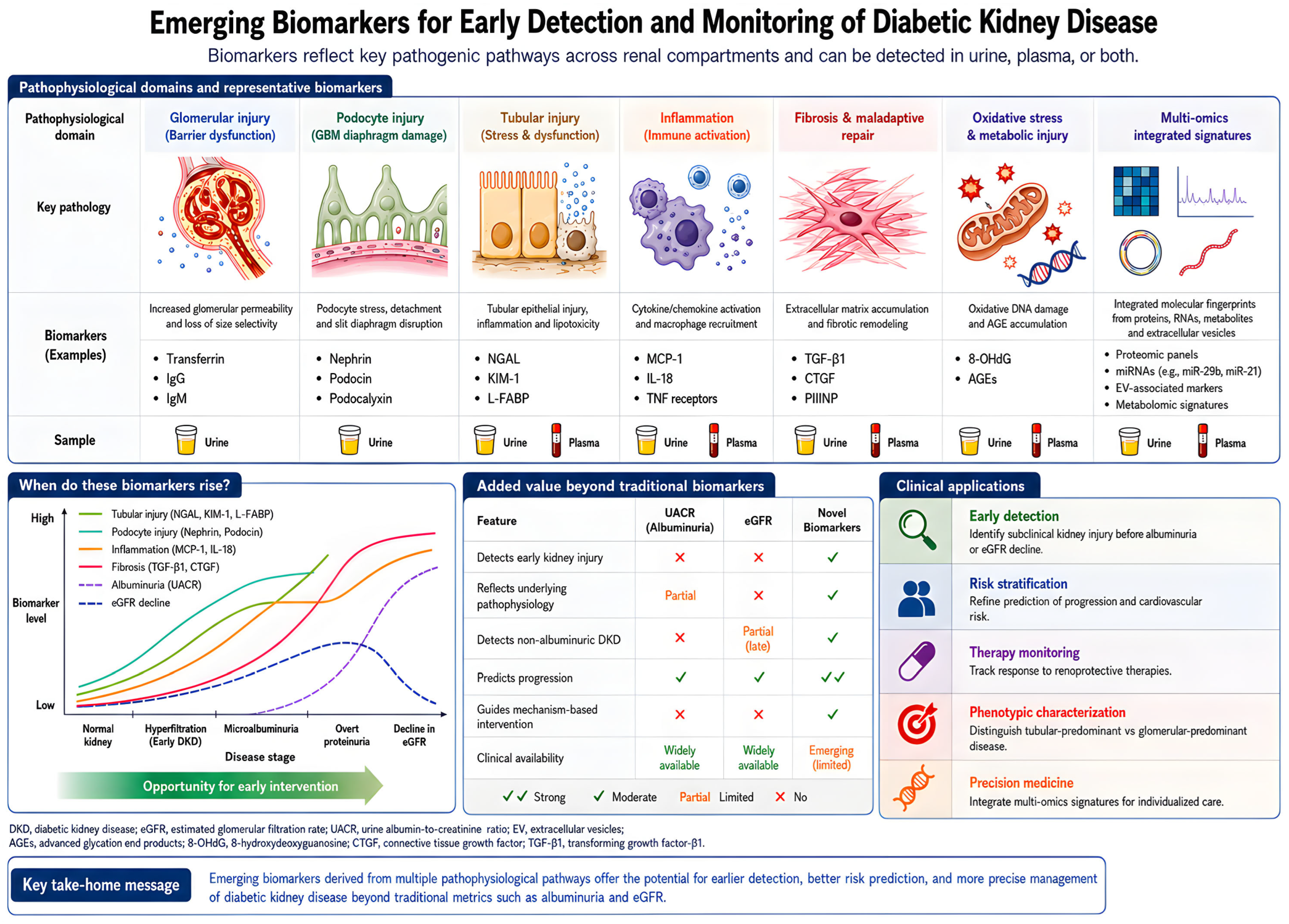
Disclaimer/Publisher’s Note: The statements, opinions and data contained in all publications are solely those of the individual author(s) and contributor(s) and not of MDPI and/or the editor(s). MDPI and/or the editor(s) disclaim responsibility for any injury to people or property resulting from any ideas, methods, instructions or products referred to in the content. |
© 2026 by the authors. Licensee MDPI, Basel, Switzerland. This article is an open access article distributed under the terms and conditions of the Creative Commons Attribution (CC BY) license.