


Abstract
Retinoblastoma (Rb) is the most common intraocular malignancy. If left untreated, Rb can result in death within 1–2 years. However, with timely detection and proper treatment, the survival rate is as high as 98%. The primary goal of Rb treatment is to eliminate cancer and save the patient’s life, while the second goal includes preserving the eye and vision. Management of Rb requires timely detection, differentiation of the tumor from similar conditions, staging, making informed decisions about the best therapeutic approach, and close follow-up to detect any signs of tumor recurrence. There are several treatment options available for Rb. Early detection and proper treatment are essential in saving the lives and vision of children affected by Rb. Progress in developing efficient diagnostic and therapeutic techniques brings hope to patients with Rb and their families. The PubMed database was utilized to identify relevant references published during the last 35 years. This article shows basic and current concepts on managing Rb, which encompasses diagnosis, evaluation, treatment, follow-up, and challenges.
1. Introduction
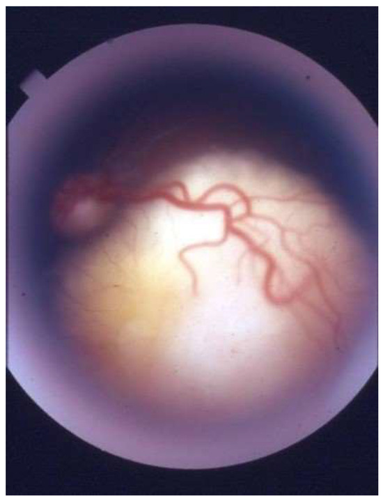
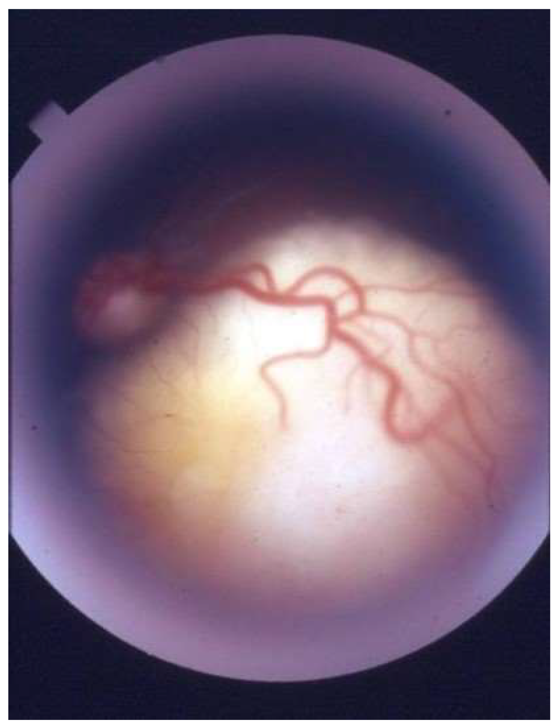
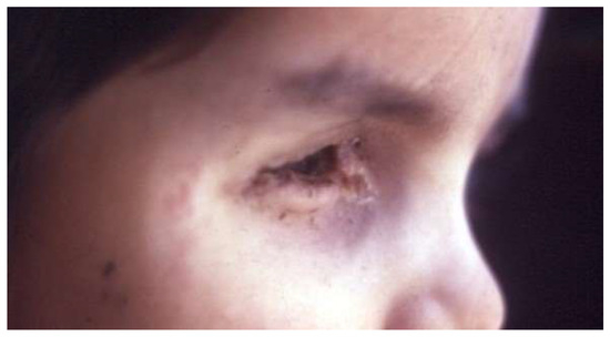
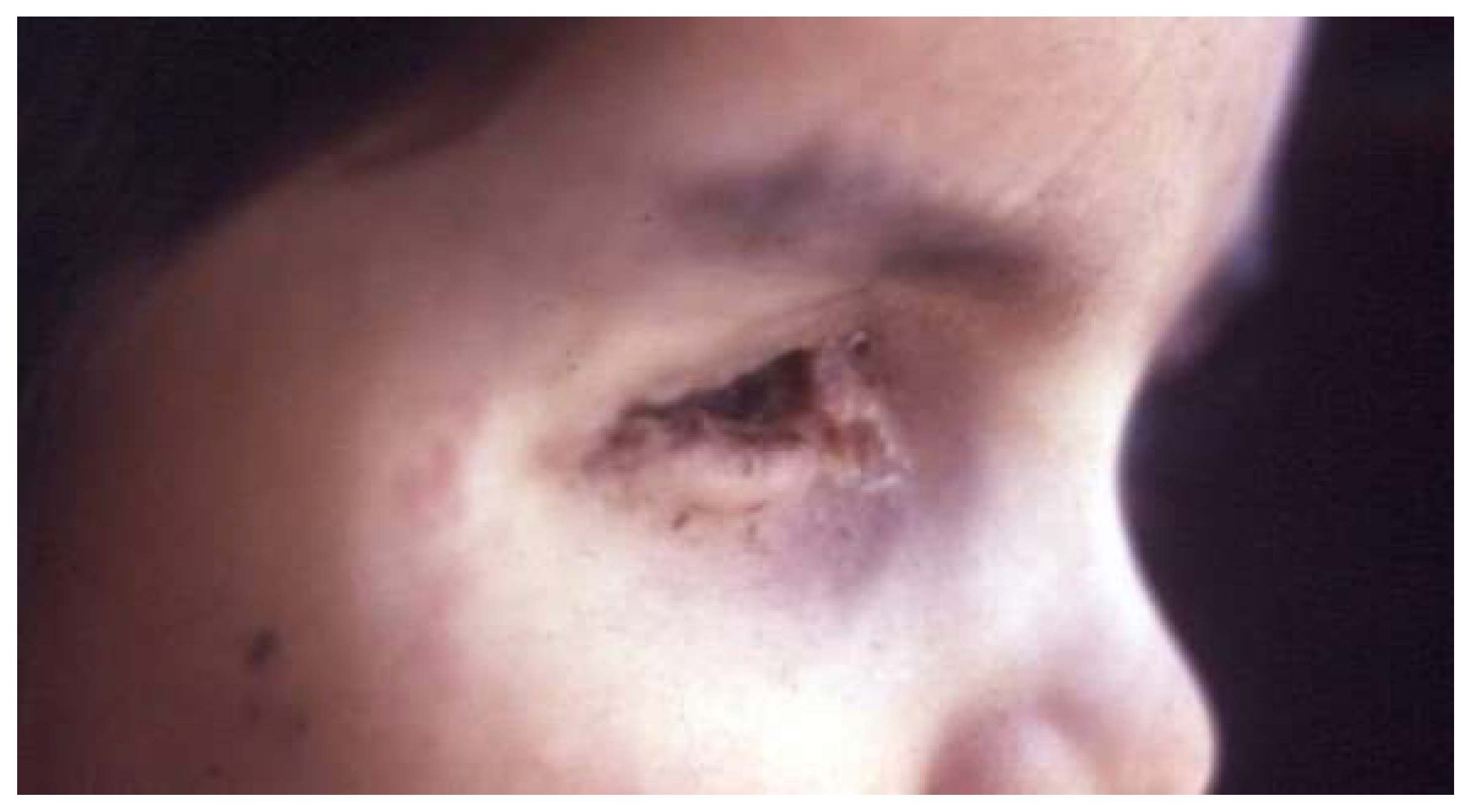
Retinoblastoma (Rb), named by Verhoeff, is the most common intraocular malignancy (Figure 1) [1,2]. The American Ophthalmological Society adopted the Rb term in 1926 [1]. Rb is caused by mutations in the retinoblastoma gene (RB1) [1,3,4,5]. Autosomal dominant inheritance is observed in 30–40% of cases, while the remaining cases are of the non-inherited sporadic type [4]. Leukocoria is the most common sign of Rb, followed by strabismus [1]. The most frequent differential diagnoses of Rb include Coats disease, persistent fetal vasculature, vitreous hemorrhage, familial exudative vitreoretinopathy, and rhegmatogenous retinal detachment [1]. Rb, if left untreated, can result in death within 1–2 years [6]. However, with timely detection and proper treatment, the survival rate can be as high as 98% [2,7]. It is noteworthy that in Low-Income Countries (LIC), the survival rate is considerably lower, only 40%, due to the limitations of healthcare [2,8]. Therefore, early detection and the correct staging (Table 1) of Rb is essential because it allows for more accessible, less expensive treatments and the best prognosis.
Figure 1.
Retinoblastoma.
Table 1.
The International Intraocular Retinoblastoma Classification (IIRC).
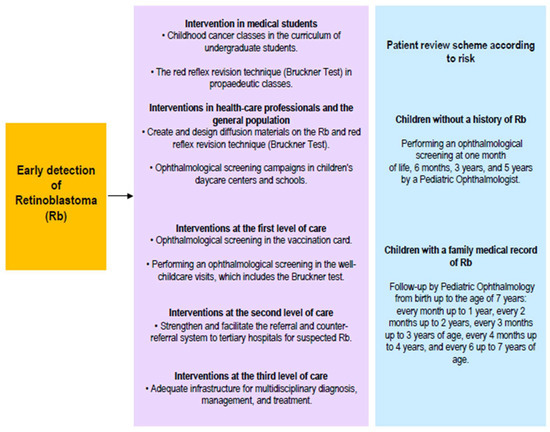
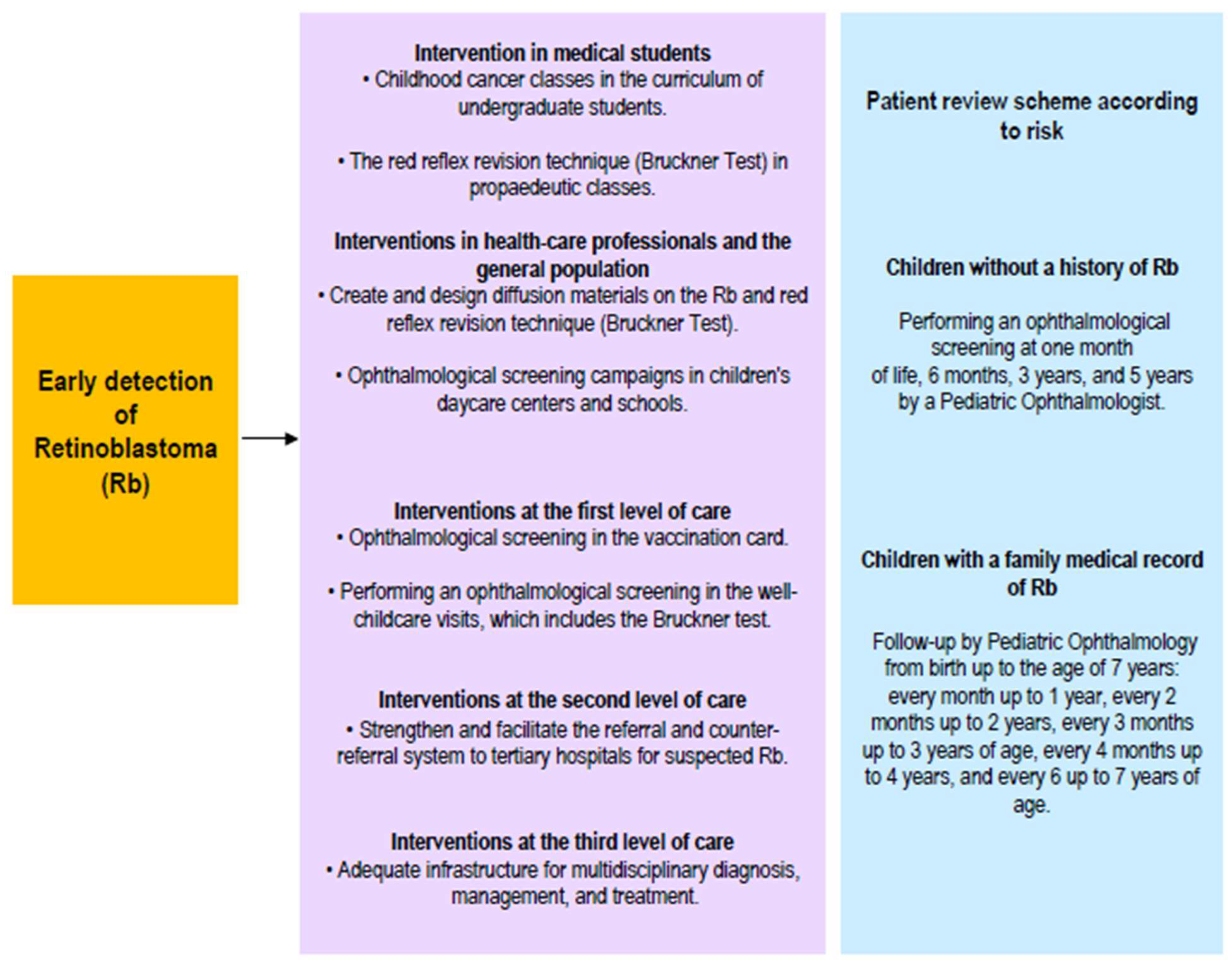
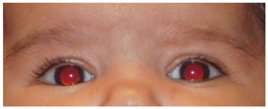
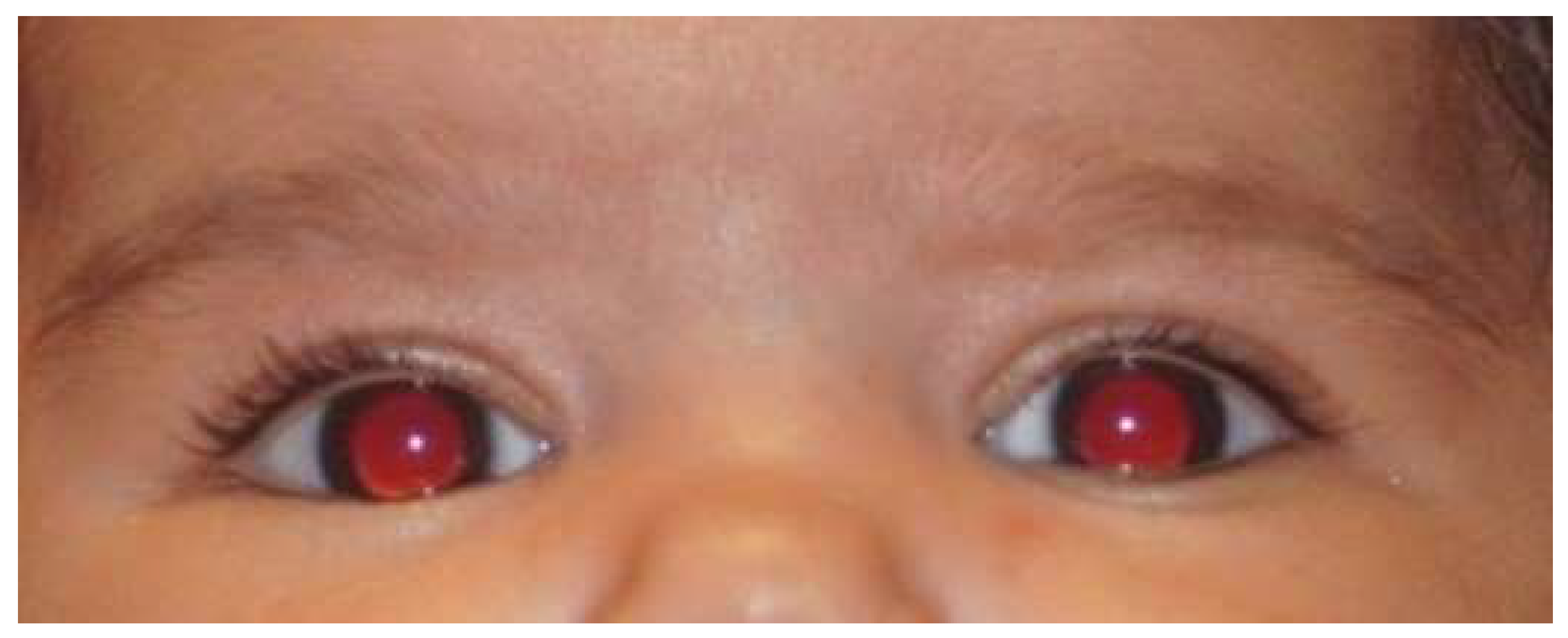
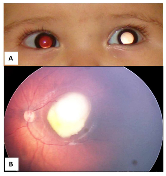
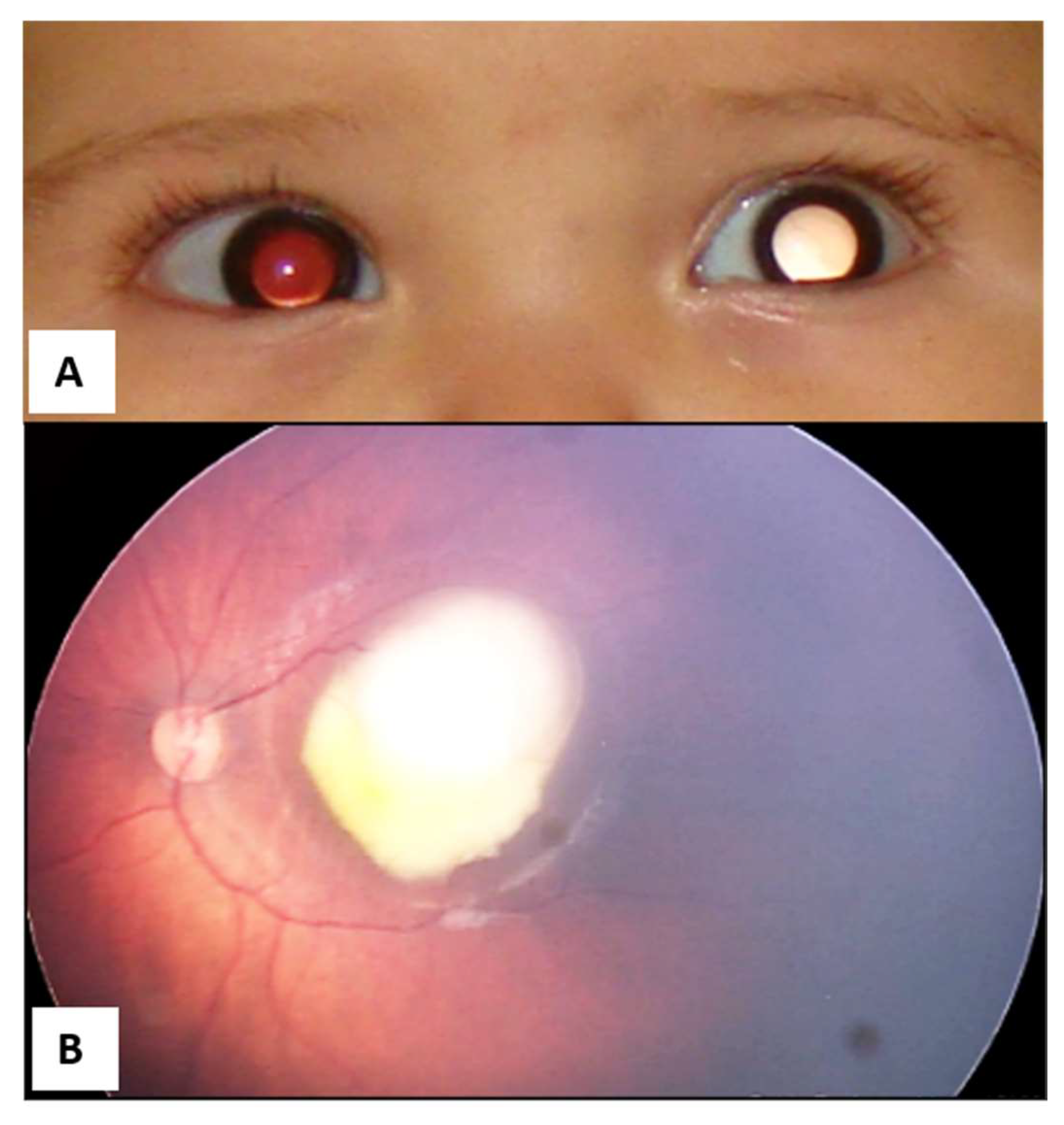
Information campaigns on Rb targeting the general population, nursing or medical students, pediatricians, and healthcare professionals can help achieve this (Figure 2) [5]. Rapid Rb detection tests, such as the Bruckner test, also contribute to early detection. The Bruckner test involves simultaneously evaluating the red reflex of both eyes using a direct ophthalmoscope (Figure 3) [9]. Performing an ophthalmological screening in the well-childcare visits, which includes the Bruckner test, is an easy, fast, and simple strategy that could generate substantial benefits in the early detection of Rb when leukocoria is evidenced (Figure 4), and even of other eye disorders or pathologies in children [9].
Figure 2.
Strategies for achieving early detection of retinoblastoma.
Figure 3.
The Bruckner test (red reflex) is normal in both eyes.
Figure 4.
(A) Leukocoria in the left eye secondary to retinoblastoma. (B) Confirmation of Group C retinoblastoma in the left eye.
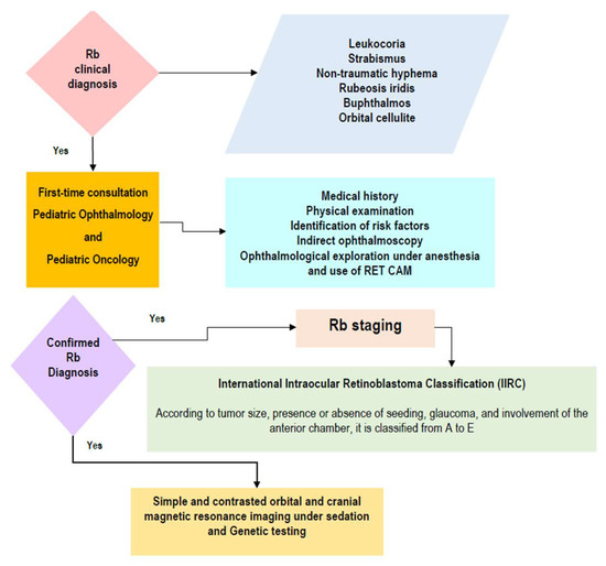
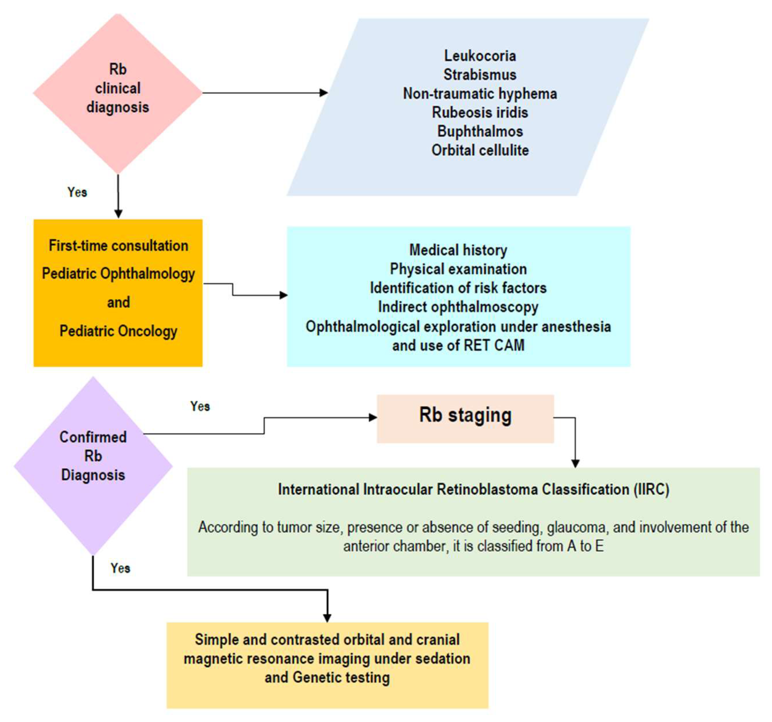
The initial approach to suspected Rb is essential for a better prognosis (Figure 5). The clinical presentation of Rb is widely recognized. In 60% of cases, leukocoria is the initial sign, which can be identified through the Bruckner test. Unfortunately, this sign is often missed by pediatricians, ophthalmologists, or general practitioners [5].
Figure 5.
The initial approach to suspected retinoblastoma.
Managing Rb requires timely detection, differentiation of the tumor from similar conditions, staging, making informed decisions about the best therapeutic approach, and close follow-up to detect any signs of tumor recurrence [6]. It is a complex process that requires a lot of attention and care, but it is critical for ensuring the best possible outcomes for patients [6].
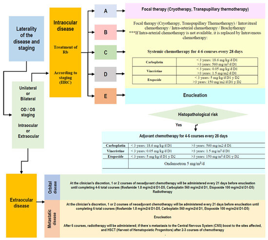
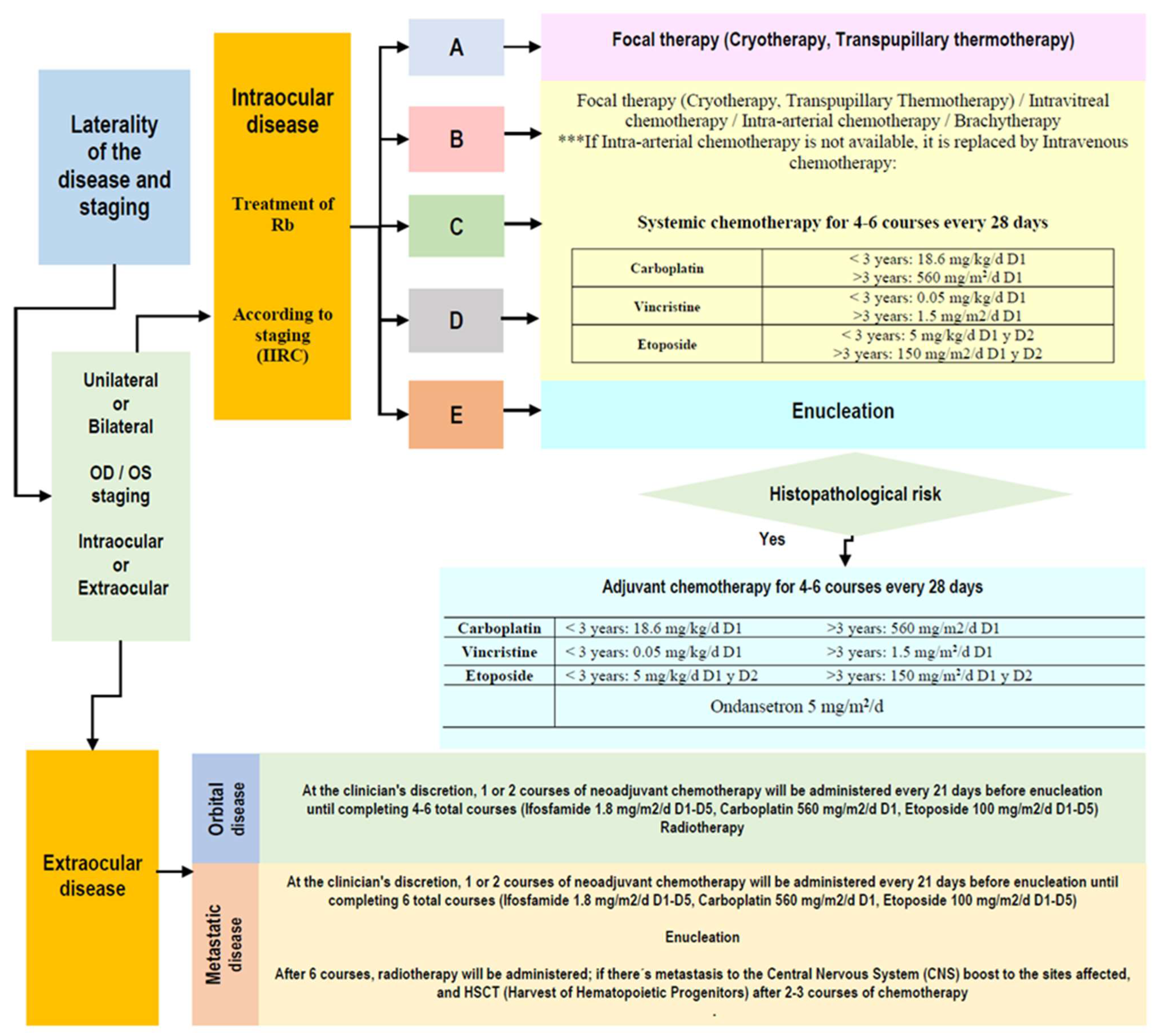
The primary goal of Rb treatment is to eliminate cancer and save the patient’s life, while the second goal includes preserving the eye and vision [6,10]. Management of intraocular Rb includes chemotherapy and focal treatments such as Transpupillary Thermotherapy (TTT), Cryotherapy (Cryo), and Brachytherapy. Enucleation and External Beam RadioTherapy (EBRT) are also treatment options [11,12]. Chemotherapy treatment for Rb includes Intravenous Chemotherapy (IVC) and local chemotherapy delivered by intracameral, intravitreal, and intra-arterial routes. If a patient is diagnosed with extraocular or metastatic Rb, specialized protocols are implemented to safeguard the life of the patient [13] (Figure 6).
Figure 6.
Algorithm for retinoblastoma treatment.
This review focuses on the management of Rb and its challenges. The PubMed database was utilized to identify relevant references published during the last 35 years. This article shows basic and current concepts on managing Rb, which encompasses diagnosis, evaluation, treatment, follow-up, and its challenges. This review seeks to encompass the essential aspects of Rb that allow an updated understanding of this pathology. This review aims to show, in a practical, summarized, and updated way, the essential aspects of Rb that allow the reader sufficient knowledge of the fundamental aspects of Rb.
2. Retinoblastoma Treatment
During this century, treatment for Rb has significantly improved. Advanced techniques have opened up new possibilities for preserving the eye and its visual function more effectively.
At the beginning of the 20th century, significant advancements were achieved through IVC for treating Rb, allowing for intraocular tumor reduction, the treatment of micrometastases, and a remarkable increase in the survival and eye-salvage rates of patients with Rb [5]. Nevertheless, IVC carries the risk of severe systemic adverse reactions: Carboplatin is known to cause ototoxicity, and Etoposide has the potential to cause leukemia [2,5,14].
Over the past decade, treating Rb has been revolutionized by targeted approaches such as Intra-arterial Chemotherapy (IAC) or Intravitreal Chemotherapy (IVitC). These methods have greatly improved the chances of saving the eye, particularly in advanced cases [12,15].
Therapies such as IAC and IVitC have changed the way we treat children with Rb in that the treatment is more focused; we have better intravitreal concentrations of chemotherapy medications with fewer side effects and better outcomes [16]. Hence, these are more effective and less toxic than conventional systemic chemotherapy [17]. Despite the variety of therapeutic methods, refractory or recurrent cases do occur [18].
Rb centers worldwide are transitioning from systemic to ocular-delivered chemotherapy for better success rates [10]. Nonetheless, these novel treatment techniques must be standardized at all Rb centers worldwide, especially in LIC. Even when results are good with the new therapies, it is expensive and challenging to implement IAC and Brachytherapy [10].
The management of Rb should be customized according to various factors, such as disease staging, for example, through the International Intraocular Retinoblastoma Classification (IIRC) [6] (Table 1), age, laterality, the presence of a germline mutation, and liquid biopsy results (if feasible).
Before making any treatment decisions, conducting a thorough ophthalmological evaluation [5,19] should include a bilateral dilated ocular fundus examination under general anesthesia [5,20] and high-resolution simple and contrast-enhanced Magnetic Resonance Imaging (MRI) orbital and cranial (after the initial approach, request as often as each case requires) [5,21]. Photographs of the tumor should be taken when possible. The Pediatric Oncologist should also schedule a consultation with the patient [21]. The frequency, studies, and procedures for follow-up visits will be adapted to each case.
Treatment of Retinoblastoma According to IIRC Staging
Group A
Focal Therapy
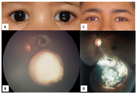
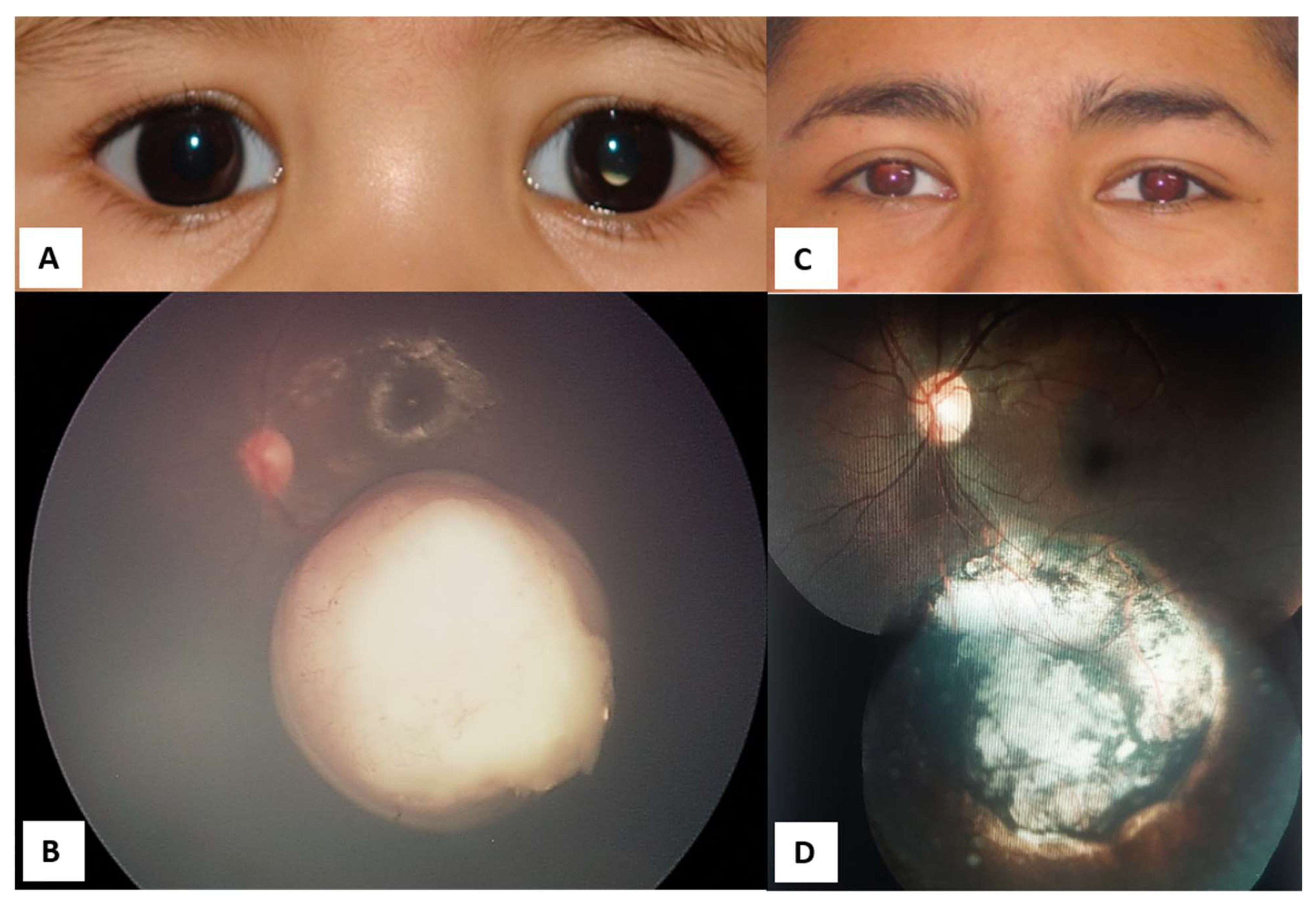
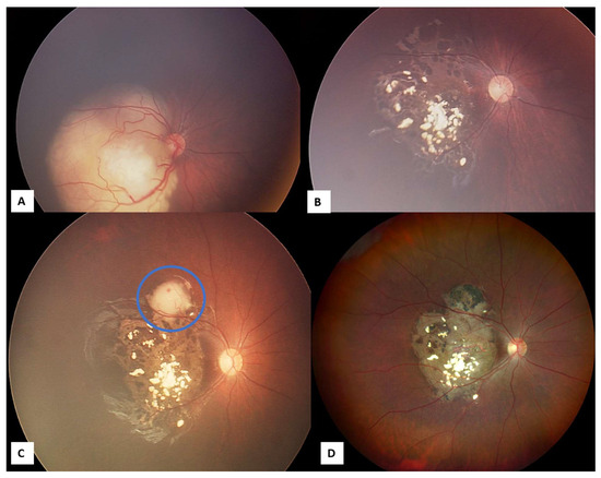
Cryotherapy (Cryo). The treatment consists of a freeze-and-thaw technique [5] repeated three times under indirect ophthalmoscopic control using scleral indentation [22,23]. For peripheral lesions, the cryotherapy probe is placed on the conjunctiva, while for more posteriorly located lesions, it is placed directly on the sclera following a conjunctival incision [21]. It treats small tumors less than 3 mm and subretinal seedings. Retinal detachment has been reported with extensive cryotherapy [21]. Cryotherapy is used in combination with IVC or IAC (Figure 7) [21].
Figure 7.
Managed retinoblastoma with intravenous chemotherapy and cryotherapy (before intra-arterial chemotherapy availability in México). (A) Four-month-old male patient with retinoblastoma in the left eye, pre-treatment. (B) Confirmation of Group C retinoblastoma in the left eye. (C) The same patient, 17 years after treatment. (D) Inactive retinoblastoma 17 years after treatments.
Transpupillary ThermoTherapy (TTT). This is the primary treatment for small tumors less than 3 mm; it can also be used in combination with IVC (Figure 8) [22]. Hyperthermia is administered using an 810 nm diode laser [22] continuously, with 30–50 s burn durations [23]. Several complications may arise from TTT, such as iris atrophy, focal cataracts, and retinal complications [21].
Figure 8.
Managed retinoblastoma with transpupillary thermotherapy.
Groups B, C, and D.
Focal therapy.
Cryo or TTT (plus other options, including IAC, IVC, IVitC, or brachytherapy) [22].
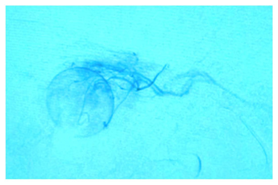
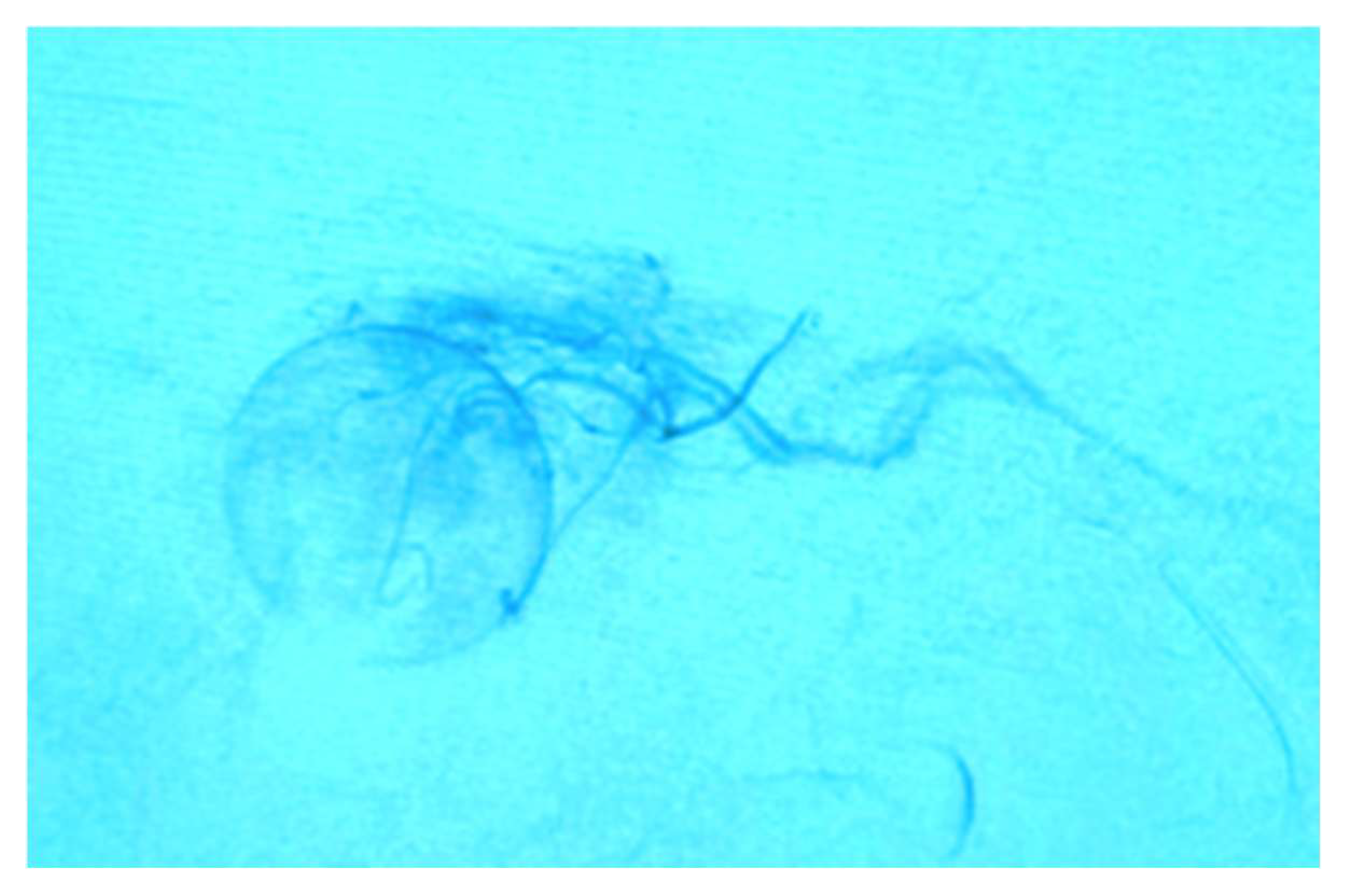
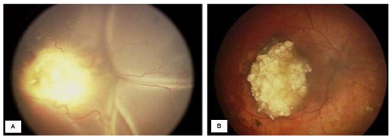
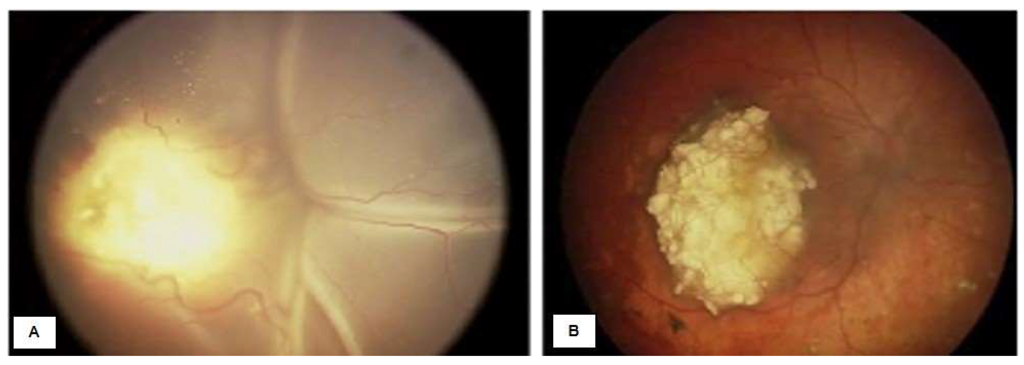
Intra-arterial Chemotherapy (IAC). This is the primary cornerstone treatment for Rb at present. The Kaneko drug-delivery method through the intra-arterial route enables direct delivery into the ophthalmic artery, limiting the systemic distribution of chemotherapy and its adverse effects [5,14]. The IAC technique was popularized by Abramson et al. [2]. The procedure starts with a microcatheter in the femoral artery that flows into the ophthalmic artery guided by fluoroscopy (Figure 9 and Figure 10). The chemotherapy drugs used in IAC are Melphalan, Topotecan, and Carboplatin [24]. One or a combination of these drugs can be employed monthly for two or three sessions [10,25,26]. IAC has proven effective for treating Rb in both unilateral and bilateral cases, even in infants as young as 3 months of age [27]. When administering chemotherapeutic drugs through IAC directly into the tumor, the concentration in the intraocular lesions is 10 times higher than when administered intravenously [14]. IAC treats advanced Rb that could not be treated with systemic chemotherapy [28]. However, it is a complex procedure that could be expensive for LIC. It must be performed in a Hemodynamics suite adapted for angiography and by an experienced interventional neuroradiologist. Ophthalmic vascular events can occur with this treatment [21].
Figure 9.
Intra-arterial chemotherapy.
Figure 10.
Managed Group D retinoblastoma with intra-arterial chemotherapy. (A) Pre-treatment; (B) post-treatment.
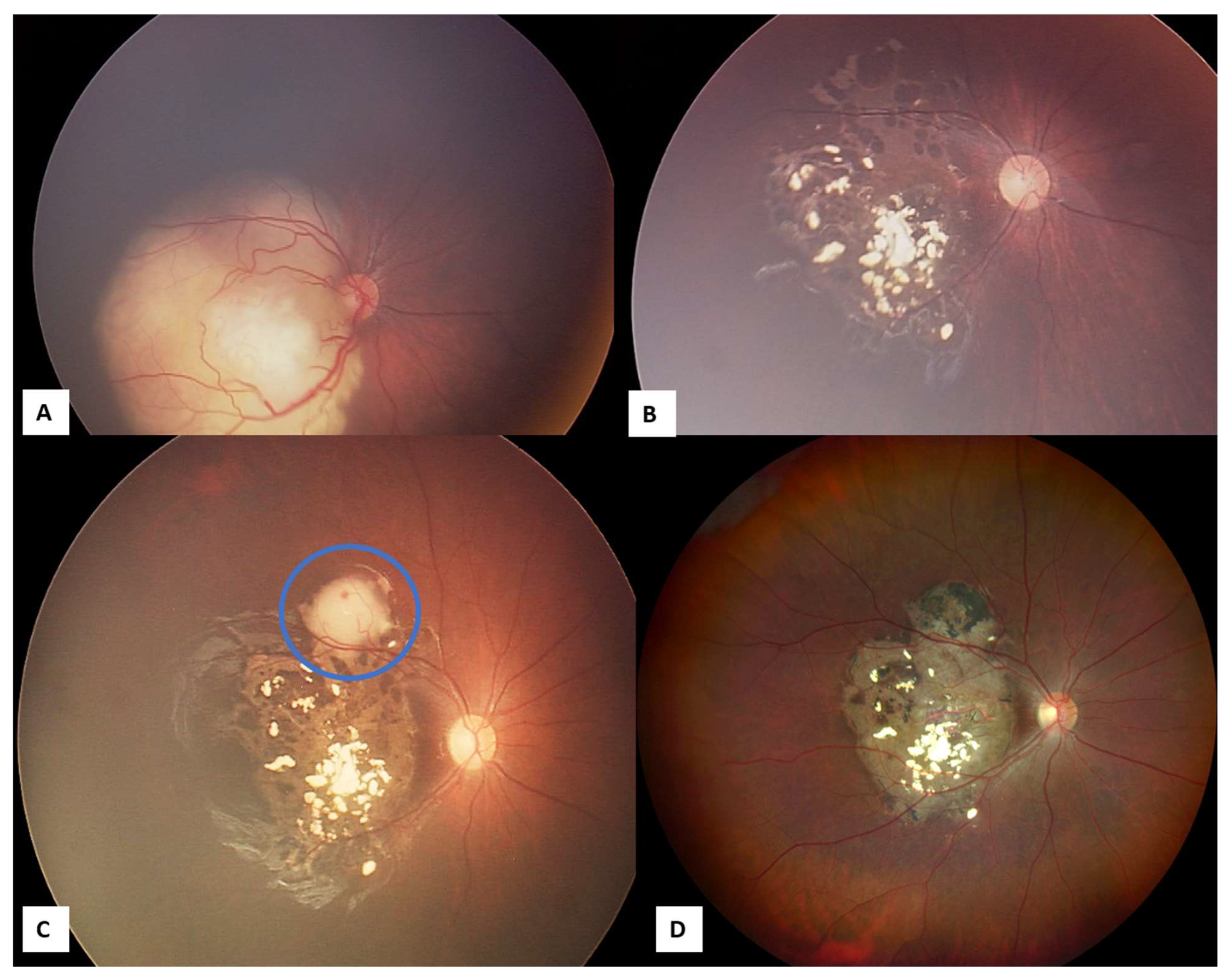
Intravenous chemotherapy (IVC). IVC was introduced during the 1990s [6]. The most frequently used agents include Carboplatin, Etoposide, and Vincristine (VEC), administered monthly [6,29]. The consecutive cycles range in number between 6 and 9 [26]. IVC continues to be employed globally to control intraocular Rb (Groups B and C) and prevent systemic metastasis [6,29]. IVC is used alone or combined with focal therapies, such as TTT, Cryo, and brachytherapy (Figure 11) [6]. IVC is indicated in bilateral cases, confirmed germline mutation, in patients with suspected optic-nerve invasion, or in tiny infants waiting to gain weight for IAC [16,21]. The adverse effects of IVC include nausea, vomiting, transient alopecia, cytopenia, and fever [21].
Figure 11.
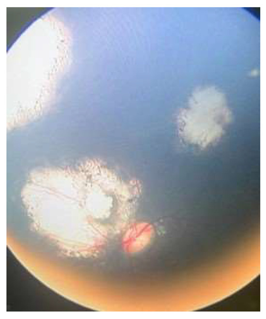
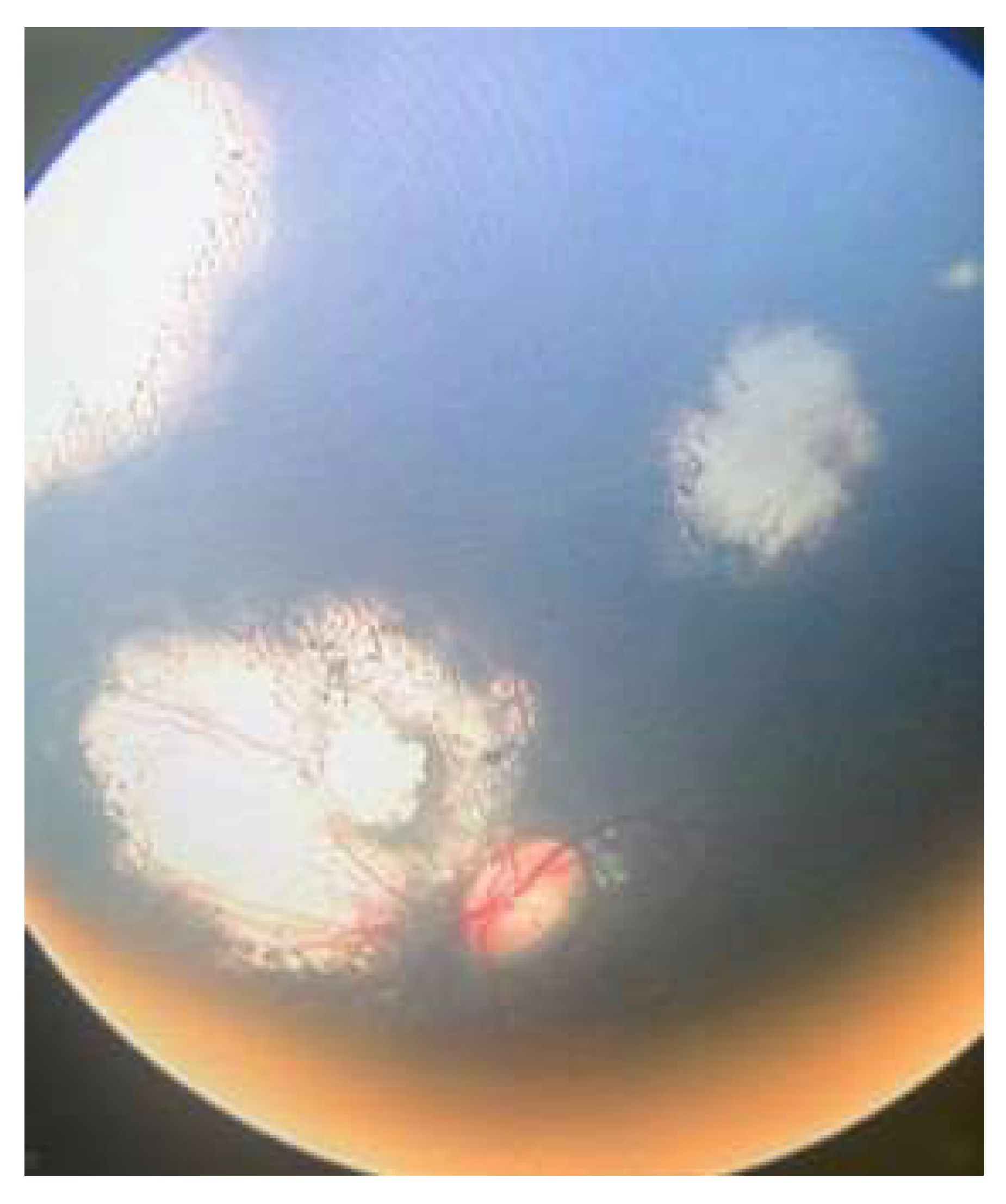
Managed retinoblastoma with intravenous chemotherapy, cryotherapy, and transpupillary thermotherapy TTT (before intra-arterial chemotherapy availability in México). (A) Eight-month-old female patient with Group C retinoblastoma in the right eye, previous intravenous chemotherapy and cryotherapy. (B) Inactive retinoblastoma. (C) Tumor activity (Blue circle) that was managed with TTT. (D) Inactive retinoblastoma 12 years after treatments.
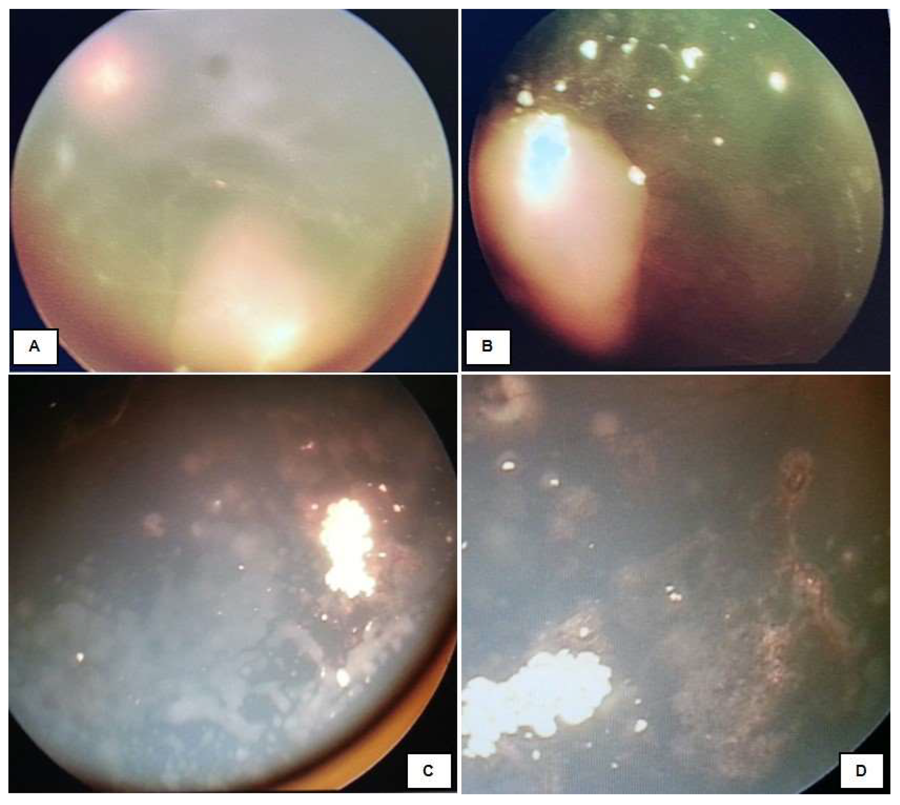
Intravitreal Chemotherapy (IVitC). Intravitreal administration of chemotherapy is an effective treatment for Rb. Several teams have described this treatment, which was formalized by Munier et al. [5]. IVitC delivers the highest drug concentration into the vitreous while minimizing systemic drug concentration [12]. IVitC is indicated for cases where vitreous seeds persist or recur despite other treatments (Figure 12) [12,21]. The drug regimen for IVitC consists of Melphalan and Topotecan, either used alone or in combination. To effectively control vitreous seeds without harmful side effects, 20–30 μg doses are recommended to be administered every 2–4 weeks [5,30,31]. Topotecan is also used in doses of 90 μg. IvitC may lead to ocular adverse events such as cataracts or vitreous and sub-retinal hemorrhage [21].
Figure 12.
Managed Group D retinoblastomas with intravitreal chemotherapy. (A) Pre-treatment; (B–D) post-treatment.
Brachytherapy. Brachytherapy for Rb was pioneered by Moore et al. and was further defined by Stallard, Sealy, Lommatzsch, and Vollmar [32]. Brachytherapy is a form of radiotherapy wherein the irradiation source is placed adjacent to or near the target tissue or organ [22]. Iodine and ruthenium plaques are currently the most frequently used plaques [32]. Brachytherapy is a secondary treatment for medium-sized tumors that do not respond to chemotherapy and that have localized vitreous or sub-retinal seeding [21]. It is typically utilized after recurrence following IVC or IAC [33]. The ocular adverse events associated with brachytherapy are cataracts and radiation-associated ophthalmic events.
Group E
Enucleation. This option continues to be used in advanced cases. Current indications could be tumor progression despite conventional treatment, active Rb without vision, unilateral Group E, optical-nerve involvement, and orbital stage [34,35,36,37]. The globe must undergo a detailed histopathological examination after enucleation. This report will provide us with important information concerning tumor extension. Pathologic features found in enucleated eyes indicate that the risk of metastases is as follows: post-laminar optic-nerve invasion; massive choroidal invasion > 3 mm; anterior chamber invasion involving the iris or ciliary body, and scleral or extra-scleral involvement. However, the definition of the features is widely debated [21,38,39]. If one of these risks is present, the pediatric oncologist should provide adjuvant IVC to prevent metastases. Parents should be correctly informed on the risk of metastases by conserving a nonfunctional eye with intensive eye-conserving treatment. It is necessary to achieve aesthetic rehabilitation.
Treatment for extraocular tumor extension and metastasis prevention.
External Beam Radiotherapy
External Beam Radiotherapy (EBRT) is rarely used at present. In cases of extraocular tumor extension, orbital recurrence, and positive optic-nerve margin following enucleation, EBRT remains a viable treatment option in conjunction with IVC [21,40]. EBRT causes osseous-growth retardation, possibly leading to facial deformity (Figure 13) [22,41,42,43]. The most severe risk associated with EBRT is the high possibility of developing second primary tumors [22]. The incidence of a second cancer inside and outside of the irradiated field has been estimated to be 51% at 50 years of age [5]. The most commonly observed subsequent malignant neoplasms (SMN) are sarcomas, melanomas, and myelogenous leukemia [2,44].
Figure 13.
After-effects of external beam radiotherapy.
3. Metastases
In Rb, the presence of metastases is devastating. In countries with access to advanced healthcare, the likelihood of metastasis in children with Rb is less than 10%, but this percentage is higher in LIC [2]. According to Kaliki et al., patients with non-high-risk Rb did not experience metastasis; however, 4% of high-risk patients died due to metastasis [45,46]. The chance of survival for patients with distant metastasis was nearly zero, employing high-dose chemotherapy and autologous stem-cell transplantation [47].
4. Retinoblastoma Characterization for Diagnosis, Prognosis, Predictive Information, and Treatment Choice
Liquid Biopsy
Aqueous humor (AH) liquid biopsy allows the in vivo detection of tumor-derived DNA in patients with Rb [48]. The AH liquid biopsy provides a wealth of genetic information specific to Rb [48]. In 2017, it was shown that the AH liquid biopsy, attained via paracentesis, comprises a robust source of tumor-derived cell-free DNA (cfDNA) [48,49,50,51]. cfDNA can be effectively isolated to detect relevant molecular biomarkers in eyes actively undergoing treatment or before treatment. Tumor DNA present in the AH of eyes with Rb can be utilized to identify the Rb gene (RB1), single-nucleotide variants (SNV), somatic copy number alterations (SCNA), and tumor methylation status to estimate tumor fraction, metabolomic signatures, and the expression of secreted peptides [48]. Liquid biopsy of aqueous humor at the time of diagnosis can provide the highest concentration of nucleic acids and proteins and may prove to be most useful for RB1 mutational testing. Even during therapy, aqueous humor biopsy can yield sufficient DNA concentrations for low-pass whole-genome sequencing and SCNA detection, which can aid in predicting prognosis and monitoring trends in tumor fraction that are associated with ocular outcomes [49]. The AH liquid biopsy in Rb could pave the way for a better understanding of mechanisms for treatment response, resistance, and recurrence in Rb and could provide specific therapeutic targets to improve globe salvage [51,52].
Genetic Counseling
Genetic counseling in Rb represents a challenge for the Treating Physician [53]. Rb genetics supports appropriate management in the clinical setting through correct information and the genetic testing of patients and families [54]. It explains the genetics of Rb appropriately and in simple language that can aid parents and patients in their adulthood in terms of their proper understanding of the disease [55]. Individuals with inherited Rb are more likely to develop ocular and non-ocular tumors [50]. Additionally, transmitting the mutated gene to their future offspring is possible [50]. Patients with heritable Rb have an increased risk of SMN [2]. It is appropriate to offer genetic counseling (including the discussion of potential risks to offspring and reproductive options) to young adults affected with or at risk of Rb [56]; in vitro fertilization with a pre-implantation diagnosis can be discussed in future family planning, in addition to pointing out the importance of a timely pediatric ophthalmologic examination from birth up to age 7 years, [57] for the future descendants of Rb survivors. Rb survivors should undergo lifelong monitoring, particularly those with a germline mutation.
Radiomics Model
Radiomics uses the high-throughput extraction of advanced quantitative features to objectively and quantitatively describe tumor phenotypes [58]. These quantitative features can provide a valuable oncological diagnosis, prognosis, or predictive information [58]. Magnetic resonance imaging (MRI) is a critical technique for detecting the presence of post-laminar optic-nerve invasion (PLONI) in patients with Rb [59,60,61,62]. PLONI is a risk factor for developing metastatic disease [62]. The MRI-based radiomics model to predict PLONI in patients with Rb was shown to be superior to visual assessment and may serve as a potential tool to guide personalized treatment [58].
Artificial Intelligence
Artificial Intelligence (AI) in medicine aids in developing treatment algorithms and predictive models via electronic medical records and Big Data, or softbots [63]. In the field of ophthalmology, AI has been capable of analyzing data and images, thus aiding in the diagnosis and staging of various ocular pathologies, including strabismus, refractive error, keratitis, keratoconus, cataracts, glaucoma, optic-disk abnormalities, diabetic retinopathy, age-related macular degeneration, or the retinopathy of prematurity [63,64,65,66,67]. AI has also been employed to develop treatment algorithms and to prognosticate ocular pathologies. In ophthalmology, AI entertains the potential for universalizing patient access to disease screening and diagnosis, for expediting treatment as appropriate, and for monitoring outcomes [67]. AI is a promising assessment tool for Rb [63]. Using an AI model, it is possible to screen children in the community for Rb by utilizing fundus images captured on an inexpensive nonmydriatic fundus camera. This approach can help diagnose the condition at an early stage [63].
Genomics
The majority of Rb cases result from biallelic RB1 gene inactivation, although a small subset (1–2%) is initiated through MYCN-gene amplification in the absence of RB1-gene inactivation [68,69,70,71]. Other genetic modifications have also been associated with Rb. Genetic modifiers, such as MDM2, MDM4, or MED4, and polymorphisms in p53, CDKN1A, CDKN2A, and BCOR could also influence hereditary Rb development [70]. These findings have diagnostic, clinical, and response-to-treatment implications.
MYCN amplifications were recently identified in a subset of aggressive Rb in infants [72] with relative resistance to typical therapeutic approaches to Rb [73]. MYCN-amplified Rb possesses distinct features compared with the RB1 pathogenic variant on MRI, including tumors in a peripheral (anterior) location with a plaque or pleomorphic shape, irregular margins, tumor–retinal folding, and peri-tumor blood [72]. miR-142-5p restricted cell proliferation, migration, invasion, and enhanced cell apoptosis in Rb cells. MYCN is adversely controlled by miR-142-5p [74]. MYCN upregulated m6A reader functions to promote Rb cell proliferation and tumor growth [75]. Another mutated gene in Rb (7–13% of cases) is the epigenetic modifier gene BCOR [76].
Investigations propose FGFR4, NQO1, ACADS, CX3CR1, GBE1, KRT85, and TYR as possible candidate genes involved in Rb oncogenesis. A significant proportion of sporadic Rb exhibits somatic mosaicism for RB1 mutations [70]. Early onset Rb tumor cells express an immune gene-expression signature followed by the accumulation of dendritic, monocyte, macrophage, and T-lymphocyte cells in the Rb tumors [77]. These findings represent potential effective targets for Rb treatment.
5. Retinoblastoma Challenges
Rb management requires a highly trained and up-to-date multidisciplinary team comprising a pediatrician, pediatric ophthalmologist, pediatric oncologist, pediatric geneticist, pediatric radiologist, and pediatric psychologist, which can be complex to integrate, especially in developing countries [21]. The prognosis of Rb is excellent if it is diagnosed early and treated appropriately, with cure rates higher than 90% [78]. Regrettably, the epidemiology of Rb reveals inequities in access to timely diagnosis and treatment between developed countries and LIC [78,79,80]. Many patients in LIC frequently present in an advanced Rb stage due to inadequate knowledge of the early signs of Rb, limited medical-care resources, and postponed referral [15,78]. This leads to a mortality rate that is significantly higher in LIC. In Asia and Africa, 40–70% of children with Rb die, while in Europe, Canada, and the U.S., the mortality rate is only 3–5% [60].
Early detection of Rb is fundamental; therefore, fast and easy detection strategies such as the Bruckner test contribute to the effective management of this disease. Rb can be detected by means of the Bruckner test, exhibiting in a patient a shining white pupil, or “Leukocoria” [9,81]. OCT (Optical Coherence Tomography) can aid in identifying very small Rb that are not detectable (that are “invisible”) by indirect ophthalmoscopy [82,83,84], and the latter should be more widely used for the timely detection of Rb. Teleophthalmology and tele-education resources have been validated in diverse ocular pathologies. In Rb, there is the option of online mentorship via Orbis Cybersight, which has shown encouraging results [85]. Early detection and proper treatment are essential in saving the lives and vision of children affected by Rb.
Today, there are several forms of Rb treatment, including using chemotherapy through different routes of administration, focal therapy, EBRT, and enucleation. Unfortunately, none of these options is foolproof [78].
IVC represents an essential part of Rb treatment, proving to be effective not only in controlling the disease but also in preventing metastasis and reducing the occurrence of second cancers in the long term [86]. Although the potential risk of IVC toxicity [87], the lower concentration of IVC achieved at the intraocular level secondary to the blood–retinal barrier, and the chemoresistance of Rb should not be ignored [9,88].
Carboplatin is one of the most important drugs in the standard chemotherapy regimen for Rb. Still, it activates the transcription factor nuclear factor-kappa B (NF-ĸB), which entails Rb cell survival [9]. Therefore, it is important to look for additional drug options that are effective per se for Rb or that enhance the effect of chemotherapy without increasing its dose. Molecularly targeted drugs may be more effective and less toxic [89].
The management of Rb will likely be substantially modified based on the more frequent use of liquid biopsy [48], molecular biomarkers [79], updated genetic testing [80], genotype–phenotype tumor correlations [90], prenatal diagnosis [91], Rb subtypes [92], and new technologies such as radiomics models and AI.
The investigation of Rb emerging treatments includes new or repositioned drugs (alone or in combination), immunotherapy, novel delivery systems, oncolytic adenovirus, gene therapy, and molecularly targeted therapies [78,86,93,94].
New intracellular targets can potentially lead to the identification of novel Rb drugs. Examples of this are Bcl-2 proteins, bromodomain and extra-terminal motif proteins (BET), MDM2/MDM4 inhibitors, NF-kB, histone deacetylase, kinesin spindle protein, STAT3, cyclin-dependent kinase (CDK) 4 and 6, p-53, MYC, and GABA receptors [95].
In vitro and in vivo studies showed synergistic inhibitory effects of the combination of nutlin-3a and topotecan. miR-129 could be a novel targeted therapy for Rb treatment. Bortezomib in vivo was mediated through the induction of apoptosis in 2 Rb cell lines by blocking the NF-κB [96].
The use of nanotechnology offers the possibility of new therapeutic strategies in Rb [97]. Organic polymers or inorganic nanoparticles loaded with synthetic or natural drugs have been developed. All systems showed an increase in the bioavailability of the drug and an ability to pass the blood–retinal barrier and reduce the side effects of anticancer conventional drugs [96].
Epigenetic markers (noncoding RNAs, DNA methylations, RNA modifications, chromatin conformations, and histone modifications) play a vital role in the inactivation of RB1. This knowledge could contribute to future Rb treatment strategies or as diagnostic biomarkers [98,99,100].
Fully understanding Rb tumorigenesis is still an ongoing process. The creation of organoid models is a strategy that enables us to understand this process in greater detail [101,102,103].
The proposal to subdivide the Rb group E could open up a new possibility for management in this group [104,105].
Recently, in Rb patients with unilateral pathological high-risk features, undergoing frontline enucleation, the administration of three cycles versus six cycles of adjuvant chemotherapy was studied, resulting in a non-inferiority of three cycles versus six cycles [106]. This results in less toxicity, less decline in quality of life, and lower treatment costs. However, it should be noted that observations have been made on this research, for example, that different types of high-risk histopathological findings involve different prognoses, so this reduced treatment should not be taken as a standardized management guide; what should prevail is the individualization of each case.
IAC and IVitC have improved conservative treatment and eye salvage in many cases of Rb. For instance, the Grupo de America Latina de Oncología Pediátrica (GALOP), through a consensus document on the treatment of Rb, found that IAC was the preferred therapy in advanced disease, and IVitC was the primary treatment for vitreous seeding [107]. Also supporting part of the previous findings, Wen et al. compared IAC vs. IVC, and based on their findings, they indicate that IAC is a first-line treatment in children with advanced unilateral Rb [108]. IAC has a high success rate that has improved over the past 15 years [109]. However, IAC and IVitC imply a more significant health expenditure to make available the necessary resources (drugs, hospitalization, hospital infrastructure, and trained personnel) [110,111,112,113,114,115].
However, even with the challenges posed by IVitC, it continues to demonstrate its importance as a therapeutic strategy in Rb. An example of this is that high-dose IVitC topotecan (90 μg), in conjunction with other therapies, has proven to be an option for recurrent Rb, which is difficult to handle [116,117].
Unfortunately, systemic metastases are the cause of significant Rb mortality in LIC, which evidences the disparity in the management of Rb [118].
Rb survivors, particularly those with hereditary Rb or those exposed to radiotherapy, are at risk of developing second primary tumors [119,120,121]; therefore, they require lifelong follow-up, which entails costs, whether at a personal or institutional level. According to Zhao et al., the second primary tumors in Rb survivors are more common in the fourth decade of life [119]. They also point out that 25% of the Rb survivors with second primary tumors in their research had a history of unilateral Rb, and approximately 25% of Rb survivors analyzed were treated with surgery alone and not with chemotherapy or radiotherapy, and yet developed second primary cancers [119].
Cases outside the usual presentation of Rb also present a challenge. Rb is rare after 5 years of age. Two mechanisms associated with this phenomenon have been described: the novo Rb and as a sequela of oncogenetic mutations in a pre-existing retinocytoma or a retinoma. There are reported cases of patients aged 14 years old or even adults [122,123]. Outside of the usual presentation of Rb, reaching a diagnosis can be more complex.
Additional comorbidities such as tuberculosis or any other infection, diseases that may require a prolonged period of recovery, human immunodeficiency virus (HIV)/acquired immune deficiency syndrome (AIDS), and malnutrition need to be taken into account in the management of unilateral Rb, for example, a reduction in chemotherapy doses of the first cycle of chemotherapy is recommended [124].
Another important aspect to consider in Rb is the psychological impact of the diagnosis on both the family and the patient, particularly due to the impact of enucleation, which can lead to an altered body image, impaired binocular vision, and even the costs associated with the need for an ocular prosthesis [125].
Even if the eyeball is saved, it is important to consider that vision in that eye could also be compromised, whether due to the underlying diagnosis or the consequences of the different treatment modalities [125]. Therefore, psychological support is important from the moment the Rb diagnosis is confirmed.
The progress in developing efficient diagnostic, staging (e.g., according to IIRC or American Joint Committee on Cancer, AJJC) (Table 1, Table 2, Table 3, Table 4, Table 5, Table 6 and Table 7), [6,126] and therapeutic strategies brings hope to patients with Rb and their families. However, it also challenges healthcare systems with respect to ensuring that every patient can access early diagnosis and timely treatment [109]. WHO Global Initiative for Childhood Cancer (CURE-ALL) seeks to respond to these needs since it has as its pillars: centers of excellence and care networks, universal health coverage, regimens for management, evaluation, and follow-up [118]. Hopefully, more initiatives like this one will be developed to reduce the differences in management and prognosis between the different scenarios in which Rb can occur.
Table 2.
Definition of Clinical Primary Tumor (cT) for retinoblastoma.
Table 3.
Definition of Heritable Trait (H) for retinoblastoma.
Table 4.
Definition of Pathological Primary Tumor (pT) for retinoblastoma.
Table 5.
Definition of Clinical Regional Lymph Node (cN) for retinoblastoma.
Table 6.
Definition of Pathological Regional Lymph Node (pN) for retinoblastoma.
Table 7.
Definition of Clinical (c) and Pathological (p) Distant Metastasis (M) for retinoblastoma.
6. Conclusions
There are several challenges when talking about Rb. Cancer is the leading cause of death worldwide, with its incidence rising steadily [127]. Rb is not the exception [128,129,130].
Retinoblastoma survival disparities remain between developed and developing countries. In the developing countries, there are more cases and worse prognosis [127].
Although there are several therapeutic options for Rb, none of them are infallible. The priority in Rb is to safeguard the patient’s life, and this premise should not be lost sight of. Early diagnosis that allows for less complex treatment strategies is a cornerstone in the management of Rb [131].
Author Contributions
V.M.V.-C., V.B.-C. and C.C.C.-G. contributed to the conception and design of the manuscript. C.C.C.-G., T.I.C.-B., M.E.C.-M. and X.G.-V. organized the database. V.B.-C., X.G.-V. and C.C.C.-G. wrote the first draft of the manuscript. J.C.O.-F., V.M.V.-C., V.B.-C., M.E.C.-M., T.I.C.-B., X.G.-V. and C.C.C.-G. wrote sections of the manuscript. All authors have read and agreed to the published version of the manuscript.
Funding
Instituto Nacional de Pediatría (INP).
Institutional Review Board Statement
Not applicable.
Informed Consent Statement
Not applicable.
Data Availability Statement
No new data were created or analyzed in this study.
Acknowledgments
This work was supported by the Instituto Nacional de Pediatría (INP).
Conflicts of Interest
The authors report no conflicts of interest in this work.
References
- Grossniklaus, H.E. Retinoblastoma. fifty years of progress. the LXXI Edward Jackson memorial lecture. Am. J. Ophthalmol. 2014, 158, 875–891. [Google Scholar] [CrossRef] [Scilit]
- Chen, Q.; Zhang, B.; Dong, Y.; Mo, X.; Zhang, L.; Huang, W.; Jiang, H.; Xia, J.; Zhang, S. Comparison between intravenous chemotherapy and intra-arterial chemotherapy for retinoblastoma: A meta-analysis. BMC Cancer 2018, 18, 486. [Google Scholar] [CrossRef] [Scilit] [PubMed] [PubMed Central]
- Global Retinoblastoma Study Group. Global retinoblastoma presentation and analysis by national income level. JAMA Oncol. 2020, 6, 685–695. [Google Scholar] [CrossRef] [Scilit] [PubMed]
- Nag, A.; Khetan, V. Retinoblastoma. A comprehensive review, update and recent advances. Indian J. Ophthalmol. 2024, 72, 778–788. [Google Scholar] [CrossRef] [Scilit]
- Cassoux, N.; Lumbroso, L.; Levy-Gabriel, C.; Aerts, I.; Doz, F.; Desjardins, L. Retinoblastoma: Update on current management. Asia Pac. J. Ophthalmol. 2017, 6, 290–295. [Google Scholar] [CrossRef] [Scilit]
- Shields, C.L.; Fulco, E.M.; Arias, J.D.; Alarcon, C.; Pellegrini, M.; Rishi, P.; Kaliki, S.; Bianciotto, C.G.; Shields, J.A. Retinoblastoma frontiers with intravenous, intra-arterial, peri-ocular, and intravitreal chemotherapy. Eye 2013, 27, 253–264. [Google Scholar] [CrossRef] [Scilit]
- Broaddus, E.; Topham, A.; Singh, A.D. Survival with retinoblastoma in the USA: 1975–2004. Br. J. Ophthalmol. 2009, 93, 24–27. [Google Scholar] [CrossRef] [Scilit]
- Rodriguez-Galindo, C.; Orbach, D.; Vanderveen, D. Retinoblastoma. Pediatr. Clin. N. Am. 2015, 62, 201–223. [Google Scholar] [CrossRef] [Scilit]
- Cruz-Gálvez, C.C.; Ordaz-Favila, J.C.; Villar-Calvo, V.M.; Cancino-Marentes, M.E.; Bosch-Canto, V. Retinoblastoma: Review and new insights. Front. Oncol. 2022, 12, 963780. [Google Scholar] [CrossRef] [Scilit] [PubMed]
- Manjandavida, F.P.; Stathopoulos, C.; Zhang, J.; Honavar, S.G.; Shields, C.L. Intra-arterial chemotherapy in retinoblastoma—A paradigm change. Indian J. Ophthalmol. 2019, 67, 740–754. [Google Scholar] [CrossRef] [Scilit] [PubMed]
- Zhu, X.Y.; Li, J.; Guan, W.B.; Chen, C.L.; Ji, X.D.; Zhao, P.Q. Pathologic comparisons of enucleated eyes with retinoblastoma after superselective ophthalmic arterial chemotherapy with or without intravenous chemotherapy. Int. J. Ophthalmol. 2020, 13, 1794–1799. [Google Scholar] [CrossRef] [Scilit] [PubMed] [PubMed Central]
- Raval, V.; Bowen, R.C.; Soto, H.; Singh, A. Chemotherapy for Retinoblastoma: Impact of Intravitreal Chemotherapy. Asia Pac. J. Ophthalmol. 2020, 10, 200–202. [Google Scholar] [CrossRef] [Scilit] [PubMed]
- Available online: https://www.cancer.gov/types/retinoblastoma/hp/retinoblastoma-treatment-pdq (accessed on 15 July 2023).
- Xu, K.; Liu, J.; Zhang, C. Intra-arterial chemotherapy combined with VEC intravenous chemotherapy in the treatment of advanced retinoblastoma. J. BUON 2020, 25, 1199–1205. [Google Scholar] [PubMed]
- Kumari, N.; Jain, N.; Saboo, S.; Parthasarathy, R.; Gupta, V.; Mahajan, A.; Das, S. Intra-arterial chemotherapy in refractory and advanced intraocular retinoblastoma. Indian J. Ophthalmol. 2023, 71, 436–443. [Google Scholar] [CrossRef] [Scilit] [PubMed] [PubMed Central]
- Schaiquevich, P.; Francis, J.H.; Cancela, M.B.; Carcaboso, A.M.; Chantada, G.L.; Abramson, D.H. Treatment of Retinoblastoma: What Is the Latest and What Is the Future. Front. Oncol. 2022, 12, 822330. [Google Scholar] [CrossRef] [Scilit] [PubMed] [PubMed Central]
- Mohanan, A.; Meel, R.; Venkatesh, P.; Kashyap, S. Retinal tumour outcome with IVC in retinoblastoma. Eye 2021, 35, 2046–2047. [Google Scholar] [CrossRef] [Scilit] [PubMed] [PubMed Central]
- Berry, J.L.; Kogachi, K.; Murphree, A.L.; Jubran, R.; Kim, J.W. A Review of Recurrent Retinoblastoma: Children’s Hospital Los Angeles Classification and Treatment Guidelines. Int. Ophthalmol. Clin. 2019, 59, 65–75. [Google Scholar] [CrossRef] [Scilit] [PubMed] [PubMed Central]
- Meel, R.; Radhakrishnan, V.; Bakhshi, S. Current therapy and recent advances in the management of retinoblastoma. Indian J. Med. Paediatr. Oncol. 2012, 33, 80–88. [Google Scholar] [CrossRef] [Scilit] [PubMed] [PubMed Central]
- Razek, A.A.; Elkhamary, S. MRI of retinoblastoma. Br. J. Radiol. 2011, 84, 775–784. [Google Scholar] [CrossRef] [Scilit] [PubMed] [PubMed Central]
- Ancona-Lezama, D.; Dalvin, L.A.; Shields, C.L. Modern treatment of retinoblastoma: A 2020 review. Indian J. Ophthalmol. 2020, 68, 2356–2365. [Google Scholar] [CrossRef] [Scilit]
- Ramasubramanian, A.; Shields, C.L. Retinoblastoma; Jaypee-Highlights Medical Publishers Inc.: New Delhi, India, 2012; pp. 1–360. [Google Scholar]
- Murphree, A.L.; Kim, J.W. Retinoblastoma: Focal Therapy—Laser Treatment, Cryotherapy, and Brachytherapy. In Clinical Ophthalmic Oncology; Singh, A., Murphree, A., Damato, B., Eds.; Springer: Berlin/Heidelberg, Germany, 2015. [Google Scholar]
- Pekacka, A. The Role of Intraarterial Chemotherapy in the Management of Retinoblastoma. J. Ophthalmol. 2020, 2020, 3638410. [Google Scholar] [CrossRef] [Scilit] [PubMed] [PubMed Central]
- Shields, C.L.; Lally, S.E.; Leahey, A.M.; Jabbour, P.M.; Caywood, E.H.; Schwendeman, R.; Shields, J.A. Targeted retinoblastoma management: When to use intravenous, intra-arterial, periocular, and intravitreal chemotherapy. Curr. Opin. Ophthalmol. 2014, 25, 374–385. [Google Scholar] [CrossRef] [Scilit]
- Jabbour, P.; Chalouhi, N.; Tjoumakaris, S.; Gonzalez, L.F.; Dumont, A.S.; Chitale, R.; Rosenwasser, R.; Bianciotto, C.G.; Shields, C. Pearls and pitfalls of intraarterial chemotherapy for retinoblastoma. J. Neurosurg. Pediatr. 2012, 10, 175–181. [Google Scholar] [CrossRef] [Scilit]
- Jiang, H.; Jiang, Y.; Liu, Z.; Guo, Y.; Zhang, J. Intra-arterial chemotherapy for treatment of very low birthweight infants with retinoblastoma: Two case reports. Medicine 2021, 14, e25870. [Google Scholar] [CrossRef] [Scilit] [PubMed] [PubMed Central]
- Zheng, Y.; Froehler, M.T.; Friedman, D.L.; Daniels, A.B. Intra-arterial Chemotherapy as Primary Treatment for Cavitary Retinoblastoma: Excellent Response in Eight Tumors. Ophthalmol. Retin. 2021, 5, 479–485. [Google Scholar] [CrossRef] [Scilit] [PubMed] [PubMed Central]
- Shields, C.L.; Mashayekhi, A.; Au, A.K.; Czyz, C.; Leahey, A.; Meadows, A.T.; Shields, J.A. The international classification of retinoblastoma predicts chemoreduction success. Ophthalmology 2006, 113, 2276–2280. [Google Scholar] [CrossRef] [Scilit] [PubMed]
- Shields, C.L.; Alset, A.E.; Say, E.A.T.; Caywood, E.; Jabbour, P.; Shields, J.A. Retinoblastoma control with primary intra-arterial chemotherapy: Outcomes before and during the intravitreal chemotherapy era. J. Pediatr. Ophthalmol. Strabismus 2016, 53, 275–284. [Google Scholar] [CrossRef] [Scilit] [PubMed]
- Shields, C.L.; Shields, J.A. Retinoblastoma management: Advances in enucleation, intravenous chemoreduction, and intra-arterial chemotherapy. Curr. Opin. Ophthalmol. 2010, 21, 203–212. 2. [Google Scholar] [CrossRef] [Scilit]
- Schlüter, S.; Bornfeld, N.; Valiyev, E.; Flühs, D.; Stuschke, M.; Bechrakis, N.E.; Kiefer, T.; Ketteler, P.; Göricke, S.; Biewald, E.M. Combination of Brachytherapy and Intravitreal Chemotherapy in the Treatment of Retinoblastoma with Vitreous Seeding. Ocul. Oncol. Pathol. 2022, 8, 64–70. [Google Scholar] [CrossRef] [Scilit] [PubMed] [PubMed Central]
- Shields, C.L.; Shields, J.A.; De Potter, P.; Minelli, S.; Hernandez, C.; Brady, L.W.; Cater, J.R. Plaque radiotherapy in the management of retinoblastoma. Use as a primary and secondary treatment. Ophthalmology 1993, 100, 216–224. [Google Scholar] [CrossRef] [Scilit]
- Shields, J.A.; Shields, C.L.; Sivalingam, V. Decreasing frequency of enucleation in patients with retinoblastoma. Am. J. Ophthalmol. 1989, 108, 185–188. [Google Scholar]
- Materin, M.A.; Bianciotto, C.G.; Wu, C.; Shields, C.L. Sector laser photocoagulation for the prevention of macular edema after plaque radiotherapy for uveal melanoma: A pilot study. Retina 2012, 32, 1601–1607. [Google Scholar] [CrossRef] [Scilit] [PubMed]
- De Potter, P. Current treatment of retinoblastoma. Curr. Opin. Ophthalmol. 2002, 13, 331–336. [Google Scholar] [CrossRef] [Scilit] [PubMed]
- Shields, C.L.; Uysal, Y.; Marr, B.P.; Lally, S.E.; Rodriques, E.; Kharod, B.; Shields, J.A. Experience with the polymer-coated hydroxyapatite implant after enucleation in 126 patients. Ophthalmology 2007, 114, 367–373. [Google Scholar] [CrossRef] [Scilit]
- Yaqoob, N.; Mansoor, S.; Aftab, K.; Kaleem, B.; Hamid, A.; Jamal, S. High risk histopathological factors in retinoblastoma in upfront enucleated eyes: An experience from a tertiary care centre of Pakistan. Pak. J. Med. Sci. 2022, 38, 369–374. [Google Scholar] [CrossRef] [Scilit] [PubMed] [PubMed Central]
- Han, J.W.; Lee, C.S.; Hahn, S.M.; Ahn, W.K.; Kim, H.S.; Yun, H.; Lee, S.C.; Kim, B.M.; Kim, D.J.; Lyu, C.J. The Efficacy of Alternate Systemic Intravenous Chemotherapy and Intra-arterial Chemotherapy Approach for Eye Globe Salvage in Retinoblastoma. Cancer Res. Treat. 2023, 55, 270–278. [Google Scholar] [CrossRef] [Scilit] [PubMed] [PubMed Central]
- Gudiseva, H.V.; Berry, J.L.; Polski, A.; Tummina, S.J.; O’Brien, J.M. Next-Generation Technologies and Strategies for the Management of Retinoblastoma. Genes 2019, 10, 1032. [Google Scholar] [CrossRef] [Scilit] [PubMed] [PubMed Central]
- Khan, S.; Maheshwari, S.; Khan, M.T.; Verma, S.K. Long term dento-facial effects of radiotherapy in a treated patient of retinoblastoma. J. Oral Biol. Craniofac. Res. 2014, 4, 214–217. [Google Scholar] [CrossRef] [Scilit] [PubMed] [PubMed Central]
- Yousef, Y.A.; Mohammad, M.; Jaradat, I.; Shatnawi, R.; Mehyar, M.; Al-Nawaiseh, I. Prognostic Factors for Eye Globe Salvage by External Beam Radiation Therapy for Resistant Intraocular Retinoblastoma. Oman J. Ophthalmol. 2020, 13, 123–128. [Google Scholar] [CrossRef] [Scilit] [PubMed]
- Mendoza, P.R.; Grossniklaus, H.E. Therapeutic Options for Retinoblastoma. Cancer Control 2016, 23, 99–109. [Google Scholar] [CrossRef] [Scilit] [PubMed]
- AlAli, A.; Kletke, S.; Gallie, B.; Lam, W.C. Retinoblastoma for Pediatric Ophthalmologists. Asia Pac. J. Ophthalmol. 2018, 7, 160–168. [Google Scholar] [CrossRef] [Scilit] [PubMed]
- Kaliki, S.; Shields, C.L.; Shah, S.U.; Eagle, R.C., Jr.; Shields, J.A.; Leahey, A. Postenucleation adjuvant chemotherapy with vincristine, etoposide, and carboplatin for the treatment of high-risk retinoblastoma. Arch. Ophthalmol. 2011, 129, 1422–1427. [Google Scholar] [CrossRef] [Scilit]
- Kaliki, S.; Shields, C.L.; Rojanaporn, D.; Al-Dahmash, S.; McLaughlin, J.P.; Shields, J.A.; Eagle, R.C., Jr. High-risk retinoblastoma based on international classification of retinoblastoma: Analysis of 519 enucleated eyes. Ophthalmology 2013, 120, 997–1003. [Google Scholar] [CrossRef] [Scilit]
- Abramson, D.H. Retinoblastoma in the 20th century: Past success and future challenges the Weisenfeld Lecture. Investig. Ophthalmol. Vis. Sci. 2005, 46, 2683–2691. [Google Scholar] [CrossRef] [Scilit] [PubMed]
- Kim, M.E.; Polski, A.; Xu, L.; Prabakar, R.K.; Peng, C.C.; Reid, M.W.; Shah, R.; Kuhn, P.; Cobrinik, D.; Hicks, J.; et al. Comprehensive Somatic Copy Number Analysis Using Aqueous Humor Liquid Biopsy for Retinoblastoma. Cancers 2021, 13, 3340. [Google Scholar] [CrossRef] [Scilit] [PubMed] [PubMed Central]
- Im, D.H.; Pike, S.; Reid, M.W.; Peng, C.-C.; Sirivolu, S.; Grossniklaus, H.E.; Hubbard, G.B.; Skalet, A.H.; Bellsmith, K.N.; Shields, C.L.; et al. A Multicenter Analysis of Nucleic Acid Quantification Using Aqueous Humor Liquid Biopsy in Retinoblastoma: Implications for Clinical Testing. Ophthalmol. Sci. 2023, 3, 100289. [Google Scholar] [CrossRef] [Scilit] [PubMed] [PubMed Central]
- Xu, L.; Shen, L.; Polski, A.; Prabakar, R.K.; Shah, R.; Jubran, R.; Kim, J.W.; Biegel, J.; Kuhn, P.; Cobrinik, D.; et al. Simultaneous identification of clinically relevant RB1 mutations and copy number alterations in aqueous humor of retinoblastoma eyes. Ophthalmic Genet. 2020, 41, 526–532. [Google Scholar] [CrossRef] [Scilit]
- Xu, L.; Kim, M.E.; Polski, A.; Prabakar, R.K.; Shen, L.; Peng, C.C.; Reid, M.W.; Chévez-Barrios, P.; Kim, J.W.; Shah, R.; et al. Establishing the Clinical Utility of ctDNA Analysis for Diagnosis, Prognosis, and Treatment Monitoring of Retinoblastoma: The Aqueous Humor Liquid Biopsy. Cancers 2021, 13, 1282. [Google Scholar] [CrossRef] [Scilit] [PubMed] [PubMed Central]
- Ghose, N.; Kaliki, S. Liquid biopsy in Retinoblastoma: A review. Semin. Ophthalmol. 2022, 37, 813–819. [Google Scholar] [CrossRef] [Scilit]
- DeBenedictis, M.J.; Singh, A.D. Retinoblastoma: Genetic Counseling and Testing. In Clinical Ophthalmic Oncology; Berry, J., Kim, J., Damato, B., Singh, A., Eds.; Springer: Cham, Switzerland, 2019. [Google Scholar] [CrossRef] [Scilit]
- Berry, J.L.; Polski, A.; Cavenee, W.K.; Dryja, T.P.; Murphree, A.L.; Gallie, B.L. The RB1 Story: Characterization and Cloning of the First Tumor Suppressor Gene. Genes 2019, 10, 879. [Google Scholar] [CrossRef] [Scilit]
- Roy, S.R.; Kaliki, S. Retinoblastoma: A Major Review. Mymensingh Med. J. 2021, 30, 881–895. [Google Scholar] [PubMed]
- Lohmann, D.R.; Gallie, B.L. Retinoblastoma. In GeneReviews®; Adam, M.P., Mirzaa, G.M., Pagon, R.A., Eds.; University of Washington: Seattle, WA, USA, 2000; pp. 1993–2023. [Google Scholar]
- Skalet, A.H.; Gombos, D.S.; Gallie, B.L.; Kim, J.W.; Shields, C.L.; Marr, B.P.; Plon, S.E.; Chévez-Barrios, P. Screening Children at Risk for Retinoblastoma: Consensus Report from the American Association of Ophthalmic Oncologists and Pathologists. Ophthalmology 2018, 125, 453–458. [Google Scholar] [CrossRef] [Scilit] [PubMed]
- Li, Z.; Guo, J.; Xu, X.; Wei, W.; Xian, J. MRI-based radiomics model can improve the predictive performance of postlaminar optic nerve invasion in retinoblastoma. Br. J. Radiol. 2022, 95, 20211027. [Google Scholar] [CrossRef] [Scilit] [PubMed] [PubMed Central]
- Hiasat, J.G.; Saleh, A.; Al-Hussaini, M.; Al Nawaiseh, I.; Mehyar, M.; Qandeel, M.; Mohammad, M.; Deebajah, R.; Sultan, I.; Jaradat, I.; et al. The predictive value of magnetic resonance imaging of retinoblastoma for the likelihood of high-risk pathologic features. Eur. J. Ophthalmol. 2019, 29, 262–268. [Google Scholar] [CrossRef] [Scilit] [PubMed]
- Dimaras, H.; Corson, T.W. Retinoblastoma, the visible CNS tumor: A review. J. Neurosci. Res. 2019, 97, 29–44. [Google Scholar] [CrossRef] [Scilit] [PubMed] [PubMed Central]
- Orman, G.; Huisman, T.A.G.M. A descriptive neuroimaging study of retinoblastoma in children: Magnetic resonance imaging features. Pol. J. Radiol. 2022, 87, e363–e368. [Google Scholar] [CrossRef] [Scilit] [PubMed]
- Jansen, R.W.; Van der Heide, S.; Cardoen, L.; Sirin, S.; de Bloeme, C.M.; Galluzzi, P.; Göricke, S.; Brisse, H.J.; Maeder, P.; Sen, S.; et al. European Retinoblastoma Imaging Collaboration. Magnetic Resonance Imaging Can Reliably Differentiate Optic Nerve Inflammation from Tumor Invasion in Retinoblastoma with Orbital Cellulitis. Ophthalmology 2022, 129, 1275–1286. [Google Scholar] [CrossRef] [Scilit] [PubMed]
- Kaliki, S.; Vempuluru, V.S.; Ghose, N.; Patil, G.; Viriyala, R.; Dhara, K.K. Artificial intelligence and machine learning in ocular oncology: Retinoblastoma. Indian J. Ophthalmol. 2023, 71, 424–430. [Google Scholar] [CrossRef] [Scilit] [PubMed]
- Ting, D.S.W.; Pasquale, L.R.; Peng, L.; Campbell, J.P.; Lee, A.Y.; Raman, R.; Tan, G.S.W.; Schmetterer, L.; Keane, P.A.; Wong, T.Y. Artificial intelligence and deep learning in ophthalmology. Br. J. Ophthalmol. 2019, 103, 167–175. [Google Scholar] [CrossRef] [Scilit] [PubMed] [PubMed Central]
- Li, J.O.; Liu, H.; Ting, D.S.J.; Jeon, S.; Chan, R.V.P.; Kim, J.E.; Sim, D.A.; Thomas, P.B.M.; Lin, H.; Chen, Y.; et al. Digital technology, tele-medicine and artificial intelligence in ophthalmology: A global perspective. Prog. Retin. Eye Res. 2021, 82, 100900. [Google Scholar] [CrossRef] [Scilit] [PubMed] [PubMed Central]
- Ting, D.S.J.; Foo, V.H.; Yang, L.W.Y.; Sia, J.T.; Ang, M.; Lin, H.; Chodosh, J.; Mehta, J.S.; Ting, D.S.W. Artificial intelligence for anterior segment diseases: Emerging applications in ophthalmology. Br. J. Ophthalmol. 2021, 105, 158–168. [Google Scholar] [CrossRef] [Scilit] [PubMed]
- Honavar, S.G. Artificial intelligence in ophthalmology—Machines think! Indian J. Ophthalmol. 2022, 70, 1075–1079. [Google Scholar] [CrossRef] [Scilit] [PubMed] [PubMed Central]
- Abu-Amero, K.K.; Kondkar, A.A.; Almontashiri, N.A.M.; Khan, A.M.; Maktabi, A.M.Y.; Hameed, S.; AlMesfer, S. Genetics of Retinoblastoma: An Overview and Significance of Genetic Testing in Clinical Practice. Genes 2025, 16, 1031. [Google Scholar] [CrossRef] [Scilit] [PubMed] [PubMed Central]
- Price, E.A.; Patel, R.; Scheimberg, I.; Kotiloglu Karaa, E.; Sagoo, M.S.; Reddy, M.A.; Onadim, Z. MYCN amplification levels in primary retinoblastoma tumors analyzed by Multiple Ligation-dependent Probe Amplification. Ophthalmic Genet. 2021, 42, 604–611. [Google Scholar] [CrossRef] [Scilit] [PubMed]
- Capasso, M.; Montella, A.; Tirelli, M.; Maiorino, T.; Cantalupo, S.; Iolascon, A. Genetic Predisposition to Solid Pediatric Cancers. Front. Oncol. 2020, 10, 590033. [Google Scholar] [CrossRef] [Scilit] [PubMed] [PubMed Central]
- Blixt, M.K.E.; Hellsand, M.; Konjusha, D.; Zhang, H.; Stenfelt, S.; Åkesson, M.; Rafati, N.; Tararuk, T.; Stålhammar, G.; All-Eriksson, C.; et al. MYCN induces cell-specific tumorigenic growth in RB1-proficient human retinal organoid and chicken retina models of retinoblastoma. Oncogenesis 2022, 11, 34. [Google Scholar] [CrossRef] [Scilit] [PubMed] [PubMed Central]
- Moulin, A.P.; Stathopoulos, C.; Marcelli, F.; Schoumans Pouw, J.; Beck-Popovic, M.; Munier, F.L. Secondary enucleated retinoblastoma with MYCN amplification. Ophthalmic Genet. 2021, 42, 354–359. [Google Scholar] [CrossRef] [Scilit] [PubMed]
- Jansen, R.W.; de Bloeme, C.M.; Cardoen, L.; Göricke, S.; van Elst, S.; Jessen, J.L.; Ramasubramanian, A.; Skalet, A.H.; Miller, A.K.; Maeder, P.; et al. MRI Features for Identifying MYCN-amplified RB1 Wild-type Retinoblastoma. Radiology 2023, 307, e222264. [Google Scholar] [CrossRef] [Scilit] [PubMed] [PubMed Central]
- Li, H.; Fan, D.; Wang, W.; Zhang, X.; Song, L.; Huang, Y. MiR-142-5p serves as a tumor suppressor in retinoblastoma cells by regulating MYCN. Biochem. Biophys. Res. Commun. 2021, 574, 20–26. [Google Scholar] [CrossRef] [Scilit] [PubMed]
- Luo, Y.; He, M.; Yang, J.; Zhang, F.; Chen, J.; Wen, X.; Fan, J.; Fan, X.; Chai, P.; Jia, R. A novel MYCN-YTHDF1 cascade contributes to retinoblastoma tumor growth by eliciting m6A -dependent activation of multiple oncogenes. Sci. China Life Sci. 2023, 66, 2138–2151. [Google Scholar] [CrossRef] [Scilit] [PubMed]
- Liu, J.; Ottaviani, D.; Sefta, M.; Desbrousses, C.; Chapeaublanc, E.; Aschero, R.; Sirab, N.; Lubieniecki, F.; Lamas, G.; Tonon, L.; et al. A high-risk retinoblastoma subtype with stemness features, dedifferentiated cone states and neuronal/ganglion cell gene expression. Nat. Commun. 2021, 12, 5578. [Google Scholar] [CrossRef] [Scilit] [PubMed] [PubMed Central]
- Sarver, A.L.; Xie, C.; Riddle, M.J.; Forster, C.L.; Wang, X.; Lu, H.; Wagner, W.; Tolar, J.; Hallstrom, T.C. Retinoblastoma tumor cell proliferation is negatively associated with an immune gene expression signature and increased immune cells. Lab. Investig. 2021, 101, 701–718. [Google Scholar] [CrossRef] [Scilit] [PubMed]
- Onugwu, A.L.; Ugorji, O.L.; Ufondu, C.A.; Ihim, S.A.; Echezona, A.C.; Nwagwu, C.S.; Onugwu, S.O.; Uzondu, S.W.; Agbo, C.P.; Ogbonna, J.D.; et al. Nanoparticle-based delivery systems as emerging therapy in retinoblastoma: Recent advances, challenges and prospects. Nanoscale Adv. 2023, 5, 4628–4648. [Google Scholar] [CrossRef] [Scilit] [PubMed] [PubMed Central]
- Mao, J.; Lu, M.; Lu, S.; Xing, Y.; Xu, X.; Chen, Y.; Xu, H.; Zuo, W.; Zhou, J.; Du, W. Retinoblastoma gene expression profiling based on bioinformatics analysis. BMC Med. Genom. 2023, 16, 101. [Google Scholar] [CrossRef] [Scilit] [PubMed] [PubMed Central]
- Marković, L.; Bukovac, A.; Varošanec, A.M.; Šlaus, N.; Pećina-Šlaus, N. Genetics in ophthalmology: Molecular blueprints of retinoblastoma. Hum. Genom. 2023, 17, 82. [Google Scholar] [CrossRef] [Scilit] [PubMed] [PubMed Central]
- Lin, F.Y.; Chintagumpala, M.M. Neonatal Retinoblastoma. Clin. Perinatol. 2021, 48, 53–70. [Google Scholar] [CrossRef] [Scilit] [PubMed]
- Berry, J.L.; Cobrinik, D.; Kim, J.W. Detection and Intraretinal Localization of an ‘Invisible’ Retinoblastoma Using Optical Coherence Tomography. Ocul. Oncol. Pathol. 2016, 2, 148–152. [Google Scholar] [CrossRef] [Scilit] [PubMed] [PubMed Central]
- McGarrey, M.; Surakiatchanukul, T.; Shields, C.L. Submillimeter retinoblastoma monitoring following transpupillary thermotherapy using hand-held optical coherence tomography. Int. J. Retin. Vitr. 2018, 4, 9. [Google Scholar] [CrossRef] [Scilit]
- Spencer, M.A.; Welch, R.J.; Shields, C.L. Hand-held optical coherence tomography monitoring of submillimeter retinoblastoma treated with indocyanine green-enhanced transpupillary therapy. Middle East Afr. J. Ophthalmol. 2018, 25, 108–110. [Google Scholar]
- Huemmer, S.E.; Patnaik, J.L.; Ybarra, S.; Congdon, N.; Cherwek, D.H.; Wilson, M.W. Teleophthalmology Through Online Mentorship Over a 20-Year Period: Education and Capacity Building. JCO Glob. Oncol. 2024, 10, e2400297. [Google Scholar] [CrossRef] [Scilit] [PubMed]
- Kritfuangfoo, T.; Rojanaporn, D. Update on chemotherapy modalities for retinoblastoma: Progress and challenges. Asia Pac. J. Ophthalmol. 2024, 13, 100061. [Google Scholar] [CrossRef] [Scilit] [PubMed]
- Katabalo, D.M.; Njile, E.; Kidenya, B.R.; Liwa, A.C.; Schroeder, K. Chemotherapy-Induced Toxicities in Pediatric Cancer Patients: A Cross-Sectional Survey Using a Child- and Caregiver-Reported Outcome Tool in Tanzania. Drug Healthc. Patient Saf. 2026, 18, 1–14. [Google Scholar] [CrossRef] [Scilit]
- Pareek, A.; Kumar, D.; Pareek, A.; Gupta, M.M.; Jeandet, P.; Ratan, Y.; Jain, V.; Kamal, M.A.; Saboor, M.; Ashraf, G.M.; et al. Retinoblastoma: An update on genetic origin, classification, conventional to next-generation treatment strategies. Heliyon 2024, 10, e32844. [Google Scholar] [CrossRef] [Scilit] [PubMed] [PubMed Central]
- Chronopoulos, A.; Babst, N.; Schiemenz, C.; Schutz, J.S.; Heindl, L.M.; Ranjbar, M.; Kakkassery, V. A Narrative Review—Therapy Options and Therapy Failure in Retinoblastoma. Neurosignals 2022, 30, 39–58. [Google Scholar] [CrossRef] [Scilit] [PubMed]
- Munier, F.L. Special Issue of Cancers: “Retinoblastoma: Current Challenges and Promising New Approaches”. Cancers 2023, 15, 2293. [Google Scholar] [CrossRef] [Scilit] [PubMed] [PubMed Central]
- Rodriguez, A.; Kelley, C.; Patel, A.; Ramasubramanian, A. Prenatal Diagnosis of Retinoblastomas: A Scoping Review. Int. J. Gen. Med. 2023, 16, 1101–1110. [Google Scholar] [CrossRef] [Scilit] [PubMed] [PubMed Central]
- Zeng, Q.; Wang, S.; Chen, L.; Wang, J. Transcriptome analysis reveals molecularly distinct subtypes in retinoblastoma. Sci. Rep. 2023, 13, 16475. [Google Scholar] [CrossRef] [Scilit] [PubMed] [PubMed Central]
- Sinenko, I.L.; Turnell-Ritson, R.C.; Munier, F.L.; Dyson, P.J. The predictive capacity of in vitro preclinical models to evaluate drugs for the treatment of retinoblastoma. Exp. Eye Res. 2023, 230, 109447. [Google Scholar] [CrossRef] [Scilit] [PubMed]
- Rathore, S.; Verma, A.; Ratna, R.; Marwa, N.; Ghiya, Y.; Honavar, S.G.; Tiwari, A.; Das, S.; Varshney, A. Retinoblastoma: A review of the molecular basis of tumor development and its clinical correlation in shaping future targeted treatment strategies. Indian J. Ophthalmol. 2023, 71, 2662–2676. [Google Scholar] [CrossRef] [Scilit] [PubMed] [PubMed Central]
- Russo, E.; Spallarossa, A.; Tasso, B.; Villa, C.; Brullo, C. Nanotechnology for Pediatric Retinoblastoma Therapy. Pharmaceuticals 2022, 15, 1087. [Google Scholar] [CrossRef] [Scilit]
- Manukonda, R.; Narayana, R.V.; Kaliki, S.; Mishra, D.K.; Vemuganti, G.K. Emerging therapeutic targets for retinoblastoma. Expert Opin. Ther. Targets 2022, 26, 937–947. [Google Scholar] [CrossRef] [Scilit]
- Byroju, V.V.; Nadukkandy, A.S.; Cordani, M.; Kumar, L.D. Retinoblastoma: Present scenario and future challenges. Cell Commun. Signal. 2023, 21, 226. [Google Scholar] [CrossRef] [Scilit]
- Zhou, M.; Tang, J.; Fan, J.; Wen, X.; Shen, J.; Jia, R.; Chai, P.; Fan, X. Recent progress in retinoblastoma: Pathogenesis, presentation, diagnosis and management. Asia Pac. J. Ophthalmol. 2024, 13, 100058. [Google Scholar] [CrossRef] [Scilit] [PubMed]
- Zhou, L.; Tong, Y.; Ho, B.M.; Li, J.; Chan, H.Y.E.; Zhang, T.; Du, L.; He, J.N.; Chen, L.J.; Tham, C.C.; et al. Etiology including epigenetic defects of retinoblastoma. Asia Pac. J. Ophthalmol. 2024, 13, 100072. [Google Scholar] [CrossRef] [Scilit] [PubMed]
- Kiosis, G.; Skourtsidis, K.; Ioannou, D.; Tseriotis, V.S.; Stergiou, K.; Akritidou, F.; Papamitsou, T.; Kourti, M.; Karachrysafi, S. Epigenetic Factors in Pathogenesis of Retinoblastoma: DNA Methylation and Histone Acetylation. Curr. Issues Mol. Biol. 2025, 47, 844. [Google Scholar] [CrossRef] [Scilit] [PubMed] [PubMed Central]
- Norrie, J.L.; Nityanandam, A.; Lai, K.; Chen, X.; Wilson, M.; Stewart, E.; Griffiths, L.; Jin, H.; Wu, G.; Orr, B.; et al. Retinoblastoma from human stem cell-derived retinal organoids. Nat. Commun. 2021, 12, 4535. [Google Scholar] [CrossRef] [Scilit] [PubMed]
- Park, J.; Cui, G.; Hong, J.; Jeong, H.; Han, M.; Choi, M.S.; Lim, J.A.; Han, S.; Lee, C.S.; Kim, M.; et al. Human Retinal Organoid Modeling Defines Developmental Window and Therapeutic Vulnerabilities in MYCN-Amplified Retinoblastoma. Int. J. Mol. Sci. 2025, 26, 8675. [Google Scholar] [CrossRef] [Scilit] [PubMed]
- Zheng, C.; Schneider, J.W.; Hsieh, J. Role of RB1 in human embryonic stem cell-derived retinal organoids. Dev. Biol. 2020, 462, 197–207. [Google Scholar] [CrossRef] [Scilit] [PubMed] [PubMed Central]
- Singh, A.; Raval, V.; Sedaghat, A.; Kaliki, S. Evolving criteria for group E retinoblastoma: Impact on outcomes. Can. J. Ophthalmol. 2024, 59, e534–e541. [Google Scholar] [CrossRef] [Scilit]
- Sedaghat, A.; Raval, V.; Jia, R.; Fan, X.; Singh, A.D. Advanced intraocular retinoblastoma: Evolving criteria for group E disease. Can. J. Ophthalmol. 2024, 59, e635–e641. [Google Scholar] [CrossRef] [Scilit] [PubMed]
- Ye, H.; Xue, K.; Zhang, P.; Chen, R.; Zhai, X.; Ling, L.; Xiao, W.; Tang, L.; Wang, H.; Mao, Y.; et al. Three vs 6 Cycles of Chemotherapy for High-Risk Retinoblastoma: A Randomized Clinical Trial. JAMA 2024, 332, 1634–1641. [Google Scholar] [CrossRef] [Scilit] [PubMed] [PubMed Central]
- Garin, J.P.L.; Sampor, C.; Grigorovski, N.; Ceciliano, A.; Fandiño, A.; Ossandon, D.; Perez, V.; Caixeta, R.; Neto, J.C.A.; Alarcón, S.; et al. How we approach conservative treatment of retinoblastoma in South America in the era of local ocular treatments: A consensus of the Grupo America Latina de Oncologia Pediatrica (GALOP). Pediatr. Blood Cancer 2023, 70, e30352. [Google Scholar] [CrossRef] [Scilit]
- Wen, X.; Fan, J.; Jin, M.; Jiang, H.; Li, J.; Han, M.; Zhang, C.; He, X.; Luo, Y.; Yang, J.; et al. Intravenous versus super-selected intra-arterial chemotherapy in children with advanced unilateral retinoblastoma: An open-label, multicentre, randomised trial. Lancet Child. Adolesc. Health 2023, 7, 613–620. [Google Scholar] [CrossRef] [Scilit] [PubMed]
- Kocharian, G.; Gobin, Y.P.; Kharas, N.; Knopman, J.; Francis, J.H.; Abramson, D.H. Advancements in super-selective catheterization and drug selection for intra-arterial chemotherapy for retinoblastoma: A 15-year evolution. J. Neurointerv. Surg. 2024, 16, 398–404. [Google Scholar] [CrossRef] [Scilit] [PubMed]
- Honavar, S.G. Intraarterial chemotherapy for retinoblastoma in low and lower-middle-income countries—Can we break the barriers? Indian J. Ophthalmol. 2023, 71, 325–326. [Google Scholar] [CrossRef] [Scilit] [PubMed] [PubMed Central]
- Vempuluru, V.S.; Maniar, A.; Kaliki, S. Global retinoblastoma studies: A review. Clin. Exp. Ophthalmol. 2024, 52, 334–354. [Google Scholar] [CrossRef] [Scilit]
- Raval, V.; Singh, A. Management of retinoblastoma: Are we there yet? Can. J. Ophthalmol. 2024, 59, 376–379. [Google Scholar] [CrossRef] [Scilit] [PubMed]
- Liang, T.; Zhang, X.; Li, J.; Hua, X.; Zhao, P.; Ji, X. Intra-Arterial Chemotherapy as Primary Treatment for Ad-vanced Unilateral Retinoblastoma in China. Front. Med. 2022, 9, 855661. [Google Scholar] [CrossRef] [Scilit]
- Chintagumpala, M.; Piao, J.; Gombos, D.; Chevez-Barrios, P.; Brock, L.; Dunkel, I.J.; Jubran, R.; Leahey, A.M.; Kim, J.; O’Brien, J.; et al. A multi-institutional feasibility study of intra-arterial chemotherapy in children with retinoblastoma. A Children’s Oncology Group study (COG ARET12P1). Pediatr. Blood Cancer 2024, 71, e30718. [Google Scholar] [CrossRef] [Scilit] [PubMed] [PubMed Central]
- Dittner-Moormann, S.; Reschke, M.; Abbink, F.C.H.; Aerts, I.; Atalay, H.T.; Fedorovna Bobrova, N.; Biewald, E.; Brecht, I.B.; Caspi, S.; Cassoux, N.; et al. Adjuvant therapy of histopathological risk factors of retinoblastoma in Europe: A survey by the European Retinoblastoma Group (EURbG). Pediatr. Blood Cancer 2021, 68, e28963. [Google Scholar] [CrossRef] [Scilit] [PubMed]
- Abramson, D.H.; Francis, J.H. High-dose topotecan (90 and 180 μG) for recurrent subretinal and vitreous seeding in retinoblastoma. Am. J. Ophthalmol. Case Rep. 2025, 40, 102437. [Google Scholar] [CrossRef] [Scilit] [PubMed] [PubMed Central]
- Shields, C.L.; Medina, R.; Evans, H.; Valdes-Perez, N.; Acar, A.B.; Bansal, R.; Lally, S.E.; Shields, J.A. High-Dose Intravitreal Topotecan for Recurrent Retinoblastoma, subretinal seeds, and vitreous seeds. Retina 2025, 45, 1–6. [Google Scholar] [CrossRef] [Scilit] [PubMed]
- Global Retinoblastoma Study Group. The Global Retinoblastoma Outcome Study: A prospective, cluster-based analysis of 4064 patients from 149 countries. Lancet Glob. Health 2022, 10, e1128–e1140. [Google Scholar] [CrossRef] [Scilit] [PubMed] [PubMed Central]
- Zhao, N.O.; Daewoo, P.; El-Hadad, C.; Debnam, J.M.; Ning, J.; Esmaeli, B. Characteristics and Survival Outcomes of Second Primary Cancers in Long-term Retinoblastoma Survivors. Asia Pac. J. Ophthalmol. 2021, 10, 366–372. [Google Scholar] [CrossRef] [Scilit] [PubMed]
- Fabius, A.W.M.; van Hoefen Wijsard, M.; van Leeuwen, F.E.; Moll, A.C. Subsequent Malignant Neoplasms in Retinoblastoma Survivors. Cancers 2021, 13, 1200. [Google Scholar] [CrossRef] [Scilit] [PubMed] [PubMed Central]
- Sun, J.; Gu, X.; Wang, L. Incidence of second primary cancers in patients with retinoblastoma: A systematic review and meta-analysis. Front. Oncol. 2024, 14, 1372548. [Google Scholar] [CrossRef] [Scilit] [PubMed] [PubMed Central]
- Temkar, S.; Kasturi, N.; Deb, A.K.; Ramachandar, H.; Murali, K.; Rajan, R.P. Late-Onset Bilateral Retinoblastoma in Adolescence with Successful Globe Salvage. Case Rep. Oncol. 2025, 18, 1063–1068. [Google Scholar] [CrossRef] [Scilit] [PubMed] [PubMed Central]
- Zhou, N.; Yang, L.; Xu, X.; Liu, Y.; Wei, W. Retinoblastoma in Adults: Clinical Features, Gene Mutations and Treatment Outcomes: A Study of Six Cases. Front. Oncol. 2022, 12, 835965. [Google Scholar] [CrossRef] [Scilit] [PubMed] [PubMed Central]
- The ARIA Guide Coordinating Center, Global Representative Panel, and Steering Committee; Department of Global Pediatric Medicine and St. Jude Global; St. Jude Children’s Research Hospital; International Society of Paediatric Oncology (SIOP); Paediatric Radiation Oncology Society (PROS); International Society of Paediatric Surgical Oncology (IPSO); Childhood Cancer International (CCI). The ARIA Guide Unilateral Retinoblastoma Adapted Management Guideline (AMG) Document (Ver 1.4); St. Jude Children’s Research Hospital: Memphis, TN, USA, 2025; p. 127. [Google Scholar]
- Warda, O.; Naeem, Z.; Roelofs, K.A.; Sagoo, M.S.; Reddy, M.A. Retinoblastoma and vision. Eye 2023, 37, 797–808. [Google Scholar] [CrossRef] [Scilit] [PubMed] [PubMed Central]
- Amin, M.B.; Edge, S.B.; Greene, F.L.; Compton, C.C.; Gershenwald, J.E.; Brookland, R.K.; Meyer, L.; Gress, D.M.; Byrd, D.R.; Winchester, D.P. (Eds.) AJCC Cancer Staging Manual, 8th ed.; Springer: New York, NY, USA, 2017; pp. 819–831. [Google Scholar]
- Faranoush, M.; Naseripour, M.; Faranoush, P.; Davoodi-Moghaddam, Z.; Jahandideh, A.; Sadighnia, N.; Daneshjou, D.; Shams, P.; Sedaghat, A.; Mirshahi, R.; et al. Delving Into Retinoblastoma Genetics: Discovery of Novel Mutations and Their Clinical Impact: Retrospective Cohort Study. Cancer Med. 2025, 14, e70922. [Google Scholar] [CrossRef] [Scilit] [PubMed] [PubMed Central]
- Wang, L.; Chen, J.; Shen, Y.; Hooi, G.L.M.; Wu, S.; Xu, F.; Pei, H.; Sheng, J.; Zhu, T.; Ye, J. Incidence, mortality, and global burden of retinoblastoma in 204 countries worldwide from 1990 to 2021: Data and systematic analysis from the Global Burden of Disease Study 2021. Neoplasia 2025, 60, 101107. [Google Scholar] [CrossRef] [Scilit] [PubMed] [PubMed Central]
- Zhu, H.; Zhao, M.; Zheng, J. Global, regional, and national burden of retinoblastoma in children aged under 10 years from 1990 to 2021 and projections for future disease burden. Sci. Rep. 2025, 15, 7488. [Google Scholar] [CrossRef] [Scilit] [PubMed] [PubMed Central]
- Li, C.; Zhang, L.; Zhang, J.; Jiao, J.; Hua, G.; Wang, Y.; He, X.; Cheng, C.; Yu, H.; Yang, X.; et al. Global, regional and national burden due to retinoblastoma in children aged younger than 10 years from 1990 to 2021. BMC Med. 2024, 22, 604. [Google Scholar] [CrossRef] [Scilit] [PubMed] [PubMed Central]
- So, J.Y.; Pershing, S.; Pollom, E.L.; Hiniker, S.M.; Afshar, A.R. Disparities in United States Retinoblastoma Presentation, Management, and Local Recurrence in the National Cancer Database, 2004–2016. Ophthalmol. Retin. 2025, 9, 465–475. [Google Scholar] [CrossRef] [Scilit] [PubMed]

Disclaimer/Publisher’s Note: The statements, opinions and data contained in all publications are solely those of the individual author(s) and contributor(s) and not of MDPI and/or the editor(s). MDPI and/or the editor(s) disclaim responsibility for any injury to people or property resulting from any ideas, methods, instructions or products referred to in the content. |
© 2026 by the authors. Licensee MDPI, Basel, Switzerland. This article is an open access article distributed under the terms and conditions of the Creative Commons Attribution (CC BY) license.