


Abstract
Evidence regarding the cognitive and circulating biomarker responses to progressive resistance exercise in older women receiving statins remains limited. This study examined cognitive performance in statin users assigned to exercise or no exercise and explored changes in biological markers. Forty-four sedentary older women completed the study. Statin users were randomly assigned to a statin-treated control group (ST; n = 14) or a statin-treated progressive resistance exercise group (STPE; n = 15). Non-statin users were recruited separately (NSPE; n = 15). The exercise groups trained three times weekly for eight weeks. Cognitive performance, pBDNF, and oxidative stress and antioxidant markers were assessed before and after the study period. Compared with ST, STPE showed more favorable changes in Stroop incongruent task completion time (p = 0.002), interference (p = 0.005), and combined incongruent and neutral task completion time (p = 0.018). Changes in Digit Span performance and the TMT-B/TMT-A ratio did not differ significantly between the randomized groups. In baseline-adjusted sensitivity analyses, the between-group difference remained statistically significant for interference (p = 0.029), but not for incongruent task completion time (p = 0.075) or combined task completion time (p = 0.151). pBDNF increased within both ST and STPE, while POX increased and total antioxidant capacity decreased within both groups. None of the biological markers, including catalase, showed a significant difference in change between ST and STPE, although catalase increased within NSPE. Progressive resistance exercise was associated with more favorable changes in selected Stroop measures among women receiving statins, without corresponding between-group differences in biological responses. These findings provide no evidence that increased circulating pBDNF contributed to the observed cognitive benefits. The sample size, multiple outcomes, and baseline differences warrant cautious interpretation and confirmation in larger trials.
1. Introduction
A growing body of research suggests that cardiovascular risk factors present in young adulthood and midlife may contribute to cognitive decline and dementia in later life, given that cardiovascular disease and cognitive aging share similar pathogenetic mechanisms [1]. Statins are a key component in the management of dyslipidemia in adults in clinical settings, and studies on the primary cardiovascular disease prevention show a significant association between their use and reduced all-cause mortality [2]. However, statin therapy is associated with adverse effects, with myopathy being the most well-documented complication [3]. Furthermore, the use of statins on cognitive function remains controversial. While the literature increasingly expresses concern about possible cognitive impairments associated with statin use [4], some reports suggest that statins have no effect on cognitive function [5]. Nevertheless, individuals who could benefit from statin therapy may discontinue due to musculoskeletal side effects, which can be worsen by the recommended level of physical activity [6]. In light of these concerns, promoting a healthy lifestyle has been suggested as an effective preventive measure for both cardiovascular disease and cognitive decline [7]. In particular, resistance training has been shown to positively affect executive functions [8]. Furthermore, progressive resistance training has been reported to restore skeletal muscle function and enhance overall exercise performance [9]. Improved brain-derived neurotrophic factor (BDNF) and reduced depressive symptoms have been observed in older adults (aged 60 years) who perform resistance [10]. The proposed mechanism connecting the positive effects of resistance exercise to cognitive function is that molecules produced by contracting muscles contribute to the cognitive improvements induced by exercise [11].
A previous study suggested that statin treatment increased BDNF, thereby improving brain plasticity in stroke patients [12]. As BDNF can easily cross the blood–brain barrier, its serum concentrations are considered an indicator of its brain expression [13]. Yang et al. suggested that statins may transiently induce BDNF expression and activate the Akt-mediated signaling pathway following intracerebral hemorrhage (ICH) in rats [14]. BDNF is closely associated with cognitive function, and its reduction has been linked to cognitive impairments, depression, and progressive neurodegeneration [15]. In addition, BDNF plays an essential role as a metabotrophin in metabolic regulation, particularly in sustaining energy balance and influencing insulin resistance [16]. The current literature reports that BDNF promotes cholesterol biosynthesis, which is essential for the development of presynaptic functions [17]. Additionally, BDNF levels were found to be positively related to fat mass, as well as energy metabolism, thus being associated with cardiometabolic risk factors [18].
The association between statin treatment and oxidative stress is unclear, as is the impact on antioxidant responsiveness. Notably, however, statin therapy has been shown to inhibit oxidative enzymes, reduce the production of malondialdehyde (a reactive by-product of polyunsaturated fatty acid lipid peroxidation) and increase the activity of antioxidant enzymes [19]. However, statin-induced ROS generation is a key contributor to the development of myotoxicity [20]. Exhaustive exercise worsens metabolic disturbances and increases ROS production in skeletal muscle, suggesting that ROS generation may reduce exercise capacity and contribute to muscle-related symptoms [21]. The pathology of statin-associated muscle symptoms is thought to be related to mitochondrial dysfunction. This is because skeletal muscles are highly energy-demanding and rely heavily on mitochondrial function [22]. The conflicting findings may result from different mechanisms underlying both the cognitive benefits and side effects of statins [23]. The role of mitochondrial function in explaining these contradictory results is not yet fully understood. It is possible that the mechanisms underlying statin-associated muscle symptoms, including ROS generation, and mitochondrial membrane depolarization, also play a part in cognitive decline [24]. Taken together, combining statin therapy with progressive resistance exercise appears to be an effective strategy for reducing cardiovascular risk factors, mitigating cognitive decline. However, evidence concerning its relationship with circulating BDNF and oxidative stress markers in this population remains limited. The primary aim of this study was to compare changes in cognitive performance between statin users randomly assigned to progressive resistance exercise or a control group. As exploratory aims, we examined changes in pBDNF and oxidative stress and antioxidant markers and described outcomes in a separately recruited, non-randomized group of non-statin users who undertook the same exercise program. We hypothesize that combining statin treatment with progressive resistance exercise will enhance cognitive performance, increase pBDNF levels, and improve the balance between oxidative stress and antioxidants.
2. Materials and Methods
2.1. Study Design, Participants, Group Allocation, and Blinding
The controlled, parallel-group experimental trial was conducted to examine the effects of an 8-week progressive resistance exercise training program (PE) on cognitive function, oxidative stress, antioxidant capacity, and pBDNF levels in community-dwelling older adults. Recruitment was carried out via community health centers and local health databases using purposive and referral-based sampling. All participants provided written informed consent prior to participation. The study protocol was approved by the Burapha University Ethics Committee for Human Research (IRB1-026/2565) on 11 March 2022, before the first participant was enrolled on 1 April 2022. The trial was retrospectively registered with the Thai Clinical Trials Registry (TCTR20250203007) on 3 February 2025. Although registration was retrospective, ethical approval was obtained prior to study initiation. This study consisted of a randomized comparison among statin users and an additional non-randomized exercise comparator group. Statin users were randomly assigned, using a computer-generated sequence, to either the statin-treated control group (ST) or the statin-treated progressive resistance exercise group (STPE). NSPE participants were recruited from the community during the same period as the statin-treated participants and assigned directly to the exercise program without randomization. Accordingly, the randomized comparison was limited to ST versus STPE; comparisons involving NSPE were observational and exploratory. Due to the nature of the intervention, participant blinding was not feasible. However, exercise trainers were blinded to the participants’ statin use status to minimize potential bias.
All outcome assessors and data analysts remained blinded to group assignment. Inclusion criteria were men or women aged ≥60 years, BMI > 15 kg/m2, and the ability to perform lower-limb movements with or without minimal assistance. Participants must not have engaged in structured aerobic or resistance training ≥ 2 times/week during the prior 6 months. For statin groups, eligibility required continuous statin use for ≥1 year, confirmed by medical records or local health provider documentation. Acceptable statins included both hydrophilic and lipophilic types: pravastatin (≥10 mg), rosuvastatin (≥5 mg), atorvastatin (≥10 mg), simvastatin (≥10 mg), fluvastatin (≥20 mg), and pitavastatin (≥1 mg). Participants in the non-statin group must not have used any statin within the past year. Exclusion criteria included uncontrolled hypertension, active inflammatory joint disease, quadriceps tendon rupture, recent corticosteroid injection (within 1 year) or long-term corticosteroid use (≥1 month), current participation in a strengthening program, medications or supplements affecting muscle adaptation, or structural lower-limb impairments (e.g., joint contracture or deformity) that limited safe training. Participants were withdrawn if they voluntarily discontinued, developed severe musculoskeletal symptoms, initiated new exercise/medication regimens affecting muscle outcomes, or discontinued statin use during the trial. Two men were enrolled in the ST group. One was subsequently lost to follow-up because he could not be contacted, and the other declined to provide a blood sample. Neither was included in the final analyses.
2.2. Treatment Protocol
The progressive resistance exercise training intervention was developed with a strong emphasis on safety, individualization, and scalability for older adults, including those classified as pre-frail or frail and those using statin therapy. The protocol was developed based on the classical DeLorme method and was designed to provide physiologically progressive loading while minimizing hemodynamic and musculoskeletal risks. The approach is well suited for individuals with higher cardiovascular risk, such as statin users, due to it being associated with low hemodynamic stress [25].
Participants in both exercise groups (STPE and NSPE) completed three training sessions per week over 8 weeks, with each session lasting 45 to 60 min. Sessions began with a 5 min warm-up and ended with a 5 min cool-down. The program focused on lower-limb, weight-bearing exercises selected for their relevance to daily functional tasks. These included bodyweight squats, standing heel raises, and chair-supported standing hip and knee movements (flexion, extension, and abduction). Resistance was applied using sandbags placed at the ankles or waist and progressed every two weeks. Each training session followed a triphasic set structure based on the DeLorme model, consisting of three sets of 10 repetitions at intensities of 50%, 75%, and 100% of the participant’s estimated 10-repetition maximum (10RM). The one-repetition maximum (1RM) was directly assessed biweekly, and the 10RM was calculated using the Brzycki equation [26]. Regular progression of training loads, informed by biweekly testing, aligns with current recommendations that highlight the importance of early and individualized intensity adjustments for maximizing adaptation in frail populations [27].
To improve both safety and scalability, a hybrid supervision model was employed. In the first week of each two-week training block, all sessions were directly supervised, either in person or via live video conferencing. This enabled real-time monitoring of exercise techniques, postural alignment, and early detection of adverse responses. In the second week, participants transitioned to telerehabilitation under synchronous remote supervision via a secure digital platform. To monitor for statin-associated muscle symptoms (SAMS) and other exercise-related adverse events, participants were contacted weekly and completed standardized check-ins regarding delayed-onset muscle soreness, fatigue, and muscle weakness. Participants in the ST group received no structured exercise intervention and were instructed to maintain their usual physical activity levels. They were asked to refrain from the beginning any new exercise or rehabilitation programs during the study period. This intervention model combines validated resistance training principles with contemporary delivery innovations, offering a safe, individualized, and evidence-based approach to enhancing cognitive outcomes, antioxidant responsiveness status, and muscle function in older adults.
Blood samples were collected in EDTA tubes at PRE and POST following an overnight fast. Samples were processed within 2 h of collection and centrifuged at 3000 rpm. The separated plasma was stored at −20 °C until analyses of pBDNF, oxidative stress, and antioxidant markers.
2.3. Cognitive Performance Assessments
The prespecified primary cognitive outcome was Stroop incongruent task completion time. Other Stroop measures, Digit Span scores, and the TMT-B/TMT-A ratio were secondary cognitive outcomes. The Digit Span Forward (DSF) test was used to assess short-term memory, while the Digit Span Backward (DSB) test evaluated working memory. Both tests were administered using a computerized system that presented randomized digit sequences, with difficulty adaptively adjusted by increasing or decreasing sequence length [28]. The procedures and scoring methods are described in Wood et al. (2011) [28]. Executive function was assessed using the Trail Making Test (TMT), specifically the ratio of Part B to Part A (TMT-B/TMT-A), which represents an index of executive functioning [29].
The Stroop app test was used to assess executive function, specifically psychomotor speed and cognitive flexibility related to attentional control [30]. The application used was the EncephalApp Stroop Test (Thai version), which was downloaded from the Google Play Store and developed by Col. Theeranun Sanpajit, MD, Gastroenterology and Hepatology Division, Medicine, Phramongkutklao Hospital, Thailand. The outcomes obtained from the EncephalApp Stroop Test are detailed below, including total off time (off state with neutral stimuli), median of 5 correct off trials, total on time (on state with incongruent stimuli), median of 5 correct on trials, total on time minus off time (interference), and total on time plus off time. Details of the measurement principles and procedures are provided by Bajaj JS et al. [31].
2.4. Plasma Brain-Derived Neurotrophic Factor (pBDNF) Measurement
pBDNF levels were measured using a commercial ELISA kit from Sigma-Aldrich (St. Louis, MO, USA; Catalog number RAB0026), following the manufacturer’s instructions. Results are expressed in ng/mL. Additional methodological details are described in Fraga et al. [32].
2.5. Oxidative and Antioxidant Measurements
Oxidative Stress Measurement
The MDA level was measured using thiobarbituric reactive substances (TBARS) assay as a marker of oxidative stress. The assay method was adapted from Nielsen et al. and Tsai et al. [33,34]. The assay solution contained plasma dilution, 0.1 mM butylated hydroxytoluene (BHT), 5 mM EDTA, 8.1% (w/v) sodium dodecyl sulfate (SDS), 10% (w/v) trichloroacetic acid (TCA), and 0.67% (w/v) thiobarbituric acid (TBA). The solution was incubated at 95 °C for 30 min and then cooled in water for 5 min. Subsequently, it was centrifuged at 3000 rpm for 15 min at room temperature. The supernatant was transferred to a glass cuvette, and the absorbance was measured at 532 nm. Tetraethoxypropane was used as the standard, with a concentration standard curve range of 0.25–2 μM.
2.6. Non-Enzymatic Antioxidant Measurement
The Total Antioxidant Capacity (TAC) of Plasma
The total antioxidant capacity of plasma samples was determined using a modified method of Ferric Reducing Antioxidant Power (FRAP) assay, based on the method of Benzie and Strain (1996) [35]. The freshly prepared working FRAP reagent was composed of 300 mM acetate buffer (pH 3.6), 10 mM 2,4,6-tri[2-pyridyl]-s-triazine (TPTZ), and 20 mM ferric chloride hexahydrate (FeCl3·6H2O) mixed in a 10:1:1 ratio. The FRAP reagent was incubated at 37 °C and deionized water was added. Subsequently, plasma sample was introduced to the reaction solution. The Ferric tripyridyltriazine (Fe3+–TPTZ) complex was reduced to the blue-colored Ferrous tripyridyltriazine (Fe2+–TPTZ) complex at low pH. The solution was proceeded incubation for 4 min. Absorbance was recorded immediately at 593 nm. Ferrous sulfate heptahydrate (FeSO4.7H2O) was adopted as the standard.
2.7. Enzymatic Antioxidant Measurements
Peroxidase Enzyme Activity (POX) Assay
POX activity was evaluated using a colorimetric assay of 3,30-diaminobenzidine tetrahydrochloride (DAB) [36]. DAB-gelatin solution and plasma were combined in a glass cuvette. The reaction was initiated by mixing 30% hydrogen peroxide (H2O2) solution. The absorbance at 465 nm was monitored at 25 °C at various time points. Horseradish peroxidase was employed as the standard, and POX activity is expressed in kilo units per liter (kU/L).
2.8. Catalase Enzyme Activity (CAT) Assay
CAT was determined spectrophotometrically by measuring the decomposition of hydrogen peroxide (H2O2). The residual H2O2 reacts with ammonium molybdate to form a stable complex, which absorbs at 405 nm [37]. Briefly, the substrate solution (65 μmol/mL H2O2 in 60 mmol/L phosphate buffer, pH 7.4) was incubated with plasma at 37 °C for 60 s. The reaction was terminated by adding 32.4 mmol/L ammonium molybdate ((NH4)6 Mo7O24·4H2O), and the resulting yellow molybdate–hydrogen peroxide complex was measured at 405 nm against a reagent blank. Plasma CAT activity was linear up to 100 kU/L. For samples exceeding this value, the plasma was diluted with phosphate buffer before measurement. One unit of catalase (CAT) represents the amount of enzyme that decomposes 1 μmol of hydrogen peroxide per minute under the assay conditions. CAT activity is expressed in kilo units per liter (kU/L).
Assay quality control included duplicate measurements, reagent blanks, and control samples. Duplicate measurements were accepted when the coefficient of variation (CV) was <15%; measurements that did not meet this criterion were reassayed. Both intra-assay and inter-assay CVs were <15%.
2.9. Data Analysis
Statistical analyses were performed using SPSS version 17.0 (IBM Corporation, Armonk, NY, USA). Data normality was assessed using the Kolmogorov–Smirnov test. Normally distributed data are presented as mean ± standard deviation (SD), and non-normally distributed data as median (interquartile range [IQR]). Changes were calculated for each participant as POST minus PRE. Within-group changes were assessed using paired-samples t-tests for normally distributed data and Wilcoxon signed-rank tests for non-normally distributed data. The principal between-group analyses compared individual change scores between the randomized statin-treated groups (ST and STPE) using the Mann–Whitney U test. For TAC and POX, between-group differences in change scores were estimated using Hodges–Lehmann location shifts (STPE minus ST), with 95% confidence intervals. Additional exploratory sensitivity analyses were performed using analysis of covariance (ANCOVA) to compare POST scores for three Stroop outcomes—incongruent task completion time, interference, and total On plus Off time—between ST and STPE, adjusting for the corresponding PRE score. Adjusted between-group differences (STPE minus ST) and 95% confidence intervals were estimated. Results are presented in Supplementary Table S1. These sensitivity analyses were added during manuscript revision and were not prespecified. The separately recruited, non-randomized NSPE group was included only in descriptive and within-group analyses, and no between-group comparisons involving NSPE were conducted. Cognitive outcomes were the main focus of interpretation, while pBDNF, oxidative stress, and antioxidant markers were considered exploratory. The sensitivity analyses adjusted for baseline cognitive performance but not for weight, height, or BMI. p-values were not adjusted for multiple testing, and findings were therefore interpreted cautiously. A two-sided p-value < 0.05 was considered statistically significant.
An a priori power analysis was conducted using G*Power 3.1 to determine the required sample size for a mixed-design (between × within) repeated-measures ANOVA, aimed at detecting a medium-sized group × time interaction effect (effect size f = 0.25). The analysis was based on an alpha level of 0.05 and statistical power of 0.80, assuming three groups and two repeated measurements (pre- and post-intervention). The effect size estimate was derived from the Digit Span Forward scores reported in a prior study [38]. The calculation indicated a minimum total sample size of 42 participants (14 per group). To account for potential dropouts, the target sample size was increased to 60 participants (20 per group). This original sample size calculation was based on the planned three-group analysis. The principal analysis reported in the revised manuscript was restricted to the randomized ST and STPE groups. Therefore, the original calculation does not establish adequate power for that two-group comparison.
3. Results
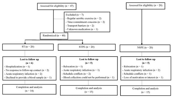
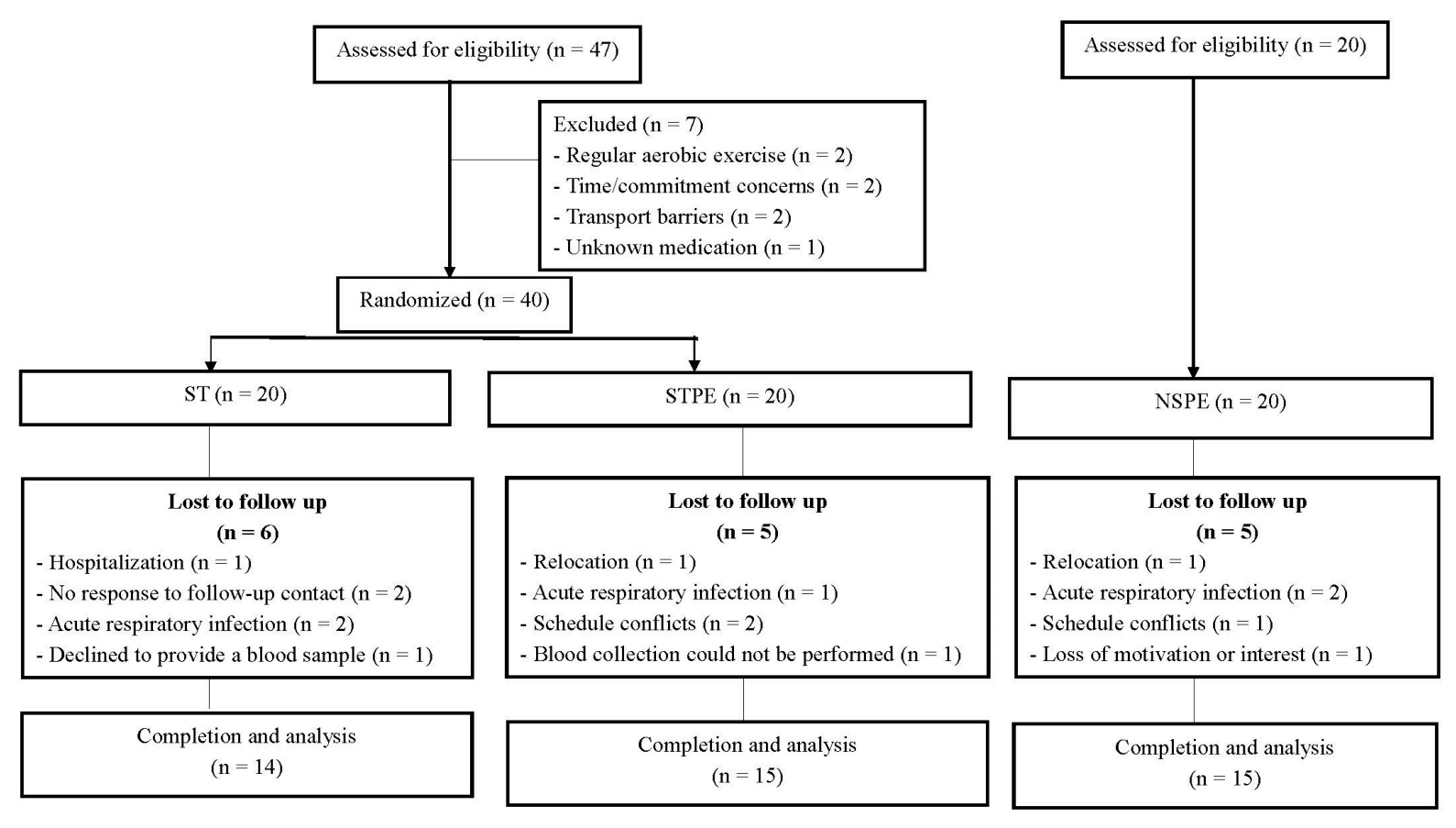
A total of 60 participants enrolled, 44 completed the PRE and POST assessments. There were 14 participants in the ST group, 15 in STPE, and 15 in NSPE. Of those in the ST group, one participant was hospitalized, one declined to provide a blood sample, two were lost to follow-up, and two experienced acute respiratory infections. In the NSPE group, one participant relocated, one reported scheduling conflicts, one lost motivation, and two developed acute respiratory infections. In the STPE group, one participant relocated, one developed an acute respiratory infection, one was unable to undergo blood collection, and two reported scheduling conflicts as illustrated in the study flow diagram (Figure 1).
Figure 1.
The flow of the study.
Baseline characteristics are shown in Table 1. Within the randomized comparison, participants in STPE had higher weight (p = 0.015) and height (p = 0.048) than those in ST. BMI did not differ significantly between ST and STPE (p = 0.225). Characteristics of the separately recruited NSPE group are presented descriptively. This may be because individuals prescribed statins are commonly present with cardiovascular risk factors (e.g., hypertension, stroke, type 2 diabetes) and tend to have higher bodyweight and BMI [39].
Table 1.
Demographic characteristics of statin-treated (ST), statin-treated with progressive resistance exercise (STPE), and non-statin-treated with progressive resistance exercise (NSPE).
Exercise attendance, training loads, and adverse events.
Participants who completed the study in both STPE and NSPE attended all 24 scheduled exercise sessions, corresponding to 100% attendance among completers. Training loads reached 100% of estimated 10RM in the final set of each exercise, following sets performed at 50% and 75% of estimated 10RM. Reported adverse events included muscle soreness and fatigue, with no withdrawals attributed to the exercise intervention.
3.1. Outcomes
3.1.1. Cognitive Outcomes
Changes in Stroop test performance are presented in Table 2. The change in incongruent task completion time differed between ST and STPE (p = 0.002). Median completion time increased in ST, from 94.3 to 109.7 s, and decreased in STPE, from 95.6 to 93.0 s. The between-group changes also differed for Stroop interference (p = 0.005) and the sum of incongruent and neutral task completion times (p = 0.018). No significant between-group differences in change were observed for neutral task completion time or the numbers of trials required for correct runs. NSPE results are shown as within-group observations and were not included in these between-group comparisons.
Table 2.
The Stroop test at beginning (PRE) and after the 8 wk training period (POST) in the statin-treated (ST), statin-treated with progressive exercise (STPE), and non-statins-treated with progressive exercise (NSPE).
Digit Span Forward, Digit Span Backward, and the TMT-B/TMT-A ratio showed no significant differences in change between ST and STPE (all p > 0.05), as in Table 3. Within STPE, Digit Span Forward scores increased from 6.4 ± 2.0 to 7.5 ± 2.2 (p = 0.015). This within-group change did not correspond to a statistically significant difference in change between ST and STPE (p = 0.105).
Table 3.
The cognitive performances (Digit Span test and Trail Making Test B/Trail Making Test A) at beginning (PRE) and after the 8 wk training period (POST) in the statin-treated (ST), statin-treated with progressive resistance exercise (STPE), and non-statin-treated with progressive resistance exercise (NSPE).
In baseline-adjusted sensitivity analyses, POST interference time was lower in STPE than in ST (adjusted difference, −10.47 s; 95% CI, −19.78 to −1.16; p = 0.029). Differences were not statistically significant for incongruent task completion time (−29.13 s; 95% CI, −61.46 to 3.20; p = 0.075) or total On plus Off time (−43.67 s; 95% CI, −104.33 to 16.99; p = 0.151). These exploratory analyses were not adjusted for multiple testing (Supplementary Table S1).
3.1.2. Exploratory Biological Outcomes
Plasma BDNF increased within ST (p = 0.023) and STPE (p = 0.019), but the change did not differ significantly between these randomized groups (p = 0.150), as in Table 4. CAT activity increased within NSPE (p = 0.020). Changes in CAT activity did not differ significantly between ST and STPE (p = 0.570).
Table 4.
The peripheral blood brain-derived neurotrophic factor in plasma (pBDNF) and catalase enzyme activity (CAT) at beginning (PRE) and after the 8 wk training period (POST) in the statin-treated (ST), statin-treated with progressive resistance exercise (STPE), and non-statin-treated with progressive resistance exercise (NSPE).
TAC decreased within ST (p = 0.040) and STPE (p = 0.028), with no significant difference in change between these groups (p = 0.485). POX activity increased within all three groups (p < 0.05), while its change did not differ significantly between ST and STPE (p = 0.827). The calculated MDA concentrations were higher at POST than at PRE in all three groups. However, review of the assay records identified substantial differences between the calibration curves used for PRE and POST samples, raising concerns about between-batch comparability. Accordingly, these values are reported descriptively and are not interpreted as evidence of a biological increase in MDA. Biological findings and within-group results for NSPE were considered exploratory.
3.2. Figures, Tables and Schemes
Table 5.
The oxidative stress and antioxidant levels at beginning (PRE) and after the 8 wk training period (POST) in the statin-treated (ST), statin-treated with progressive resistance exercise (STPE), and non-statin-treated with progressive resistance exercise (NSPE).
4. Discussion
The main finding of this study was that, among statin users, the STPE group showed more favorable changes than the ST group in selected Stroop measures after eight weeks, including incongruent task completion time, interference, and the sum of incongruent and neutral task completion times. Changes in Digit Span performance and the TMT-B/TMT-A ratio did not differ significantly between the two groups. The biological findings were exploratory, although several markers changed within groups, their changes did not differ significantly between ST and STPE. Baseline-adjusted sensitivity analyses supported the between-group difference in Stroop interference, but not the differences in incongruent task completion time or total On plus Off time. The cognitive findings should therefore be interpreted as exploratory and were not consistently supported across analytical approaches.
A single bout of high-intensity resistance exercise in healthy young adults has been shown to improve behavioral performance, as evidenced by faster reaction times [40]. Similarly, 12 months of resistance training in older adults was associated with significant improvements in executive function, as measured by the Stroop test [41]. Moreover, 16 weeks of training in healthy older adults resulted in enhanced Stroop interference performance and faster reaction times [42].
The more favorable changes in selected Stroop measures observed in STPE are consistent with previous reports of improved cognitive performance following resistance training [41,42]. However, baseline-adjusted sensitivity analyses supported the between-group difference only for interference. Similarly, Baptista et al. demonstrated that a combination of statins and exercise training, or exercise training alone, provided superior outcomes in terms of functional performance, cholesterol management and cardiovascular risk mitigation among older adults with dyslipidemia and comorbidities, compared to statin therapy alone [43]. Toyama et al. also observed that combining statin therapy with aerobic exercise for five months may improve cognitive dysfunction in patients with coronary artery disease and pre-existing mild cognitive impairment [44]. On the other hand, Saillant et al. observed that statin users with aerobic training for 12 weeks appear to limit the cognitive benefits derived from aerobic training [45]. The proposed mechanism is that simvastatin may impair the adaptations of mitochondria to exercise. These adaptations are essential for enhancing the oxygen consumption of skeletal muscle and represent a key component of the improvements in cardiorespiratory fitness that result from exercise [45,46]. Resistance training induced substantial functional brain changes, particularly in the frontal lobe, which were accompanied by improvements in executive function [47]. Statin use was significantly associated with short-term cognitive impairment, but not with long-term cognitive outcomes [48]. Similarly, lower performance was demonstrated in global cognition and episodic memory among statin users [49]. Six months of simvastatin therapy in randomized controlled trials (RCTs) was also observed to reduce cognitive function in patients [50]. However, the three-city study, with a 7-year follow-up period, found no association between baseline statin use and the incidence of dementia [51]. A meta-analysis and systemic review indicated that statin treatment was not associated with cognitive impairment in RCTs [5,52]. Repeated exposure to the cognitive tests may have produced practice effects. Although ST and STPE were assessed at the same time points, the present study did not include additional assessments to characterize the learning curve or determine whether practice effects differed between groups.
It has been proposed that statins induce an increase in BDNF, which enhance brain plasticity [12]. In the present study, pBDNF increased within both statin-treated groups, but changes in pBDNF did not differ significantly between these groups. More favorable changes in selected Stroop measures were observed in STPE, whereas the increase in pBDNF within ST was not accompanied by cognitive improvement. Thus, the present findings provide no evidence that increased circulating pBDNF contributed to the observed cognitive benefits. Resistance exercise was found to be a modifiable factor that influences cognition [53]. Plasma BDNF in the study increased within both ST and STPE, but its change did not differ significantly between these randomized groups. Thus, the data do not support attributing the difference in Stroop performance to a differential pBDNF response. BDNF is also associated with fat mass, energy metabolism and cardiometabolic risk factors [18]. Evidently, patients receiving statin treatment are believed to be determined by cardiovascular risk factors such as hypertension, stroke, coronary heart disease, type 2 diabetes and BMI, all of which are susceptible to cognitive function [39]. Moreover, the mechanisms involved in statin-associated muscle symptoms, such as ROS generation and mitochondrial membrane depolarization, may be associated with cognitive impairment [24]. In the ST group, pBDNF increased without a corresponding improvement in cognitive performance. A compensatory response has been proposed as a possible explanation for increased circulating BDNF [54]. However, the present study cannot confirm this mechanism and provides no evidence that the observed increase in pBDNF contributed to improved cognitive performance. Circulating pBDNF may also be influenced by factors other than the intervention, including differences in participant characteristics. The biological meaning of the observed increase remains unclear, and the present measurements cannot establish whether it reflects changes in brain BDNF, neuroplasticity, a compensatory response, or another process.
Statin therapy has been linked to oxidative stress and antioxidant responses [19,20]. In the present study, POX activity increased within each group, but its change did not differ significantly between ST and STPE. TAC decreased within both statin-treated groups, again without a significant between-group difference in change. CAT increased within NSPE, but its change did not differ significantly between ST and STPE. Previous studies have reported changes in antioxidant enzymes during statin treatment [19] and have proposed that exercise-related oxidative stress may influence enzymatic antioxidant responses [55]. Experimental studies have also described increased reactive oxygen species, lipid peroxidation, and impaired mitochondrial function following exposure to certain statins [56,57]. However, these proposed mechanisms cannot be established from the present data. The observed biomarker changes do not demonstrate that adding progressive resistance exercise altered oxidative stress or antioxidant responses differently among statin users. Furthermore, because NSPE was recruited separately, its within-group changes cannot establish an effect of statin exposure on the response to exercise. Review of the original assay records identified substantial differences between the calibration curves used for PRE and POST MDA measurements. Although different calibration curves do not necessarily invalidate an assay, comparability between these batches could not be established from the available records. We therefore withdrew the interpretation of the higher calculated POST values as a biological increase in lipid peroxidation. The MDA results cannot support conclusions about changes in oxidative stress or the effects of statin treatment or exercise.
TAC decreased within both ST and STPE over the eight-week period, but the magnitude of change did not differ significantly between them. The FRAP assay used to assess TAC reflects the combined reducing capacity of several circulating substances, including uric acid, ascorbic acid, alpha-tocopherol, proteins, and bilirubin [35]. Previous studies have reported changes in antioxidant vitamin levels during statin treatment [58] and reductions in serum uric acid following atorvastatin treatment [59]. Urate can contribute to antioxidant capacity directly and through its interaction with ascorbate [60]. However, the present study did not measure the contributions of individual substances to FRAP values. Therefore, the decrease in TAC cannot be attributed specifically to changes in uric acid or antioxidant vitamins. The increase in POX alone does not establish an adaptive antioxidant response or identify its cause.
Overall, this study provides a preliminary view of oxidative stress and antioxidant responses alongside cognitive outcomes in older women receiving statins, with or without progressive resistance exercise. POX increased within all three groups, while TAC decreased within both randomized statin-treated groups. CAT increased within the separately recruited NSPE group. However, none of these biomarker changes differed significantly between ST and STPE. Thus, although several markers changed over eight weeks, the data do not show that adding progressive resistance exercise altered these responses among statin users. Although this study assessed enzymatic and non-enzymatic antioxidant measures within the same randomized exercise comparison, the physiological meaning of the observed patterns remains uncertain.
Several limitations should be considered when interpreting these findings. First, 44 of the 60 enrolled participants completed the study. The small, completed sample limits the precision and generalizability of the findings, and the 16 withdrawals may have introduced attrition bias because analyses included only participants with available PRE and POST measurements. The inclusion of women only further limits generalizability. Second, randomization was limited to statin users, whereas NSPE participants were recruited separately. Observations within NSPE may therefore reflect differences in participant characteristics and cannot establish the effect of statin use on exercise responses. Third, weight and height differed at baseline between ST and STPE. Although the sensitivity analyses adjusted for the corresponding baseline cognitive score, they did not account for these anthropometric differences. Fourth, multiple cognitive and biological outcomes were examined without adjustment for multiple testing, increasing the possibility of chance findings. The conventional ANCOVA results for incongruent task completion time were limited by marked residual non-normality and unequal variances. Diet, sleep, and physical activity outside the intervention were not fully controlled. Practice effects from repeated cognitive testing could not be fully assessed, and biological markers were measured at only two time points. Finally, creatine kinase and lactate were not measured, so biochemical evidence of muscle injury and lactate responses to training could not be assessed. These limitations warrant cautious interpretation of the findings, particularly the exploratory biological results and observations within NSPE. The main strength of the study is the inclusion of a homogeneous cohort of sedentary elderly women with a history of statin use for more than one year, which helps minimize variability related to lifestyle and treatment duration. In addition, the progressive exercise program was implemented using a hybrid supervised model, delivered both in person and through live video conferencing, which has proven feasible, safe, and acceptable for older adults with different physical abilities. Lastly, the assessment of cognitive function, pBDNF levels, and physiological responses reflecting oxidative stress and antioxidant balance allowed us to investigate potential interrelationships. Assessing cognitive performance alongside circulating biological markers also provides information for planning future studies. Larger trials should specify a primary cognitive outcome in advance, account for expected attrition, and assess potential confounders and practice effects. Additional biomarker measurements would be needed to investigate proposed biological mechanisms.
5. Conclusions
This study shows that, among statin users, those assigned to progressive resistance exercise had more favorable changes in selected Stroop measures than those assigned to the non-exercise group. However, baseline-adjusted sensitivity analyses supported the difference in interference only, warranting caution in interpreting the broader Stroop findings. Changes in Digit Span performance and the TMT-B/TMT-A ratio did not differ significantly between the randomized groups. Although pBDNF increased within both statin-treated groups, the present findings provide no evidence that this increase contributed to improved cognitive performance. TAC and POX changed within the statin-treated groups, but their changes did not differ significantly between ST and STPE. CAT increased within the separately recruited NSPE group, but this finding cannot establish an effect of statin use on the response to exercise. Given the sample size, multiple outcomes, and unadjusted baseline differences, these findings should be interpreted with appropriate caution. Larger trials with prespecified cognitive outcomes are needed to confirm the Stroop findings and investigate their underlying mechanisms.
Supplementary Materials
The following supporting information can be downloaded at: https://www.mdpi.com/article/10.3390/life16111865/s1, Table S1: Sensitivity analyses of Stroop outcomes comparing ST and STPE after adjustment for baseline performance.
Author Contributions
Design of the study, data collection, data analysis, first draft of this manuscript, K.K.; design of the study, data collection, laboratory analysis, S.S.; design of the study and data collection B.P.S., N.L., and C.T.; data analysis and supervised the manuscript, M.B.; design of the study, data collection, data analysis, first draft of this manuscript, and administration of the grant, S.K. All authors contribute to the manuscript writing. All authors have read and agreed to the published version of the manuscript.
Funding
This research was funded by The Research Grant of Faculty of Allied Health Sciences, Burapha University, Thailand, grant number AHS06/2564.
Institutional Review Board Statement
The study was conducted in accordance with the Declaration of Helsinki and was approved by the Burapha University Ethics Committee of Human Research (IRB No. IRB1-026/2565; Protocol No. HS069/2564, Approval Date: 11 March 2022). The study was also registered with the Thai Clinical Trials Registry (TCTR20250203007).
Informed Consent Statement
Informed consent was obtained from all subjects involved in the study. Written informed consent has been obtained from the patient(s) to publish this paper.
Data Availability Statement
The datasets obtained and/or evaluated during the present study are restricted from public access due to ethical regulations but may be provided by the corresponding author following a reasonable request and approval from the relevant ethics committee.
Acknowledgments
The authors have reviewed and edited the output and take full responsibility for the content of this publication.
Conflicts of Interest
The authors declare no conflicts of interest. The funders had no role in the design of the study; in the collection, analyses, or interpretation of data; in the writing of the manuscript; or in the decision to publish the results.
Abbreviations
The following abbreviations are used in this manuscript:
| PE | Progressive resistance exercise |
| pBDNF | Plasma brain-derived neurotrophic factor |
| ST | Statin-treated |
| STPE | Statin-treated with progressive resistance exercise |
| NSPE | Non-statin-treated with progressive resistance exercise |
| ICH | Intracerebral hemorrhage |
| ROS | Reactive oxygen species |
| BMI | Body mass index |
| RM | Repetition maximum |
| SAMS | Statin-associated muscle symptoms |
| EDTA | Ethylenediaminetetraacetic acid |
| DSF | Digit Span Forward |
| DSB | Digit Span Backward |
| TMT | Trail Making Test |
| TMT-A | Trail Making Test Part A |
| TMT-B | Trail Making Test Part B |
| TBARS | Thiobarbituric reactive substances |
| BHT | Butylated hydroxytoluene |
| SDS | Sodium dodecyl sulfate |
| TCA | Trichloroacetic acid |
| TBA | Thiobarbituric acid |
| TAC | The total antioxidant capacity |
| FRAP | Ferric Reducing Antioxidant Power |
| TPTZ | 2,4,6-tri[2-pyridyl]-s-triazine |
| DAB | Diaminobenzidine tetrahydrochloride |
| POX | Peroxidase enzyme activity |
| CAT | Catalase enzyme activity |
| SD | Standard deviation |
| IQR | Interquartile range |
| RCTS | Randomized controlled trials |
| GPX | Glutathione peroxidase |
| MDA | Malondialdehyde |
References
- Qiu, C.; Fratiglioni, L. A major role for cardiovascular burden in age-related cognitive decline. Nat. Rev. Cardiol. 2015, 12, 267–277. [Google Scholar] [CrossRef] [Scilit] [PubMed]
- Lolekha, P.; Khovidhunkit, W.; Deerochanawong, C.; Thongtang, N.; Boonyasirinant, T.; Rattarasarn, C.; Chutinet, A.; Ophascharoensuk, V.; Somlaw, N.; Sitthisook, S.; et al. 2024 The Royal College of Physicians of Thailand (RCPT) clinical practice guidelines on management of dyslipidemia for atherosclerotic cardiovascular disease prevention. Asian BioMed. 2024, 18, 246–267. [Google Scholar] [CrossRef] [Scilit] [PubMed]
- Bruckert, E.; Hayem, G.; Dejager, S.; Yau, C.; Bégaud, B. Mild to moderate muscular symptoms with high-dosage statin therapy in hyperlipidemic patients--the PRIMO study. Cardiovasc Drugs Ther. 2005, 19, 403–414. [Google Scholar] [CrossRef] [Scilit] [PubMed]
- Richardson, K.; Schoen, M.; French, B.; Umscheid, C.A.; Mitchell, M.D.; Arnold, S.E.; Heidenreich, P.A.; Rader, D.J.; M deGoma, E. Statins and cognitive function: A systematic review. Ann. Intern Med. 2013, 159, 688–697. [Google Scholar] [PubMed]
- Adhikari, A.; Tripathy, S.; Chuzi, S.; Peterson, J.; Stone, N.J. Association between statin use and cognitive function: A systematic review of randomized clinical trials and observational studies. J. Clin. Lipidol. 2021, 15, 22–32. [Google Scholar] [CrossRef] [Scilit] [PubMed]
- Deichmann, R.E.; Lavie, C.J.; Asher, T.; DiNicolantonio, J.J.; O’Keefe, J.H.; Thompson, P.D. The Interaction Between Statins and Exercise: Mechanisms and Strategies to Counter the Musculoskeletal Side Effects of This Combination Therapy. Ochsner J. 2015, 15, 429–437. [Google Scholar] [PubMed]
- Klimova, B.; Valis, M.; Kuca, K. Cognitive decline in normal aging and its prevention: A review on non-pharmacological lifestyle strategies. Clin. Interv. Aging 2017, 12, 903–910. [Google Scholar] [CrossRef] [Scilit] [PubMed]
- Landrigan, J.F.; Bell, T.; Crowe, M.; Clay, O.J.; Mirman, D. Lifting cognition: A meta-analysis of effects of resistance exercise on cognition. Psychol. Res. 2020, 84, 1167–1183. [Google Scholar] [CrossRef] [Scilit] [PubMed]
- Pu, C.T.; Johnson, M.T.; Forman, D.E.; Hausdorff, J.M.; Roubenoff, R.; Foldvari, R.M.; Fielding, R.A.; Singh, M.A.F. Randomized trial of progressive resistance training to counteract the myopathy of chronic heart failure. J. Appl. Physiol. 2001, 90, 2341–2350. [Google Scholar] [CrossRef] [Scilit] [PubMed]
- Setayesh, S.; Mohammad Rahimi, G.R. The impact of resistance training on brain-derived neurotrophic factor and depression among older adults aged 60 years or older: A systematic review and meta-analysis of randomized controlled trials. Geriatr. Nurs. 2023, 54, 23–31. [Google Scholar] [CrossRef] [Scilit] [PubMed]
- Vints, W.A.J.; Gökçe, E.; Langeard, A.; Pavlova, I.; Çevik, O.S.; Ziaaldini, M.M.; Todri, J.; Lena, O.; Sakkas, G.K.; Jak, S.; et al. Myokines as mediators of exercise-induced cognitive changes in older adults: Protocol for a comprehensive living systematic review and meta-analysis. Front Aging Neurosci. 2023, 15, 1213057. [Google Scholar] [CrossRef] [Scilit] [PubMed]
- Zhang, J.; Mu, X.; Breker, D.A.; Li, Y.; Gao, Z.; Huang, Y. Atorvastatin treatment is associated with increased BDNF level and improved functional recovery after atherothrombotic stroke. Int. J. Neurosci. 2017, 127, 92–97. [Google Scholar] [CrossRef] [Scilit] [PubMed]
- Laske, C.; Stransky, E.; Leyhe, T.; Eschweiler, G.W.; Maetzler, W.; Wittorf, A. BDNF serum and CSF concentrations in Alzheimer’s disease, normal pressure hydrocephalus and healthy controls. J. Psychiatr. Res. 2007, 41, 387–394. [Google Scholar] [CrossRef] [Scilit] [PubMed]
- Yang, D.; Han, Y.; Zhang, J.; Chopp, M.; Seyfried, D.M. Statins Enhance Expression of Growth Factors and Activate the PI3K/Akt-mediated Signaling Pathway after Experimental Intracerebral Hemorrhage. World J. Neurosci. 2012, 2, 74–80. [Google Scholar] [PubMed]
- Nagahara, A.H.; Tuszynski, M.H. Potential therapeutic uses of BDNF in neurological and psychiatric disorders. Nat. Rev. Drug Discov. 2011, 10, 209–219. [Google Scholar] [CrossRef] [Scilit] [PubMed]
- Gomez-Pinilla, F.; Vaynman, S.; Ying, Z. Brain-derived neurotrophic factor functions as a metabotrophin to mediate the effects of exercise on cognition. Eur. J. Neurosci. 2008, 28, 2278–2287. [Google Scholar] [CrossRef] [Scilit] [PubMed]
- Suzuki, S.; Kiyosue, K.; Hazama, S.; Ogura, A.; Kashihara, M.; Hara, T.; Koshimizu, H.; Kojima, M. Brain-derived neurotrophic factor regulates cholesterol metabolism for synapse development. J. Neurosci. 2007, 27, 6417–6427. [Google Scholar] [CrossRef] [Scilit] [PubMed]
- Suwa, M.; Kishimoto, H.; Nofuji, Y.; Nakano, H.; Sasaki, H.; Radak, Z.; Kumagai, S. Serum brain-derived neurotrophic factor level is increased and associated with obesity in newly diagnosed female patients with type 2 diabetes mellitus. Metabolism 2006, 55, 852–857. [Google Scholar] [CrossRef] [Scilit] [PubMed]
- Zinellu, A.; Mangoni, A.A. A Systematic Review and Meta-Analysis of the Effect of Statins on Glutathione Peroxidase, Superoxide Dismutase, and Catalase. Antioxidants 2021, 10, 1841. [Google Scholar] [CrossRef] [Scilit] [PubMed]
- Liu, A.; Wu, Q.; Guo, J.; Ares, I.; Rodríguez, J.; Martínez-Larrañaga, M.; Yuan, Z.; Anadón, A.; Wang, X.; Martínez, M.-A. Statins: Adverse reactions, oxidative stress and metabolic interactions. Pharmacol. Ther. 2019, 195, 54–84. [Google Scholar] [CrossRef] [Scilit] [PubMed]
- Bouitbir, J.; Charles, A.; Rasseneur, L.; Dufour, S.; Piquard, F.; Geny, B.; Zoll, J. Atorvastatin treatment reduces exercise capacities in rats: Involvement of mitochondrial impairments and oxidative stress. J. Appl. Physiol. 2011, 111, 1477–1483. [Google Scholar] [CrossRef] [Scilit] [PubMed]
- Chan, K.; Truong, D.; Shangari, N.; O’Brien, P.J. Drug-induced mitochondrial toxicity. Expert Opin. Drug Metab. Toxicol. 2005, 1, 655–669. [Google Scholar] [CrossRef] [Scilit] [PubMed]
- Schultz, B.G.; Patten, D.K.; Berlau, D.J. The role of statins in both cognitive impairment and protection against dementia: A tale of two mechanisms. Transl. Neurodegener. 2018, 7, 5. [Google Scholar] [CrossRef] [Scilit] [PubMed]
- Mollazadeh, H.; Tavana, E.; Fanni, G.; Bo, S.; Banach, M.; Pirro, M.; von Haehling, S.; Jamialahmadi, T.; Sahebkar, A. Effects of statins on mitochondrial pathways. J. Cachexia Sarcopenia Muscle 2021, 12, 237–251. [Google Scholar] [CrossRef] [Scilit] [PubMed]
- Della Corte, J.; Paz, G.; Brandão Pinto de Castro, J.; Miranda, H. Hypotensive effect induced by strength training adopting DeLorme and Oxford methods in trained men. Pol. J. Sport Tour. 2018, 25, 23–30. [Google Scholar] [CrossRef] [Scilit]
- Brzycki, M. Strength Testing—Predicting a One-Rep Max from Reps-to-Fatigue. J. Phys. Educ. Recreat. Dan. 1993, 64, 88–90. [Google Scholar] [CrossRef] [Scilit]
- Lai, X.; Zhu, H.; Wu, Z.; Chen, B.; Jiang, Q.; Du, H.; Huo, X. Dose-response effects of resistance training on physical function in frail older Chinese adults: A randomized controlled trial. J. Cachexia Sarcopenia Muscle 2023, 14, 2824–2834. [Google Scholar] [CrossRef] [Scilit] [PubMed]
- Woods, D.L.; Kishiyamaa, M.M.; Lund, E.W.; Herron, T.J.; Edwards, B.; Poliva, O.; Hink, R.F.; Reed, B. Improving digit span assessment of short-term verbal memory. J. Clin. Exp. Neuropsychol. 2011, 33, 101–111. [Google Scholar] [CrossRef] [Scilit] [PubMed]
- Arbuthnott, K.; Frank, J. Trail making test, part B as a measure of executive control: Validation using a set-switching paradigm. J. Clin. Exp. Neuropsychol. 2000, 22, 518–528. [Google Scholar] [CrossRef] [Scilit] [PubMed]
- Amodio, P.; Schiff, S.; Del Piccolo, F.; Mapelli, D.; Gatta, A.; Umiltà, C. Attention dysfunction in cirrhotic patients: An inquiry on the role of executive control, attention orienting and focusing. Metab. Brain Dis. 2005, 20, 115–127. [Google Scholar] [CrossRef] [Scilit] [PubMed]
- Bajaj, J.S.; Thacker, L.R.; Heuman, D.M.; Fuchs, M.; Sterling, R.K.; Sanyal, A.J.; Puri, P.; Siddiqui, M.S.; Stravitz, R.T.; Bouneva, I.; et al. The Stroop smartphone application is a short and valid method to screen for minimal hepatic encephalopathy. Hepatology 2013, 58, 1122–1132. [Google Scholar] [CrossRef] [Scilit] [PubMed]
- Fraga, I.; Weber, C.; Galiano, W.B.; Iraci, L.; Wohlgemuth, M.; Morales, G.; Cercato, C.; Rodriguez, J.; Pochmann, D.; Dani, C.; et al. Effects of a multimodal exercise protocol on functional outcomes, epigenetic modulation and brain-derived neurotrophic factor levels in institutionalized older adults: A quasi-experimental pilot study. Neural Regen. Res. 2021, 16, 2479–2485. [Google Scholar] [CrossRef] [Scilit] [PubMed]
- Nielsen, F.; Mikkelsen, B.B.; Nielsen, J.B.; Andersen, H.R.; Grandjean, P. Plasma malondialdehyde as biomarker for oxidative stress: Reference interval and effects of life-style factors. Clin. Chem. 1997, 43, 1209–1214. [Google Scholar] [CrossRef] [Scilit]
- Tsai, L.Y.; Lee, K.T.; Tsai, S.M.; Lee, S.C.; Yu, H.S. Changes of lipid peroxide levels in blood and liver tissue of patients with obstructive jaundice. Clin. Chim. Acta 1993, 215, 41–50. [Google Scholar] [CrossRef] [Scilit] [PubMed]
- Benzie, I.F.; Strain, J.J. The ferric reducing ability of plasma (FRAP) as a measure of “antioxidant power”: The FRAP assay. Anal. Biochem 1996, 239, 70–76. [Google Scholar] [CrossRef] [Scilit] [PubMed]
- Herzog, V.; Fahimi, H.D. A new sensitive colorimetric assay for peroxidase using 3,3’-diaminobenzidine as hydrogen donor. Anal. Biochem 1973, 55, 554–562. [Google Scholar] [CrossRef] [Scilit] [PubMed]
- Goth, L. A simple method for determination of serum catalase activity and revision of reference range. Clin. Chim. Acta 1991, 196, 143–151. [Google Scholar] [CrossRef] [Scilit] [PubMed]
- Singsanan, S.; Luangpon, N.; Kiatkulanusorn, S.; Boonsiri, P.; Burtscher, M.; Klarod, K. Qigong Training Effects on Brain-Derived Neurotrophic Factor and Cognitive Functions in Sedentary Middle-Aged and Elderly Females With Type 2 Diabetes. WSPAJ 2024, 32. [Google Scholar] [CrossRef] [Scilit]
- Grundy, S.M.; Stone, N.J.; Bailey, A.L.; Beam, C.; Birtcher, K.K.; Blumenthal, R.S.; Braun, L.T.; de Ferranti, S.; Faiella-Tommasino, J.; Forman, D.E.; et al. 2018 AHA/ACC/AACVPR/AAPA/ABC/ACPM/ADA/AGS/APhA/ASPC/NLA/PCNA Guideline on the Management of Blood Cholesterol: A Report of the American College of Cardiology/American Heart Association Task Force on Clinical Practice Guidelines. J. Am. Coll. Cardiol. 2019, 73, e285–e350. [Google Scholar] [PubMed]
- Chang, H.; Kim, K.; Jung, Y.J.; Kato, M. Effects of acute high-Intensity resistance exercise on cognitive function and oxygenation in prefrontal cortex. J. Exerc Nutr. Biochem 2017, 21, 1–8. [Google Scholar] [CrossRef] [Scilit] [PubMed]
- Liu-Ambrose, T.; Nagamatsu, L.S.; Voss, M.W.; Khan, K.M.; Handy, T.C. Resistance training and functional plasticity of the aging brain: A 12-month randomized controlled trial. Neurobiol. Aging 2012, 33, 1690–1698. [Google Scholar] [CrossRef] [Scilit] [PubMed]
- Coetsee, C.; Terblanche, E. Cerebral oxygenation during cortical activation: The differential influence of three exercise training modalities. A randomized controlled trial. Eur. J. Appl. Physiol. 2017, 117, 1617–1627. [Google Scholar] [CrossRef] [Scilit] [PubMed]
- Baptista, L.C.; Veríssimo, M.T.; Martins, R.A. Statin combined with exercise training is more effective to improve functional status in dyslipidemic older adults. Scand. J. Med. Sci. Sports 2018, 28, 2659–2667. [Google Scholar] [CrossRef] [Scilit] [PubMed]
- Toyama, K.; Sugiyama, S.; Oka, H.; Hamada, M.; Iwasaki, Y.; Horio, E.; Rokutanda, T.; Nakamura, S.; Spin, J.M.; Tsao, P.S.; et al. A Pilot Study: The Beneficial Effects of Combined Statin-exercise Therapy on Cognitive Function in Patients with Coronary Artery Disease and Mild Cognitive Decline. Intern Med. 2017, 56, 641–649. [Google Scholar] [CrossRef] [Scilit] [PubMed]
- Saillant, K.; Langeard, A.; Kaushal, N.; Minh Vu, T.T.; Pothier, K.; Langlois, F.; Nigam, A.; Juneau, M.; Bherer, L. Statin use moderates the beneficial effects of aerobic exercise on older adults’ performances on the Stroop test: A subanalysis. Exp. Gerontol. 2021, 147, 111277. [Google Scholar] [CrossRef] [Scilit] [PubMed]
- Mikus, C.R.; Boyle, L.J.; Borengasser, S.J.; Oberlin, D.J.; Naples, S.P.; Fletcher, J.; Meers, G.M.; Ruebel, M.; Laughlin, M.H.; Dellsperger, K.C.; et al. Simvastatin impairs exercise training adaptations. J. Am. Coll. Cardiol. 2013, 62, 709–714. [Google Scholar] [CrossRef] [Scilit] [PubMed]
- Herold, F.; Torpel, A.; Schega, L.; Muller, N.G. Functional and/or structural brain changes in response to resistance exercises and resistance training lead to cognitive improvements—A systematic review. Eur. Rev. Aging Phys. Act. 2019, 16, 10. [Google Scholar] [CrossRef] [Scilit] [PubMed]
- Gentreau, M.; Rukh, G.; Miguet, M.; Clemensson, L.E.; Alsehli, A.M.; Titova, O.E.; Schiöth, H.B. The Effects of Statins on Cognitive Performance Are Mediated by Low-Density Lipoprotein, C-Reactive Protein, and Blood Glucose Concentrations. J. Gerontol. A Biol. Sci. Med. Sci. 2023, 78, 1964–1972. [Google Scholar] [CrossRef] [Scilit] [PubMed]
- Zhou, Z.; Ryan, J.; Ernst, M.E.; Zoungas, S.; Tonkin, A.M.; Woods, R.L.; McNeil, J.J.; Reid, C.M.; Curtis, A.J.; Wolfe, R.; et al. Effect of Statin Therapy on Cognitive Decline and Incident Dementia in Older Adults. J. Am. Coll. Cardiol. 2021, 77, 3145–3156. [Google Scholar] [CrossRef] [Scilit] [PubMed]
- Muldoon, M.F.; Ryan, C.M.; Sereika, S.M.; Flory, J.D.; Manuck, S.B. Randomized trial of the effects of simvastatin on cognitive functioning in hypercholesterolemic adults. Am. J. Med. 2004, 117, 823–829. [Google Scholar] [CrossRef] [Scilit] [PubMed]
- Ancelin, M.L.; Carrière, I.; Barberger-Gateau, P.; Auriacombe, S.; Rouaud, O.; Fourlanos, S.; Berr, C.; Dupuy, A.-M.; Ritchie, K. Lipid lowering agents, cognitive decline, and dementia: The three-city study. J. Alzheimers Dis. 2012, 30, 629–637. [Google Scholar] [CrossRef] [Scilit] [PubMed]
- Ott, B.R.; Daiello, L.A.; Dahabreh, I.J.; Springate, B.A.; Bixby, K.; Murali, M.; Trikalinos, T.A. Do statins impair cognition? A systematic review and meta-analysis of randomized controlled trials. J. Gen. Intern Med. 2015, 30, 348–358. [Google Scholar] [CrossRef] [Scilit] [PubMed]
- Yoon, D.H.; Lee, J.-Y.; Song, W. Effects of Resistance Exercise Training on Cognitive Function and Physical Performance in Cognitive Frailty: A Randomized Controlled Trial. J. Nutr. Health Aging 2018, 22, 944–951. [Google Scholar] [CrossRef] [Scilit] [PubMed]
- Gao, L.; Zhang, Y.; Sterling, K.; Song, W. Brain-derived neurotrophic factor in Alzheimer’s disease and its pharmaceutical potential. Transl. Neurodegener. 2022, 11, 4. [Google Scholar] [CrossRef] [Scilit] [PubMed]
- Urso, M.L.; Clarkson, P.M. Oxidative stress, exercise, and antioxidant supplementation. Toxicology 2003, 189, 41–54. [Google Scholar] [CrossRef] [Scilit] [PubMed]
- Abdoli, N.; Azarmi, Y.; Eghbal, M.A. Protective Effects of N-acetylcysteine Against the Statins Cytotoxicity in Freshly Isolated Rat Hepatocytes. Adv. Pharm. Bull. 2014, 4, 249–254. [Google Scholar] [CrossRef] [Scilit] [PubMed]
- Pal, S.; Sarkar, A.; Pal, P.B.; Sil, P.C. Protective effect of arjunolic acid against atorvastatin induced hepatic and renal pathophysiology via MAPK, mitochondria and ER dependent pathways. Biochimie 2015, 112, 20–34. [Google Scholar] [CrossRef] [Scilit] [PubMed]
- Vasankari, T.; Ahotupa, M.; Viikari, J.; Nuotio, I.; Strandberg, T.; Vanhanen, H.; Gylling, H.; Miettinen, T.; Tikkanen, M.J. Effect of 12-month statin therapy on antioxidant potential of LDL and serum antioxidant vitamin concentrations. Ann. Med. 2004, 36, 618–622. [Google Scholar] [CrossRef] [Scilit] [PubMed]
- Milionis, H.J.; Kakafika, A.I.; Tsouli, S.G.; Athyros, V.G.; Bairaktari, E.T.; Seferiadis, K.I.; Elisaf, M.S. Effects of statin treatment on uric acid homeostasis in patients with primary hyperlipidemia. Am. Heart J. 2004, 148, 635–640. [Google Scholar] [CrossRef] [Scilit] [PubMed]
- Sevanian, A.; Davies, K.J.; Hochstein, P. Serum urate as an antioxidant for ascorbic acid. Am. J. Clin. Nutr. 1991, 54, 1129s–1134s. [Google Scholar] [CrossRef] [Scilit] [PubMed]

Disclaimer/Publisher’s Note: The statements, opinions and data contained in all publications are solely those of the individual author(s) and contributor(s) and not of MDPI and/or the editor(s). MDPI and/or the editor(s) disclaim responsibility for any injury to people or property resulting from any ideas, methods, instructions or products referred to in the content. |
© 2026 by the authors. Licensee MDPI, Basel, Switzerland. This article is an open access article distributed under the terms and conditions of the Creative Commons Attribution (CC BY) license.