


Abstract
Various markers and indicators have been examined to reveal the presence and complexity of coronary artery disease (CAD). One of these biomarkers that has been the subject of research in cardiovascular diseases recently is the Endothelial Activation and Stress Index (EASIX) value. Calcification of costal cartilage (CCC) is one of the indicators of tissue calcification and can show different characteristics according to age and gender. In the present study, we investigated the significance of CCC and EASIX values in demonstrating the presence and complexity of CAD. Consecutive patients scheduled for cardiac surgery between 10 January 2025 and 15 June 2026 were included retrospectively. Firstly, patients were divided into two groups: those without CAD and those with CAD. To further assess the data distributions, patients were first split into two groups based on their SYNTAX scores. The control group consisted of 206 patients with a median age of 53 (18–87), while the CAD patient group consisted of 550 patients with a median age of 62 (30–86) (p < 0.001). Subsequently, 550 CAD patients were divided into groups according to the severity of coronary artery disease. Multivariable logistic regression analyses were performed to identify factors playing a role in demonstrating the presence or severity of coronary artery disease. Advanced age, hypertension, smoking, presence of carotid artery disease, high HbA1c values, and CCC volume were associated with CAD. Also, high HbA1c, a high EASIX value, and CCC volume were associated with complex CAD. The current study has shown that the CCC evaluation in these CT images may help to raise suspicion of CAD, thus favoring early diagnosis. Furthermore, our analyses have shown that we may raise suspicion of complex CAD using the EASIX value and CCC evaluation.
1. Introduction
Atherosclerosis is a systemic disease that can affect all organ systems. When the coronary artery system is affected, coronary artery bypass graft (CABG) surgery is considered a valuable treatment method, and despite endovascular advancements, it remains relevant and can be performed with high success rates. Thoracic computed tomography (CT) can be used for preoperative planning purposes prior to these surgeries [1].
Various markers and indicators have been examined to reveal the presence and complexity of coronary artery disease (CAD). These include carotid intima-media thickness, various blood values, the ankle-brachial index, as well as the calcium score by computed tomography [2]. The role of parameters such as ankle-arm index, aortic pulse wave velocity, interleukin-6, and tumor necrosis factor-α has been investigated [3]. These studies aim to diagnose the possible presence of CAD early; by identifying risky groups with these biomarkers, earlier coronary imaging can be performed [4]. One of these biomarkers that has been the subject of research in cardiovascular diseases recently is the Endothelial Activation Stress Index (EASIX) value. In a recent study investigating coronary collateral circulation in patients with CAD, high EASIX values were found to be associated with poor collateral circulation [5]. In another recent study, high EASIX values were also found to be associated with morbidity after CABG surgeries [6].
Calcification of costal cartilage (CCC) is one of the indicators of tissue calcification and can show different characteristics according to age and gender [7]. In a clinical study, it was shown that even in individuals with a coronary calcium score of zero, the presence of CCC was associated with fasting blood glucose exposure [8]. In a recent study published in early 2026, it was shown that CCC assessment in adult diabetic patients could refine clinical decisions and improve individualized risk stratification in older adults with longstanding diabetes [9].
In the present study, we first investigated the significance of CCC and EASIX values in demonstrating the presence of CAD. Subsequently, we aimed to examine their relationship with coronary artery disease complexity in patients scheduled for CABG surgery.
2. Methods
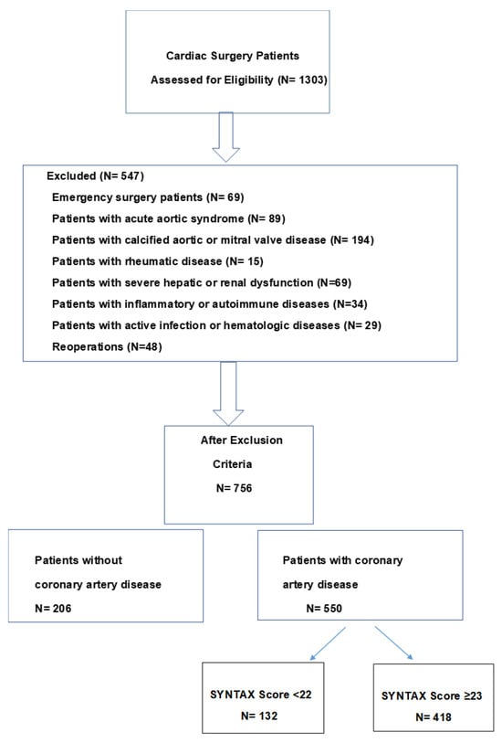
Consecutive patients scheduled for cardiac surgery between 10 January 2025 and 15 June 2026 were included retrospectively. The study was approved by the Bursa Yuksek Ihtisas Training and Research Hospital Clinical Research Ethics Committee (Approval number: 2024-TBEK 2026/08-01, date: 5 August 2026). Every method used in this investigation was carried out in compliance with the 1964 Declaration of Helsinki and its subsequent amendments, or similar ethical standards, as well as the institutional and/or national research committee’s ethical guidelines. Patient data were obtained from the hospital’s data system and patient files. Preoperative blood counts (complete blood count and biochemical parameters), demographic characteristics (age, gender, etc.), echocardiography data, coronary angiography characteristics, and additional chronic disease status were recorded for all patients. Patients scheduled for cardiac surgery at our clinic who had undergone a thoracic CT scan and coronary angiography were included in the study. Reoperations, patients undergoing emergency surgery, those with acute aortic syndrome, those with calcified aortic or mitral valve disease, those with inflammatory or autoimmune diseases, those with severe hepatic and renal dysfunction, those with active infection or hematologic diseases, and those with rheumatic disease were excluded from the study. Following the exclusion criteria, 550 patients consecutively scheduled for CABG were included in the study. To investigate the role of CCC status in indicating the presence of atherosclerotic heart disease, 206 patients scheduled for cardiac surgery within the specified date range were also included as a control group (Figure 1). This patient group consisted of patients scheduled for elective cardiac surgery other than CABG who did not have aortic-mitral valve calcification. Firstly, patients were divided into two groups: those without CAD and those with CAD; the data were then analyzed.
Figure 1.
Flowchart of the study.
2.1. Patient Variables
All patients had their demographics, hypertension, smoking, ejection fraction, and HbA1c values documented. All patients underwent carotid artery Doppler ultrasonography imaging. Carotid stenosis was defined as 50% or greater stenosis in at least one carotid artery. Additionally, biochemical tests such as urea, creatinine, blood lipids, albumin, and C-reactive protein levels were assessed, along with complete blood counts that included white blood cell, hemoglobin, and platelet values.
2.2. Blood Value Analysis
After at least eight hours of fasting upon hospital admission, blood samples were drawn from the antecubital veins of study participants. Automated analyzers (Coulter LH 780 Analyzer, CA, USA, and Coulter Erythrolyse II Reagent Kit, Beckman Coulter, Co. Clare, Ireland) were used to assess all blood values. The EASIX value was calculated according to the following formula:
EASIX = (LDH [U/L] × Creatinine [mg/dL])/Platelet count 103/µL
2.3. Calculation of SYNTAX Score I Value [10]
All patients included in the study underwent coronary angiography. The Judkins approach was used to perform coronary angiography imaging for the participants. The femoral or radial arteries were used for the procedures. Lesions in all coronary arteries with a diameter of 1.5 mm or greater that caused stenosis of 50% or more were included in the study and used to calculate the SYNTAX score. It was calculated using the online program (https://www.syntaxscore.com/). An experienced cardiovascular specialist (SY), blinded to the study results, made the calculations. To further assess the data distributions, CABG patients were first split into two groups based on their SYNTAX scores. A SYNTAX score between 0 and 22 was classified as low, and ≥23 as medium-high [11] (Figure 1). The control group also consisted of other patients scheduled for cardiac surgery; however, these patients did not have coronary lesions requiring CABG surgery that could be assessed using the SYNTAX score.
2.4. Thoracic CT Scan and CCC Evaluation (Figure 2)
Thoracic CT scans were performed using a 128-detector multislice CT scanner (Aquilion, Canon Medical Systems, Otawara, Japan) with a slice thickness of 2.5 mm (within 48 h following coronary angiography during the preoperative period). Acquisition parameters were 100 kV and variable mA based on the patient’s body size. To assess costal cartilage calcification (CCC), the costochondral region was defined according to previously defined anatomical criteria. In axial CT sections, this area started from the costochondral junction and was located medial to the line extending from the mid-sternal line to the most lateral concave contour. The area was lateral to the sternum cortex and anterior to the lung parenchyma. A single reference level was selected for each patient. This level corresponded to the first pair of cartilages fully visualized in the axial plane at the levels of the 5th to 7th ribs. This pair was preferred for quantifying CCC due to its flat configuration and increased reproducibility of this measurement. The area of calcification was classified as CCC by applying a lower threshold of 180 Hounsfield units (HU) within the defined area [8,12]. In our study, we selected the side with the higher CCC value for evaluation.
Figure 2.
Segmentation of costal cartilage calcification in axial thoracic CT sections using 3D Slicer software (version 5.10.0, slicer.org). Calcified cartilage tissue was segmented using a threshold of 180 HU (≥180 HU) and is shown in purple. (A) Section with an average HU value of 596 (red arrow); (B) Section with an average HU value of 210 (red arrow).
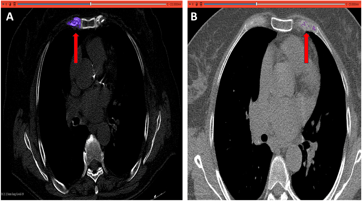
Images were analyzed using 3D Slicer (version 5.10.0, slicer.org), an open-source medical image processing software package. Since high CCC values have been found to be associated with atherosclerosis [8,12], the side with the higher CCC value (right or left) was considered for evaluation. For each segment, the CCC volume (mm3) and mean attenuation values (HU) were obtained. All measurements were performed by an experienced radiologist (NKK) blinded to the patients’ clinical information.
2.5. Statistical Analysis
The variables in the study were analyzed using SPSS 21.0 (IBM Statistical Package for the Social Sciences Statistics Inc. Version 21.0, Chicago, IL, USA). For both continuous and ordinal numerical variables, the median, mean, standard deviations, and minimum–maximum values were computed. The Shapiro–Wilk and Kolmogorov–Smirnov tests were used to analyze the normality of these data. The Student’s t-test was applied to variables that were normally distributed, and the results were reported as the mean and standard deviation. The Mann–Whitney U test was used to analyze non-normally distributed data, and the results were displayed as median (minimum–maximum). For nominal data, frequency and dispersion analyses were carried out. The chi-square test was used to analyze these numbers. CAD predictors and complex CAD predictors were examined using multivariable logistic regression analysis. In univariate analyses, variables with a p-value below 0.01 were included in multivariable analysis. Although diabetes mellitus met this criterion, the HbA1c value was used in the analyses. ROC curve analyses were performed for the CCC volume and EASIX values to reveal CAD and the complexity of CAD. Statistical significance was defined as a p-value of less than 0.05.
3. Results
A total of 756 patients were included in the study; their demographic characteristics and blood parameters are presented in Table 1. The control group consisted of 206 patients with a median age of 53 (18–87), while the CAD patient group consisted of 550 patients with a median age of 62 (30–86) (p < 0.001). There were no differences between the groups in terms of gender, left ventricular ejection fraction, white blood cell count, hemoglobin, platelet count, urea, creatinine, lactate dehydrogenase, albumin, C-reactive protein, EASIX, and triglyceride levels. In patients with CAD, the frequency of hypertension (HT), diabetes mellitus (DM), carotid artery disease, and smoking was statistically significantly higher than in the control group (p < 0.001, p < 0.001, p < 0.001, and p = 0.001, respectively). HDL-C levels were significantly lower in patients with CAD (p = 0.022). HbA1c values, LDL-C values, CCC volume, and CCC values were statistically significantly higher in the CAD patient group (p < 0.001 for all variables).
Table 1.
Evaluation of preoperative characteristics of patients with and without coronary artery disease.
Subsequently, 550 CAD patients were divided into groups according to the severity of coronary artery disease: those with low SYNTAX scores and those with medium-high SYNTAX scores, and analyses were performed (Table 2). There were 132 patients in the low SYNTAX score group with a mean age of 61.8 ± 9.8 and 418 patients in the moderate-high SYNTAX score group with a mean age of 61.6 ± 8.7 (p = 0.861). There were no differences between the groups in terms of gender, hypertension, smoking, carotid artery disease frequency, left ventricular ejection fraction, white blood cell counts, hemoglobin, platelet count, urea, creatinine, lactate dehydrogenase, albumin, C-reactive protein, LDL-C, HDL-C, triglycerides, and CCC values. HbA1c, EASIX, CCC volume values, and DM rate were statistically significantly lower in patients with low SYNTAX scores (p < 0.001, p = 0.006, p = 0.001, and p = 0.015, respectively).
Table 2.
Evaluation of preoperative characteristics of coronary artery disease patients according to the complexity of coronary artery disease.
Multivariable logistic regression analyses were performed to identify factors playing a role in demonstrating the presence or severity of coronary artery disease (Table 3). In the analysis conducted to identify CAD predictors, advanced age (Odds ratio (OR): 1.06, confidence interval (CI) 95%: 1.04–1.07, p < 0.001), hypertension (OR: 1.96, CI 95%: 1.34–2.87, p = 0.001), smoking (OR: 1.62, CI 95%: 1.08–2.43, p = 0.018), presence of carotid artery disease (OR: 1.69, CI 95%: 1.09–2.60, p = 0.018), high HbA1c values (OR: 1.34, CI 95%: 1.17–1.52, p < 0.001) and CCC volume (OR: 1.42, CI 95%: 1.35–1.66, p < 0.001, per 50 mm3) were identified as significant risk factors. High HbA1c (OR: 1.35, CI 95%: 1.18–1.55, p < 0.001), a high EASIX value (OR: 1.72, CI 95%: 1.11–2.67, p = 0.015) and CCC volume (OR: 1.39, 95% CI: 1.27–1.54, p < 0.001, per 50 mm3) were identified as significant risk factors in a multivariable analysis performed to reveal the presence of more complex CAD.
Table 3.
Multivariable logistic regression analysis of factors playing a role in indicating the presence or severity of coronary artery disease.
For predicting the presence of CAD, the cutoff level in the ROC curve analysis was 105 mm3 for CCC volume (AUC: 0.718, 95% CI: 0.677–0.758, p < 0.001, 69.3% sensitivity and 62.1% specificity, negative predictive value: 43.1%, positive predictive value: 83%). For predicting complexity of CAD, the cutoff levels in the ROC curve analysis were 0.64 for EASIX (AUC: 0.579, 95% CI: 0.523–0.634, p = 0.006, 56.7% sensitivity and 54.5% specificity, negative predictive value: 30.4%, positive predictive value: 80.6%) and 165 mm3 for CCC volume (AUC: 0.595, 95% CI: 0.544–0.646, p = 0.001, 51.8% sensitivity and 72.7% specificity, negative predictive value: 32%, positive predictive value: 85.6%).
4. Discussion
Detecting the presence or complexity of CAD using markers other than coronary angiography and other coronary interventions is popular at present. Suspected patient groups may be identified with simpler tests, and preventive measures may be instituted. Thoracic CT scans are the third most frequently used tomography scans in the world after abdominal and cranial CT scans. Moreover, their use is increasing correspondingly with the increasing number of lung diseases [13]. In our current study, we showed that the presence of CCC obtained simply from thoracic CT images may reveal the presence of coronary atherosclerosis. Thoracic CT is widely used in emergency departments and outpatient clinics for the detection of numerous diseases. By simply evaluating CCC from these examinations, necessary precautions may be taken in patients suspected of having CAD.
Soft tissues in the human body can reveal calcification and ossification conditions. These tissues include spinal discs, the aorta, heart valves, connective tissues, articular cartilage structures, and rib cartilage [14]. Costal cartilage structures connect the sternum and ribs and play an important role in respiratory functions. It is known that calcifications in these structures increase with age [15]. The incidence of CCC exceeds 50% in those over 60 years of age, and this usually begins with the alkaline phosphatase secreted by chondrocytes breaking down phosphates into free ions. Precipitation and calcification develop as these ions combine with calcium ions in the tissue matrix. In addition to these physiological changes, it is known that calcifications can also develop after trauma [16,17]. Various studies have shown a relationship between CCC and cardiometabolic diseases [8,9,18].
In this context, a clinical study by Ghotbi et al. investigated the relationship between CCC and coronary artery calcification in young adults (<50 years). In the study, which included 402 participants, patients were stratified by CCC into below- and above-median groups (n = 201 each). Clinical follow-up revealed that coronary calcification development was higher (27% vs. 42%) in patients with high CCC [18]. In a 2021 study by Shabani et al., it was suggested that increased CCC status might be related to high blood glucose exposure. In this study, which included 2305 participants, high CCC values were found to be associated with increased blood glucose levels and uncontrolled long-term blood sugar, particularly in female patients. This clinical finding was also observed in patients with zero coronary artery calcium levels [8]. Another study encompassing this main idea was published in early 2026. In this study, participants aged 65 years and older with DM for at least five years (n = 231) and without DM (n = 1148) were examined. Cardiovascular event development was monitored in patients followed for an average of 8.7 years. At the end of the analyses, it was concluded that evaluating CCC values together with the coronary artery calcium score increased the predictive power of cardiovascular event risk in patients with DM. At the end of their study, the authors emphasized that CCC values can be added to cardiovascular event risk scores in appropriate patient cohorts [9]. In our current study, we found that the CCC volume value not only indicates the presence of CAD but also serves as a marker that may raise suspicion of the existence of more complex CAD in CAD patients.
Many non-invasive imaging and blood biomarker studies have been conducted to reveal the presence of CAD. In a study involving 475 patients with stable chest pain, the role of non-invasive methods such as coronary computed tomographic angiography, myocardial perfusion imaging, and wall motion imaging in predicting CAD was investigated. As a result of the analyses, CT angiography was shown to be more accurate than non-invasive functional testing for detecting significant CAD [19]. The process has indeed begun to be used more frequently in recent years. In the imaging process, parameters such as perivascular fat tissue or epicardial fat tissue have also been evaluated as markers in risk classification [20]. In another clinical study, 190 patients without cardiac symptoms were included. Multidetector CT scans were performed on the patients included in the study, and cardiac atherosclerosis was evaluated. As a result of the analyses, it was shown that increased epicardial fat thickness was associated with >50% coronary stenosis [21]. In another clinical study involving 5743 consecutive patients, it was emphasized that epicardial fat volume can be used in coronary atherosclerosis risk prediction algorithms, together with conventional risk factors and coronary calcium score [22].
Various biomarkers have also been studied to investigate the presence and severity of coronary artery disease. For example, leucine-rich alpha-2 glycoprotein-1 (LRG1) is a serum biomarker associated with inflammation and vascular damage. A recent study found significantly higher serum LRG1 levels in patients with SYNTAX scores ≥ 23 [23]. Another study investigating various biomarkers showed that serum interleukin-8 levels can also indicate the presence of CAD [24]. However, the lack of routine use of these parameters and their high relative cost may pose a challenge to practitioners. Conversely, some markers obtained from routine blood tests have become increasingly prevalent in research; the EASIX value has recently become a subject of research in cardiovascular diseases.
A study by Liu et al. included 38,794 participants, and the cardiovascular disease rate in this population was found to be 10.4%. As a result of the analyses, high EASIX values were found to be associated with high CAD rates, myocardial infarction, and heart failure [25]. In another study involving 4478 participants, a significant relationship was found between high EASIX values and the risk of developing atherosclerotic cardiovascular disease within 10 years [26]. In contrast to these studies, our study revealed similar EASIX values between patients with CAD and those without.
A study published in 2026, involving 343 non-ST-elevation myocardial infarction patients who underwent CABG surgery and saphenous vein intervention, investigated the relationship between EASIX values and no-reflow. In this study, no-reflow developed in 20.7% of the patients, and a high EASIX value (OR: 3.151, p = 0.001) was shown to be a significant independent predictor of no-reflow development [27]. Similarly, in a study of patients who developed acute coronary syndrome, high EASIX values were found to be associated with early mortality [28]. In our research, we also detected higher EASIX values in patients with CAD, although not statistically significant. Further investigations in patients with CAD revealed that high EASIX values may be associated with more complex coronary artery disease.
While our study yielded significant findings, it had some significant limitations. Firstly, this was a single-center study with a limited number of patients, and the control group could not be composed entirely of a normal population. We selected the control group in this way because our study required thoracic CT scans; the presence of valvular disease in the control group may affect inflammatory markers. To mitigate this effect, patients with calcific mitral and aortic valve disease were excluded from the study. Nevertheless, these patients were younger. This age difference may also affect the marker levels in the study. This limitation could be corrected in future multicenter studies by including patients from other medical fields. Also, it should not be forgotten that calcification is only one component of coronary risk and that clinically important non-calcified plaques may occur even with a zero coronary calcium score [29]. In nine of the patients in the control group, HU values were 0. Subgroup analyses including these patients could be conducted in future studies with larger patient cohorts. And there were no interobserver reproducibility analyses for CCC measurements and SYNTAX score calculations in our study. Additionally, coronary CT angiography evaluations were not performed on the patients in our study. The relationships between CCC and coronary calcium burden could also be investigated in current studies that include coronary CT angiography evaluations.
5. Conclusions
One of the most critical issues regarding CAD today is how to identify at-risk groups. Thoracic CT imaging is widely used in many medical applications. The current study demonstrated that the CCC evaluation in these CT images may help to raise suspicion of CAD, thus favoring early diagnosis. Furthermore, our analyses have shown that we may raise suspicion of complex CAD using the EASIX value and CCC evaluation. We believe future prospective multicenter studies could yield improved data on these values.
Author Contributions
Conceptualization, N.K.K.; methodology, N.K.K. and M.E.; software, N.K.K.; validation, M.E., N.C.G. and A.K.A.; formal analysis, N.K.K. and M.E.; investigation, N.K.K., M.E., N.C.G., A.K.A. and S.Y.; resources, N.K.K.; data curation, M.E., N.C.G., A.K.A. and S.Y.; writing—original draft preparation, N.K.K., M.E., N.C.G., A.K.A. and S.Y.; writing—review and editing, N.K.K., M.E., N.C.G., A.K.A. and S.Y.; visualization, N.K.K., M.E., N.C.G., A.K.A. and S.Y.; supervision, N.K.K., M.E., N.C.G., A.K.A. and S.Y.; project administration, N.C.G. and A.K.A.; funding acquisition, N.C.G. and A.K.A. All authors have read and agreed to the published version of the manuscript.
Funding
This research received no external funding.
Institutional Review Board Statement
The study was approved by the Bursa Yuksek Ihtisas Training and Research Hospital Clinical Research Ethics Committee (2024-TBEK 2026/08-01). Every method used in this investigation was carried out in compliance with the 1964 Declaration of Helsinki and its subsequent amendments, or similar ethical standards, as well as the institutional and/or national research committee’s ethical guidelines.
Informed Consent Statement
Written informed consent was waived due to the retrospective nature of the study.
Data Availability Statement
All data produced here are available and can be produced upon reasonable request.
Conflicts of Interest
The authors declare no conflict of interest.
References
- Miller, C.L.; Kocher, M.; Koweek, L.H.; Zwischenberger, B.A. Use of computed tomography (CT) for preoperative planning in patients undergoing coronary artery bypass grafting (CABG). J. Card. Surg. 2022, 37, 4150–4157. [Google Scholar] [CrossRef] [Scilit] [PubMed]
- Guimarães, J.; de Almeida, J.; Mendes, P.L.; Ferreira, M.J.; Gonçalves, L. Advancements in non-invasive imaging of atherosclerosis: Future perspectives. J. Clin. Lipidol. 2024, 18, e142–e152. [Google Scholar] [CrossRef] [Scilit] [PubMed]
- Amarasekera, A.T.; Chang, D.; Schwarz, P.; Tan, T.C. Does vascular endothelial dysfunction play a role in physical frailty and sarcopenia? A systematic review. Age Ageing 2021, 50, 725–732. [Google Scholar] [CrossRef] [Scilit] [PubMed]
- Meng, H.; Ruan, J.; Yan, Z.; Chen, Y.; Liu, J.; Li, X.; Meng, F. New Progress in Early Diagnosis of Atherosclerosis. Int. J. Mol. Sci. 2022, 23, 8939. [Google Scholar] [CrossRef] [Scilit] [PubMed]
- Yalcin, M.U.; Gurses, K.M.; Aydoğan, C.; Butun, S.; Tunçez, A.; Tezcan, H.; Ozen, Y.; Demir, K.; Aygul, N.; Kirmizigul, M.; et al. Endothelial Activation and Stress Index Predicts Poor Coronary Collateral Development in Chronic Total Occlusion. J. Cardiovasc. Dev. Dis. 2026, 13, 124. [Google Scholar] [CrossRef] [Scilit] [PubMed]
- Krombholz-Reindl, P.; Vötsch, A.; Linni, K.; Seitelberger, R.; Gottardi, R.; Lichtenauer, M.; Hammerer, M.; Boxhammer, E.; Winkler, A. Endothelial Activation and Stress Index-A Novel and Simple Prognostic Tool in Coronary Artery Bypass Grafting. J. Clin. Med. 2025, 14, 2857. [Google Scholar] [CrossRef] [Scilit] [PubMed]
- Shehu, V.; Gandhi, O.H.; Glennan, P.; Gujral, J.; Singh, S.B.; Amanullah, A.A.; Patil, S.; Gujral, K.; Raynor, W.Y.; Park, P.S.U.; et al. Quantification of Costal Cartilage Calcification Using 18F-NaF-PET/CT. J. Imaging 2026, 12, 206. [Google Scholar] [CrossRef] [Scilit] [PubMed]
- Shabani, M.; Pishgar, F.; Akhtarkhavari, S.; Quinaglia, T.; Budoff, M.J.; Bluemke, D.A.; Barr, G.R.; Post, W.S.; Wu, C.O.; Arbab-Zadeh, A.; et al. Association of Quantified Costal Cartilage Calcification and Long-Term Cumulative Blood Glucose Exposure: The Multi-Ethnic Study of Atherosclerosis. Front Endocrinol. 2021, 12, 785957. [Google Scholar] [CrossRef] [Scilit] [PubMed]
- Ghotbi, E.; Bluemke, D.A.; Hathaway, Q.A.; Klein, J.G.; Shabani, M.; Akhtarkhavari, S.; Barr, R.G.; Bancks, M.P.; Post, W.S.; Budoff, M.; et al. Use of CT-derived non-cardiovascular calcification marker for predicting cardiovascular events among diabetic older adults: The multi-ethnic study of atherosclerosis. Eur. Radiol. 2026, 36, 1366–1378. [Google Scholar] [CrossRef] [Scilit] [PubMed]
- Dragomir, T.L.; Branea, H.S.; Andor, M.; Cîtu, I.M.; Căruntu, F.R.; Șușan, M.M.; Oprean, C.M.; Dragomir, R.T.; Șușan, R.M.; Huțanu, D.; et al. Association of the ApoB/ApoA1 Ratio with Angiographically Documented Coronary Artery Disease: Comparison with Conventional Lipid Markers. Diagnostics 2026, 16, 3245. [Google Scholar] [CrossRef] [Scilit] [PubMed]
- Wang, T.; Wang, Z.; Luo, P. Positive Association Between Ultrasonographic Fatty Liver Indicator and the Severity of Coronary Artery Disease. Diagnostics 2025, 15, 1208. [Google Scholar] [CrossRef] [Scilit] [PubMed]
- Ghotbi, E.; Hathaway, Q.A.; Bluemke, D.A.; Ibad, H.A.; Shabani, M.; Akhtarkhavari, S.; Barr, R.G.; Post, W.S.; Budoff, M.; Mathur, A.; et al. CT-derived Noncardiovascular Calcifications May Improve Coronary Heart Disease Prediction in Chronic Kidney Disease: The Multi-Ethnic Study of Atherosclerosis. Radiol. Cardiothorac. Imaging 2025, 7, e240546. [Google Scholar] [CrossRef] [Scilit] [PubMed]
- Hess, E.P.; Haas, L.R.; Shah, N.D.; Stroebel, R.J.; Denham, C.R.; Swensen, S.J. Trends in computed tomography utilization rates: A longitudinal practice-based study. J. Patient Saf. 2014, 10, 52–58. [Google Scholar] [CrossRef] [Scilit] [PubMed]
- Lau, A.G.; Kindig, M.W.; Salzar, R.S.; Kent, R.W. Micromechanical modeling of calcifying human costal cartilage using the generalized method of cells. Acta Biomater. 2015, 18, 226–235. [Google Scholar] [CrossRef] [Scilit] [PubMed]
- van Dam, V.S.; Berzenji, D.; van den Berg, A.S.; van Zijl, F.V.W.J.; Nagtegaal, A.P.; Kremer, B.; Datema, F.R. Costal Cartilage Calcification in a Caucasian Population: Machine Learning Recommendations for Chest CT-guided Rhinoplasty Planning. Facial Plast. Surg. 2026, 42, 496–501. [Google Scholar] [CrossRef] [Scilit] [PubMed]
- Kayo, A.; Tsuchiya, N.; Yonemoto, K.; Nakamura, M.; Murayama, S.; Uechi, M.; Kinjo, S.; Sato, M.; Moromizato, H.; Toyosato, S.; et al. Detection of Costal Cartilage Fractures on CT Images With Computer-aided Detection System for Rib Fractures. In Vivo 2025, 39, 390–395. [Google Scholar] [CrossRef] [Scilit] [PubMed]
- Seng, J.J.B.; Kho, Z.C.; Kaur, N. Premature Calcification of Costochondral Cartilage: A Scoping Review of the Literature. Cureus 2024, 16, e75328. [Google Scholar] [CrossRef] [Scilit] [PubMed]
- Ghotbi, E.; Nasir, K.; Bancks, M.P.; Subhas, N.; Akhtarkhavari, S.; Shabani, M.; Hadidchi, R.; Bluemke, D.A.; Post, W.S.; Budoff, M.; et al. Non-cardiovascular Calcification Measures and Warranty Period of a Zero CAC in Young Adults: The Multi-ethnic Study of Atherosclerosis. Acad. Radiol. 2025, 32, 5724–5732. [Google Scholar] [CrossRef] [Scilit] [PubMed]
- Neglia, D.; Rovai, D.; Caselli, C.; Pietila, M.; Teresinska, A.; Aguadé-Bruix, S.; Pizzi, M.N.; Todiere, G.; Gimelli, A.; Schroeder, S.; et al. Detection of significant coronary artery disease by noninvasive anatomical and functional imaging. Circ. Cardiovasc. Imaging 2015, 8, e002179. [Google Scholar] [CrossRef] [Scilit] [PubMed]
- Cundari, G.; Marchitelli, L.; Pambianchi, G.; Catapano, F.; Conia, L.; Stancanelli, G.; Catalano, C.; Galea, N. Imaging biomarkers in cardiac CT: Moving beyond simple coronary anatomical assessment. Radiol. Med. 2024, 129, 380–400. [Google Scholar] [CrossRef] [Scilit] [PubMed]
- Bachar, G.N.; Dicker, D.; Kornowski, R.; Atar, E. Epicardial adipose tissue as a predictor of coronary artery disease in asymptomatic subjects. Am. J. Cardiol. 2012, 110, 534–538. [Google Scholar] [CrossRef] [Scilit] [PubMed]
- Zhou, J.; Chen, Y.; Zhang, Y.; Wang, H.; Tan, Y.; Liu, Y.; Huang, L.; Zhang, H.; Ma, Y.; Cong, H. Epicardial Fat Volume Improves the Prediction of Obstructive Coronary Artery Disease Above Traditional Risk Factors and Coronary Calcium Score. Circ. Cardiovasc. Imaging 2019, 12, e008002. [Google Scholar] [CrossRef] [Scilit] [PubMed]
- Yang, Z.; Liu, Z.; Xu, Y.; Wang, H.; Lu, Y.; Zou, C. Serum LRG1 as a non-invasive biomarker for identifying complex coronary anatomy in pre-angiography risk stratification. Front Cardiovasc. Med. 2026, 13, 1818245. [Google Scholar] [CrossRef] [Scilit] [PubMed]
- Wojciechowska, M.; Nizio, M.; Wróbel, K.; Momot, K.; Czarzasta, K.; Flis, K.; Zarębiński, M. Predictive Value of Selected Plasma Biomarkers in the Assessment of the Occurrence and Severity of Coronary Artery Disease. Int. J. Mol. Sci. 2025, 26, 537. [Google Scholar] [CrossRef] [Scilit] [PubMed]
- Liu, W.; Xu, X.; Zhang, M.; Jin, Y. Endothelial activation and stress index and cardiovascular disease among American population-findings from NHANES 1999-2018. BMC Cardiovasc. Disord. 2025, 25, 681. [Google Scholar] [CrossRef] [Scilit] [PubMed]
- Huang, Y.; Lan, H.; Yang, H. Regulating effect of genistein on the association of endothelial activation and stress index with atherosclerotic cardiovascular disease risk: A cross-sectional study of the NHANES database. Naunyn Schmiedebergs Arch. Pharmacol. 2026, 399, 6791–6802. [Google Scholar] [CrossRef] [Scilit] [PubMed]
- Hancıoğlu, E.; Özcan, S.; Faideci, E.M.; Bekler, Ö.; Karaçöp, E.; Enhoş, A.; Okuyan, E. Endothelial Activation and Stress Index (EASIX) as a Predictor of No-Reflow in Patients Undergoing Saphenous Vein Graft Percutaneous Coronary Intervention. Catheter Cardiovasc. Interv. 2026, 108, 67–75. [Google Scholar] [CrossRef] [Scilit] [PubMed]
- Tekin, I.; Büber, İ.; Avcı, E. Endothelial stress indices in acute coronary syndrome: Comparative prognostic performance of EASIX and its variants. J. Lab. Med. 2026, 50, 120–127. [Google Scholar] [CrossRef] [Scilit]
- Mátyás, B.B.; Gerculy, R.; Rat, N.; Blîndu, E.; Stănescu, A.G.; Roșca, A.; Buicu, C.F.; Benedek, I.; Benedek, T. Highly Inflamed Non-Calcified Coronary Plaques Sealed with Stents in Patients with Zero Calcium Score—A Case Series and Review of the Literature. J. Cardiovasc. Emerg. 2024, 10, 38–44. [Google Scholar] [CrossRef] [Scilit]

Disclaimer/Publisher’s Note: The statements, opinions and data contained in all publications are solely those of the individual author(s) and contributor(s) and not of MDPI and/or the editor(s). MDPI and/or the editor(s) disclaim responsibility for any injury to people or property resulting from any ideas, methods, instructions or products referred to in the content. |
© 2026 by the authors. Licensee MDPI, Basel, Switzerland. This article is an open access article distributed under the terms and conditions of the Creative Commons Attribution (CC BY) license.