


Abstract
Pathology has always been a spatial science: for more than 150 years, diagnosis has rested on reading the architecture of cells and tissues. Three technological streams are now converging on this founding principle. Spatially resolved omics can measure thousands of transcripts and over a hundred proteins in intact, formalin-fixed paraffin-embedded (FFPE) tissue; multiplexed imaging quantifies tissue microenvironments cell by cell; and computational pathology has progressed from slide digitization to foundation models that can predict molecular features from morphology. The clinical evidence for spatial readouts is real but heterogeneous: a few assays have reached guideline mention or prospective use, while most findings remain retrospective, platform-dependent, and unevenly validated. Yet the field remains fragmented across “spatial biology”, “digital pathology”, and “computational pathology”, without a unified framework for standards, training, and diagnostics. Here we propose, for discussion rather than as a settled matter, that this convergence be treated as a coherent branch of pathology, “spatial pathology”, which we define as the use of spatially resolved measurements of tissue to derive diagnostic, prognostic, and predictive information. We set out which problems existing structures do not yet address, acknowledge the counterarguments, and outline a consensus-building agenda. We also suggest “spathology” as an optional shorthand, and we disclose openly that all authors are co-founders of a company, SPATHO AB, whose name derives from the same contraction.
1. Pathology Has Always Been Spatial
Modern pathology was founded on a spatial claim. Virchow’s cellular pathology located disease in cells and in the tissue architecture they compose, to be read under the microscope [1]. Much of what is durable in diagnostic practice since then has been, at its core, codified spatial pattern recognition: Gleason grading scores the architectural growth patterns of prostate cancer [2]; TNM staging formalizes the anatomic extent of disease [3]; HER2 immunohistochemistry is scored by the completeness, intensity, and distribution of membrane staining across tumor cells [4]. Even immunohistochemistry itself—descended from the first fluorophore-conjugated antibody of 1941 [5]—is a spatial molecular assay, albeit one that has remained essentially single-plex in the clinic for eight decades.
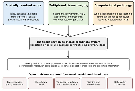
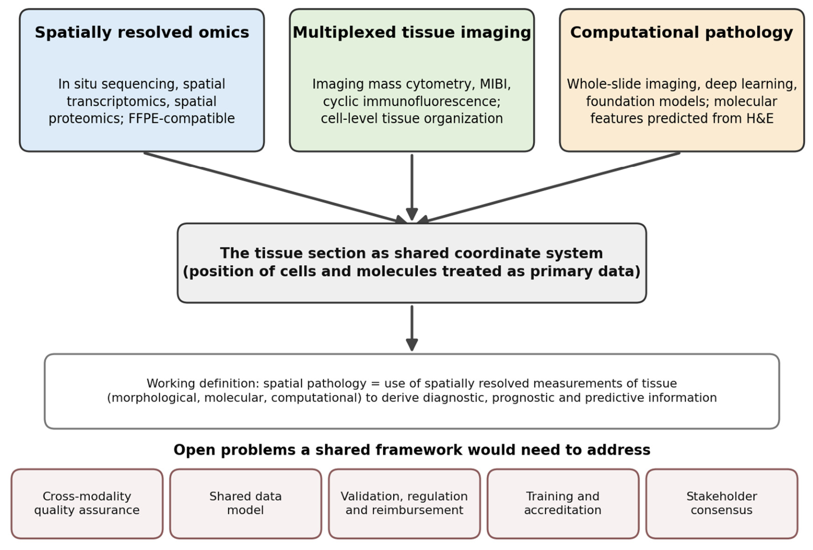
Pathology, in other words, has practiced spatial biology since its founding. What has changed is not the principle but the instrumentation: the number of molecular species that can be measured at each position in a tissue section has grown from one to tens of thousands, and the computational capacity to interpret such measurements has grown with it. Whether this change warrants a shared framework, and possibly a name, for a distinct branch of pathology is a question for the community, not for three authors. In this short opinion article, we set out the case for and against, propose a working definition, and suggest what a consensus process would need to address. Figure 1 summarizes the argument.
Figure 1.
Three technological streams—spatially resolved omics, multiplexed tissue imaging, and computational pathology—converge on the tissue section as the shared coordinate system. The working definition of spatial pathology proposed here and the open problems it would need to address are shown below the confluence.
2. Three Streams, One Confluence
2.1. Spatially Resolved Omics
The molecular stream begins with single-molecule fluorescence in situ hybridization, which first rendered individual transcripts visible in fixed cells [6]. In situ sequencing then showed that RNA could be decoded directly inside morphologically preserved tissue, amplified by padlock probes and rolling-circle amplification and sequenced in place; it was demonstrated from the outset in human breast cancer sections, with point-mutation sequencing shown in cultured cells [7]. Combinatorial barcoding scaled imaging-based profiling to as many as a thousand genes per cell [8], while spatial barcoding of capture arrays made unbiased, whole-transcriptome measurement possible in tissue sections and gave the field its name, spatial transcriptomics [9]. Successive generations of array chemistry pushed resolution to the subcellular scale and fields of view to whole organs [10].
Two developments moved these technologies from atlas-building toward the pathology department. The first is commercialization into robust platforms designed around routine specimens—region-of-interest digital spatial profiling [11], cyclic-hybridization imagers measuring ~1000 RNAs or >100 proteins at subcellular resolution [12], in situ sequencing platforms benchmarked directly on clinical tumor material [13]. The second, and arguably decisive, development is compatibility with formalin-fixed, paraffin-embedded (FFPE) tissue [14]: FFPE is the universal substrate of diagnostic practice and of the world’s tissue archives, with decades of outcome annotation attached. By 2024, many major commercial platforms were FFPE-compatible, and whole-transcriptome profiling at single-cell scale is now feasible on routine FFPE sections [15]. The field’s coming of age has been formally recognized twice in five years: spatially resolved transcriptomics was Nature Methods’ Method of the Year 2020 [16], and spatial proteomics its Method of the Year 2024 [17]. The technology stack continues to broaden into spatial epigenomics, metabolomics, and three-dimensional approaches [18,19], as well as unbiased mass-spectrometry-based spatial proteomics performed directly on archived diagnostic material [20].
2.2. Multiplexed Tissue Imaging
The protein stream extended immunohistochemistry from one marker to dozens. Imaging mass cytometry [21] and multiplexed ion beam imaging [22]—published simultaneously in 2014 and both demonstrated on breast cancer tissue—used metal-tagged antibodies to escape the spectral limits of fluorescence, while DNA-barcoded cyclic immunofluorescence achieved comparable plex on conventional microscopes [23]. These methods did more than add markers: they made the organization of tissue quantifiable. Cellular-neighborhood analysis showed that recurrent multicellular structures at the colorectal cancer invasive front stratify patient outcome [24], and single-cell pathology atlases of breast cancer demonstrated that spatially defined tissue phenotypes carry prognostic information beyond conventional subtyping [25]. Deep-learning segmentation trained on more than a million annotated cells now converts such images into single-cell data at human-level accuracy [26]—an early example of the mutual dependence of spatial assays and artificial intelligence.
2.3. Computational Pathology
The computational stream is the youngest but has moved the fastest. Whole-slide imaging was authorized for primary diagnosis in the United States in 2017 on the strength of a 1992-case noninferiority trial [27], creating the data substrate for machine learning. Within a few years, deep learning matched or exceeded pathologists on defined diagnostic tasks [28], predicted driver mutations [29] and microsatellite instability [30] directly from hematoxylin-and-eosin (H&E) morphology, reached pathologist-level Gleason grading in blinded international validation [31], and produced an AI tool for assisting prostate cancer detection on biopsy images authorized by the U.S. Food and Drug Administration (FDA) [32]. The current foundation-model era—self-supervised encoders pretrained on hundreds of thousands to over a million whole-slide images [33,34,35]—provides general-purpose representations of tissue morphology that transfer across organs, tasks, and institutions. What these morphology-only models still largely lack is molecular grounding, and what still separates benchmark performance from clinical deployment is prospective validation, generalization, and workflow integration [36].
Recent work applies machine learning directly to spatial omics data. Foundation models pretrained on tens of millions of dissociated and spatially resolved cells learn representations of spatial context and can transfer spatial information to dissociated data, although models trained on dissociated data alone fail to recover the complexity of spatial microenvironments [37]. Open-source frameworks such as Squidpy have made spatial statistics and image-feature analysis routine [38], and new methods predict spatial gene expression at near-single-cell resolution from histology [39,40,41]. Open problems remain. Benchmarks report limited accuracy and spatial resolution for many histology-to-expression methods [40], and predictive performance depends strongly on the sparsity and noise of the underlying molecular data, with limited gains from imputation, as shown in a recent preprint [42].
3. What Spatial Readouts Have Shown, and How Mature the Evidence Is
The claim that spatial context carries diagnostic information beyond bulk or single-marker assays rests on examples of very different maturity. Table 1 lists the main ones and, for each, what has and has not been established. We do not claim that spatial readouts are generally superior to non-spatial biomarkers, or that spatial assays are clinically ready; we claim that a small number of spatial biomarkers have been taken further than the rest, and that they show what is possible.
Table 1.
Examples of spatial biomarkers and the current level of evidence.
The Immunoscore is the founding example: the type, density, and—critically—location of T cells in colorectal cancer predicted outcome better than the histopathological staging systems in use at the time [43]. An international 2681-patient validation study subsequently showed that this digitally quantified, location-aware assay contributed more to recurrence-risk prediction than any conventional parameter, including TNM stage itself, with inter-observer and inter-center reproducibility of r = 0.97 [44]. The European Society for Medical Oncology guideline for localized colon cancer notes that the Immunoscore may be used alongside TNM to refine prognosis, while stating that its role in predicting chemotherapy benefit is uncertain [45]. Tertiary lymphoid structures, organized immune aggregates identifiable on tissue sections, were associated in 2020 with checkpoint-inhibitor benefit in melanoma, renal cell carcinoma, and sarcoma [46,47,48]. One phase 2 sarcoma cohort was then selected on the presence of such structures: the six-month non-progression rate was 40%, compared with 4.9% in earlier unselected cohorts, a cross-cohort comparison from a single small trial [49]. A meta-analysis of 8135 patients reported a higher pooled discrimination for multiplexed, spatially resolved immunohistochemistry/immunofluorescence (area under the curve, AUC, 0.79) than for PD-L1 immunohistochemistry (0.65), gene-expression profiling (0.65), and tumor mutational burden (0.69) for response to PD-1/PD-L1 blockade [50]; the underlying studies were heterogeneous and largely retrospective, and a whole-slide multiplexed platform later reported external validation of spatial predictors in melanoma [51].
Highly multiplexed and spatially resolved studies have gone on to show that tumor–immune architecture, not merely cellular composition, is associated with survival in triple-negative breast cancer [52]; that recurrent microenvironmental structures link genotype to outcome across hundreds of breast tumors [53]; and that recurrent stromal–immune niches, co-localized in spatial data, underpin clinically distinct ecotypes [54]. Spatial profiling has also begun to unsettle diagnostic categories themselves: in ductal carcinoma in situ, the state of the surrounding stroma and myoepithelium—more than tumor-cell features—tracks progression risk, sometimes in unexpected directions [55], and organ-wide spatial transcriptomics of the prostate has revealed clonal copy-number expansions in histologically benign epithelium, challenging the morphological definition of “normal” [56]. Nor is the case confined to oncology: spatial multi-omics has mapped the reorganization of human myocardial infarction [57], and organ-level roadmaps now explicitly co-locate spatial omics and digital pathology as the future of tissue diagnostics [58]. Most of these studies are discovery-stage and retrospective; they motivate assay development rather than replace it.
4. The Convergence Loop: Morphology–Molecule Inference
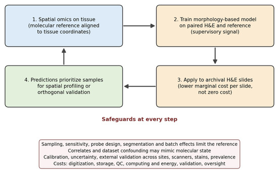
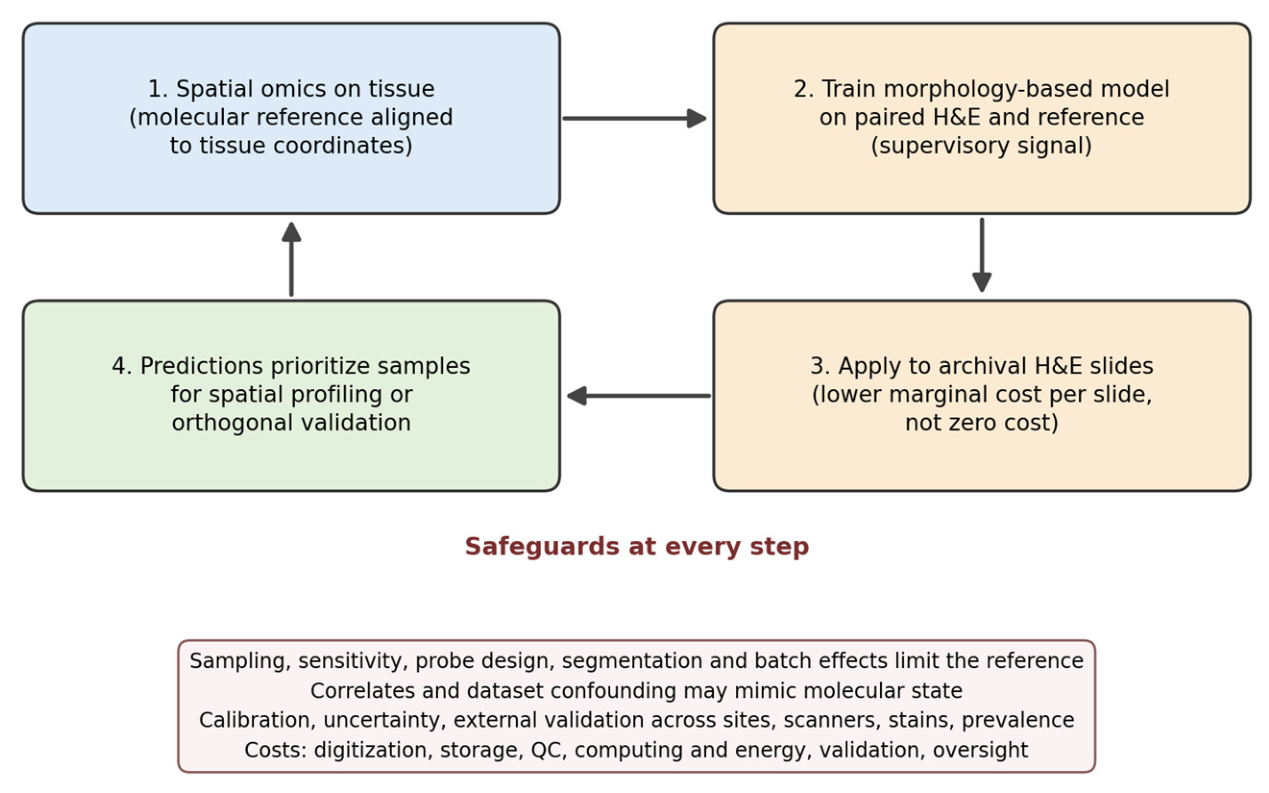
One specific mechanism links the three streams. Spatially resolved omics can provide a molecular reference, aligned to tissue coordinates, against which morphology-based models are trained; the trained models can then be applied to archival H&E slides, with lower marginal cost per slide than new spatial profiling; and the resulting predictions can help prioritize which samples justify spatial profiling or orthogonal validation, which in turn refines the reference (Figure 2).
Figure 2.
The morphology–molecule loop and its safeguards. Spatial omics provides a molecular reference (a supervisory signal, not an unqualified ground truth); morphology-based models trained on it are applied to archival slides; predictions are used to prioritize samples for orthogonal validation, which feeds back on the reference. Each step carries uncertainty that must be measured and reported.
This loop is already running. Deep learning trained on spatial transcriptomics spots matched to histology predicted the local expression of more than 100 genes from breast cancer H&E [60]; transcriptome-scale prediction from whole-slide images followed, with “virtual spatialization” of the inferred expression [61]; integration of spatial transcriptomics with high-resolution histology now yields near-single-cell expression inference, including in sections for which only H&E exists [39]; and a visual–omics foundation model aligning histology with spatial transcriptomics has generalized the approach [62].
The loop should not be over-read. Spatial omics measurements are themselves affected by sampling, sensitivity and specificity, probe design, tissue processing, cell segmentation, spatial resolution, batch effects, and platform-dependent error [59]; they are a molecular reference, not ground truth. Predictions of molecular state from H&E may reflect morphological correlates or dataset-specific confounding rather than recovery of the molecular state itself, and performance can fall across institutions, scanners, staining protocols, and disease prevalence [36]. Any use of such predictions beyond research would require calibration, uncertainty estimates, external validation, and explicit attention to causal interpretation. Nor is the marginal cost of applying a trained model to archival slides zero: digitization, storage, quality control, computation and its energy footprint, model validation, integration, and professional oversight all carry costs. The claim is therefore modest: spatial omics and computational pathology are becoming interdependent, which is a reason for the two communities to share standards and vocabulary.
6. The Agenda a Discipline Would Need to Deliver
If the community agrees that a shared framework is worth building, the following items head the agenda.
Consensus and scope. A multi-stakeholder process, for example, a modified Delphi exercise including practicing diagnostic pathologists, laboratory-medicine and quality-assurance specialists, clinicians, computational scientists, industry, regulators, and patient representatives, should decide whether a distinct framework is needed, what it covers, and what it is called.
Standardization and external quality assurance. Head-to-head comparisons of imaging-based spatial transcriptomics platforms on serial FFPE sections reveal substantial inter-platform differences, with data quality strongly dependent on probe-panel design [59]. Consensus guidance now exists for multiplexed immunofluorescence staining, validation, image analysis, and data sharing [71,72], and minimum-information standards for multiplexed tissue images have been established [73]—but nothing yet resembles the external quality assessment schemes that made clinical immunohistochemistry reliable. Such a framework would need proficiency-testing infrastructure, reference materials, and inter-laboratory ring trials.
Data acquisition, stewardship, and infrastructure. Spatial experiments produce multimodal, multi-gigapixel datasets whose value depends on how they are acquired, stored, structured, and shared. Acquisition needs common metadata on tissue handling, ischemia and fixation times, panel design, and instrument settings. Stewardship needs clear provenance, consent, and access rules, including for archival clinical material, together with FAIR (findable, accessible, interoperable, reusable) practices and community-wide sharing and coordination, building on consortium data standards. Storage and processing need scalable, interoperable, and auditable pipelines: open frameworks for spatial omics data [38,63] are the beginning of what must become the equivalent of radiology’s picture archiving and communication systems (PACS) and DICOM (Digital Imaging and Communications in Medicine) infrastructure, integrated with pathology information systems rather than bolted onto them, with automated processing that is validated and version-controlled.
Validation and clinical delivery. Most spatial signatures remain retrospective; the exceptions—large-scale international validation [44] and prospective, biomarker-selected enrollment [49]—prove the concept but also illustrate the difficulty: even clinically validated spatial assays have struggled to achieve durable commercial and reimbursement pathways, and regulatory frameworks for laboratory-developed and AI-enabled tests remain in flux worldwide. The realistic near-term route, as recent roadmaps argue, is to use high-plex spatial discovery to define biology, then distill it into scalable, low-plex or H&E-inferred assays that fit existing laboratory economics [77], each validated analytically and clinically in its own right.
Training. Molecular pathology matured with societies, fellowships, and accreditation. A spatial framework would require pathologists conversant with molecular data science, and computational scientists conversant with tissue; neither is produced systematically today, and calls for such training are appearing in pathology’s own journals [74]. Fellowship tracks, core curricula, and shared vocabularies between pathology, genomics, and machine-learning communities are the necessary scaffolding.
7. Conclusions
Every previous re-instrumentation of pathology—the microscope, the immunostain, the sequencer, the scanner—was eventually matched by an institutional response. Spatially resolved measurement is a substantial new re-instrumentation, and a small number of spatial biomarkers have been taken to guideline mention or prospective use, but the evidence overall is heterogeneous and most spatial findings are not yet clinical tools. Whether pathology needs a distinct, named branch for this work, or whether existing bodies can absorb it, is a question that should be settled by the pathology community through an open and independent process. We offer “spatial pathology” as a description, “spathology” as an optional shorthand with the caveats and conflicts stated above, and the agenda in Section 6 as a starting point for that discussion.
Author Contributions
Conceptualization, M.L., T.H. and M.N.; writing—original draft preparation, M.L. and T.H.; writing—review and editing, M.L., T.H. and M.N.; supervision, M.N. All authors have read and agreed to the published version of the manuscript.
Funding
This research received no external funding.
Institutional Review Board Statement
Not applicable.
Informed Consent Statement
Not applicable.
Data Availability Statement
No new data were created or analyzed in this study. Data sharing is not applicable to this article.
Acknowledgments
During the preparation of this manuscript, the authors used Claude (Anthropic) for the purposes of literature searching, drafting and language editing of the text, and preparing the schematic figures. The authors conceived the article and its central arguments, have reviewed and edited the output, and take full responsibility for the content of this publication.
Conflicts of Interest
Mengping Long, Mats Nilsson, and Taobo Hu are co-founders of SPATHO AB and have financial interests in the company. SPATHO AB is a Swedish company developing technologies and analytical methods in spatial pathology, and its name derives from the same contraction as the term “spathology” discussed in this article. The authors’ advocacy of the term is therefore a potential conflict of interest, discussed in Section 5.3. The article is a purely academic discussion and does not describe, evaluate, or promote any product or service of the company. T.H. serves as Guest Editor of the Special Issue “Advances in Computational and Spatial Pathology” and had no role in the editorial handling of this manuscript, the selection of reviewers, or the decision on it.
References
- Bignold, L.P.; Coghlan, B.L.D.; Jersmann, H.P.A. Virchow’s “cellular pathology” 150 years later. Semin. Diagn. Pathol. 2008, 25, 140–146. [Google Scholar] [CrossRef] [Scilit] [PubMed]
- Gleason, D.F.; Mellinger, G.T. Prediction of prognosis for prostatic adenocarcinoma by combined histological grading and clinical staging. J. Urol. 1974, 111, 58–64. [Google Scholar] [CrossRef] [Scilit] [PubMed]
- Amin, M.B.; Greene, F.L.; Edge, S.B.; Compton, C.C.; Gershenwald, J.E.; Brookland, R.K.; Meyer, L.; Gress, D.M.; Byrd, D.R.; Winchester, D.P. The Eighth Edition AJCC Cancer Staging Manual: Continuing to build a bridge from a population-based to a more “personalized” approach to cancer staging. CA Cancer J. Clin. 2017, 67, 93–99. [Google Scholar] [CrossRef] [Scilit] [PubMed]
- Wolff, A.C.; Somerfield, M.R.; Dowsett, M.; Hammond, M.E.H.; Hayes, D.F.; McShane, L.M.; Saphner, T.J.; Spears, P.A.; Allison, K.H. Human Epidermal Growth Factor Receptor 2 Testing in Breast Cancer: ASCO-College of American Pathologists Guideline Update. J. Clin. Oncol. 2023, 41, 3867–3872. [Google Scholar] [CrossRef] [Scilit] [PubMed]
- Coons, A.H.; Creech, H.J.; Jones, R.N. Immunological Properties of an Antibody Containing a Fluorescent Group. Proc. Soc. Exp. Biol. Med. 1941, 47, 200–202. [Google Scholar] [CrossRef] [Scilit]
- Femino, A.M.; Fay, F.S.; Fogarty, K.; Singer, R.H. Visualization of single RNA transcripts in situ. Science 1998, 280, 585–590. [Google Scholar] [CrossRef] [Scilit] [PubMed]
- Ke, R.; Mignardi, M.; Pacureanu, A.; Svedlund, J.; Botling, J.; Wählby, C.; Nilsson, M. In situ sequencing for RNA analysis in preserved tissue and cells. Nat. Methods 2013, 10, 857–860. [Google Scholar] [CrossRef] [Scilit] [PubMed]
- Chen, K.H.; Boettiger, A.N.; Moffitt, J.R.; Wang, S.; Zhuang, X. Spatially resolved, highly multiplexed RNA profiling in single cells. Science 2015, 348, aaa6090. [Google Scholar] [CrossRef] [Scilit] [PubMed]
- Ståhl, P.L.; Salmén, F.; Vickovic, S.; Lundmark, A.; Navarro, J.F.; Magnusson, J.; Giacomello, S.; Asp, M.; Westholm, J.O.; Huss, M.; et al. Visualization and analysis of gene expression in tissue sections by spatial transcriptomics. Science 2016, 353, 78–82. [Google Scholar] [CrossRef] [Scilit] [PubMed]
- Chen, A.; Liao, S.; Cheng, M.; Ma, K.; Wu, L.; Lai, Y.; Qiu, X.; Yang, J.; Xu, J.; Hao, S.; et al. Spatiotemporal transcriptomic atlas of mouse organogenesis using DNA nanoball-patterned arrays. Cell 2022, 185, 1777–1792.e21. [Google Scholar] [CrossRef] [Scilit] [PubMed]
- Merritt, C.R.; Ong, G.T.; Church, S.E.; Barker, K.; Danaher, P.; Geiss, G.; Hoang, M.; Jung, J.; Liang, Y.; McKay-Fleisch, J.; et al. Multiplex Digital Spatial Profiling of Proteins and RNA in Fixed Tissue. Nat. Biotechnol. 2020, 38, 586–599. [Google Scholar] [CrossRef] [Scilit] [PubMed]
- He, S.; Bhatt, R.; Brown, C.; Brown, E.A.; Buhr, D.L.; Chantranuvatana, K.; Danaher, P.; Dunaway, D.; Garrison, R.G.; Geiss, G.; et al. High-Plex Imaging of RNA and Proteins at Subcellular Resolution in Fixed Tissue by Spatial Molecular Imaging. Nat. Biotechnol. 2022, 40, 1794–1806. [Google Scholar] [CrossRef] [Scilit] [PubMed]
- Janesick, A.; Shelansky, R.; Gottscho, A.D.; Wagner, F.; Williams, S.R.; Rouault, M.; Beliakoff, G.; Morrison, C.A.; Oliveira, M.F.; Sicherman, J.T.; et al. High Resolution Mapping of the Tumor Microenvironment Using Integrated Single-Cell, Spatial and In Situ Analysis. Nat. Commun. 2023, 14, 8353. [Google Scholar] [CrossRef] [Scilit] [PubMed]
- Gracia Villacampa, E.; Larsson, L.; Mirzazadeh, R.; Kvastad, L.; Andersson, A.; Mollbrink, A.; Kokaraki, G.; Monteil, V.; Schultz, N.; Appelberg, K.S.; et al. Genome-Wide Spatial Expression Profiling in Formalin-Fixed Tissues. Cell Genom. 2021, 1, 100065. [Google Scholar] [CrossRef] [Scilit] [PubMed]
- Oliveira, M.F.; Romero, J.P.; Chung, M.; Williams, S.R.; Gottscho, A.D.; Gupta, A.; Pilipauskas, S.E.; Mohabbat, S.; Raman, N.; Sukovich, D.J.; et al. High-Definition Spatial Transcriptomic Profiling of Immune Cell Populations in Colorectal Cancer. Nat. Genet. 2025, 57, 1512–1523. [Google Scholar] [CrossRef] [Scilit] [PubMed]
- Marx, V. Method of the Year: Spatially Resolved Transcriptomics. Nat. Methods 2021, 18, 9–14. [Google Scholar] [CrossRef] [Scilit] [PubMed]
- Method of the Year 2024: Spatial Proteomics. Nat. Methods 2024, 21, 2195–2196. [CrossRef] [Scilit] [PubMed]
- Bressan, D.; Battistoni, G.; Hannon, G.J. The Dawn of Spatial Omics. Science 2023, 381, eabq4964. [Google Scholar] [CrossRef] [Scilit] [PubMed]
- Deng, Y.; Bartosovic, M.; Ma, S.; Zhang, D.; Kukanja, P.; Xiao, Y.; Su, G.; Liu, Y.; Qin, X.; Rosoklija, G.B.; et al. Spatial Profiling of Chromatin Accessibility in Mouse and Human Tissues. Nature 2022, 609, 375–383. [Google Scholar] [CrossRef] [Scilit] [PubMed]
- Mund, A.; Coscia, F.; Kriston, A.; Hollandi, R.; Kovács, F.; Brunner, A.-D.; Migh, E.; Schweizer, L.; Santos, A.; Bzorek, M.; et al. Deep Visual Proteomics Defines Single-Cell Identity and Heterogeneity. Nat. Biotechnol. 2022, 40, 1231–1240. [Google Scholar] [CrossRef] [Scilit] [PubMed]
- Giesen, C.; Wang, H.A.O.; Schapiro, D.; Zivanovic, N.; Jacobs, A.; Hattendorf, B.; Schüffler, P.J.; Grolimund, D.; Buhmann, J.M.; Brandt, S.; et al. Highly multiplexed imaging of tumor tissues with subcellular resolution by mass cytometry. Nat. Methods 2014, 11, 417–422. [Google Scholar] [CrossRef] [Scilit] [PubMed]
- Angelo, M.; Bendall, S.C.; Finck, R.; Hale, M.B.; Hitzman, C.; Borowsky, A.D.; Levenson, R.M.; Lowe, J.B.; Liu, S.D.; Zhao, S.; et al. Multiplexed ion beam imaging of human breast tumors. Nat. Med. 2014, 20, 436–442. [Google Scholar] [CrossRef] [Scilit] [PubMed]
- Goltsev, Y.; Samusik, N.; Kennedy-Darling, J.; Bhate, S.; Hale, M.; Vazquez, G.; Black, S.; Nolan, G.P. Deep Profiling of Mouse Splenic Architecture with CODEX Multiplexed Imaging. Cell 2018, 174, 968–981.e15. [Google Scholar] [CrossRef] [Scilit] [PubMed]
- Schürch, C.M.; Bhate, S.S.; Barlow, G.L.; Phillips, D.J.; Noti, L.; Zlobec, I.; Chu, P.; Black, S.; Demeter, J.; McIlwain, D.R.; et al. Coordinated Cellular Neighborhoods Orchestrate Antitumoral Immunity at the Colorectal Cancer Invasive Front. Cell 2020, 182, 1341–1359.e19. [Google Scholar] [CrossRef] [Scilit] [PubMed]
- Jackson, H.W.; Fischer, J.R.; Zanotelli, V.R.T.; Ali, H.R.; Mechera, R.; Soysal, S.D.; Moch, H.; Muenst, S.; Varga, Z.; Weber, W.P.; et al. The single-cell pathology landscape of breast cancer. Nature 2020, 578, 615–620. [Google Scholar] [CrossRef] [Scilit] [PubMed]
- Greenwald, N.F.; Miller, G.; Moen, E.; Kong, A.; Kagel, A.; Dougherty, T.; Fullaway, C.C.; McIntosh, B.J.; Leow, K.X.; Schwartz, M.S.; et al. Whole-cell segmentation of tissue images with human-level performance using large-scale data annotation and deep learning. Nat. Biotechnol. 2022, 40, 555–565. [Google Scholar] [CrossRef] [Scilit] [PubMed]
- Mukhopadhyay, S.; Feldman, M.D.; Abels, E.; Ashfaq, R.; Beltaifa, S.; Cacciabeve, N.G.; Cathro, H.P.; Cheng, L.; Cooper, K.; Dickey, G.E.; et al. Whole Slide Imaging Versus Microscopy for Primary Diagnosis in Surgical Pathology: A Multicenter Blinded Randomized Noninferiority Study of 1992 Cases (Pivotal Study). Am. J. Surg. Pathol. 2018, 42, 39–52. [Google Scholar] [CrossRef] [Scilit] [PubMed]
- Ehteshami Bejnordi, B.; Veta, M.; Johannes van Diest, P.; van Ginneken, B.; Karssemeijer, N.; Litjens, G.; van der Laak, J.A.W.M.; Hermsen, M.; Manson, Q.F.; Balkenhol, M.; et al. Diagnostic Assessment of Deep Learning Algorithms for Detection of Lymph Node Metastases in Women with Breast Cancer. JAMA 2017, 318, 2199–2210. [Google Scholar] [CrossRef] [Scilit] [PubMed]
- Coudray, N.; Ocampo, P.S.; Sakellaropoulos, T.; Narula, N.; Snuderl, M.; Fenyö, D.; Moreira, A.L.; Razavian, N.; Tsirigos, A. Classification and mutation prediction from non-small cell lung cancer histopathology images using deep learning. Nat. Med. 2018, 24, 1559–1567. [Google Scholar] [CrossRef] [Scilit] [PubMed]
- Kather, J.N.; Pearson, A.T.; Halama, N.; Jäger, D.; Krause, J.; Loosen, S.H.; Marx, A.; Boor, P.; Tacke, F.; Neumann, U.P.; et al. Deep learning can predict microsatellite instability directly from histology in gastrointestinal cancer. Nat. Med. 2019, 25, 1054–1056. [Google Scholar] [CrossRef] [Scilit] [PubMed]
- Bulten, W.; Kartasalo, K.; Chen, P.C.; Ström, P.; Pinckaers, H.; Nagpal, K.; Cai, Y.; Steiner, D.F.; van Boven, H.; Vink, R.; et al. Artificial Intelligence for Diagnosis and Gleason Grading of Prostate Cancer: The PANDA Challenge. Nat. Med. 2022, 28, 154–163. [Google Scholar] [CrossRef] [Scilit] [PubMed]
- U.S. Food and Drug Administration. De Novo Classification Request DEN200080 (Paige Prostate). Available online: https://www.accessdata.fda.gov/scripts/cdrh/cfdocs/cfpmn/denovo.cfm?id=DEN200080 (accessed on 20 August 2026).
- Chen, R.J.; Ding, T.; Lu, M.Y.; Williamson, D.F.K.; Jaume, G.; Song, A.H.; Chen, B.; Zhang, A.; Shao, D.; Shaban, M.; et al. Towards a General-Purpose Foundation Model for Computational Pathology. Nat. Med. 2024, 30, 850–862. [Google Scholar] [CrossRef] [Scilit] [PubMed]
- Vorontsov, E.; Bozkurt, A.; Casson, A.; Shaikovski, G.; Zelechowski, M.; Severson, K.; Zimmermann, E.; Hall, J.; Tenenholtz, N.; Fusi, N.; et al. A Foundation Model for Clinical-Grade Computational Pathology and Rare Cancers Detection. Nat. Med. 2024, 30, 2924–2935. [Google Scholar] [CrossRef] [Scilit] [PubMed]
- Xu, H.; Usuyama, N.; Bagga, J.; Zhang, S.; Rao, R.; Naumann, T.; Wong, C.; Gero, Z.; González, J.; Gu, Y.; et al. A Whole-Slide Foundation Model for Digital Pathology from Real-World Data. Nature 2024, 630, 181–188. [Google Scholar] [CrossRef] [Scilit] [PubMed]
- van der Laak, J.; Litjens, G.; Ciompi, F. Deep Learning in Histopathology: The Path to the Clinic. Nat. Med. 2021, 27, 775–784. [Google Scholar] [CrossRef] [Scilit] [PubMed]
- Tejada-Lapuerta, A.; Schaar, A.C.; Gutgesell, R.; Palla, G.; Halle, L.; Minaeva, M.; Vornholz, L.; Dony, L.; Drummer, F.; Richter, T.; et al. Nicheformer: A foundation model for single-cell and spatial omics. Nat. Methods 2025, 22, 2525–2538. [Google Scholar] [CrossRef] [Scilit] [PubMed]
- Palla, G.; Spitzer, H.; Klein, M.; Fischer, D.; Schaar, A.C.; Kuemmerle, L.B.; Rybakov, S.; Ibarra, I.L.; Holmberg, O.; Virshup, I.; et al. Squidpy: A scalable framework for spatial omics analysis. Nat. Methods 2022, 19, 171–178. [Google Scholar] [CrossRef] [Scilit] [PubMed]
- Zhang, D.; Schroeder, A.; Yan, H.; Yang, H.; Hu, J.; Lee, M.Y.Y.; Cho, K.S.; Susztak, K.; Xu, G.X.; Feldman, M.D.; et al. Inferring super-resolution tissue architecture by integrating spatial transcriptomics with histology. Nat. Biotechnol. 2024, 42, 1372–1377. [Google Scholar] [CrossRef] [Scilit] [PubMed]
- Fu, X.; Cao, Y.; Bian, B.; Wang, C.; Graham, D.; Pathmanathan, N.; Patrick, E.; Kim, J.; Yang, J.Y.H. Spatial gene expression at single-cell resolution from histology using deep learning with GHIST. Nat. Methods 2025, 22, 1900–1910. [Google Scholar] [CrossRef] [Scilit] [PubMed]
- Schmauch, B.; Herpin, L.; Olivier, A.; Duboudin, T.; Dubois, R.; Gillet, L.; Filiot, A.; Schiratti, J.-B.; Di Proietto, V.; Le Corre, D.; et al. A deep learning-based multiscale integration of spatial omics with tumor morphology. Nat. Commun. 2025, 16, 11674. [Google Scholar] [CrossRef] [Scilit] [PubMed]
- Hallinan, C.; Lucas, C.-H.G.; Fan, J. Impact of data quality on deep learning prediction of spatial transcriptomics from histology images. bioRxiv 2025. [Google Scholar] [CrossRef] [Scilit] [PubMed]
- Galon, J.; Costes, A.; Sanchez-Cabo, F.; Kirilovsky, A.; Mlecnik, B.; Lagorce-Pagès, C.; Tosolini, M.; Camus, M.; Berger, A.; Wind, P.; et al. Type, Density, and Location of Immune Cells within Human Colorectal Tumors Predict Clinical Outcome. Science 2006, 313, 1960–1964. [Google Scholar] [CrossRef] [Scilit] [PubMed]
- Pagès, F.; Mlecnik, B.; Marliot, F.; Bindea, G.; Ou, F.S.; Bifulco, C.; Lugli, A.; Zlobec, I.; Rau, T.T.; Berger, M.D.; et al. International Validation of the Consensus Immunoscore for the Classification of Colon Cancer: A Prognostic and Accuracy Study. Lancet 2018, 391, 2128–2139. [Google Scholar] [CrossRef] [Scilit] [PubMed]
- Argilés, G.; Tabernero, J.; Labianca, R.; Hochhauser, D.; Salazar, R.; Iveson, T.; Laurent-Puig, P.; Quirke, P.; Yoshino, T.; Taieb, J.; et al. Localised colon cancer: ESMO Clinical Practice Guidelines for diagnosis, treatment and follow-up. Ann. Oncol. 2020, 31, 1291–1305. [Google Scholar] [CrossRef] [Scilit] [PubMed]
- Cabrita, R.; Lauss, M.; Sanna, A.; Donia, M.; Skaarup Larsen, M.; Mitra, S.; Johansson, I.; Phung, B.; Harbst, K.; Vallon-Christersson, J.; et al. Tertiary Lymphoid Structures Improve Immunotherapy and Survival in Melanoma. Nature 2020, 577, 561–565. [Google Scholar] [CrossRef] [Scilit] [PubMed]
- Helmink, B.A.; Reddy, S.M.; Gao, J.; Zhang, S.; Basar, R.; Thakur, R.; Yizhak, K.; Sade-Feldman, M.; Blando, J.; Han, G.; et al. B Cells and Tertiary Lymphoid Structures Promote Immunotherapy Response. Nature 2020, 577, 549–555. [Google Scholar] [CrossRef] [Scilit] [PubMed]
- Petitprez, F.; de Reyniès, A.; Keung, E.Z.; Chen, T.W.; Sun, C.M.; Calderaro, J.; Jeng, Y.M.; Hsiao, L.P.; Lacroix, L.; Bougoüin, A.; et al. B cells are associated with survival and immunotherapy response in sarcoma. Nature 2020, 577, 556–560. [Google Scholar] [CrossRef] [Scilit] [PubMed]
- Italiano, A.; Bessede, A.; Pulido, M.; Bompas, E.; Piperno-Neumann, S.; Chevreau, C.; Penel, N.; Bertucci, F.; Toulmonde, M.; Bellera, C.; et al. Pembrolizumab in soft-tissue sarcomas with tertiary lymphoid structures: A phase 2 PEMBROSARC trial cohort. Nat. Med. 2022, 28, 1199–1206. [Google Scholar] [CrossRef] [Scilit] [PubMed]
- Lu, S.; Stein, J.E.; Rimm, D.L.; Wang, D.W.; Bell, J.M.; Johnson, D.B.; Sosman, J.A.; Schalper, K.A.; Anders, R.A.; Wang, H.; et al. Comparison of Biomarker Modalities for Predicting Response to PD-1/PD-L1 Checkpoint Blockade: A Systematic Review and Meta-analysis. JAMA Oncol. 2019, 5, 1195–1204. [Google Scholar] [CrossRef] [Scilit] [PubMed]
- Berry, S.; Giraldo, N.A.; Green, B.F.; Cottrell, T.R.; Stein, J.E.; Engle, E.L.; Xu, H.; Ogurtsova, A.; Roberts, C.; Wang, D.; et al. Analysis of multispectral imaging with the AstroPath platform informs efficacy of PD-1 blockade. Science 2021, 372, eaba2609. [Google Scholar] [CrossRef] [Scilit] [PubMed]
- Keren, L.; Bosse, M.; Marquez, D.; Angoshtari, R.; Jain, S.; Varma, S.; Yang, S.R.; Kurian, A.; Van Valen, D.; West, R.; et al. A Structured Tumor-Immune Microenvironment in Triple Negative Breast Cancer Revealed by Multiplexed Ion Beam Imaging. Cell 2018, 174, 1373–1387.e19. [Google Scholar] [CrossRef] [Scilit] [PubMed]
- Danenberg, E.; Bardwell, H.; Zanotelli, V.R.T.; Provenzano, E.; Chin, S.F.; Rueda, O.M.; Green, A.; Rakha, E.; Aparicio, S.; Ellis, I.O.; et al. Breast tumor microenvironment structures are associated with genomic features and clinical outcome. Nat. Genet. 2022, 54, 660–669. [Google Scholar] [CrossRef] [Scilit] [PubMed]
- Wu, S.Z.; Al-Eryani, G.; Roden, D.L.; Junankar, S.; Harvey, K.; Andersson, A.; Thennavan, A.; Wang, C.; Torpy, J.R.; Bartonicek, N.; et al. A single-cell and spatially resolved atlas of human breast cancers. Nat. Genet. 2021, 53, 1334–1347. [Google Scholar] [CrossRef] [Scilit] [PubMed]
- Risom, T.; Glass, D.R.; Averbukh, I.; Liu, C.C.; Baranski, A.; Kagel, A.; McCaffrey, E.F.; Greenwald, N.F.; Rivero-Gutiérrez, B.; Strand, S.H.; et al. Transition to invasive breast cancer is associated with progressive changes in the structure and composition of tumor stroma. Cell 2022, 185, 299–310.e18. [Google Scholar] [CrossRef] [Scilit] [PubMed]
- Erickson, A.; He, M.; Berglund, E.; Marklund, M.; Mirzazadeh, R.; Schultz, N.; Kvastad, L.; Andersson, A.; Bergenstråhle, L.; Bergenstråhle, J.; et al. Spatially resolved clonal copy number alterations in benign and malignant tissue. Nature 2022, 608, 360–367. [Google Scholar] [CrossRef] [Scilit] [PubMed]
- Kuppe, C.; Ramirez Flores, R.O.; Li, Z.; Hayat, S.; Levinson, R.T.; Liao, X.; Hannani, M.T.; Tanevski, J.; Wünnemann, F.; Nagai, J.S.; et al. Spatial multi-omic map of human myocardial infarction. Nature 2022, 608, 766–777. [Google Scholar] [CrossRef] [Scilit] [PubMed]
- Yoshikawa, T.; Bülow, R.D.; Boor, P.; Yanagita, M. Next-generation kidney tissue analysis—Spatial omics and digital pathology. Nat. Rev. Nephrol. 2026, 22, 603–621. [Google Scholar] [CrossRef] [Scilit] [PubMed]
- Ozirmak Lermi, N.; Molina Ayala, M.; Hernandez, S.; Lu, W.; Khan, K.; Serrano, A.; Lubo, I.; Hamana, L.; Tomczak, K.; Barnes, S.; et al. Comparison of imaging based single-cell resolution spatial transcriptomics profiling platforms using formalin-fixed paraffin-embedded tumor samples. Nat. Commun. 2025, 16, 8499. [Google Scholar] [CrossRef] [Scilit] [PubMed]
- He, B.; Bergenstråhle, L.; Stenbeck, L.; Abid, A.; Andersson, A.; Borg, Å.; Maaskola, J.; Lundeberg, J.; Zou, J. Integrating spatial gene expression and breast tumour morphology via deep learning. Nat. Biomed. Eng. 2020, 4, 827–834. [Google Scholar] [CrossRef] [Scilit] [PubMed]
- Schmauch, B.; Romagnoni, A.; Pronier, E.; Saillard, C.; Maillé, P.; Calderaro, J.; Kamoun, A.; Sefta, M.; Toldo, S.; Zaslavskiy, M.; et al. A deep learning model to predict RNA-Seq expression of tumours from whole slide images. Nat. Commun. 2020, 11, 3877. [Google Scholar] [CrossRef] [Scilit] [PubMed]
- Chen, W.; Zhang, P.; Tran, T.N.; Xiao, Y.; Li, S.; Shah, V.V.; Cheng, H.; Brannan, K.W.; Youker, K.; Lai, L.; et al. A visual-omics foundation model to bridge histopathology with spatial transcriptomics. Nat. Methods 2025, 22, 1568–1582. [Google Scholar] [CrossRef] [Scilit] [PubMed]
- Marconato, L.; Palla, G.; Yamauchi, K.A.; Virshup, I.; Heidari, E.; Treis, T.; Vierdag, W.-M.; Toth, M.; Stockhaus, S.; Shrestha, R.B.; et al. SpatialData: An open and universal data framework for spatial omics. Nat. Methods 2025, 22, 58–62. [Google Scholar] [CrossRef] [Scilit] [PubMed]
- Weinstein, R.S. Prospects for telepathology. Hum. Pathol. 1986, 17, 433–434. [Google Scholar] [CrossRef] [Scilit] [PubMed]
- Fuchs, T.J.; Buhmann, J.M. Computational pathology: Challenges and promises for tissue analysis. Comput. Med. Imaging Graph. 2011, 35, 515–530. [Google Scholar] [CrossRef] [Scilit] [PubMed]
- Louis, D.N.; Feldman, M.; Carter, A.B.; Dighe, A.S.; Pfeifer, J.D.; Bry, L.; Almeida, J.S.; Saltz, J.; Braun, J.; Tomaszewski, J.E.; et al. Computational Pathology: A Path Ahead. Arch. Pathol. Lab. Med. 2016, 140, 41–50. [Google Scholar] [CrossRef] [Scilit] [PubMed]
- Abels, E.; Pantanowitz, L.; Aeffner, F.; Zarella, M.D.; van der Laak, J.; Bui, M.M.; Vemuri, V.N.P.; Parwani, A.V.; Gibbs, J.; Agosto-Arroyo, E.; et al. Computational pathology definitions, best practices, and recommendations for regulatory guidance: A white paper from the Digital Pathology Association. J. Pathol. 2019, 249, 286–294. [Google Scholar] [CrossRef] [Scilit] [PubMed]
- Zhao, N.; Rosen, J.M. Breast cancer heterogeneity through the lens of single-cell analysis and spatial pathologies. Semin. Cancer Biol. 2022, 82, 3–10. [Google Scholar] [CrossRef] [Scilit] [PubMed]
- Xiao, Y.; Ding, Y.; Ji, K. Decoding the breast cancer microenvironment by spatial multi-omics: From architecture to clinical translation. Clin. Transl. Oncol. 2026; online ahead of print. [CrossRef] [Scilit] [PubMed]
- Eloy, C.; Fraggetta, F.; van Diest, P.J.; Polónia, A.; Curado, M.; Temprana-Salvador, J.; Zlobec, I.; Purqueras, E.; Weis, C.-A.; Matias-Guiu, X.; et al. Digital transformation of pathology—The European Society of Pathology expert opinion paper. Virchows Arch. 2025, 487, 971–981. [Google Scholar] [CrossRef] [Scilit] [PubMed]
- Taube, J.M.; Akturk, G.; Angelo, M.; Engle, E.L.; Gnjatic, S.; Greenbaum, S.; Greenwald, N.F.; Hedvat, C.V.; Hollmann, T.J.; Juco, J.; et al. The Society for Immunotherapy of Cancer statement on best practices for multiplex immunohistochemistry (IHC) and immunofluorescence (IF) staining and validation. J. Immunother. Cancer 2020, 8, e000155. [Google Scholar] [CrossRef] [Scilit] [PubMed]
- Taube, J.M.; Sunshine, J.C.; Angelo, M.; Akturk, G.; Eminizer, M.; Engle, L.L.; Ferreira, C.S.; Gnjatic, S.; Green, B.; Greenbaum, S.; et al. Society for Immunotherapy of Cancer: Updates and best practices for multiplex immunohistochemistry (IHC) and immunofluorescence (IF) image analysis and data sharing. J. Immunother. Cancer 2025, 13, e008875. [Google Scholar] [CrossRef] [Scilit] [PubMed]
- Schapiro, D.; Yapp, C.; Sokolov, A.; Reynolds, S.M.; Chen, Y.-A.; Sudar, D.; Xie, Y.; Muhlich, J.; Arias-Camison, R.; Arena, S.; et al. MITI minimum information guidelines for highly multiplexed tissue images. Nat. Methods 2022, 19, 262–267. [Google Scholar] [CrossRef] [Scilit] [PubMed]
- Sadanandappa, M.K. Spatial Pathobiology in the Omics Era: Transforming Modern Pathology. Am. J. Pathol. 2026, 196, 1403–1405. [Google Scholar] [CrossRef] [Scilit] [PubMed]
- Agrawal, A.; Thomann, S. Redefining Pathobiology with Spatial Multi-Omics at the Intersection of Biology, Computation, and Histopathological Assessment. Am. J. Pathol. 2026, 196, 1406–1426. [Google Scholar] [CrossRef] [Scilit] [PubMed]
- Gagné, A.; Lin, J.-R. Moving Beyond Morphology to Multiplexed Molecular Imaging as the Next Frontier in Diagnostic Pathology. Am. J. Pathol. 2026, 196, 1458–1470. [Google Scholar] [CrossRef] [Scilit] [PubMed]
- Liu, Y.; Dai, Y.; Wang, L. Spatial omics at the forefront: Emerging technologies, analytical innovations, and clinical applications. Cancer Cell 2026, 44, 24–49. [Google Scholar] [CrossRef] [Scilit] [PubMed]

Disclaimer/Publisher’s Note: The statements, opinions and data contained in all publications are solely those of the individual author(s) and contributor(s) and not of MDPI and/or the editor(s). MDPI and/or the editor(s) disclaim responsibility for any injury to people or property resulting from any ideas, methods, instructions or products referred to in the content. |
© 2026 by the authors. Licensee MDPI, Basel, Switzerland. This article is an open access article distributed under the terms and conditions of the Creative Commons Attribution (CC BY) license.