


Abstract
Additive manufacturing (AM) offers new opportunities for biomedical device design; however, its translation to soft-tissue reinforcement remains challenging, particularly in pelvic organ prolapse (POP) applications requiring mechanical performance and tissue compatibility. In this study, a hybrid AM approach combining melt electrowriting (MEW) and controlled post-processing was developed to fabricate biodegradable poly(ε-caprolactone) (PCL) cog threads for minimally invasive pelvic reinforcement. This integrated fabrication workflow enables the precise deposition of microscale fibers via MEW followed by localized mechanical modification, offering a versatile platform for tailoring graft architecture and anchoring geometry. Smooth filaments were first produced via MEW and subsequently post-processed to introduce barbs for mechanical anchorage. The resulting structures were mechanically characterized through uniaxial tensile testing and evaluated as reinforcement elements in ex vivo sow vaginal tissue using ball burst testing. The MEW-fabricated cog threads increased the ultimate load of vaginal tissue from 83 ± 20 N (control) to 126 ± 15 N, corresponding to a 51.8% improvement (p = 0.0477). Compared with commercial PCL cog threads reported in the literature (177.0 ± 5.4 N), the reinforced specimens achieved approximately 71% of the benchmark load. Owing to their intermediate stiffness profile, the MEW-fabricated cog threads reduced mechanical mismatch with soft tissue compared to high-stiffness commercial alternatives. These findings demonstrate the feasibility of hybrid MEW-based additive manufacturing strategies for engineering mechanically compatible, application-driven soft-tissue reinforcement systems.
1. Introduction
Additive manufacturing (AM) has emerged as a transformative approach for the fabrication of functional components with complex geometries, enabling unprecedented control over architecture, material distribution, and design customization [1,2,3,4]. In particular, the shift toward hybrid manufacturing—integrating high-precision deposition with specialized post-processing—has emerged as an important direction in AM process integration, enabling the creation of geometrically and functionally complex features that single-process systems cannot readily achieve. In recent years, advances in AM systems and materials have extended their applicability beyond rigid mechanical components toward soft, compliant, and biofunctional structures, opening new opportunities in biomedical device development [5,6,7,8,9]. However, the translation of AM technologies to load-bearing soft-tissue reinforcement applications remains challenging, particularly when fine geometrical features, mechanical compliance, and strict manufacturing repeatability must be simultaneously achieved [10,11,12].
Among advanced AM techniques, melt electrowriting (MEW) has gained increasing attention due to its ability to produce microscale filaments with high spatial precision and controlled deposition [13,14]. MEW enables the fabrication of continuous polymeric structures with well-defined diameters and architectures, making it especially suitable for applications where mechanical performance is strongly linked to geometry [15,16]. Nevertheless, components fabricated via standard MEW systems are typically limited to smooth filamentous structures, and the integration of functional features—such as anchoring elements or surface modifications—often requires additional post-processing steps [17,18,19]. Therefore, developing hybrid manufacturing strategies that integrate MEW with secondary processing operations represents an important emerging direction in AM research [20].
One application domain where such hybrid AM approaches may offer substantial benefits is pelvic floor reinforcement. Pelvic organ prolapse (POP) is a highly prevalent condition affecting millions of women worldwide, particularly those of advanced age or with a history of multiple vaginal deliveries [21,22,23]. Clinically, POP is characterized by the descent of pelvic organs into or beyond the vaginal canal, often leading to significant impairment of quality of life [24,25]. Although synthetic meshes were historically introduced to improve surgical durability, their associated complications [26,27] and subsequent regulatory restrictions [28,29] have created an urgent need for alternative reinforcement strategies that balance mechanical efficacy with tissue compatibility [30].
In this context, minimally invasive anchoring devices such as cog threads represent a promising alternative to traditional mesh-based implants [31,32,33,34]. Cog threads consist of filamentary structures with barbs along their length, enabling mechanical anchorage within soft tissue without extensive fixation [34,35]. Clinically, this approach is particularly well-suited for minimally invasive transvaginal techniques, potentially allowing procedures to be performed under local anesthesia with shorter operative times and reduced surgical morbidity [33,34]. Such a strategy may be especially advantageous for younger patients or those wishing to preserve sexual function and fertility, as biodegradable threads may reduce long-term implant-related complications compared to non-absorbable meshes [36,37]. Notably, the efficacy of commercially available cog threads has been previously investigated by the authors, confirming their ability to provide significant mechanical reinforcement in in vivo animal models [32,38].
While these characteristics make them attractive candidates for pelvic reconstructive applications, commercially available cog threads are produced using standardized industrial processes, which limit opportunities for customization and innovation. The integration of AM into cog thread fabrication offers several potential benefits, including precise control over filament dimensions and barb geometry. It also facilitates the use of advanced materials, such as poly(ε-caprolactone) (PCL), which is noted for its excellent biocompatibility, slow degradation rate, and mechanical flexibility [38,39,40].
Despite this potential, the feasibility of producing cog threads via AM and the mechanical performance of such devices when reinforcing soft tissue have not been systematically investigated. In particular, there is a lack of studies addressing hybrid MEW-based fabrication workflows that combine microscale filament deposition with post-processing techniques to introduce functional anchoring features. Addressing this gap is essential for advancing the role of AM in soft-tissue reinforcement devices.
In this study, we present the design, fabrication, and mechanical evaluation of biodegradable PCL cog threads produced through a hybrid AM approach. Smooth filaments were fabricated using a custom-built MEW system, followed by a controlled cutting process to introduce barbs that replicate the anchoring features of commercial cog threads. The mechanical performance of the resulting structures was assessed through uniaxial tensile testing and ball burst testing using ex vivo sow vaginal tissue. Beyond the specific requirements of pelvic floor repair, this work underscores the broader significance of hybrid MEW-based manufacturing workflows as a versatile fabrication strategy. By decoupling fiber deposition from functional surface modification, this approach provides a scalable and transferable framework for producing microstructured filamentary systems requiring precise geometric control and localized mechanical features.
2. Materials and Methods
2.1. Cog Thread Fabrication via Melt Electrowriting (MEW)
To address the mechanical requirements for vaginal wall reinforcement, customized cog threads were fabricated using medical-grade PCL (Facilan PCL 100, Haarlem, The Netherlands), characterized by a mean molecular weight (Mw) of 50,000 g/mol, a density of 1.1 g/cm3, and a melting point of 58 to 60 °C [41]. PCL was selected for its biodegradability, FDA approval, and mechanical flexibility, which are essential for integration with the dynamic environment of the pelvic floor [42]. The design of these cog threads was specifically engineered to replicate the dimensional and structural profile of commercial 360° 4D barbed threads (PCL-19G-100, Shanghai Yastrid Industry Co., Shanghai, China). These commercial references were chosen as the study’s benchmark because they have been previously validated in literature for their capacity to reinforce vaginal tissue and improve the structural integrity of the vaginal canal [31,33,38].
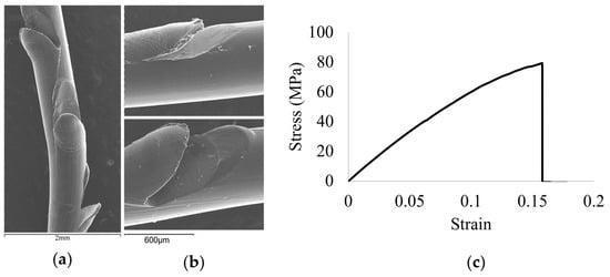
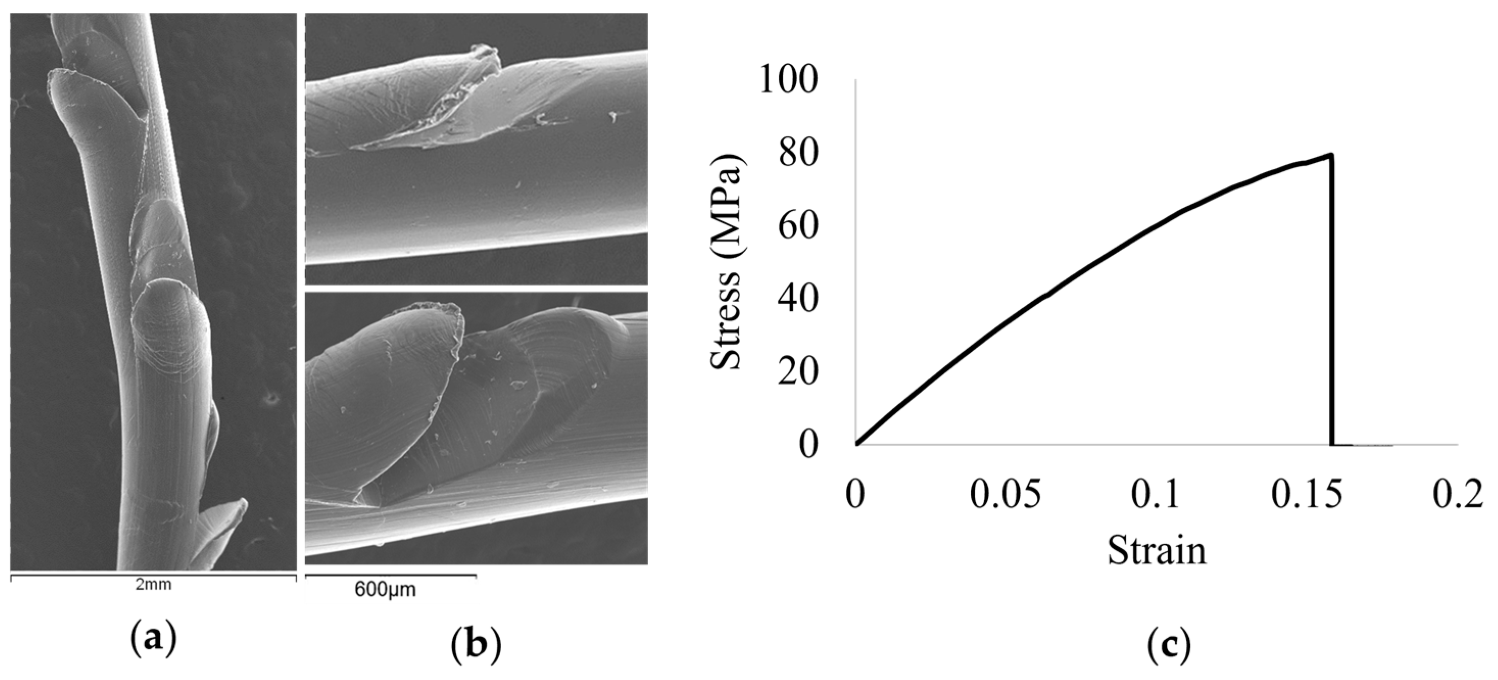
Preliminary geometric characterization of these commercial threads via scanning electron microscopy (SEM), as illustrated in Figure 1a,b, and their representative mechanical behavior under uniaxial tensile loading, shown in Figure 1c, established the target engineering and performance parameters required for effective tissue anchoring [31,34]. These parameters include a nominal diameter of 630 ± 4 µm, a cog cut angle of ≈135°, a cutting depth of 200 ± 6 µm, and an inter-barb distance of 1600 ± 11 µm.
Figure 1.
SEM micrographs of commercial biodegradable PCL cog threads used as benchmarks. (a) Low-magnification (25×) view of the outer surface showing the longitudinal distribution of the barbs (scale bar: 2 mm); (b) Higher-magnification (100×) image highlighting the surface morphology and cog architecture (scale bar: 600 µm); (c) Representative stress–strain curve of the commercial cog thread, illustrating its mechanical response under uniaxial tensile loading [34].
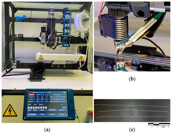
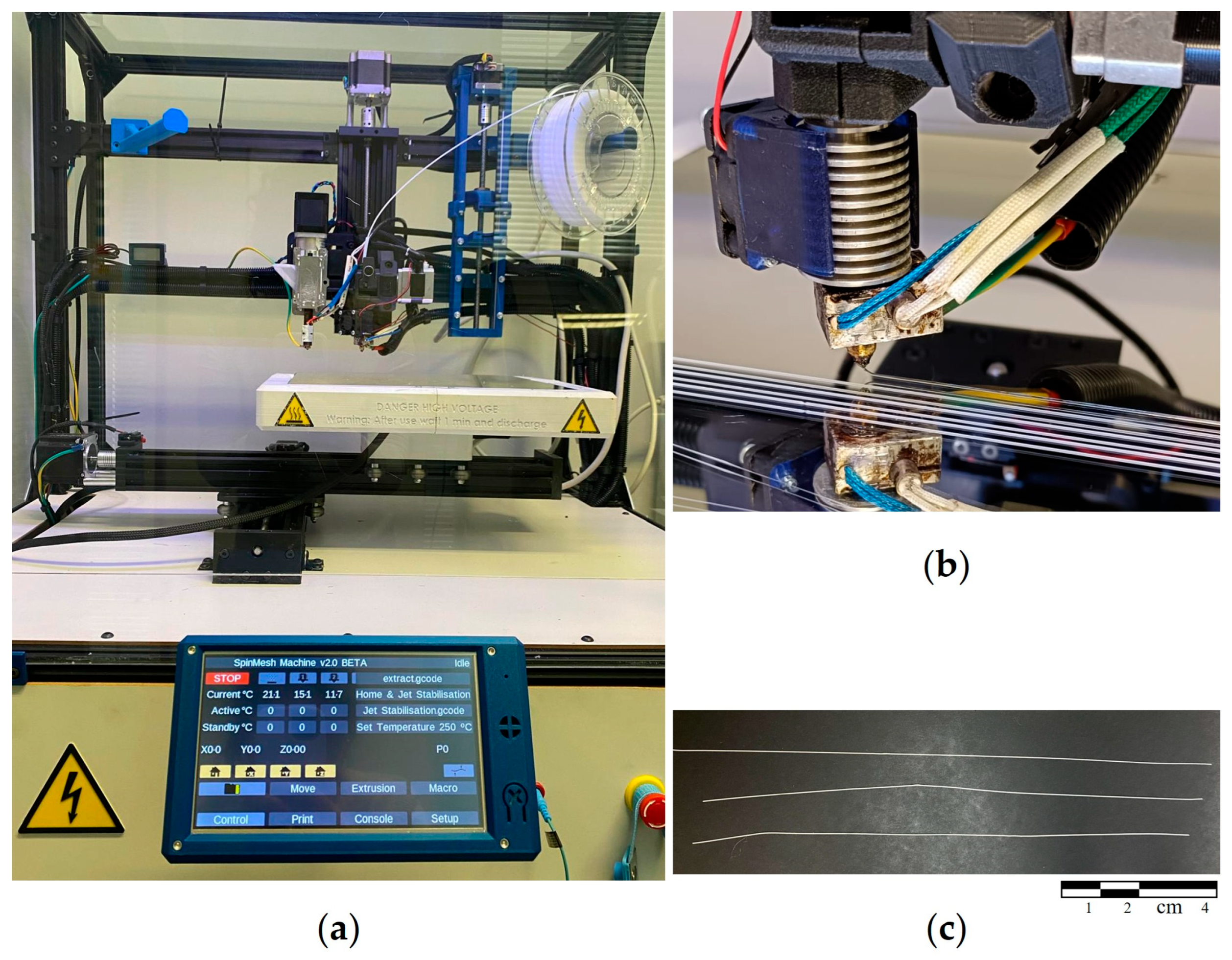
To replicate these specifications through an additive manufacturing approach, a custom MEW prototype was used (Figure 2a,b) to fabricate the targeted PCL threads (Figure 2c) [15]. This system features a modular Cartesian architecture, providing independent motion for the aluminum collector (X and Y directions) and the spinneret head (Z direction) via linear actuators and stepper motors, managed by a Duet 3D 2.0 Ethernet controller board.
Figure 2.
PCL thread production. (a) Custom-developed SPINMESH electro-injection system used for the controlled fabrication of PCL threads. This hybrid platform combines electrospinning and extrusion to enable specific morphological control; (b) Close-up of the extruded thread during collection, prior to the post-processing barbing cuts; (c) Macroscopic view of the final printed threads.
The manufacturing process was optimized to ensure that the produced threads achieved a structural consistency comparable to the core of the commercial standards. The raw 1.75 mm PCL thread was fed by a Bondtech QR direct extruder (Värnamo, Sweden) and melted through an E3D V6 Hotend equipped with a 400 µm orifice nozzle. Optimized printing variables included an extrusion temperature of 180 °C, a collector temperature of 35 °C, a printing speed of 1000 mm/min, and a nozzle-to-collector distance of 1 cm, with a high voltage of 4.2 kV applied via a Linari Engineering generator (Pisa, Italy). The resulting threads achieved a calibrated mean diameter of approximately 550 µm and a total length of 10 cm (Figure 2b). This diameter was specifically targeted to account for the material removal during the subsequent barbing process, ensuring the final functionalized thread aligned with the industrial profiles previously tested for POP treatment.
2.2. Cutting Tool Prototype
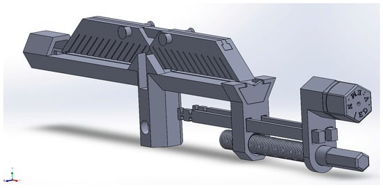
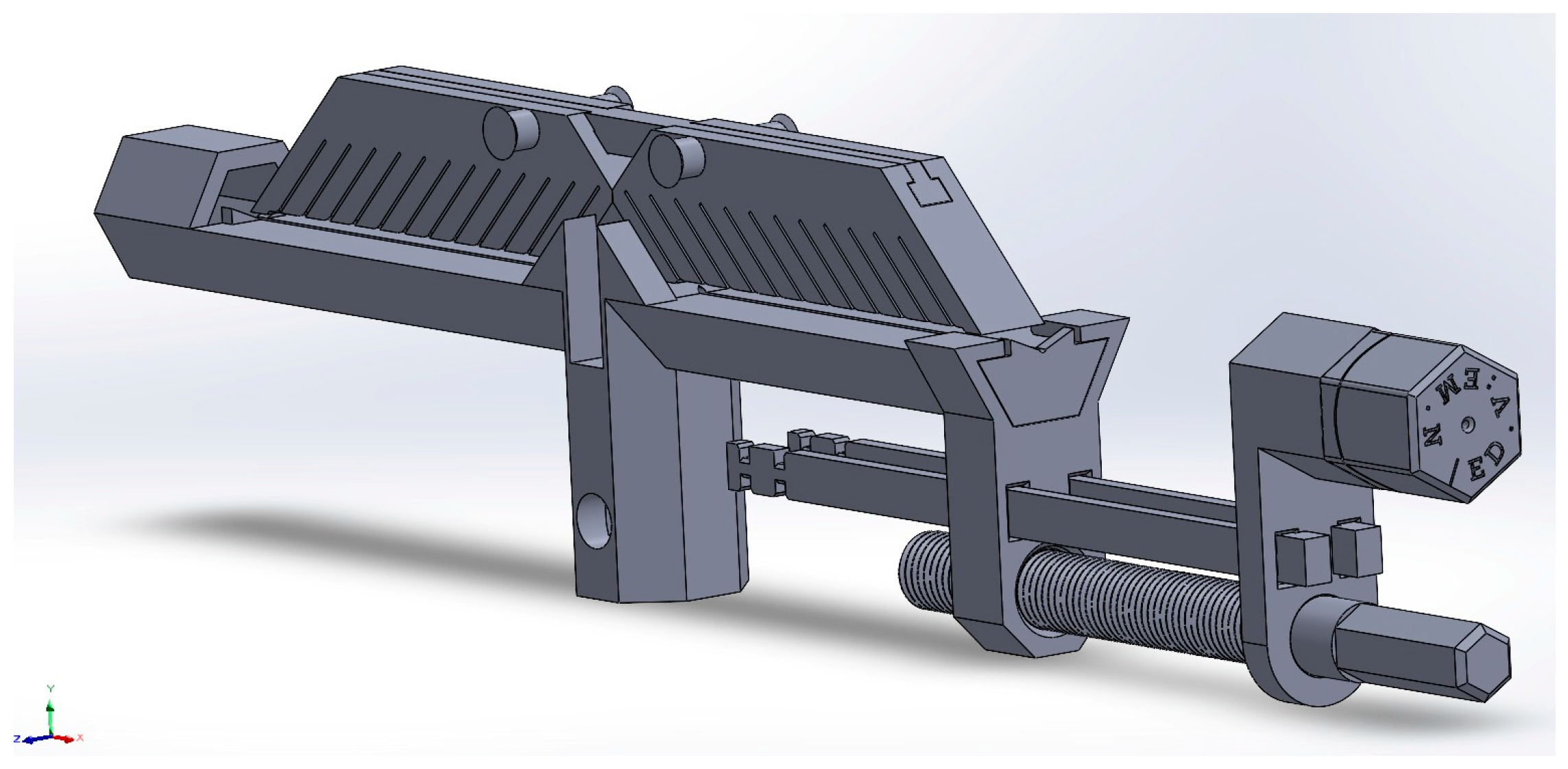
The conversion of smooth MEW-printed threads into functional cog threads was achieved using a custom-designed cutting tool prototype. The tool was designed in SolidWorks 2025 (Dassault Systèmes, Waltham, MA, USA) to ensure geometric repeatability and high precision during the barbing process, as illustrated in Figure 3. Physical components were fabricated in Polylactic Acid (PLA) via fused deposition modeling (FDM) on a Prusa MK2 3D printer (Prague, Czechia). The barbing apparatus was integrated onto a universal tensile testing machine (Mecmesin Multitest, West Sussex, UK), which served as the precision actuator for the cutting process. The device incorporates a thread chuck to stabilize the 60 µm PCL thread and a precision blade housing that governs the incision parameters.
Figure 3.
Design and components of the custom 3D-printed barbing tool. View the custom tool designed in SolidWorks, illustrating the principal components necessary for precise thread alignment and stabilization.
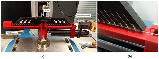
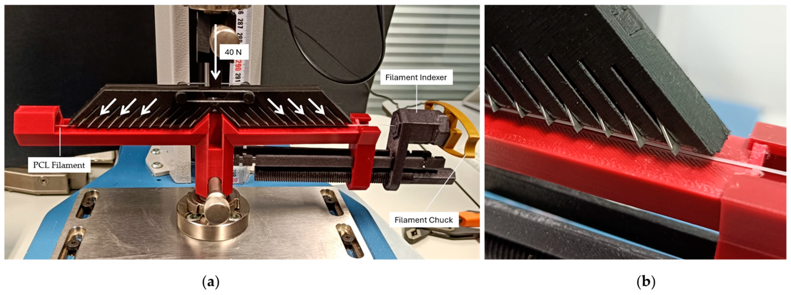
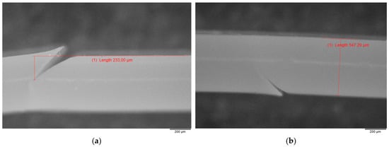
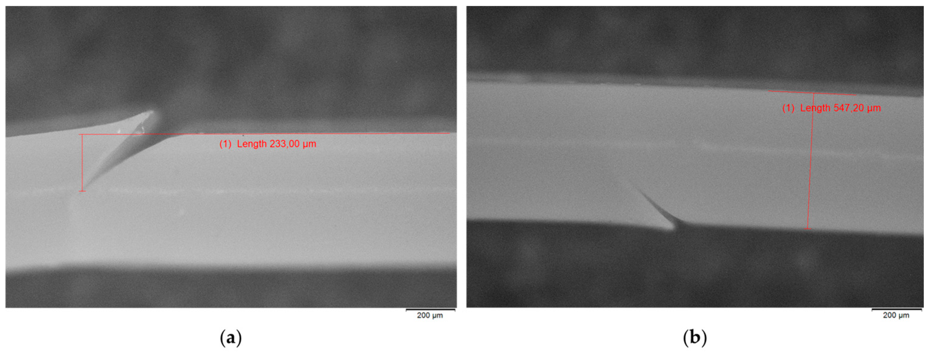
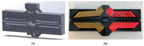
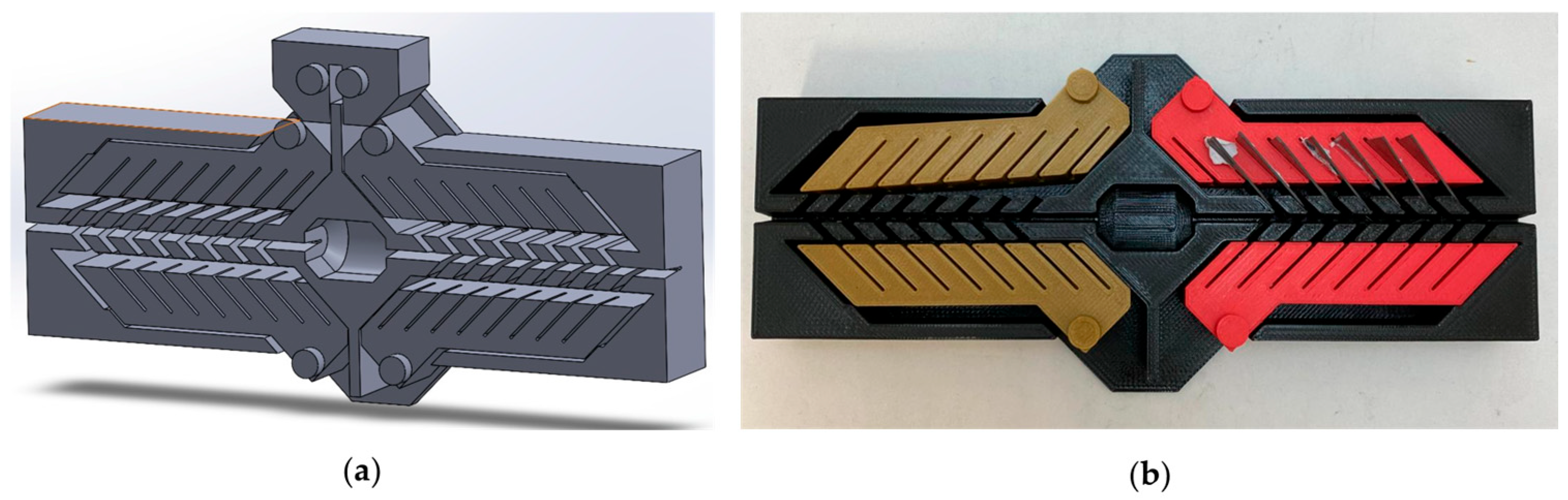
To perform the barbing, the testing machine was programmed to apply a controlled downward force of 40 N onto the device, driving the blades to create incisions at a 45° angle with a controlled 200 µm penetration depth, as schematically demonstrated in Figure 4a,b. Following the initial cut, the thread was rotated three additional times using a rotary thread indexer to achieve a 360° 4D configuration, effectively replicating the mechanical features and spatial distribution of commercial cog threads. The resulting morphology of the MEW threads after the barbing process was characterized via microscopy imaging, using a Axiophot optical microscope (Carl Zeiss, Oberkochen, Germany) equipped with a DP22 digital camera (Olympus Corporation, Tokyo, Japan), to verify the accuracy and consistency of the incisions. Dimensional analysis revealed an average incision depth of approximately 230 ± 15 µm (Figure 5a), while the thread diameter was measured at approximately 545 ± 10 µm (Figure 5b). This mechanism allows for the creation of uniform barbs designed to optimize tissue anchoring and reinforcement, aligning with established engineering targets for POP treatment. Parallel to the development of this automated system, an alternative standalone manual barbing device is currently under prototyping. Unlike the apparatus integrated into the testing machine, this novel design is engineered for handheld operation, intending to simplify the fabrication process and eliminate the dependency on heavy instrumentation. The computer-aided design (CAD) model of this concept is presented in Figure 6a, with the corresponding physical prototype, fabricated via 3D printing for preliminary functional testing and design validation, shown in Figure 6b. This manual approach is currently undergoing evaluation to assess its feasibility for producing consistent cog geometries independent of automated machinery.
Figure 4.
Experimental setup and mechanism for automated barb fabrication. (a) Overview of the custom tool integrated into the Multitest machine, with arrows indicating the load application and the direction of the cutting process; (b) Close-up view of the cutting interface, illustrating the physical contact between the precision blades and the stabilized PCL thread during the application of the 40 N force.
Figure 5.
Optical micrographs of the MEW-printed PCL cog threads after the barbing process (mag.—5×, scale—200 µm. (a) Morphological characterization highlighting the measurement of the incision depth to verify cutting precision; (b) Measurement of the cog thread diameter to confirm dimensional consistency.
Figure 6.
Development of a standalone manual barbing prototype. (a) SolidWorks CAD model illustrating the design concept for handheld operation; (b) Photograph of the 3D-printed PLA prototype currently undergoing preliminary functional testing.
2.3. Animal Vaginal Tissue Preparation
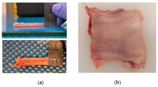
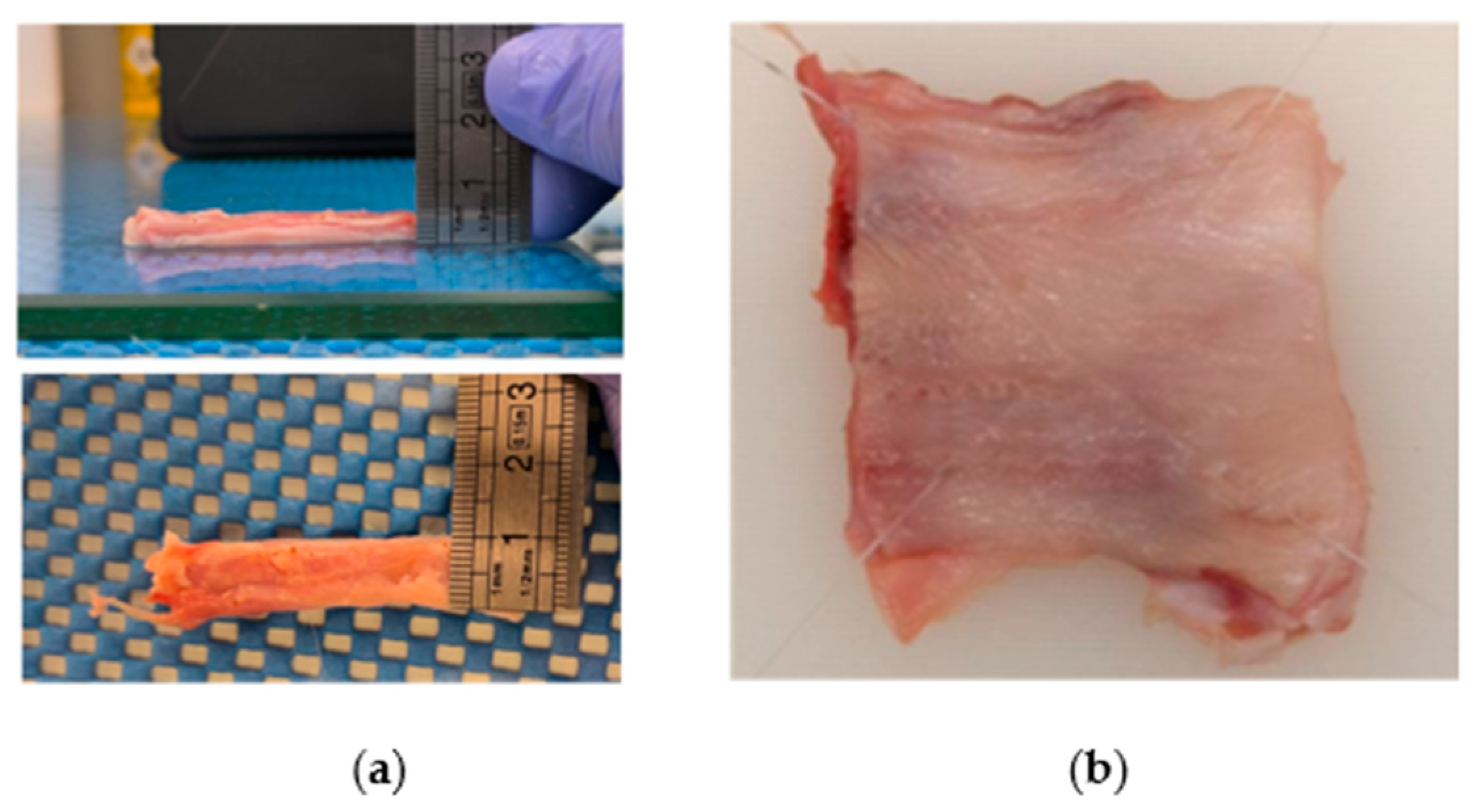
Vaginal soft tissues were obtained from eight young nulliparous sows (~6 months of age) at a certified slaughterhouse (Euroabate—Matadouro Industrial, Lda, NCV D 42, Marco de Canaveses, Portugal) for research purposes. These specimens were specifically selected to represent the baseline structural characteristics of the porcine reproductive tract prior to any gestational or dilatational changes. All procedures complied with Regulation (EC) No. 1069/2009 of 21 October. The tissues were provided as a single anatomical block, comprising the vaginal canal, rectum, pelvic musculature and surrounding fat. After collection, the vaginal canal was dissected and opened longitudinally along the urethra. From its distal portion, square specimens (~50 × 50 mm, ~4 mm thick) were prepared. Specimens were divided into two groups: control (unreinforced, n = 3) and cog (reinforced with printed cog threads, n = 3), and subsequently tested using the ball burst method (Figure 7b).
Figure 7.
Sow vaginal tissue preparation and reinforcement. (a) Representative tissue specimen prepared for uniaxial tensile testing; (b) Sow vaginal tissue preparation and reinforcement. Ex vivo insertion of PCL cog threads into a square-shaped specimen for ball burst testing, illustrating the perpendicular intersecting configuration of the cog threads.
Additionally, three rectangular specimens (50 × 10 mm) (n = 3) were prepared for uniaxial tensile testing to establish a mean stress–strain curve, which served as a basis for comparison with the uniaxial results obtained from the printed cog threads, as shown in Figure 7a.
2.4. Uniaxial Tensile Testing of Printed Cog Threads and Vaginal Tissue
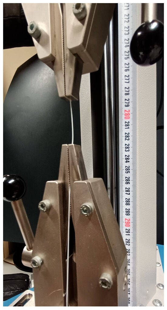
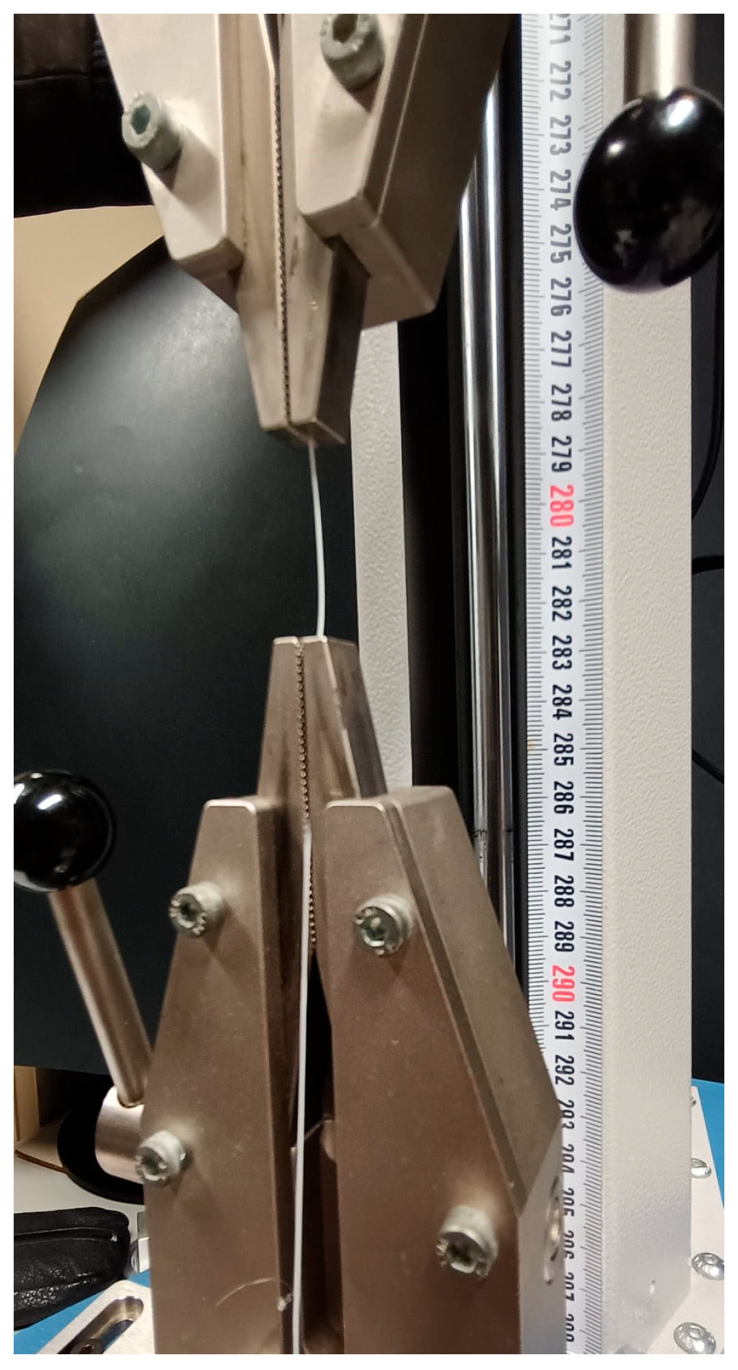
The tensile behavior of melt electrowriting (MEW)-fabricated barbed PCL cog threads was characterized using a Mecmesin Multitest testing system (Mecmesin Ltd., West Sussex, UK) equipped with a 500 N load cell and controlled via VectorPro 6.1.0 software, as shown in Figure 8. All tests were conducted at room temperature (22 °C) to ensure consistent experimental conditions. A total of four specimens (n = 4) were tested. Printed cog threads were mounted using pneumatic grips and loaded under uniaxial tension at a constant displacement rate of 10 mm/min until complete failure. To ensure consistency and allow for a direct comparison, the rectangular sow’s vaginal tissue specimens (n = 3) were tested using the same experimental setup, including the 500 N load cell and the constant displacement rate of 10 mm/min. The objective of these tests was to establish a representative mean stress–strain curve for both the printed cog threads and the native sow’s tissue, enabling a direct mechanical comparison with performance benchmarks of commercial cog threads available on the market.
Figure 8.
Mechanical characterization of the printed cog threads.
To contextualize the mechanical response of the MEW-fabricated cog threads within both a physiological and clinical framework, the experimental results were qualitatively compared with uniaxial tensile data of sow vaginal tissue and with data, previously reported, from commercial PCL-based surgical cog threads [31,33]. This comparative analysis aimed to verify that the mechanical response of the MEW cog threads falls within a biomechanically relevant range—positioned between the native tissue’s properties and high-stiffness commercial alternatives—to promote effective load sharing with the host tissue while avoiding excessive stiffness. Such an approach seeks to minimize localized stress concentrations and reduce the risk of adverse mechanical interactions, including tissue cutting or stress shielding, thereby supporting the development of mechanically compatible intravaginal reinforcement strategies.
2.5. Ex Vivo Minimally Invasive Insertion of the PCL Cog Threads
The insertion of the lab-made PCL cog threads into the biological tissue was performed following a minimally invasive protocol previously established for commercial cog threads. The insertion procedure involved an initial surface perforation using a needle. A cannula containing the PCL cog thread was then inserted until the end of the sample, turned, and carefully removed, ensuring that the lab-made barbs remained fixed within the tissue. To create a non-rigid reinforcement structure, two cog threads were inserted into the tissue at a 90° angle, intersecting each other. This intersecting pattern is designed to provide structural support while matching the physiological compliance of the vaginal wall.
2.6. Ball Burst Test
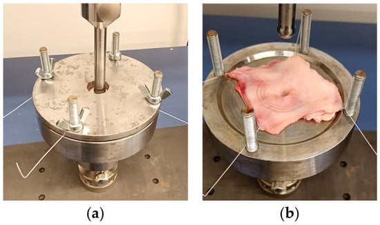
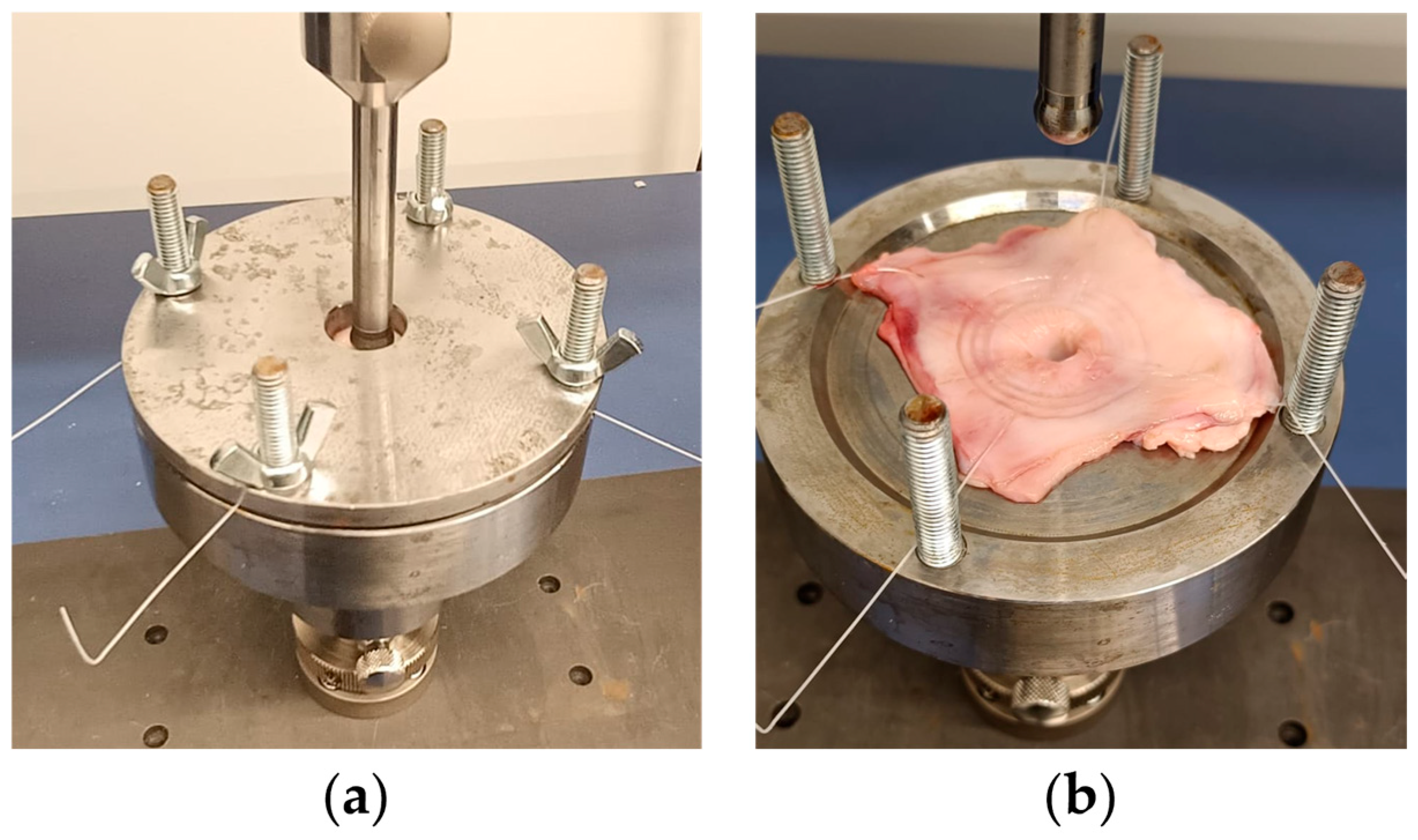
The multiaxial mechanical properties were evaluated by comparing unreinforced sow’s vaginal wall specimens (control) with those integrated with MEW-fabricated PCL cog threads (Figure 9a,b), using ball burst testing to provide an indirect measurement of material strength and the overall structural reinforcement effect. Testing was conducted on a Mecmesin Multitest (West Sussex, UK) machine equipped with a 500 N load cell, and utilized the proprietary VectorPro software (Mecmesin Ltd., West Sussex, UK) for high-precision data acquisition. The experimental setup featured a custom 3D-printed prototype based on established literature designs, consisting of a ring clamp with an internal diameter of 20.0 mm. For each test, the square tissue specimens were secured within the clamping mechanism, and a polished spherical plunger with a diameter of 11.50 mm was positioned at the center of the sample. To eliminate any initial slack in the tissue, a preload of 0.1 N was applied before the commencement of the test. The plunger was then driven through the specimen at a constant displacement rate of 10 mm/min until complete tissue rupture occurred. The output data, including peak force (N) and ball displacement (mm), were recorded to analyze the ultimate load and mechanical behavior in both the physiological (comfort) and supra-physiological (stress) zones.
Figure 9.
Ball burst test performed on a sow’s vaginal wall specimen reinforced with a PCL cog thread. (a) Initial stage of the test, showing the first contact between the spherical plunger and the reinforced tissue; (b) Final stage of the test, illustrating complete tissue rupture and deformation of the integrated cog thread.
2.7. Statistical Analysis
Statistical analyses were conducted using IBM SPSS Statistics 28.0 (IBM Corp., New York, NY, USA). The normality of the data distribution was assessed using the Kolmogorov–Smirnov test. Quantitative results are reported as mean ± standard deviation (SD). To evaluate the mechanical reinforcement effect, unpaired Student’s t-tests were used to compare the performance of the lab-made PCL cog threads against the native tissue condition and against commercial cog thread benchmarks reported in the literature. All statistical tests were performed with a 95% confidence level, and results were considered statistically significant when p < 0.05.
3. Results
3.1. Uniaxial Tensile Performance and Biomechanical Compatibility
The uniaxial tensile behavior of the MEW-fabricated barbed PCL cog threads was evaluated under monotonic loading until failure. All specimens exhibited a consistent load–displacement response characterized by an initial increase in load with increasing displacement, followed by a peak load and subsequent rupture. The ultimate tensile force measured for each individual specimen is reported in Table 1. Peak loads ranged from 2.1 N to 3.1 N, with a mean ultimate tensile force of 2.8 ± 0.4 N.
Table 1.
Individual ultimate tensile force (N) for each MEW-fabricated cog thread sample.
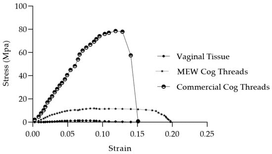
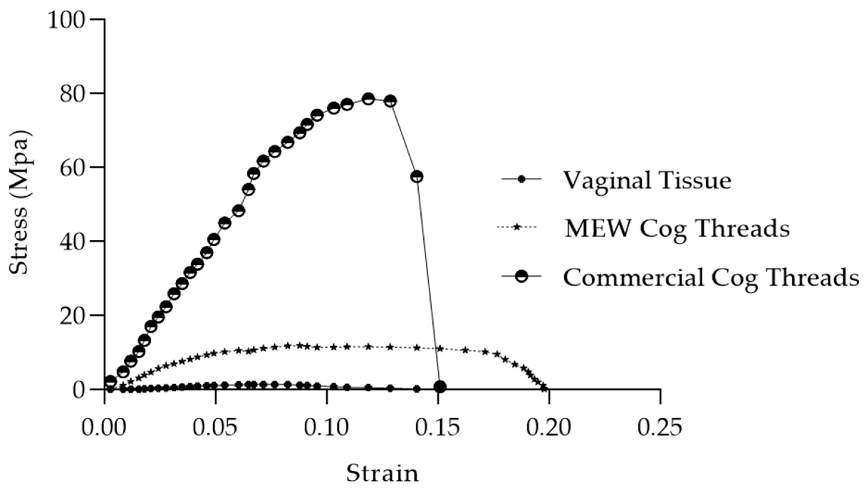
The MEW-fabricated cog threads exhibited a mean ultimate tensile strength of 14.1 ± 1.1 MPa and a strain at peak load of 9.6 ± 1%. The comparative stress–strain response—comprising original experimental data for MEW cog threads (n = 4) and sow’s vaginal tissue (n = 3), alongside literature-derived data from commercial cog threads [33]—is illustrated in Figure 10. The plot highlights the intermediate mechanical behavior of MEW cog threads, which provide structural reinforcement while minimizing the mechanical mismatch compared to high-strength commercial alternatives. This intermediate profile suggests a more biomimetic approach to intravaginal reinforcement, balancing the need for structural support with mechanical compatibility with the host tissue.
Figure 10.
Biomechanical compatibility analysis. Comparative stress–strain profiles of MEW-fabricated cog threads, sow vaginal tissue and commercial PCL cog threads.
3.2. Ball Burst Performance
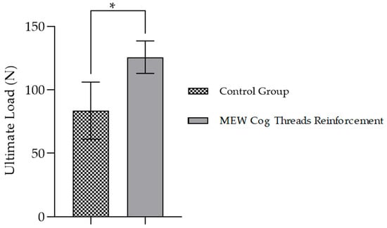
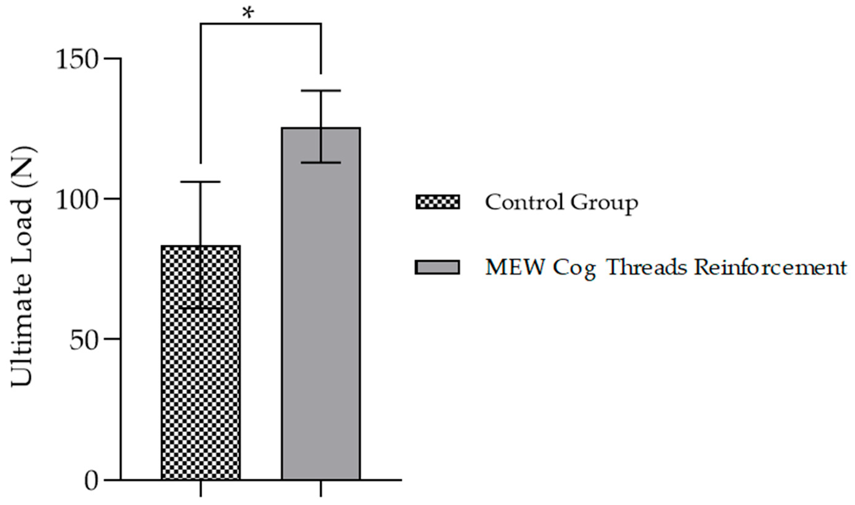
The multiaxial mechanical performance of the vaginal tissue under ball burst loading was assessed to evaluate the reinforcement provided by the MEW-fabricated PCL cog threads. In this study, the term “control” corresponds to unreinforced native tissue. Table 2 summarizes the ultimate load for the control specimens, the MEW-reinforced specimens, and the commercial cog thread benchmark (literature data [33]). Integration of the cog threads resulted in a significant increase in load-bearing capacity, with the mean ultimate load rising from 83 ± 20 N (control) to 126 ± 15 N (reinforced) (n = 3 per condition).
Table 2.
Mean ultimate load (N) for Ball Burst testing across the three experimental groups. Values are reported as mean ± standard deviation.
Statistical analysis (unpaired Student’s t-test) confirmed a significant difference between these two groups (p = 0.0477), corresponding to an improvement of approximately 51.8%. For technological validation, these results were compared to the commercial cog threads (177.0 ± 5.4 N), with the MEW-fabricated cog threads achieving approximately 71% of their reported ultimate load. Additional increases in resistance to deformation were observed in both comfort and stress zones for the reinforced condition. The complete comparative profile is shown in Figure 11.
Figure 11.
Reinforcement efficacy assessed via ball burst testing. Comparison of the mean ultimate load between control (unreinforced sow vaginal tissue) and tissue reinforced with MEW-fabricated cog threads. Asterisk denotes statistical significance (p < 0.05).
4. Discussion
The diagnosis and treatment of POP remain major clinical challenges, particularly following the regulatory restrictions on transvaginal synthetic meshes due to their association with severe complications [28]. In this context, the prevailing assumption that higher mechanical strength inherently leads to better clinical outcomes deserves reconsideration. The results of the present study demonstrate that PCL cog threads fabricated via MEW provide an intermediate mechanical profile between native vaginal tissue and commercially available cog threads, achieving substantial reinforcement while reducing mechanical mismatch. However, a direct material comparison is constrained by the unavailability of specific data regarding the molecular weight (Mn, Mw) and polydispersity index (PDI) of the commercial cog threads, as these are proprietary specifications. While the PCL utilized in this work (Facilan™ PCL100) is characterized by a mean molecular weight of 50,000 g/mol, the exact specifications of the commercial benchmark remain unknown. Since mechanical properties are strongly affected by molecular weight and thermal processing conditions (such as the 180 °C extrusion temperature used here), the higher mechanical resistance observed in the commercial benchmark is likely attributed to the use of a polymer with a higher molecular weight or different chain characteristics, rather than geometric superiority. Consequently, the MEW-based approach offers significant tunability: while higher mechanical strength could be achieved, if required, by using PCL grades with higher molecular weights or narrower PDIs, the mechanical response achieved in the present work is comparatively more compliant. This may be advantageous in applications where excessive stiffness could increase tissue damage, while still preserving effective anchoring functionality.
In the present study, PCL was selected primarily due to its availability and suitability for processing using the MEW platform available in our laboratory. While alternative PCL grades will be explored in future work to further tailor the mechanical response, it is important to emphasize that the objective of this work is not to replicate the stiffness of commercially available cog threads. Instead, a softer mechanical behavior is intentionally pursued, as excessive stiffness in anchoring devices may lead to increased local stress concentrations, tissue trauma, and inflammatory responses.
With a mean tensile strength of 14.1 ± 1.1 MPa and a 51.8% increase in ultimate load under ball burst testing, the MEW-fabricated cog threads offer sufficient structural reinforcement without the excessive rigidity characteristic of high-strength commercial alternatives. From a biomechanical perspective, this balanced profile supports a load-sharing mechanism with the host tissue rather than load-bearing dominance. Such behavior may mitigate localized stress concentrations, which have been implicated in tissue damage, erosion, and the so-called “cheese-wire effect” often observed with overly stiff sutures or meshes [38]. By occupying this intermediate mechanical window, the printed threads may reduce tissue trauma while maintaining functional support, aligning with emerging trends that emphasize mechanical compatibility over maximal strength.
From a biological standpoint, prior studies suggest that the implantation of biodegradable threads can stimulate a localized healing response, including neocollagenesis and elastin formation in surrounding tissues [43,44]. Given that collagen is the primary determinant of soft tissue tensile strength, such biostimulation may contribute to a supportive biological environment that complements the mechanical function of the implant [45,46]. Regarding the degradation timeframe, although the specific PCL grade used in this study may exhibit faster hydrolysis than higher-molecular-weight counterparts, it is expected to follow the characteristic slow degradation profile of PCL. Literature indicates that PCL’s hydrophobic nature typically allows for the maintenance of structural integrity for over 12 to 24 months in biological environments [47,48]. This degradation window is considered compatible with the critical phases of tissue healing and remodeling, during which mechanical load may be progressively transferred from the synthetic reinforcement to the host tissue. In this context, the primary function of the cog thread is intended to provide temporary mechanical reinforcement rather than permanent structural support. As degradation proceeds, progressive tissue integration may contribute to maintaining functional stability through a load-sharing mechanism. Furthermore, the presence of multiple anchoring features may promote a more distributed load transfer compared to conventional sutures, which could help reduce localized stress concentrations and support effective fixation during the early healing phase. Nevertheless, in vivo studies are required to validate these mechanisms and their long-term implications.
Regarding the morphological features, it is important to acknowledge that the current post-processing protocol resulted in barbs with a slit-like configuration, contrasting with the more open, bud-like structure of the commercial benchmark. The commercial design, characterized by a greater distance between the barb and the thread shank, likely facilitates superior soft tissue ingress, thereby potentially enhancing fixation security. However, despite this geometric variance, the MEW-fabricated threads demonstrated effective functional performance, providing a 51.8% increase in tissue load-bearing capacity. Future optimization of the manufacturing process will focus on increasing the barb opening angle, potentially through auxiliary thermal treatment or mechanical lifting steps post-cutting, to further mimic the commercial geometry and maximize tissue integration.
Nevertheless, several limitations of the present study must be acknowledged. The mechanical characterization was performed using ex vivo models and static uniaxial tensile testing, which do not fully capture the complex anisotropic, hyperelastic, and viscoelastic behavior of living vaginal tissue under dynamic physiological loading. The porcine tissue model used in this study was obtained from young nulliparous animals and was selected to provide a consistent substrate to quantify the reinforcement effect of the cog thread under controlled conditions. Therefore, this model is not intended to replicate the pathological tissue weakening typically observed in advanced POP. It should be noted that the commercial cog thread data used for comparison were obtained in a previous study by the same authors using an identical experimental setup, including the same custom-made ball burst testing rig and loading parameters. In both studies, vaginal tissue samples were collected at a slaughterhouse and subsequently frozen prior to testing, following the same preparation and storage protocol. As the tissue samples were prepared using identical procedures, the resulting mechanical responses are expected to be comparable. Nevertheless, as is inherent to experiments involving biological tissues, complete equivalence cannot be guaranteed due to intrinsic biological variability. For this reason, the comparative analysis presented here focuses on relative functional performance trends rather than absolute mechanical values. Additionally, the absence of cellular, vascular, and immune interactions limits the ability to predict in vivo degradation kinetics, inflammatory responses, and long-term tissue remodeling. These factors are critical for assessing both safety and efficacy in a clinical setting.
Future research should therefore focus on in situ mechanical evaluations, including pull-out strength and anchoring efficiency within native vaginal tissue. In vivo studies will be essential to assess host response, degradation behavior, and functional outcomes over time. Moreover, the integration of antimicrobial agents or surface modifications within the PCL matrix warrants investigation as a strategy to reduce the risk of infection and biofilm formation. Collectively, these future directions will be critical to determining whether MEW-fabricated PCL cog threads can successfully achieve the intended balance between mechanical support and biological integration, ultimately contributing to safer and more effective pelvic floor rehabilitation strategies.
5. Conclusions
In conclusion, this study demonstrates that biodegradable PCL cog threads fabricated via MEW constitute a viable and technologically robust approach for vaginal wall reinforcement in the context of POP. By exhibiting an intermediate mechanical profile between native vaginal tissue and high-stiffness commercial cog threads, the MEW-fabricated threads achieved a 51.8% increase in load-bearing capacity while reducing mechanical mismatch. This balanced mechanical behavior supports a load-sharing reinforcement strategy rather than load-bearing dominance, which may be critical for minimizing tissue damage associated with overly rigid devices. The customized barbed architecture enables effective self-anchoring, offering the potential to simplify surgical procedures, reduce operative time, and support less invasive, patient-tailored treatment strategies.
Future work should focus on in situ and in vivo evaluations to assess anchoring efficiency, host response, degradation behavior, and long-term tissue remodeling. Additionally, further technological optimization—such as tuning degradation kinetics or integrating antimicrobial functionalities—may enhance the clinical relevance of this approach. Collectively, these findings position MEW-fabricated PCL cog threads as a promising platform for the development of mechanically compatible and minimally invasive reinforcement solutions in the post-mesh era of pelvic floor rehabilitation.
Author Contributions
Conceptualization, F.P., A.T.S., H.L.B., M.E.S. and M.F.V.; methodology, F.P., H.L.B., M.E.S. and A.A.F.; validation, F.P., M.E.S. and A.A.F.; formal analysis, F.P., M.E.S., A.A.F., A.C.M. and N.A.; investigation F.P., M.E.S. and A.A.F.; resources, M.E.S., A.T.S., J.P.M., A.A.F., N.M.F. and A.C.M.; writing—original draft preparation, F.P., M.E.S., M.F.V., H.L.B., A.T.S. and A.A.F.; writing—review and editing, All authors; supervision, M.E.S., A.A.F., A.C.M. and N.A.; project administration, M.E.S. and A.A.F.; funding acquisition, M.E.S., A.A.F., A.C.M. and N.A. All authors have read and agreed to the published version of the manuscript.
Funding
This research was funded by Fundação para a Ciência e a Tecnologia (FCT), through project LA/P/0079/2020 (associated with INEGI/LAETA). This work was also financially supported by the Individual Scientific Employment Stimulus grant 2021.00077.CEECIND and by the PhD grants 2023.00640.BD and 2024.00925.BD, all financed by FCT/MCTES through national funds.
Institutional Review Board Statement
Ethical review and approval were waived for this study since INEGI and DGAV (Direção-Geral da Alimentação e Veterinária, Portugal), celebrated protocol with registry number N19129UDER. Such protocol authorizes INEGI to collect animal tissue in slaughterhouses for research/educational purposes. Moreover, all procedures respected the conditions imposed by Regulation (EC) No. 1069/2009 of 21 October, on health rules concerning animal by-products and derived products not intended for human consumption.
Data Availability Statement
The original contributions presented in this study are included in the article. Further inquiries can be directed to the corresponding author.
Acknowledgments
The authors gratefully acknowledge the funding by Ministério da Ciência Tecnologia, e Ensino Superior, FCT, Portugal, under grants 2023.00640.BD and 2024.00925.BD, and from Stimulus of Scientific Employment 2021.00077.CEEIND. This work was supported by FCT, through INEGI, under LAETA, project LA/P/0079/2020.
Conflicts of Interest
The authors declare that there is no financial, professional or other personal interest of any nature or kind in any product, service and/or company that could be constructed as influencing the position.
References
- Zhou, L.; Miller, J.; Vezza, J.; Mayster, M.; Raffay, M.; Justice, Q.; Tamimi, Z.A.; Hansotte, G.; Sunkara, L.D.; Bernat, J. Additive Manufacturing: A Comprehensive Review. Sensors 2024, 24, 2668. [Google Scholar] [CrossRef] [Scilit]
- Bai, W.; Fang, H.; Wang, Y.; Zeng, Q.; Hu, G.; Bao, G.; Wan, Y. Academic Insights and Perspectives in 3D Printing: A Bibliometric Review. Appl. Sci. 2021, 11, 8298. [Google Scholar] [CrossRef] [Scilit]
- Bera, T. A Review on Significant Role of Additive Manufacturing in Biomedical Applications. Adv. Mater. Sustain. Manuf. 2024, 1, 10002. [Google Scholar] [CrossRef] [Scilit]
- Jin, Y.; Li, X.; Campbell, R.I.; Ji, S. Visualizing the Hotspots and Emerging Trends of 3D Printing through Scientometrics. Rapid Prototyp. J. 2018, 24, 801–812. [Google Scholar] [CrossRef] [Scilit]
- Heidari, B.S.; Dodda, J.M.; El-Khordagui, L.K.; Focarete, M.L.; Maroti, P.; Toth, L.; Pacilio, S.; El-Habashy, S.E.; Boateng, J.; Catanzano, O.; et al. Emerging Materials and Technologies for Advancing Bioresorbable Surgical Meshes. Acta Biomater. 2024, 184, 1–21. [Google Scholar] [CrossRef] [Scilit]
- Jiang, D.; Thissen, H.; Hughes, T.C.; Yang, K.; Wilson, R.; Murphy, A.B.; Nguyen, V. Advances in Additive Manufacturing of Auxetic Structures for Biomedical Applications. Mater. Today Commun. 2024, 40, 110045. [Google Scholar] [CrossRef] [Scilit]
- Peng, X.; Wu, S.; Sun, X.; Yue, L.; Montgomery, S.M.; Demoly, F.; Zhou, K.; Zhao, R.R.; Qi, H.J. 4D Printing of Freestanding Liquid Crystal Elastomers via Hybrid Additive Manufacturing. Adv. Mater. 2022, 34, 2204890. [Google Scholar] [CrossRef] [Scilit]
- Zhu, Z.; Park, H.S.; McAlpine, M.C. 3D Printed Deformable Sensors. Sci. Adv. 2020, 6, eaba5575. [Google Scholar] [CrossRef] [Scilit] [PubMed]
- Javaid, M.; Haleem, A. 3D Printed Tissue and Organ Using Additive Manufacturing: An Overview. Clin. Epidemiol. Glob. Health 2020, 8, 586–594. [Google Scholar] [CrossRef] [Scilit]
- Liaw, C.-Y.; Guvendiren, M. Current and Emerging Applications of 3D Printing in Medicine. Biofabrication 2017, 9, 024102. [Google Scholar] [CrossRef] [Scilit] [PubMed]
- Dedeloudi, A.; Bertelli, P.M.; Martinez-Marcos, L.; Quinten, T.; Lengyel, I.; Andersen, S.K.; Lamprou, D.A. Development of Bioceramic Bone-Inspired Scaffolds Through Single-Step Melt-Extrusion 3D Printing for Segmental Defect Treatment. J. Funct. Biomater. 2025, 16, 358. [Google Scholar] [CrossRef] [Scilit]
- Kumar, A.; Nune, K.C.; Murr, L.E.; Misra, R.D.K. Biocompatibility and Mechanical Behaviour of Three-Dimensional Scaffolds for Biomedical Devices: Process–Structure–Property Paradigm. Int. Mater. Rev. 2016, 61, 20–45. [Google Scholar] [CrossRef] [Scilit]
- Vaz, M.F.R.; Martins, J.A.P.; Pinheiro, F.; Ferreira, N.M.; Brandão, S.; Alves, J.L.; Fernandes, A.A.; Parente, M.P.L.; Silva, M.E.T. Medical- and Non-Medical-Grade Polycaprolactone Mesh Printing for Prolapse Repair: Establishment of Melt Electrowriting Prototype Parameters. Appl. Sci. 2024, 14, 9670. [Google Scholar] [CrossRef] [Scilit]
- Yin, Y.; Yang, H.; Han, W.; Guo, C.; Mu, Q.; Yang, H.; Chen, D. Melt Electrowriting for Biomimetic Tissue Engineering: Advances in Scaffold Design, Materials, and Multifunctional Applications. Polym. Adv. Techs 2025, 36, e70067. [Google Scholar] [CrossRef] [Scilit]
- Rynkevic, R.; Silva, M.E.T.; Martins, P.; Mascarenhas, T.; Alves, J.L.; Fernandes, A.A. Characterisation of Polycaprolactone Scaffolds Made by Melt Electrospinning Writing for Pelvic Organ Prolapse Correction—A Pilot Study. Mater. Today Commun. 2022, 32, 104101. [Google Scholar] [CrossRef] [Scilit]
- Sarker, M.T.; Rahaman, M.A. Advancements in 3D Printing Techniques for Polymer Fiber-Reiforced Textile Composites: A Systematic Literature Review. Am. J. Interdiscip. Stud. 2022, 3, 32–60. [Google Scholar] [CrossRef] [Scilit]
- Pien, N.; Bartolf-Kopp, M.; Parmentier, L.; Delaey, J.; De Vos, L.; Mantovani, D.; Van Vlierberghe, S.; Dubruel, P.; Jungst, T. Melt Electrowriting of a Photo-Crosslinkable Poly(ε-caprolactone)-Based Material into Tubular Constructs with Predefined Architecture and Tunable Mechanical Properties. Macro. Mater. Eng. 2022, 307, 2270028. [Google Scholar] [CrossRef] [Scilit]
- Sandoval Salaiza, D.A.; Valsangiacomo, N.; Dinç, N.U.; Yildirim, M.; Madrid-Wolff, J.; Bertsch, A.; Jiguet, S.; Dalton, P.D.; Brugger, J.; Moser, C. Electrowriting of SU-8 Microfibers. Polymers 2024, 16, 1630. [Google Scholar] [CrossRef] [Scilit]
- Darroch, C.; Asaro, G.A.; Gréant, C.; Suku, M.; Pien, N.; Van Vlierberghe, S.; Monaghan, M.G. Melt Electrowriting of a Biocompatible Photo-crosslinkable Poly(D,L-lactic Acid)/Poly(Ε-caprolactone)-based Material with Tunable Mechanical and Functionalization Properties. J. Biomedical. Mater. Res. 2023, 111, 851–862. [Google Scholar] [CrossRef] [Scilit]
- Said, L.B.; Ayadi, B.; Alharbi, S.; Dammak, F. Recent Advances in Additive Manufacturing: A Review of Current Developments and Future Directions. Machines 2025, 13, 813. [Google Scholar] [CrossRef] [Scilit]
- Wang, B.; Chen, Y.; Zhu, X.; Wang, T.; Li, M.; Huang, Y.; Xue, L.; Zhu, Q.; Gao, X.; Wu, M. Global Burden and Trends of Pelvic Organ Prolapse Associated with Aging Women: An Observational Trend Study from 1990 to 2019. Front. Public Health 2022, 10, 975829. [Google Scholar] [CrossRef] [Scilit]
- Walker, G.J.A.; Gunasekera, P. Pelvic Organ Prolapse and Incontinence in Developing Countries: Review of Prevalence and Risk Factors. Int. Urogynecol. J. 2011, 22, 127–135. [Google Scholar] [CrossRef] [Scilit]
- Tunn, R.; Baeßler, K.; Knüpfer, S.; Hampel, C. Urinary Incontinence and Pelvic Organ Prolapse in Women. Dtsch. Ärzteblatt Int. 2023, 120, 71. [Google Scholar] [CrossRef] [Scilit]
- Raju, R.; Linder, B.J. Evaluation and Management of Pelvic Organ Prolapse. Mayo Clin. Proc. 2021, 96, 3122–3129. [Google Scholar] [CrossRef] [Scilit] [PubMed]
- Zumrutbas, A.E. Understanding Pelvic Organ Prolapse: A Comprehensive Review of Etiology, Epidemiology, Comorbidities, and Evaluation. Société Int. D’urologie J. 2025, 6, 6. [Google Scholar] [CrossRef] [Scilit]
- Houshyar, S.; Quigley, A.; Cole, I.; Yin, H.; Zizhou, R.; Saha, T.; Pirogova, E.; Yeung, J.M.; Mohsenipour, M.; Mirabedini, A.; et al. Bioengineered Visible Polymeric Mesh to Enhance Urogynaecological Health. bioRxiv 2025. [Google Scholar] [CrossRef] [Scilit]
- Khan, A.; Elmadbouh, S.; Khalid, S.A.; Ayub, M.; Mahmood, S.; Ravendran, K. Efficacy of Biological Versus Synthetic Mesh in Ventral Hernia Repair: A Systematic Review and Meta-Analysis of Long-Term Outcomes and Recurrence Rates. Cureus 2025, 17, e92557. [Google Scholar] [CrossRef] [Scilit]
- U.S. Food and Drug Administration. FDA Takes Action to Protect Women’s Health, Orders Manufacturers of Surgical Mesh Intended for Transvaginal Pelvic Organ Prolapse Repair to Stop Sales and Distribution; U.S. Food and Drug Administration: Silver Spring, MD, USA, 2019.
- Zargar, N.; Carr, A. The Regulatory Ancestral Network of Surgical Meshes. PLoS ONE 2018, 13, e0197883. [Google Scholar] [CrossRef] [Scilit]
- Guan, Y.; Han, J. Pelvic Organ Prolapse According to Educational Level. Res. Sq. 2022. [Google Scholar] [CrossRef] [Scilit]
- Pinheiro, F.; Jesus, A.M.P.; de Maurício, A.C.; Alves, N.; Fernandes, A.A.; Silva, M.E.T. da Biomechanical Evaluation of Biodegradable PCL Cog Threads for Prolapse Rehabilitation. Proc. Inst. Mech. Eng. Part H J. Eng. Med. 2025, 239, 687–696. [Google Scholar] [CrossRef] [Scilit]
- Ferreira, N.M.; Silva, M.E.T.; Parente, M.P.L.; Pinheiro, F.; Mascarenhas, T.; Fernandes, A.A. Evaluation of Mechanical Biocompatibility of Cog Threads for Prolapse Repair. Proc. Inst. Mech. Eng. Part H J. Eng. Med. 2025, 239, 155–164. [Google Scholar] [CrossRef] [Scilit]
- Soares, C.; Martins, P.; Silva, E.; Hympanova, L.; Rynkevic, R. Cog Threads for Transvaginal Prolapse Repair: Ex-Vivo Studies of a Novel Concept. Surgeries 2022, 3, 101–110. [Google Scholar] [CrossRef] [Scilit]
- Silva, A.T.; Ferreira, N.M.; Bastos, H.L.; Vaz, M.F.; Martins, J.P.; Pinheiro, F.; Fernandes, A.A.; Silva, E. Development and Simulation-Based Validation of Biodegradable 3D-Printed Cog Threads for Pelvic Organ Prolapse Repair. Materials 2025, 18, 3638. [Google Scholar] [CrossRef] [Scilit]
- Hong, G.-W.; Wan, J.; Yoon, S.-E.; Wong, S.; Yi, K.-H. Pre- and Post-Procedural Considerations and Thread Types for Thread Lifting. Life 2025, 15, 85. [Google Scholar] [CrossRef] [Scilit]
- Sukgen, G.; Altunkol, A.; Yiğit, A. Effects of Mesh Surgery on Sexual Function in Pelvic Prolapse and Urinary Incontinence. Int. Braz. J. Urol. 2021, 47, 82–89. [Google Scholar] [CrossRef] [Scilit]
- Achtari, C.; Dwyer, P.L. Sexual Function and Pelvic Floor Disorders. Best Pract. Res. Clin. Obstet. Gynaecol. 2005, 19, 993–1008. [Google Scholar] [CrossRef] [Scilit] [PubMed]
- Pinheiro, F.; Fernandes, A.; Maurício, A.C.; Alves, N.; Silva, M.E. In Vivo Assessment of PCL Cog Thread Reinforcement Using an Intravaginal Force Measurement Device. Appl. Sci. 2025, 15, 12677. [Google Scholar] [CrossRef] [Scilit]
- Ntrivala, M.A.; Pitsavas, A.C.; Lazaridou, K.; Baziakou, Z.; Karavasili, D.; Papadimitriou, M.; Ntagkopoulou, C.; Balla, E.; Bikiaris, D.N. Polycaprolactone (PCL): The Biodegradable Polyester Shaping the Future of Materials—A Review on Synthesis, Properties, Biodegradation, Applications and Future Perspectives. Eur. Polym. J. 2025, 234, 114033. [Google Scholar] [CrossRef] [Scilit]
- Krumov, N.; Atanasova, N.; Boyadzhieva, I.; Petrov, K.; Petrova, P. Biodegradation of Poly(ε-Caprolactone): Microorganisms, Enzymes, and Mechanisms. Int. J. Mol. Sci. 2025, 26, 5826. [Google Scholar] [CrossRef] [Scilit]
- 3D4Makers. FacilanTM PCL 100 (Polycaprolactone) Filament; 3D4Makers: Haarlem, The Netherlands, 2011. [Google Scholar]
- U.S. Food and Drug Administration. Polycaprolactone (PCL) Safety Profile; ECRI Corporate Governance: Plymouth Meeting, PA, USA, 2021.
- Kress, D. The History of Barbed Suture Suspension: Applications, and Visions for the Future. In Simplified Facial Rejuvenation; Springer: Berlin/Heidelberg, Germany, 2008; pp. 247–256. [Google Scholar]
- Burko, P.; Miltiadis, I. Evolution of Thread Lifting: Advancing Toward Bioactive Polymers and Sustained Hyaluronic Acid Delivery. Cosmetics 2025, 12, 127. [Google Scholar] [CrossRef] [Scilit]
- Ponzo, M.; La Padula, S.; Lombardi, M.; Cavallini, M.; Santorelli, A. Pilot Study: Efficacy and Safety of Polycaprolactone Collagen Stimulator for Middle Third of the Face. Plast. Reconstr. Surg.—Glob. Open 2025, 13, e7285. [Google Scholar] [CrossRef] [Scilit] [PubMed]
- Cho, S.W.; Shin, B.H.; Heo, C.Y.; Shim, J.H. Efficacy Study of the New Polycaprolactone Thread Compared with Other Commercialized Threads in a Murine Model. J. Cosmet. Dermatol. 2021, 20, 2743–2749. [Google Scholar] [CrossRef] [Scilit] [PubMed]
- Dwivedi, R.; Kumar, S.; Pandey, R.; Mahajan, A.; Nandana, D.; Katti, D.S.; Mehrotra, D. Polycaprolactone as Biomaterial for Bone Scaffolds: Review of Literature. J. Oral Biol. Craniofacial Res. 2020, 10, 381–388. [Google Scholar] [CrossRef] [Scilit] [PubMed]
- Deshpande, M.V.; Girase, A.; King, M.W. Degradation of Poly(ε-Caprolactone) Resorbable Multifilament Yarn under Physiological Conditions. Polymers 2023, 15, 3819. [Google Scholar] [CrossRef] [Scilit]

Disclaimer/Publisher’s Note: The statements, opinions and data contained in all publications are solely those of the individual author(s) and contributor(s) and not of MDPI and/or the editor(s). MDPI and/or the editor(s) disclaim responsibility for any injury to people or property resulting from any ideas, methods, instructions or products referred to in the content. |
© 2026 by the authors. Licensee MDPI, Basel, Switzerland. This article is an open access article distributed under the terms and conditions of the Creative Commons Attribution (CC BY) license.