


Abstract
The separation of ultrafine apatite from quartz through reverse flotation is a challenge due to its low mass and high specific surface area. In this work, xanthan gum–Fe3+ (XG–Fe3+) was explored as a novel and efficient depressant–flocculant for the selective flotation separation of apatite and quartz. The flotation performance and separation selectivity of XG–Fe3+ were systematically assessed using micro-flotation tests. The mechanisms were revealed through turbidity analysis, microscopic observation, contact angle measurement, zeta potential, and Fourier Transform Infrared Spectroscopy (FTIR) analysis. Flotation results illustrated that XG-Fe3+ strongly depressed the flotation of apatite, while it hardly affected the flotation of quartz. Turbidity analysis and microscopic observation indicated the flocculation of fine apatite in the presence of XG-Fe3+, while no flocculation was found for quartz. Contact angle, zeta potential and FTIR results indicated that XG-Fe3+ was chemically adsorbed onto the apatite surface, hindering the adsorption of dodecylamine (DDA) and maintaining the hydrophilicity of apatite. XG-Fe3+ enhanced the flotation separation of ultrafine apatite from quartz by maintaining the hydrophilicity of ultrafine apatite while simultaneously reducing its entrainment into the froth via selective flocculation.
1. Introduction
As the main natural source of phosphorus, phosphate ore plays a crucial role in modern agriculture and chemical industries. It provides the essential raw material for the production of phosphate fertilizers, phosphoric acid, food additives, and a wide range of phosphorus-based chemical products [1]. With the increasing demand for agricultural productivity and phosphorus-containing industrial products, the efficient utilization of phosphate resources has become increasingly important [2]. The progressive depletion of high-grade and readily beneficial phosphate ores has made it necessary to turn attention to low-grade and finely disseminated complex phosphate ores [3]. In such ores, apatite, as the valuable mineral, is often closely associated with gangue minerals such as quartz. Due to their fine particle size and complex intergrowth relationships, issues such as poor selectivity, low separation efficiency, and high reagent consumption have become increasingly prominent in conventional flotation processes [4,5].
Reverse froth flotation has emerged as one of the most widely adopted approaches for silica removal from low-grade ores due to its high efficiency and practical applicability. This technique has been successfully implemented in industrial mineral processing, particularly for the beneficiation of iron ores and magnesite ores [6,7]. However, since there is no effective reagent for fine-grained apatite and quartz, the application of reverse flotation in apatite desilication remains limited. The development of novel apatite modifiers has received substantial attention in recent years, as such reagents are critical to the reverse flotation of phosphate ores by selectively rendering apatite hydrophilic, suppressing its collector affinity, and providing a favorable physico-chemical environment for flotation [8,9]. Conventional modifiers include inorganic acids, phosphates, starch, and starch derivatives; however, their selectivity and effectiveness may be limited, especially in ultrafine particle systems [4,8,9,10]. Therefore, the development of efficient and environmentally benign modifiers remains an important research direction for fine-grained phosphate flotation.
Polymers have emerged as a versatile class of reagents in mineral flotation due to their ability to selectively inhibit the flotation of gangue minerals while leaving the target minerals unaffected [11]. These high-molecular-weight organic compounds, including polysaccharides such as xanthan gum, starch derivatives, and cellulose-based polymers, function primarily by adsorbing onto the surface of unwanted minerals, thereby increasing their hydrophilicity and preventing collector attachment [12,13,14,15]. The adsorption mechanisms can involve hydrogen bonding, electrostatic interactions, and, in some cases, chemical adsorption, which enhances the binding affinity of the polymer to specific mineral surfaces [16,17,18]. In fact, polymers can influence not only true flotation phenomena but also mechanical entrainment through flocculation or dispersion behavior, which is critically important for fine particles where entrainment effects dominate recovery [19]. The dual functions of polymer depressants make them superior to conventional inorganic depressants.
Xanthan gum (XG) is a natural anionic polysaccharide polymer with abundant hydroxyl and carboxyl groups, giving it a highly branched, anionic, and flexible conformation suitable for strong interactions with mineral surfaces [12]. Zhong et al. investigated xanthan gum as a polysaccharide depressant in scheelite–apatite flotation, showing that XG selectively adsorbs onto the apatite surface and hinders collector adsorption, enabling effective separation [20]. Guo et al. demonstrated that xanthan gum serves as an effective depressant for fluorite with a negligible effect on bastnaesite, a selectivity that arises from its –COO− and –OH groups bonding chemically and via hydrogen bonds to the Ca and F species present on the fluorite surface [21]. Pan et al. demonstrated that xanthan gum could serve as a selective depressant for talc during chalcopyrite flotation. The preferential adsorption of xanthan gum on the talc surface, driven by physical interactions and hydrophobic forces, was identified as the main mechanism responsible for the selective depression effect [22]. Yan et al. found that XG selectively adsorbed on molybdenite surfaces other than chalcopyrite, leading to selective flotation separation of Cu-Mo sulfides [23]. In addition, researchers have made effort on using combined depressant consisting of cation ions and polymers [24,25,26]. However, the role of XG-Fe3+ in the flotation separation of apatite and quartz remains unclear.
One of the key factors responsible for the ineffective separation of ultrafine apatite and quartz is the lack of a suitable and efficient modifier. In this study, the flotation performance of the novel XG-Fe3+ complex on ultrafine apatite and quartz was evaluated via micro-flotation experiments. Its dual functions as a depressant and flocculant were systematically elucidated using turbidity measurement, microscopic observations, contact angle measurement, zeta potential measurement, and FTIR analysis. The findings provide new insights into the design and application of multifunctional reagents for the efficient flotation separation of ultrafine phosphate minerals.
2. Materials and Methods
2.1. Materials
Although reverse flotation using cationic collectors has been widely recognized as an effective approach for apatite–quartz separation, the presence of ultrafine particles often leads to a considerable decline in flotation selectivity and separation efficiency. Therefore, in this study, the −10 μm size fraction was selected as the target material to investigate potential approaches for improving flotation performance. Natural apatite and quartz samples were obtained from Guizhou Province, China. The raw samples were initially crushed and manually sorted to remove impurities and obtain representative mineral fragments for contact angle measurements. Subsequently, the selected samples were further crushed, ground, and sieved to prepare particles with a size fraction finer than 10 μm, which were used for X-ray fluorescence (XRF, Axios, Malvern Panalytical, Almelo, The Netherlands), quantitative X-ray diffraction (XRD, Bruker AXS GmbH, Karlsruhe, Germany), micro-flotation, turbidity, zeta potential, and Fourier Transform Infrared Spectroscopy (FTIR) analyses.
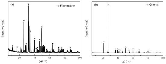
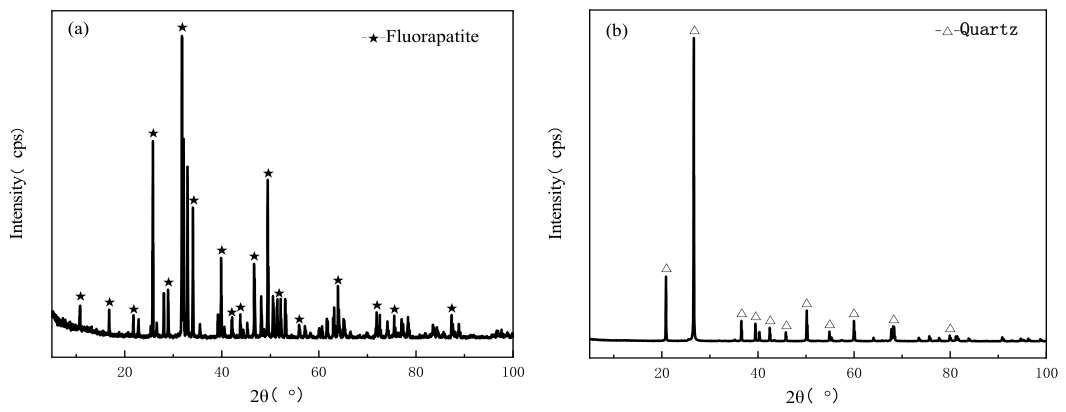
The chemical compositions of the apatite and quartz samples determined by XRF are summarized in Table 1 and Table 2. To enhance the reliability and reproducibility of elemental analysis, the samples were prepared using the fused-bead method instead of direct powder measurement. The phase compositions of the samples were identified by quantitative XRD analysis, with the corresponding diffraction patterns presented in Figure 1 and Figure 2. The results of XRF and quantitative XRD analyses confirmed that the purities of apatite and quartz samples were 93.51% and 96.17%, respectively, indicating that the prepared mineral samples were suitable for subsequent flotation and surface interaction studies. To prepare the XG–Fe3+ complex, XG powder (Aladdin, Shanghai, China) was dissolved in deionized water under stirring to obtain a homogeneous solution with a concentration of 2 wt%. Thereafter, a 10 wt% FeCl3·6H2O solution (Xilong Chemical Co., Ltd., Guangzhou, China) was introduced, and the mixture, at a mass ratio of 1:9 (XG and FeCl3·6H2O), was stirred for 2 h at room temperature. Dodecylamine (DDA, Aladdin, Shanghai, China) was selected as the flotation collector. Due to its limited solubility in water, the DDA solution was prepared through protonation with acetic acid. Briefly, DDA was dissolved in deionized water with an equimolar quantity of acetic acid (1:1 molar ratio), and the solution was stirred at 40 °C until complete dissolution was achieved. Analytical-grade NaOH and HCl solutions were used to adjust the pH of the mineral suspension when required. All experiments were conducted using ultrapure water.
Table 1.
Chemical composition of apatite wt%.
Table 2.
Chemical composition of quartz wt%.
Figure 1.
XRD patterns of apatite (a) and quartz (b).
Figure 2.
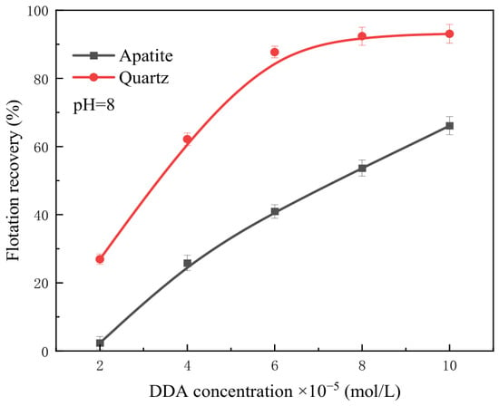
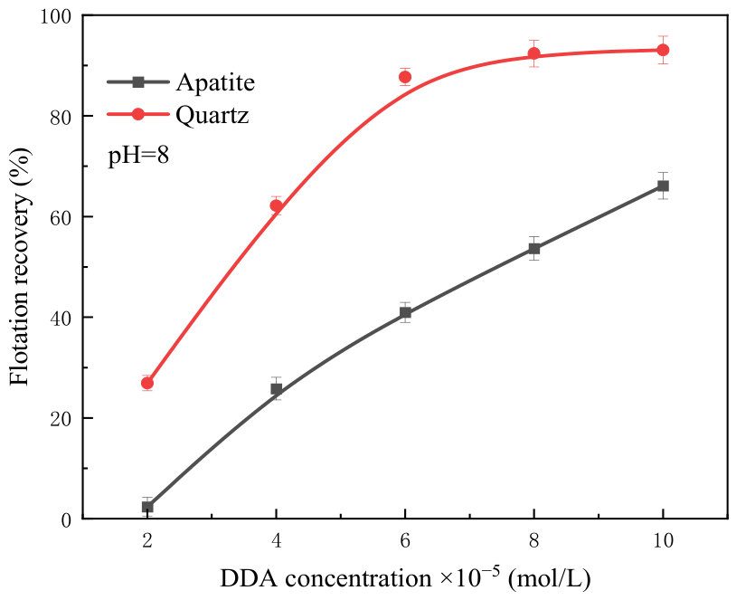
Effect of DDA concentration on flotation recovery of apatite and quartz.
2.2. Methods
2.2.1. Micro-Flotation
Micro-flotation experiments were conducted using an XFG-II flotation machine equipped with a 40 mL plexiglass cell (Wuhan Exploration Machinery Plant, Wuhan, China). Prior to flotation, 2.0 g of mineral samples were dispersed in 40 mL of ultrapure water using an ultrasonic cleaner (KQ300E, 40 kHz, 150 W, Kunshan Ultrasonic Instruments Co., Ltd., Kunshan, China) for 5 min to minimize particle aggregation and ensure a homogeneous suspension. The prepared slurry was then transferred into the flotation cell, and the impeller speed was maintained at 1800 rpm throughout the experiments. After adjusting the slurry pH to the desired value, XG–Fe3+ and DDA were sequentially added into the suspension. The conditioning times for the pH modifier, XG–Fe3+, and DDA were set at 2, 3, and 1 min, respectively. Subsequently, flotation was carried out for 3 min under an airflow rate of 0.1 L/min.
For the flotation tests of artificial mixed minerals, a binary mixture of apatite and quartz with a mass ratio of 1:1 (1.0 g of each mineral) was prepared, and the same experimental procedure was adopted. After flotation, the froth and tailing products were separately collected, filtered, dried, and weighed. The P2O5 content in the concentrate was determined, and the corresponding recovery was calculated based on the assay results.
2.2.2. Turbidity Analysis
Turbidity measurements were performed using a WZB-4000 turbidimeter (LeiCi, Shanghai INESA Scientific Instrument Co., Ltd., Shanghai, China) equipped with a cylindrical measuring cell (28 mm × 70 mm). Briefly, 2.0 g of mineral samples were first dispersed in 100 mL of ultrapure water by ultrasonic treatment to obtain a uniform suspension. Subsequently, a predetermined amount of XG–Fe3+ was introduced into the slurry, followed by sufficient conditioning. The treated suspensions were then transferred into 150 mL graduated cylinders and allowed to settle for 3 min. After sedimentation, 30 mL of the supernatant was carefully withdrawn for turbidity analysis [27].
2.2.3. Microscopic Observation
The formation and morphological features of mineral flocs were characterized using an XP-500E polarizing microscope (Shanghai Changfang Optical Instrument Co., Ltd., Shanghai, China) coupled with a digital camera. After treatment with XG–Fe3+, the apatite and quartz suspensions were deposited onto glass slides, followed by gentle placement of a coverslip to immobilize the samples for microscopic observation. Representative micrographs were collected to compare the aggregation states of different mineral particles.
2.2.4. Contact Angle Measurement
Contact angle measurements were performed using a goniometer (JC2000, Zhongchen Digital Technology Equipment Co., Ltd., Shanghai, China) via the sessile drop technique at an ambient temperature (25–30 °C), following the procedure described in our previous work [28]. All measurements were repeated in triplicate, and the average values were reported. To evaluate the wettability of reagent-treated mineral surfaces, the prepared samples were immersed in solutions containing XG–Fe3+ and/or DDA at pH 8 for approximately 2 min prior to measurement.
2.2.5. Zeta Potential Measurement
Zeta potential measurements were carried out using a Zetasizer Nano ZS90 (Malvern Panalytical, Malvern, UK). In each measurement, 0.05 g of mineral sample was ultrasonically dispersed in 80 mL of 0.001 mol/L KCl solution. Subsequently, predetermined amounts of XG–Fe3+ and DDA were sequentially added to the suspension, with conditioning times of 3 min and 2 min, respectively. After conditioning, approximately 1 mL of the suspension was extracted and transferred into the sample cell for zeta potential analysis. All measurements were performed at least in triplicate, and the reported values represent the mean of replicate determinations.
2.2.6. FTIR Analysis
Fourier Transform Infrared (FTIR) spectra were acquired using an FTIR-740 spectrometer (Nicolet Instrument Corp., Madison, WI, USA) via the standard KBr pellet technique. Mineral samples (apatite or quartz) were first conditioned in DDA solution at pH 8, either in the presence or absence of XG–Fe3+. Following 5 min of agitation, the suspensions were centrifuged, and the solid residues were washed three times with distilled water. The washed samples were then dried under vacuum freeze-drying conditions prior to spectral acquisition. For each measurement, 1.0 mg of the dried mineral sample was thoroughly mixed with 100 mg of KBr, and the resulting mixture was pressed into a pellet (8 mm in diameter) under a pressure of 10 MPa. Background spectra of pure KBr were recorded beforehand for baseline correction.
3. Results and Discussion
3.1. Single Mineral Flotation
3.1.1. Effect of DDA Concentration on Single Mineral Flotation Performance
The effect of DDA concentration on the flotation recovery of apatite and quartz is illustrated in Figure 2. With increasing DDA concentration, more collector was adsorbed onto the mineral surfaces, leading to a corresponding increase in the flotation recovery of both minerals. Notably, when the DDA concentration reached 6 × 10⁻⁵ mol/L, the disparity in flotation recovery between apatite and quartz attained its maximum, with the recovery of quartz exceeding that of apatite by 41.27%. Throughout the investigated DDA concentration range, the flotation recovery of quartz was consistently higher than that of apatite, indicating that DDA is more readily adsorbed onto the quartz surface. This finding is consistent with the observations reported by Li et al. [29].
3.1.2. Effect of XG-Fe3+ Concentration on Single Mineral Flotation Performance
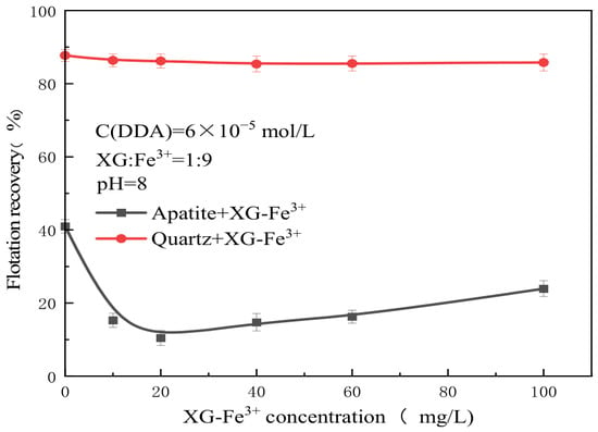
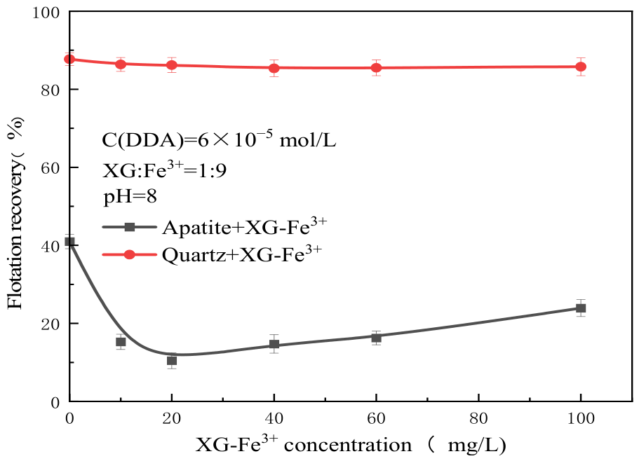
Preliminary experiments revealed that xanthan gum (XG) alone exhibited limited selectivity in the ultrafine apatite-quartz flotation system. Drawing on the documented synergy between metal ions and polymeric depressants [30,31,32,33], we tested the XG–Fe3+ combination and observed optimal separation performance at an XG-to-FeCl3·H2O mass ratio of 1:9. Based on this finding, the influence of the XG–Fe3+ concentration on the flotation behavior of ultrafine apatite and quartz was systematically examined, and the results are presented in Figure 3. As the XG–Fe3+ concentration increased, the flotation recovery of quartz remained essentially unchanged. In contrast, the recovery of apatite initially decreased and then increased, reaching a minimum at an XG–Fe3+ concentration of 20 mg/L, where the flotation recovery of apatite was 10.45%. In the absence of XG–Fe3+, the difference in flotation recovery between apatite and quartz was 41.27%. After the addition of XG–Fe3+, this difference in flotation recovery increased to 66.9%, indicating that the introduction of XG–Fe3+ expanded the flotation recovery difference between apatite and quartz. Thus, XG-Fe3+ may selectively adsorb onto the apatite surface, thereby depressing its flotation.
Figure 3.
Effect of XG-Fe3+ concentration on flotation recovery of apatite and quartz.
3.1.3. Effect of pH on Single Mineral Flotation Performance
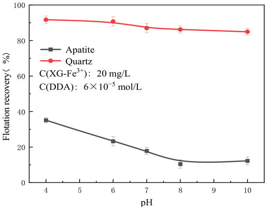
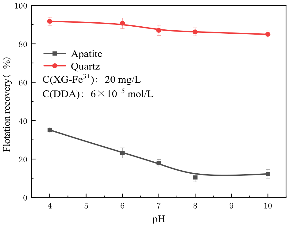
Figure 4 shows the effect of flotation pH on the flotation recovery of apatite and quartz. It was observed that within the investigated pH range, the flotation recovery of quartz remained largely constant with increasing pH. In contrast, the flotation recovery of apatite initially decreased and then remained nearly constant. At pH 8, the flotation recovery of apatite was 11.91%, and the difference in flotation recovery between the two minerals reached its maximum, indicating the most favorable separation performance at this pH. Therefore, a pH of 8 was selected for all subsequent experiments.
Figure 4.
Effect of pH on flotation recovery of apatite and quartz.
3.2. Artificial Mixed-Mineral Flotation
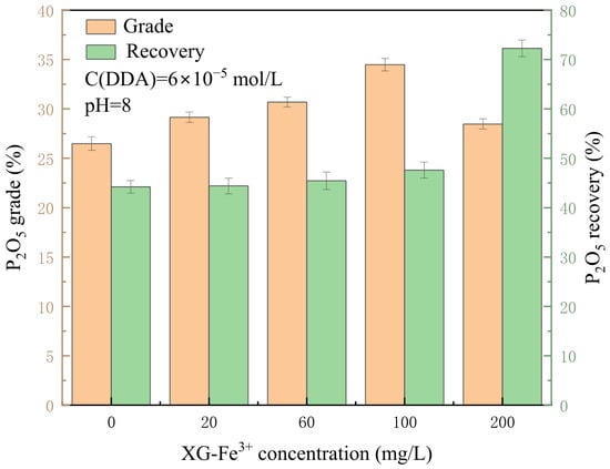
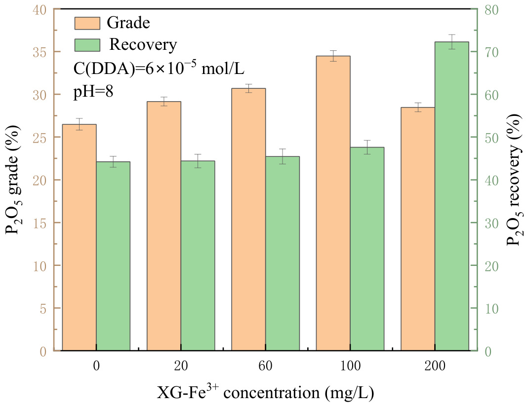
The influence of XG–Fe3+ concentration on the flotation performance of artificial apatite–quartz mixtures is illustrated in Figure 5. In the absence of XG–Fe3+, the flotation separation exhibited relatively poor selectivity, with the concentrate achieving a P2O5 grade and recovery of only 26.49% and 44.21%, respectively. This is consistent with the findings of Liu et al., who reported that DDA exhibits poor flotation performance in the separation of apatite from quartz [34]. After treatment with XG-Fe3+, the P2O5 grade of the concentrate first increased and then decreased with increasing XG-Fe3+ concentration, reaching a maximum at an XG-Fe3+ concentration of 100 mg/L. At this concentration, the grade of P2O5 in the concentrate was 34.48%, with a corresponding P2O5 recovery of 47.61%. At an XG-Fe3+ concentration of 200 mg/L, the grade of P2O5 in the concentrate was 28.47%, with a P2O5 recovery of 72.27%. The addition of 200 mg/L XG-Fe3+ increased the concentrate grade by 1.98% while simultaneously increasing the recovery by 28.06%. These results indicate that XG-Fe3+ can selectively depress the flotation of apatite in the flotation of the artificial mixed minerals, which is consistent with the single mineral flotation results.
Figure 5.
Effect of XG-Fe3+ concentration on the flotation of artificial mixed minerals.
3.3. Selective Flocculation of Apatite Evidenced by Turbidity Analysis
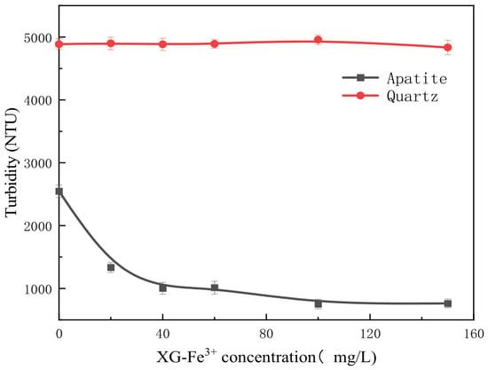
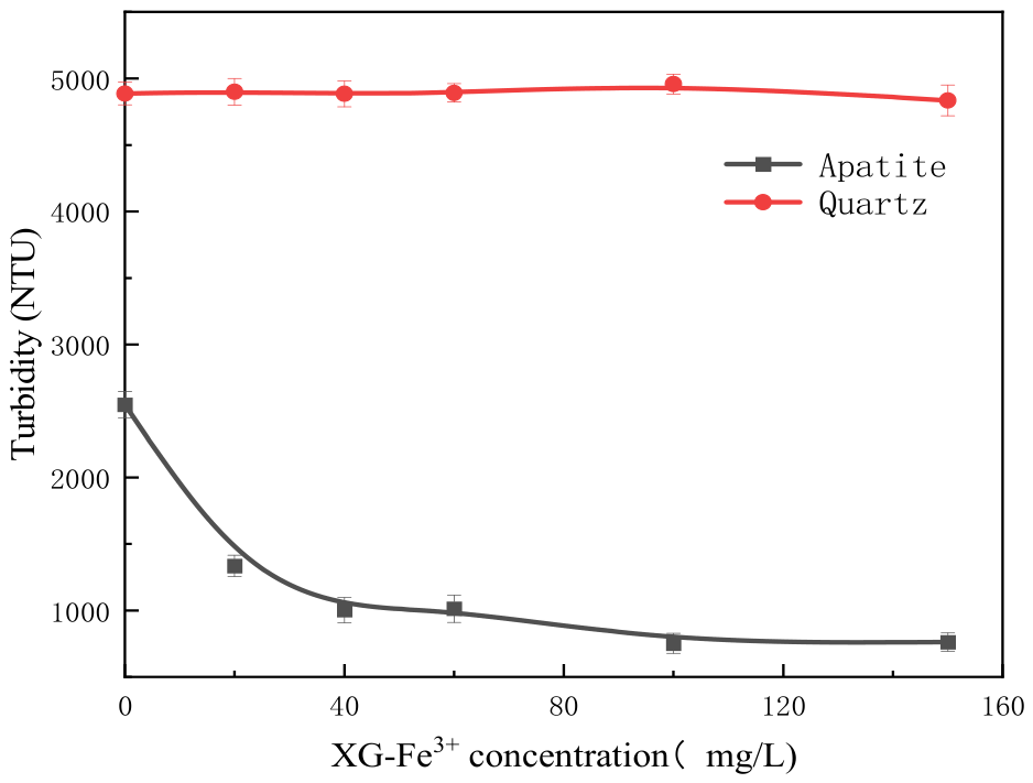
Turbidity analysis serves as an effective indicator of particle flocculation, as a reduction in suspension turbidity directly reflects the aggregation of fine particles into larger flocs, which settle more rapidly under gravity [35]. Figure 6 shows the turbidity of apatite and quartz suspensions under the action of different concentrations of XG-Fe3+. As the concentration of XG-Fe3+ increased, XG-Fe3+ adsorbed only weakly onto the quartz surface. Therefore, the turbidity of the quartz suspension changed only slightly. In contrast, the turbidity of the apatite suspension decreased significantly with increasing XG-Fe3+ concentration, indicating that XG-Fe3+ could selectively adsorb onto the apatite surface and flocculate ultrafine apatite, increase the apparent particle size of apatite, and make it more prone to settling.
Figure 6.
Effect of XG-Fe3+ concentration on turbidity of quartz and apatite suspension.
3.4. Selective Flocculation of Apatite Evidenced by Microscopic Observation
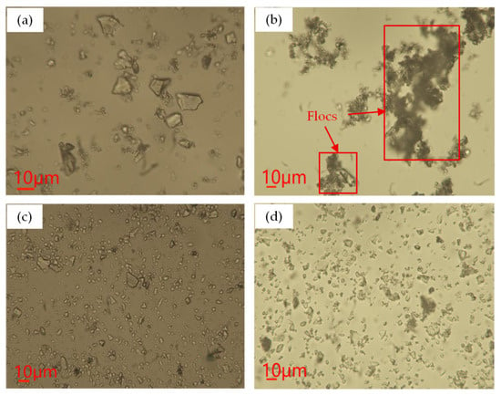
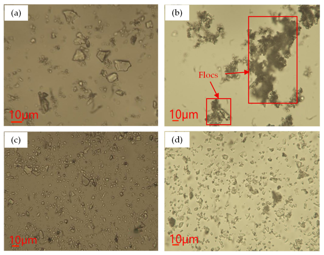
Microscopy images of apatite and quartz untreated and treated with 100 mg/L XG-Fe3+ are presented in Figure 7. A comparison between Figure 7a,b revealed that apatite formed larger flocs after treatment with XG-Fe3+, clearly indicating that ultrafine apatite particles could be flocculated together in the presence of XG-Fe3+. The floc morphology in Figure 7b is similar to that reported by Song et al. [36]. A comparison of Figure 7c,d showed that quartz remained dispersed both before and after treatment with XG-Fe3+. Microscopic observation confirmed that the addition of XG-Fe3+ induced selective flocculation of apatite. This finding is consistent with the turbidity measurements, which indicated an increase in apatite apparent particle size in the presence of XG-Fe3+. The findings from turbidity measurements and microscopic examination are in good agreement with the flotation results obtained for the artificial mixed minerals. Specifically, the flocculation of fine hydrophilic particles markedly mitigated their mechanical entrainment, which is a key factor governing the separation efficiency [19].
Figure 7.
Microscopic images of quartz and apatite with and without the treatment of XG-Fe3+: (a) apatite, (b) apatite+XG-Fe3+, (c) quartz, and (d) quartz+XG-Fe3+.
3.5. Effect of XG-Fe3+ on Wettability of Apatite and Quartz
Contact angle measurements were carried out to assess the surface hydrophobicity of apatite and quartz as a function of reagent treatment, with the results for apatite shown in Figure 8a–d. The untreated apatite surface exhibited a contact angle of 53.75°, a value lying between those reported by Wang et al. [37] and Liu et al. [38]. Upon treatment with XG-Fe3+ alone, the contact angle decreased to 32.75°, whereas treatment with DDA alone increased the contact angle to 73°. When apatite was treated with the combination of XG-Fe3+ and DDA, the contact angle was measured at 35.75°, which is close to that observed with XG-Fe3+ alone. The observed contact angle results are in good agreement with the micro-flotation data, indicating that demonstrating that the pre-adsorption of XG–Fe3+ effectively hinders DDA adsorption on apatite and thereby preserves its hydrophilicity.
Figure 8.
Contact angles of apatite under different conditions: (a) apatite, (b) apatite+XG-Fe3+, (c) apatite+DDA, (d) apatite+XG-Fe3++DDA.
The contact angles of quartz under different reagent conditions are presented in Figure 9a–d. The untreated quartz surface exhibited a contact angle of 49.25°, which falls within the range of 35.50° to 63.75° reported by Can et al. for the {1010} and {1120} crystal faces, respectively [39]. Upon treatment with XG-Fe3+ alone, the contact angle decreased to 33.75°, whereas treatment with DDA alone increased the contact angle to 69.29°. When quartz was treated with a combination of XG-Fe3+ and DDA, the contact angle was measured at 71.00°, which was close to that observed with DDA alone. These contact angle results indicated that XG-Fe3+ adsorbed only weakly onto the quartz surface and did not hinder the adsorption of DDA, thereby rendering quartz hydrophobic. This finding was in good agreement with the micro-flotation results.
Figure 9.
Contact angles of quartz under different conditions: (a) quartz, (b) quartz+XG-Fe3+, (c) quartz+DDA, (d) quartz+XG-Fe3++DDA.
3.6. Effect of XG-Fe3+ on Zeta Potential of Apatite and Quartz
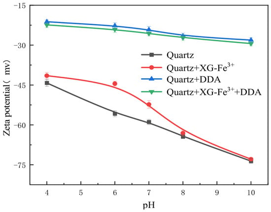
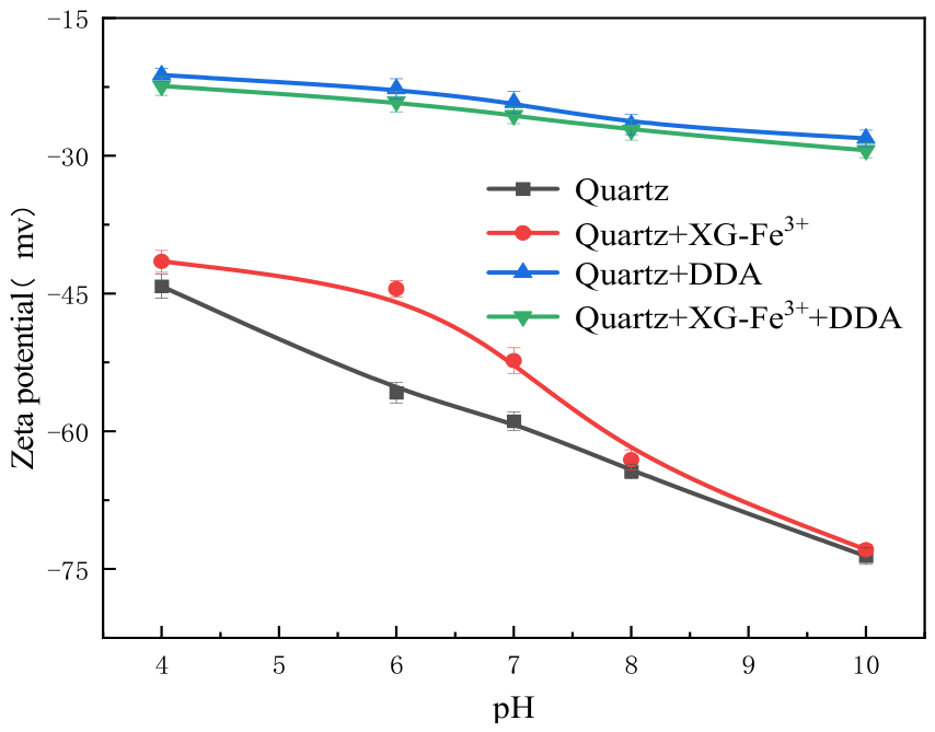
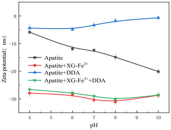
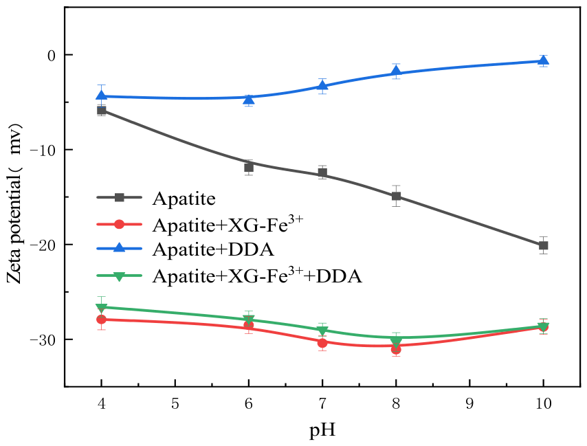
To elucidate the interfacial interactions between reagents and mineral surfaces, the zeta potentials of apatite and quartz were measured as a function of pH in both the absence and presence of various reagent systems, with the results presented in Figure 10 and Figure 11. For pristine quartz, the zeta potential exhibited a decreasing trend with increasing pH, which is consistent with previous observations reported by Sun et al. [40]. Upon the addition of XG–Fe3+, the zeta potential of quartz shifted toward more positive values across the pH range of 4–10, indicating adsorption of the XG–Fe3+ species onto the mineral surface. Notably, only marginal variations in zeta potential were detected between pH 8 and 10, suggesting relatively limited adsorption of XG–Fe3+ onto quartz under weakly alkaline conditions (e.g., pH 8). In contrast, the introduction of DDA resulted in a pronounced positive shift in the zeta potential of quartz over the entire pH range examined, implying a substantial adsorption affinity of DDA for the quartz surface. When XG–Fe3+ and DDA were sequentially added, the zeta potential curve of quartz closely resembled that obtained with DDA alone, indicating that the presence of XG–Fe3+ did not interfere with DDA adsorption on the quartz surface. This observation is consistent with the flotation and contact angle results, which showed that XG–Fe3+ exerted negligible influence on either the contact angle or flotation recovery when DDA was used as the collector.
Figure 10.
Zeta potential of quartz in the absence and presence of different reagents.
Figure 11.
Zeta potential of apatite in the absence and presence of different reagents.
The zeta potential of apatite under different conditions is shown in Figure 11. In the pH range of 4–10, the zeta potential of apatite gradually decreased with increasing pH, similar to that reported by Zhou et al. [41]. After treatment with XG-Fe3+ or DDA alone, the zeta potential shifted to varying degrees, indicating that both XG-Fe3+ and DDA were adsorbed onto the apatite surface. When XG-Fe3+ and DDA were added sequentially, the resulting zeta potential curve largely coincided with that of XG-Fe3+ alone, suggesting that XG-Fe3+ preferentially adsorbed onto the apatite surface, thereby hindering the adsorption of DDA. Therefore, the contact angle of apatite decreased, accompanied by a decline in its flotation recovery. The addition of XG-Fe3+ alone resulted in a negative shift in the overall zeta potential, which increased the electrostatic repulsion between particles and was unfavorable for particle sedimentation. This finding contradicts the flocculation observed in Figure 7. Given that XG-Fe3+ is a macromolecular reagent, it is inferred that XG-Fe3+ flocculates apatite via adsorption bridging.
3.7. FTIR Analysis of Reagent Adsorption on Apatite and Quartz Surfaces
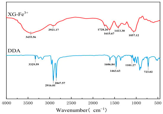
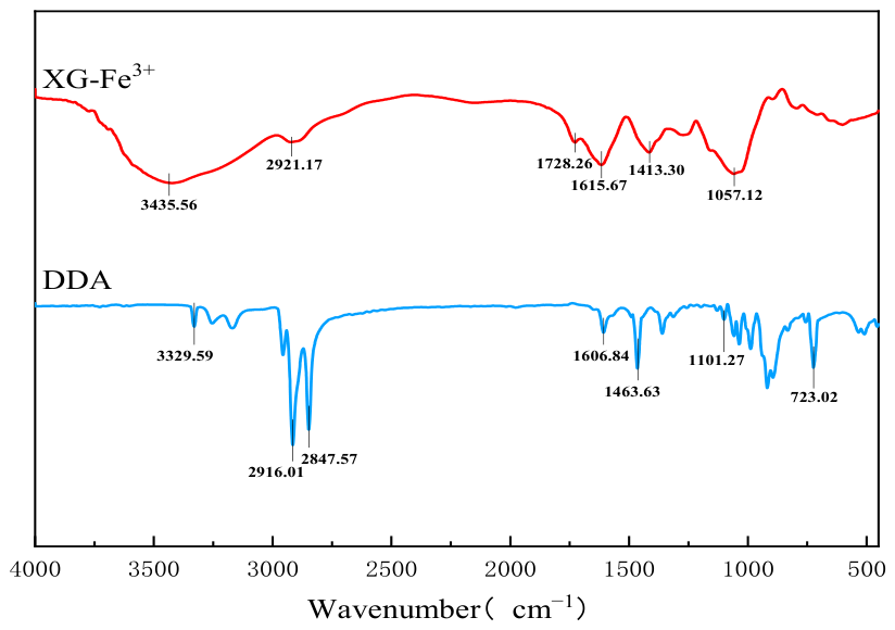
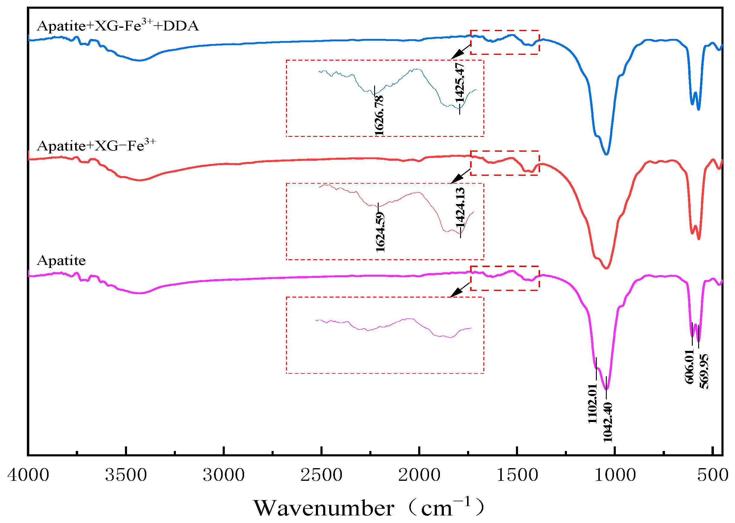
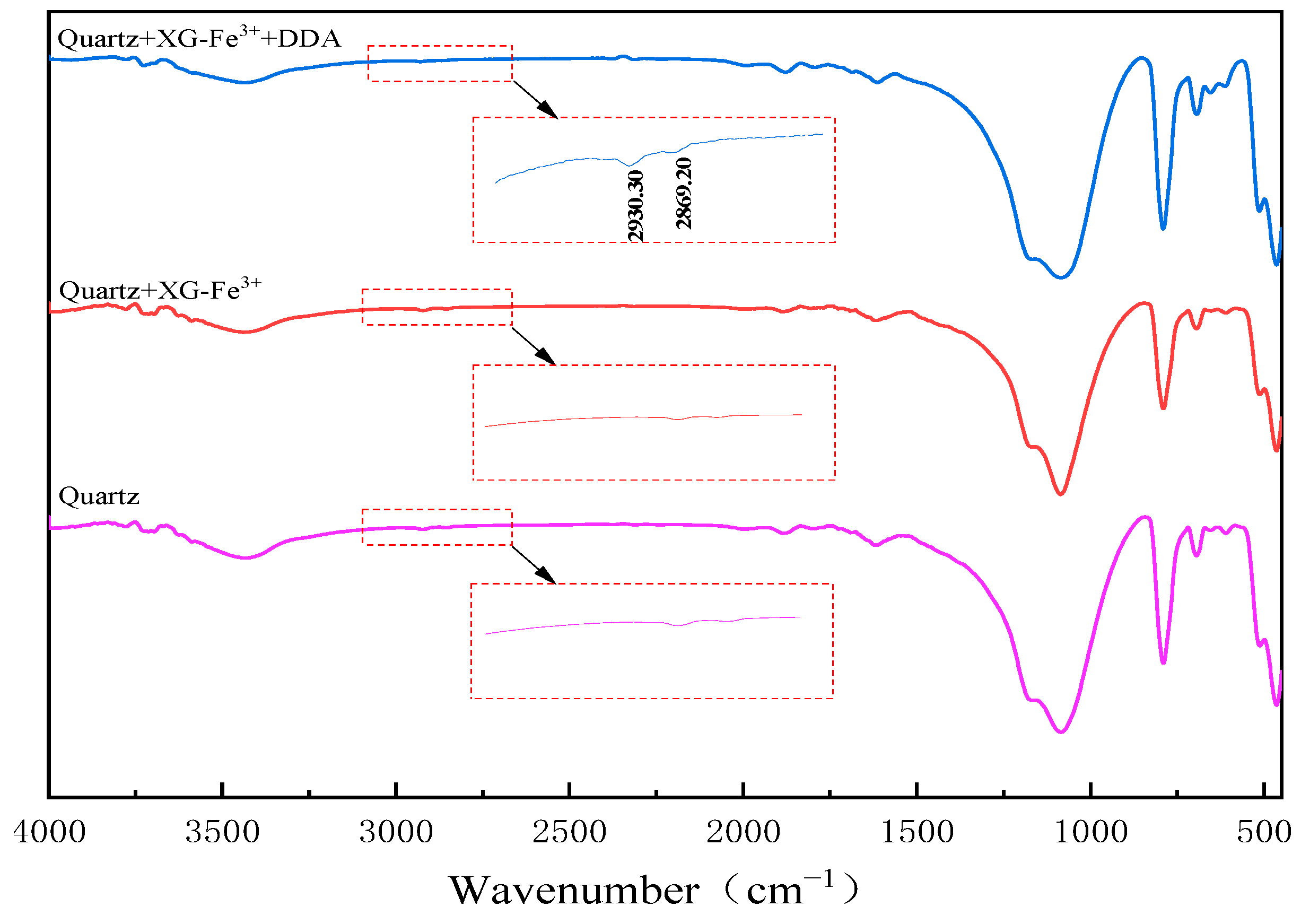
To elucidate the adsorption mechanism of the XG–Fe3+ complex, Fourier Transform Infrared (FTIR) spectroscopy was conducted on XG–Fe3+, DDA, minerals, and minerals treated with different reagents, and the results are presented in Figure 12, Figure 13 and Figure 14.
Figure 12.
FTIR spectra of XG-Fe3+ and DDA.
Figure 13.
FTIR spectra of apatite treated with various reagents.
Figure 14.
FTIR spectra of quartz treated with various reagents.
The FTIR spectra presented in Figure 12 reveal characteristic vibrational bands for both dodecylamine (DDA) and XG–Fe3+. For DDA, the absorption band at 3329 cm−1 corresponds to the N–H stretching vibration of the –NH2 group [42], while the band at 1606 cm−1 is assigned to the N–H in-plane bending mode [43]. The asymmetric and symmetric C–H stretching vibrations of the –CH2 and –CH3 groups appeared at 2916 and 2847 cm−1, respectively. The band at 1463 cm−1 is attributable to the –CH2 bending vibration, and the absorption at 1101 cm−1 arises from the C–N stretching vibration [29]. Additionally, the band at 723 cm−1 is characteristic of the in-plane rocking vibration of the long-chain aliphatic –(CH2)n– backbone. For the XG–Fe3+ complex, a broad and intense band centered at 3435 cm−1 is ascribed to O–H stretching vibrations, indicative of hydrogen bonding between xanthan gum molecules and associated water [44]. The band at 2921 cm−1 corresponds to the C–H stretching vibration of the –CH2– groups [45]. The absorption at 1728 cm−1 is attributed to the C=O stretching vibration of carboxylic acid groups, whereas the bands at 1615 and 1413 cm−1 are assigned to the asymmetric and symmetric stretching vibrations of the carboxylate (COO−) moiety, respectively. Furthermore, the strong band at 1057 cm−1 is associated with the C–O stretching vibration of the ether C–O–C linkage [46].
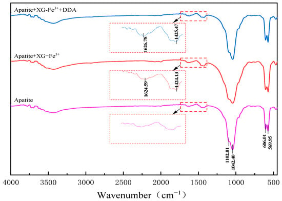
As shown in Figure 13, the characteristic peaks at 1102.01 cm−1 and 1042.40 cm−1 corresponded to the asymmetric stretching vibrations of PO43- in apatite, while the peaks at 606.01 cm−1 and 569.95 cm−1 were assigned to the bending vibrations of PO43− [47].
After treatment with XG–Fe3+, noticeable changes in the FTIR spectrum of apatite were observed. The C=O stretching vibration peak of the carboxyl group at 1728 cm−1 disappeared, while new peaks appeared at 1424.13 cm−1 and 1624.59 cm−1, which are attributed to the symmetric and asymmetric stretching vibrations of COO−, respectively [48,49]. According to the empirical criteria proposed by Deacon and Phillips, the coordination mode of carboxylate groups can be evaluated from the separation between the asymmetric and symmetric stretching vibrations of COO−. A Δν value greater than approximately 200 cm−1 was generally indicative of monodentate coordination [50]. Therefore, it could be inferred that the XG–Fe3+ complex underwent monodentate chemical adsorption at the calcium active sites on the apatite surface. After the sequential addition of XG–Fe3+ and DDA, the FTIR spectrum of apatite showed no significant difference compared to that treated with XG–Fe3+ alone, and no new characteristic peaks were observed. This indicates that the XG–Fe3+ complex effectively inhibited the adsorption of DDA on the apatite surface.
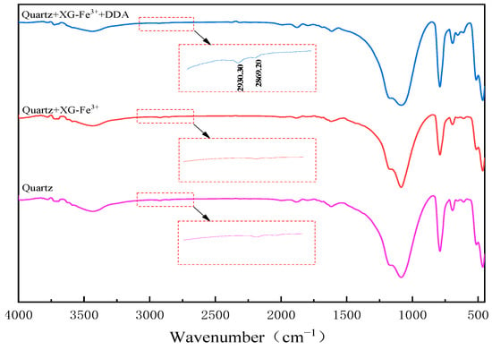
Figure 14 presents the FTIR spectra of quartz. After treatment with XG–Fe3+ alone, no noticeable changes were observed in the characteristic peaks of quartz. However, after sequential treatment with XG–Fe3+ and DDA, two new peaks appeared at 2930.30 cm−1 and 2869.20 cm−1, which were attributed to the stretching vibration of –CH2– and the bending vibration of –CH3, respectively [51]. This indicates that DDA was adsorbed onto the quartz surface. Meanwhile, no significant changes were observed in the peaks associated with Si–O stretching vibrations, suggesting that DDA adsorption on quartz was dominated by electrostatic physical adsorption rather than chemical interaction.
3.8. The Depression Mechanism
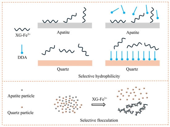
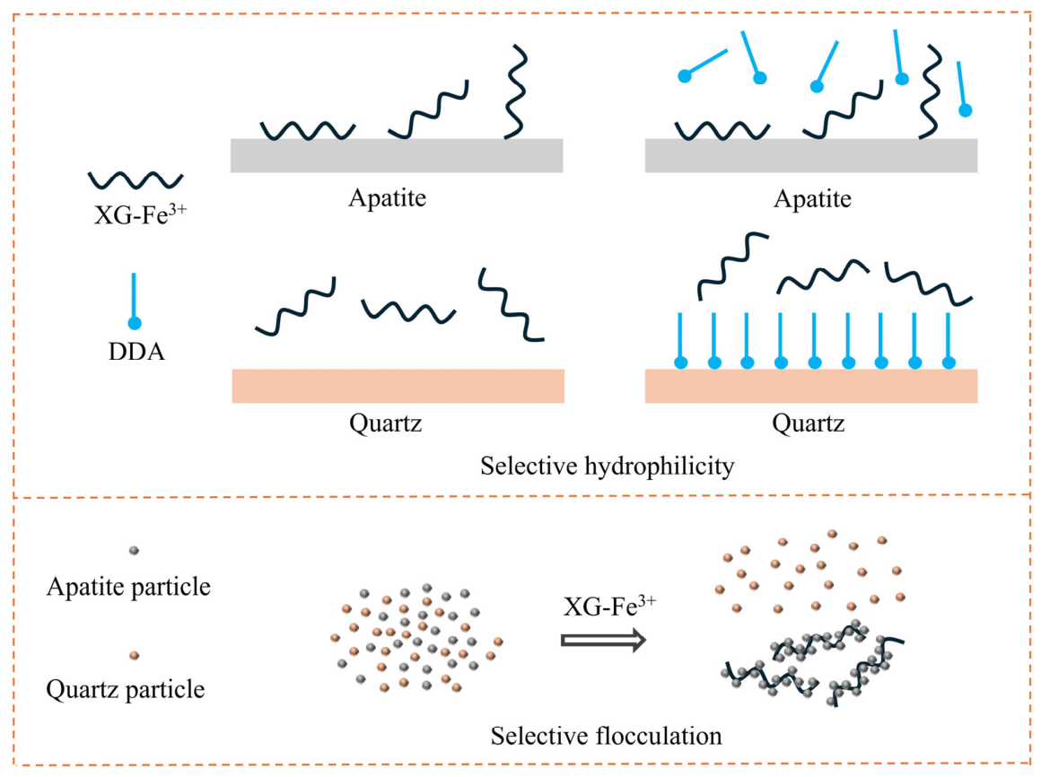
In view of the foregoing, a schematic diagram of depression mechanism of XG-Fe3+ on the flotation of ultrafine apatite from quartz at pH 8 is illustrated in Figure 15. XG-Fe3+ enhanced the separation of ultrafine apatite from quartz in two aspects. First, XG-Fe3+ chemically adsorbed on apatite, hindering the adsorption of DDA and maintaining the hydrophilicity of apatite. Furthermore, as a long-chain macromolecular reagent, XG–Fe3+ induced selective flocculation of ultrafine apatite through an adsorption bridging mechanism. This flocculation increased the apparent particle size of apatite, thereby reducing its mechanical entrainment into the froth phase during flotation of artificial mixed minerals. Consequently, the separation efficiency of ultrafine apatite from quartz was effectively enhanced. In contrast, XG–Fe3+ exhibited weak interaction with the quartz surface, and thus did not interfere with either the adsorption of DDA on quartz or the flotation response of quartz.
Figure 15.
Proposed mechanism of XG–Fe3+ in the flotation separation of ultrafine apatite from quartz.
4. Conclusions
To improve the reverse flotation separation of ultrafine apatite from quartz, XG-Fe3+ was introduced as a novel modifier. The flotation behavior of both minerals in the presence of XG-Fe3+ was systematically examined, and the underlying mechanisms were further elucidated.
- (1)
- At pH 8, the addition of XG–Fe3+ significantly enhanced the flotation separation of apatite from quartz. In single-mineral flotation tests, the flotation recovery difference between apatite and quartz increased from 41.27% to 66.9%. In artificial mixed-mineral flotation, the P2O5 grade of the concentrate improved from 26.49% to 34.48%, with the increase in P2O5 recovery by 3.40%.
- (2)
- The improved separation of ultrafine apatite from quartz by XG–Fe3+ occurs via two complementary mechanisms. First, XG–Fe3+ chemically adsorbed on the apatite surface, inhibiting DDA adsorption and preserving apatite hydrophilicity. Second, it induced selective flocculation of ultrafine apatite through adsorption bridging, reducing the mechanical entrainment of ultrafine apatite and thereby enhancing separation efficiency. In contrast, XG–Fe3+ exhibited minimal adsorption on quartz, leaving DDA adsorption and quartz flotation largely unaffected.
Author Contributions
Conceptualization, Z.L., W.X. and D.H.; methodology, Z.L. and G.L.; validation, Z.L., D.H. and Y.T. (Yun Tang); formal analysis, W.X., Y.T. and W.L.; investigation, H.L., Z.Z. and G.L.; resources, L.S.; data curation, W.X. and H.S.; writing—original draft preparation, H.L. and Z.Z.; writing—review and editing, Z.L., H.S. and Y.F.; visualization, Y.T. (Yuan Tang) and W.L.; supervision, H.S., Y.T. (Yun Tang) and Y.F.; project administration, L.S.; funding acquisition, Z.L. and D.H. All authors have read and agreed to the published version of the manuscript.
Funding
This research was funded by Scientific and Technological Innovation Platform Research Project of Guizhou Province (CXPTXM [2025] 016), Open foundation of State Key Laboratory of Efficient Utilization for Low-Grade Phosphate Rock and Its Associated Resources (WFKF(2023)005), Hubei Provincial Science and Technology Program (2024DJC089; 2025BCB065), The National Natural Science Foundation of China (No. 52204279), The Central Government’s Special Project for Supporting Local Science and Technology Development in Hubei Province (2025CSA015).
Data Availability Statement
Data is contained within the article.
Acknowledgments
We would like to express our gratitude to Postdoctoral Research Workstation of Wengfu (Group) Co., Ltd. for their support in this project.
Conflicts of Interest
Zhili Li received financial support from Wengfu (Group) Co., Ltd. for this study. The funding sponsors had no role in the design of the study; in the collection, analyses, or interpretation of data; in the writing of the manuscript; or in the decision to publish the results.
References
- Cordell, D.; Drangert, J.O.; White, S. The story of phosphorus: Global food security and food for thought. Glob. Environ. Change 2009, 19, 292–305. [Google Scholar] [CrossRef] [Scilit]
- Zou, T.; Zhang, X.; Davidson, E.A. Global trends of cropland phosphorus use and sustainability challenges. Nature 2022, 611, 81–87. [Google Scholar] [CrossRef] [Scilit]
- Vaccari, D.A. Phosphorus: A looming crisis. Sci. Am. 2009, 300, 54–59. [Google Scholar] [CrossRef] [Scilit]
- Farrokhpay, S.; Filippov, L.; Fornasiero, D. Flotation of fine particles: A review. Miner. Process. Extr. Metall. Rev. 2021, 42, 473–483. [Google Scholar] [CrossRef] [Scilit]
- Santana, C.R.; Duarte, R.C.; Ataíde, H.C.; Barrozo, M.A.S. Flotation selectivity of phosphate ore: Effects of particle size and reagent concentration. Sep. Sci. Technol. 2011, 46, 1511–1518. [Google Scholar] [CrossRef] [Scilit]
- Wills, B.A.; Finch, J.A. Wills’ Mineral Processing Technology; Butterworth-Heinemann: Oxford, UK, 2015. [Google Scholar]
- Fuerstenau, M.C.; Han, K.N. Principles of Mineral Processing; SME: Littleton, CO, USA, 2003. [Google Scholar]
- Chander, S.H. Reagents used in the flotation of phosphate ores: A critical review. Miner. Eng. 2003, 16, 577–585. [Google Scholar] [CrossRef] [Scilit]
- Yu, L.; Yu, P.; Bai, S.A. Critical review on the flotation reagents for phosphate ore beneficiation. Minerals 2024, 14, 828. [Google Scholar] [CrossRef] [Scilit]
- Talebi, E.; Maleki, H.; Langari, S.; Noaparast, M.; Shafaei, S.Z.; Jozanikohan, G. Comparative evaluation of different collector–depressant systems application in the reverse flotation of apatite and collophane from a sedimentary phosphate ore sample. Results Eng. 2026, 30, 110544. [Google Scholar] [CrossRef] [Scilit]
- Bulatovic, S.M. Use of organic polymers in the flotation of polymetallic ores: A review. Miner. Eng. 1999, 12, 341–354. [Google Scholar] [CrossRef] [Scilit]
- Ming, P.; Xie, Z.; Guan, Y.; Wang, Z.; Li, F.; Xing, Q. The effect of polysaccharide depressant xanthan gum on the flotation of arsenopyrite from chlorite. Miner. Eng. 2020, 157, 106551. [Google Scholar] [CrossRef] [Scilit]
- Beattie, D.A.; Huynh, L.; Kaggwa, G.B.; Ralston, J. Influence of adsorbed polysaccharides and polyacrylamides on talc flotation. Int. J. Miner. Process. 2005, 78, 238–249. [Google Scholar] [CrossRef] [Scilit]
- Xia, L.; Zhong, H.; Liu, G.; Wang, S. Utilization of soluble starch as a depressant for the reverse flotation of diaspore from kaolinite. Miner. Eng. 2009, 22, 560–565. [Google Scholar] [CrossRef] [Scilit]
- Laitinen, O.; Kemppainen, K.; Ammala, A.; Sirviö, J.A.; Liimatainen, H.; Niinimäki, J. Use of chemically modified nanocelluloses in flotation of hematite and quartz. Ind. Eng. Chem. Res. 2014, 53, 20092–20098. [Google Scholar] [CrossRef] [Scilit]
- Morris, E.G.; Fornasiero, D.; Ralston, J. Polymer depressants at the talc–water interface: Adsorption isotherm, microflotation and electrokinetic studies. Int. J. Miner. Process. 2002, 67, 211–227. [Google Scholar] [CrossRef] [Scilit]
- Evan, S.; Biesheuvel, P.M.; Wiebe, M. Adsorption of charged and neutral polymer chains on silica surfaces: The role of electrostatics, volume exclusion, and hydrogen bonding. Phys. Rev. E Stat. Nonlinear Soft Matter Phys. 2015, 91, 012601. [Google Scholar] [CrossRef] [Scilit]
- Xie, L.; Wang, J.; Huang, J.; Cui, X.; Wang, X.; Liu, Q.; Zhang, H.; Liu, Q.; Zeng, H. Anisotropic polymer adsorption on molybdenite basal and edge surfaces and interaction mechanism with air bubbles. Front. Chem. 2018, 6, 361. [Google Scholar] [CrossRef] [Scilit]
- Liu, Q.; Wannas, D.; Peng, Y. Exploiting the dual functions of polymer depressants in fine particle flotation. Int. J. Miner. Process. 2006, 80, 244–254. [Google Scholar] [CrossRef] [Scilit]
- Zhong, C.; Wang, H.; Feng, B.; Zhang, L.; Chen, Y.; Gao, Z. Flotation separation of scheelite and apatite by polysaccharide depressant xanthan gum. Miner. Eng. 2021, 170, 107045. [Google Scholar] [CrossRef] [Scilit]
- Guo, C.; Hou, S.; Wang, W.; Jin, H. Surface chemistry of xanthan gum interactions with bastnaesite and fluorite during flotation. Miner. Eng. 2022, 189, 107891. [Google Scholar] [CrossRef] [Scilit]
- Pan, G.; Shi, Q.; Zhang, G.; Huang, G. Selective depression of talc in chalcopyrite flotation by xanthan gum: Flotation response and adsorption mechanism. Colloids Surf. A Physicochem. Eng. Asp. 2020, 600, 124902. [Google Scholar] [CrossRef] [Scilit]
- Yan, H.; Yang, B.; Zeng, M.; Huang, P.; Teng, A. Selective flotation of Cu-Mo sulfides using xanthan gum as a novel depressant. Miner. Eng. 2020, 156, 106486. [Google Scholar] [CrossRef] [Scilit]
- Yue, T.; Wu, X. Depressing iron mineral by metallic-starch complex (MSC) in reverse flotation and its mechanism. Minerals 2018, 8, 85. [Google Scholar] [CrossRef] [Scilit]
- Mai, Q.; Zhou, H.; Ou, L. Flotation separation of chalcopyrite and talc using calcium ions and calcium lignosulfonate as a combined depressant. Metals 2021, 11, 651. [Google Scholar] [CrossRef] [Scilit]
- Wang, Q.; Yang, S.; Huang, L.; Liu, S.; Liu, C.; Xu, J. Research progress of application and interaction mechanism of polymers in mineral flotation: A review. Polymers 2024, 16, 3335. [Google Scholar] [CrossRef] [Scilit]
- Asgari, K.; Khoshdast, H.; Nakhaei, F.; Garmsiri, M.R.; Huang, Q.; Hassanzadeh, A. A review on floc-flotation of fine particles: Technological aspects, mechanisms, and future perspectives. Miner. Process. Extr. Metall. Rev. 2024, 45, 669–696. [Google Scholar] [CrossRef] [Scilit]
- Li, Z.; Rao, F.; Song, S.; Uribe-Salas, A.; López-Valdivieso, A. Effects of common ions on adsorption and flotation of malachite with salicylaldoxime. Colloids Surf. A Physicochem. Eng. Asp. 2019, 577, 421–428. [Google Scholar] [CrossRef] [Scilit]
- Li, X.; Zhang, Q.; Hou, B.; Ye, J.; Mao, S.; Li, X. Flotation separation of quartz from collophane using an amine collector and its adsorption mechanisms. Powder Technol. 2017, 318, 224–229. [Google Scholar] [CrossRef] [Scilit]
- Wang, R.; Sun, W.; Han, H.; Tao, D.; Zhang, H. New insight into the structure nature for Pb-starch colloidal depressant from experiments and DFT calculations. Colloids Surf. A Physicochem. Eng. Asp. 2023, 675, 132105. [Google Scholar] [CrossRef] [Scilit]
- Wang, R.; Sun, W.; Han, H.; Sun, W.; Wei, Z.; Peng, J. Al-caustic starch coordination compounds: A new depressant for fine calcite. Colloids Surf. A Physicochem. Eng. Asp. 2022, 648, 129268. [Google Scholar] [CrossRef] [Scilit]
- Fu, Y.; Zhu, Z.; Yao, J.; Han, H.; Yin, W.; Yang, B. Improved depression of talc in chalcopyrite flotation using a novel depressant combination of calcium ions and sodium lignosulfonate. Colloids Surf. A Physicochem. Eng. Asp. 2018, 558, 88–94. [Google Scholar] [CrossRef] [Scilit]
- Huang, S.; Huang, L.; Zuo, Q.; Shen, L.; Hu, B. Interfacial regulation of calcite by a Cu (II)-carboxymethyl cellulose synergistic depressant for selective flotation separation from cassiterite. Colloids Surf. A Physicochem. Eng. Asp. 2026, 749, 141124. [Google Scholar] [CrossRef] [Scilit]
- Liu, W.; Ge, R.; Guo, Y.; Xia, Y.; Zhao, S.; Liu, W.; Cui, B.; Shen, Y. Study on the performance and mechanism of novel hydroxyl quaternary ammonium collector for separation of quartz from apatite. J. Mol. Liq. 2025, 421, 126868. [Google Scholar] [CrossRef] [Scilit]
- Lv, J.; Zhang, C.; Li, J.; Qian, H. Selective flocculation mechanism of sodium oleate in flotation separation of cassiterite and calcite with cetylpyridinium bromide as a dispersant. Miner. Eng. 2026, 238, 110079. [Google Scholar] [CrossRef] [Scilit]
- Song, S.; Zhang, X.; Yang, B.; Lopez, A. Flotation of molybdenite fines as hydrophobic agglomerates. Sep. Purif. Technol. 2012, 98, 451–455. [Google Scholar] [CrossRef] [Scilit]
- Wang, T.; Feng, B.; Guo, Y.; Zhang, W.; Rao, Y.; Zhong, C.; Zhang, L.; Cheng, C.; Wang, H.; Luo, X. The flotation separation behavior of apatite from calcite using carboxymethyl chitosan as depressant. Miner. Eng. 2020, 159, 106635. [Google Scholar] [CrossRef] [Scilit]
- Liu, S.; Han, B.; Zhao, T. The effect of various surfactants on fatty acid for apatite flotation and their adsorption mechanizm. Physicochem. Probl. Miner. Process. 2021, 57, 46–56. [Google Scholar] [CrossRef] [Scilit]
- Gungoren, C.; Ozdemir, O.; Ozkan, S.G. Surface properties and beneficiation of quartz with flotation. Minerals 2025, 15, 846. [Google Scholar] [CrossRef] [Scilit]
- Sun, Y.; Yao, J.; Yin, W.; Sun, H.; Yang, S. Esterquat EQ-90 as a green novel collector for effective desilication in magnesite flotation: Adsorption mechanisms and selectivity. Miner. Eng. 2025, 232, 109543. [Google Scholar] [CrossRef] [Scilit]
- Zhou, F.; Liu, Q.; Liu, X.; Li, W.; Feng, J.; Chi, R. Surface electrical behaviors of apatite, dolomite, quartz, and phosphate ore. Front. Mater. 2020, 7, 35. [Google Scholar] [CrossRef] [Scilit]
- Yang, L.; Li, X.; Li, W.; Yan, X.; Zhang, H. Intensification of interfacial adsorption of dodecylamine onto quartz by ultrasonic method. Sep. Purif. Technol. 2019, 227, 115701. [Google Scholar] [CrossRef] [Scilit]
- Wang, X.; Liu, J.; Du, H.; Miller, J.D. States of adsorbed dodecyl amine and water at a silica surface as revealed by vibrational spectroscopy. Langmuir 2010, 26, 3407–3414. [Google Scholar] [CrossRef] [Scilit]
- Mudgil, D.; Barak, S.; Khatkar, B.S. X-ray diffraction, IR spectroscopy and thermal characterization of partially hydrolyzed guar gum. Int. J. Biol. Macromol. 2012, 50, 1035–1039. [Google Scholar] [CrossRef] [Scilit]
- Kacurakova, M.; Belton, P.S.; Wilson, R.H.; Hirsch, J.; Ebringerova, A. Hydration properties of xylan-type structures: An FTIR study of xylooligosaccharide. J. Sci. Food Agric. 1998, 77, 38–44. [Google Scholar] [CrossRef] [Scilit]
- Jin, R.; Hu, W.; Hou, X. Mechanism of selective flocculation of hematite quartz with hydrolyzed polyacrylamide. Colloids Surf. 1987, 26, 317–331. [Google Scholar] [CrossRef] [Scilit]
- Veiderma, M.; Knubovets, R.; Tonsuaadu, K. Structural properties of apatites from Finland studied by FTIR spectroscopy. Bull.-Geol. Soc. Finl. 1998, 70, 69–75. [Google Scholar] [CrossRef] [Scilit]
- Duczmal, K.; Darowska, M.; Raczyńska, D.E. Spectral (DFT-IR, FT-IR and UV) similarities and differences between substrate (pyruvate) and inhibitor (oxamate) of lactic dehydrogenase (LDH). Vib. Spectrosc. 2004, 37, 77–82. [Google Scholar] [CrossRef] [Scilit]
- Mittal, H.; Alili, A.A.; Morajkar, P.P.; Alhassan, S.M. Graphene oxide crosslinked hydrogel nanocomposites of xanthan gum for the adsorption of crystal violet dye. J. Mol. Liq. 2021, 323, 115034. [Google Scholar] [CrossRef] [Scilit]
- Sutton, C.C.R.; Silva, G.D.; Franks, G.V. Modeling the IR spectra of aqueous metal carboxylate complexes: Correlation between bonding geometry and stretching mode wavenumber shifts. Chemistry 2015, 21, 6801–6805. [Google Scholar] [CrossRef] [Scilit]
- Xu, H.; Zhong, H.; Tang, Q.; Wang, S.; Zhao, G.; Liu, G. A novel collector 2-ethyl-2-hexenoic hydroxamic acid: Flotation performance and adsorption mechanism to ilmenite. Appl. Surf. Sci. 2015, 353, 882–889. [Google Scholar] [CrossRef] [Scilit]





Disclaimer/Publisher’s Note: The statements, opinions and data contained in all publications are solely those of the individual author(s) and contributor(s) and not of MDPI and/or the editor(s). MDPI and/or the editor(s) disclaim responsibility for any injury to people or property resulting from any ideas, methods, instructions or products referred to in the content. |
© 2026 by the authors. Licensee MDPI, Basel, Switzerland. This article is an open access article distributed under the terms and conditions of the Creative Commons Attribution (CC BY) license.