


Abstract
Saltworks are anthropogenic environments providing important ecosystem services ranging from biodiversity support to ecotourism. The traditional management of small Mediterranean coastal saltworks is slowly declining. Consequently, there is a need to deepen the study of these active evaporitic basins and valorize the byproducts of the production cycle. In accordance with circular economy principles, these byproducts are traditionally reused as essential ingredients for the purification of edible salt. The study area is a small Mediterranean saltwork, “Chiusicella,” located within the Trapani–Paceco Saltworks Nature Reserve (Sicily, Italy). Sediments from various ponds and a waste material used by salt workers, locally known as “mamma caura”, were analyzed using X-ray diffraction (XRD) and Scanning Electron Microscopy coupled with Energy-Dispersive X-Ray Spectroscopy (SEM-EDS). Additionally, brines from high-salinity ponds were used to analyze the precipitation sequence following total evaporation. Results demonstrate a clear relationship between the stability fields of mineral phases typical of evaporitic environments and the pond typologies. Furthermore, the interaction and synergy between bacterial activity in the water and the precipitating mineral phases appear more evident, thus outlining a clearer profile of the role of byproduct reuse within the saltwork’s annual production cycle.
1. Introduction
Solar saltworks are anthropogenic ecosystems that provide a product of vital economic importance (NaCl, mineral halite) while offering significant ecosystem services, such as biodiversity maintenance, carbon sequestration, and ecotourism [1,2]. However, traditional small-scale saltworks in the Mediterranean have faced a sharp decline since the 1950s, leading to major shifts in salt production patterns. The higher efficiency of mechanized operations in industrial saltworks—better suited for large-scale production—has driven a trend toward increasing pond dimensions, with a consequent decrease in the number of saltworks, resulting in the progressive abandonment and economic failure of smaller, artisanal facilities [3,4]. This shift also led to the loss of traditional production practices, such as the use of the so-called ‘mamma caura’, a mixed-phase, fine-grained sediment derived from saltworks cultivation and traditionally seasonally reused. This dense, dark, and muddy byproduct is reused in artisanal salt cultivation processes, aligning this ancient production method with the principles of the circular economy. Indeed, in modern or large-scale industrial saltworks, this sediment is typically disregarded and excluded from the production cycle, unlike in artisanal salt making, which relies on the maintenance and preservation of traditional methods.
Mineral extraction in saltworks relies on two fundamental resources: solar energy, which drives evaporation, and seawater, the primary source of salts. Seawater contains a complex suite of dissolved ions and metals, with major components including Cl−, Na+, SO42−, Mg2+, Ca2+, K+, HCO3− and Br− [5]. Among these, the bromide anion (Br−) does not typically form its own crystalline phases during early evaporation but acts as a co-variant, substituting within the crystal lattice of halite by isomorphic replacement of chloride ion (Cl−) at advanced stages of evaporation [6,7]. Other cations, such as Sr2+, Fe2+, and Al3+, are minor components associated with rarer mineral phases [8,9]. Notably, many of these elements are classified as Critical Raw Materials (CRMs), representing strategic resources for the global energy transition [10]. The most common precipitates forming evaporite sequences are chlorides, sulfates, and calcium-magnesium carbonates. The three minerals constituting the bulk of evaporitic deposits are gypsum (CaSO4 · 2H2O), anhydrite (CaSO4), and halite (NaCl) [11]. K-Mg salts are rarer and precipitate at higher densities; these include sylvite (KCl), carnallite (KMgCl3 · 6H2O), langbeinite (K2Mg2(SO4)3), kainite (KMg(SO4)Cl · 3H2O), and polyhalite (K2Ca2Mg(SO4)4 · 2H2O) [12]. However, evaporitic crystallization is influenced by many factors: temperature and its variations, humidity, wind action and consequently evaporation and crystallization rate, depth of water, pH, local variations of the water composition [13], and biological activity [14,15]. Salt precipitation via evaporation is a polythermal process with daily and seasonal temperature fluctuations [13], and the precipitation mode is termed equilibrium crystallization when the salts remain in contact with the solution from which they precipitated [16]. The pioneering experiment on the complete evaporation of water (collected from the Mediterranean coast of France) at 40 °C to study the precipitation sequence was conducted by Usiglio (1849a,b) [17,18]. This study established a sequence inversely proportional to the solubility of the precipitated salts, known as the “Usiglio sequence”. Subsequent experiments and observations in marine saltworks [10,11,12,13,14,15,16,17,18,19,20,21] have repeated these findings, and all converge on the same results regarding the initial stages of precipitation:
- First stage: Characterized by the precipitation of calcium carbonate (CaCO3), whose stability field lies within the salinity range of approximately 35‰ to 140–200‰.
- Second stage: When salinity exceeds ≈200‰, calcium sulfate precipitates as gypsum (CaSO4 · 2H2O). Initially fine-grained, the crystal progressively coarsens; when individual crystals reach 2 mm in size, they are termed “selenites” [22].
- Third stage: The gypsum stability field terminates when salinity reaches approximately 290–325‰, marking the entry into the halite (NaCl) stability field.
Beyond these first three stages, the sequence enters the stability field of potassium and magnesium salts, where differences exist for slightly different conditions of evaporation and chemical composition of the waters. Many studies converge on the precipitation of epsomite (MgSO4 · 7H2O) as the salt following halite [23,24]. Valyashko (1962) [25] reports the following precipitation sequence from Black Sea waters: epsomite (which subsequently transforms into sakiite, MgSO4 · 6H2O), followed by carnallite (KMgCl3 · 6H2O), and finally, bischofite (MgCl2 · 6H2O) as the last phase. Furthermore, the study notes the presence of sylvite (KCl), which begins to precipitate with sakiite and disappears at the onset of carnallite precipitation. However, another study by Charuit and Genty (1980) [26] on Mediterranean bitterns at a temperature of approximately 28–35 °C reports a different sequence: epsomite alone, followed by epsomite with kainite (KMg(SO4)Cl · 3H2O), then kainite alone, and finally kainite associated with bischofite and/or carnallite. The theoretical evaporation of seawater proceeds until the so-called “eutonic point”, where the brine reaches an invariant composition and multiple solid phases co-crystallize in equilibrium [27,28]. The theoretical crystallization sequence was first established through solubility studies by Jacobus Henricus vant’Hoff and numerical calculations based on the Pitzer model [29,30,31,32]. While new models yielding different sequences have subsequently been proposed, one of the reference theoretical precipitation sequences at a constant temperature of 25 °C, reported by Braitsch and Kinsman (1978) [33], is as follows:
- Initial precipitation phases characterized by calcite, gypsum and halite;
- Precipitation of Mg-sulfates or Na-Mg sulfates (without potassium);
- Precipitation of Mg and K salts (specifically the sylvite field);
- Precipitation of carnallite;
- Precipitation of bischofite.
This study presents a characterization of the crystalline phases along the sediments of each pond and analyzes the crystalline phases that precipitate from the brines (known as bittern) after total evaporation in order to obtain a mineralogical characterization of the various environments and a precipitation sequence of the most soluble salts. The research focuses on the “Chiusicella” saltwork within the Trapani and Paceco Nature Reserve (Sicily, Italy), a site that still employs traditional harvesting methods. Another key objective of this work is the characterization of a strategic yet under-investigated byproduct locally known as “mamma caura”. Often described by traditional salt workers as the “salt yeast” or “mother lye”, this sludge is considered the essential ingredient for producing refined, high-purity salt for the food industry. While this material has been largely discarded in industrial-scale production, this study aims to scientifically validate its role as a natural bio-georeactor. We hypothesize that the synergistic action of the microbial communities and the specific mineral phases within the “mamma caura” allows it to adsorb and absorb impurities, effectively “cleaning” the final salt product. To achieve this, solid samples and sludge were subjected to a multi-analytical approach, including X-ray Diffraction (XRD), Scanning Electron Microscopy (SEM-EDS), and Raman Spectroscopy, to reconstruct the geochemical and mineralogical evolution across the saltern system.
2. Study Area
Saltpans, also called salinas or saltworks, are considered an anthropogenic ecosystem, and for this reason, they are well-known to a restricted fraction of the scientific community. Saltworks are part of the cultural heritage of the Mediterranean, but in addition to economic production, they play an important role in maintaining biodiversity [3].
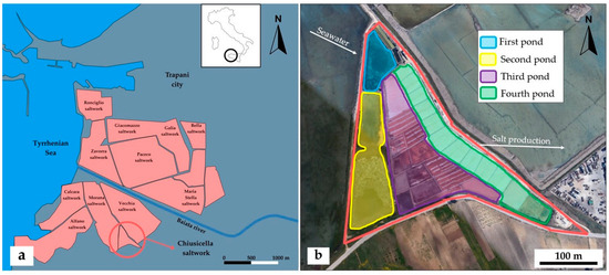
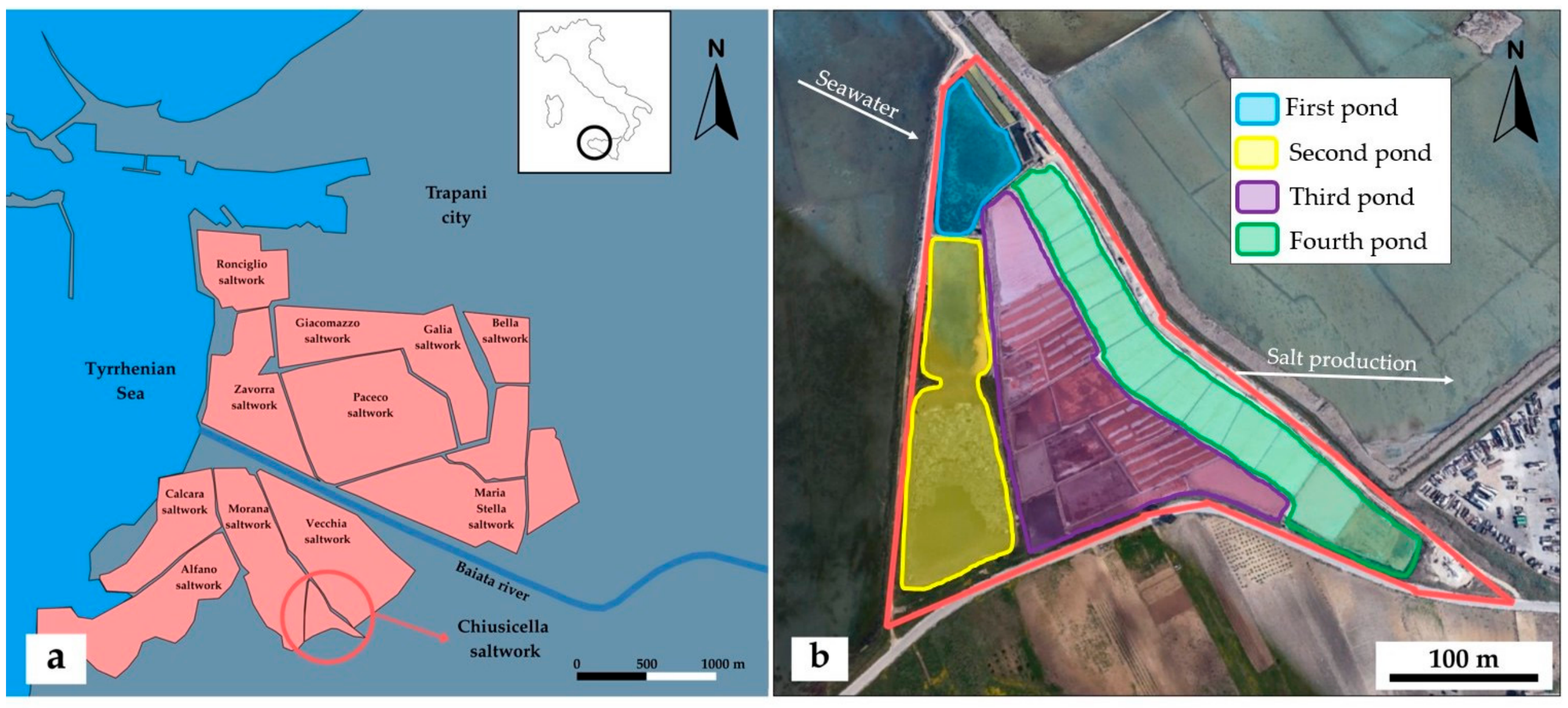
The samples were collected from the Chiusicella saltwork (also known as Salina Chiusa), located in the Nubia district of Paceco, Trapani, Sicily (Figure 1a). The site is situated within the “Saline di Trapani e Paceco” Nature Reserve, managed by the WWF. The traditional and little saltwork Chiusicella (Figure 1b) covers an area of ≈73.75 m2 and is dedicated exclusively to the production of food-grade salt. The primary objective of the saltwork management is to ensure the maximum recovery of high-purity halite (NaCl) through a controlled fractional crystallization process. The saltwork is organized into four distinct types of ponds, each characterized by a progressively increasing range of salinity. This interconnected system acts as a natural laboratory, mirroring the classic evaporitic sequence described by Usiglio (1849a,b) [17,18].
Figure 1.
(a) Simplified map of the major saltworks of the Nature Reserve located near Trapani city. The study area, Chiusicella saltwork, is the small environment in the red circle on the south shore of the Baiata river. (b) Satellite image of the Chiusicella saltwork, in which it is possible to distinguish the four typologies of ponds inside, differentiated by colors and the flow of water from the seawater to the salt production.
Every solar saltworks consists of a variable number of interconnected ponds. In the Chiusicella saltwork, as well as in other saltworks, each pond can be classified into four main categories (Figure 1b), sequentially arranged along an increasing density gradient. Brine density has been traditionally described by salt workers using an empirical measurement dating back to 1768, known as the Baumé scale. The measured values (°Bé) were converted to density (ρ, expressed in g/cm3) using the standard conversion formula for liquids heavier than water at the reference temperature of 16 °C: ρ = 145/(145 − °Bé) ([34,35] see Tables 1–11). Based on this non-linear conversion, the starting seawater concentration of 3.5 °Bé corresponds to an initial density of 1.025 g/cm3, while the halite saturation point at 26 °Bé corresponds to a density of 1.218 g/cm3.
Each type of pond corresponds to an evaporitic stage, and the brines become progressively denser from the first basin to the last pond. The terms “cold” and “warm” are purely empirical and traditional: salt workers historically observed that the initial ponds, containing mostly raw seawater, are generally cooler than the highly concentrated brines in the ponds immediately preceding halite precipitation. Hence, the distinction between the “cold pond” and the “warm pond” is fundamentally based on brine density. They are defined as follows:
- Cold pond: This is the initial basin where seawater is introduced via tidal action. These ponds are typically larger and deeper than the subsequent ones, with a density slightly higher than that of natural seawater, ranging from about 3.5 °Bé to 5 °Bé (≈1.025–1.036 g/cm3). For simplicity, from now on, this pond will be referred to as the first pond.
- Second pond: In this second stage, the salinity increases up to 12 °Bé (≈1.090 g/cm3).
Ecologically, these first two environments closely resemble coastal lagoons, hosting marine mollusks and gastropods; additionally, they are often utilized by locals for fish farming.
- 3.
- Warm pond: Here, the brine concentration reaches up to 26 °Bé (≈1.218 g/cm3), bringing the solution close to halite saturation. For simplicity, from now on, this pond will be referred to as the third pond.
- 4.
- Crystallizing pond: This is the final, shallowest basin where the food-grade salt precipitates and is ultimately harvested. For simplicity, from now on, this pond will be referred to as the fourth pond.
In the third and fourth ponds, compared to the first and second ponds, biodiversity decreases significantly due to hypersaline conditions and high temperature. The ecosystem hosts organisms adapted to these extreme conditions, such as unicellular green microalgae of the genus Dunaliella, particularly the species D. salina, which is characteristic of Mediterranean hypersaline environments and contributes to the water coloration ranging from pink to red due to the production of pigments such as carotenoids [36]. However, the water coloration is not entirely due to the production of secondary metabolites by algae, but primarily to halophilic bacterial communities and Archaea [37,38,39]. Prokaryotic diversity in highly saline ponds is dominated by extremely halophilic Archaea such as the genus Halobacterium (whose best-known species is H. salinarum) and Haloquadratum, as well as halophilic bacteria such as Salinibacter ruber and others of the genus Halomonas [40,41,42].
The salt cultivation cycle is annual, and the harvesting period varies locally between August and September. Salt workers explain that it is necessary to wait a few years from the start of cultivation for the saltworks to reach full capacity, which corresponds to the time required for the formation of “mamma caura”. At the end of each season, this sediment, which accumulates in the third ponds, is not discarded but rather preserved for use the following year. It is traditionally referred to as “salt yeast”, as its application acts as a biological primer that promotes the coloration of the brines. This coloration, in turn, increases the absorption of solar radiation, thereby accelerating evaporation. Furthermore, its use appears to be linked to the production of a cleaner and whiter artisanal salt. Finally, being a gypsum-rich sediment, it is also utilized to waterproof the ponds and their embankments.
3. Materials and Methods
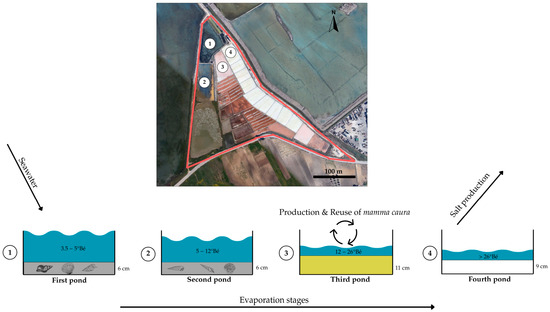
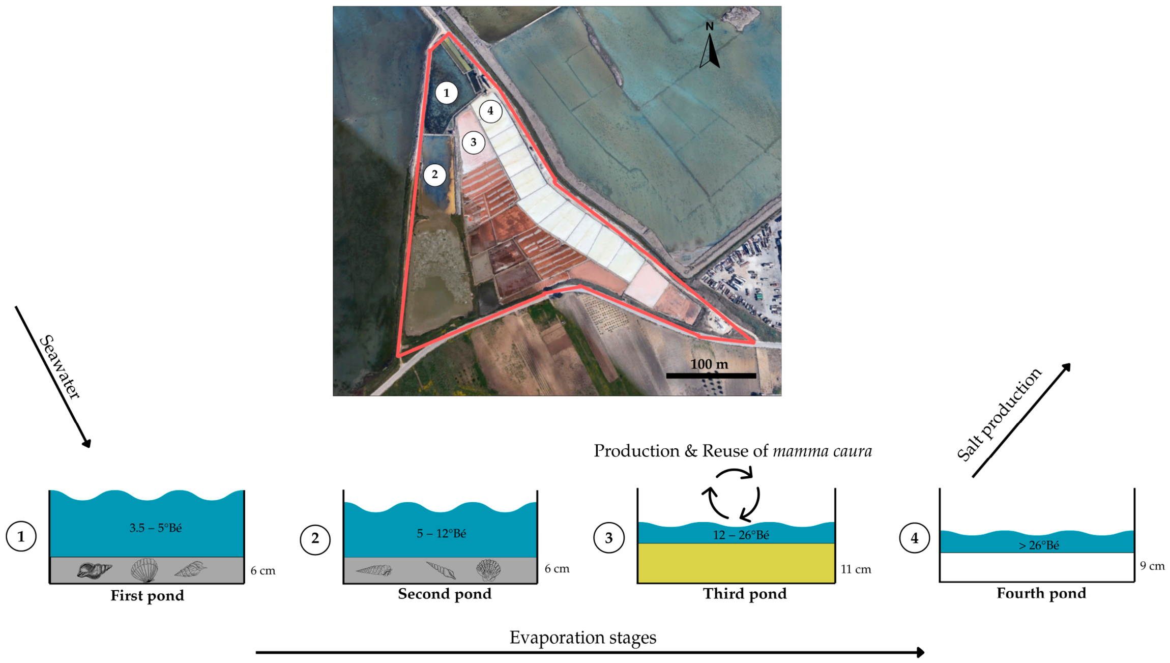
Samples were collected in September, at the end of the saltwork’s production cycle and during the salt harvesting phase. Sediment samples were collected from each pond of the Chiusicella saltwork (Figure 2). Sediment cores were collected using a custom-made PVC manual corer (inner diameter ≈ 10 cm). The coring tube was manually driven into the salt pan sediments. To ensure full recovery and prevent sample loss during retrieval, the top of the tube was hermetically capped to create a vacuum. Once extracted, the sediment core was upwardly extruded using a custom-fitted piston. The core was then carefully sectioned at 1 cm intervals using a caliper to ensure high depth resolution. Each slice was immediately transferred into individual, uniquely labeled containers for subsequent analysis. Solid samples were immediately refrigerated and transported to the Department of Earth and Marine Sciences of the University of Palermo (Sicily, Italy). Each sediment centimeter was labeled with the corresponding pond ID and depth range (i.e., the first centimeter was labeled 0–1, the second 1–2, and so on). A total of 33 solid samples were collected: 6 cm from the first pond, 6 cm from the second pond, 11 cm from the third pond, 9 cm from the fourth pond and 1 sample of mamma caura. Concurrently, 1 L brine samples in contact with the sediments were collected from the fourth and third ponds, stored in polypropylene containers, refrigerated, and transported to the research laboratories. The solid samples were subsequently oven-dried at 40 °C for 48 h to remove pore water. This type of treatment induces the precipitation of residual salts in all samples due to the increase in supersaturation during the evaporation of the water solvent. Therefore, a rigorous quantitative XRD analysis of the highly soluble crystalline phases is not possible, as it is affected by an overestimation of the concentrations of these salts. For this reason, the quantitative analysis for the first and second ponds was not considered reliable and was not performed. In the third pond, the analysis was carried out, but the overestimation (ranging between 0.54 and 3.24% as NaCl) of the more soluble phases due to evaporation effects must be considered. This overestimation can be quantified based on the known salinity of the water and the amount of water removed from the sample during the drying procedure. In the fourth pond, the overestimation is still present, but the quantitative analysis was not performed, as this pond was the most subject to anthropogenic intervention. All samples were promptly sent to the research laboratories of the Department of Earth Sciences at the University of Turin (Turin, Italy) for chemical and mineralogical analyses.
Figure 2.
Satellite image of the “Chiusicella saltwork” (red perimeter) in which the numbers indicate the points of sampling, and they correspond to the four typologies of ponds. The diagram below illustrates the water flow from seawater to salt harvesting (black arrows). In the third pond, the “mamma caura” is produced, which will subsequently be used in the next production cycle. Additionally, the thickness of the sampled sediments and the number of samples are reported: 6 in the first pond, 6 in the second, 11 in the third, and 9 in the fourth. Note that the water level in the pond represents brine density.
A 50 mL aliquot of the residual brine was transferred into a 50 mL flask containing a glass slide placed at the bottom. The solution was evaporated to dryness at a constant temperature of 50 °C. Upon complete evaporation, the glass slide featuring the precipitated crystals was recovered, coated with a thin gold layer, and analyzed using a Tescan Vega Scanning Electron Microscope (SEM) (TESCAN ORSAY HOLDING a.s., Brno, Czech Republic) equipped with Oxford Instruments Xplore 15 EDS microanalysis (Oxford Instruments, High Wycombe, UK).
For all solid samples, mineralogical identification was performed via X-ray powder diffraction (XRD). Samples were finely ground using an agate mortar and spiked with a known amount of metallic silicon as an internal standard. This standard was employed to perform full pattern fitting and to calibrate peak shift corrections, ensuring high precision in d-spacing measurements.
Data were acquired using a MiniFlex 600 diffractometer (Rigaku Corporation, Tokyo, Japan) (λ = 1.54 Å, Cu-Kα radiation) across a 2θ range of 2.0–100.0°, with a step size of 0.02° and a scanning speed of 0.6°/min. Phase identification was carried out using the Match! software (version 4.2) package [43] coupled with the Crystallography Open Database (COD) [44]. The Reference Intensity Ratio (RIR) method was selected for semi-quantitative analysis, as it proved more suitable than Rietveld refinement for these specific samples, which are characterized by complex multi-phase matrices (often exceeding four distinct mineral phases) and varying degrees of crystallinity and chemistry. Semi-quantitative analysis was performed only for the sediments of the third pond.
Morphological and micro-chemical investigations of the sediment were conducted by Scanning Electron Microscopy (TESCAN ORSAY HOLDING a.s., Brno, Czech Republic) equipped with an Energy-Dispersive X-ray Spectroscopy 230 detector (Oxford Instruments, High Wycombe, UK) (SEM-EDS).
To preserve the internal fabric and textural relationships and associated sediments, samples were embedded in epoxy resin (R&G Faserverbundwerkstoffe GmbH, Composite Technology, Waldenbuch, Germany). Once cured, they were sectioned and polished to expose a representative cross-section. Finally, the polished surfaces were carbon-coated to provide the necessary conductivity for high-resolution secondary electron imaging and quantitative EDS microanalysis.
Furthermore, Raman spectroscopy was employed to analyze the organic pigments present in the “mamma caura” sample. Measurements were carried out using a Horiba Jobin Yvon HR800 Raman spectrometer (Horiba Jobin Yvon, Longjumeau, France) equipped with grating (600 grooves/mm) and an air-cooled CCD detector (−70 °C). The excitation sources consisted of polarized lasers: a red HeNe laser (633 nm wavelength, 20 mW power) and a solid-state green Nd laser (532 nm wavelength, 250 mW power). The system was fitted with Edge filters for the 633 nm and 532 nm laser lines, along with a series of interference filters. The spectral resolution with the 600 grooves/mm grating was 1.5 cm−1 for the red laser. The spectrometer is coupled to an Olympus BX41 polarized light petrographic microscope (Olympus Corporation, Tokyo, Japan), configured for reflected light observations.
4. Results and Discussion
4.1. Mineralogical Characterization
4.1.1. First Pond and Second Pond
X-ray diffraction (XRD) analysis performed on sediments from the first two ponds (first and second, see Figure 2) reveals a qualitatively similar composition, dominated by a terrigenous fraction and carbonates, with mainly quantitative variations. This similarity reflects the nature of these environments, which act as a bridge between the coastal marine environment (aragonite from the shells), detrital fraction (sand and clay such as quartz and kaolinite), and the first stage of evaporation within the carbonate stability field (inorganic calcium carbonate such as calcite). Diffractograms for all sediment horizons from the first and second ponds are available in the Supplementary Information File (Figure S1).
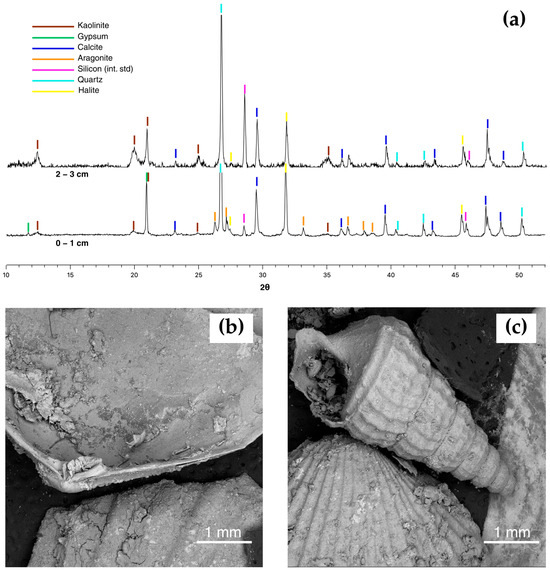
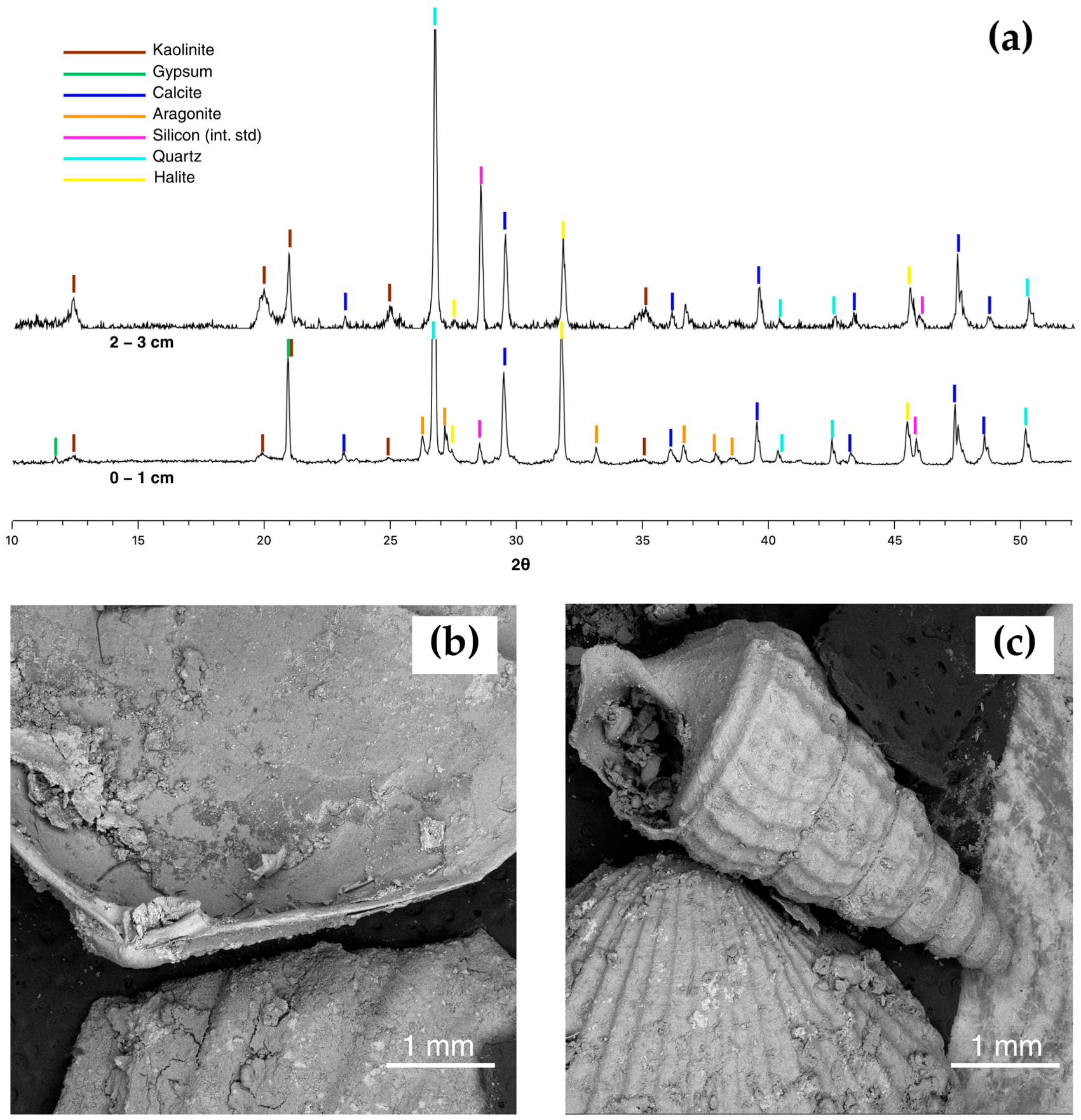
In the upper sediment layers of the first pond and second pond (0–1 and 1–2 cm), the coexistence of calcite and aragonite was detected. However, analysis of the vertical profiles highlights a distinctive trend: while calcite is persistent, aragonite tends to disappear in deeper layers (Figure 3a). This evidence is attributable to the predominantly biogenic origin of aragonite in this system, linked to the presence of bioclasts (fragments of gastropod and mollusk shells) that colonize the bottom of low-salinity ponds but are not present in the underlying horizons (Figure 3b,c).
Figure 3.
XRPD of the first centimeter of sediment from the first pond (bottom, 0–1) and the third centimeter of the same pond (top, 2–3), showing the identified phases. (a) The presence of aragonite (CaCO3, in orange) is evident in the first centimeter, whereas its main diagnostic peaks disappear from the third centimeter onwards. (b,c) SEM photomicrographs of gastropod and bivalve shells present in the first pond and second pond.
The silicoclastic component consists of quartz (sandy fraction) and phyllosilicates, such as kaolinite in this case. A rise in the background was also observed in the 12–20 Å range, probably indicating the presence of poorly crystalline smectites and disordered mixed-layer clays [45] (Figure S2). The origin of this detrital fraction is strictly connected to the geological setting of the area, characterized by the deposits of the Capo Plaia Synthem (Holocene), which constitutes the recent sedimentary cover of the Trapani and Paceco coastal plain [46]. Specifically, the saltworks are located on marshy and coastal plain deposits (AFL unit in the official geological cartography, specifically the “Xitta-Salina Grande” deposits), historically fed by the solid transport of the Baiata Stream and the local hydrographic system. These watercourses erode and transport coastalward the weathering products of the clayey-marly substrates of the hinterland (e.g., Terravecchia Formation), justifying the presence of clay minerals and quartz in the pond bottom sediments [46,47].
Finally, the presence of gypsum and halite does not represent primary precipitation from the water column (as the first ponds are still undersaturated) but is interpreted as secondary efflorescence resulting from the evaporation of interstitial water (pore water) during sample drying and preparation.
4.1.2. Third Pond and Fourth Pond
In the third pond and fourth pond, the physicochemical parameters of the brine shift significantly, entering the stability fields of sulfates and chlorides. The mineralogical assemblage of the third pond is primarily characterized by gypsum precipitation. However, a distinctive discontinuity is observed in the surface layer (0–1 cm), where halite prevails over sulfates. The presence of sodium chlorides at the very beginning of this sequence, theoretically reserved for calcium sulfates, stems from two fundamental factors. Firstly, changes in traditional saltworks management have led to the abandonment of an intermediate pond (historically known as Ruffiana) located between the second and the third pond. Its specific function was to monitor and regulate brine concentration through controlled dilution: by introducing less concentrated brines, this step prevented premature chloride supersaturation [48] before the final harvesting stage.
Secondly, the massive accumulation of halite in these horizons is a direct consequence of the brine density reaching and exceeding the halite saturation threshold (approx. 26 °Bé). In these advanced stages of evaporation, high-salinity conditions allow for the abundant crystallization of halite, which can synchronously co-precipitate with calcium sulfates. This co-precipitation is characteristic of natural highly evaporative environments, where specific chemical conditions allow multiple minerals to reach or maintain saturation simultaneously [49].
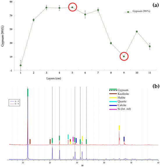
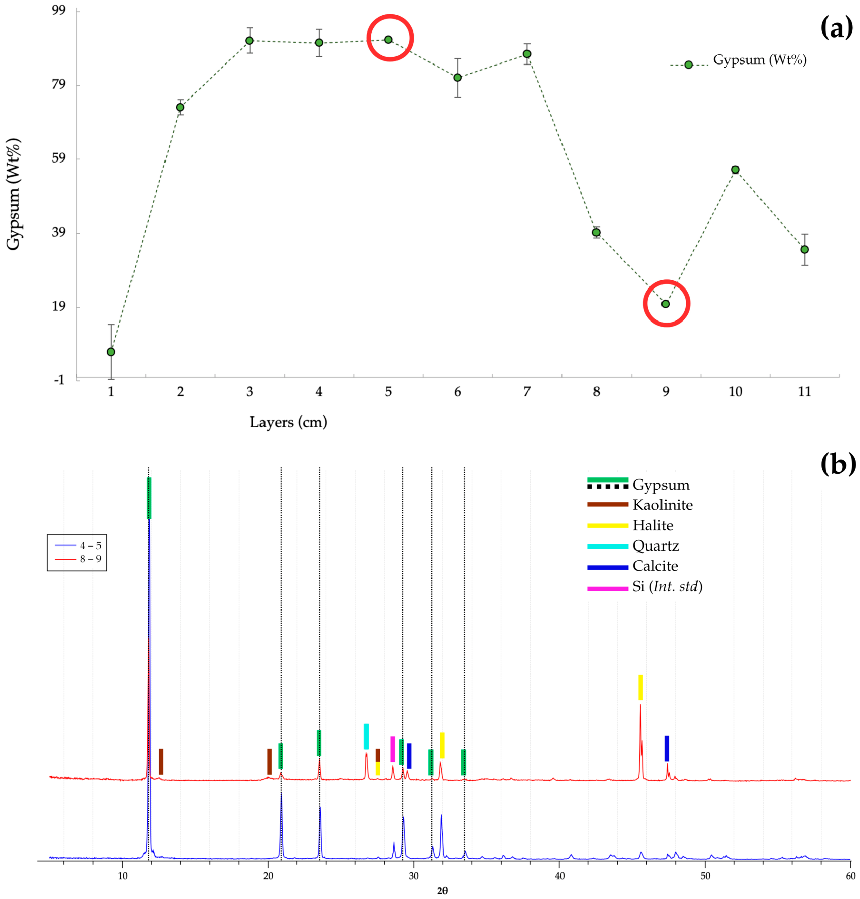
Below the surface crust, the mineralogical composition aligns with the expected evaporitic sequence. The gypsum content progressively increases with depth, reaching a peak concentration of approximately 91 wt% at the 5 cm horizon (Figure 4). Finally, a drastic shift occurs at the 8 cm depth, where the mineralogy reverts to the terrigenous-carbonates association typical of the initial ponds (see Section 4.1.1). This discontinuity marks the lithological boundary of the evaporitic sequence, between the 7th and 8th centimeters, identifying the horizon where the gypsum precipitation of the current annual production cycle began.
Figure 4.
(a) The graph displays the trend of gypsum (CaSO4 · 2H2O) weight percentage throughout the third pond sediments. A sharp discontinuity is observed between the 7th and 8th centimeters, marking the boundary between the pond’s authigenic precipitation and the allochthonous clayey and sandy substrate. Marked in red are the horizons, whose respective XRPD are shown below in (b). (b) XRPD of the 5th centimeter (in blue) and the 9th centimeter (in red) are shown. The peaks of the identified phases can be distinguished (by different colors shown in the legend), highlighting the progressive decrease in gypsum and the appearance of the other minerals, such as quartz, calcite, and kaolinite, in the deeper horizons.
All diffraction patterns of the third pond and their semi-quantitative analysis are reported in the Supplementary Material (Figure S3 and Table S1, respectively).
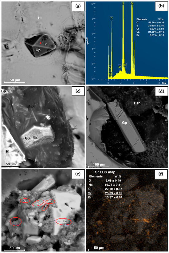
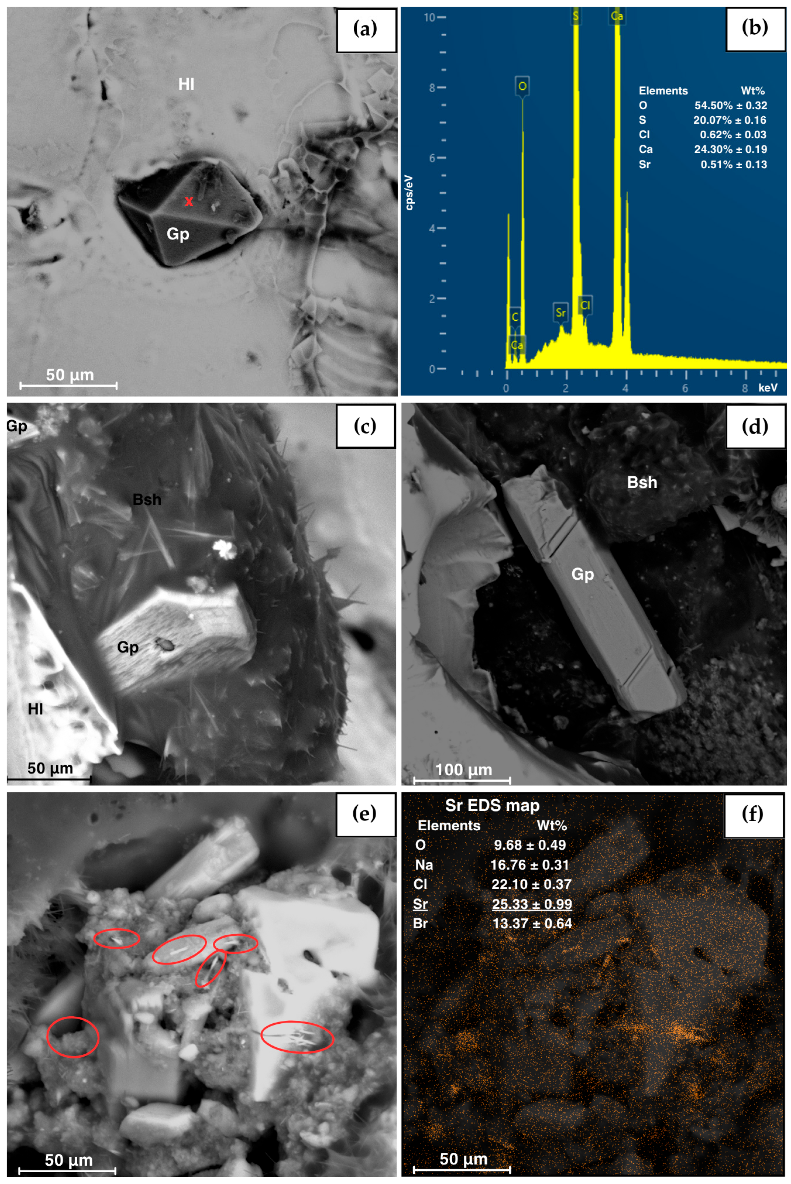
The SEM-EDS investigation focused on the surface layers (0–3 cm) of the third pond to refine the mineralogical characterization. While the analysis confirmed the dominant presence of halite and gypsum (Figure 5a,b), the occurrence of minor phases in the 2–3 cm interval that were not clearly detectable by X-ray diffraction due to their low abundance (below the detection limit) is also highlighted. The EDS maps revealed phases of strontium and magnesium chlorides. Given the evaporitic context and the exclusive presence of Mg and Cl signals (e.g., absence of K), this latter phase is interpreted as likely bischofite (MgCl2 · 6H2O) (Figure 5c,d). The strontium crystals exhibit an acicular to elongated morphology. While acicular celestite (SrSO4) has been documented in several studies [50,51], in high-salinity environments, this habit also suggests the presence of a hydrated precursor phase, such as SrSO4 · 0.5H2O [52], rather than the typical tabular anhydrous celestite. Given the analytical limits of EDS in determining hydration states, both phases remain possible. Furthermore, due to the weak sulfur signal (<1%), the possibility that it is strontianite (SrCO3) cannot be ruled out. Finally, although gypsum, which is abundantly found in these sediments (Figure 5a–d), may contain a certain amount of strontium [53], it is excluded as the strontium-bearing phase due to both the high concentration of strontium (~26%) and the low calcium content (<1%). The presence of these phases suggests the existence of micro-environmental conditions locally reaching higher supersaturation degrees and enriched in strontium (Figure 5e,f).
Figure 5.
Scanning Electron Microscopy (SEM) photomicrographs of the top (0–1 and 1–2 cm) of the third pond sediments; chemical composition was determined via spot microanalyses and EDS mapping. (a) Gypsum crystal (Gp) surrounded by halite crystals (Hl). Red cross represents the position of the EDS spectrum reported in (b). (c) Gypsum crystal (Gp) with several magnesium chloride crystals, presumably bischofite (Bsh). (d) Gypsum crystal (Gp) surrounded by magnesium chloride (Bsh) and halite (Hl) crystals. (e) Small strontium salt needle crystals, highlighted in red, and relative EDS compositional map (f).
The characterization of the fourth pond reflects the operational conditions at the time of sampling, which coincided with salt harvesting operations. Consequently, the uppermost portion of the evaporitic sequence (the salt crusts intended for food use) is largely absent from the analyzed sedimentary record. Nevertheless, the first centimeter (0–1 cm) preserves the signature of the final precipitation phase, showing a mineralogy dominated by halite, which is clearly detectable by X-ray diffraction (XRD).
Below this superficial crust, the stratigraphy reveals a rapid transition. The second centimeter (1–2 cm) is predominantly enriched in gypsum. However, starting from the third centimeter, evaporitic purity decreases with the appearance of calcite and a quartz-rich sandy fraction, while the clay fraction reappears from the seventh centimeter downwards. The interpretation of this vertical profile suggests two distinct processes:
- The presence of gypsum down to the fourth centimeter (0–4 cm), associated with low calcite concentrations, indicates authigenic precipitation (in situ) that occurred within the fourth pond during the early stages of supersaturation.
- From the fifth centimeter downwards (5–9 cm), the concomitant presence of gypsum, calcite, halite, quartz, and clay minerals does not follow a natural depositional sequence but identifies the bottom horizon manipulated annually by salt workers during pond maintenance.
A crucial detail emerges from the morphological observation of the crystals: the gypsum crystals precipitated in this pond (transparent and clear) differ markedly from those precipitated in the third pond and constituting the “mamma caura”, which appear highly pigmented (see Section 4.2). All diffraction patterns for the fourth pond are available in the Supplementary Materials (Figure S4).
Only the dominant phases are reported in Table 1; these are the phases present in weight concentrations significantly higher (approximately >6 wt%) than the added internal standard.
Table 1.
Lists the primary mineral phases identified by XRD in each centimeter of sediment from each pond. The Figure reports the main mineral phases identified by X-ray diffraction (XRD) that characterize each centimeter of the sediments from the respective ponds, present in significant concentrations (>6 wt%).
4.1.3. Precipitation Sequence
Although X-ray diffraction (XRD) analyses performed on precipitates obtained from the isothermal evaporation of brine (T ≈ 50 °C) indicate an absolute predominance of halite, micro-structural and chemical investigations (SEM-EDS) revealed a more complex and polyphasic mineral assemblage.
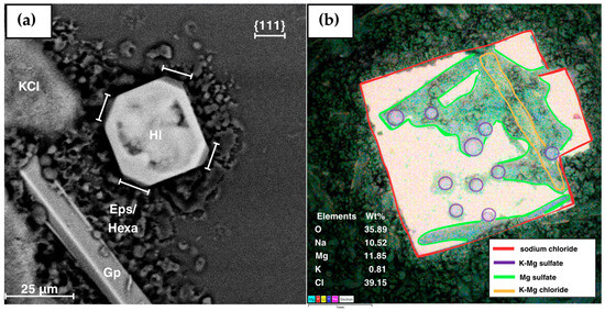
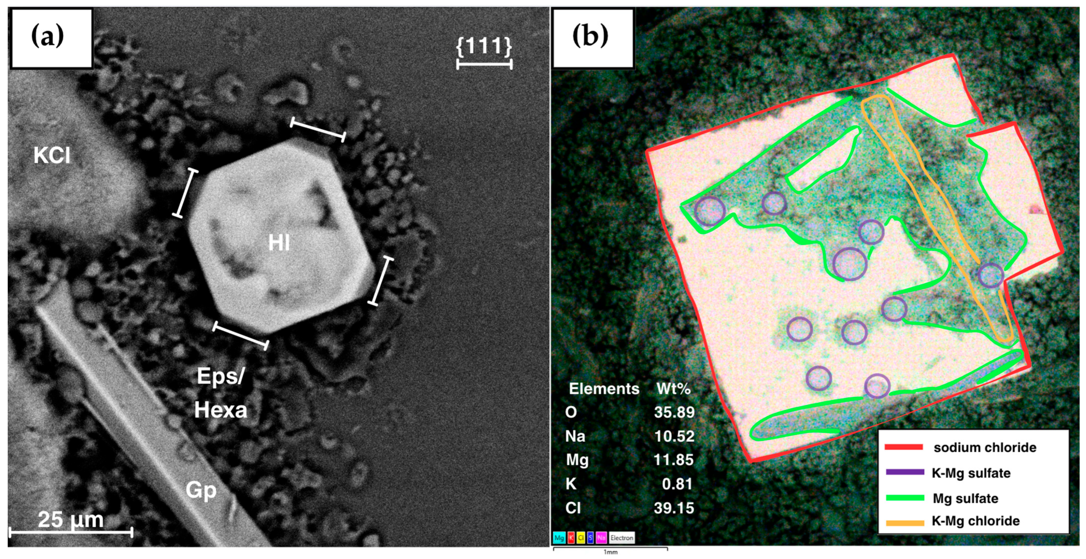
In the sample derived from the third pond (Figure 6a), the presence of halite crystals with well-developed octahedral faces is remarkable. The stabilization of the {111} form at the expense of the cubic faces is a phenomenon attributable to the selective adsorption of organic molecules present in the biological matrix of the brine or to kinetic conditions of high supersaturation [54,55,56,57,58]. The formation of the {111} form may depend on the extent of thermal gradient during crystallization [55,56]; however, under identical thermal gradient conditions, only the halite precipitated from the third pond exhibits the {111}-octahedral form. This finding supports the hypothesis that, in this instance, organic matter is responsible for stabilizing octahedral faces [57,58]. Furthermore, chemical maps (EDS) (available in Supplementary Materials) confirm the accessory precipitation of potassium chlorides (sylvite, KCl) and calcium and magnesium sulfate hydrates (gypsum and hexahydrite).
Figure 6.
(a) SEM photomicrograph of precipitates from the “third pond” brines, displaying a halite crystal (Hl) with distinct octahedral {111} form, surrounded by magnesium sulfate crystals (epsomite (Eps) and/or hexahydrite (Hexa), depending on the hydration state) and sylvite crystals (KCl). (b) EDS map of salts precipitated from the fourth pond brines, revealing a large halite crystal (in red) accompanied by K-Mg chlorides (in orange) and magnesium sulfates (in green), and small Mg-K sulfate crystals (in purple), presumably leonite or langbeinite.
From the fourth pond brine (Figure 6b), the mineral association evolves, reflecting a higher degree of concentration. Above the halite matrix, crusts of magnesium sulfates (epsomite or hexahydrite, respectively MgSO4 · 7H2O and MgSO4 · 6H2O), K-Mg double chlorides (carnallite, KMgCl3 · 6H2O), and K-Mg double sulfates, chemically compatible with leonite or langbeinite (respectively K2Mg(SO4)2 · 4H2O and K2Mg2(SO4)3), are observed (Figure 6).
The absence of thermodynamically stable phases such as kainite or kieserite suggests that the experimental evaporation process followed a metastable path, kinetically controlled. The kinetic conditions induced by rapid evaporation inhibit the nucleation of kainite or kieserite in favor of other soluble salts [12,27,59,60]. It is crucial to note that, within the operational context of the saltworks, these highly soluble salts are not typically found in the ponds. This is because standard anthropogenic practices intentionally interrupt the natural precipitation sequence: salt workers actively drain the residual mother brines (bitterns) and harvest the halite before the brine reaches the eutonic point or dries up completely. This controlled discharge is necessary to prevent the co-precipitation of magnesium and potassium salts, thereby avoiding the chemical contamination of the commercial sodium chloride harvest.
4.2. “Mamma Caura”
“Mamma caura” samples were collected from the exposed banks of the third pond. Macroscopically, the deposit exhibits a distinct vertical zonation: a bleached, oxidized surface crust overlies a dark, viscous layer (below 2 cm depth). The latter maintains higher temperatures, a phenomenon attributable to both thermal absorption by the dark substrate and the exothermic microbial decomposition of organic matter. This byproduct is described by salt workers as a material traditionally linked to salt production. Salt workers say its use is linked to the “good” production of clean, white salt, and that without it, the salt is gray or dirty.
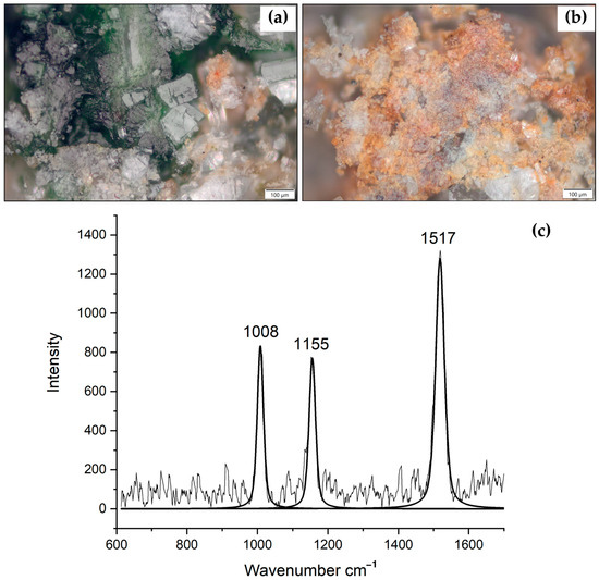
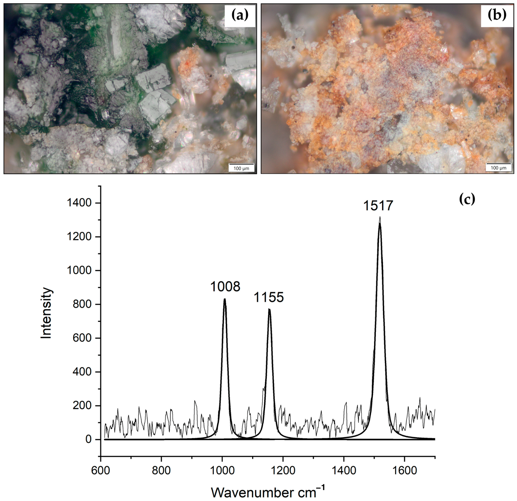
XRD analysis reveals a mineralogical assemblage overwhelmingly dominated by gypsum, with subordinate amounts of halite and traces of calcite and quartz (Figure S6). The most striking feature, however, lies in the habit and color of the gypsum crystals. Unlike the transparent crystals of the fourth pond, the “mamma caura” crystals are intensely pigmented, displaying hues ranging from pink to brick-red and green (Figure 7a,b).
Figure 7.
Photomicrographs of “mamma caura” crystals showing green (a) and orange (b) pigments; Raman spectrum of the orange pigment after background subtraction, showing three diagnostic peaks at 1517, 1155 and 1008 cm−1 (c).
This coloration is not superficial but due to the physical occlusion of biomass and metabolic products within the internal structure of the crystals during mineral growth. These occlusions provide evidence of endoevaporitic communities composed of halophilic bacteria, Archaea, and microalgae colonizing crystals. As widely documented in both modern and ancient evaporites, gypsum crystals (selenites) act as an “optical window”: they offer physical protection against desiccation and UV radiation while allowing the light penetration necessary for photosynthesis, effectively creating an ideal intracrystalline photic zone [61,62,63,64].
The pigments responsible for the coloration are the metabolic products of these extremophiles, primarily identified as bacteriorhodopsins, retinal proteins characteristic of Halobacteria (Archaea); spirilloxanthins, carotenoids typically produced by anoxygenic photosynthetic purple bacteria; and carotenoids, antioxidant pigments produced by algae [38,64,65].
Raman spectroscopy performed on the pigmented crystals provided molecular confirmation of this biological activity. The Raman spectrum of the orange layer in the gypsum of the “mamma caura” crust was acquired using 633 nm excitation and contains a very strong fluorescence signal. In the spectrum, the strongest diagnostic bands are clearly visible, specifically at 1517, 1155, and 1008 cm−1 (Figure 7c). The fluorescent Raman spectrum without background subtraction and deconvolution operations can be found in the supplementary material in Figure S7. Generally, Raman bands at around 1515, 1155 and 1007 cm−1 are diagnostic of carotenoid pigments, and they are respectively attributed to υ1 (C=C), υ2 (C–C) stretching vibrations and to rocking modes δ (C=CH) of the CH3 groups [66].
However, as reported by Edwards et al. (2005) [65], one typical carotenoid Raman band (at 1006 cm−1) is often severely obscured by the strong sulfate stretching mode of the matrix (at ~1008 cm−1). Indeed, in our spectrum, the band observed around 1008 cm−1 perfectly overlaps with the υ1 symmetric stretch vibration mode of the SO4 tetrahedra of the embedding gypsum crystals (υ1 at 1008 cm−1 for gypsum) [67,68].
The second Raman band at around 1155 cm−1 could be attributed to υ2 (C–C) stretching vibrations of carotenoids.
However, the most important and reliable Raman band for our analysis is the υ1 position (C=C stretching) at 1517 cm−1, which is completely free from sulfate overlap. This band contains crucial information about the number of double bonds in the polyene chain (N) and is normally used for discrimination [39,63,64]. However, discriminating the exact biological source of this carotenoid based solely on the υ1 value through spectroscopic analysis is difficult. The υ1 value obtained for β-carotene in different biological systems is in the range of 1513–1531 cm−1 [69]. Nevertheless, our results are close to the values reported for salinixanthin extracted from Salinibacter ruber (at 1510–1513 cm−1, N = 11) and spirilloxanthin extracted from Ectothiorhodospira marismortui (at 1509–1513 cm−1, N = 13) in the study by Culka et al. (2022) [39].
The evidence of pigments found exclusively within the gypsum crystals of the “mamma caura” in the third pond provides crucial insights into the interactions between the bacterial component and gypsum mineralization processes [61,62,63,64,65,66]. This complex interaction suggests the existence of a bio-filtration mechanism driven by this predominantly gypsum-matrix geomaterial. This byproduct, far from being discarded, finds a fundamental reuse within the artisanal production cycle, ensuring a natural and effective filtration of the brines before their transfer to the final salt-harvesting ponds. Although salt workers have historically utilized this material on a purely empirical basis, observing that its application yields a visibly cleaner and higher-quality salt, these analyses now scientifically confirm its filtering power. This result allows for the definitive valorization of ancient traditional knowledge, demonstrating how biological efficacy supports and validates centuries-old artisanal practice.
5. Conclusions
Each pond in the saltworks system represents a distinct micro-ecosystem, although connected both mineralogically and hydrodynamically. The mineralogical characterization reflects the theoretical fractional precipitation stages: from the dominance of carbonates in the initial ponds (first and second), transitioning to calcium sulfates in the third pond, and finally to sodium chloride in the last pond. This zonation allows for the precise identification of stability fields for mineral phases relative to the salinity gradient.
A key finding is the dominant role of anthropogenic management in shaping the basin mineralogy. Stratigraphic analysis allowed for the identification of mineralogical discontinuities separating reworked bottom horizons from authigenic precipitation layers in equilibrium with the brines. In contrast, the stability field of soluble K-Mg salts is not represented in the natural ponds due to the artificial interruption of the precipitation cycle for harvesting. Laboratory experiments demonstrated that the formation of these highly soluble phases is strongly dependent on kinetic factors, which differ significantly between controlled laboratory conditions and the natural environment subject to thermal fluctuations. This suggests that future research should focus on the crystallization kinetics of bittern salts up to the eutonic point using different methodology.
Finally, this study provides the first analysis of “mamma caura”, a byproduct of artisanal salt production traditionally integrated into the production cycle. It clarifies and confirms its mineral composition, which consists primarily of gypsum and is rich in organic secondary metabolites produced by microorganisms living in the ponds and sediments, allowing it to be classified as a bio-mineral deposit. Specifically utilized in the third pond, this material acts as a bio-accumulator through the synergistic action of the mineral matrix and bacterial activity. This performs a crucial “filtering” function, explaining the underlying mechanisms of its longstanding empirical use to ensure the high purity of the salt harvested in the final ponds.
Supplementary Materials
The following supporting information can be downloaded at: https://www.mdpi.com/article/10.3390/min16070681/s1, Figure S1: Mineralogical phases recognized by XRD: kaolinite (Kln), halite (Hl), calcite (Cal), aragonite (Arg), quartz (Qz); (Hl and Gp are considered artifacts resulting from sample preparation). (a) XRPDs of all horizons of the first pond; (b) XRPDs of all horizons of the second pond; Figure S2: Detail of XRPD of the 1st and 4th horizons of the second pond in which it is observed a rise in the background in the 12–20 Å d range; Figure S3: Mineralogical phases recognized by XRD: kaolinite (Kln), halite (Hl), gypsum (Gp), calcite (Cal), quartz (Qz) in the XRPDs from the 1st to 11th cm of the third pond; Table S1: Semi-quantitative analysis of the sediments of the third pond. Overconcentration of soluble salts was estimated as described in the Materials and Methods section of the main text; Figure S4: Mineralogical phases recognized by XRD: kaolinite (Kln), halite (Hl), gypsum (Gp), calcite (Cal), quartz (Qz), bassanite (Bss) in the XRPDs from the 1st to 11th cm of the third pond (Bss is considered artifact resulting from sample preparation); Figure S5: EDS compositional maps of precipitates from the third pond brine; Figure S6: XRPD pattern of mamma caura highlighting the characteristic diffraction peaks of gypsum (Gp), halite (Hl) and traces of calcite (Cal) and quartz (Qz); Figure S7: Original Raman spectrum with strong fluorescence signal of the orange pigment.
Author Contributions
Conceptualization, A.L.M., L.P. (Linda Pastero) and M.M.; methodology, A.L.M., L.P. (Linda Pastero), A.C. and L.P. (Luca Pellegrino); software, A.L.M., L.P. (Linda Pastero) and A.C.; validation, A.L.M., L.P. (Luca Pellegrino) and L.P. (Linda Pastero); formal analysis, A.L.M. and L.P. (Linda Pastero); investigation, A.L.M. and L.P. (Linda Pastero); resources, L.P. (Linda Pastero) and L.P. (Luca Pellegrino); data curation, A.L.M., L.P. (Linda Pastero), M.M. and A.C.; writing—original draft preparation, A.L.M.; writing—review and editing, A.L.M.; visualization, A.L.M., L.P. (Linda Pastero) and A.C.; supervision, L.P. (Linda Pastero) and M.M.; project administration, L.P. (Linda Pastero) and M.M.; funding acquisition, L.P. (Linda Pastero) and A.L.M. All authors have read and agreed to the published version of the manuscript.
Funding
This research was funded by the University of Palermo, grant number 118.TDA.UNIPA and University of Torino, Ricerca locale ex-60% 2024 (PASL_RILO_24_01).
Data Availability Statement
The data presented in this study are available on request from the corresponding author.
Acknowledgments
The authors of this work thank Gerardo Cortellaro, the technical staff, and Silvana Piacentino, director of the WWF Oasis of the Saltworks of Trapani–Paceco, for their support and theoretical advice, which has been invaluable for understanding the “Saline di Trapani-Paceco” Nature Reserve (TP). The authors also wish to thank Marianna Cangemi, INGV researcher (Catania, Italy), and Paolo Madonia, INGV researcher (Palermo, Italy), for technical support in the environmental matrix sampling phase.
Conflicts of Interest
The authors declare no conflicts of interest.
Abbreviations
The following abbreviations are used in this manuscript:
| Arg | Aragonite |
| Bsh | Bischofite |
| Cal | Calcite |
| EDS | Energy-Dispersive X-ray Spectroscopy |
| Eps | Epsomite |
| Gp | Gypsum |
| Hexa | Hexahydrite |
| Hl | Halite |
| Ilt | Illite |
| Kln | Kaolinite |
| Qz | Quartz |
| SEM | Scanning Electron Microscopy |
| XRD | X-Ray Diffraction |
References
- Korovessis, N.A.; Lekkas, T.D. Solar saltworks’ wetland function. Glob. NEST J. 2009, 11, 49–57. [Google Scholar] [CrossRef] [Scilit]
- Soares, R.H.R.d.M.; Assunção, C.A.; Fernandes, F.O.; Marinho-Soriano, E. Identification and analysis of ecosystem services with biodiversity of saltworks. Ocean Coast. Manag. 2018, 163, 278–284. [Google Scholar] [CrossRef] [Scilit]
- Sadoul, N.; Walmsley, J.; Charpentier, B. Salinas and Nature Conservation; MedWet Series No. 9; Tour du Valat: Arles, France, 1998; 96p, Available online: https://medwet.org/wp-content/uploads/2016/06/N9_Salinas_and_nature_conservation.pdf (accessed on 28 February 2026).
- Crisman, T.L.; Takavakoglou, V.; Alexandridis, T.; Antonopoulos, V.; Zalidis, G. Rehabilitation of abandoned saltworks to maximize conservation, ecotourism and water treatment potential. Glob. NEST J. 2009, 11, 24–31. [Google Scholar] [CrossRef] [Scilit]
- Hay, W.H.; Migdisov, A.; Balukhovsky, A.N.; Wold, C.N.; Flögel, S.; Söding, E. Evaporites and the salinity of the ocean during the Phanerozoic: Implications for climate, ocean circulation and life. Palaeogeogr. Palaeoclimatol. Palaeoecol. 2006, 240, 3–46. [Google Scholar] [CrossRef] [Scilit]
- Siemann, M.G.; Schramm, M. Thermodynamic modelling of the Br partition between aqueous solutions and halite. Geochim. Cosmochim. Acta 2000, 64, 1681–1693. [Google Scholar] [CrossRef] [Scilit]
- Sun, S.; Li, M.; Yan, M.; Fang, X.; Zhang, G.; Liu, X. Bromine content and Br/Cl molar ratio of halite in a core from Laos: Implications for origin and environmental changes. Carbonates Evaporites 2019, 34, 1107–1117. [Google Scholar] [CrossRef] [Scilit]
- Pueyo, J.J. (Ed.) Génesis de Formaciones Evaporíticas: Modelos Andinos e Ibéricos; Publicación No. 2; Universitat de Barcelona: Barcelona, Spain, 1991. [Google Scholar]
- Sonnenfeld, P. Brines and Evaporites; Academic Press: Orlando, FL, USA, 1984. [Google Scholar]
- European Commission. Study on the Critical Raw Materials for the EU 2023-Final Report; Publications Office of the European Union: Luxembourg, 2023. [Google Scholar] [CrossRef]
- Warren, J.K. Evaporites: Sediments, Resources and Hydrocarbons, 1st ed.; Springer: Berlin/Heidelberg, Germany, 2006. [Google Scholar] [CrossRef] [Scilit]
- Stewart, F.H. Marine Evaporites. In U.S. Geological Survey Professional Paper 440-Y, 6th ed.; U.S. Geological Survey: Washington, DC, USA, 1963; pp. 1–52. [Google Scholar]
- Bąbel, M.; Schreiber, B.C. Geochemistry of Evaporites and Evolution of Seawater. In Treatise on Geochemistry, 2nd ed.; Holland, H.D., Turekian, K.K., Eds.; Elsevier: Oxford, UK, 2014; Volume 9, pp. 483–560. [Google Scholar] [CrossRef] [Scilit]
- Bontognali, T.R.R.; McKenzie, J.A.; Warthmann, R.J.; Vasconcelos, C. Microbially influenced formation of Mg-calcite and Ca-dolomite in the presence of exopolymeric substances produced by sulphate-reducing bacteria. Terra Nova 2014, 26, 72–77. [Google Scholar] [CrossRef] [Scilit]
- Riding, R. Microbial carbonates: The geological record of calcified bacterial-algal mats and biofilms. Sedimentology 2000, 47, 179–214. [Google Scholar] [CrossRef] [Scilit]
- Harvie, C.E.; Weare, J.H. Prediction of mineral solubilities in natural waters: The Na K Mg Ca Cl SO4 H2O system from zero to high concentration at 25 °C. Geochim. Cosmochim. Acta 1980, 44, 981–997. [Google Scholar] [CrossRef] [Scilit]
- Usiglio, J. Analyse de l’eau de la Méditerranée sur les côtes de France. Ann. Chim. Phys. (Sér. 3) 1849, 27, 92–107. [Google Scholar]
- Usiglio, J. Etudes sur la composition de l’eau de la Méditerranée et sur l’exploitation des sels qu’elle contient. Ann. Chim. Phys. (Sér. 3) 1949, 27, 172–191. [Google Scholar]
- McCaffrey, M.A.; Lazar, B.; Holland, H.D. The evaporation path of seawater and coprecipitation of Br- and K+ with halite. J. Sediment. Petrol. 1987, 57, 928–937. [Google Scholar] [CrossRef] [Scilit] [PubMed]
- Baseggio, G. The composition of sea water and its concentrates. In Fourth Symposium on Salt; Coogan, A.H., Ed.; Northern Ohio Geological Society: Cleveland, OH, USA, 1974; Volume 2, pp. 351–358. [Google Scholar]
- Ortí, F.; Busson, G. (Eds.) Introduction to the sedimentology of the coastal salinas of Santa Pola (Alicante, Spain). Rev. Investig. Geol. 1984, 38–39, 9–229. [Google Scholar]
- Warren, J.K. The hydrological setting, occurrence and significance of gypsum in late Quaternary salt lakes in South Australia. Sedimentology 1982, 29, 609–637. [Google Scholar] [CrossRef] [Scilit]
- Cohen-Adad, R.; Balarew, C.; Tepavitcharova, S.; Rabadjieva, D. Sea-water solubility phase diagram. Application to an extractive process. Pure Appl. Chem. 2002, 74, 1811–1821. [Google Scholar] [CrossRef] [Scilit]
- Ortí, F.; Pueyo, J.J.; Geisler-Cussey, D.; Dulau, N. Evaporitic sedimentation in the coastal salinas of Santa Pola (Alicante, Spain). Rev. Investig. Geol. 1984, 38–39, 169–220. [Google Scholar]
- Valyashko, M.G. Geochemical Rules of the Potassium Salt Deposits Formation; Izdatelstvo Moskovskovo Universiteta: Moscow, Russia, 1962; 398p. (In Russian) [Google Scholar]
- Charuit, P.; Genty, P. Experimental study covering production of secondary salts extracted from bitterns of solar salt works. In Proceedings of the Fifth International Symposium on Salts, Hamburg, Germany, 29 May–1 June 1979; Coogan, A.H., Ed.; Northern Ohio Geological Society: Cleveland, OH, USA, 1980; pp. 251–258. [Google Scholar]
- Mullin, J.W. Crystallization, 4th ed.; Butterworth-Heinemann: Oxford, UK, 2001; 594p. [Google Scholar]
- Usdowki, E.; Dietzel, M. Atlas and Data of Solid-Solution Equilibria of Marine Evaporites; Springer: Berlin/Heidelberg, Germany, 1998; 316p. [Google Scholar] [CrossRef] [Scilit]
- Al-Droubi, A.; Fritz, B.; Gac, J.Y.; Tardy, Y. Generalized residual alkalinity concept. Am. J. Sci. 1980, 280, 560–572. [Google Scholar] [CrossRef] [Scilit]
- Bea, S.A.; Carrera, J.; Ayora, C.; Batlle, F. Modeling of concentrated aqueous solutions: Efficient implementation of Pitzer equations in geochemical and reactive transport models. Comput. Geosci. 2010, 36, 526–538. [Google Scholar] [CrossRef] [Scilit]
- Keller, A.; Burger, J.; Hasse, H.; Kohns, M. Application of the Pitzer model for describing the evaporation of seawater. Desalination 2021, 503, 114866. [Google Scholar] [CrossRef] [Scilit]
- Turner, D.R.; Achterberg, E.P.; Chen, C.-T.A.; Clegg, S.L.; Hatje, V.; Maldonado, M.T.; Sander, S.G.; van den Berg, C.M.G.; Wells, M. Toward a quality-controlled and accessible Pitzer model for seawater and related systems. Front. Mar. Sci. 2016, 3, 139. [Google Scholar] [CrossRef] [Scilit]
- Braitsch, O.; Kinsman, D.J. Physicochemical conditions of origin. In The Encyclopedia of Sedimentology; Fairbridge, R.W., Bourgeois, J., Eds.; Dowden, Hutchinson and Ross, Inc.: Stroudsburg, PA, USA, 1978; pp. 303–311. [Google Scholar]
- UNIDO. Guidelines for the Establishment of Solar Salt Facilities from Seawater, Underground Brines and Salted Lakes; United Nations Industrial Development Organization: Vienna, Austria, 1982; Available online: https://downloads.unido.org/ot/47/93/4793176/10001-15000_12271.pdf (accessed on 28 February 2026).
- Green, D.W.; Southard, M.Z. Perry’s Chemical Engineers’ Handbook, 9th ed.; McGraw-Hill Education: New York, NY, USA, 2018; Available online: https://iem.ca/pdf/resources/Perrys%20Chemical%20Engineers%20Handbook.pdf (accessed on 28 February 2026).
- Oren, A. A hundred years of Dunaliella research: 1905–2005. Saline Syst. 2005, 1, 2. [Google Scholar] [CrossRef] [Scilit] [PubMed]
- Oren, A. On the red coloration of saltern crystallizer ponds. Int. J. Salt Lake Res. 1992, 1, 77–89. [Google Scholar] [CrossRef] [Scilit]
- Oren, A. The ecology of the extremely halophilic archaea. FEMS Microbiol. Rev. 1994, 13, 415–439. [Google Scholar] [CrossRef]
- Culka, A.; Jehlička, J.; Oren, A.; Rousaki, A.; Vandenabeele, P. Fast outdoor screening and discrimination of carotenoids of halophilic microorganisms using miniaturized Raman spectrometers. Spectrochim. Acta Part A Mol. Biomol. Spectrosc. 2022, 276, 126316. [Google Scholar] [CrossRef] [Scilit] [PubMed]
- Antón, J.; Rosselló-Mora, R.; Rodríguez-Valera, F.; Amann, R. Extremely Halophilic Bacteria in Crystallizer Ponds from Solar Salterns. Appl. Environ. Microbiol. 2000, 66, 3052–3057. [Google Scholar] [CrossRef] [Scilit] [PubMed]
- Oren, A. Microbial life at high salt concentrations: Phylogenetic and metabolic diversity. Saline Syst. 2008, 4, 2. [Google Scholar] [CrossRef] [Scilit] [PubMed]
- Villanova, V.; Galasso, C.; Fiorini, F.; Lima, S.; Brönstrup; Sansone, C.; Brunet, C.; Brucato, A.; Scargiali, F. Biological and chemical characterization of new isolated halophilic microorganisms from saltern ponds of Trapani, Sicily. Algal Res. 2021, 54, 102192. [Google Scholar] [CrossRef] [Scilit]
- Putz, H.; Brandenburg, K. Match!—Phase Analysis Using Powder Diffraction; Versione 3.X; Crystal Impact GbR: Bonn, Germany, 2025. [Google Scholar]
- Gražulis, S.; Chateigner, D.; Downs, R.T.; Yokochi, A.F.T.; Quirós, M.; Lutterotti, L.; Manakova, E.; Butkus, J.; Moeck, P.; Le Bail, A. Crystallography Open Database—An open-access collection of crystal structures. J. Appl. Crystallogr. 2009, 42, 726–729. [Google Scholar] [CrossRef] [Scilit] [PubMed]
- Moore, D.M.; Reynolds, R.C., Jr. X-Ray Diffraction and the Identification and Analysis of Clay Minerals, 2nd ed.; Oxford University Press: Oxford, UK, 1997; 378p. [Google Scholar]
- Catalano, R.; Di Maggio, C.; Agate, M.; Lena, G.; Valenti, V.; Moscariello, A. Note Illustrative della Carta Geologica d’Italia Alla Scala 1:50.000, Foglio 605 Paceco; ISPRA: Roma, Italy, 2019. [Google Scholar]
- Agate, M.; Basilone, L.; Di Maggio, C.; Contino, A.; Pierini, S.; Catalano, R. Quaternary marine and continental unconformity-bounded stratigraphic units of the NW Sicily coastal belt. J. Maps 2017, 13, 425–437. [Google Scholar] [CrossRef] [Scilit]
- Herrmann, A.G.; Knake, D.; Schneider, J.; Peters, H. Geochemistry of modern seawater and brines from salt pans: Main components and bromine distribution. Contrib. Mineral. Petrol. 1973, 40, 1–24. [Google Scholar] [CrossRef] [Scilit]
- Ebner, R.M.; Meijer, P.T. A question of time and space: A model approach to the synchronous precipitation of gypsum and halite during the Messinian Salinity Crisis. Clim. Past 2025, 21, 705–718. [Google Scholar] [CrossRef] [Scilit]
- Real-Fernández, E.; Pozo, M.; De Ignacio, C.; Sánchez-Malo, Á.; Sanz-Rubio, E.; Villa, L. Fluid-Rock Interaction Signature in Palomares Fault Zone—New Mineralogical and Geochemical Insights into the Tectono-Magmatic Águilas Arc Geothermal System (SE Spain). Appl. Sci. 2026, 16, 1420. [Google Scholar] [CrossRef] [Scilit]
- Vicari, F.; Randazzo, S.; López, J.; Fernández de Labastida, M.; Vallès, V.; Micale, G.; Tamburini, A.; D’Alì Staiti, G.; Cortina, J.L.; Cipollina, A. Mining minerals and critical raw materials from bittern: Understanding metal ions fate in saltwork ponds. Sci. Total Environ. 2022, 847, 157544. [Google Scholar] [CrossRef] [Scilit] [PubMed]
- Pina, C.M.; Tamayo, Á. Crystallisation of strontium sulphates from Si-bearing aqueous solutions. Geochim. Cosmochim. Acta 2012, 92, 220–232. [Google Scholar] [CrossRef] [Scilit]
- Kushnir, J. The coprecipitation of strontium, magnesium, sodium, potassium and chloride ions with gypsum. An experimental study. Geochim. Cosmochim. Acta 1980, 44, 1471–1482. [Google Scholar] [CrossRef] [Scilit]
- Bruno, M.; Aquilano, D.; Pastero, L.; Prencipe, M. Structures and Surface Energies of (100) and Octopolar (111) Faces of Halite: An Ab initio Quantum-Mechanical and Thermodynamical Study. Cryst. Growth Des. 2008, 8, 2163–2170. [Google Scholar] [CrossRef] [Scilit]
- Aquilano, D.; Pastero, L.; Bruno, M.; Rubbo, M. {100} and {111} forms of the NaCl crystals coexisting in growth from pure aqueous solution. J. Cryst. Growth 2009, 311, 399–403. [Google Scholar] [CrossRef] [Scilit]
- Censi, P.; Sirota, I.; Zuddas, P.; Lensky, N.; Merli, M.; Saiano, F.; Piazzese, D.; Sposito, F.; Venturelli, M. Trace elements fractionation through halite crystallisation: Geochemical mechanisms and environmental implications. Sci. Total Environ. 2020, 723, 137926. [Google Scholar] [CrossRef] [Scilit] [PubMed]
- Pastero, L.; Aquilano, D.; Moret, M. Selective adsorption/absorption of formamide in NaCl crystals growing from solution. Cryst. Growth Des. 2012, 12, 2306–2314. [Google Scholar] [CrossRef] [Scilit]
- Pastero, L.; Aquilano, D. A Three-modal Epitaxial Adsorption of Formamide (HO-C-NH2) on the {111} Surfaces of f.c.c. Alkali Halides Crystals Growing from Aqueous Solutions. CrystEngComm 2016, 18, 9067–9076. [Google Scholar] [CrossRef] [Scilit]
- Braitsch, O. Salt Deposits: Their Origin and Composition; Springer: Berlin, Germany, 1971; 297p. [Google Scholar] [CrossRef] [Scilit]
- Shalev, N.; Lazar, B.; Köbberich, M.; Halicz, L.; Gavrieli, I. The chemical evolution of brine and Mg-K-salts along the course of extreme evaporation of seawater—An experimental study. Geochim. Cosmochim. Acta 2018, 241, 164–179. [Google Scholar] [CrossRef] [Scilit]
- Oren, A.; Kühl, M.; Karsten, U. An endoevaporitic microbial mat within a gypsum crust: Zonation of phototrophs, photopigments, and light penetration. Mar. Ecol. Prog. Ser. 1995, 128, 151–159. [Google Scholar] [CrossRef] [Scilit]
- Oren, A.; Sørensen, K.B.; Canfield, D.E.; Teske, A.P.; Ionescu, D.; Lipski, A.; Alterndorf, K. Microbial communities and processes within a hypersaline gypsum crust in a saltern evaporation pond (Eilat, Israel). Hydrobiologia 2009, 626, 15–26. [Google Scholar] [CrossRef] [Scilit]
- Nemečková, K.; Jehlička, J.; Culka, A. Fast screening of carotenoids of gypsum endoliths using portable Raman spectrometer (Messinian gypsum, Sicily). J. Raman Spectrosc. 2020, 51, 1127–1137. [Google Scholar] [CrossRef] [Scilit]
- Jehlička, J.; Oren, A.; Vítek, P.; Wierzchos, J. Microbial colonization of gypsum: From the fossil record to the present day. Front. Microbiol. 2024, 15, 1397437. [Google Scholar] [CrossRef] [Scilit] [PubMed]
- Edwards, H.G.M.; Jorge Villar, S.E.; Parnell, J.; Cockell, C.S.; Lee, P. Raman spectroscopic analysis of cyanobacterial gypsum halotrophs and relevance for sulfate deposits on Mars. Analyst 2005, 130, 917–923. [Google Scholar] [CrossRef] [Scilit] [PubMed]
- Withnall, R.; Chowdhry, B.Z.; Silver, J.; Edwards, H.G.M.; de Oliveira, L.F.C. Raman spectra of carotenoids in natural products. Spectrochim. Acta Part A Mol. Biomol. Spectrosc. 2003, 59, 2207–2212. [Google Scholar] [CrossRef] [Scilit] [PubMed]
- Liu, Y.; Wang, A.; Freeman, J.J. Raman, MIR, and NIR spectroscopic study of calcium sulfates: Gypsum, bassanite, and anhydrite. In Proceedings of the 40th Lunar and Planetary Science Conference, The Woodlands, TX, USA, 23–27 March 2009; Available online: https://www.lpi.usra.edu/meetings/lpsc2009/pdf/2128.pdf (accessed on 18 April 2026).
- White, S.N. Laser Raman spectroscopy as a technique for identification of seafloor hydrothermal and cold seep minerals. Chem. Geol. 2009, 259, 240–252. [Google Scholar] [CrossRef] [Scilit]
- Tschirner, N.; Schenderlein, M.; Brose, K.; Schlodder, E.; Mroginski, M.A.; Hildebrandts, P.; Thomsen, C. Raman excitation profiles of b-carotene—Novel insights into the nature of the u1-band. Phys. Status Solidy B 2008, 245, 2225. [Google Scholar] [CrossRef] [Scilit]

Disclaimer/Publisher’s Note: The statements, opinions and data contained in all publications are solely those of the individual author(s) and contributor(s) and not of MDPI and/or the editor(s). MDPI and/or the editor(s) disclaim responsibility for any injury to people or property resulting from any ideas, methods, instructions or products referred to in the content. |
© 2026 by the authors. Licensee MDPI, Basel, Switzerland. This article is an open access article distributed under the terms and conditions of the Creative Commons Attribution (CC BY) license.