


Abstract
The TaiLing Mausoleum in Western Qing Tombs has great aesthetic value and a rich history. In this study, we conducted an analysis of the materials used in the architectural polychrome paintings of the TaiLing Mausoleum. Optical microscopy (OM), portable X-ray fluorescence (p-XRF), scanning electron microscopy with energy-dispersive X-ray spectroscopy (SEM–EDX), micro-Raman spectroscopy (μ-RS), and X-ray diffraction (XRD) were used to analyze the paintings of Long’en Gate in TaiLing Mausoleum. The results indicate that the main minerals in the ground layer are quartz, augite, feldspars and illite. The gilding materials employed gold leaf. The red pigment is hematite, and the black pigment is carbon black. The green pigment is emerald green with barium sulfate as an extender. The blue pigments are smalt and synthetic ultramarine. In some areas, emerald green is observed overlaying smalt, suggesting that the paintings at Long’en Gate underwent overlay restoration or repainting from the late Qing Dynasty to modern times. These results can support future conservation of the polychrome paintings at the TaiLing Mausoleum.
1. Introduction
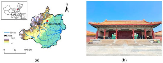
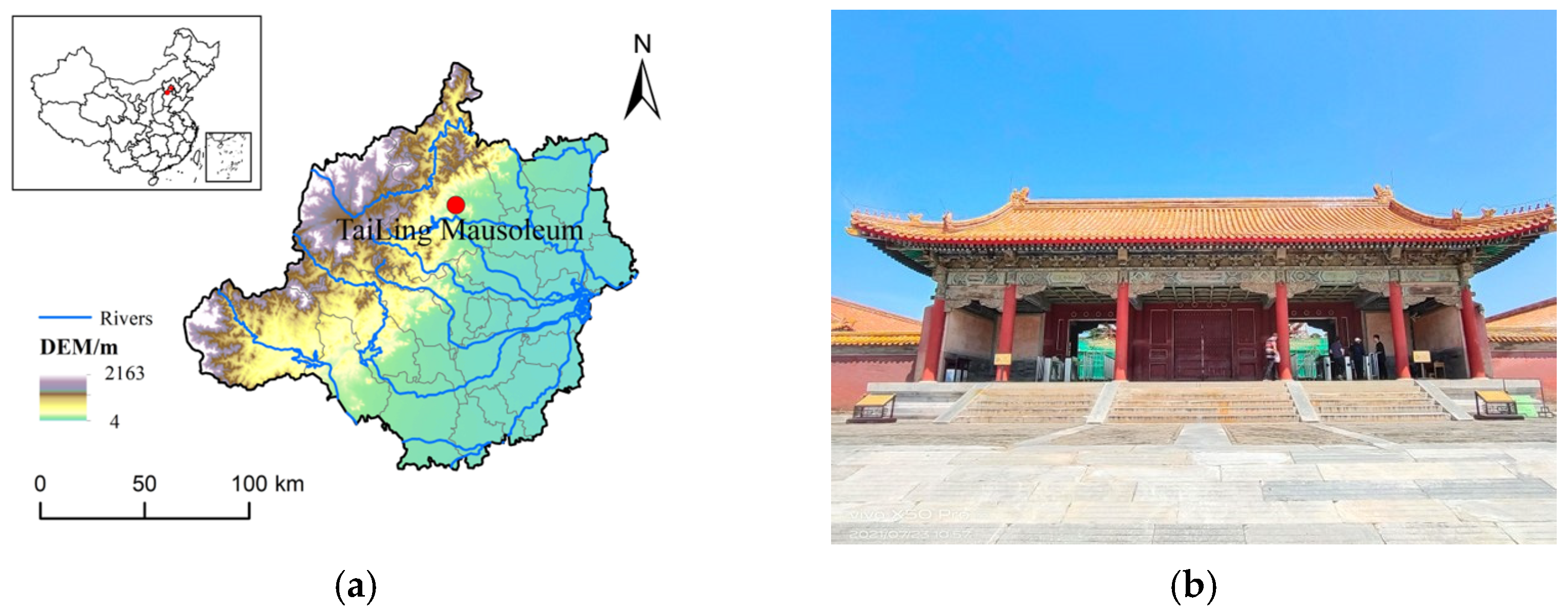
The Western Qing Tombs are in Yi County, Hebei Province, China (Figure 1a). Along with the Eastern Qing Tombs, they comprise the two principal imperial necropolises of the Qing Dynasty. The complex was inscribed on the UNESCO World Heritage List in 2000 [1].
Figure 1.
Location of the study area. (a) Location of the TaiLing Mausoleum in Yi County, Hebei Province. (b) Front view of the Long’en Gate at TaiLing, the sampling site.
TaiLing, constructed between 1730 and 1737, is the first imperial mausoleum of the Western Qing Tombs. It serves as the final resting place of the Yongzheng Emperor and is a key part of the scenic and ritual landscape of the Western Tombs. As the first mausoleum of the Western Qing Tombs, TaiLing established the spatial layout and ritual order of the complex during the Yongzheng reign, signaling the Qing dynasty’s expansion of imperial burial practice from east to west. The Long’en Gate (Figure 1b), situated along TaiLing’s central axis, functions as a formal and symbolic threshold separating the sacrificial precinct from the rear burial chambers. The Xuanzi polychrome paintings on the gate are highly representative of the official style in terms of their patterns, colors, and craftsmanship. These paintings serve decorative, fireproofing, and moisture-proofing functions; they also reflect the architectural hierarchy [2,3]. However, these paintings are located on the outermost layer of the architectural components. They are directly exposed to the atmosphere. Consequently, they are highly susceptible to erosion from temperature fluctuations, humidity, light, and pollutants [4].
Polychrome paintings generally consist of a ground layer and a paint layer, with gold leaf for decoration on the surface in some areas. While this basic structure is relatively straightforward, the material composition of individual layers can be considerably heterogeneous and variable. In addition, historical interventions further complicate the material composition. Different materials exhibit distinct sensitivities to light, humidity, pollutants, and salt-driven degradation processes, which can promote deterioration. Therefore, identifying the materials used in the ground, paint, and gilded layers is essential. It can also provide important evidence for dating, analyzing craftsmanship [5], detecting restoration traces [6] and creating a scientific basis for future conservation [7,8,9].
Recently, scholars have increasingly employed multi-technique approaches to determine the material compositions of polychrome paintings [10]. Microscopic observation records pigment morphology, stratigraphic interfaces, and deterioration features [11]. XRF and SEM-EDX are utilized to acquire the characteristic elemental composition and its spatial distribution [12]. μ-RS is applied to identify the molecular vibrational features of pigments and their associated components [13]. XRD is used to determine the crystalline phases of the ground layer and selected paint-layer materials [14].
Previous studies indicate that the Qing Dynasty architectural polychrome paintings generally relied on traditional mineral pigments. Modern synthetic pigments were gradually introduced during the late Qing to the early Republic of China period [7,15]. Fu et al. (2020) [16] conducted a preliminary analysis of the polychrome materials at the Taidong Tomb within the Western Qing Tombs. However, systematic data for the TaiLing Tomb in the primary mausoleum remain lacking.
This study aims to characterize the materials of the polychrome paintings at the TaiLing Mausoleum. Representative microsamples were collected from the Long’en Gate from areas of severe flaking. Techniques including OM, XRF, SEM-EDX, μ-RS, and XRD were employed to analyze the paint, ground, and gilded layers. The study identifies the primary composition of each layer. This provides the necessary scientific basis for the tomb’s conservation.
2. Materials and Methods
2.1. Materials
Representative micro-sampling followed the principle of minimum intervention. The samples included the ground, gilded, and representative paint layers (green, blue, red, and black). Detailed sampling information (sample ID, layer type, analytical methods, and remarks) is provided in Appendix A, Table A1.
2.2. Methods
Multiple techniques are used to analyze the samples, including OM, XRD, p-XRF, SEM-EDX, and μ-RS.
OM recorded surface morphology and deterioration features. p-XRF obtained on-site elemental data. Subsequently, representative microsamples underwent laboratory testing. XRD analyzed the ground-layer minerals. SEM-EDX obtained microscopic morphology and elemental composition, while μ-RS identified molecular structures. Taken together, these results allowed for a more reliable interpretation of the material composition.
2.2.1. OM
Surface imaging employed an Anyty MSA600S portable digital microscope (3R Eddytek Corp., Beijing, China) (200×), equipped with built-in white LED illumination. This recorded pigment morphology, surface color, and deterioration forms.
2.2.2. p-XRF
p-XRF (Thermo Scientific/Niton XL3 series, Thermo Fisher Scientific, Waltham, MA, USA) was used for in situ elemental analysis of individual layers. The instrument is equipped with a silver (Ag) target X-ray tube operating at a maximum excitation voltage of 50 kV and a power of 2 W, with an 8 mm beam spot diameter. The gilding sample was measured in the Precious Metals mode (60 s). All other samples were analyzed in Test All Geo mode (120 s). The target element lists for each mode are provided in Appendix A, Table A2. As the analytical depth of p-XRF depends on material density and composition, the detected signal integrates contributions from multiple layers rather than reflecting the composition of any single layer in isolation; moreover, light elements such as carbon and oxygen are beyond the effective detection range and are subsumed into the balance (Bal) value. In accordance with this, all results are semi-quantitative in nature: ground-layer data are interpreted in conjunction with the XRD mineralogical results, while data for the gilding and red paint layers are treated qualitatively to identify characteristic elemental associations only. Elemental identification and quantification were performed using the Thermo Scientific NDT software (Version 8.4.4) supplied with the instrument.
2.2.3. SEM-EDX
A Phenom Desktop SEM (Thermo Fisher Scientific, Eindhoven, The Netherlands) was used to observe micro-morphology and perform EDX microanalysis. BSE images were acquired in the compositional mode at 15 kV. An integrated EDX detector (Thermo Fisher Scientific, Eindhoven, The Netherlands) was used for spot analyses and elemental mapping with 20–30 s counting time per measurement. The working distance was kept within 7.5–9.4 mm. The analyses were performed on heterogeneous pigment fragments and localized surface micro-regions rather than polished cross-sections, the SEM–EDX results are treated as local semi-quantitative data and are used primarily for a relative comparison of elemental enrichment between micro-areas.
2.2.4. μ-RS
Pigments in the paint layer were characterized by micro-Raman spectroscopy (Horiba Scientific, Palaiseau, France) using an XploRA system equipped with 532, 638, and 785 nm excitation lasers. Microsamples were analyzed directly without any pretreatment. The spectra were collected over the range of 100–2000 cm−1. The excitation wavelength was selected according to pigment color to improve the spectral quality and to reduce luminescence background. The laser power at the sample surface was kept below 1 mW to minimize photothermal alteration of the pigments. Acquisition parameters were 10–30 s per accumulation with 2–5 accumulations, depending on signal quality. Spectral acquisition and processing were performed using LabSpec 6 (Version 6.0, Horiba Scientific, Palaiseau, France).
2.2.5. XRD
Powdered ground-layer samples were analyzed using an X-ray diffractometer (SmartLab, Rigaku Corporation, Tokyo, Japan) with Cu Kα radiation (40 kV, 150 mA). The data were collected over 2θ = 2.6–45.0° with a step size of 0.02° and a counting time of 0.15 s per step. The diffraction patterns were processed in line with SmartLab Guidance (Version 2.1.0.0, Rigaku Corporation, Tokyo, Japan) for phase identification.
3. Results and Discussion
3.1. Composition of the Ground Layer
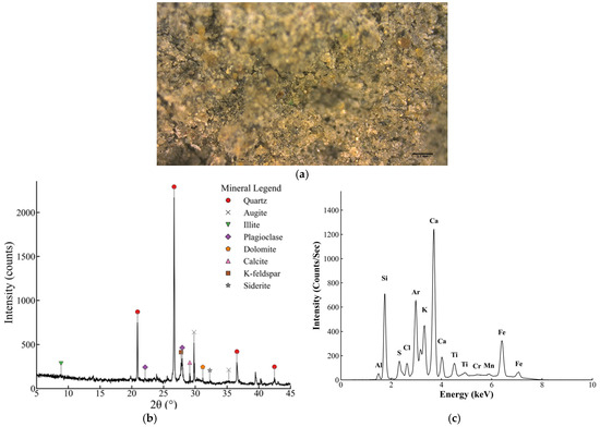
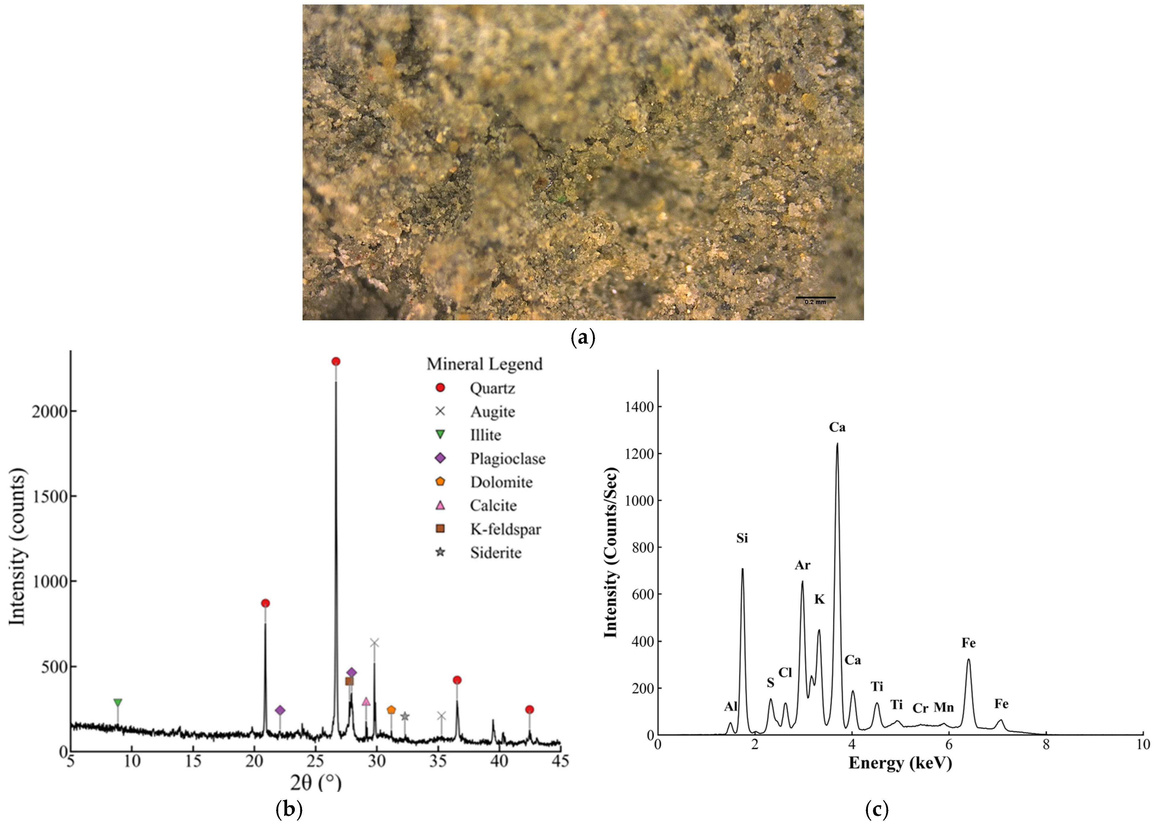
The ground layer (Sample 01; Appendix A, Table A1) is composed of coarse sand-sized grains within a fine-grained clay matrix. At 200× (Figure 2a), numerous colorless, translucent, angular detrital grains with sharp boundaries are present. The absence of cleavage and the irregular, local conchoidal fracture are characteristic of quartz, indicating that quartz-rich grains constitute the dominant skeletal aggregate.
Figure 2.
Micromorphology and compositional characterization of the ground layer: (a) digital micrograph at 200×; (b) XRD patternand (c) XRF spectrum.
The XRD quantitative results for the ground-layer sample (Figure 2b, Table 1) indicate that quartz is the dominant phase (46.6 wt.%), followed by augite (21.0 wt.%). Feldspars are mainly plagioclase (9.8 wt.%) with minor K-feldspar (1.1 wt.%). Illite accounts for 12.6 wt.%, indicating a substantial fine clay fraction. Carbonates occur as minor phases, including dolomite (5.8 wt.%) and calcite (2.3 wt.%), with trace siderite (0.8 wt.%). Overall, this assemblage is consistent with a sandy soil-based mixture in which natural sand was added as the main aggregate.
Table 1.
XRD analysis results of the ground layer (wt.%).
p-XRF elemental screening results for the ground layer are presented in Figure 2c and Table 2. Si is the most abundant detected element and is likely derived mainly from quartz, with additional contributions from other Si-bearing phases identified by XRD, including augite, feldspar, and illite. Ca, Al, Fe, and K are also major constituents and are consistent with the XRD-identified mineral assemblage. Minor Ti is consistent with the presence of accessory minerals that are commonly associated with sandy soils. Mg is detected but carries relatively high uncertainty and is therefore interpreted with caution; its presence is nonetheless consistent with the dolomite identified by XRD. An S signal was detected, but no corresponding crystalline phase was identified by XRD; it may reflect minor sulfate-containing surface deposits or contamination or a low-abundance/non-crystalline sulfur-bearing component that is below the detection limit of XRD. The balance value represents the residual fraction, including unreported light elements and matrix contributions. Overall, the p-XRF elemental pattern is in general agreement with the XRD results and supports the mineralogical interpretation of the ground layer.
Table 2.
Semi-quantitative p-XRF elemental screening results for the ground layer (wt.%).
The ground layer’s mineralogy has conservation implications. Illite is moisture-sensitive. Humidity cycles may cause volumetric changes that stress the paint-ground interface, leading to flaking and delamination [17,18]. In addition, repair materials must be substrate compatible. Those with mismatched moisture behavior or low vapor permeability can trap salts and worsen deterioration [19,20,21].
3.2. Composition of the Gilding Material
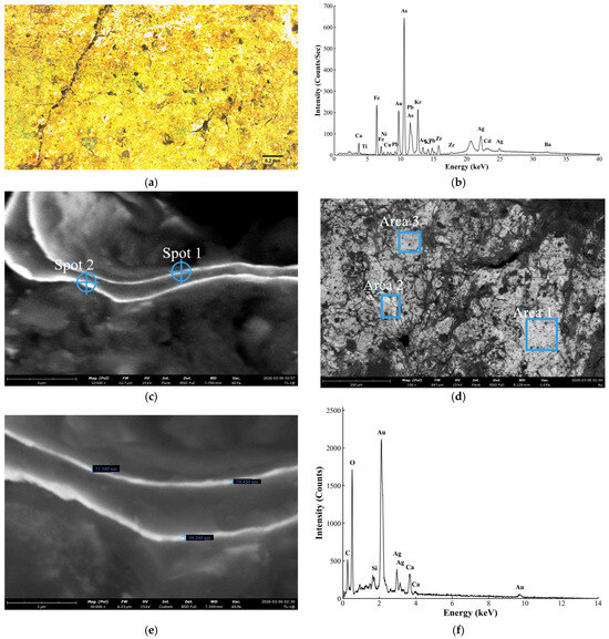
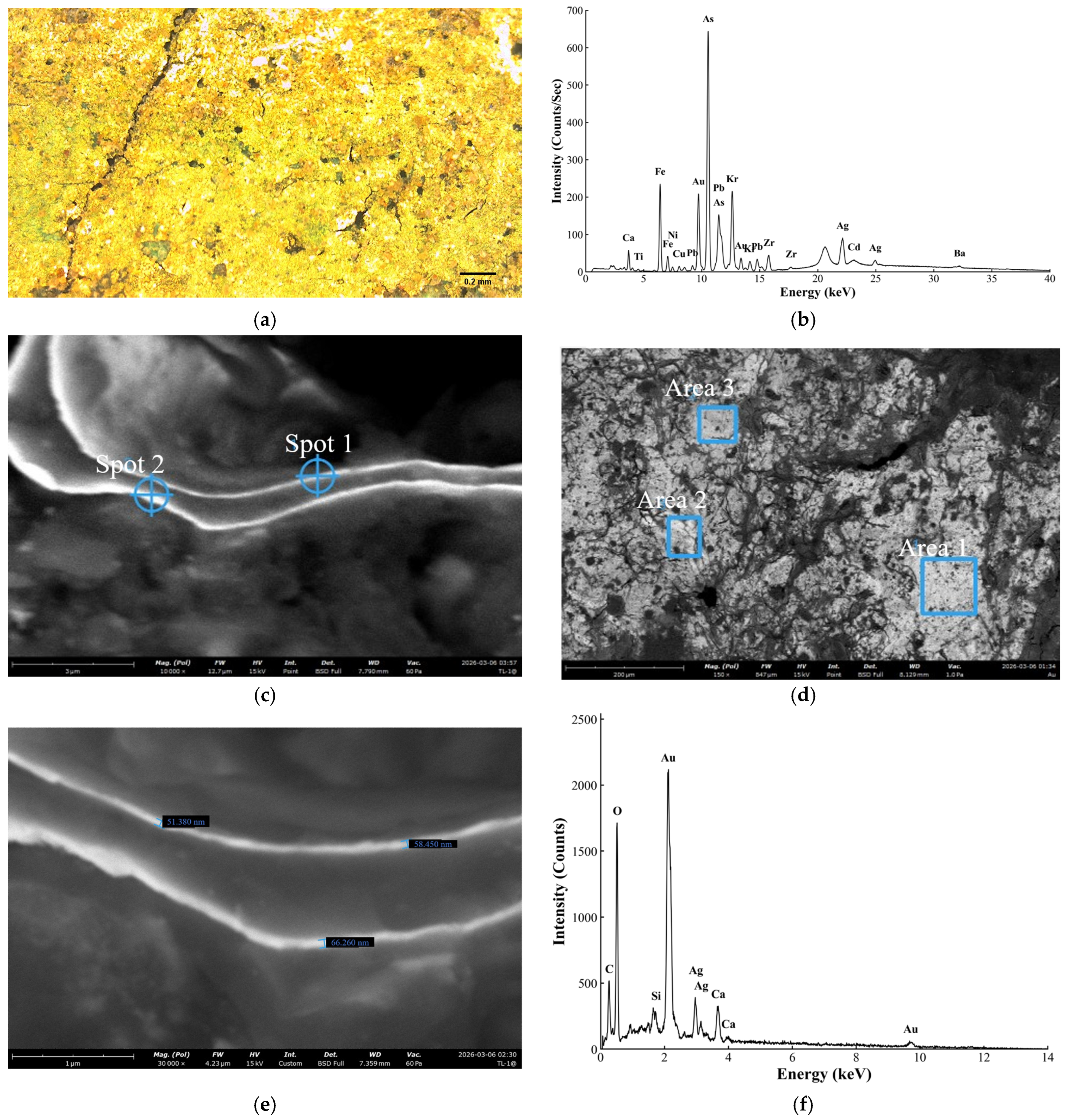
At 200× (Figure 3a), the layer (Sample 02; Appendix A, Table A1) shows high reflectance, a fine crack network, and occasional overlap seams, features which are consistent with gold-leaf gilding [22].
Figure 3.
Micromorphology and elemental characteristics of the gilding layer: (a) a digital micrograph of the gilded area at 200×; (b) XRF spectrum; (c) a cross-sectional SEM image indicating the analyzed spots (spots 1 and 2); (d) a surface SEM image indicating the analyzed areas (Region 1–3); (e) a high-magnification cross-sectional SEM image showing the thickness of the metallic leaf (approximately 51–66 nm); and (f) a representative SEM–EDX spectrum of the gilded layer (Region 1).
The portable XRF detected Au in the gilded area (Figure 3b), supporting the identification of the surface decoration as gold-leaf gilding. Because the p-XRF interaction depth substantially exceeds the foil thickness, the detected signal integrates contributions from multiple underlying layers; in accordance with this, the spectrum is only used here to confirm the presence of Au and to qualitatively describe the mixed elemental signals of the layered structure. The prominent feature in the As/Pb energy region is attributed mainly to underlying paint layers rather than to the metallic leaf itself; given the overlap of As and Pb signals in this region, these peaks are interpreted with caution. The As signal most plausibly originates from an underlying Cu–As green layer, while Pb, Fe, Ca, and Ti are consistent with contributions from adjacent paint and ground layers. The Ag peaks near 22 and 25 keV, so should also be interpreted with caution, as they may partly reflect the Ag-anode source rather than the silver in the leaf. Additional SEM–EDX analyses (Figure 3c,d) were carried out on both the gilded surface and the cross-section. These analyses detected Au as the dominant metallic element, with minor Ag also present, providing more direct microanalytical support for the identification of an Au-rich gold leaf. The cross-sectional SEM observation further showed that the metallic leaf is extremely thin, with a thickness of approximately 51–66 nm (Figure 3e). Because this thickness is much smaller than the SEM–EDX interaction volume under the analytical conditions used, both surface and cross-sectional spectra inevitably include contributions from adjacent and underlying materials. In the surface analyses, the electron beam may penetrate the metallic leaf and reach the underlying layers, whereas in the cross-sectional analyses, the signal may additionally spread laterally across the interface and include contributions from surrounding materials. In accordance with this, minor elements such as Pb and As, detected only in some analyzed areas, are interpreted cautiously and are not taken as primary indicators of the composition of the metallic leaf. A representative SEM–EDX spectrum is shown in Figure 3f, and the detailed quantitative results for all analyzed areas and spots are provided in Appendix A (Table A3). The gilding is therefore identified here as a Au-rich gold leaf, with minor Ag detected, although the present data are not sufficient to assign a precise compositional grade or purity class.
3.3. Composition of the Green Pigment
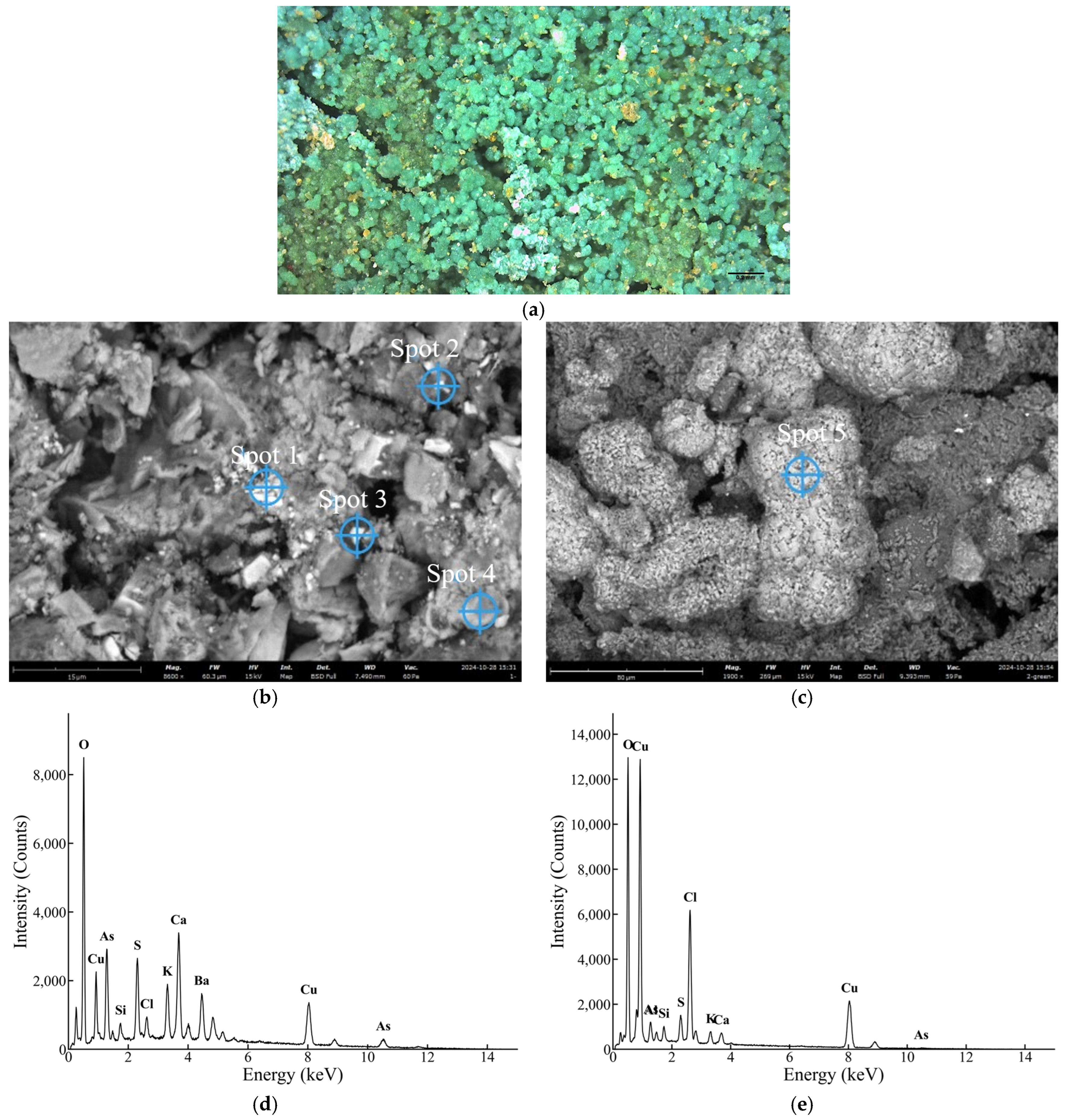
At 200× (Figure 4a), the pigment particles of the green paint layer (Sample 03; Appendix A, Table A1) are predominantly rounded to sub-rounded, with diffuse outlines and poorly developed crystal facets, occurring as dense grape-like agglomerates. This morphology is more typical of synthetic pigments [23,24]. In contrast, natural mineral pigments often manifest as fragmental or flake-like particles with relatively clear crystal faces and distinct edges [25].
Figure 4.
Micromorphology and SEM–EDX characterization of the green layer: (a) a digital micrograph at 200×; (b) an SEM image showing EDX spot locations (spots 1–4); (c) an SEM image showing the EDX spot location (spot 5); (d) the EDX spectrum of spot 3; and (e) the EDX spectrum of Spot 5. Detailed elemental compositions for all spots are listed in Table 3.
SEM–EDX spot analyses (Figure 4 b–d) confirm that Cu and As are consistently co-detected across the green layer (Table 3), providing the primary elemental evidence for a Cu–As pigment system. The spot showing the highest Cu and As concentrations (spot 4) most likely reflects the main pigment phase, while the spots with elevated Ba and S are consistent with the presence of a BaSO4 extender. Minor Cl is detected at most measurement points. An apparent W signal is present at one spot but is interpreted as a spectral overlap artifact between W M lines and Si K lines in this compositionally complex matrix rather than as a genuine W-bearing phase. Notably, one micro-region (Figure 4e) shows a pronounced Cu–Cl co-enrichment accompanied by a marked relative depletion in As compared with the other spots, pointing to a localized Cu–Cl-rich phase that may be indicative of alteration products derived from the Cu–As pigment.
Table 3.
The EDX results for the green pigment (wt.%).
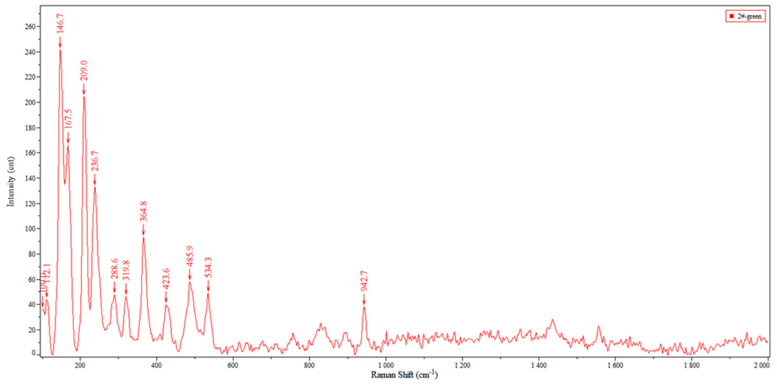
Raman and elemental evidence support identification of the green pigment as emerald green (Cu(C2H3O2)2·3Cu(AsO2)2). The Raman spectrum (Figure 5) shows diagnostic bands at 146, 209, and 364 cm−1, together with a prominent acetate-related band near 950 cm−1 (942 cm−1 in this sample) [26]. This ~950 cm−1 feature is attributed to acetate-group vibrations and distinguishes emerald green from other Cu–As green pigments such as Scheele’s green [27]. Emerald green was first produced commercially in 1814 and has been widely reported in Chinese architectural polychromy since the late Qing period (often after the 1890s) [28,29]. Therefore, its presence provides a terminus post quem for restoration at the TaiLing site. A late Qing or later intervention is the most plausible interpretation when considering stratigraphy and its associated pigments.
Figure 5.
Micro-Raman spectrum of the green paint layer.
Spot 5 shows Cu–Cl enrichment accompanied by depleted As (Table 3). Considering the identified pigment (emerald green) and published evidence, this pattern may reflect localized alteration. Emerald green is susceptible to degradation under light, moisture, and salt-bearing conditions, with processes involving oxidation and mobilization of arsenic species and the formation of secondary Cu-bearing phases [24,29]. A plausible explanation is that As(III) in emerald green oxidizes and migrates outward as more mobile arsenate species under humid and salt-affected conditions, thereby lowering the local As/Cu ratio. Synchrotron-based studies have documented such ion-migration pathways at the molecular scale [30,31], which is consistent with the depleted As/Cu ratio observed at spot 5. In parallel, released Cu2+ may be retained locally and combine with environmental chloride to form Cu–Cl-rich secondary phases. Related studies on Chinese cave and architectural polychromy have reported chloride-bearing alteration products (e.g., lavendulan) that have derived from emerald green [29].
No complementary phase identification was performed on this micro-region; the discussion of secondary phases remains tentative. Nevertheless, the localized Cu–Cl anomaly suggests that the micro-environment at this site may be influenced by moisture fluctuation and soluble-salt transport. Future conservation-oriented investigations should prioritize phase-specific identification of the alteration products in this micro-region—for example, by micro-XRD or targeted Raman mapping—before treatment decisions are made. In the interim, conservation efforts should focus on humidity stabilization, a reduction in soluble-salt input, and an avoidance of chloride-bearing cleaning or consolidation materials.
3.4. Composition of the Blue Pigment
3.4.1. Surface Blue Pigment
At 200× (Figure 6a), the blue layer (Sample 04; Appendix A, Table A1) is dominated by homogeneous fine particles with a narrow size distribution. Coarse mineral grains and obvious impurities typical of natural pigments are not observed. The dense packing and high hiding power are consistent with a synthetic pigment [32].
Figure 6.
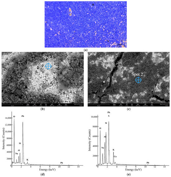
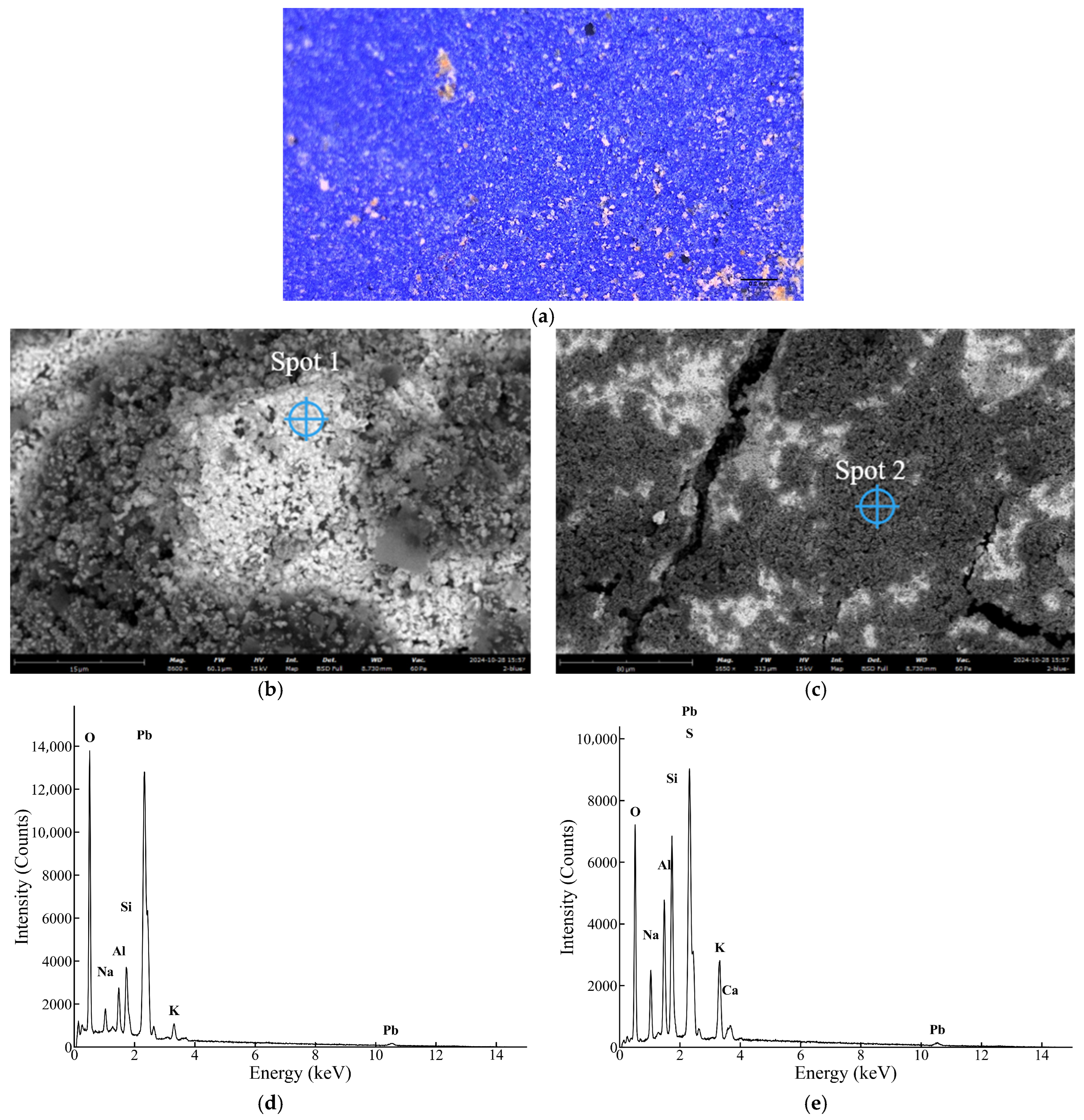
Micromorphology and SEM–EDX spot-analysis results of the upper blue paint layer: (a) a digital micrograph at 200×; (b) an SEM image showing the EDX spot location (spot 1); (c) an SEM image showing the EDX spot location (spot 2); (d) the EDX spectrum of spot 1; and (e) the EDX spectrum of spot 2.
SEM–EDX spot analyses (Table 4, Figure 6b–e) indicate mixing has occurred between blue and Pb-rich white components. Spot 1 is strongly enriched in Pb, with negligible S and only minor light elements, consistent with a locally concentrated lead white-rich phase. By contrast, spot 2 shows a Na–Al–Si–S association that is consistent with ultramarine, together with substantial Pb and K. This spot-to-spot heterogeneity suggests an intentional mixing of the blue pigment with a lead-based white component, broadly interpreted here as lead white, to modify hue and opacity [32,33].
Table 4.
EDX results for the surface blue paint layer (wt.%).
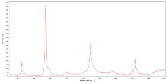
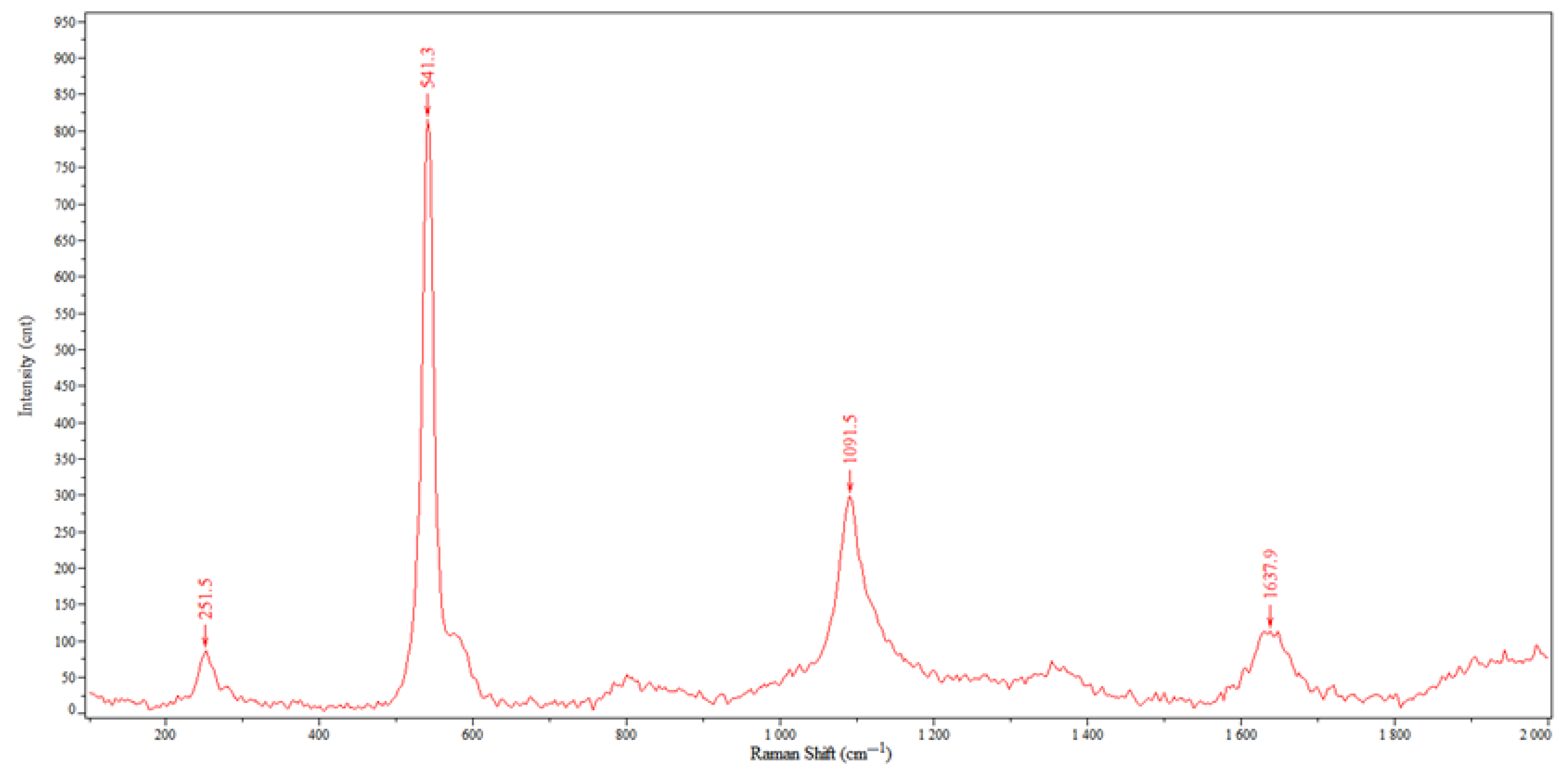
Raman spectroscopy confirms the identification (Figure 7). Strong bands at 251.5 and 541.3 cm−1 are observed; the 541.3 cm−1 band is assigned to the symmetric stretching vibration of the S3− radical anion, which is a diagnostic of ultramarine [31,34]. A band at 1091.5 cm−1 indicates carbonates, likely from calcite fillers or the underlying substrate [35]. A weak band near 1637.9 cm−1 may relate to the adsorbed water or organic matrix contributions [36]. Although the Raman spectrum does not show a complete diagnostic set of carbonate bands for lead white, this does not exclude the presence of a lead-white component in the paint layer. In the present interpretation, the lead-white assignment is based primarily on the Pb-rich SEM–EDX results and the observed micro-scale heterogeneity, while the Raman data are insufficient to distinguish the specific lead-bearing phase in the analyzed micro-area.
Figure 7.
Micro-Raman spectrum of the upper blue paint layer.
Synthetic ultramarine was invented in Europe in 1826–1828 and quickly replaced expensive natural ultramarine due to its vibrant color and low cost. It was commonly used in combination with lead white [37]. The pigment was introduced to China via European trade routes in the mid-to-late 19th century and became widely used in architectural polychromy and mural restoration during the late Qing period [38]. Its presence therefore provides a terminus post quem for dating interventions; synthetic ultramarine has been identified in conservation studies of sites including the Forbidden City and the Summer Palace in Beijing [39,40,41].
3.4.2. Bottom Blue Pigment
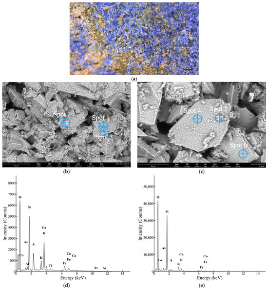
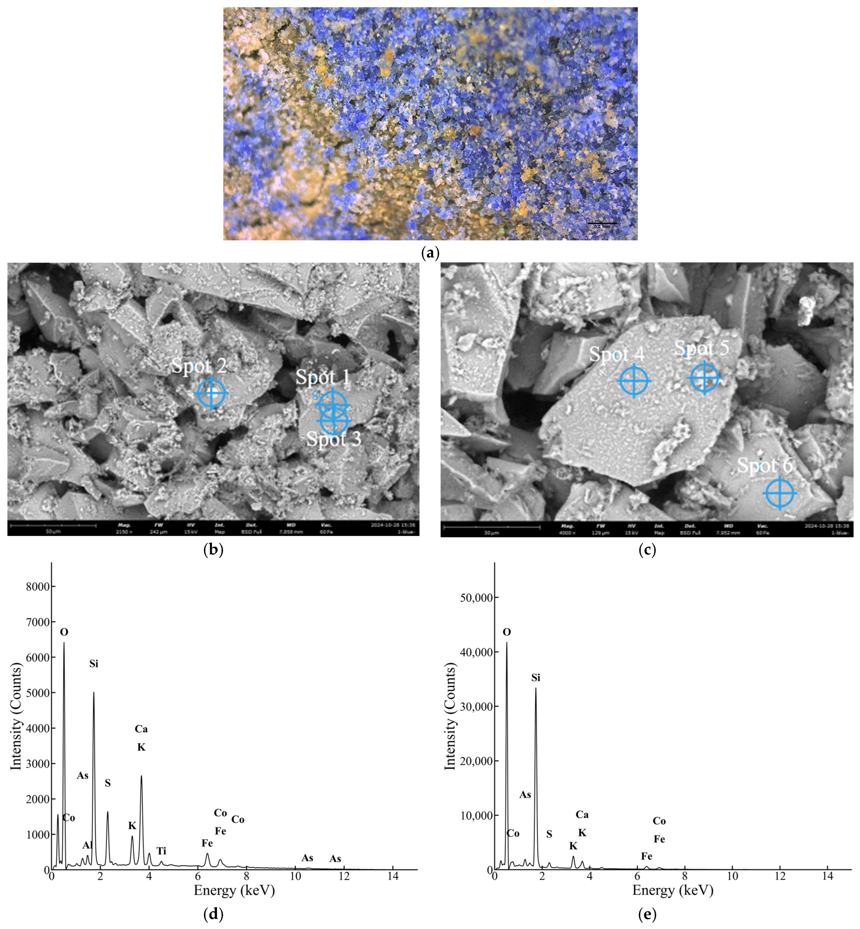
At 200× (Figure 8a), most blue grains of the bottom blue layer (Sample 05; Appendix A, Table A1) are angular fragments with sharp boundaries and a broad size distribution; some are translucent with a glassy luster, consistent with crushed glassy material [42].
Figure 8.
Micromorphology and SEM–EDX spot-analysis results of the lower blue paint layer: (a) a digital micrograph at 200×; (b) an SEM image showing EDX spot locations (spots 1–3); (c) an SEM image showing EDX spot locations (spots 4–6); (d) the EDX spectrum of Spot 1; and (e) the EDX spectrum of Spot 4. Detailed elemental compositions for all spots are listed in Table 5.
Given the compositional heterogeneity, identification of the lower blue layer is based primarily on the angular glass-like particle morphology observed in OM and SEM, together with the SEM–EDX spot analyses (Figure 8b–e; Table 5). Blue-rich micro-areas consistently show Co, K, and Si, with frequent As signals. K and Si indicate a potash silicate glass matrix, Co is the chromophore, and the Co–As association is consistent with reported smalt compositions [43]. These features together support the identification of the lower blue pigment as smalt.
Table 5.
EDX results for the bottom blue paint layer (wt.%).
Smalt was widely used in European paintings and polychrome decoration from the fifteenth to the nineteenth centuries but has often been overlooked in Chinese studies. Recent surveys trace its earliest confirmed use in China to at least Kara Khoto [42]. It became one of the more commonly used blue pigments in architectural polychrome paintings and murals from the late Ming to the Qing dynasties (16th–19th centuries). It is found in both royal architecture [44] and local temples. This indicates a sustained supply and use rather than occasional importation [45].
In China, smalt has been reported in combination with indigo or azurite and in some cases in lower blue layers that are associated with azurite-bearing upper layers [42]. However, in the present lower-blue sample, azurite was not detected in the analyzed micro-areas, indicating that the analyzed layer is dominated by smalt.
Smalt, as a K-rich silicate glass, is moisture-sensitive: humid conditions can promote K leaching, which changes the local coordination environment of Co and shifts the color toward fading or graying [46]. Similar K mobility has been discussed in oil-paint systems, where K migration and secondary potassium salts/soaps formed by reactions with organic acids have been reported [45,47]. Taken together, these observations point to a higher alteration risk for smalt-bearing layers when moisture and soluble salts are present. For treatment, the implication is straightforward: avoid keeping these layers wet for long periods, and be cautious with cleaning/consolidation systems that introduce soluble ions or promote ion exchange. Maintaining stable humidity and limiting salt input/redistribution are generally more effective than repeated surface retouching.
3.5. Composition of the Red Pigment
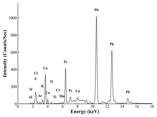
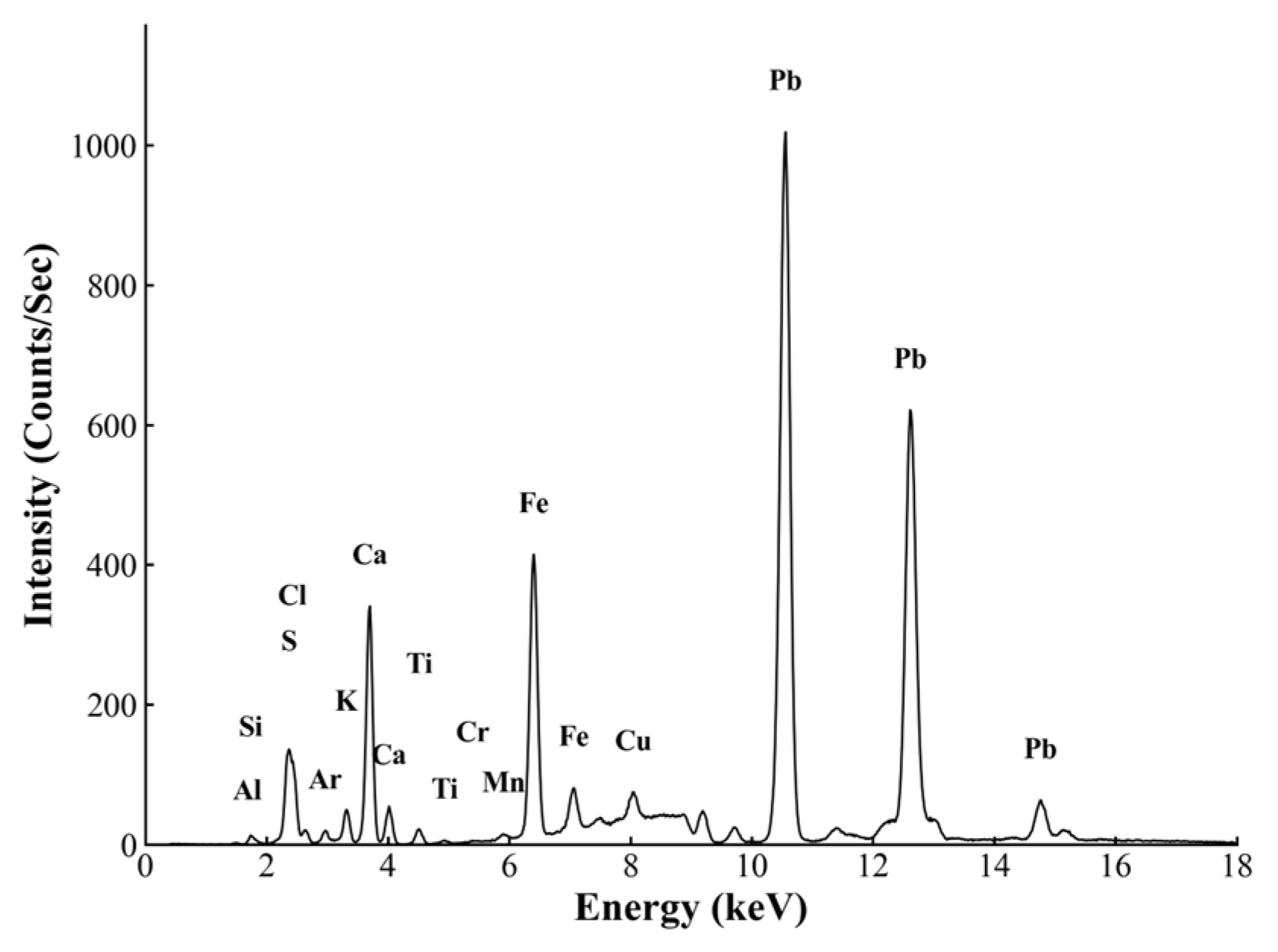
The red sample (Sample 06; Appendix A, Table A1) was examined by p-XRF and μ-RS. The p-XRF spectrum (Figure 9) shows a distinct Fe signal, indicating the presence of an iron-based red pigment. Signals of Al, Si, S, K, Ca, Ti, and Pb are also present. Because the interaction depth of p-XRF exceeds the thickness of the surface paint layer, these signals likely reflect contributions not only from the red paint layer itself but also from underlying or adjacent layers within the local stratigraphy. In particular, the Pb signal is most plausibly attributed to a lead-bearing white material, most likely lead white, while Ti is more likely to be associated with Ti-bearing mineral components in the underlying layer.
Figure 9.
The p-XRF spectrum of the red layer.
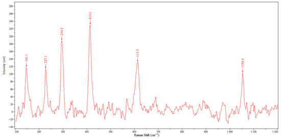
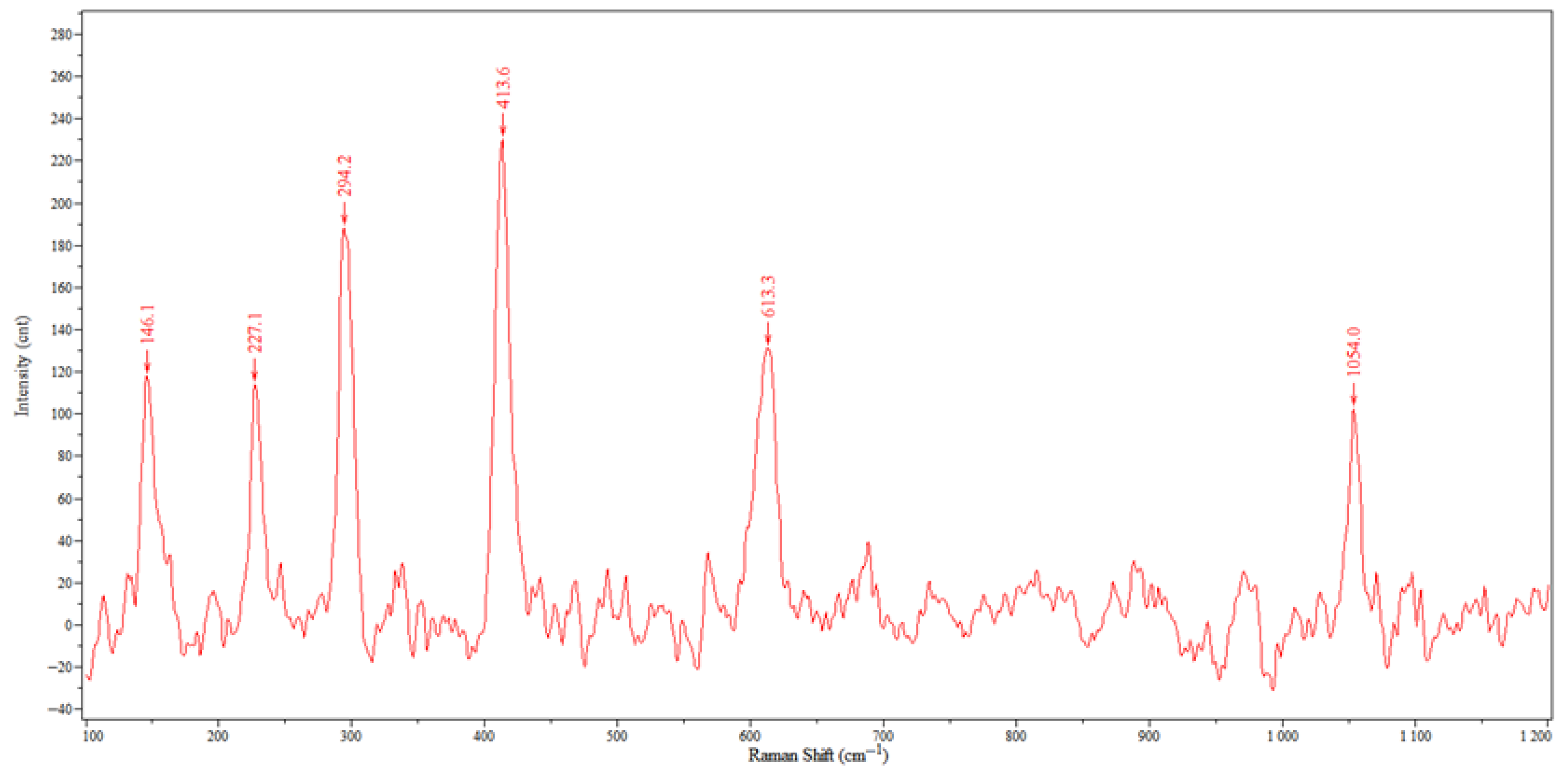
Raman spectra of the red sample show distinct bands at 146.1, 227.1, 294.2, 413.6, and 613.3 cm−1 (Figure 10), matching the lattice modes of hematite. In accordance with this, the red chromophore is identified as iron red dominated by hematite [48].
Figure 10.
Micro-Raman spectrum of the red paint layer.
3.6. Composition of the Black Pigment
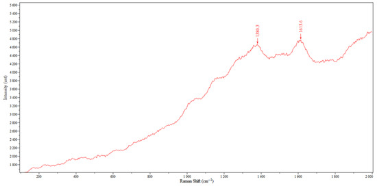
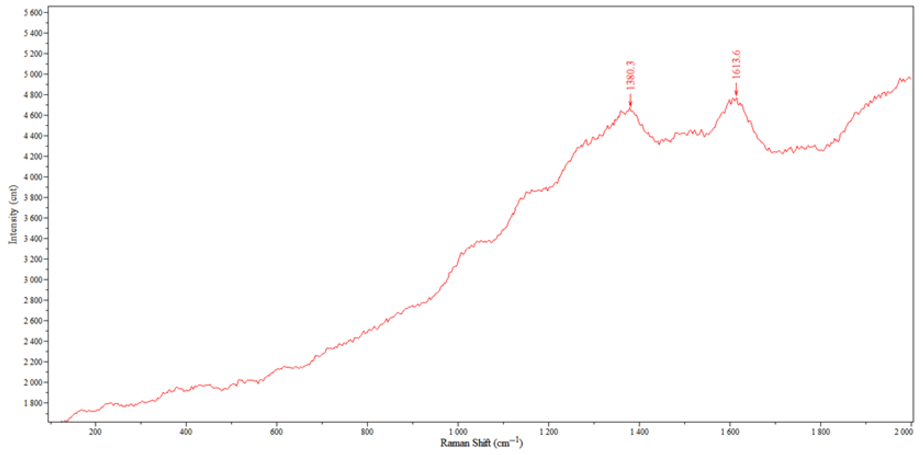
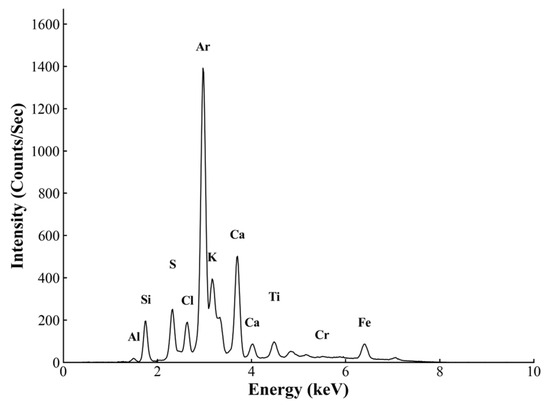
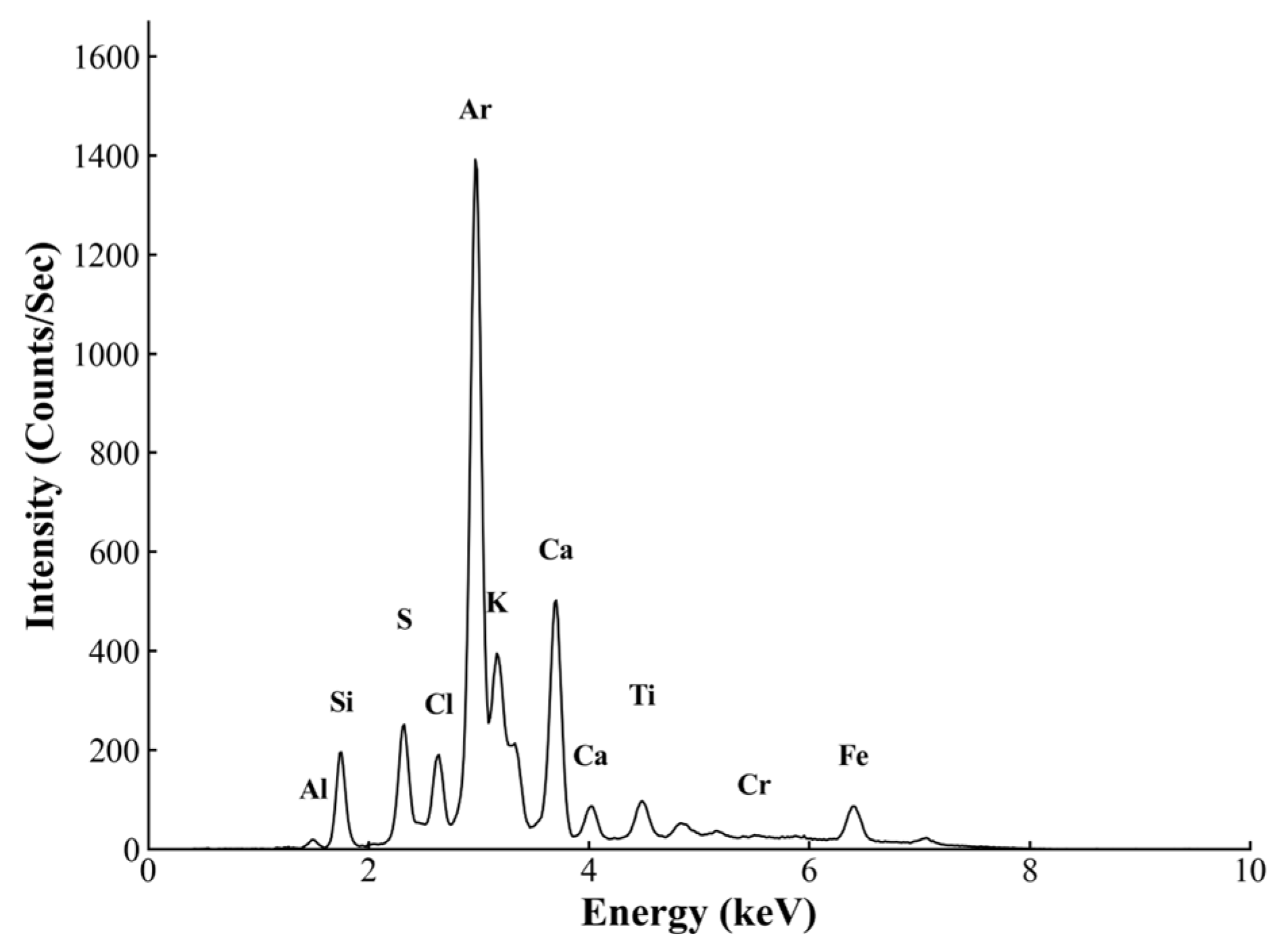
Raman spectrum (Figure 11) of black pigment (Sample 07; Appendix A, Table A1) is dominated by luminescence with a rising baseline, suggesting an organic binder or surface contamination. After background subtraction, broad bands at ~1380 and ~1614 cm−1 correspond to the D and G bands of carbonaceous material, respectively, which is characteristic of carbon black [28]. No diagnostic low-wave number peaks of inorganic black pigments (e.g., magnetite and manganese oxides) are observed, confirming that the black component is carbon-based. In ancient China, carbon black was commonly produced from the incomplete combustion of wood or bone [16,49]. However, micro-Raman spectroscopy does not always allow a secure distinction between these sources because the phosphate symmetric stretching band near 960 cm−1, often associated with bone black, is not invariably visible. In the present sample, no distinct band is observed near 960 cm−1. In addition, p-XRF analysis did not detect a clear P signal (Figure 12). Taken together, these results suggest that the black pigment is more likely to be plant-derived carbon black rather than bone black.
Figure 11.
Micro-Raman spectrum of the black paint layer.
Figure 12.
XRF spectrum of the black layer.
4. Conclusions
Focusing on the TaiLing Long’en Gate polychrome paintings, this paper integrated multiple analytical means such as OM, XRD, XRF, SEM-EDX, and μ-RS to study their material composition and manufacturing techniques. The main conclusions are as follows:
- Material Composition: The ground layer of the TaiLing Long’en Gate polychrome paintings is composed of soil mixed with sand. Its mineral composition is dominated by quartz and augite, and it contains a relatively high content of illite. The blue pigments are smalt and synthetic ultramarine (locally mixed with lead-containing components for tinting). The green pigment is emerald green and is accompanied by barium sulfate fillers. The red pigment is hematite, the black pigment is carbon black, and the gold color is gold-leaf gilding. In addition, chlorine enrichment phenomena appeared locally in green-related samples; the chlorine may originate from the degradation products of the emerald green pigment.
- Restoration History: The superimposition phenomenon of localized emerald green overlying the blue layer, as well as the use of modern pigments such as synthetic ultramarine, indicates the existence of modern restoration or repainting phenomena.
- Conservation Implications: Given that the TaiLing Long’en Gate polychrome paintings contain components that are relatively sensitive to moisture, such as illite and smalt, and the emerald green locally presents the phenomenon of Cl enrichment and co-occurrence with Cu, subsequent restoration should prioritize controlling dampness and seepage and reducing the risk of soluble salt migration and avoid the introduction of chlorine-containing cleaning agents or materials and processes that may bring in chloride ions.
Author Contributions
Conceptualization, Z.Z., L.L. and W.W.; methodology, W.W.; software, W.W.; validation, Z.Z.; formal analysis, W.W. and Y.S.; investigation, W.W., Y.S., M.Y. and Z.S.; resources, M.Y. and Z.S.; data curation, W.W.; writing—original draft preparation, W.W.; writing—review and editing, Z.Z. and L.L.; visualization, W.W. and Y.S.; supervision, Z.Z.; project administration, Z.Z.; funding acquisition, Z.Z. All authors have read and agreed to the published version of the manuscript.
Funding
This research was funded by the financial support from the National Natural Science Foundation of China (Key Project, No. 42530718) and the National Natural Science Foundation of China (No. 42272336).
Data Availability Statement
The data presented in this study are available on request from the corresponding author.
Acknowledgments
The authors would like to thank the Administration of Western Qing Tombs for providing the research samples and access to the site. We are grateful to Saimijiang Wulamu for his assistance with the transportation and on-site sampling. We also appreciate Junjie Guo from Phenom Scientific for his help with the SEM-EDX analysis and Shiying Yang for her assistance in optimizing the figures. We thank Zhenyu Long for his constructive suggestions on the revision of this manuscript. Finally, we express our gratitude to the anonymous reviewers for their valuable comments that helped improve this paper.
Conflicts of Interest
The authors declare no conflicts of interest.
Appendix A
Table A1.
Sampling information.
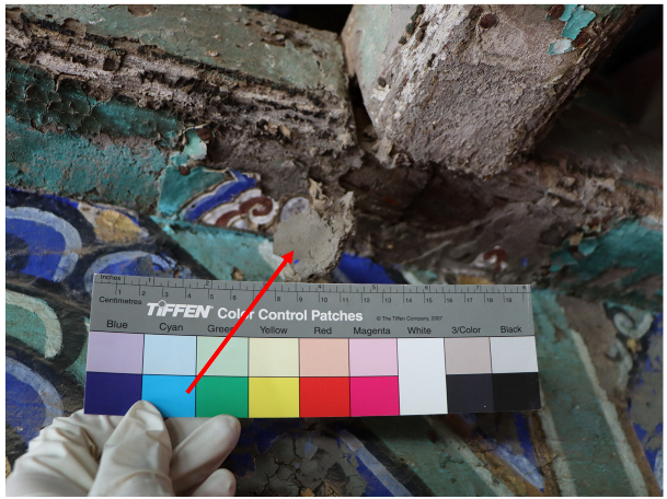
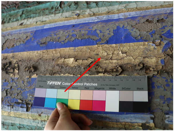
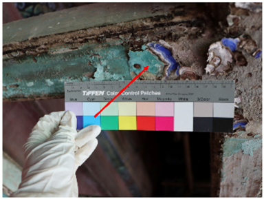
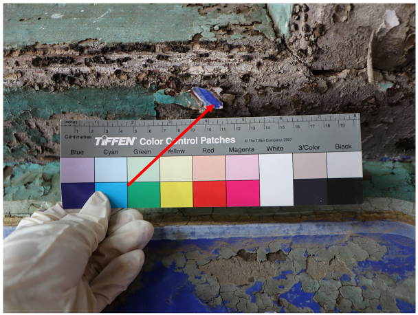
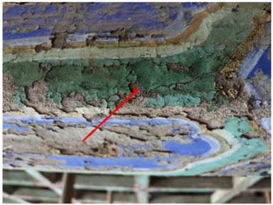
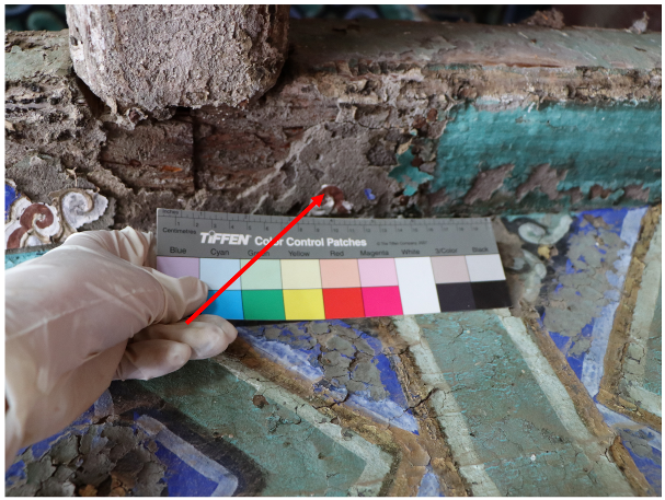
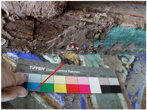
Table A2.
The target element lists of p-XRF for each mode.
Table A3.
EDX results for the Gilding Material (wt.%).
References
- Dang, X.; Hong, Q.; Liu, W.; Wang, Y. China’s heritage governance blueprint: Revisiting evolutionary trajectories, reframing institutional priorities and mapping the national registry. Habitat Int. 2025, 165, 103541. [Google Scholar] [CrossRef] [Scilit]
- Shen, L.; Hua, D.; Nan, B.; Yao, Y.; Duan, H.; Wang, J. Material and Technique Analysis of Qing Dynasty Official Style Architectural Polychrome Paintings in Hangzhou, Zhejiang, China. Crystals 2025, 15, 92. [Google Scholar] [CrossRef] [Scilit]
- Liu, L.Y.; Zhang, B.J.; Yang, H.; Zhang, Q. The Analysis of the Colored Paintings from the Yanxi Hall in the Forbidden City. Spectrosc. Spectr. Anal. 2018, 38, 2054–2063. [Google Scholar]
- Stamboliyska, B.; Tapanov, S.; Kovacheva, D.; Atanasova-Vladimirova, S.; Ranguelov, B.; Yancheva, D.; Velcheva, E.; Stoyanov, S.; Guncheva, M.; Fischer, D.; et al. Characterization of art materials and degradation processes in the exterior wall paintings of the main church of Rila Monastery, Bulgaria. Vib. Spectrosc. 2023, 128, 103580. [Google Scholar] [CrossRef] [Scilit]
- He, L.; Wang, N.; Zhao, X.; Zhou, T.; Xia, Y.; Liang, J.; Rong, B. Polychromic structures and pigments in Guangyuan Thousand-Buddha Grotto of the Tang Dynasty (China). J. Archaeol. Sci. 2012, 39, 1809–1820. [Google Scholar] [CrossRef] [Scilit]
- Clark, R.J.H. Raman microscopy: Application to the identification of pigments on medieval manuscripts. Chem. Soc. Rev. 1995, 24, 187–196. [Google Scholar] [CrossRef] [Scilit]
- McCarthy, B.; Giaccai, J. (Eds.) Scientific Studies of Pigments in Chinese Paintings; Archetype Publications: London, UK, 2021. [Google Scholar]
- Yang, H.R.; Lee, C.H.; Yi, J. Analysis of pigments and damages for the 19th century White-robed Water-moon Avalokitesvara Painting in Gongju Magoksa Temple, Republic of Korea. Herit. Sci. 2021, 9, 139. [Google Scholar] [CrossRef] [Scilit]
- Sá, S.; Hendriks, L.; Cardoso, I.P.; Hajdas, I. Radiocarbon dating of lead white: Novel application in the study of polychrome sculpture. Sci. Rep. 2021, 11, 13210. [Google Scholar] [CrossRef] [Scilit]
- Ma, C.; Dou, H.; Zhao, Z.; Qiu, X.; Li, H.; Wang, X. Review of in-situ non-and micro-destructive techniques for pigment analysis in architectural heritage. npj Herit. Sci. 2025, 13, 222. [Google Scholar] [CrossRef] [Scilit]
- do Nascimento Campos, G.; Granato, M.; Middea, A.; de Souza Gonçalves Vasques, F.; da Fonseca Martins Gomes, O. Multitechnique Characterization of Pigments Used in Paintings by Léon Pallière. Microsc. Microanal. 2023, 29, 1315–1327. [Google Scholar] [CrossRef] [Scilit] [PubMed]
- Valadas, S.; Candeias, A.; Dias, C.; Schiavon, N.; Cotovio, M.; Pestana, J.; Gil, M.; Mirão, J. A multi-analytical study of the fifteenth century mural paintings of the Batalha Monastery (Portugal) in view of their conservation. Appl. Phys. A 2013, 113, 989–998. [Google Scholar] [CrossRef] [Scilit]
- Cheilakou, E.; Troullinos, M.; Koui, M. Identification of pigments on Byzantine wall paintings from Crete (14th century AD) using non-invasive Fiber Optics Diffuse Reflectance Spectroscopy (FORS). J. Archaeol. Sci. 2014, 41, 541–555. [Google Scholar] [CrossRef] [Scilit]
- Araya, C.; Jaque, J.; Naranjo, N.; Icaza, M.; Clavijo, R.E.; Aguayo, T.; Campos-Vallette, M.M. Raman characterization of pigments in painted beams and a wall painting discovered in the San Francisco church in Santiago, Chile. Spectrosc. Lett. 2014, 47, 177–183. [Google Scholar] [CrossRef] [Scilit]
- Li, X. Research on the evolution and technical appraisal of pigment use in Chinese Painting in the Late Ming and Early Qing Dynasties. Mediterr. Archaeol. Archaeom. 2024, 24, 140–153. [Google Scholar]
- Fu, P.; Teri, G.L.; Li, J.; Li, J.-X.; Li, Y.-H.; Yang, H. Investigation of ancient architectural painting from the Taidong tomb in the western qing tombs, Hebei, China. Coatings 2020, 10, 688. [Google Scholar] [CrossRef] [Scilit]
- Elert, K.; Rodriguez-Navarro, C. Degradation and conservation of clay-containing stone: A review. Constr. Build. Mater. 2022, 330, 127226. [Google Scholar] [CrossRef] [Scilit]
- Avrami, E.; Guillaud, H.; Hardy, M. Terra Literature Review: An Overview of Research in Earthen Architecture Conservation; The Getty Conservation Institute: Los Angeles, CA, USA, 2008. [Google Scholar]
- Iafrate, S.; Giandomenico, M.; Cucchietti, R.; Russo, C.; Bartolini, M.; Conti, L.; De Angelis, S.; Fontani, V.; Kumbaric, A.; Sidoti, G.; et al. Towards the Definition of Guidelines for the Conservation of Mural Paintings in Hypogea. Heritage 2025, 8, 472. [Google Scholar] [CrossRef] [Scilit]
- Arnold, A.; Zehnder, K. Monitoring Wall Paintings Affected by Soluble Salts; Getty Conservation Institute: Marina Del Rey, CA, USA, 1991. [Google Scholar]
- Hu, T.; Brimblecombe, P.; Zhang, Z.; Song, Y.; Liu, S.; Zhu, Y.; Duan, J.; Cao, J.; Zhang, D. Capillary rise induced salt deterioration on ancient wall paintings at the Mogao Grottoes. Sci. Total Environ. 2023, 881, 163476. [Google Scholar] [CrossRef] [Scilit] [PubMed]
- Darque-Ceretti, E.; Felder, E.; Aucouturier, M. Foil and leaf gilding on cultural artifacts: Forming and adhesion. Matéria 2011, 16, 540–559. [Google Scholar] [CrossRef] [Scilit]
- Dong, S.; Xiang, J.; Ji, J.; Wang, Y.; Zhang, G.; Fu, P.; Han, J.; Li, L. Multi-method analysis of painting materials in murals of the north mosque (Linqing, China). Coatings 2023, 13, 1298. [Google Scholar] [CrossRef] [Scilit]
- Li, Z.; Wang, L.; Chen, H.; Ma, Q. Degradation of emerald green: Scientific studies on multi-polychrome Vairocana Statue in Dazu Rock Carvings, Chongqing, China. Herit. Sci. 2020, 8, 64. [Google Scholar] [CrossRef] [Scilit]
- Artesani, A.; Lamuraglia, R.; Menegazzo, F.; Bonetti, S.; Traviglia, A. Terahertz time-domain spectroscopy in reflection configuration for inorganic and mineral pigment identification. Appl. Spectrosc. 2023, 77, 74–87. [Google Scholar] [CrossRef] [Scilit]
- Bell, I.M.; Clark, R.J.H.; Gibbs, P.J. Raman spectroscopic library of natural and synthetic pigments (pre-≈ 1850 AD). Spectrochim. Acta A 1997, 53, 2159–2179. [Google Scholar] [CrossRef] [Scilit] [PubMed]
- Pantoja Munoz, L. Hidden in plain sight: Revisiting the synthesis, characterisation, degradation and the intricate relationship between Scheele’s green and Emerald green. Herit. Sci. 2024, 12, 94. [Google Scholar] [CrossRef] [Scilit]
- Shen, A.G.; Wang, X.H.; Xie, W.; Shen, J.; Li, H.Y.; Liu, Z.A.; Hu, J.M. Pigment identification of colored drawings from Wuying Hall of the Imperial Palace by micro-Raman spectroscopy and energy dispersive X-ray spectroscopy. J. Raman Spectrosc. 2006, 37, 230–234. [Google Scholar] [CrossRef] [Scilit]
- Shen, L.; Wang, C.; Zhang, J.; Cui, B.; Zhu, S.; Mao, J. Cu and As containing pigments in Zhejiang architecture polychrome paintings: A case study of degradation products of emerald green. Herit. Sci. 2023, 11, 9. [Google Scholar] [CrossRef] [Scilit]
- Sun, F.; Wang, R.; Qi, D.; Yan, H. Degradation of emerald green pigment in painted grottoes in Sichuan, China. J. Cult. Herit. 2024, 69, 1–9. [Google Scholar] [CrossRef] [Scilit]
- Carboni Marri, S.; Monico, L.; Rosi, F.; Miliani, C.; Vivani, R.; Janssens, K.; De Meyer, S.; Mathon, O.; Cotte, M.; Burghammer, M.; et al. Discovering the dual degradation pathway of emerald green in oil paints: The effects of light and humidity. Sci. Adv. 2025, 11, eady1807. [Google Scholar] [CrossRef] [Scilit]
- Osticioli, I.; Mendes, N.F.C.; Nevin, A.; Gil, F.P.; Becucci, M.; Castellucci, E. Analysis of natural and artificial ultramarine blue pigments using laser induced breakdown and pulsed Raman spectroscopy, statistical analysis and light microscopy. Spectrochim. Acta A 2009, 73, 525–531. [Google Scholar] [CrossRef] [Scilit]
- Li, K.; Han, K.; Teri, G.; Tian, Y.; Cui, M.; Qi, Y.; Li, Y. A Study on the Materials Used in Ancient Wooden Architectural Paintings at DaZhong Gate in Confucius Temple, Qufu, Shandong, China. Materials 2024, 17, 2170. [Google Scholar] [CrossRef] [Scilit] [PubMed]
- Li, Y.; Wang, F.; Ma, J.; He, K.; Zhang, M. Study on the pigments of Chinese architectural colored drawings in the Altar of Agriculture (Beijing, China) by portable Raman spectroscopy and ED-XRF spectrometers. Vib. Spectrosc. 2021, 116, 103291. [Google Scholar] [CrossRef] [Scilit]
- González-Cabrera, M.; Arjonilla, P.; Domínguez-Vidal, A.; Ayora-Cañada, M. Natural or synthetic? Simultaneous Raman/luminescence hyperspectral microimaging for the fast distinction of ultramarine pigments. Dyes Pigm. 2020, 178, 108349. [Google Scholar] [CrossRef] [Scilit]
- Gambardella, A.A.; Cotte, M.; de Nolf, W.; Schnetz, K.; Erdmann, R.; van Elsas, R.; Gonzalez, V.; Wallert, A.; Iedema, P.D.; Eveno, M.; et al. Sulfur K-edge micro-and full-field XANES identify marker for preparation method of ultramarine pigment from lapis lazuli in historical paints. Sci. Adv. 2020, 6, eaay8782. [Google Scholar] [CrossRef] [Scilit] [PubMed]
- Pozzi, F.; Lombardi, J.R.; Bruni, S.; Leona, M. Sample treatment considerations in the analysis of organic colorants by surface-enhanced Raman scattering. Anal. Chem. 2012, 84, 3751–3757. [Google Scholar] [CrossRef] [Scilit]
- Mertens, J. The history of artificial ultramarine (1787–1844): Science, industry and secrecy. Ambix 2004, 51, 219–244. [Google Scholar] [CrossRef]
- Zhang, C.; Huang, J.; Zhu, T.; Zhang, R. Guangzhou Tongcao painting in late China Qing Dynasty (1840–1912 AD): Technology revealed by analytical approaches. Herit. Sci. 2021, 9, 9. [Google Scholar] [CrossRef] [Scilit]
- Ma, Z.; Wang, L.; Yan, J.; Zhou, W.; Pitthard, V.; Bayerova, T.; Krist, G. Chromatographic, microscopic, and spectroscopic characterization of a wooden architectural painting from the Summer Palace, Beijing, China. Anal. Lett. 2019, 52, 1670–1680. [Google Scholar] [CrossRef] [Scilit]
- Lei, Z.; Wu, W.; Shang, G.; Wu, Y.; Wang, J. Study on colored pattern pigments of a royal Taoist temple beside the Forbidden City (Beijing, China). Vib. Spectrosc. 2017, 92, 234–244. [Google Scholar] [CrossRef] [Scilit]
- Xia, Y.; Xi, N.; Huang, J.; Wang, N.; Lei, Y.; Fu, Q.; Wang, W. Smalt: An under-recognized pigment commonly used in historical period China. J. Archaeol. Sci. 2019, 101, 89–98. [Google Scholar] [CrossRef] [Scilit]
- Cavallo, G.; Riccardi, M.P. Glass-based pigments in painting: Smalt blue and lead–tin yellow type II. Archaeol. Anthropol. Sci. 2021, 13, 199. [Google Scholar] [CrossRef] [Scilit]
- Lei, Y.; Yang, H.; Qu, L.; Wang, S. Technical study of architectural paintings (Caihua) in the Forbidden City, Beijing, China. Stud. Conserv. 2014, 59, S242–S243. [Google Scholar] [CrossRef] [Scilit]
- Ping, M.; Jin, H.; Zhang, Z.; Zhang, L.; Sun, M.; Cao, M.; Cui, J. Application of smalt in building glazed tiles from Yuanmingyuan Park, Beijing in Qing dynasty. npj Herit. Sci. 2025, 13, 282. [Google Scholar] [CrossRef] [Scilit]
- Ricciardi, P.; Dooley, K.A.; MacLennan, D.; Bertolotti, G.; Gabrieli, F.; Patterson, C.S.; Delaney, J.K. Use of standard analytical tools to detect small amounts of smalt in the presence of ultramarine as observed in 15th-century Venetian illuminated manuscripts. Heritage 2022, 5, 1234–1256. [Google Scholar] [CrossRef] [Scilit]
- Giannini, R.; Freestone, I.C.; Shortland, A.J. European cobalt sources identified in the production of Chinese famille rose porcelain. J. Archaeol. Sci. 2017, 80, 27–36. [Google Scholar] [CrossRef] [Scilit]
- De Faria, D.L.A.; Venâncio Silva, S.; de Oliveira, M.T. Raman microspectroscopy of some iron oxides and oxyhydroxides. J. Raman Spectrosc. 1997, 28, 873–878. [Google Scholar] [CrossRef]
- Coccato, A.; Jehlicka, J.; Moens, L.; Vandenabeele, P. Raman spectroscopy for the investigation of carbon-based black pigments. J. Raman Spectrosc. 2015, 46, 1003–1015. [Google Scholar] [CrossRef] [Scilit]

Disclaimer/Publisher’s Note: The statements, opinions and data contained in all publications are solely those of the individual author(s) and contributor(s) and not of MDPI and/or the editor(s). MDPI and/or the editor(s) disclaim responsibility for any injury to people or property resulting from any ideas, methods, instructions or products referred to in the content. |
© 2026 by the authors. Licensee MDPI, Basel, Switzerland. This article is an open access article distributed under the terms and conditions of the Creative Commons Attribution (CC BY) license.