


Abstract
A multimethodic approach based on infrared, Raman, electron spin resonance and photoluminescence spectroscopy, absorption spectroscopy in near infrared, visible and ultraviolet regions, single-crystal X-ray diffraction as well as electron microprobe analyses was applied to the characterization of a new commensurately modulated cubic haüyne analogue with the modulation parameter of 0.2 and unit-cell parameter of 45.3629(3) Å (designated as haüyne-45Å) from the Malobystrinskoe lazurite deposit, in the Baikal Lake area, Siberia, Russia, as well as associated SO32−-bearing afghanite. Haüyne-45Å is the second member, after vladimirivanovite, of the sodalite group with a commensurately modulated structure. The average structure is based on the tetrahedral aluminosilicate sodalite-type framework with sodalite cages of different sizes. The simplified formula of haüyne-45Å is Na6Ca2−x(Si6Al6O24)(SO42−,HS−,S2●−,S4,S3●−,S52−)2−y. The structural modulations of the haüyne-45Å framework are presumably related to the regular alternation of SO42− anions with polysulfide S2●−, S3●−, S4, and S52− groups detected by the spectroscopic methods. Mechanisms of thermal conversions of S-bearing groups in haüyne-45Å under oxidizing and reducing conditions at temperatures up to 800 °C are studied, and their geochemical importance is discussed.
1. Introduction
Minerals of the sodalite group and synthetic crystalline compounds whose structures are based on frameworks of the sodalite topological type are prototypes of numerous advanced materials that can be used as matrices for immobilization of heavy metals and radioactive waste products, catalysts in organic synthesis, materials for storage and separation of gases, purification of water and air, phosphors, pigments, materials with various electro-conductivity characteristics, magnetic properties, etc. [1].
The presence of various S-containing groups (SO42−, SO32−, S2●−, S3●−, S4●−, cis- and trans-S4, S6, HS−, S52− and COS, in some cases together), H2O and CO2 molecules and their proportions in feldspathoids are considered as important geochemical markers indicating temperature and redox conditions of crystallization [2,3,4,5].
Sodalite-group aluminosilicates containing sulfide sulfur are typically characterized by incommensurately modulated structures. This feature is associated with irregular alternation of sodalite cavities containing sulfate anions and various polysulfide groups (radical anions S2●−, S3●−, S4●− as well as neutral groups S4 and/or S6), which can lead to symmetry lowering from the archetypic (basic) cubic to orthorhombic (for vladimirivanovite), monoclinic (for so-called “monoclinic lazurite”) or triclinic (for slyudyankaite) [6].
In this work, a new, unusual haüyne-like feldspathoid with commensurate modulated structure and a fivefold increased cubic unit cell parameter, equal to 45.36 Å (below it is designated as haüyne-45Å) was studied using a complex approach involving single-crystal X-ray diffraction (XRD), electron microprobe analysis (EMPA), electron spin resonance (ESR), infrared (IR), and Raman spectroscopy as well as luminescence spectroscopy and absorption spectroscopy in near IR, visible and ultraviolet (NIR–Vis–UV) regions. The products of its thermal transformations under different conditions were characterized using Raman spectroscopy, which proved to be a very effective method for identifying extra-framework anionic groups in sodalite-group minerals.
2. Studied Sample
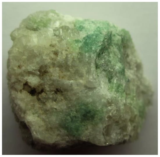
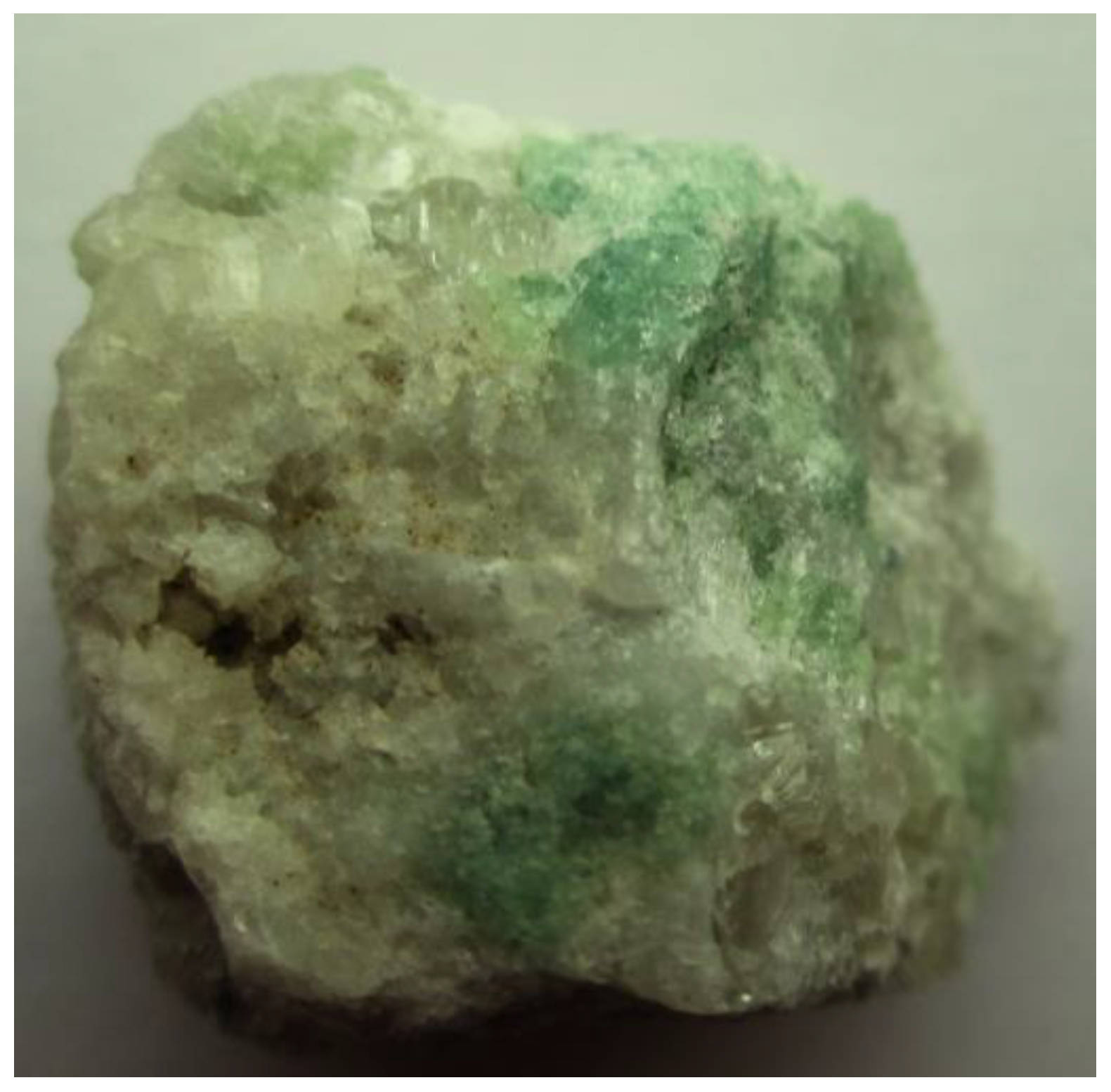
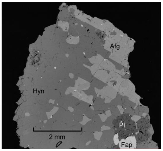
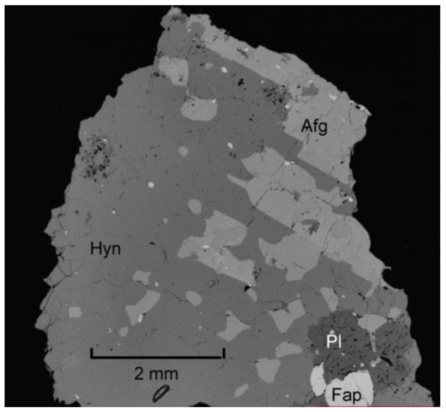
The sample studied in this work (Figure 1 and Figure 2) originates from the Malobystrinskoe gem lazurite deposit, in the Baikal Lake area, Siberia, Russia. Haüyne-45Å forms green grains up to 5 mm across in metasomatic rock formed in the contact zone between alkaline rocks and marbles. The associated minerals are calcite, afghanite, alkali plagioclase, subordinate diopside as well as minor fluorapatite and pyrite.
Figure 1.
Green grains of haüyne-45Å in a rock mainly composed of calcite and afghanite. Field of view width: 25 mm.
Figure 2.
Association of haüyne-45Å (Hyn) with afghanite (Afg), alkali plagioclase (Pl), fluorapatite (Fap) and pyrite (multiple white spots). SEM (BSE) image of a polished section.
Afghanite forms separate colorless elongate grains up to 3 mm long in the rock and subhedral or anhedral inclusions in haüyne-45Å (Figure 2).
3. Methods
The IR spectrum of haüyne-45Å was measured in the Federal Research Center of Problems of Chemical Physics and Medicinal Chemistry, Russian Academy of Sciences, Chernogolovka, Russia. In order to obtain the IR absorption spectrum, powdered samples were mixed with anhydrous KBr (in the KBr to mineral ratio of about 150:1), pelletized, and analyzed using an ALPHA FTIR spectrometer (Bruker Optics, Ettlingen, Germany) with a resolution of 4 cm−1. A total of 16 scans were collected for each spectrum. The time of each scan was 3 s. No baseline correction and no smoothing were applied. The IR spectrum of an analogous pellet of pure KBr was used as a reference.
The Raman spectra were obtained at the Dept. of Mineralogy, Faculty of Geology, Lomonosov Moscow State University for randomly oriented grains using an EnSpectr R532 spectrometer based on an OLYMPUS CX 41 microscope (Enhanced Spectrometry, San Jose, CA, USA) coupled with a diode laser (λ = 532 nm) at room temperature. Haüyne-45Å has a cubic symmetry; therefore, no significant anisotropy was expected. The spectra were recorded in the range of 100 to 4000 cm−1 with a diffraction grating (1800 gr mm−1) and a spectral resolution of about 6 cm−1. The output power of the laser beam was in the range of 5 to 13 mW. The diameter of the focal spot on the sample was 5–10 μm. The backscattered Raman signal was collected with a 40× objective; signal acquisition time for a single scan of the spectral range was 1 s, and the signal was averaged over 50 scans. Crystalline silicon was used as a standard.
The transmission spectra in the near infrared, visible, and ultraviolet (NIR–Vis–UV) ranges were measured in the Vinogradov Institute of Geochemistry, Siberian Branch of the Russian Academy of Sciences, Russia, at room temperature using a Lambda 950 spectrophotometer (Perkin-Elmer, Shelton, CT, USA).
The photoluminescence spectra were measured in the Vinogradov Institute of Geochemistry, Siberian Branch of the Russian Academy of Sciences, Russia, using a spectrometer based using an SDL-1 monochromator with a 600 lines per mm threaded diffraction grating (LOMO, St. Petersburg, Russia). The spectral slit width was 0.2 nm. Registration was carried out using a Hamamatsu H10721-04 photomodule (Hamamatsu, Sendai, Japan). Excitation was performed using a semiconductor laser with a wavelength of 405 nm and a power of 40 mW. The sample was fixed in a filling nitrogen cryostat, which was placed in a vacuum chamber. Photoluminescence spectra were measured at 7 K.
The ESR spectrum was measured on raw grains in the Vinogradov Institute of Geochemistry, Siberian Branch of the Russian Academy of Sciences, Russia, at room temperature with a RE-1306 X-band spectrometer (KBST, Smolensk, Russia) with a frequency of 9.3841 GHz at room temperature and 9.1841 GHz.
The chemical composition of haüyne-45Å and associated afghanite was studied in the Institute of Experimental Mineralogy RAS on an analytical suite including a digital scanning electron microscope Tescan VEGA-II XMU equipped with an energy-dispersive spectrometer (EDS) INCA Energy 450 with semiconducting Si (Li) detector Link INCA Energy and wave-dispersive spectrometer (WDS) Oxford INCA Wave 700, produced by Tescan Orsay Hld., Brno, Czech Republic. The samples for analysis were prepared as polished sections covered with graphite. The analyses were performed at an accelerating voltage of 20 kV, a current of 120 to 150 pA, and a beam diameter of 120 nm. The diameter of the excitation zone was below 5 μm. The following standards were used: CaF2 for F, albite for Na, synthetic Al2O3 for Al, wollastonite for Ca, potassium feldspar for K, SiO2 for Si, Fe metal for Fe, and FeS2 for S.
The H2O content in haüyne-45Å was measured in the Federal Research Center of Problems of Chemical Physics and Medicinal Chemistry, Russian Academy of Sciences, Chernogolovka, Russia, by gas chromatography of the products of heating at 1200 °C using a vario Micro CUBE automatic analyzer (Elementar Analysensysteme GmbH, Nidderau, Germany).
Thermal conversions of haüyne-45Å were studied in the Federal Research Center of Problems of Chemical Physics and Medicinal Chemistry, Russian Academy of Sciences, Chernogolovka, Russia. For annealing haüyne-45Å in vacuum, a shaft chamber electric resistance furnace T-1-1300-250 of the company Termionika (Podolsk, Russia). The samples were maintained at temperatures of 800 °C, 600 °C, or 400 °C for 4 h. In the first case, preliminary heating was performed to 700 °C for 70 min, after which the temperature was slowly increased to 800 °C for 35 min. The 800 °C was held for 4 h, and then the cooling process to 350 °C began, after which the chamber was cooled to a temperature of 140 °C by blowing 99.999 purity argon into the chamber. After reaching this temperature, the furnace with the sample was cooled to room temperature. The total process time before cooling was 5.75 h. Annealing of samples at 400 °C and 600 °C was carried out in a similar manner. In addition, haüyne-45Å was heated in air in a muffle furnace SNOL 10/1300 LSM from the company SNOL-TERM (Tver, Russia) at temperatures of 800 °C, 600 °C, and 400 °C for 4 h. The furnace temperature was set before firing.
Single-crystal XRD data were collected at the Faculty of Geology, Lomonosov Moscow State University at room temperature using an Xcalibur S CCD diffractometer (MoKα radiation) (Oxford Diffraction, Oxford, UK). More than a hemisphere of three-dimensional data was collected. Data reduction was performed using the CrysAlisPro program system, version 1.171.42.49 [7]. The data were corrected for Lorentz factor and polarization effects. Single-crystal structure analysis was performed using SHELX (version 2018/3) [8].
4. Results
4.1. Chemical Composition
Chemical data of the studied haüyne-45Å and associated afghanite are given in Table 1. Contents of other elements with atomic numbers > 8 are below their detection limits. Both minerals are almost homogenous in composition: standard deviations do not exceed 0.13 apfu for Na and 0.07 apfu for other components.
Table 1.
Chemical composition (wt. %) of studied haüyne-45Å and associated afghanite.
The total sums of the analyses were calculated under the assumption that all sulfur belongs to sulfate groups that significantly exceed 100%. This fact indicates that a significant part of the sulfur in the studied feldspathoids occurs in the form of sulfide and/or polysulfide groups.
4.2. Infrared Spectroscopy
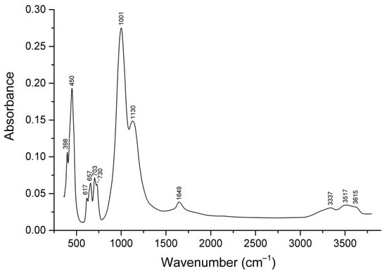
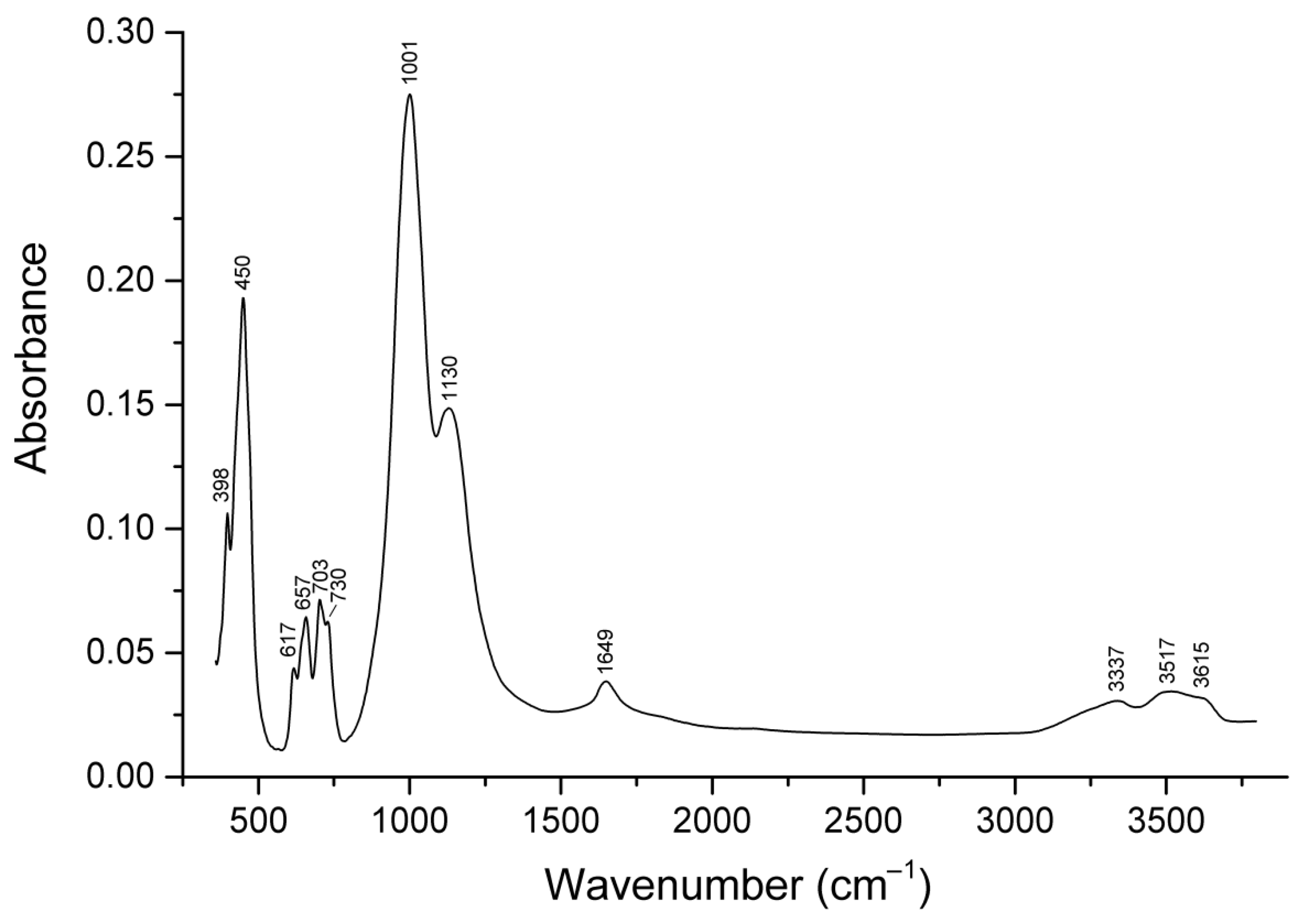
The IR spectrum of haüyne-45Å is given in Figure 3. The bands at 1001 cm−1 and below 500 cm−1 correspond to collective stretching and bending vibrations of the aluminosilicate framework. The band at 1130 cm−1 is due to asymmetric stretching vibrations of extra-framework SO42− anionic groups [F2(ν3) mode] The band at 617 cm−1 corresponds to bending vibrations of the SO42− groups [F2(ν4) mode] Other bands in the so-called finger-print region (610–730 cm−1) are due to mixed vibrations of the aluminosilicate framework.
Figure 3.
IR spectrum of haüyne-45Å.
Bands of stretching and bending vibrations of H2O molecules are observed in the range of 3300–3700 cm−1 and at 1649 cm−1. Splitting of the band of H2O stretching vibrations indicates the presence of several nonequivalent states of water molecules in the structure of the mineral.
The band of stretching vibrations of extra-framework CO2 molecules (in the range of 2339–2341 cm−1), which is typical for sodalite-group minerals [1,2,3,4,5], is not observed. This fact shows that crystallization of the studied haüyne took place under highly reducing conditions.
4.3. Raman Spectroscopy
The Gaussian03 software package (Gaussian version 03) [9] was used to calculate Raman spectra as well as UV-Vis-NIR absorption spectra of different S52− conformers using the density functional theory DFT with the B3LYP functional. Before calculating the absorption and Raman spectra, the found geometry was optimized. The set of basis functions 6-31G ** was used for the calculation.
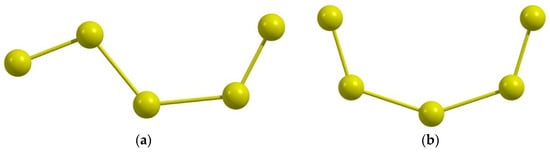
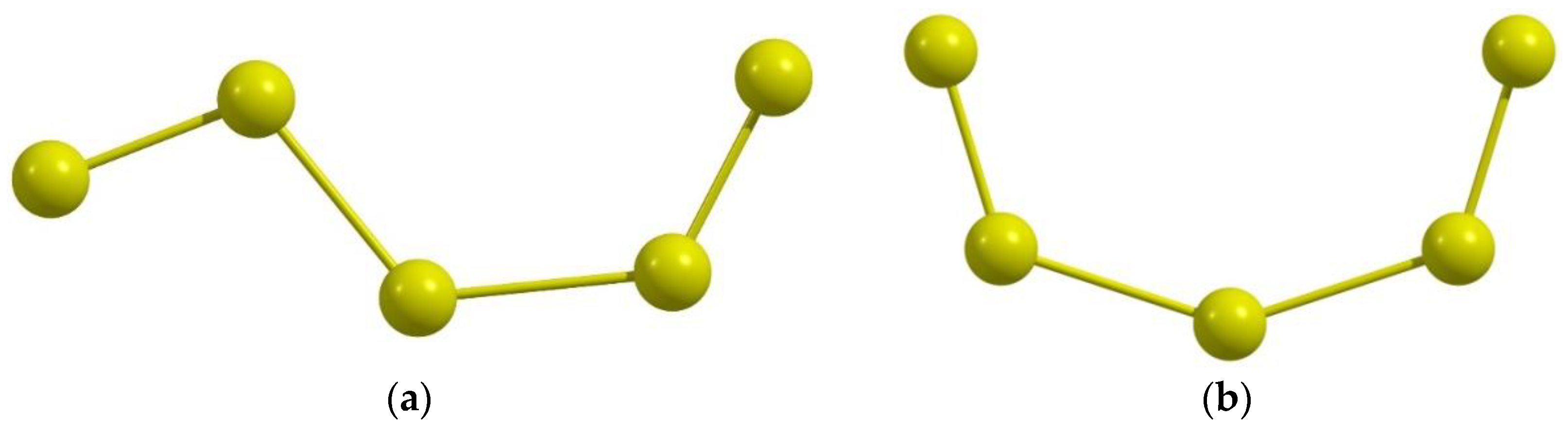
Local energy minima were found for two gauche-S52− conformers (Figure 4), trans-S52− conformer, and the ring S52− anion. The trans-S52− conformer was excluded from further consideration due to the large distance between the terminal sulfur atoms (7.12 Å). For isolated gauche-S52−-1, gauche-S52−-2, and cyclic S52− anions, these distances are equal to 6.74, 5.67, and 3.5 Å, respectively.
Figure 4.
Gauche-conformers of S52−. (a) Gauche-S52−-1; (b) Gauche-S52—2.
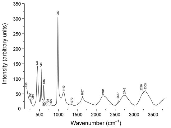
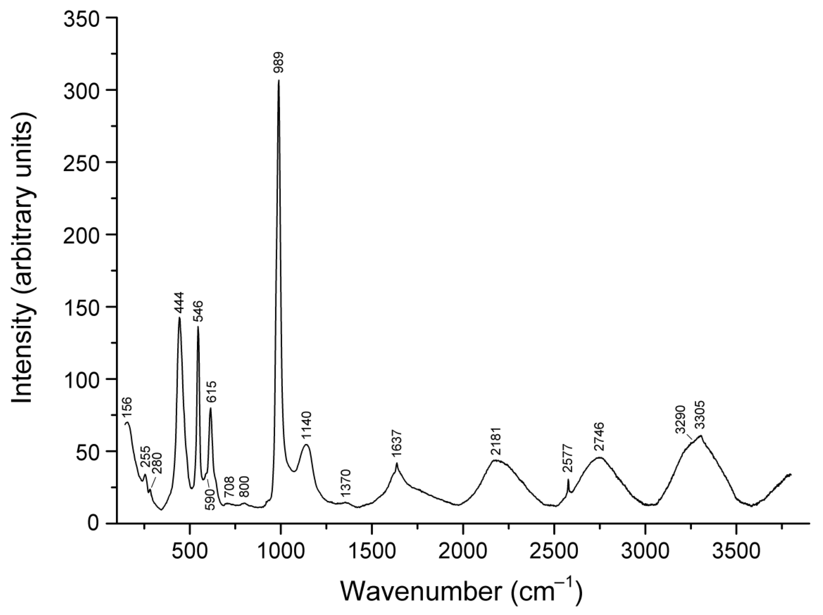
The Raman spectra of haüyne-45Å and associated afghanite are given in Figure 5 and Figure 6. The assignment of Raman bands (Table 2), except those of S52−, was made in accordance with [3,4,5,10,11,12,13,14,15,16,17,18].
Figure 5.
Raman spectrum of haüyne-45Å.
Figure 6.
Raman spectrum of afghanite associated with haüyne-45Å.
Table 2.
Assignment of Raman bands of the studied feldspathoids.
The calculated wavenumbers are (in cm−1; relative intensities are given in parentheses): 197(1), 287(0.61), 337(0.25), 579(0.40), and 593(0.47) for gauche-S52−-1; 236(1), 259(0.26), 284(0.06), 585(0.07), and 608(0.29) for gauche-S52−-2; 209(0.82), 243(0.45), 266(0.64), 267(0.03), 380(0.14), 429(1), and 479(0.32) for cyclic S52−. As one can see from these data, calculated wavenumbers of gauche-S52−-2 and cyclic S52− are rather close to the wavenumbers of Raman bands of haüyne-45Å. Minor differences may be due to the influence of the crystal field. Most probably, the broad band at 444 cm−1 is a superposition of overlapping bands of stretching vibrations of S52− or S4 and the band of framework bending vibrations.
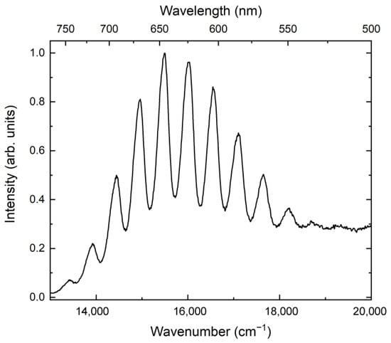
The specific features of the Raman spectrum of haüyne-45Å are a strong luminescence having a distinct vibration structure (with a period of ~ 550 cm−1) caused by stretching vibrations of S2●− and corresponding S–S stretching band at 546 cm−1. The distinct peak at 615 cm−1 is a characteristic band of S2●− and presumably corresponds to the combination (stretching + libration) mode of this radical anion. The weak band at 1370 cm−1 [(2ν1 + ν2) S3●− combination mode] [2,3,4,5,15,16,18] indicates that S3●− may have a minor contribution to Raman scattering at 546 cm−1. As one can see from these data, a major part of sulfur in haüyne-45Å belongs to the sulfate anions. The main extra-framework sulfide component in haüyne-45Å is the S2●− radical anion whereas other polysulfide groups (S3●−, S4●−, S52−, and/or S4) as well as the H2S anion occur in this mineral in minor amounts.
4.4. UV–Vis–near IR Absorption Spectroscopy
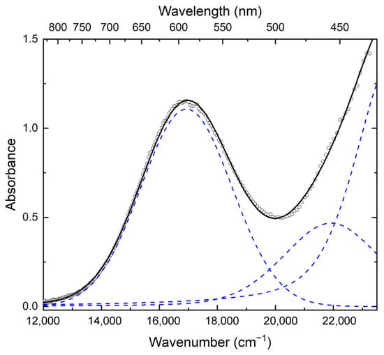
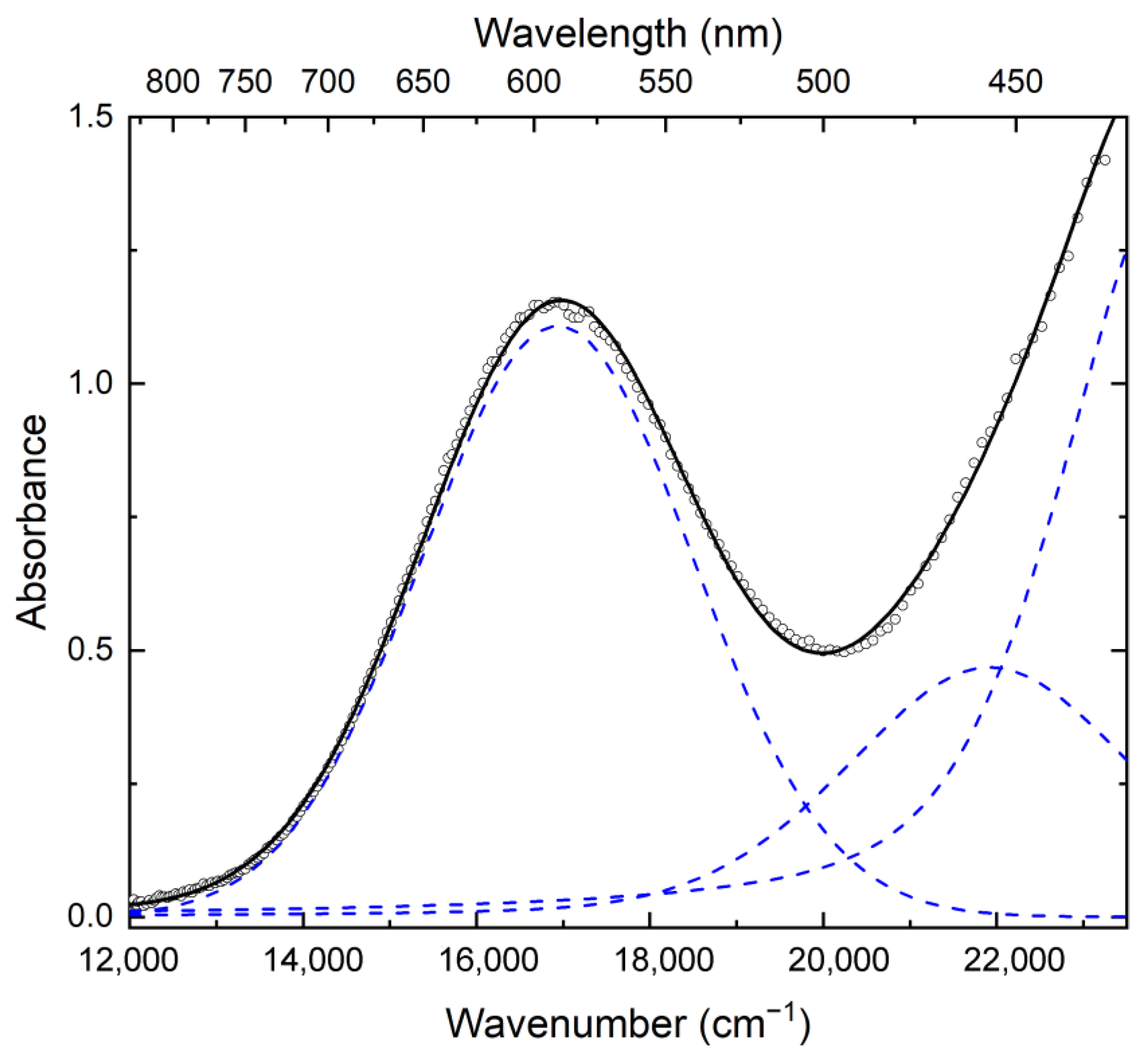
The UV-Vis-NIR spectrum of haüyne-45Å can be described as a superposition of three bands with the maxima at 16,900 cm−1 (590 nm), 21,900 cm−1 (455 nm), and 25,600 cm−1 (390 nm) (Figure 7). The strongest band at 25,600 cm−1 corresponds to S2●− radical anions [19]. The band at 16,900 cm−1 can be related to the absorption of S3●− radical anions and/or simultaneous absorption of two conformers of tetrasulfur neutral molecules, cis-S4 and trans-S4 [3,16].
Figure 7.
The UV-Vis-NIR spectrum of haüyne-45Å. The dots show the experimental data, the solid line is the result of decomposing the spectrum into three components, which are shown by dashed lines.
The band at 21,900 cm−1 is in the range of absorption of different conformers of the S52− anion. As shown by quantum chemical calculations carried out using the Gaussian03 software package for isolated pentasulfur anions, the wavenumbers of the strongest absorption bands of the gauche-S52−-1 and gauche-S52−-2 conformers are equal to 21,916 and 21,422 cm−1, respectively. The latter band of gauche-S52−-2 is accompanied by a weak band at 22,477 cm−1.
4.5. ESR Spectroscopy
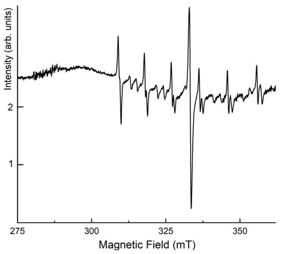
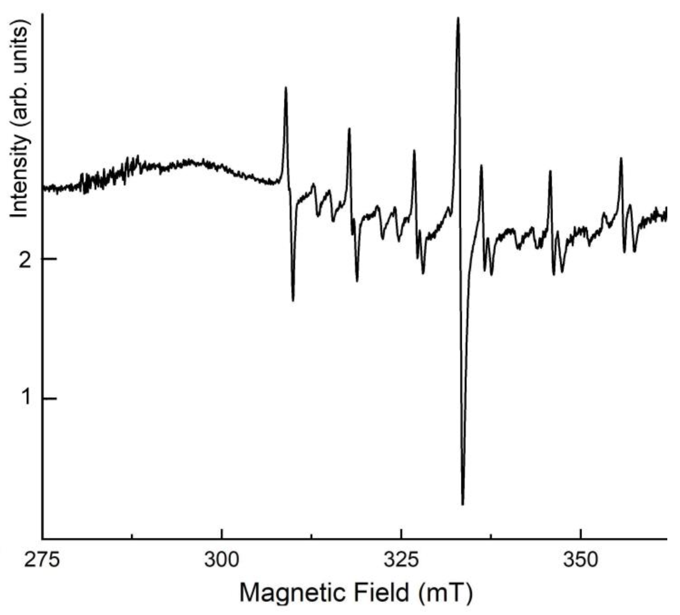
In the ESR spectrum of haüyne-45Å (Figure 8), a distinct signal with the components g1 = 2.34 and g2 = 2.00 is caused by the presence of the S2●− radical anions [20]. A sextet of weaker lines corresponds to Mn2+ in admixed calcite. Additional (very weak) sextets may correspond to forbidden transitions in Mn2+ impurity in calcite or trace amounts of Mn2+ in haüyne-45Å. Thus, S2●− is the main S-bearing paramagnetic species in haüyne-45Å, in accordance with the UV-Vis-NIR and Raman spectroscopy data.
Figure 8.
The ESR spectrum of haüyne-45Å.
4.6. Luminescence Spectroscopy
Upon excitation of haüyne-45Å by laser radiation with a wavelength of 405 nm, luminescence was observed with a maximum at about 16,000 cm−1. Upon cooling below 80 K, an oscillatory structure with a period of 546 cm−1 appears in the luminescence spectrum (Figure 9). This period coincides with the wave number of the S2●− stretching vibration band in the Raman spectrum (Figure 5).
Figure 9.
Photoluminescence spectrum of a haüyne sample measured at 7 K.
4.7. Structural Features
The crystal structure model of haüyne-45Å including the framework and partly extra-framework filling was obtained by direct methods in the frames of non-standard space group I-1 despite the unit cell parameters [a = 45.3629(3) Å and V = 93,347.4(11) Å3] and preliminary analysis pointed the cubic space group I-43m (#217). Unfortunately, all attempts to obtain the structure model directly in the cubic system as well as to increase the symmetry using the obtained model were unsuccessful. We should also note that analysis of the reciprocal space revealed the presence of additional weak satellite reflections, which additionally triple the parameter of the cubic unit cell. These reflections were not taken into account. Thus, we can consider the obtained result only as an averaged structure model. At the same time, the analysis of interatomic distances in the tetrahedra and the positions of the tetrahedra of the framework showed that the model of the framework consisting of sodalite cages is correct and reliable.
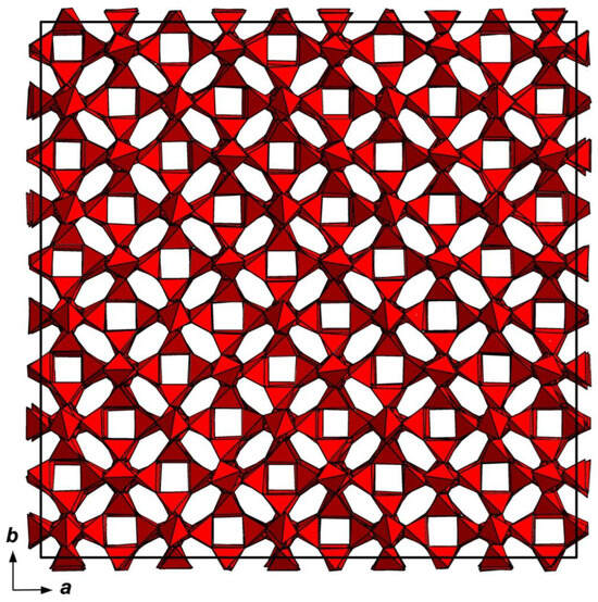
The structure model of the studied haüyne-45Å sample (Figure 10) is based on the sodalite-type aluminosilicate framework, where tetrahedrally coordinated Si and Al atoms are connected via common vertices to form an open framework. Low quality of the model did not allow for the determination of Al and Si ordering. The cavities of the framework (so-called sodalite cages) are mainly occupied by sodium and calcium cations, water molecules, Cl anions, and SO4 tetrahedra. Minor amounts of extra-framework species could not be localized.
Figure 10.
Tetrahedral framework in the averaged structure model of the studied haüyne-45Å.
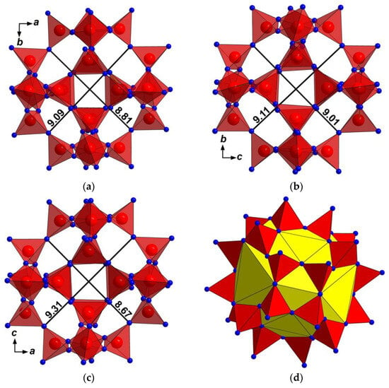
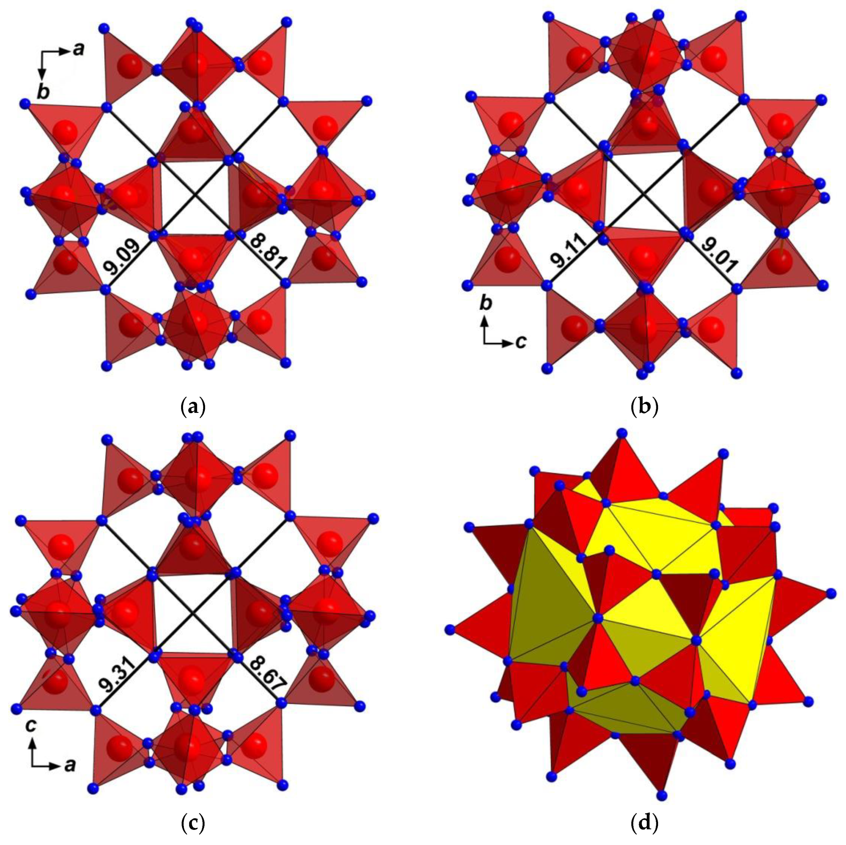
To assume possible cages that could host large polysulfide groups, we have measured some of the sodalite cages. The measurements were performed not for traditional sodalite cages (β-cages) limited by Si- and Al-sites, but for cages that also include O atoms of tetrahedra. For each considered cage, we have measured diameters and the volume of the hypothetical 36-fold polyhedron inside the cage. Figure 11 demonstrates the measurements performed for one cage.
Figure 11.
One sodalite cage with measured diameters in three directions (a–c) and hypothetical 36-fold polyhedron inside the cage (d).
Such measurements were performed for 25 random cages and five cages along the a axis. The results are summarized in Table 3. It is important that the anomalous large Sod 3 cage does not host cations and anions. Most probably, it is populated by the S4 neutral molecules, which could not be localized because of the orientational splitting of the corresponding S-sites.
Table 3.
Diameters of sodalite cages and the volume of the hypothetical 36-fold polyhedron inside the cage in haüyne-45Å.
4.8. Thermal Conversions
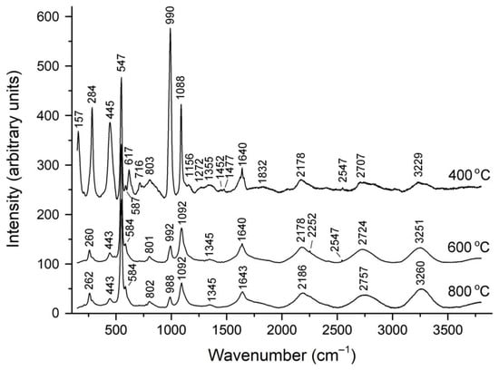
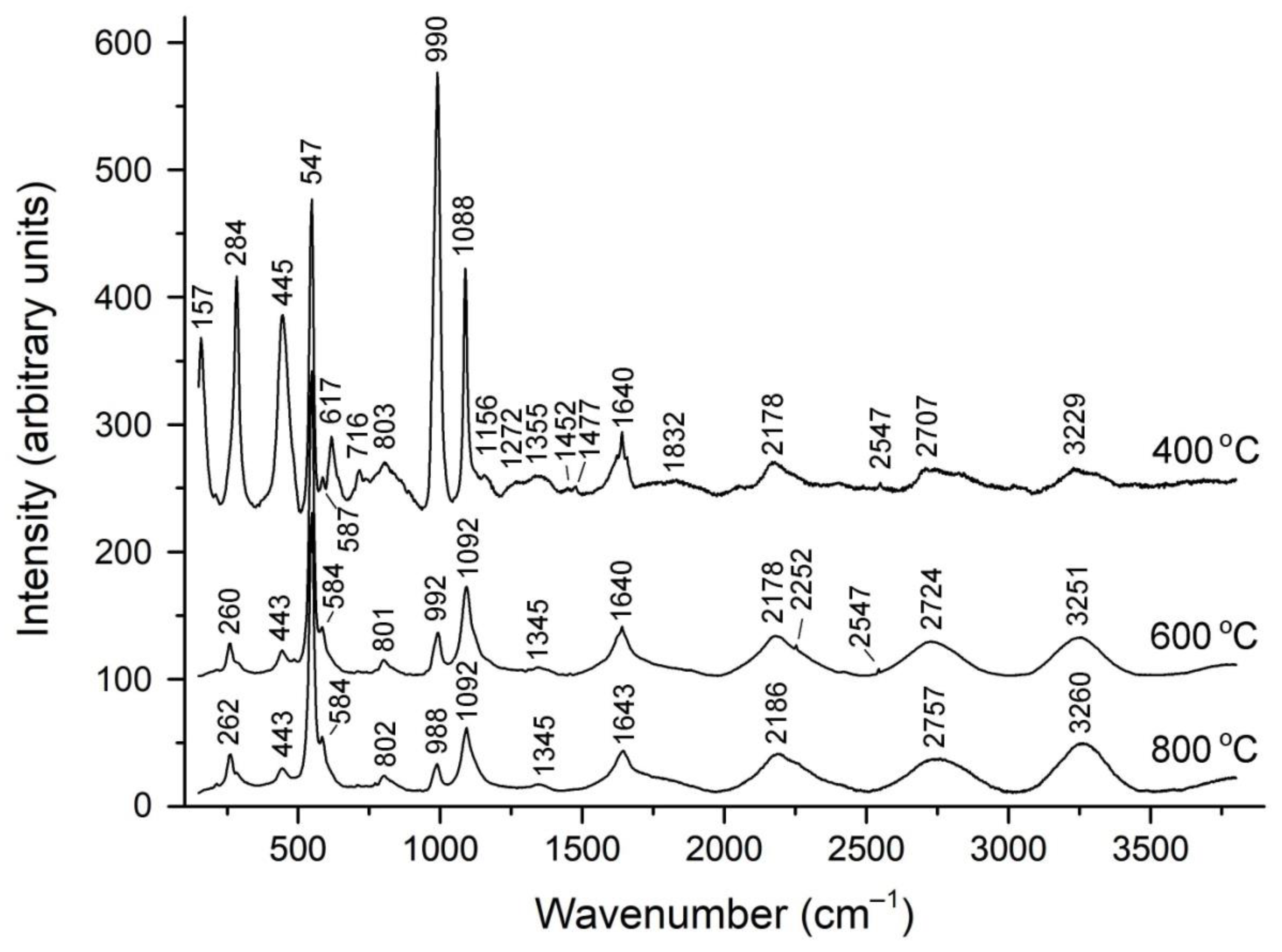
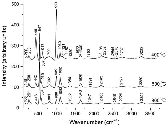
To study the mutual transformations of S-containing groups during the annealing of haüyne-45Å, Raman spectroscopy was used since it allows the most reliable identification of these groups. The Raman spectra of the annealing products are shown in Figure 12 and Figure 13, and the assignment of the Raman bands is given in Table 4 and Table 5.
Figure 12.
Raman spectra of haüyne-45Å annealed at different temperatures in vacuum.
Figure 13.
Raman spectra of haüyne-45Å annealed at different temperatures in air.
Table 4.
Assignment of Raman bands of the products of heating of haüyne-45Å in vacuum.
Table 5.
Assignment of Raman bands of the products of heating of haüyne-45Å in air.
The product of annealing in a vacuum at 400 °C has a deep green color. Its Raman spectrum differs from the spectrum of the initial sample by weaker luminescence with the vibrational structure of S2●−, a weaker peak of the HS− anion, more intense S3●− bands and, most importantly, by the presence of an intense band at 284 cm−1, which can be attributed to both the S4●− radical anion and/or the S52− anion. The latter seems more likely, taking into account the deep green color of the calcination product, which can be associated with the simultaneous presence of two strong chromophores: blue (S3●−) and yellow (S52−). The most intense band in the Raman spectrum of this sample is the SO42− band at 990 cm−1.
The Raman spectra of the product of the annealing of haüyne-45Å in vacuum at 600 °C and 800 °C are almost identical. They are characterized by strong luminescence with the vibrational structure of S2●−, dominance of bands related to S3●− and the presence of a relatively weak band SO42−. The band at 284 cm−1, presumably assigned to S52−, is absent in these spectra, which may be the cause of the color change from green to blue.
The heating of haüyne-45Å in air leads to changes in the Raman spectrum that differ significantly from those observed when heating this mineral in a vacuum. Already at 400 °C, the luminescence with the vibrational structure of S2●− disappears. Heating at higher temperatures leads to a gradual weakening of the bands SO42− and S52− as well as an increase in the intensity of the bands related to S3●−. These changes are accompanied by a change in color from bluish-green (for the original sample) through light blue to dark blue.
5. Discussion
5.1. Extra-Framework Components
The application of a multimethodic approach based on IR, Raman, UV-Vis-NIR absorption, ESR and photoluminescence spectroscopy methods as well as chemical data made it possible to identify various S-bearing extra-framework groups occurring in haüyne-45Å (namely, SO42−, S2●−, S3●−, S4, S52−, and HS−). In addition, based on the available spectroscopic data, the presence of the S4●− radical anion in haüyne-45Å cannot be excluded. The simplified formula of haüyne-45Å is Na6Ca2−x(Si6Al6O24)(SO42−,HS−,S2●−,S4,S3●−,S52−)2−y.
Apparently, the simultaneous presence of yellow chromophores (S2●− and S52−) and blue chromophore (S3●−) in haüyne-45Å is the cause of its green color. In S-bearing minerals of the sodalite group, regular or irregular alternation of sulfate anions with polysulfide groups is the cause of commensurate or incommensurate structural modulations, respectively [6,21,22]. The commensurate modulations of the haüyne-45Å framework are presumably related to the regular alternation of SO42− anions with S2●−, S3●−, S4, and S52− groups. Commensurate modulations of the hauyne-45A framework are discussed in the next section and in Supplementary Materials.
5.2. Structure Modulations
In the powder X-ray diffraction pattern of haüyne-45Å, main reflections with integer indices corresponding to the basic cubic lattice with a parameter of about 9.13 Å are accompanied by weaker satellites with fractional indices (Figure S1 in Supplementary Materials). The value of the modulation parameter n = 0.2013 allows us to consider the modulation to be almost commensurate and to study a superstructure with a fivefold lattice parameter of about 45.36 Å (5n = 1.0065 ≈ 1).
The measured X-ray diffraction pattern from the single crystal (see Figure S2 in Supplementary Materials) consists of strong main reflections corresponding to the basic lattice with a parameter of about 9.13 Å and less intense satellites located in six <110> directions from the main reflections at distances equal, within the experimental accuracy, to 1/5 of the face diagonal of the basic reciprocal lattice. Commensurate and incommensurate structure modulations are very typical for lazurite-related sodalite-group minerals with extra-framework polysulfide groups. For comparison, the previously studied structures of “monoclinic lazurite” are incommensurately modulated (with a wave vector length of about 0.215 of the cube-face diagonal in a cubic setting), whereas the triclinic lazurite-related mineral slyudyankaite and orthorhombic lazurite-related mineral vladimirivanovite have commensurately modulated structures [6].
5.3. Mechanisms of Thermal Conversions and Their Geochemical Importance
The S4 molecule is unstable above 400 °C [23]. Based on the Raman spectroscopy data obtained in this work, one can conclude that the mechanisms of thermal conversions of S-bearing groups in haüyne-45Å depend both on the temperature and redox conditions. The experimental data show that in the absence of oxygen at 400 °C only a partial decomposition of S52−, accompanied by the transformation of H2S to HS− and possible appearance of the S2●− radical anion which corresponds to the following hypothetic scheme: 2S52− + H2S → S5●− + S4●− + HS−. At a higher temperature of 600 °C, the S52− anion decomposes in accordance with the scheme S52− → S2●− + S3●−. At 800 °C, in a vacuum, partial reduction of sulfate sulfur occurs, which is manifested in a sharp decrease in the intensity of the Raman band of SO42− stretching vibrations. This process can be described by the scheme SO42− → S2− + 2O2(gas), which corresponds to the only possible reaction channel in which the charge balance is maintained, since the transfer of an electron to S2●− or S3●− would be accompanied by the appearance of additional bands in the Raman spectrum. Note that the S2− anion is the only sulfide species that has no internal vibrational modes.
In an oxidizing environment, S52−, S4, and, to a lesser extent, S2●− are unstable already at 400 °C, whereas minor amounts of S2●− and SO42− occur in the solid phase even at 800 °C. Corresponding transformations of S-bearing groups can be described by the following scheme: S52− → S2●− + S3●−; 3S4 + 4e− → 4 S3●−; 2S2●− + 2O2 + e− → SO42− + S3●− where e− is an electron. The reaction 3H2O → 2H3O+ + 0.5O2 + 2 e− may serve as a source of electrons.
Thus, the results of the study of the calcination products of haüyne-45Å under different conditions show that in the absence of an external oxygen source, the radical anions S2●− and S3●− are stable up to 800 °C, but in the presence of air, S2●− gradually transforms into a mixture of SO42− and S3●−. The decomposition of S52− and S4 begins already at 400 °C under both oxidizing and non-oxidizing conditions. According to our observations, S52− and/or HS− groups, characteristic of feldspathoids crystallized under strongly reducing conditions, are never accompanied by the presence of CO2 molecules in these minerals, which are easily detected by IR spectroscopy [1,2,3,4,5,6,21,22]. At the same time, the joint presence of CO2 and other polysulfide species, including S3●−, S3●−, S4●−, S4, and/or S6, in minerals of the cancrinite and sodalite groups is rather common. These data can be used to reconstruct the temperature and oxidation-reduction conditions of mineral formation.
It should be noted that afghanite found in the association with the studied haüyne-45Å is characterized by the space group P31c (#159), unit-cell parameters a = 12.7664(6), c = 21.4544(9) Å, V = 3028.2(3) Å3 and formula Na5.22Ca2.56K0.13(Si6Al6O24)(SO4)0.5(SO3)0.5[(SO4)0.78(SO3)0.22]0.5Cl1.5 (Z = 4). The presence of sulfite anion, (SO3)2−, found during the crystal structure refinement, as well as the presence of the groups HS−, S4, and S52− detected by spectroscopic methods in haüyne-45Å confirm the conclusion about the reducing conditions of mineral formation.
6. Conclusions and Implications
Haüyne-45Å is the third commensurately modulated counterpart of haüyne found in nature, after vladimirivanovite and slyudyankaite. Structural modulations in these minerals arise as a result of regular alternation of sodalite cages containing SO42− anions and large polysulfide groups (mainly, S4, S3●− and S6, respectively).
Based on the chemical, structural, and spectroscopic data obtained in this work, the simplified formula of haüyne-45Å is Na6Ca2−x(Si6Al6O24)(SO42−,HS−,S2●−,S4,S3●−,S52−)2−y. Regular alternation of sodalite cages containing relatively small anionic species (SO42−, HS−, and S2●−) and larger polysulfide groups (mainly, S4 as well as minor S3●− and S52−) is the cause of commensurate structure modulations.
Haüyne-45Å crystallized under low-temperature reducing conditions (below 400 °C). This conclusion follows from the fact that the HS− and S52− anions, as well as the absence of extra-framework CO2 molecules, indicate reducing conditions, whereas the S4 molecule is unstable above 400 °C.
Supplementary Materials
The following supporting information can be downloaded at: https://www.mdpi.com/article/10.3390/min15070709/s1, Figure S1: The powder X-ray diffraction pattern of the studied mineral with a modulated structure. Integer indices of the main reflections and fractional indices of the satellites correspond to the basic cubic lattice with a parameter of about 9.13 Å. The indices of the main reflections are given in brackets. Figure S2: Sections of the diffraction pattern by planes l = 9 (a), l = 10 (b), l =11 (c).
Author Contributions
Conceptualization, N.V.C., A.N.S., I.V.P. and D.Y.P.; Methodology, N.B.B., N.V.Z., D.A.V., D.A.K. and D.Y.P.; Investigation, N.V.C., R.Y.S., N.V.Z., N.A.C., D.A.V., N.B.B. and M.F.V.; Original Manuscript Draft Preparation, N.V.C.; Manuscript Review and Editing, N.V.Z. and I.V.P.; Figures, N.V.C., N.V.Z., R.Y.S. and D.A.V. All authors have read and agreed to the published version of the manuscript.
Funding
IR spectroscopy investigation, chemical data processing, and study of thermal conversions were performed in accordance with the state task, registration number 124013100858-3 (for N.V.C. and N.A.C.). A Raman spectroscopy study was conducted under the state assignment of Lomonosov Moscow State University (state task No. 121061600049-4: M.F.V. and I.V.P). Luminescence and ESR spectroscopy studies were performed at the Center of Isotope and Geochemical Research for Collective Use (Vinogradov Institute of Geochemistry of the Siberian Branch of the Russian Academy of Sciences) and supported by Research Program 0284-2021-0004 (for R.Y.S.).
Data Availability Statement
The original contributions presented in this study are included in the article/supplementary material. Further inquiries can be directed to the corresponding author.
Acknowledgments
The authors are grateful to anonymous referees for valuable comments.
Conflicts of Interest
The authors declare no conflicts of interest.
References
- Chukanov, N.V.; Aksenov, S.M. Crystal chemistry, chemical diversity and physical properties of microporous sodalite-type materials: A review. Intern. J. Molec. Sci. 2024, 25, 10218. [Google Scholar] [CrossRef] [Scilit]
- Sapozhnikov, A.N.; Tauson, V.L.; Lipko, S.V.; Shendrik, R.Y.; Levitskii, V.I.; Suvorova, L.F.; Chukanov, N.V.; Vigasina, M.F. On the crystal chemistry of sulfur-rich lazurite, ideally Na7Ca(Al6Si6O24)(SO4)(S3)−·nH2O. Amer. Miner. 2021, 106, 226–234. [Google Scholar] [CrossRef] [Scilit]
- Chukanov, N.V.; Shendrik, R.Y.; Vigasina, M.F.; Pekov, I.V.; Sapozhnikov, A.N.; Shcherbakov, V.D.; Varlamov, D.A. Crystal chemistry, isomorphism, and thermal conversions of extra-framework components in sodalite-group minerals. Minerals 2022, 12, 887. [Google Scholar] [CrossRef] [Scilit]
- Chukanov, N.V.; Vigasina, M.F.; Shendrik, R.Y.; Varlamov, D.A.; Pekov, I.V.; Zubkova, N.V. Nature and isomorphism of extra-framework components in cancrinite- and sodalite-related minerals: New data. Minerals 2022, 12, 729. [Google Scholar] [CrossRef] [Scilit]
- Chukanov, N.V.; Shchipalkina, N.V.; Shendrik, R.Y.; Vigasina, M.F.; Tauson, V.L.; Lipko, S.V.; Varlamov, D.A.; Shcherbakov, V.D.; Sapozhnikov, A.N.; Kasatkin, A.V.; et al. Isomorphism and mutual transformations of S-bearing components in feldspathoids with microporous structures. Minerals 2022, 12, 1456. [Google Scholar] [CrossRef] [Scilit]
- Bolotina, N.B.; Sapozhnikov, A.N.; Chukanov, N.V.; Vigasina, M.F. Structure modulations and symmetry of lazurite-related sodalite-group minerals. Crystals 2023, 13, 768. [Google Scholar] [CrossRef] [Scilit]
- Oxford Diffraction CrysAlisPro Software System; v. 1.171.42.49; Rigaku Corporation: Oxford, UK, 2022.
- Sheldrick, G.M. Crystal structure refinement with SHELXL. Acta Crystallogr. 2015, C71, 3–8. [Google Scholar] [CrossRef] [Scilit]
- Frisch, M.J.; Trucks, G.W.; Schlegel, H.B.; Scuseria, G.E.; Robb, M.A.; Cheeseman, J.R.; Scalmani, G.; Barone, V.; Mennucci, B.; Petersson, G.A.; et al. Gaussian 09, Revision D.01; Gaussian, Inc.: Wallingford, CT, USA, 2009. [Google Scholar]
- Dubessy, J.; Boiron, M.-C.; Moissette, A.; Monnion, C.; Sretenskaya, N. Determination of water, hydrates and pH in fluid inclusions by micro-Raman spectrometry. Eur. J. Min. 1992, 4, 885–894. [Google Scholar] [CrossRef] [Scilit]
- Eckert, B.; Steudel, F. Molecular spectra of sulfur molecules and solid sulfur allotropes. Top. Curr. Chem. 2003, 231, 31–97. [Google Scholar] [CrossRef] [Scilit]
- Wong, M.W.; Steudel, R. Structure and spectra of tetrasulfur S4—An ab initio MO study. Chem. Phys. Lett. 2003, 379, 162–169. [Google Scholar] [CrossRef] [Scilit]
- Ling, Z.C.; Wang, A.; Jolliff, B.L. Mineralogy and geochemistry of four lunar soils by laser-Raman study. Icarus 2011, 211, 101–113. [Google Scholar] [CrossRef] [Scilit]
- Hettmann, K.; Wenzel, T.; Marks, M.; Markl, G. The sulfur speciation in S-bearing minerals: New constraints by a combination of electron microprobe analysis and DFT calculations with special reference to sodalite-group minerals. Amer. Miner. 2012, 97, 1653–1661. [Google Scholar] [CrossRef] [Scilit]
- Steudel, R.; Chivers, T. The role of polysulfide dianions and radical anions in the chemical, physical and biological sciences, including sulfur-based batteries. Chem. Soc. Rev. 2019, 48, 3279–3319. [Google Scholar] [CrossRef] [Scilit]
- Rejmak, P. Computational refinement of the puzzling red tetrasulfur chromophore in ultramarine pigments. Phys. Chem. Chem. Phys. 2020, 22, 22684–22698. [Google Scholar] [CrossRef] [Scilit]
- Caggiani, M.C.; Mangone, A.; Aquafredda, P. Blue coloured haüyne from Mt. Vulture (Italy) volcanic rocks: SEM-EDS and Raman investigation of natural and heated crystals. J. Raman Spectrosc. 2022, 53, 956–968. [Google Scholar] [CrossRef] [Scilit]
- Chivers, T.; Oakley, R.T. Structures and spectroscopic properties of polysulfide radical anions: A theoretical perspective. Molecules 2023, 28, 5654. [Google Scholar] [CrossRef] [Scilit]
- Rolfe, J. Emission spectra of S2−, Se2−, and SeS− ions in KI Crystals. J. Chem. Phys. 1968, 49, 4193–4197. [Google Scholar] [CrossRef] [Scilit]
- Kowalak, S.; Jankowska, A.; Zeidler, S.; Wiećkowski, A.B. Sulfur radicals embedded in various cages of ultramarine analogs prepared from zeolites. J. Solid State Chem. 2007, 180, 1119–1124. [Google Scholar] [CrossRef] [Scilit]
- Sapozhnikov, A.N.; Bolotina, N.B.; Chukanov, N.V.; Shendrik, R.Y.; Kaneva, E.V.; Vigasina, M.F.; Ivanova, L.A.; Tauson, V.L.; Lipko, S.V. Slyudyankaite, Na28Ca4(Si24Al24O96)(SO4)6(S6)1/3(CO2)·2H2O, a new sodalite group mineral from the Malo-Bystrinskoe lazurite deposit, Baikal Lake area, Russia. Amer. Miner. 2023, 108, 1805–1817. [Google Scholar] [CrossRef] [Scilit]
- Bolotina, N.B.; Chukanov, N.V.; Sapozhnikov, A.N.; Zubkova, N.V.; Pekov, I.V.; Varlamov, D.A.; Vigasina, M.F.; Bulakh, M.O.; Yapaskurt, V.O.; Ksenofontov, D.A. Vladimirivanovite revised: General crystal chemistry and isomorphous substitutions of extra-framework species. Minerals 2024, 14, 883. [Google Scholar] [CrossRef] [Scilit]
- Sapozhnikov, A.N.; Tauson, V.L.; Lipko, S.V.; Danilov, B.S.; Chukanov, N.V. On the temperature conditions of formation of pink S4-containing haüyne from the Malo-Bystrinskoe lazurite deposit (East Siberia, Russia). Zap. Russ. Miner. Soc. 2025, 154, 107–117. [Google Scholar] [CrossRef]

Disclaimer/Publisher’s Note: The statements, opinions and data contained in all publications are solely those of the individual author(s) and contributor(s) and not of MDPI and/or the editor(s). MDPI and/or the editor(s) disclaim responsibility for any injury to people or property resulting from any ideas, methods, instructions or products referred to in the content. |
© 2025 by the authors. Licensee MDPI, Basel, Switzerland. This article is an open access article distributed under the terms and conditions of the Creative Commons Attribution (CC BY) license (https://creativecommons.org/licenses/by/4.0/).