


Abstract
A Cl-rich annitic mica is present in zones in taxitic gabbro–dolerite enriched in base metal sulfides in the eastern portion of the Oktyabrsky deposit in the Norilsk complex (Russia). Other Cl-enriched minerals in the assemblage include hastingsite (4.06 wt.% Cl), ferro-hornblende (2.53 wt.%), and chlorapatite (>6 wt.%). New wavelength-dispersive electron probe analyses reveal compositions with up to 7.75 wt.% Cl, corresponding to the formula K0.742Na0.047Ca0.007)Σ0.796 (Fe2+2.901Mg0.078Mn0.047Ti0.007Cr0.003)Σ3.036 (Si3.190Al0.782)Σ3.972O10 (Cl1.105OH0.854F0.041)Σ2.000 based on 22 negative charges per formula unit, in which OH(calc.) = 2 − (Cl + F). Unfortunately, the grain size of the Cl-dominant mica precluded a single-crystal X-ray diffraction study even though its EBSD pattern confirms its identity as a member of the Mica group. We present results of a refinement of a crystal from the same mineralized sample containing 0.90(6) apfu Cl [R1 = 7.89% for 3720 unique reflections]. The mica is monoclinic, space group C2/m, a 5.3991(4), b 9.3586(6), c 10.2421(10) Å, β 100.873(9)°, V = 508.22(7) Å3, Z = 2. We also describe physical properties and provide a Raman spectrum. Among the mica compositions acquired from the same sample, a high Cl content is correlated with relative enrichment in Si, Mn, and Na and with a depletion in Al, Mg (low Mg#), K, Cr, and Ti. The buildup in Cl in the ore-forming environment is ultimately due to efficient fractional crystallization of the basic magma, with possible contributions from the Devonian metasedimentary sequences that it intruded.
1. Introduction
The latest mineralogical surprise to come from the Norilsk complex, located along the northwestern margin of the Siberian Platform in Russia, is an annitic mica in which the Cl ion predominates at the anion site. The relatively large ionic radius of the chlorine ion was long considered to preclude compositions exceeding 50 mol.% at the anion site. The chlorine-rich assemblages are found in sulfide-rich zones containing minerals of Pd-Pt, Au-Ag, REE, Y, Zr, U, and Th along the eastern portion of the Oktyabrsky deposit [1]. The atypical mineralized zones are associated with Cl-rich amphiboles and a trioctahedral mica enriched in Cl. The early results [1] were confirmed in follow-up analyses; they show that a Cl-rich annitic mica is fairly widespread in the mineralized drill core. At a small number of points, the mole fraction of Cl clearly exceeds that of OH and F. The separation of a single crystal having Cl > (OH, F), however, proved to be impossible owing to grain-size limitations. Confirmation as a new mineral species by CNMNC-IMA must await the discovery and characterization of larger domains of Cl-dominant annitic mica.
In this contribution, we describe the occurrence and geological setting of the Cl-rich annitic mica in the Norilsk ore assemblages, provide representative compositions, and list the coexisting minerals. We document many physical properties of the mica and present representative compositions, some of which satisfy the criterion Cl > (OH, F). We provide the Raman spectrum of the Cl-enriched mica. Its structure demonstrates that it is indeed a member of the Mica group [2]. The compositions acquired lead to an assessment of the factors that favor the incorporation of Cl in a trioctahedral mica.
2. Occurrence, Geological Setting, and Mineral Association
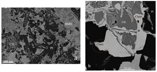
The Cl-rich annitic mica occurs in specimens of mineralized taxitic gabbro–dolerite collected in drill hole EF67. The drill hole crosses sulfide zones in the eastern section of the Oktyabrsky Cu-Ni-PGE deposit (N 69°32′38,2055″, E 88°25′41,0459″) in the Norilsk complex of Permo-Triassic age, northern Krasnoyarskiy kray, Russia [1]. The taxitic host (Table 2 in [3]) is a heterogeneous gabbro or olivine gabbro with wide variability in grain size (Figure 1). It consists of sulfide minerals (up to 20 vol.%), plagioclase, clinopyroxene, calcic amphiboles, and micas, members of the chlorite and apatite groups, with the OH-bearing phases locally Cl-enriched. Maximum Cl contents recorded are: hastingsite (4.06 wt.% Cl), ferro-hornblende (2.53%), chlorapatite (>6%), chamosite (0.96%), and serpentine (0.79%). The apatite-group minerals display extensive ternary solid-solution involving OH, F, and Cl.
Figure 1.
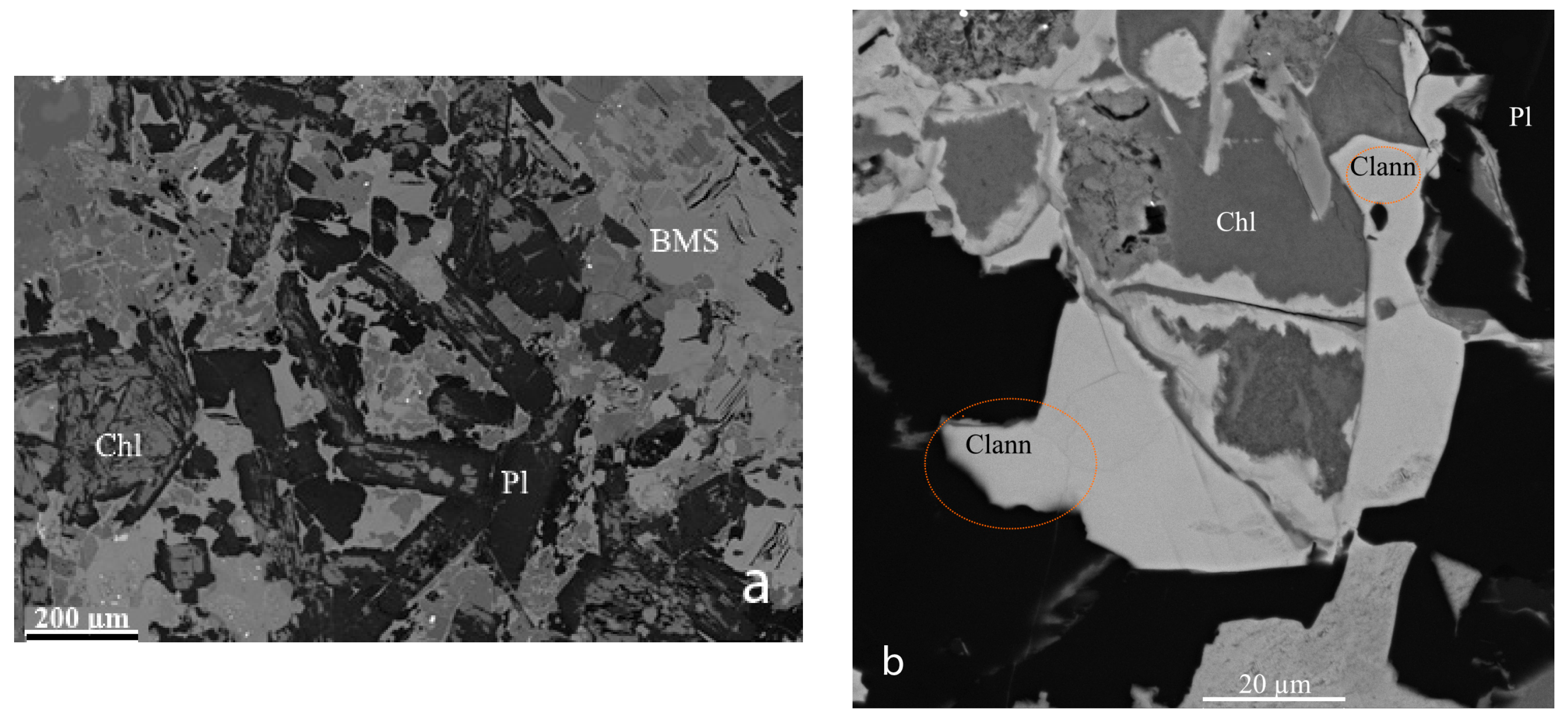
(a) SEM–BSE panoramic image of the section containing the Cl-rich annitic mica associated with chlorite (Chl) and plagioclase (Pl). (b) A high-magnification view shows the two areas from which a fragment of the Cl-rich annitic mica (Clann) was extracted. A front of replacement of chlorite by the annitic mica is clearly related to prominent fractures.
The host rock is relatively enriched in ore species, particularly platinum-group minerals. The associated minerals are chalcopyrite, pyrrhotite, pentlandite, ilmenite, magnetite, apatite, argentopentlandite, Au–Ag alloy, members of the galena–clausthalite series, members of the kotulskite–sobolevskite series, paolovite, michenerite, merenskyite, mertieite-II, sopcheite, thorite, unnamed Pd6Sn2As (or an As-enriched variety of paolovite), and unnamed (Y,Ca,REE)2Zr2(Ti,Nb)2TiFe2+O14 (metamict; likely related to zirconolite).
The mineralized rocks are marginal to the main Oktyabrsky deposit and of relatively low grade. They are genetically related to large-scale sills of gabbro–dolerite that attain 3.5 km in thickness. Their emplacement was largely controlled by the NNE-trending Norilsk–Kharayelakh fault.
In thin section, Cl-enriched mica forms a rim on chlorite, a member of the chamosite–clinochlore series (Figure 1) that seems to have replaced plagioclase. The disposition of the mica with respect to two prominent cracks supports the hypothesis that it grew by replacement. The textural relationship of the Cl-rich mica to coexisting minerals is illustrated in several figures in [1].
3. Physical and Optical Properties
Single flakes of the chlorine-rich mica are grayish brown. Their streak is colorless, and they have a silky luster. The transparent flakes are brittle and have an inferred Mohs hardness of less than 3; hardness was not measured owing to the unavailability of suitable crystals. The flakes exhibit a perfect {001} cleavage and an uneven fracture. No parting was observed. Density cannot be determined because of the small grain-size. Calculated on the basis of the empirical formula and single-crystal X-ray data, the density is 3.421 g·cm−3.
Flakes of the Cl-rich annitic mica are anhedral to subhedral and translucent. They display a weak pleochroism, from ocher to pale greenish ocher in basal section. Color along the direction perpendicular to the lamellae could not be observed because of the thinness of the flakes. The optical properties could not be determined because of the small grain-size.
4. Raman Spectroscopy
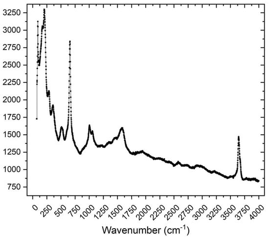
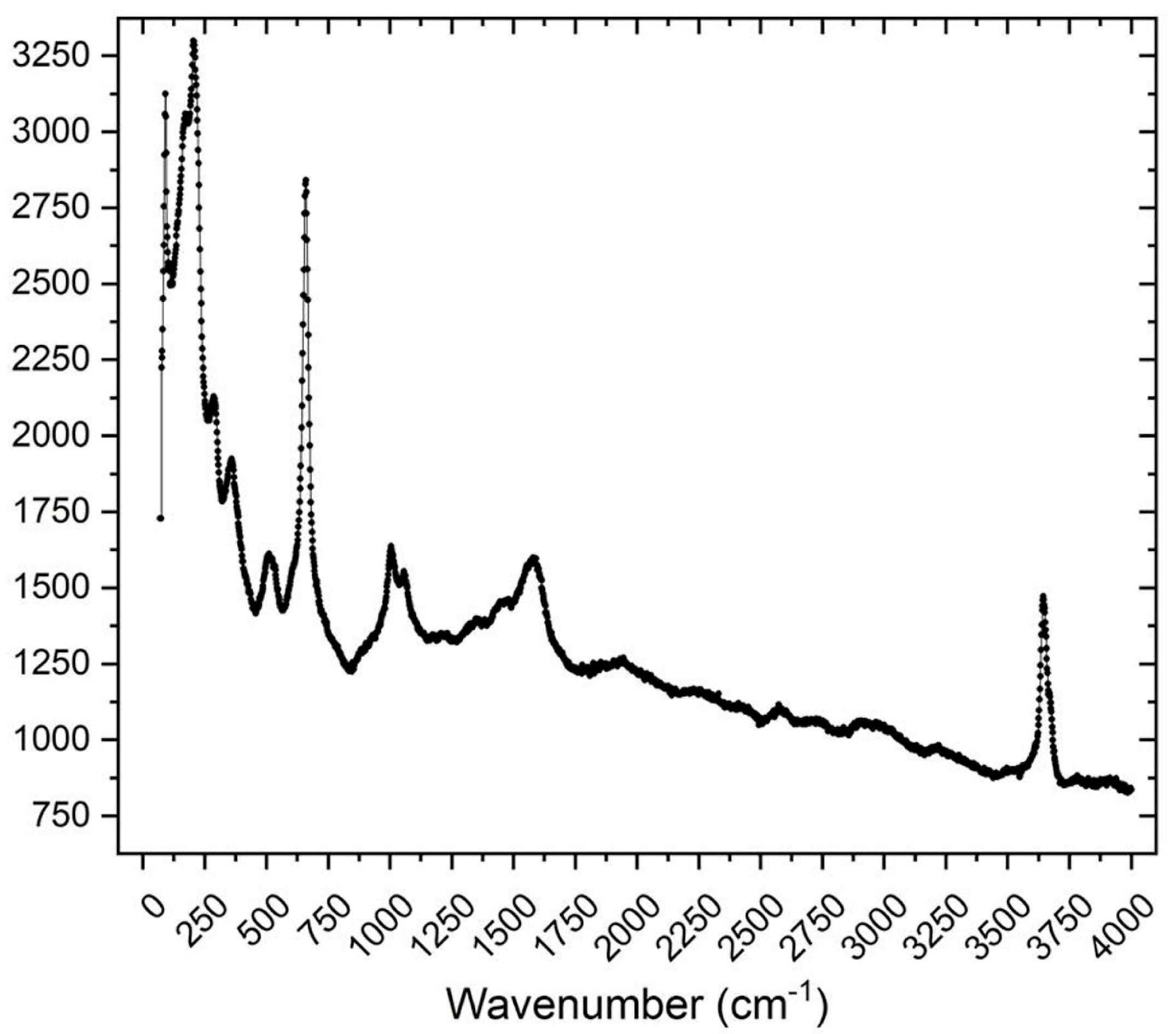
The Raman spectrum of Cl-rich annite (Figure 2) was acquired using a Horiba Jobin-Yvon LabRam HR 800 spectrometer equipped with a motorized x–y stage and an Olympus microscope. The backscattered Raman signal was collected with 50× objective; the spectrum was obtained on an unoriented crystal. The 632.8 nm line of a He–Ne laser was used as excitation; laser power was controlled with a series of density filters. The minimum lateral and depth resolution was about 2 and 5 μm, respectively. The system was calibrated using the 520.6 cm−1 Raman band of silicon before the experimental session. The spectrum was recorded from 70 to 4000 cm−1. Intensive bands at 92, 172, and a strong one at 206 cm−1 are attributed to translational vibrations. Bands at 288 and 360 cm−1 are related to the mixed O–Si–O and O–Al–O bending vibrations in the TO4 tetrahedra and bending vibrations of Me–O–Si and Me–O–Fe bonds, where Me is Fe and Mg [4]. The bands at 511 and 660 cm−1 are assigned to the asymmetric bending vibrations of Si–O–Si bonds [5]. The relatively weak bands at 1006 and 1058 cm−1 correspond to the Si–O–Si asymmetric stretching vibrations. The bands at 1348, 1449, and 1580 cm−1 have a luminescent character. The marked peak in the OH-stretching band (~3644 cm−1) is consistent with the presence of OH in the structure.
Figure 2.
The Raman spectrum of Cl-rich annitic mica.
5. Chemical Composition
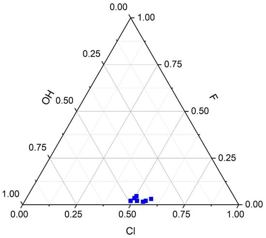
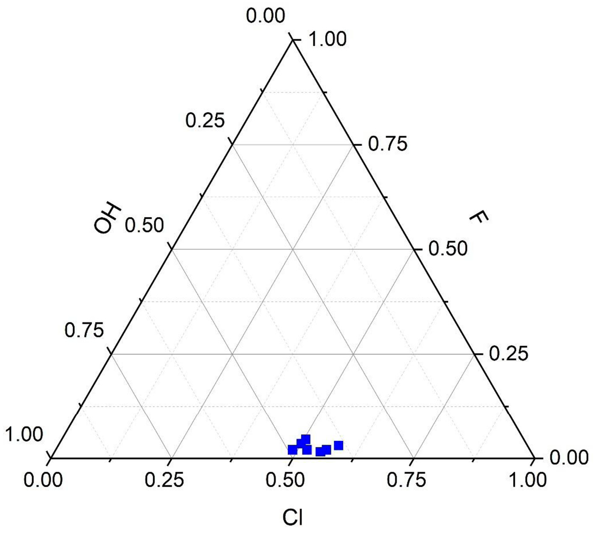
Quantitative analyses of the Cl-rich mica were made at 14 points in the mineralized sample at the Analytical Center for Multi-Elemental and Isotope Studies of the Institute of Geology and Mineralogy, SB RAS, in Novosibirsk. The mica was analyzed using a JEOL JXA-8230 instrument (JEOL Ltd., Akishima, Tokyo, Japan) in wavelength-dispersion spectrometry mode (WDS; V.N. Korolyuk, analyst). The operating conditions were 15 kV and 10 nA, with a focused beam of 1 µm. The counting times were 20 s for peak and 10 s for background. The Kα lines were used for all the elements sought. The four compositions with the highest Cl content are juxtaposed with the three with the least amount of Cl in Table 1, where we list the probe standards used. The quantitative analytical data confirm the energy-dispersion results of [1]. The distribution of Cl, F, and OH (Figure 3) measured in the single-point analyses highlights the prevalence of Cl in some compositions. The fourteen compositions are listed in Supplementary Table S1. The empirical formula of the highest-Cl mica (based on 22 negative charges) is (K0.742Na0.047Ca0.007)Σ0.796 (Fe2+2.901Mg0.078Mn0.047Ti0.007Cr0.003)Σ3.036 (Si3.190Al0.782)Σ3.972O10 (Cl1.105OH0.854F0.041)Σ2.000.
Table 1.
Composition of annite-type mica enriched in chlorine in a PGE-mineralized sample collected in the eastern portion of the Oktyabrsky deposit, Norilsk.
Figure 3.
The distribution of the Cl, F, and OH in the mica, as indicated by results of the EPMA single-point analyses. The data highlight the local predominance of Cl over OH and the low content of fluorine.
6. Crystallography
The small size of the domains of Cl-dominant annite precluded its characterization by single-crystal X-ray diffraction. We did obtain, however, X-ray data suitable for a structure refinement of a crystal from the same specimen. The crystal fragment was found to be homogeneous within analytical error. The refinement indicates 0.90(6) atoms of Cl per formula unit.
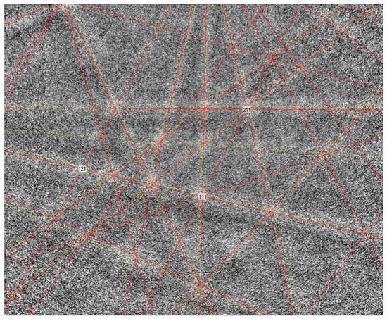
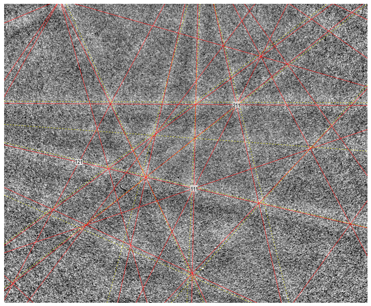
The Cl-dominant annite was verified to possess the mica structure by means of electron backscatter diffraction. The EBSD analyses were performed using a high-performance CMOS Oxford Symmetry S3 EBSD system working on a ZEISS EVO 15-MA SEM, operating at 15 kV and 9 nA nominal current in focused beam mode with a 70° tilted stage. The EBSD pattern for phase identification and analysis was acquired with maximum (1244 × 1024) pixel resolution. Structural data were obtained, and cell constants were derived by matching the experimental EBSD patterns (Figure 4) with that of the Cl-rich annite obtained from the single-crystal X-ray diffraction study. The EBSD patterns gave a good fit with the annite structure with a MAD (mean angular deviation) of 0.88° on nine detected bands.
Figure 4.
EBSD pattern of Cl-dominant annitic mica. The detected bands are yellow in the light gray bands and the simulated bands are red.
The single-crystal X-ray study was carried out using an XtaLAB Synergy-S diffractometer (Rigaku Corporation, Tokyo, Japan) equipped with a HyPix-6000HE at the Centre of the Collective Use of Equipment, Kola Science Centre using MoKα radiation (0.71073 Å). More than half of the diffraction sphere was collected with scanning step 1°, and exposure time 0.5–1 s. The intensity data were integrated and corrected by means of the CrysAlisPro program package [6], which was also used to apply an empirical absorption correction using spherical harmonics, as implemented in the SCALE3 ABSPACK scaling algorithm. Ionized scattering curves were used and a rescaling of reflections with k ≠ 3n was performed to account for the possible presence of twinning and mixed 1M and 2M1 stacking sequences. Information about data collection and refinement is listed in Table 2. The SHELXL program [7] was used for the crystal-structure refinement. The calculated diffraction pattern was obtained with the atom coordinates reported in Table 3 using the software VESTA, version 3.5.8 [8].
Table 2.
Cl-rich annite: crystal data, data collection, and refinement.
Table 3.
Cl-rich annite: calculated X-ray powder-diffraction data.
The structure of Cl-rich annite was refined to an R1 value of 7.89% on the basis of 3720 reflections having Fo > 4σ(Fo), and 938 unique reflections. The Cl-rich annite is monoclinic, space group C2/m (12). The unit-cell parameters are a 5.3991(4), b 9.3586(6), c 10.2421(10) Å, β 100.873(9)°, V 508.22(7) Å3, Z = 2. The calculated X-ray powder-diffraction data are given in Table 3. Statistical tests on the distribution of |E| values indicate the presence of an inversion center. Systematic absences conform to the requirements of space group C2/m. Given the similarity of the unit-cell values, space group and overall stoichiometry, the structure was refined using the atom coordinates of annite [9]. Final atomic coordinates and equivalent isotropic displacement parameters are given in Table 4, whereas selected bond distances are shown in Table 5; selected structural parameters are reported in Table 6.
Table 4.
Cl-rich annite: Wyckoff positions, site occupancy factors (s.o.f.), atom coordinates, and equivalent isotropic displacement parameters (Å2).
Table 5.
Selected bond distances (Å) for Cl-rich annite and synthetic annite.
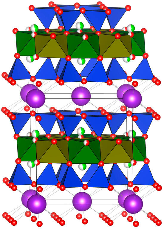
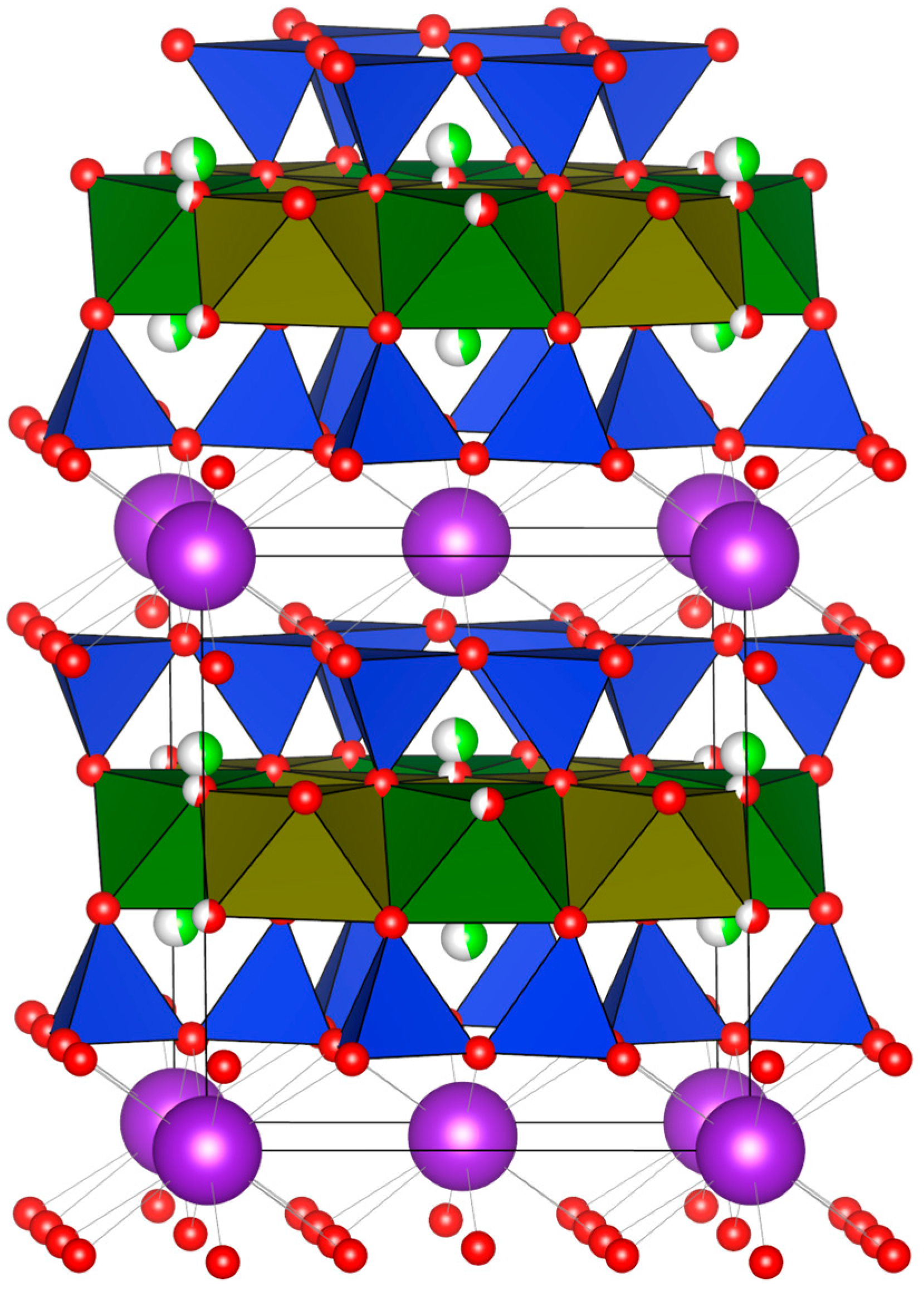
The structure of the Cl-rich annite (Figure 5) is identical to that of the other trioctahedral micas that crystallize as the 1M polytype. The interlayer site is occupied by K atoms. The T site is occupied by Si and Al; M1 and M2 sites are occupied by Fe (~90%) and minor Mg. The total electron density at the octahedral sites obtained from the structural refinement (74.11 e−) is in fair agreement with that calculated from the EPMA data (77.74 e−) even though they were measured on a different grain. The analysis of site-scattering values, mean M-O distances, and flattening parameters in M1 and M2 indicate a homo-octahedral nature [11]. On the other hand, the bond-length distortion (BLD) value is higher in M1 than in M2. No sign of vacancies at octahedrally coordinated sites can be inferred from the crystal-structure refinement nor from the chemical composition. The presence of Cl substituting for O is made evident by the splitting of the O4 site. Of the two positions, one is partially occupied by O and the other, slightly displaced approximately along csinβ, is partially occupied by Cl (Figure 5), as was already observed in a synthetic mica on the phlogopite–celadonite join [12]. The valence sums for O4 (Table 7) are relatively low and agree with the presence of H bonded to this atom, as indicated by the Raman spectroscopy results.
Figure 5.
The structure of Cl-rich annite viewed down ~[100]. The M1 and M2 octahedra are shown in green and olive green, respectively; the TO4 tetrahedra are blue, the O atoms are red, the Cl atoms, green, and the K atoms, purple. The projection was obtained using the software VESTA 3 [8].
Table 6.
Selected structural parameters for Cl-rich annite and synthetic annite calculated as indicated in [13] (and references therein).
Table 7.
Bond valence summations (v.u.) for Cl-rich annite.
7. Discussion
The search is on for coarser crystals of Cl-enriched ferruginous trioctahedral mica. The compositions that are the most promising will be similar to the ones in the left columns of Table 1. The relatively large size of the Cl ion requires a structure in which the octahedral sites are populated by relatively large cations. The ferruginous mica should thus be enriched in Mn2+ (ionic radius, high spin: 0.83 Å [16]) and have minimal amounts of Ti4+ (0.605 Å) and Cr3+ (0.58 Å), all elements that substitute for Fe2+ (high spin: 0.78 Å). The high-Cl compositions in Table 1 contain more Si (0.26 Å) and less IVAl (0.39 Å). In this case, one might predict that, the more aluminous the mica, the better, e.g., [17]. On the contrary, an increase in IVAl causes an increase in kinking of the hexagonal ring of tetrahedra, which will not be consistent with an expanded layer of octahedra. A reduction in the rotation angle among tetrahedra α will thus favor the incorporation of Cl, as found by [9]. As a matter of fact, the tetrahedral sheet of this Cl-rich annite shows nearly perfect hexagonal rings with a remarkable α value of 0.08° (Table 6), the lowest ever reported in natural micas [18,19,20]. Consequently, the interlayer is occupied by highly regular KO₁₂ polyhedra, with <K-Oinner> being very close to <K-Oouter>. The presence of chlorine substituting for OH directly affects also the interlayer thickness owing to the increased attraction between K and Cl, which yields, in a manner like what is observed in F-rich or hydrogen- depleted micas, e.g., [21,22,23], a decrease in the interlayer thickness and a consequent shortening of the c parameter. In agreement with this mechanism, the synthetic Cl-free annite structural parameters [9,10] reported in Table 2, Table 5 and Table 6 show an increased α value, leading to more distorted KO₁₂ polyhedra, and an expanded interlayer thickness, leading to a longer c parameter.
A Cl-rich annitic mica is not only widespread in the mineralized gabbro–dolerite along the eastern portion of the Oktyabrsky deposit in the Norilsk complex. It has also been described in zones of disseminated sulfide mineralization of the Vologochan–Pyasinskiy intrusion, located 25 km west–southwest of the Oktyabrsky deposit, as well as along the western flank of the Oktyabrsky orebody [24]. As on the eastern flank, fractional crystallization of the basic magma led to a considerable range in the composition of all rock-forming minerals. The volatile components (Cl, F, and H2O) accumulated progressively along with S species as a result of efficient fractional crystallization and were responsible for mineralization below the solidus. In the Norilsk camp, there is also evidence of a locally important contribution of sulfur generated by the assimilation of adjacent Devonian metasedimentary units that have an evaporitic component [25]. With such an additional source of chlorine in the circulating aqueous fluid, the environment is likely to yield more mineralogical surprises in the future.
Supplementary Materials
The following supporting information can be downloaded at: https://www.mdpi.com/article/10.3390/min15060640/s1. Table S1: Composition of annite-type mica enriched in chlorine in a PGE-mineralized sample collected in the eastern portion of the Oktyabrsky deposit, Norilsk.
Author Contributions
A.Y.B. (investigation; project administration; writing—original draft preparation); G.O.L. (investigation; writing—original draft preparation; writing—review and editing); L.B. (investigation; writing—original draft preparation; writing—review and editing); R.F.M. (investigation; writing—original draft preparation; writing—review and editing); T.P. (investigation; original draft preparation); I.I.N. (investigation; project administration; original draft preparation); S.A.S. (investigation; formal analysis; data curation). All authors have read and agreed to the published version of the manuscript.
Funding
This research received no external funding.
Data Availability Statement
All available information and results are included in this article.
Acknowledgments
We gratefully acknowledge the contribution of Vladimir N. Korolyuk, who provided the compositions of Cl-rich annitic mica reported here, and of Tiziano Catelani, who helped with the EBSD study. We also extend our appreciation to Vladimir N. Bocharov for recording the Raman spectrum. We acknowledge the constructive suggestions for improvement made by an anonymous referee and by Robert M. Hazen.
Conflicts of Interest
The authors declare no conflicts of interest.
References
- Barkov, A.Y.; Nikulin, I.I.; Nikiforov, A.A.; Lobastov, B.M.; Silyanov, S.A.; Martin, R.F. Atypical mineralization involving Pd–Pt, Au–Ag, REE, Y, Zr, Th, U, and Cl–F in the Oktyabrsky deposit, Norilsk Complex, Russia. Minerals 2021, 11, 1193. [Google Scholar] [CrossRef] [Scilit]
- Rieder, M.; Cavazzini, G.; D’yakonov, Y.S.; Frank-Kamenetskii, V.A.; Gottardi, G.; Guggenheim, S.; Koval, P.W.; Müller, G.; Neiva, A.M.R.; Radoslovich, E.W.; et al. Nomenclature of the micas. Clays Clay Miner. 1998, 46, 586–595. [Google Scholar] [CrossRef] [Scilit]
- Barnes, S.J.; Malitch, K.N.; Yudovskaya, M.A. Introduction to a special issue on the Norilsk−Talnakh Ni–Cu–platinum group element deposits. Econ. Geol. 2020, 115, 1157–1172. [Google Scholar] [CrossRef] [Scilit]
- Panikorovskii, T.L.; Yakovenchuk, V.N.; Yanicheva, N.Y.; Pakhomovsky, Y.A.; Shilovskikh, V.V.; Bocharov, V.N.; Krivovichev, S.V. Crystal chemistry of ivanyukite-group minerals, A3–xH1+x[Ti4O4(SiO4)3](H2O)n (A = Na, K, Cu), (n = 6–9, x = 0–2): Crystal structures, ion-exchange, chemical evolution. Mineral. Mag. 2021, 85, 607–619. [Google Scholar] [CrossRef] [Scilit]
- Tlili, A.; Smith, D.C.; Beny, J.-M.; Boyer, H. A Raman microprobe study of natural micas. Mineral. Mag. 1989, 53, 165–179. [Google Scholar] [CrossRef] [Scilit]
- CrysAlisPro, Version 1.171.36.20; Data Collection and Processing Software for Agilent X-Ray Diffractometers; Agilent Technologies: Tokyo, Japan, 2012.
- Sheldrick, G.M. Crystal structure refinement with SHELXL. Acta Crystallogr. 2015, 71, 3–8. [Google Scholar] [CrossRef] [Scilit]
- Momma, K.; Izumi, F. VESTA: A three-dimensional visualization system for electronic and structural analysis. J. Appl. Crystallogr. 2008, 41, 653–658. [Google Scholar] [CrossRef] [Scilit]
- Hazen, R.M.; Burnham, C.W. The crystal structures of one-layer phlogopite and annite. Am. Mineral. 1973, 58, 889–900. [Google Scholar]
- Redhammer, G.J.; Beran, A.; Schneider, J.; Amthauer, G.; Lottermoser, W. Spectroscopic and structural properties of synthetic micas on the annite-siderophyllite binary: Synthesis, crystal structure refinement, Mössbauer, and infrared spectroscopy. Am. Mineral. 2000, 85, 449–465. [Google Scholar] [CrossRef] [Scilit]
- Ďurovič, S. OD-Charakter, Polytypie und Identifikation von Schichtsilikaten. Fortschritte Mineral. 1981, 59, 191–226. [Google Scholar]
- Nazzareni, S.; Comodi, P.; Bindi, L.; Safonov, O.G.; Litvin, Y.A.; Perchuk, L.L. Synthetic hypersilicic Cl-bearing mica in the phlogopite–celadonite join: A multi-methodical characterization of the missing link between di- and tri-octahedral micas at high pressures. Am. Mineral. 2008, 93, 1429–1436. [Google Scholar] [CrossRef] [Scilit]
- Lepore, G.O.; Bindi, L.; Pedrazzi, G.; Conticelli, S.; Bonazzi, P. Structural and chemical variations in phlogopite from lamproitic rocks of the Central Mediterranean region. Lithos 2017, 286, 191–205. [Google Scholar] [CrossRef] [Scilit]
- Gagné, O.C.; Hawthorne, F.C. Comprehensive derivation of bond-valence parameters for ion pairs involving oxygen. Acta Crystallogr. 2015, B71, 562–578. [Google Scholar] [CrossRef] [Scilit]
- Brese, N.E.; O’Keeffe, M. Bond-valence parameters for solids. Acta Crystallogr. 1991, B47, 192–197. [Google Scholar] [CrossRef] [Scilit]
- Shannon, R.D. Revised effective ionic radii and systematic studies of interatomic distances in halides and chalcogenides. Acta Crystallogr. 1976, A32, 751–767. [Google Scholar] [CrossRef] [Scilit]
- Henry, D.J.; Daigle, N.M. Chlorine incorporation into amphibole and biotite in high-grade iron-formations: Interplay between crystallography and metamorphic fluids. Am. Mineral. 2018, 103, 55–68. [Google Scholar] [CrossRef] [Scilit]
- Brigatti, M.F.; Guggenheim, S. Mica crystal chemistry and the influence of pressure, temperature, and solid solution on atomistic models. Rev. Mineral. Geochem. 2002, 46, 1–97. [Google Scholar] [CrossRef] [Scilit]
- Mercier, P.H.; Rancourt, D.G.; Redhammer, G.J.; Lalonde, A.E.; Robert, J.L.; Berman, R.G.; Kodama, H. Upper limit of the tetrahedral rotation angle and factors affecting octahedral flattening in synthetic and natural 1M polytype C2/m space group micas. Am. Mineral. 2006, 91, 831–849. [Google Scholar] [CrossRef] [Scilit]
- Lepore, G.O.; Bindi, L.; Zanetti, A.; Ciriotti, M.E.; Medenbach, O.; Bonazzi, P. Balestraite, KLi2VSi4O10O2, the first member of the mica group with octahedral V5+. Am. Mineral. 2015, 100, 608–614. [Google Scholar] [CrossRef] [Scilit]
- Cruciani, G.; Zanazzi, P.F. Cation partitioning and substitution mechanisms in 1M phlogopite: A crystal chemical study. Am. Mineral. 1994, 79, 289–301. [Google Scholar]
- Gianfagna, A.; Scordari, F.; Mazziotti Tagliani, S.; Ventruti, G.; Ottolini, L. Fluorophlogopite from Biancavilla (Mt. Etna, Sicily, Italy): Crystal structure and crystal chemistry of a new F-dominant analog of phlogopite. Am. Mineral. 2007, 92, 1601–1609. [Google Scholar] [CrossRef] [Scilit]
- Schingaro, E.; Lacalamita, M.; Scordari, F.; Brigatti, M.F.; Pedrazzi, G. Crystal chemistry of Ti-rich fluorophlogopite from Presidente Olegario, Alto Paranaíba igneous province, Brazil. Am. Mineral. 2011, 96, 732–743. [Google Scholar] [CrossRef] [Scilit]
- Barkov, A.Y.; Nikulin, I.I.; Martin, R.F.; Nikiforov, A.A.; Silyanov, S.A.; Lobastov, B.M. Ore assemblages, platinum-group minerals and behavior of Cl in low-sulfide zones of the Vologochan–Pyasinskiy suites, Norilsk complex, Russia. Can. J. Mineral. Petrol. 2024, 62, 747–764. [Google Scholar] [CrossRef] [Scilit] [PubMed]
- Malitch, K.N. Forecasting criteria for sulphide PGE−Cu−Ni deposits of the Noril’sk province. Lithosphere 2021, 21, 660–682. [Google Scholar] [CrossRef] [Scilit]

Disclaimer/Publisher’s Note: The statements, opinions and data contained in all publications are solely those of the individual author(s) and contributor(s) and not of MDPI and/or the editor(s). MDPI and/or the editor(s) disclaim responsibility for any injury to people or property resulting from any ideas, methods, instructions or products referred to in the content. |
© 2025 by the authors. Licensee MDPI, Basel, Switzerland. This article is an open access article distributed under the terms and conditions of the Creative Commons Attribution (CC BY) license (https://creativecommons.org/licenses/by/4.0/).