


Abstract
This study demonstrates that the generation of excess heat and isotopic water were replicated via a nuclear-like reaction or Low-Energy Nuclear Reactions (LENRs) in 11 reactors. Cross-verification using mass spectrometry (MS), Nuclear Magnetic Resonance (NMR), and cavity ring-down spectroscopy (CRDS) confirmed that the reacted water (byproduct of LENR) does indeed contain the 17O isotope. For mass production of 17O-enriched water, a batch-type internal circulation system integrating a storage tank and a circulation pump was developed. This setup allows continuous manufacturing of 17O-enriched water, with concentrations controlled via circulation time. Specifically, a 32 L system could yield an estimated concentration of 30 mol% 17O-enriched water within 290 h. To measure the high concentration of 17O-enriched water (H217O), we have developed a dilution method, which analyzes 17O NMR data of water produced by the reactor at different dilution ratios. Moreover, it was found that the LENR process produces stable, 200 nm nanobubbles (density 9.33 × 10⁶ particles/mL for the sample #185r12), which possibly consists of non-condensable gases (O2 and CO2) containing 17O. These persistent nanobubbles could enhance the long-term storage stability of isotopic water. A preliminary in vitro cell assay (MTT) using 17O-enriched water (0.35 mol%) revealed alterations in cancer cell metabolic activity, though further studies are required to evaluate potential biological mechanisms. Overall, this cost-effective production technology may provide 17O-enriched water for physical and biological research.
1. Introduction
The phenomenon of cold fusion, initially reported in 1989 using deuterated heavy water as an electrolyte solution during electrolysis [1,2], garnered widespread attention. Subsequent studies demonstrated that similar nuclear fusion-like phenomena could also be induced using light water (ordinary water) as the electrolyte [3,4,5,6,7,8,9]. Furthermore, anomalous energy yields have been observed in water-arc-induced fog generation [10], as well as during micron-sized droplet formation via electrohydrodynamic atomization (EHDA) of pure water [11]. These findings strongly imply that deuterated heavy water is not a strict prerequisite for inducing fusion-like phenomena, and that light water poses a viable alternative.
Hydrodynamic cavitation in liquid water generates localized tensile stress and transient low-pressure microenvironments, inducing rapid bubble nucleation, growth, and energetic collapse [12]. Under such extreme hydrodynamic pressure gradients, the physical and thermodynamic properties of liquid water deviate significantly from standard bulk equilibrium states [13]. Our energy engineering research team has focused on investigating technologies that utilize cavitation in light water to generate excess heat via Low-Energy Nuclear Reactions (LENRs)—a nuclear fusion-like phenomenon. Our previous experimental findings [14] included calorimetric measurements of excess heat, as well as material analyses of fragments from the destroyed reactor caused by abnormal reactions which reveal signatures of nuclear transmutation. Subsequent experiments across a series of diverse reactors revealed that water can be triggered to undergo nuclear-like reactions that yield excess heat. Concurrently, this process generates 22Ne and 17O isotope compounds, including H217O, 17O2, and 12C17O2 etc. [15]. To advance this technology, we have designed a variety of reactors to systematically explore how water cavitation can be leveraged to excite nuclear reactions and generate both excess heat and isotopic compounds [16]. The primary focus of the present study is the replication of LENR in water, confirmation of 17O production in LENR by cross-verification using different instruments, production of 17O-enriched water from cavitation reactors and its characterization.
Recent advancements in quantum biology [17,18,19] suggest that because 17O possesses a non-zero nuclear spin, its magnetic isotope effect (MIE) may influence free radical generation or cellular signaling pathways, potentially modulating cellular metabolic activity. However, clinical and translational evaluation has historically been constrained by the prohibitively high cost and scarcity of 17O-enriched water. Utilizing the low-cost H217O mass-produced by the cavitation reactor, we conducted a pioneer and preliminary in vitro MTT assay on cancer cells to provide a reference for future biological research.
2. Materials and Methods
2.1. Reproducibility of Low-Energy Nuclear Reactions (LENRs) in Water
We have previously reported that cavitation in pure water can generate excess heat, and anomalous isotopic signatures associated with the treated water [14,15]. To rigorously verify the physical reproducibility of these findings under standardized operating conditions, 11 distinct reactors were designed, fabricated, and tested. Beyond measuring the excess heat yield (expressed as the coefficient of performance, COPx), we employed multiple analytical techniques to characterize the effluents, including:
- Mass Spectrometry (MS) Analysis: Conducted to determine the molecular and isotopic composition of the reaction effluents.
- 17O Nuclear Magnetic Resonance (17O-NMR) Spectroscopy: Utilized to analyze and quantify the specific 17O components within the generated products.
- Cavity Ring-Down Spectroscopy (CRDS): Implemented for the high-precision detection and analysis of 17O, 18O, and D (deuterium) isotopes.
2.2. Reactor Design
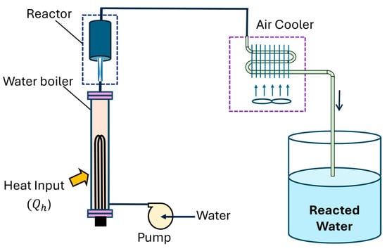
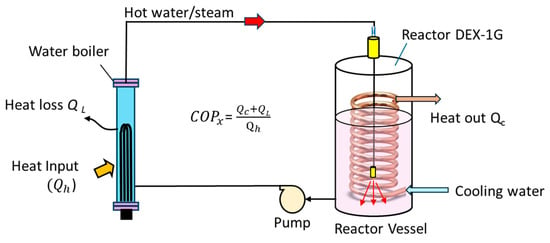
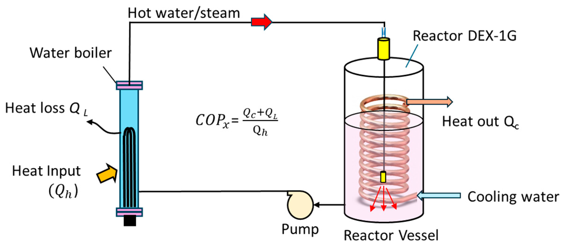
To achieve high energy output efficiency, previous investigations utilized either a triple-pipe heat exchanger (VCS/THX series) that employed refrigerant vapor discharged from a refrigerant compressor to heat water, or a double-pipe heat exchanger (DHX series) that used boiler steam to heat water [14]. Although these two designs achieved remarkable thermal efficiencies (COPx > 4.2), intense, localized cavitation and thermal stress induced material degradation, resulting in short reactor lifespan (<1 week). Due to their structural complexity and low reliability, these reactor configurations are impractical for real-world engineering applications such as power generation. To address these limitations, the present study adopts a resonant cavity structure design, as shown in Figure 1. This new configuration facilitates large-scale scale-up while confining the cavitation phenomena to a region safely away from the reactor walls. Critical operational parameters were continuously monitored and maintained across test runs: fluid flow rates were regulated at 80 ± 5 LPH, operational temperatures were held between 40 °C and 120 °C and circulation pressure differentials were controlled between 1 and 20 bars, depending on batch mode.
Figure 1.
Schematic of the simplified LENR reactor.
2.3. Collection of Reaction Water
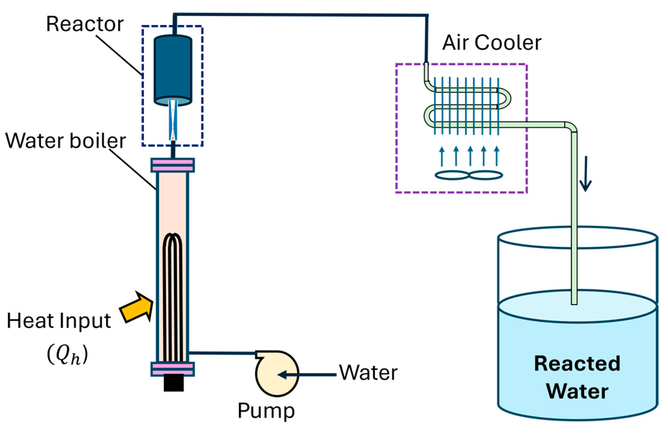
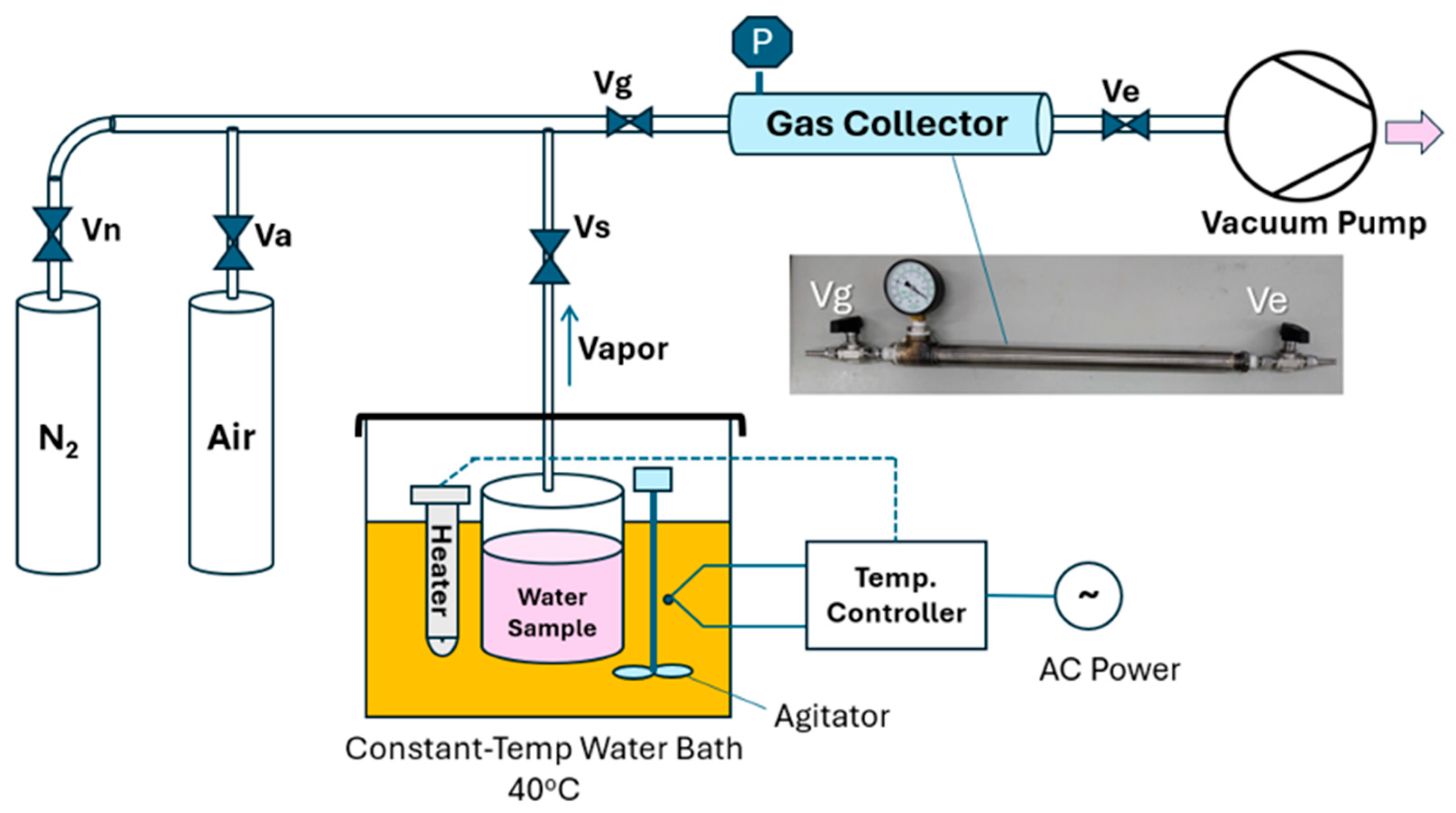
To facilitate the collection and subsequent analysis of the nuclear reaction products, the gas–liquid two-phase water flow discharged from the reactor is routed through an air-cooled radiator for heat dissipation. Upon reaching thermal equilibrium, the effluent condenses into stable liquid water suitable for storage, utilization, or analytical testing, and is subsequently collected in a water tank, as illustrated in Figure 2.
Figure 2.
LENR system.
2.4. Gas Collection for Mass Spectrometry Analysis
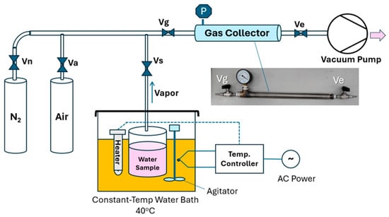
The mass spectrometer employed in this study (Extorr XT300, Extorr Inc., New Kensington, PA, USA) is designed exclusively for gas-phase analysis; therefore, the reaction water stored in the reservoir must be converted into a gaseous state prior to measurement. The analytical results from mass spectrometry can be significantly influenced by sampling techniques and operational parameters, such as the vaporization process of the water sample, mass spectrometer inlet flow rates, system pressures, and interference caused by condensation. To ensure the consistency and reproducibility of the mass spectrometry measurements, a gas collection system, as illustrated in Figure 3, was utilized along with a standard operating procedure (SOP) to convert the reaction water into water vapor. The sequential steps are outlined below.
Figure 3.
Gas collection system configured for reaction water vapor analysis.
2.4.1. Temperature Control of Water Samples
The glass jar containing the water sample was connected to the gas collector, vacuum pump, valves, nitrogen cylinder, and air cylinder as illustrated in Figure 3. The glass jar was immersed in a constant-temperature water bath equipped with an electric heater and a stirrer, and a temperature controller was employed to maintain a constant temperature of 40 ± 1 °C.
2.4.2. Gas Collector Evacuation (Isolated from the Sample Jar)
The vacuum pump was turned on. Valves Vn and Va were closed, while valves Vg, Vs, and Ve were opened. Evacuation was conducted for at least 1 min before switching off the pump and closing Ve.
2.4.3. Gas Collection in the Collector (Connected to the Sample Jar)
Valves Ve, Vn, and Va were closed, while valves Vg and Vs were opened and maintained for 20 min. The vacuum state was utilized to vaporize the liquid water sample, allowing the water vapor to flow into the gas collector. The process was stopped by closing Vg and Vs once the gas pressure reached 30 torr.
2.4.4. Injection of 99.999% N2
Valve Vn was opened until the pressure reached 630 torr. This step was primarily implemented to reduce the humidity of the collected gas, thereby preventing water vapor from condensing due to ambient temperature drops, which could otherwise interfere with the mass spectrometry operation.
2.4.5. Injection of Standard Air
Valves Va and Vg were opened, while Vn, Vs, and Ve were closed, and the process was stopped by closing Va and Vg when the pressure reached 760 torr. Because the concentration of 40Ar in water is extremely low, the injection of standard air provided a sufficient amount of 40Ar components to enhance the intensity of the m/z 40 internal standard signal in the mass spectrometry.
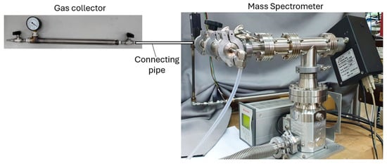
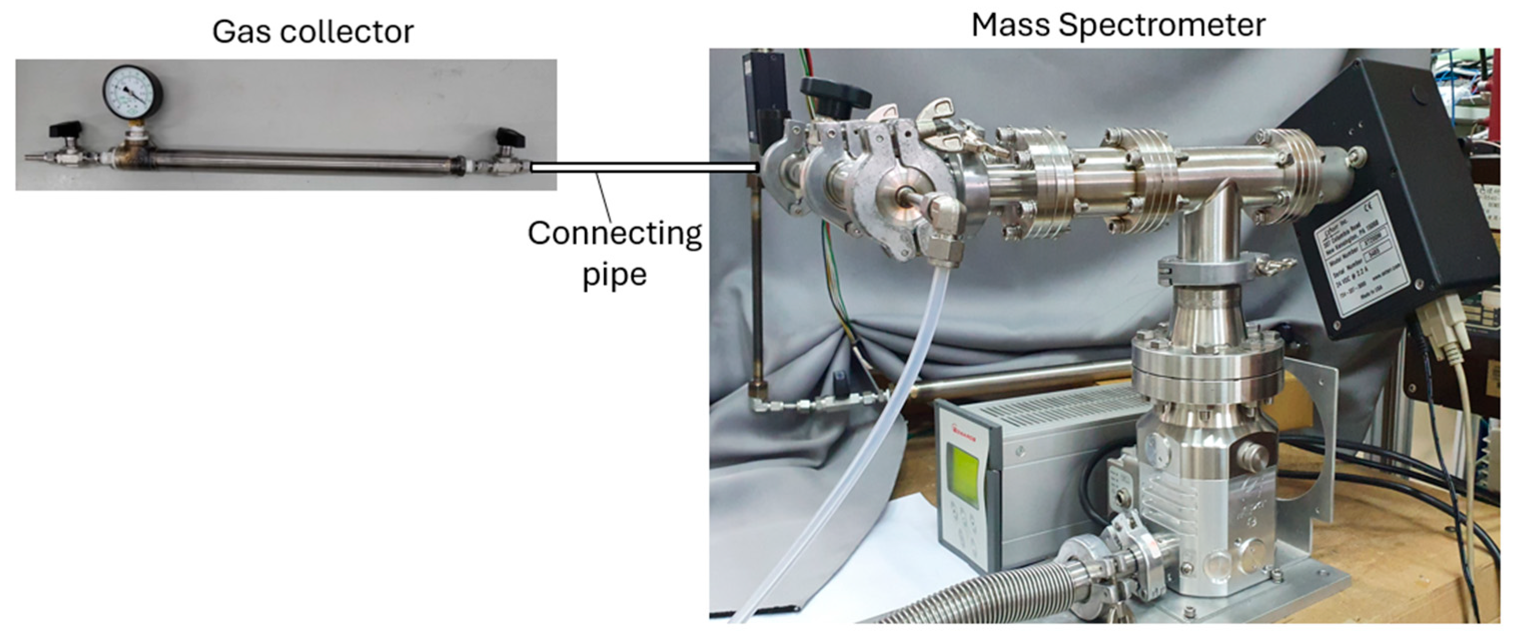
This gas collection procedure was optimized through extensive experimental experience. The connection between the gas collector and the mass spectrometer is shown in Figure 4. To ensure the consistency and reliability of the analytical results, this study simultaneously collected and compared the water after the LENR reaction with the unreacted background water (Reverse Osmosis water, RO water). The differences between the data sets obtained from both samples can effectively determine whether additional components were generated by nuclear reactions.
Figure 4.
Experimental setup showing the connection between the gas collector and the mass spectrometer (Extorr XT300M).
As for the isotope fractionation during vaporization (controlled at 40 °C) in gas collection, thermodynamics analysis shows that the decrease in 17O concentration of vapor phase is minimal (relatively around 3.0%, from 30 mol% in liquid to 29.1 mol% in vapor) for 30 mol% concentration of 17O in liquid phase.
2.5. Analytical Methods for the Composition (Qualitative) and Concentration (Quantitative) of LENR Products
Following the aforementioned vaporization and collection processes, both the reaction water obtained after the LENR process and the unreacted background RO water were introduced into a mass spectrometer (model: Extorr XT300M) for analysis. By comparing the resulting mass spectra with those of the unreacted water, it is possible to qualitatively determine whether the LENR process generated any anomalous or additional components.
2.5.1. Qualitative Analysis of Product Composition via MS
For non-condensable gases dissolved in water, m/z 40 was utilized as the internal standard, defined as follows:
where # represents the mass number of the element, and BG denotes the unreacted water (background water). A value of LL#(gas) > 1.0 indicates that the cavitation reactor generated m/z 19 elements (H217O) exceeding the background water baseline. For water isotopes in the liquid phase, such as H217O, m/z 18 was employed as the internal standard (ordinary water), defined as follows:
where BG denotes the unreacted water (background water). A value of LL198 > 1.0 indicates that the cavitation reactor generated m/z 19 elements (H217O) exceeding the background water baseline.
To minimize experimental error, two water samples—the reaction water and the unreacted background RO water (BG)—were sampled and analyzed simultaneously. This dual-sampling approach effectively eliminates interference arising from variability in water quality.
2.5.2. Quantitative Analysis of Product Concentration via MS
The measured mass spectrometry signal intensity, I#, can be utilized to obtain a quantitative approximation of the concentration (expressed in mol%). Based on the definition in Equation (1), I# represents the ratio of the mass-to-charge (m/z) signal of element # to that of 40Ar. Within the linear range of the mass spectrometer, this ratio can be assumed to represent the concentration ratio of the two elements within the gaseous sample, given by
where Cx # is the concentration of the element with mass number #, and C40Ar is the concentration of 40Ar in the gas sample. The gas sample in the gas collector was charged with water vapor (30 torr, pure nitrogen gas (630 torr), and standard air (100 torr, note: adjusted to match standard total atmospheric pressure of 760 torr). Given that standard air contains 40Ar at a concentration of 0.00934 (0.934 mol%), the concentration of 40Ar within the gas sample is determined as
CAr40 = (130/760) × 0.934 %mol = 0.16 %mol
Consequently, the concentration of the target element can be calculated as
which can be explicitly determined from the measured value of I#. This estimation of non-condensable gas concentration remains valid provided that the operational parameters fall within the linear detection range of the mass spectrometer.
Cx# = I# × C40Ar
2.5.3. Qualitative Verification of 17O Components in Water Samples via NMR and CRDS
The 17O isotope can be detected and characterized utilizing 17O Nuclear Magnetic Resonance (NMR) spectroscopy. To ensure cross-institutional validation and scientific rigor, the water samples in this study were submitted to three independent research facilities for analysis using three distinct 17O -NMR spectrometers:
- National Taiwan University: Employed a Bruker AVIII HD 400 MHz spectrometer (A525, operating at 54.2 MHz) and a Bruker AVIII 400 MHz spectrometer (B662, operating at 54.2MHz).
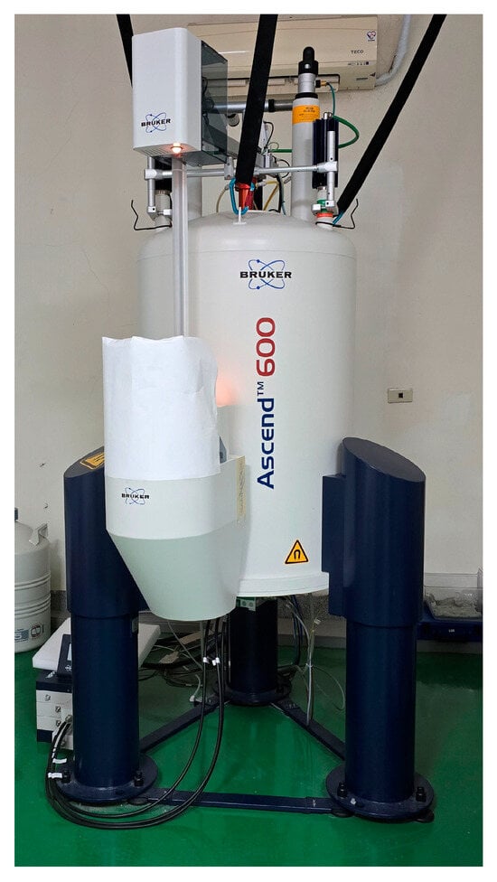
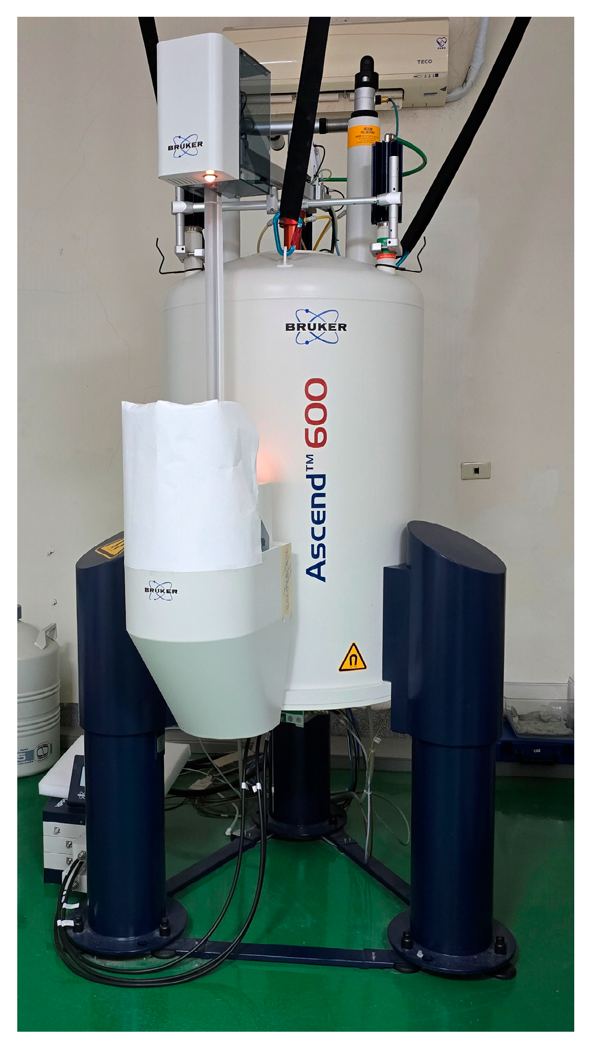
- National Central University: Employed a Bruker AVIII HD 600 MHz spectrometer (operating at 81.37 MHz), conducted both with and without an inner tube configuration, as illustrated in Figure 5.
- Rewave Tech Co (Bruker BioSpin), Ltd.: Employed a Bruker AVIII HD 400 MHz NMR spectrometer.
Figure 5.
Verification of 17O isotopes using different NMR spectrometers.
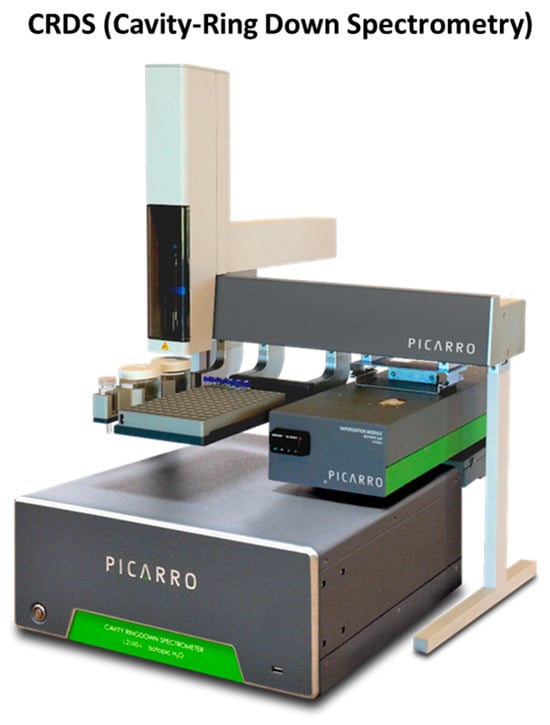
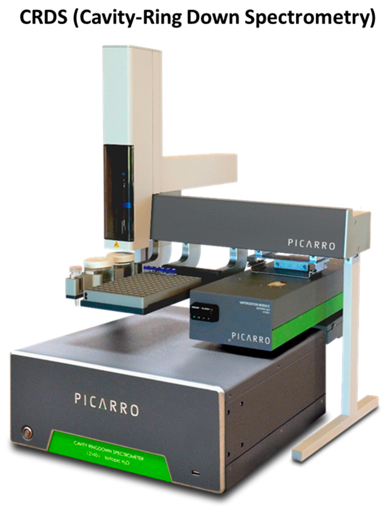
Furthermore, isotopic verification was conducted using a Cavity Ring-Down Spectrometer (CRDS; Picarro L2140-i isotope analyzer, Picarro, Santa Clara, CA, USA) located at National Chung Hsing University, as structurally illustrated in Figure 6.
Figure 6.
Isotopic product verification via CRDS: 17O, 18O, D.
2.6. Enrichment and Mass Production of 17O-Enriched Water
By incorporating a storage tank (capacity V) and a circulation pump into the LENR reaction configuration from Figure 2, a closed circulation system was constructed to continuously manufacture 17O water (Figure 7). The accumulation of H217O content in the water volume V after each circulation loop can be determined via Equation (6), derived from Equation (2):
Figure 7.
Experimental setup for the continuous mass production of 17O-enriched water.
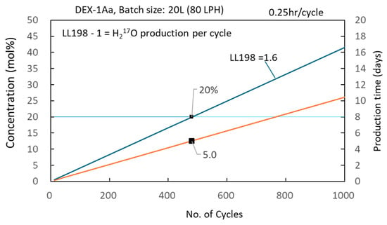
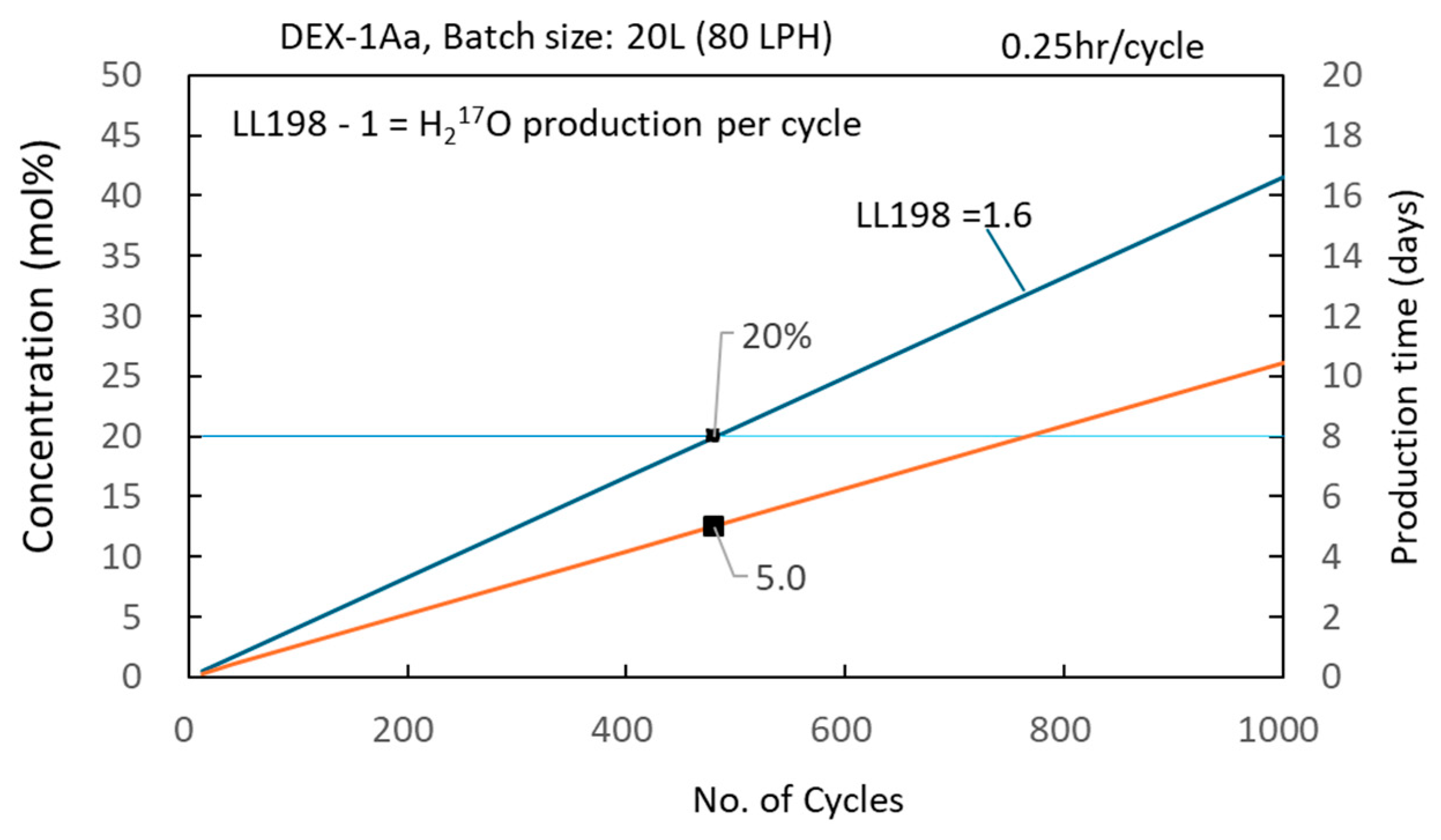
Figure 8 shows the calculated concentration as a function of circulation cycles at LL198 = 1.6 (DEX-1Aa). With a 20 L batch size, reaching 20 mol% takes about 5 days (120 h).
Figure 8.
Relationship between concentration and time in mass production.
3. Results
3.1. Replication of LENR in Water
Eleven distinct LENR reactors were designed and fabricated for the experiments. Water samples were collected both before and after the reactor treatments, and the samples were subsequently gathered according to the gas collection process illustrated in Figure 3. These gas samples were then subjected to mass spectrometry (MS) analysis to qualitatively determine and compare the composition of the reaction products with the unreacted one. The results summarized in Table 1 reveal noteworthy phenomena:
Table 1.
LENR replication results across eleven different reactors.
- Excess 22Ne consistently co-occurs with four 12C-containing compounds and five 17O-containing compounds, aligning with the conjectured reaction models proposed in [12].The black box in Equations (7) and (8) represents an unknown in detail mechanism.
- For the CO2 containing single 17O (i.e., 12C16O17O) with m/z = 45, the MS index consistently follows the trend of LL45 > LL46, except for reactors No. 10 and No. 11. This implies that the generation of CO2 containing single 17O via LENR is more favorable than that containing double 17O.
- For the O2 containing single 17O (i.e., 16O17O) with m/z = 33, the MS index always exhibits LL33 > LL34, implying that the generation of O2 containing single 17O via LENR is more favorable.
These consistent isotopic shifts across multiple reactor configurations demonstrate physical reproducibility under specified operating conditions.
3.2. Quantitative Determination of Product Concentrations via MS Analysis
By analyzing the MS data using Equations (3)–(5), the concentrations of the reaction products can be estimated, as summarized in Table 2. For sample #178r3 (DEX-1Aa, 48 cycles), estimated concentrations of isotopic species were notably higher than in untreated background water. Signals corresponding to four distinct oxygen isotopic gas species (O2) were detected in the reacted water, with 17O17O displaying the highest concentration of 0.016 mol% about 1.34 times of unreacted RO water. The concentration of 16O17O is 0.007 mol%, about 2.92 times of unreacted RO water.
Table 2.
Concentrations of LENR products calculated from mass spectrometry data.
3.3. Qualitative Verification of 17O in Water Samples via NMR Spectroscopy
The liquid water products collected from three reactors (DEX-1, DEX-1Aa, and VCS-5RT) operated under different conditions were analyzed utilizing three distinct 17O-NMR spectrometers across three independent organizations, and the findings were compared with the MS data. As summarized in Table 3, the results demonstrate high consistency among the different organizations. This cross-verification robustly substantiates the MS findings, further confirming that excess 17O is generated within the cavitation reactor.
Table 3.
17O-NMR analysis of water samples.
3.4. Isotopic Composition Analysis of Water Samples via CRDS
The third analytical technique employed for isotopic identification of the LENR products was cavity ring-down spectroscopy (CRDS). The δ17O, δ18O, and δ2H values in the Tube116 and 190r1 samples reveal a significant enrichment of 17O/16O, 18O/16O and 2H/1H ratios relative to the background (unreacted) water, i.e., Dev = (Sample-BG)/SD > 10. Conversely, sample 185r12 exhibits enrichment in 17O/16O and 18O/16O only, Dev > 2. Notably, as summarized in Table 4, these results show 17O enrichment alongside observed shifts in 18O and HDO isotope ratios relative to background water.
Table 4.
CRDS identification of 17O, 18O, 2H isotopes in water samples from different reactors.
3.5. Mass Production of 17O-Enriched Water
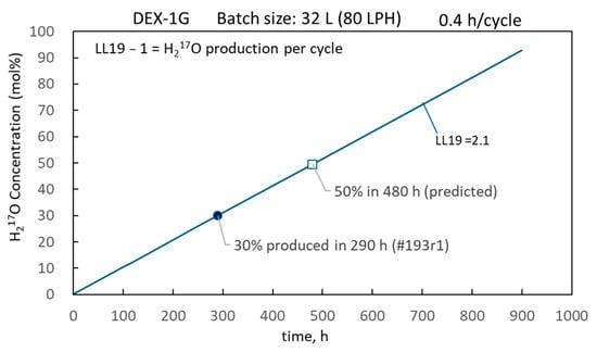
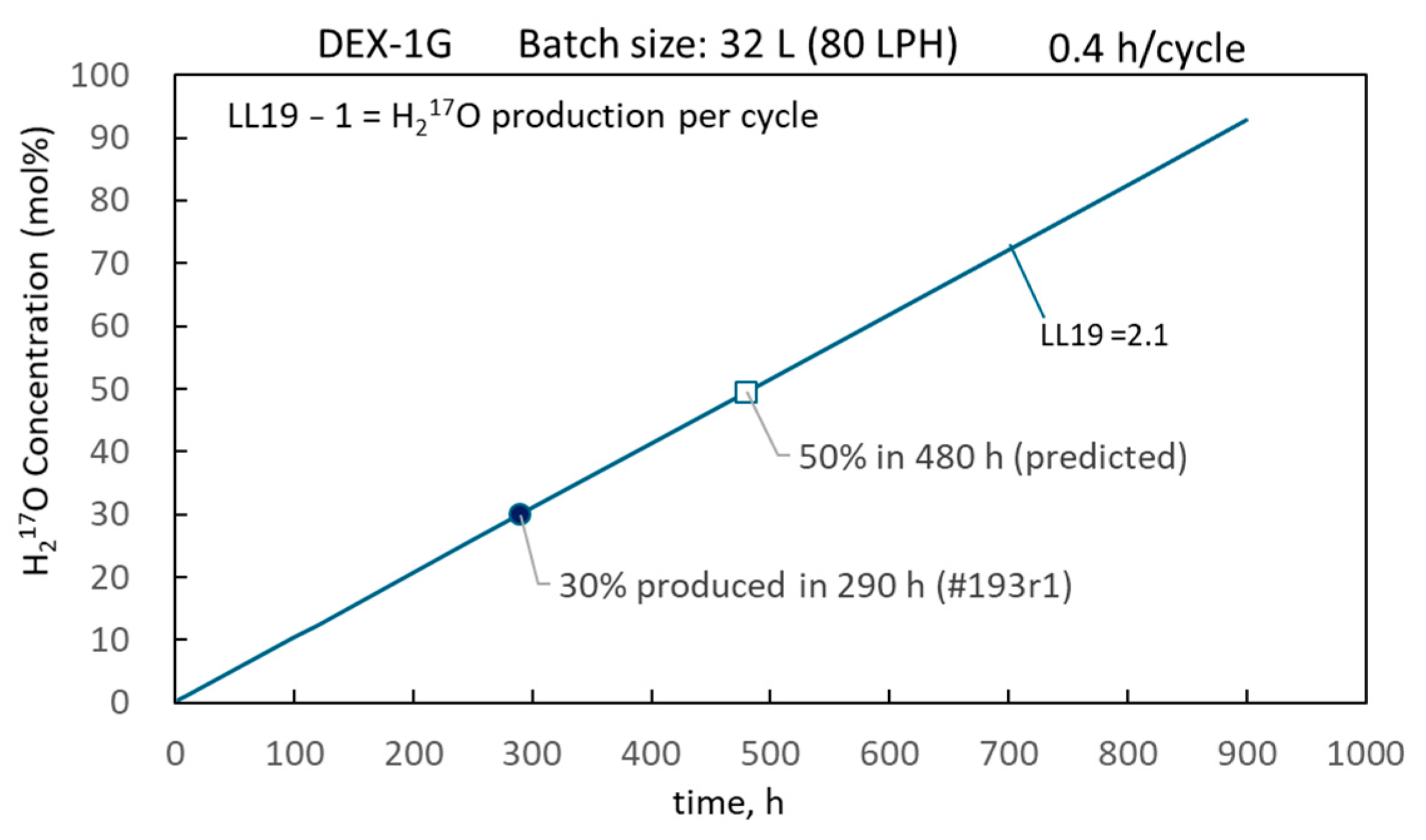
The LENR closed-loop circulation system configuration based on the DEX-1G reactor, as illustrated in Figure 7, was employed for the production of 17O-enriched water. For a batch volume (V) of 32 L, the incremental increase in H217O content per cycle can be calculated using Equation (6). Continuous operation of the DEX-1G system for 290 h (~12 days) yielded 32 L of 17O-enriched water with a concentration of 30 mol%, corresponding to an average daily production rate of 2.6 L. Furthermore, based on model extrapolation (Figure 9), extending continuous operation to 480 h is projected to yield a 17O-enriched water concentration of 50 mol%, representing a predicted benchmark subject to extended empirical validation.
Figure 9.
Mass production curve of DEX-1G—predicted data needing empirical validation.
3.6. Determination of 17O-Enriched Water Concentration via 17O-NMR Spectroscopy
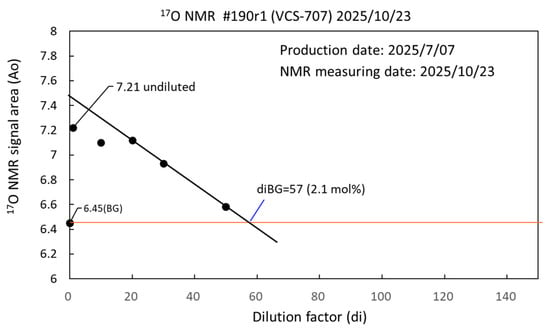
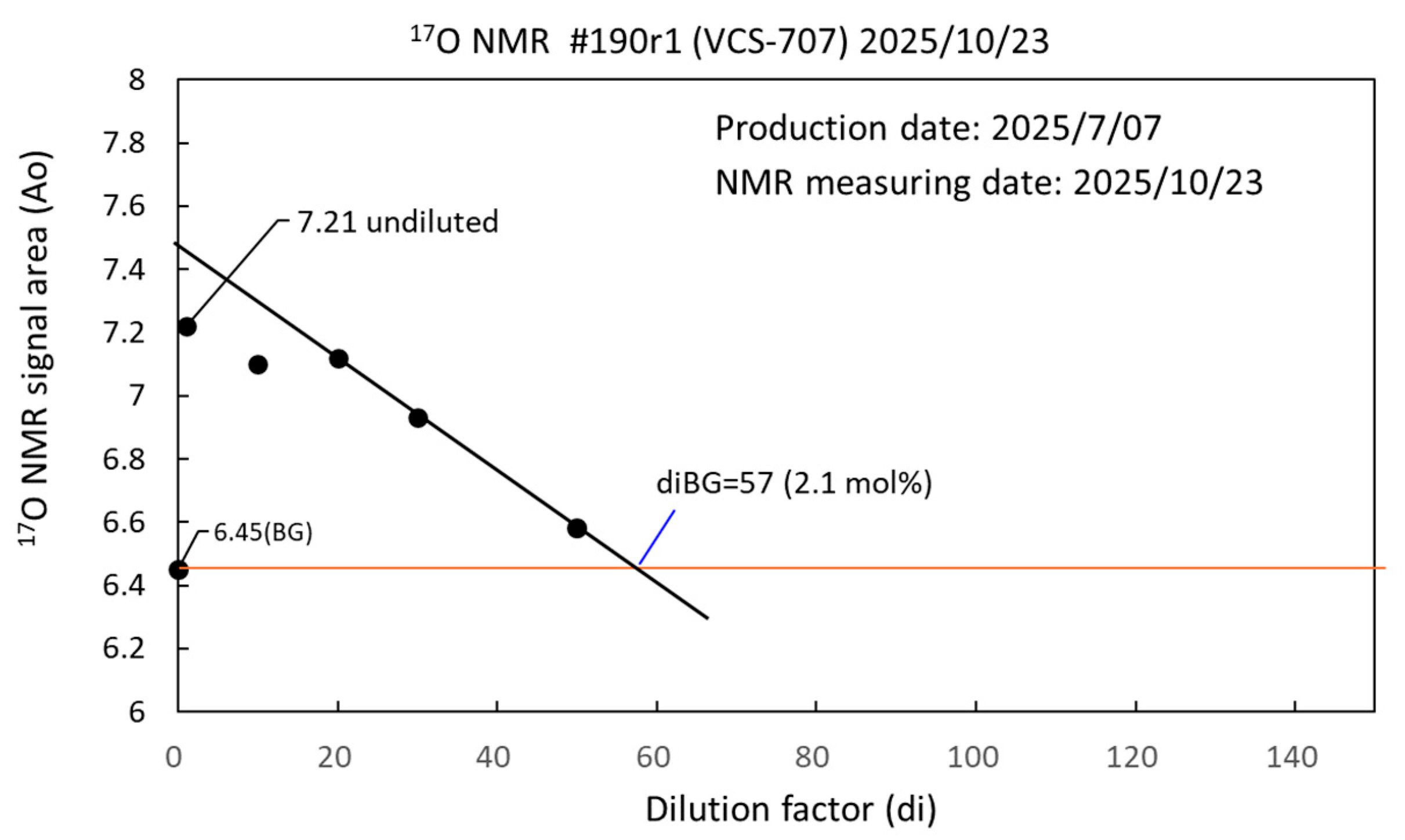
The natural abundance of 17O in natural water is 0.0375 mol%. Because the 17O concentrations produced in this study could exceed 5 mol% (more than 133 times the natural abundance), the precise determination of such high 17O-enriched water concentrations is exceptionally challenging. A dilution method was developed in this study to estimate the 17O concentration. By analyzing the 17O-NMR signals across various dilution factors (di), the dilution factor (diBG) at which the 17O-NMR signal becomes identical to that of the background water (unreacted reverse osmosis RO water) can be identified. This specific dilution factor closely correlates with the concentration of the undiluted stock solution (Cx), which is expressed as follows:
Cx = diBG × 0.0375 mol% (natural abundance)
Here, the natural abundance of 17O in natural water (0.0375 mol%) is used as a natural calibration standard. Figure 10 presents the measured concentration of sample #190r1 (2.1 mol%) using Brucker Ascend 600 MHz (NCU).
Figure 10.
17O concentration measurement of sample #190r1.
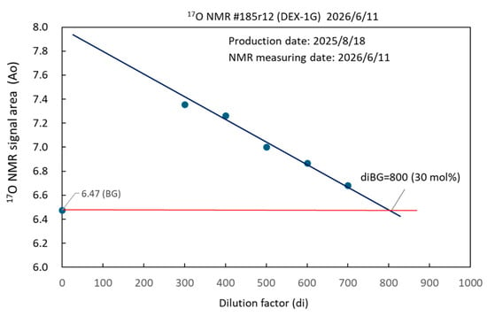
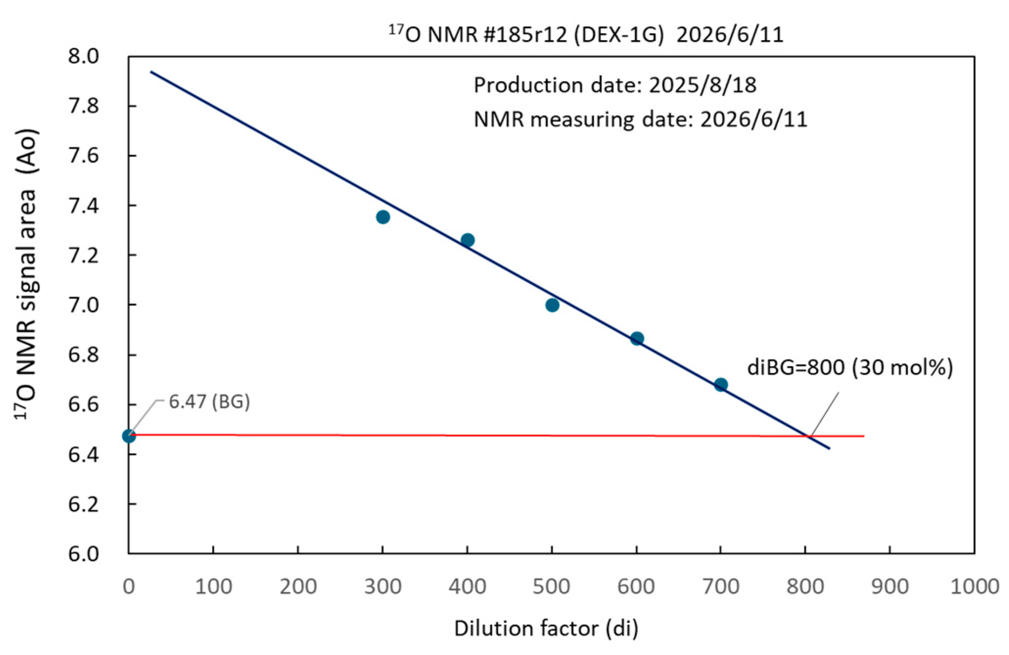
Figure 11 presents the concentration determination of sample #185r12 (30 mol%). Notably, sample #185r12 was obtained using the DEX-1Aa reactor with a water volume of 32 L, an operating temperature of 120 °C, and a flow rate of 80 LPH across 485 continuous cycles (each cycle consisting of 40 min on and 60 min off). A high 17O concentration of 30 mol% was obtained. Furthermore, this water sample was analyzed after being stored at room temperature for nearly a year (10 months), demonstrating the long-term stability of the water sample.
Figure 11.
17O concentration measurement of sample #185r12.
It should be noted that the use of the dilution method, combined with NMR measurements, to quantitatively determine the concentration of 17O-enriched water is a pioneering technique. This study is only a preliminary attempt. As 17O is a magnetic isotope, further research is required to determine whether it may interfere with the EM waves of NMR and cause measurement errors.
3.7. Size Distribution Analysis of Bulk Nanobubbles in 17O-Enriched Water
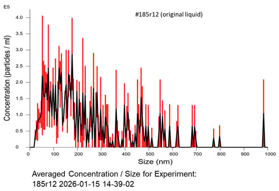
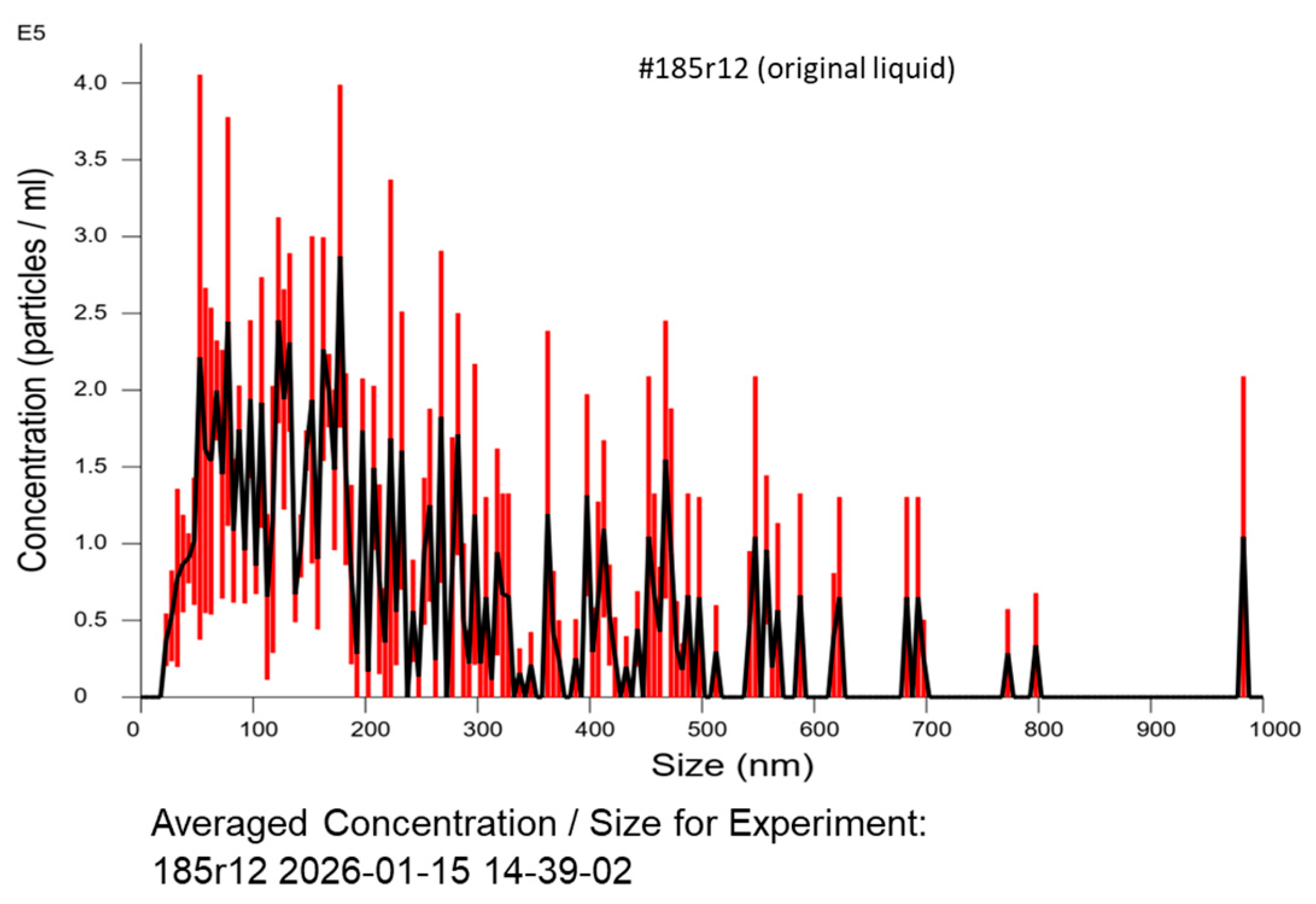
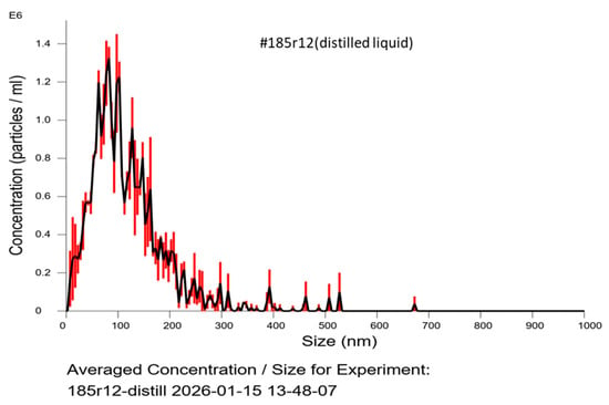
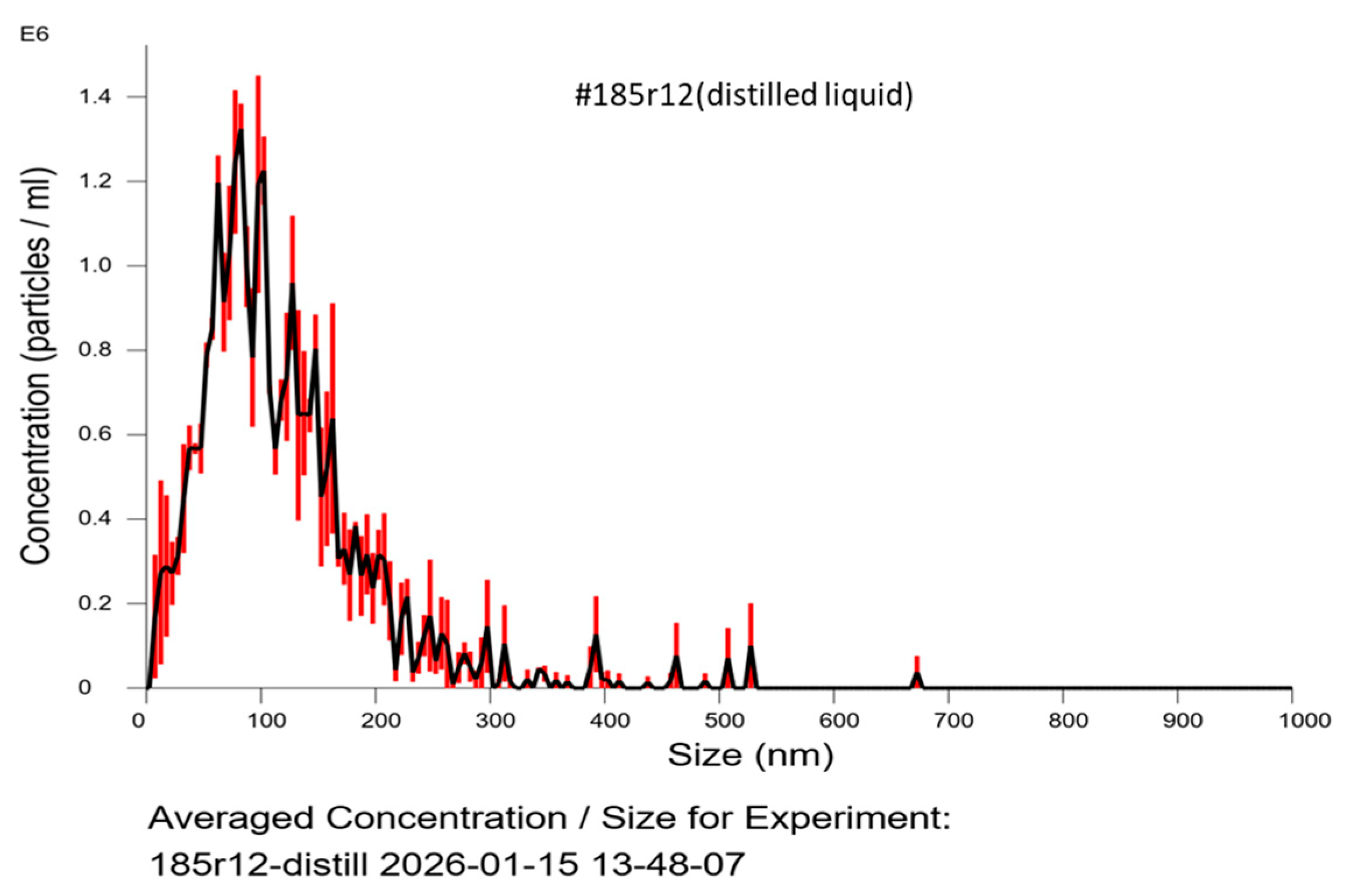
The 17O-enriched water molecules produced directly from the reactor is a liquid outflux that rapidly reaches a state of thermodynamic equilibrium with the bulk H216O water matrix. Notably, bubble evolution was observed during the cavitation process. Bulk nanobubbles generated via aqueous cavitation typically possess negative interfacial charges (zeta potential) and high surface tension resistance, which effectively inhibit bubble coalescence and suppress rapid buoyancy-driven degassing [20,21]. Nanoparticle tracking analysis (NanoSight) of sample #185r12 revealed a mean bubble diameter of 242.6 ± 18.5 nm and a bubble density of (9.33 ± 0.49) × 106 particles/mL (Figure 12). Following distillation of sample #185r12, the mean bubble diameter shrank to 119.4 ± 3.4 nm, whereas the bubble density increased to (2.83 ± 0.12) × 107 particles/mL (Figure 13). Detailed comparison is shown in Table 5. These bulk nanobubbles are inferred to encapsulate dissolved non-condensable gas species, including 17O-labeled O2 and CO2 isotopes identified in mass spectrometry analyses (Table 2).
Figure 12.
Bubble size distribution of sample #185r12 (original liquid).
Figure 13.
Bubble size distribution of sample #185r12 (distilled liquid).
Table 5.
Particle size distribution of nanobubbles in sample #185r12.
Due to their sub-micron dimensions and colloidal behavior, these nanobubbles resist rapid buoyancy-driven degassing. It is hypothesized that gas encapsulation within bulk nanobubbles retards gas loss and contributes to the prolonged retention of 17O species. Because 17O-NMR spectroscopy records the superimposed signal from both the aqueous phase (H217O) and encapsulated gaseous species, the relative mass distribution cannot be individually differentiated. This working hypothesis aligns with colloidal stabilization models, such as that described by Takahashi et al. [22] and Yasuda et al. [23], who reported that bulk nanobubbles (BNBs) achieve exceptional multi-month stability via a “condensed ionic cloud” that induces local precipitation, forming a topologically rigid solid nanobubble shell. Therefore, bulk nanobubbles demonstrate multi-month persistence in aqueous solutions. Consequently, gas encapsulation provides a plausible physical mechanism consistent with the empirical 10-month room-temperature stability observed for sample #185r12 (Figure 11).
3.8. MTT Assay Using 17O-Enriched Water
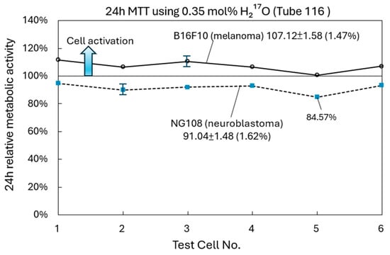
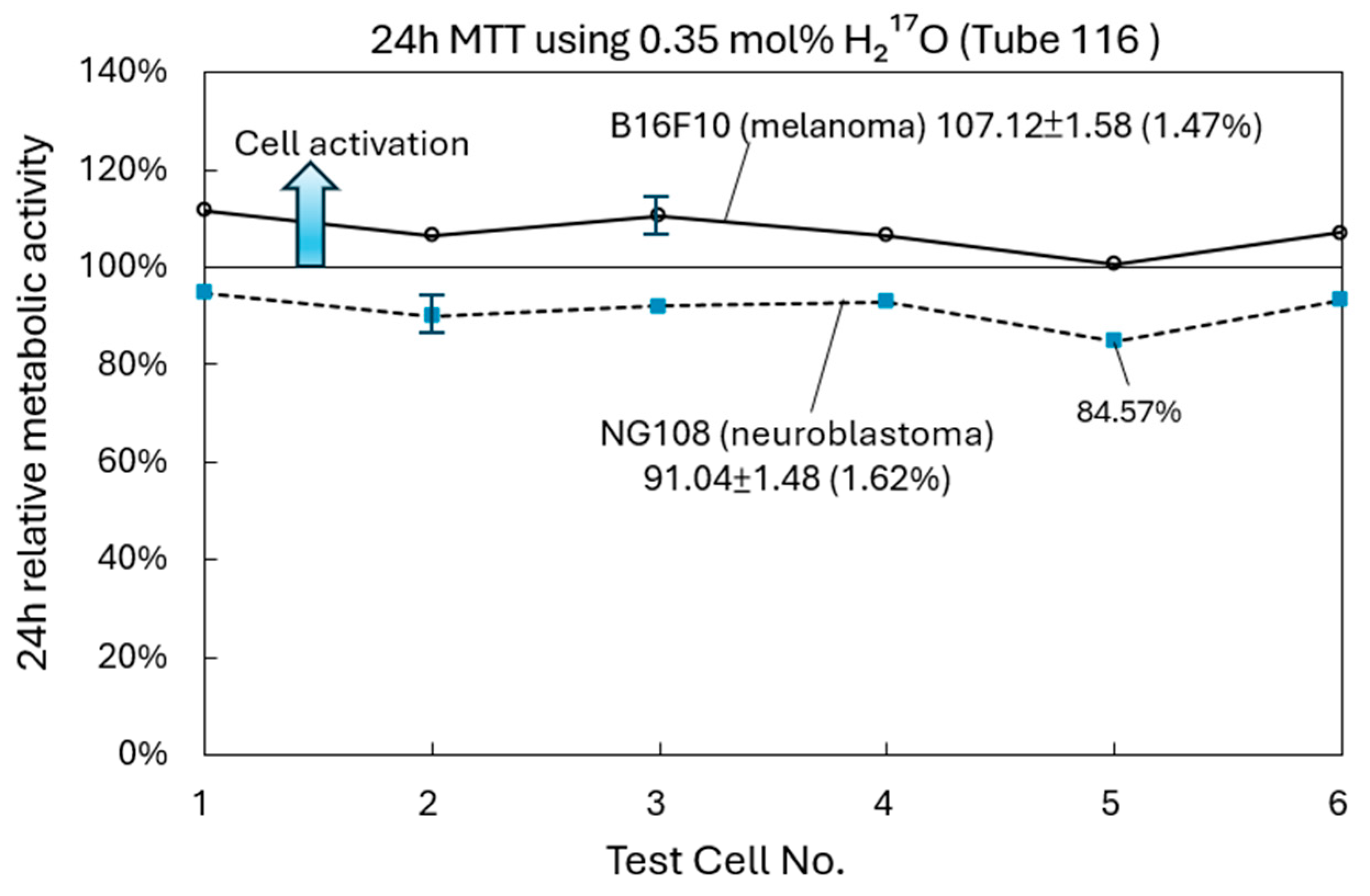
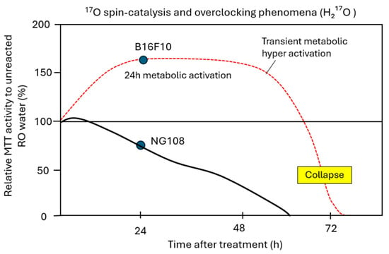
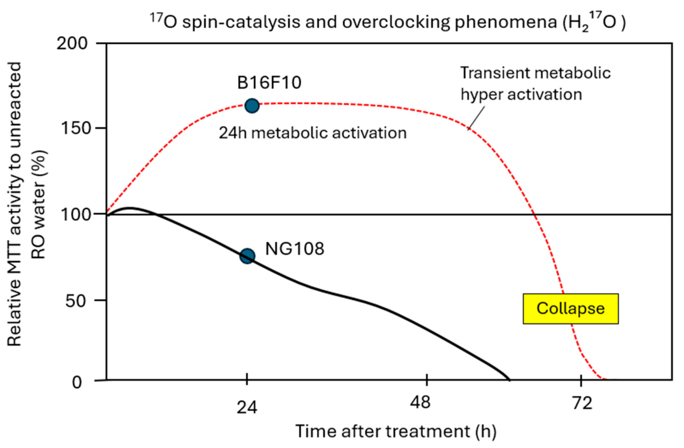
Quantum biology studies suggest that 17O may interfere with cancer cell metabolism [17,18,19]; however, experimental validation has been constrained by the scarcity of 17O. In this study, preliminary exploration in vitro MTT cell-culture assays were conducted on cancer cell lines using low-dose 17O-enriched water (0.35 mol%, Tube116). As shown in Figure 14, following treatment with 0.35% H217O, NG108 cells (neuroblastoma) exhibited mild metabolic inhibition (%) at 91.04 ± 1.48 (1.62% variation), whereas B16F10 cells (melanoma) exhibited mild metabolic activation (%) at 107.12 ± 1.58 (1.47% variation). Although this experiment involved only six samples—which is insufficient to draw statistically significant conclusions—the results showed that both cell types exhibited consistent and precise changes (SD < 2 per cent). Given the consistency of the findings, these preliminary experimental results are still of value as a reference, demonstrating that 17O-water may alter the activity of cancer cells. The concept of an “overclocking” effect and the hypothetical curve in Figure 15 represent tentative working models [17,18] requiring longer time duration, multi-dose, concentration-dependent verification. Overall, these preliminary in vitro results indicate that low-concentration 17O-enriched water can alter cancer cell metabolic activity, establishing an empirical baseline for future biological research.
Figure 14.
MTT assay of cancer cells treated with 17O-enriched water (± SD represents the standard deviation of the data of 6 samples).
Figure 15.
Hypothetical reaction curves corresponding to the overclocking phenomenon [17,18].
4. Discussions
In this study, the results of experiments conducted using 11 water cavitation reactors have once again confirmed that isotopic enrichment occurs with the occurrence of the excess heat phenomenon. By employing cross-verification using three different kinds of instrument MS, NMR, and CRDS, 17O enrichment was indeed observed concurrently with excess heat, eliminating potential measurement bias arising from the use of a single measurement method. While MS, NMR, and CRDS confirm isotopic 17O enrichment, the hypothesized reaction pathways (Equations (7) and (8)) remain as working hypotheses pending direct experimental proof. Future work can integrate gamma or neutron detectors to capture prompt radiation signatures to identify the pathways of LENR in water cavitation reactor.
From thermodynamics point of view, the reactor-yielded liquid H217O spontaneously reaches a thermodynamic equilibrium state with regular H216O water. It was also noted that the cavitation process of water also generates nanobubbles with an average diameter of approximately 200 nm and particle density of 9.33 × 10⁶ particles/mL, containing non-condensable O2 and CO2 gas species, both including 17O. The colloidal persistence of these nanobubbles is hypothesized to retard gas degassing, thereby contributing to sample stability. While 17O-NMR measurement captures the combined signal from liquid and gaseous phases without resolving phase-specific contributions, the non-zero nuclear spin (I = 5/2) of 17O offers a physical mechanism via the magnetic isotope effect (MIE) [16,17,18], which has been proposed in quantum biology to influence biochemical reactions of cancer cells.
Utilizing the mass production of 17O-enriched water, a preliminary MTT assay was performed on malignant cell lines. The MTT cell-culture assays show that exposure to 0.35 mol% 17O-enriched water may alter metabolic activity in specific cancer cell lines in vitro. These initial findings provide an empirical baseline for future biological inquiry. However, clinical, therapeutic, and translational conclusions remain premature; comprehensive multi-dose evaluations and mechanistic pathway studies are required before medical applications can be established.
5. Conclusions
This study provides an empirical replication of isotopic shifts and mass enrichment of 17O in water cavitation systems across 11 distinct reactors. Cross-verification using MS, NMR, and CRDS confirmed consistent 17O enrichment occurring concurrently with excess heat. A batch-type internal circulation system achieved mass production of 17O-enriched water up to 30 mol% concentration. The formation of persistent bulk nanobubbles (~200 nm) is proposed as a working mechanism contributing to extended sample stability. Preliminary in vitro MTT assays revealed altered cancer cell metabolic activity, establishing a baseline platform for further physical and biological exploration of 17O-enriched water.
Author Contributions
M.-Y.L.: investigation, nuclear magnetic resonance data preparation, validation, data analysis. S.-L.C.: project administration, resources, supervision. L.X.: conceptualization, methodology. Y.-H.P.: investigation, data preparation, nuclear magnetic resonance validation, data analysis. X.-Y.W.: investigation, data preparation, validation, data analysis. P.-H.W.: data preparation, validation, data analysis. J.-F.Y.: conceptualization, methodology, investigation, project administration, supervision. Y.-Y.H.: project administration, supervision. K.-C.L.: supervision, validation. Y.-T.C.: supervision, validation. T.-R.T.: supervision, validation, mass spectrometry data analysis. F.-W.K.: supervision, validation, mass spectrometry data analysis. T.-F.T.: supervision, validation, mass spectrometry data analysis. B.-J.H.: conceptualization, methodology, investigation, project administration, supervision, resources, validation, data analysis, writing—original draft, funding acquisition. All authors have read and agreed to the published version of the manuscript.
Funding
This research work was supported by National Taiwan Normal University, through Project No. 112EE00118.
Data Availability Statement
The original contributions presented in this study are included in the article. Further inquiries can be directed to the corresponding author.
Acknowledgments
The authors extend their appreciation to the researchers supporting this study.
Conflicts of Interest
Yu-Hsiang Pan, Xin-Yuan Wu, Po-Hsien Wu, Jong-Fu Yeh and Yu-Yuan Hsieh were employed by ATD Inc., Konglin Group, Taiwan. They helped build the reactors and set up experimental systems for testing; they also performed NMR analysis. Tong-Rong Tseng, Fang-Wei Kang and Tan-Feng Tsai were employed by Mastek Technologies, Inc., Taiwan. They conducted MS measurements and analysis in this study.
References
- Fleischmann, M.; Pons, S. Electrochemically induced nuclear fusion of deuterium. J. Electroanal. Chem. Interfacial Electrochem. 1989, 261, 301–308. [Google Scholar] [CrossRef] [Scilit]
- Jones, S.; Palmer, E.; Czirr, J.; Decker, D.L.; Jensen, G.L.; Thorne, J.M.; Taylor, F.S.; Rafelski, J. Observation of cold nuclear fusion in condensed matter. Nature 1989, 338, 737–740. [Google Scholar] [CrossRef] [Scilit]
- Matsumoto, T. Cold Fusion Observed with Ordinary Water. Fusion Technol. 1990, 17, 490–492. [Google Scholar] [CrossRef] [Scilit]
- Notoya, R.; Noninski, V.C. Excess heat during the electrolysis of a light water solution of K2CO3 with a nickel cathode. Fusion Technol. 1992, 21, 163–167. [Google Scholar] [CrossRef] [Scilit]
- Notoya, R. Cold Fusion by Electrolysis in a Light Water-Potassium Carbonate Solution with a Nickel Electrode. Fusion Technol. 1993, 24, 202–204. [Google Scholar] [CrossRef] [Scilit]
- Pinaev, A.V. The analyses of test data of cavitation heat-generator of TMK-25 type. In Proceedings of the 13th International Conference on Condensed Matter Nuclear Science (ICCF-13), Sochi, Russia, 25 June–1 July 2007; pp. 291–294. [Google Scholar]
- Pavlovoch, T. Nuclear Energy in Vortex Cavitation Generators. 2024. Available online: https://www.researchgate.net/publication/383093910 (accessed on 1 June 2026).
- Kanarev, P.M.; Mizuno, T. Cold Fusion by Plasma Electrolysis of Water. 2003. Available online: https://www.researchgate.net/publication/237276063 (accessed on 1 June 2026).
- Ross, G.; Anwar, D.; Sedgwick, D.; Fustero, S. Experiments demonstrating the creation of elements via pulsed electric field in water and deuterated water: Application to the production of fluorine. arXiv 2022, arXiv:2202.03744. [Google Scholar]
- Sundaresan, R.; Bockris, J.O. Anomalous Reactions During Arcing Between Carbon Rods in Water. Fusion Technol. 1994, 26, 261–265. [Google Scholar] [CrossRef] [Scilit]
- Graneau, P.; Graneau, J.; Li, X. Renewable energy liberation by nonthermal intermolecular bond dissociation in water and ethanol. J. Appl. Phys. 2011, 109, 034908. [Google Scholar] [CrossRef] [Scilit]
- Brennen, C.E. Cavitation and Bubble Dynamics; Cambridge University Press: New York, NY, USA, 2014. [Google Scholar] [CrossRef] [Scilit]
- Caupin, F.; Arvengas, A.; Davitt, K.; Azouzi, M.E.M.; Shmulovich, K.I.; Ramboz, C.; Sessoms, D.A.; Stroock, A.D. Exploring water and other liquids at negative pressure. J. Phys. Condens. Matter 2012, 24, 284110. [Google Scholar] [CrossRef] [Scilit] [PubMed]
- Huang, B.-J.; Tso, M.-L.; Liu, Y.-H.; Yeh, J.-F.; Wu, L.; Chen, I.-F.; Pan, Y.-H.; Huang, C.-K.; Liao, M.-Y.; Chen, Y.-C.; et al. Excess energy from heat-exchange systems. J. Condens. Matter Nucl. Sci. 2022, 36, 247–265. [Google Scholar] [CrossRef] [Scilit]
- Huang, B.-J.; Pan, Y.-S.; Wu, P.-H.; Yeh, J.-F.; Tso, M.-L.; Liu, Y.-H.; Wu, L.; Huang, C.-H.; Chen, I.-F.; Lin, C.-H.; et al. Water can trigger nuclear reaction to produce energy and isotope gases. Sci. Rep. 2024, 14, 214. [Google Scholar] [CrossRef] [Scilit] [PubMed] [PubMed Central]
- Huang, B.-J.; Liao, M.-Y.; Pan, Y.-H.; Wu, P.-H. Replication of Low-Energy Nuclear Reaction in Water. In Proceedings of the 26th International Conference on Condensed Matter Nuclear Science, Morioka, Japan, 26–30 May 2025; Available online: https://www.researchgate.net/publication/392160175_Replication_of_Low-Energy_Nuclear_Reaction_in_Water (accessed on 5 June 2026).
- Buchachenko, A.L.; Kuznetsov, D. Magnetic Control of the DNA Synthesis by Nuclear Magnetic Ions of Mg, Ca and Zn as a Powerful and Universal Means to Kill Cancer Cells. J. Phys. Chem. Biophys. 2018, 8, 1000272. [Google Scholar] [CrossRef]
- Koltover, V.K. Magnetic isotope effects and nuclear spin catalysis in living cells and biomolecular motors: Recent advances and future outlooks—Nuclear spin catalysis. Biophys. Rev. 2023, 15, 999–1006. [Google Scholar] [CrossRef] [Scilit] [PubMed]
- Little, R.B. On the Atomic Carcinogenic Mechanism and Cure for Cancer: Ferrochemistry for Cause of Warburg Effect; BP International: Kolkata, India, 2022; pp. 1–185. [Google Scholar] [CrossRef] [Scilit]
- Alheshibri, M.; Qian, J.; Jehannin, M.; Craig, V.S. A history of nanobubbles. Langmuir 2016, 32, 11086–11100. [Google Scholar] [CrossRef] [Scilit] [PubMed]
- Temesgen, T.; Bui, T.T.; Han, M.; Kim, T.I.; Park, H. Micro and nanobubble technologies as a new horizon for water-treatment techniques: A review. Adv. Colloid Interface Sci. 2017, 246, 40–51. [Google Scholar] [CrossRef] [Scilit] [PubMed]
- Takahashi, M.; Shirai, Y.; Sugawa, S. Free-Radical Generation from Bulk Nanobubbles in Aqueous Electrolyte Solutions: ESR Spin-Trap Observation of Microbubble-Treated Water. Langmuir 2021, 37, 5005−5011. [Google Scholar] [CrossRef] [Scilit] [PubMed]
- Yasuda, K.; Asakura, Y.; Ning, L.; Kozuka, T. Generation and reduction of bulk nanobubbles by ultrasonic irradiation/hydrodynamic cavitation in water. Chem. Eng. Sci. 2019, 195, 455–461. [Google Scholar] [CrossRef] [Scilit]



Disclaimer/Publisher’s Note: The statements, opinions and data contained in all publications are solely those of the individual author(s) and contributor(s) and not of MDPI and/or the editor(s). MDPI and/or the editor(s) disclaim responsibility for any injury to people or property resulting from any ideas, methods, instructions or products referred to in the content. |
© 2026 by the authors. Licensee MDPI, Basel, Switzerland. This article is an open access article distributed under the terms and conditions of the Creative Commons Attribution (CC BY) license.