


Abstract
Antibiotic-resistant bacteria are a potential concern in coastal environments, particularly those used for human activity such as beaches. We used a combination of culture and culture-independent 16S rRNA gene approaches to characterize antibiotic-resistant bacterial communities in seawater at seven beaches along the Mississippi Gulf Coast, USA. Antibiotic-resistant bacteria were present at all beaches sampled, with those resistant to amoxicillin being most common followed by those resistant to erythromycin. The composition and diversity of mixed culture bacterial communities varied more based on the antibiotic used in the culture media than the beach sampled. Cultured bacterial communities differed significantly from those in uncultured seawater and were substantially less diverse, but community 16S rRNA gene sequencing revealed more sequence types than identified through isolation. A total of 59 bacterial isolates were obtained and identified, with the most common genera being Pseudomonas, Vibrio, and Aeromonas. Over 80% of isolates showed some level of multi-drug resistance. These results show that antibiotic resistance is common and widespread among bacterial populations in seawater along the Mississippi Gulf Coast and that a combination of culture and molecular approaches can reveal a greater diversity of antibiotic-resistant taxa than identified by either approach alone.
1. Introduction
Antibiotic resistance began to emerge in the 1940s but is still regarded as one of the most pressing public health challenges [1,2,3]. The emergence and spread of antibiotic-resistant bacteria threaten the effectiveness of antibiotics that have been foundational to modern medicine [3]. While antibiotic resistance is often associated with clinical settings, evidence shows that resistance is not confined to hospitals or healthcare environments [4,5]. Natural systems serve as important reservoirs of antibiotic-resistant bacteria and the antibiotic resistance genes identified in clinical settings can be found in environments with no history of antibiotic contamination [4,6,7,8]. This is particularly true of aquatic ecosystems, and numerous studies have revealed the presence of antibiotic-resistant bacteria in such environments [8,9,10,11].
There has been extensive research on the presence of antibiotic-resistant bacteria or antibiotic-resistance genes in freshwater systems, but studies on their presence in coastal marine environments have been more scattered [12]. Coastal and estuarine environments connect terrestrial and marine ecosystems and are locations where freshwater and saltwater mix and interconnect [12,13]. As such, they provide valuable ecosystem services and are important both ecologically and economically [14,15]. Coastal waters also include beaches that are used for recreation, and the presence of antibiotic-resistant bacteria in such locations can lead to human exposure [16]. While the presence of potential pathogens or indicator bacteria suggesting the presence of pathogens has been examined and is often monitored at beach environments [17,18,19,20], the presence of antibiotic-resistant bacteria is not routinely assessed. More broadly, much of the research on antibiotics and antibiotic resistance patterns in coastal marine systems has occurred in Asia or Europe [12], although studies in North America have increased and the presence of antibiotic-resistant bacteria has been reported for a variety of coastal systems, including beaches of the northern Gulf of Mexico in the southeastern United States [21,22].
The Mississippi Gulf Coast is a dynamic and heavily utilized region of the northern Gulf of Mexico that is characterized by tourism, transportation, and commercial and recreational fisheries that increase human interaction with the environment. Beaches along the Mississippi Gulf Coast support various recreational activities while also receiving input from urbanized areas and river systems [23,24]. The latter, combined with the presence of barrier islands, means that much of the Mississippi Gulf Coast is at lower salinity than typical seawater, although this can fluctuate in response to environmental and anthropogenic changes [25]. Bacterial communities in sand and seawater at beaches along the Mississippi Gulf Coast have been described in recent studies, particularly in terms of their spatial and temporal patterns [26,27,28]. These studies used culture-independent high-throughput 16S rRNA gene sequencing, and while they provide extensive characterization of the diversity and structure of the natural bacterial community, such approaches tend to not focus on bacteria of potential human concern and provide no assessment of the presence of antibiotic resistance. Current knowledge of recent antibiotic resistance patterns along the Mississippi Gulf Coast is limited to a study that assessed the presence of antibiotic resistance in isolates of fecal indicator bacteria Escherichia coli and Enterococcus [22]. That study found high levels of multi-drug resistance with almost 95% of E. coli and almost 90% of Enterococcus isolates tested showing resistance to two or more antibiotics [22]. However, knowledge of antibiotic resistance in the broader bacterial community found in seawater along the Mississippi coast, or the northern Gulf Coast region as a whole, is limited.
In this study we used a combination of culture-dependent approaches and culture-independent 16S rRNA gene sequencing to characterize the antibiotic-resistant bacterial community at beaches along the Mississippi Gulf Coast. We analyzed the seawater bacterial community at three levels of focus: (1) the bacterial community in natural, unenriched seawater, (2) the bacterial communities obtained following culture on a variety of antibiotic-amended media, and (3) the identity of specific antibiotic-resistant bacterial isolates, including those showing multi-drug resistance. Sequencing mixed cultures of bacteria in combination with culture-independent sequencing of the natural community has been done for a few environments [29,30,31], but such a multi-tiered approach has rarely been used to study patterns of antibiotic resistance even though it may give a truer picture of how antibiotic-resistant bacteria relate to the broader bacterial community. We predicted that the diversity of antibiotic-resistant bacteria obtained through culturing would be less than the diversity of the natural bacterial community as a whole, a reflection of the well-known difficulty in culturing much more than 1–20% of the bacterial cells observed in nature [32,33]. However, we predicted that the diversity of bacteria in mixed cultures obtained on antibiotic-amended agar would still exceed that of isolates obtained from such plates, as even with quite exhaustive sampling of isolates from those cultures, some taxa would be missed. While we expected that some isolates would show multi-drug resistances, we also expected fundamental differences in the composition of the mixed cultures obtained on media with different types of antibiotics, and that all bacterial communities derived from culturing would differ substantially from the natural seawater bacterial community. The combination of culture and molecular approaches revealed that antibiotic resistance was common and widespread in bacterial communities in seawater at beaches along the Mississippi Gulf Coast and revealed a greater diversity of antibiotic-resistant taxa than would have been identified by either approach alone. Multidrug resistance was suggested for a variety of taxa, suggesting that the natural seawater bacterial community is a potential reservoir for antibiotic resistance that could be transferred to bacterial species of human concern.
2. Materials and Methods
2.1. Sample Collection and Processing
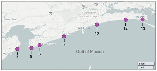
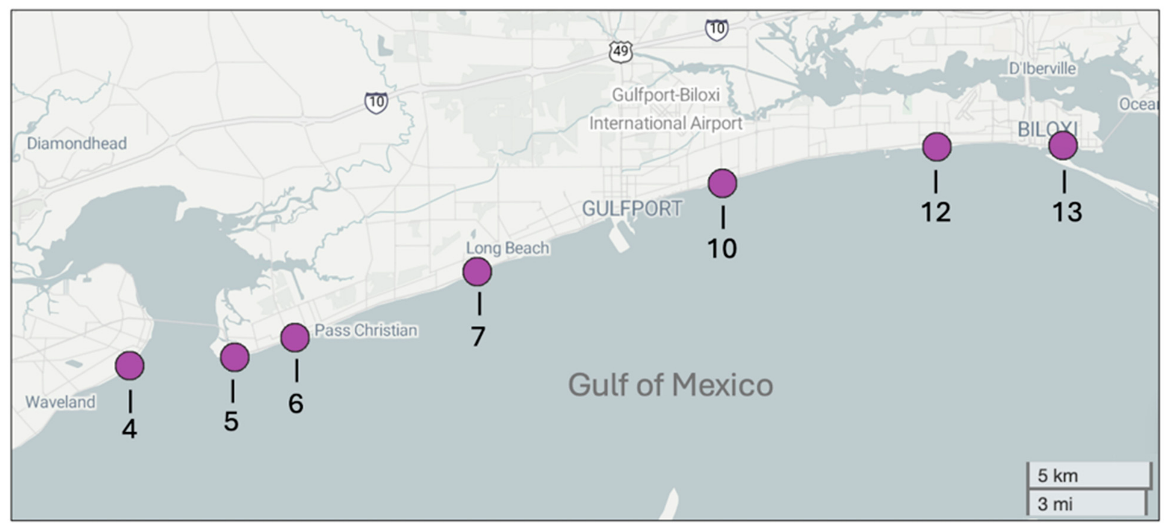
Samples of seawater were collected from seven beach sites spanning approximately 48 km of the Mississippi Gulf Coast, USA. Beach sites were locations that we have studied previously in terms of spatial and temporal patterns in seawater and sand bacterial community structure [27,28] and correspond to seven of the ten sites described by Vaughn et al. [27]. Beaches were identified based on the numbering system of the Mississippi Department of Environmental Quality (MDEQ) Beach Monitoring Program, and from west to east were beaches 4 (Bay St. Louis), 5 (Pass Christian West), 6 (Pass Christian Central), 7 (Long Beach), 10 (East Courthouse), 12 (Biloxi West), and 13 (Biloxi East) (Figure 1). Beaches were sampled from west to east, with all samples collected on the same day (20 April 2025) between 11:00 and 13:00. Water temperature (22.6–24.7 °C), salinity (8.9–15.0 ppt), pH (8.1–8.3), and dissolved oxygen content (198–200%S) were determined at the time of sampling using handheld probes. More information on the physicochemistry of these beaches as well as longer terms patterns in the composition of their natural bacterial communities is in Vaughn et al. [27].
Figure 1.
Beaches along the Mississippi Gulf Coast, USA, that were sampled for the presence of antibiotic-resistant bacteria. Beach sites are numbered in correspondence with the Mississippi Department of Environmental Quality (MDEQ) Beach Monitoring Program and correspond to Bay St. Louis beach (beach 4), Pass Christian West beach (beach 5), Pass Christian Central beach (beach 6), Long Beach beach (beach 7), East Courthouse Road beach (beach 10), Biloxi West beach (beach 12), and Biloxi East beach (beach 13).
At each beach, seawater from the surf zone (0–0.3 m depth) was collected from the shoreline into a sterile 500 mL Nalgene bottle. Samples were stored on ice until processed approximately 18 h later at the University of Mississippi. Aseptic/sterile techniques were followed for all sample processing. Prior to culturing, a subsample (200 mL) of seawater from each site was filtered through a sterile 0.2 µm filter, and the filter frozen at −20 °C for subsequent characterization of the natural seawater bacterial community by high-throughput 16S rRNA gene sequencing.
2.2. Determination of Counts of Antibiotic-Resistant Bacteria
Each seawater sample was diluted using sterile buffered saline following a 1/100 dilution series to give dilutions of 100 (the initial sample), 10−2, 10−4, and 10−6. A subsample (0.1 mL) of each dilution from each sample was then spread-plated onto Mueller–Hinton (MH) agar plates, a growth medium commonly used for antibiotic resistance testing [34]. Six different plates were inoculated per dilution of each seawater sample: unamended MH agar (controls), and MH agar plates amended with either amoxicillin (to a final concentration of 10 µg mL−1), ciprofloxacin (to 5 µg mL−1), erythromycin (to 15 µg mL−1), kanamycin (to 25 µg mL−1), or tetracycline (to 15 µg mL−1). Antibiotics were added from concentrated stocks mixed into the cooled liquid agar before plates were poured. The specific antibiotics were selected to represent different classes of drugs with the concentration used based on minimum inhibitory concentrations used in prior studies of susceptibility testing of clinical and environmental water samples [35,36,37]. Following inoculation, plates were incubated at 23 °C for 2–5 days, with growth checked daily, colonies counted manually, and counts updated based on any growth on subsequent days to reflect the presence of slower growing taxa. Final counts were determined as colony-forming units per mL of seawater (CFU mL−1). Plates were subsequently processed for isolation of antibiotic-resistant bacteria and identification of cultured bacterial communities.
2.3. Isolation and Multidrug Resistance Testing of Antibiotic-Resistant Bacteria
Representative colonies of antibiotic-resistant bacteria were picked from plates and transferred to new MH plates containing the same antibiotic, to verify that they had resistance to that antibiotic in pure culture. Isolates were selected based on morphology (including size, color, texture) with the goal of obtaining a representative of each type of colony while minimizing the number of identical colony types that were picked more than once. Plates were incubated (23 °C) until growth was observed (typically 48 h). Each isolate was subsequently tested for multidrug resistance by replating onto each of the four types of MH plates used for the initial cultures: amoxicillin (10 µg mL−1), erythromycin (15 µg mL−1), kanamycin (25 µg mL−1), or tetracycline (15 µg mL−1). Each isolate was plated onto all four plates, including the type of antibiotic plate that it was isolated from. No growth was observed from the initial seawater on ciprofloxacin plates, so those were not used in the multidrug resistance testing. Following incubation (23 °C, 48 h), growth for each isolate on each type of antibiotic plate tested was recorded as either positive or negative. Cultures demonstrating growth on more than one type of antibiotic were classified as multidrug resistant.
2.4. Identification of Initial and Cultured Bacterial Communities Through High-Throughput 16S rRNA Gene Sequencing
After representative isolates had been picked from the initial plates inoculated with seawater, the cultured bacterial communities on plates showing growth were characterized through 16S rRNA gene sequencing. Each plate was washed with 800 µL of solution CD1 from a PowerSoil DNA extraction kit (Qiagen, Germantown, MD, USA) and all bacterial growth scraped into the buffer using a sterilized cell spreader. The bacterial suspension was recovered and transferred into a DNA extraction bead tube for cultured community DNA extraction following the manufacturers protocol. DNA was also extracted from the filters obtained from the initial seawater at each site, each of which was placed into a DNA extraction bead tube and the same extraction kit and protocol followed. Thus, for each beach sampled, community DNA extractions were obtained for the entire bacterial community in the initial seawater, and the mixed cultured bacterial community obtained following growth on each type of MH agar.
Community DNA was sequenced following the procedures used by Vaughn et al. [27]. A dual-index 8-nucleotide barcoding approach was used to amplify a 250-bp section of the V4 region of the bacterial 16S rRNA [38] and the amplified barcoded products then normalized using SequalPrep plates (Life Technologies, Grand Island, NY, USA). Normalized DNA amplicons were pooled, spiked with 20% PhiX [39] and sequenced on an Illumina NextSeq (Illumina, San Diego, CA, USA) in the Molecular and Genomics Core Facility at the University of Mississippi Medical Center. Bioinformatics processing of the resulting fastq sequence files followed the protocol of Vaughn et al. [27] using the standard 16S rRNA pipeline of the DADA2 package version 1.26.0, within R version 4.2.2. Taxonomy was assigned using the SILVA database [40] v138.1 (released August 2020) to be consistent with other studies in this system [26,27,28] but nomenclature of bacterial phyla was updated to follow that of Oren and Garrity [41] used in more recent versions of the SILVA database. Sequences identified as potential chimeras, chloroplasts, mitochondria, Archaea, or Eukarya were removed and remaining unique sequences categorized as amplicon sequence variants (ASVs). Singleton ASVs were removed prior to later analyses and data from each sample rarefied by subsampling to the lowest sequence count obtained for any sample (55,216 sequences).
Identification of the bacteria present in each seawater or cultured bacterial community was determined to the level of phylum, family and genus. Alpha diversity of each community was determined as species richness (number of ASVs observed in the rarefied data) or species diversity (the Inverse Simpson’s index) of the rarefied data. Analysis of variance (ANOVA) was used to assess differences in each metric between communities followed by Tukey HSD post hoc analyses. Bray–Curtis dissimilarity scores were calculated and used to assess differences between all pairs of bacterial communities. Significant differences in bacterial community composition were assessed through permutational analysis of variance (PERMANOVA) based on Bray–Curtis scores and visualized using non-metric multidimensional scaling (NMDS) ordinations.
2.5. Identification of Antibiotic-Resistant Bacterial Isolates Through 16S rRNA Gene Sequencing
Bacterial isolates obtained and tested for multidrug resistance were identified through traditional Sanger 16S rRNA gene sequencing. Colonies were picked from the second round of culture on the type of antibiotic agar on which they were isolated and used directly as the source of template DNA in PCR. Amplifications used the primers 5′-AGAGTTTGATCCTGGCTCAG-3′ and 5′-GGTTACCTTGTTACGACTT-3′ that target most of the 16S rRNA gene and followed established protocols [42]. Amplification success was assessed by agarose gel electrophoresis and amplicons sequenced through a commercial sequencing service (Functional Biosciences, Inc., Madison, WI, USA). Sequence fasta files were trimmed manually for quality and used to identify isolates by comparisons to the NCBI nucleotide database using the online Basic Local Alignment Search Tool (BLAST) (https://blast.ncbi.nlm.nih.gov/Blast.cgi, accessed on 20 September 2026). For each sequence file, sequence matches were evaluated based on percent identity and overall BLAST alignment score.
3. Results
3.1. Counts of Cultured Antibiotic-Resistant Bacteria in Seawater
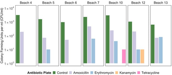
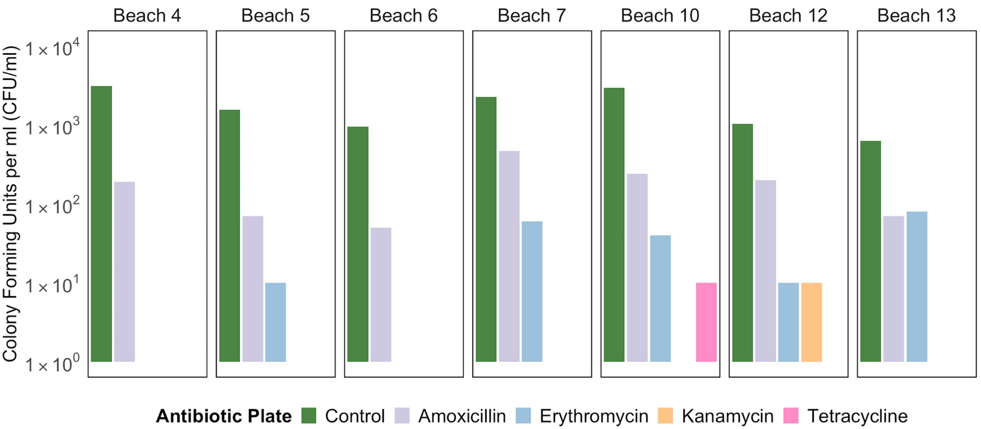
Seawater from all seven of the beach sites sampled yielded bacterial cultures on MH control plates and plates amended with amoxicillin (Figure 2). Growth on plates containing erythromycin was obtained from seawater inoculations from five sites (beaches 5, 7, 10, 12 and 13), while growth on plates containing tetracycline only occurred with seawater from beach 10 and growth on kanamycin only occurred with seawater from beach 12. No growth was obtained on plates containing ciprofloxacin, regardless of the beach from which seawater was collected.
Figure 2.
Colony counts of bacteria (CFU mL−1) obtained on Mueller–Hinton (MH) agar plates inoculated with seawater from seven beaches along the Mississippi Gulf Coast. Beaches are numbered based on the numbering system of the Mississippi Department of Environmental Quality. Counts were obtained on unamended MH agar (controls) and MH agar amended with amoxicillin (10 µg mL−1), erythromycin (15 µg mL−1), kanamycin (25 µg mL−1), or tetracycline (15 µg mL−1).
Counts on control (non-antibiotic containing) plates exceeded those on antibiotic-amended plates for all beach sites and ranged from 6.3 × 10² CFU mL−1 at beach 13 to 3.1 × 10³ CFU mL−1 at beach 4. Counts on amoxicillin were approximately an order of magnitude lower, ranging from 5.0 × 101 CFU mL−1 at beach 6 to 4.7 × 10² CFU mL−1 at beach 7. Growth on tetracycline or kanamycin amended plates were limited to just one beach for each, with 1.0 × 101 CFU mL−1 counted on tetracycline plates inoculated with seawater from beach 10 and 1.0 × 101 CFU mL−1 on kanamycin plates for beach 12 (Figure 2). There was no consistent pattern across different media of certain beaches having higher culturable counts than others, although beach 6 had the lowest count of amoxicillin-resistant bacteria and showed no growth on any of the other antibiotic plates. Counts on control plates and amoxicillin plates were moderately correlated (R = 0.53), as were counts on controls and tetracycline plates (R = 0.51), although the latter were limited to just one beach. Other correlations between counts were low to non-existent (R for all less than ±0.33).
3.2. Diversity of Natural and Cultured Bacterial Communities
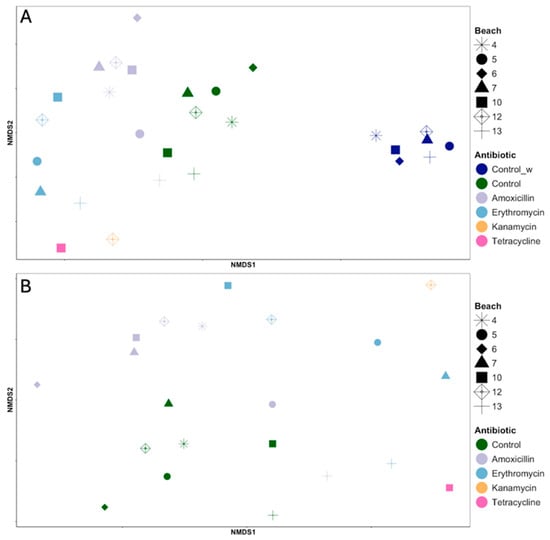
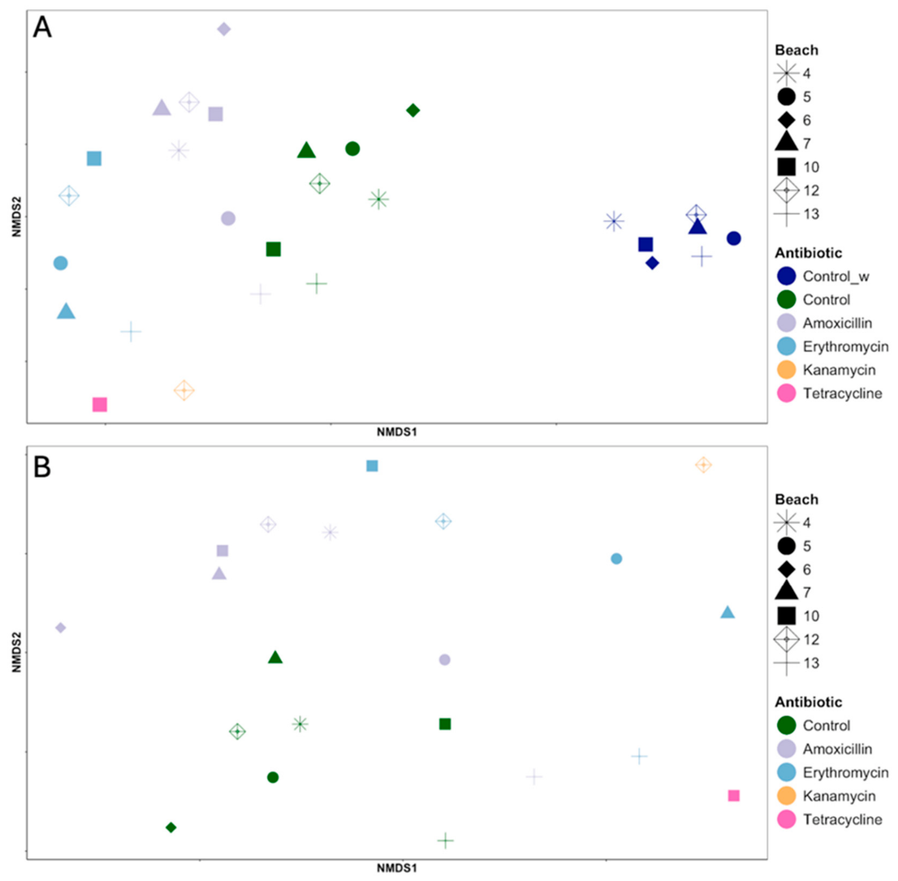
Natural seawater bacterial communities differed substantially from those obtained in mixed cultures, regardless of the growth medium used (Figure 3A). The type of community sampled (natural, type of growth medium) significantly affected community structure (PERMANOVA; R2 = 0.44, F = 3.96, p < 0.001). There were differences in the bacterial communities obtained from the different beaches but, while significant, this explained less variation in community structure than the type of community (R2 = 0.20, F = 1.51, p < 0.05). Excluding the natural seawater samples from this analysis revealed that bacterial communities in mixed cultures differed significantly based on the growth medium used (Figure 3B; R2 = 0.28, F = 1.92, p < 0.001) and by the beach from which they were sampled (R2 = 0.37, F = 1.70, p < 0.01).
Figure 3.
Non-metric multidimensional scaling ordinations of natural or cultured bacterial communities from seawater collected from seven beaches along the Mississippi Gulf Coast based on Bray–Curtis dissimilarities of 16S rRNA gene amplicon sequence variants (ASVs). Points are colored based on community type and show the natural seawater community (Control_w) and communities in mixed cultures on unamended MH agar (Control) and MH agar amended with amoxicillin, erythromycin, kanamycin, or tetracycline. Beaches are numbered based on the numbering system of the Mississippi Department of Environmental Quality. Cultured communities are shown in both panels either with (A) or without (B) the natural seawater community.
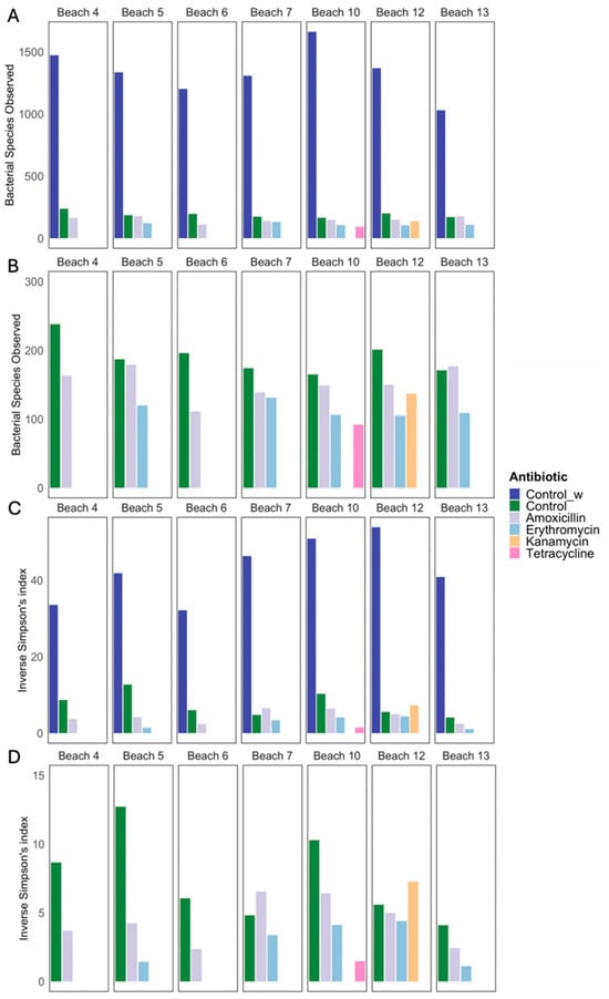
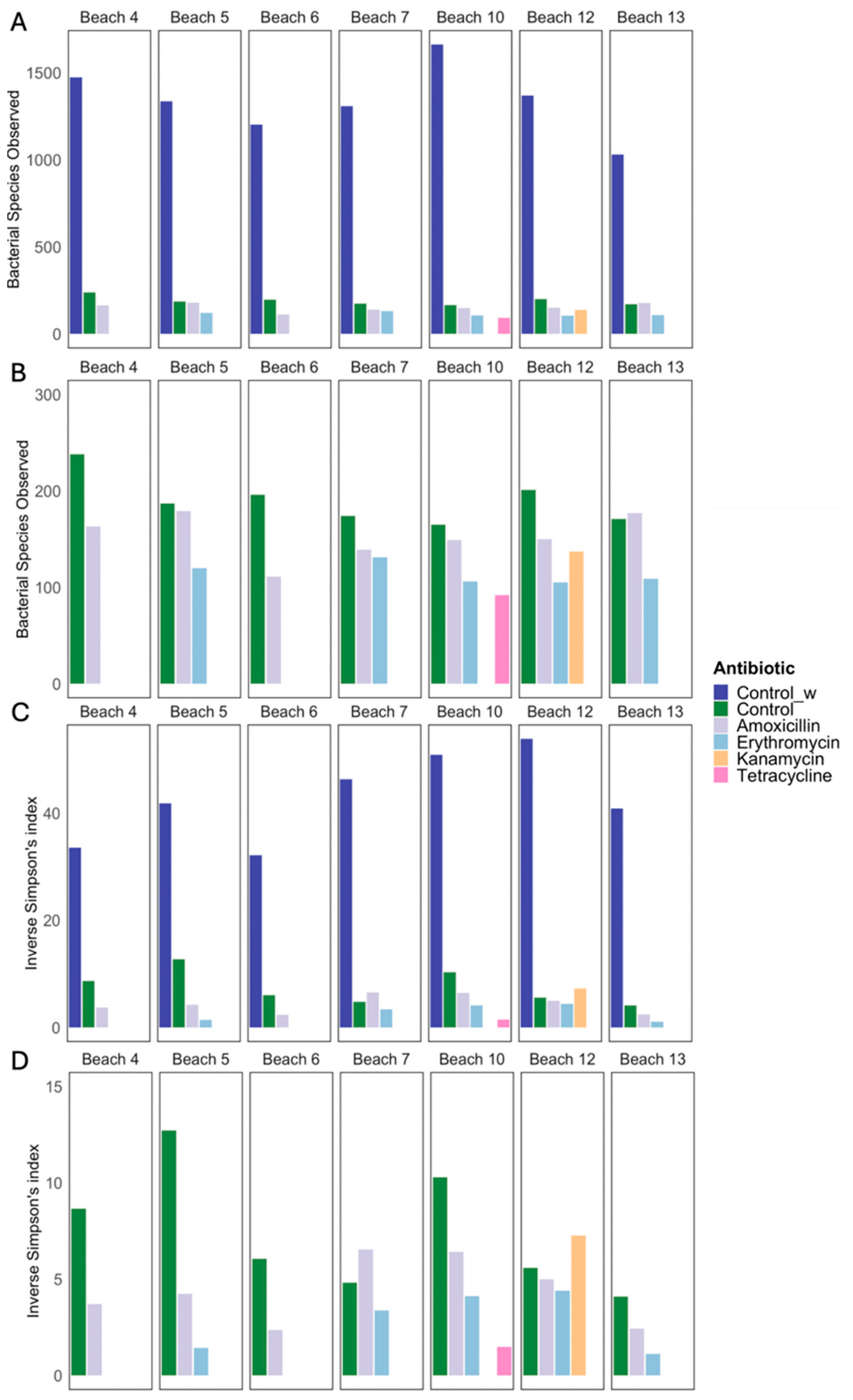
The natural seawater bacterial community was significantly richer than that obtained after culturing on different types of MH agar (ANOVA; F = 132.4, p < 0.001; Tukey’s HSD p-adj < 0.001). Species richness (as species observed) in seawater ranged from 1031 at beach 13 to 1662 at beach 10, but the effect of beach was not significant (p > 0.05; Figure 4A). For the bacterial communities assessed from mixed cultures on the different agar, species richness differed by the growth medium used (Figure 4B; F = 9.64, p < 0.01) and was significantly greater in cultures on control, non-antibiotic containing plates compared to cultures on amoxicillin (p-adj < 0.05) or erythromycin (p-adj < 0.01). Species richness on control plates was also greater than that on tetracycline or kanamycin plates, but those plates only yielded positive results from one beach each, so statistical comparisons are not possible. Beach was not a significant factor affecting species richness of cultured bacterial communities (p > 0.05). Even the lowest estimated species richness for a mixed culture (on tetracycline plates inoculated with water from beach 10) was still 92, although species diversity in this culture was low (inverse Simpson’s index of 1.5).
Figure 4.
Alpha diversity of natural (Control_w) or cultured bacterial communities from seawater collected from seven beaches along the Mississippi Gulf Coast as based on bacterial species richness ((A,B); species observed) or species diversity ((C,D); Inverse Simpson’s index). Cultured communities were obtained on MH agar (Control) and MH agar amended with amoxicillin, erythromycin, kanamycin, or tetracycline. Beaches are numbered based on the numbering system of the Mississippi Department of Environmental Quality. Species richness is shown for communities with (A) and without (B) the natural seawater community, as is species diversity (C,D).
The same patterns seen for species richness were seen for species diversity (calculated as the inverse Simpson’s index; Figure 4C) and the two measures of alpha diversity were strongly correlated (R = 0.97). Bacterial communities differed based on the sampled community (natural, type of growth medium; F = 87.2, p < 0.001) with the natural seawater bacterial community more diverse than any of the cultured communities (p-adj < 0.001). As with species richness, the beach from which seawater was sampled did not significantly affect bacterial species diversity (p > 0.05). Excluding the natural seawater from analyses (Figure 4D), showed that species diversity differed between the mixed cultures obtained on different growth media (F = 5.03, p < 0.05), with control plates yielding significantly greater species diversity than erythromycin plates (p-adj < 0.05). The effect of beach sampled on species diversity of cultures was not significant (p > 0.05).
3.3. Composition of Natural and Cultured Bacterial Communities
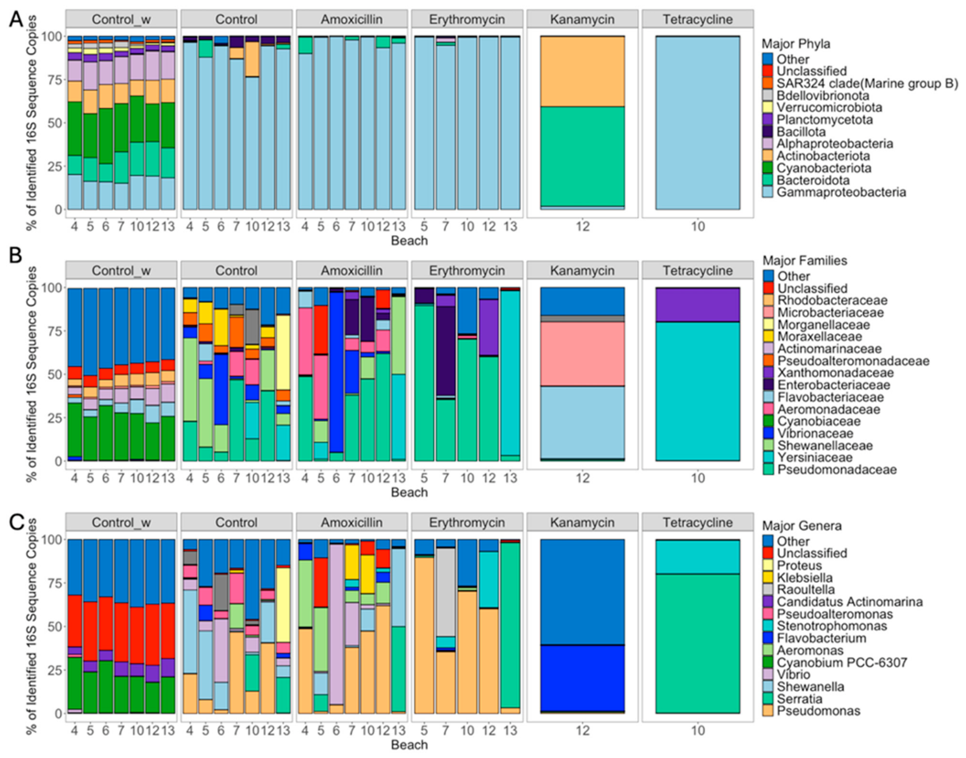
Based on the percentage of 16S rRNA gene amplicons, the most prevalent phyla or major taxonomic groups in the natural, uncultured bacterial communities in seawater were Cyanobacteriota, Bacteroidota, Gammaproteobacteria, Alphaproteobacteria, and Actinobacteriota (Figure 5A). The composition of cultured communities was dramatically different, with mixed cultures on control (non-antibiotic) MH plates and those amended with amoxicillin, erythromycin, or tetracycline all dominated by Gammaproteobacteria, regardless of the beach sampled. The sole kanamycin plate that showed growth (inoculated with seawater from beach 12) was dominated by Bacteroidota and Actinobacteriota (Figure 5A). The diversity of taxonomic groups represented in cultured bacterial communities was noticeably higher when viewed at the level of family (Figure 5B), but still less than that of bacterial communities in uncultured seawater, where many families represented <1% of the 16S rRNA gene sequences recovered. Variation in the composition of mixed cultures of bacteria obtained from seawater from different beaches was apparent at this taxonomic level, particularly in the composition of cultures on control and amoxicillin plates, the media that yielded cultures from all beaches sampled (Figure 5B). At the finer taxonomic level of genus, differences between the dominant taxa in seawater and those in mixed cultures were even more apparent. Roughly a third of the 16S rRNA gene sequences obtained from the natural seawater communities could not be classified to this level, whereas almost all the sequences obtained from cultures could (Figure 5C). Of the dominant (>1% of the community) sequences from seawater that could be classified to genus, most were either Cyanobium or Candidatus Actinomarina, genera that were absent in cultured communities. Major genera identified in mixed cultures varied by the type of growth medium used, and the beach sampled (Figure 5C). Commonly detected genera included Pseudomonas, Stenotrophomonas, Vibrio, Serratia, and Shewanella. The kanamycin plate yielding cultures from beach 12 was dominated by Flavobacterium and various less dominant genera each accounting for <1% of the sequences obtained, while the tetracycline plate yielding growth from beach 10 was dominated by Serratia and Stenotrophomonas (Figure 5C).
Figure 5.
Composition of natural or cultured bacterial communities from seawater collected from seven beaches along the Mississippi Gulf Coast at the level of bacterial phylum (A), family (B), and genus (C). Each bar represents the percentage of 16S rRNA gene sequence copies in one community sampled, with only taxa accounting for >1% of the community shown. ‘Other’ collectively represents bacterial sequences identified to a phylum/family that did not represent >1% relative abundance and ‘Unclassified’ represents bacterial sequences that were not identified to that taxonomic level. Communities are natural seawater (Control_w) and cultured communities obtained on MH agar (Control) and MH agar amended with amoxicillin, erythromycin, kanamycin, or tetracycline. Beaches are numbered based on the numbering system of the Mississippi Department of Environmental Quality, and bars for each type of community differ in size because not all beaches yielded cultures on each type of growth medium.
At the level of specific 16S rRNA sequence types, five ASVs were dominant in uncultured seawater, each found in seawater from all seven of the beaches sampled and each accounting for >1% of the 16S rRNA gene sequences obtained from a given bacterial community. These represented just two bacterial taxa, Cyanobium PCC-6307 (ASVs 57, 25 and 22) and Candidatus Actinomarina (ASVs 41 and 37; Table 1). Neither of these taxa were dominant ASVs in cultured communities. Cultured communities on control MH agar showed five dominant ASVs, with one (ASV 15, identified as Pseudoalteromonas) dominant in cultured obtained from all beaches. Two ASVs (ASV 32, Pseudomonas oleovorans, and ASV 3, Shewanella baltica) were dominant in MH agar cultures from beaches 12, 5, 6, and 4, while ASV 76 (Rheinheimera) was dominant in MH cultures from beaches 7, 5, and 10, and ASV 74 (Vibrio) was dominant in MH cultures from beaches 6, 10, and 13.
Table 1.
Dominant amplicon sequence variants (ASVs) as determined from 16S rRNA gene sequencing of bacterial communities in seawater from seven beaches along the Mississippi Gulf Coast and in cultured bacterial communities obtained by incubating seawater on Mueller–Hinton (MH) agar (Control) or MH agar amended with one of four antibiotics (amoxicillin, erythromycin, kanamycin or tetracycline). ASVs are numbered (ordered by decreasing number of 16S rRNA gene sequences in the full dataset) and taxonomy is based on the closest match to the SILVA database v138.1. Beach indicates which of the seven beaches yielded that ASV where it accounted for >1% of sequences recovered from that community and are ordered from the beach where that ASV accounted for the highest percentage of sequences to the lowest. Beaches are numbered according to the Mississippi Department of Environmental Quality Beach Monitoring Program.
Some of the dominant ASVs in mixed bacterial cultures obtained on antibiotic-containing MH media were also recovered from multiple beaches (Table 1). ASVs identified as Aeromonas (ASV 2) and Pseudomonas (ASV 10) were common in the communities cultured on amoxicillin plates, being detected on plates from five (ASV 2) or four (ASV 10) of the seven beaches sampled, with both ASVs being dominant sequence types in cultures from beaches 7, 10, and 12. A different ASV (ASV 4) also identified as a species of Pseudomonas was found in cultures on amoxicillin plates from these three beaches. ASV 4 and ASV 10 were also detected in mixed cultures on erythromycin plates inoculated with seawater from beaches 10 and 12, two of the beaches that yielded these same ASVs on amoxicillin plates (Table 1). ASV 16 (identified as Pseudomonas anguilliseptica) was also a dominant sequence type in erythromycin cultures from these two beaches, while ASV 23 (Aeromonas) was dominant on erythromycin plates from beaches 5 and 13. The kanamycin culture from beach 12 had three ASVs identified as Flavobacterium and ASVs identified as Olleya marilimosa and Paenarthrobacter as its dominant ASVs, while the tetracycline culture from beach 10 was dominated by ASV 1 (identified as Serratia) and ASV17 (Stenotrophomonas). None of the dominant ASVs in the kanamycin or tetracycline cultures were dominant in cultures on media amended with other antibiotics. More broadly, none of the dominant ASVs in cultures on antibiotic-containing media were dominant in cultures on non-antibiotic media, and none of the dominant ASVs in any cultured community were dominant in the natural seawater bacterial community.
3.4. Identification of Antibiotic-Resistant Isolates and Multi-Drug Resistance
An initial total of 76 antibiotic-resistant bacterial colonies were transferred for isolation, of which seven (four originally cultured on amoxicillin plates; three on erythromycin) did not subsequently grow. Of the remaining 69, ten grew on antibiotic-amended agar but failed to amplify and/or sequence after two attempts, leaving a final total of 59 for identification and antibiotic resistance testing (Table 2). Most (41) of these isolates came from amoxicillin cultures, 15 came from erythromycin cultures, and three from kanamycin cultures. Based on top matches to the NCBI database, the 59 isolates represented a total of 23 different bacterial genera. Pseudomonas was the most prevalent genus of bacteria isolated, with 22 isolates classified to that genus. Other genera that were obtained as multiple isolates were Vibrio (five isolates), Aeromonas and Ectopseudomonas (four isolates each), Stenotrophomonas (three isolates), and Flavobacterium, Shewanella, and Raoultella (two isolates each).
Table 2.
Antibiotic-resistant isolates of bacteria cultured from seawater from seven beaches (numbered according to Mississippi Department of Environmental Quality designations) along the Mississippi Gulf Coast. Isolate ID is organized by a letter indicating the antibiotic (A = amoxicillin, E = erythromycin, K = kanamycin, T = tetracycline), a number indicating beach, followed by an identifier number, with the NCBI accession for its partial 16S rRNA gene sequence in parentheses. The closest match in the NCBI nucleotide database based on BLAST searches of partial 16S rRNA gene sequences is shown, while %Match (bp) indicates the percentage match and the sequence length used in the search. The last four columns indicate whether that isolate showed growth on MH agar amended with each of the four antibiotics, with + indicating growth on media containing that antibiotic. While the table shows the closest match to the database, true species-level resolution is difficult for many of these taxa.
Based on growth on MH agar containing each antibiotic, four isolates showed resistance to all four of the antibiotics tested. Two of these isolates yielded partial 16S rRNA gene sequences that matched to different species of Stenotrophomonas; isolates A7-13 and E7-5. The other two isolates showing resistance to all four antibiotics were isolates A12-4, (closest partial 16S rRNA gene sequence match to Chryseobacterium cucumeris) and K12-1 (closest match to Sphingobacterium kitahiroshimense). Seven isolates showed resistance to three antibiotics, four of which showed the closest matches to different species of Pseudomonas (isolates A4-6, A7-5, E7-1 and E10-3). The other isolates showing resistance to three antibiotics were A7-2 (showing a closest partial 16S rRNA gene sequence match to Klebsiella variicola), A12-11 (closest match to Flavobacterium aquimarinum), and E12-5 (closest match to Ectopseudomonas khazarica). A total of 37 isolates were resistant to two of the antibiotics, giving a total of 48 isolates (81% of those identified and tested) that showed some level of multi-drug resistance under the test conditions.
4. Discussion
Freshwater and marine ecosystems can be important reservoirs for antibiotic resistance [8,9,10,11], although studies on the presence of antibiotic resistance in coastal environments are more limited than other systems [12]. This is particularly true of the northern coast of the Gulf of Mexico which despite its importance has had few studies investigate the presence of antibiotic-resistant taxa [21,22]. In this study we used a combination of molecular and culture-based approaches to assess the seawater bacterial community at seven beaches along the Mississippi Gulf Coast, USA, with a specific focus on antibiotic-resistant bacteria. Molecular characterization of the natural seawater community expanded on prior work [27,28] and in combination with a culture approach that targeted antibiotic-resistant bacteria allowed us to make comparisons at multiple levels.
Mixed-culture bacterial communities had much less biodiversity than the bacterial community of natural seawater, which showed species richness estimates of about 1000–1600 species, similar to those we have previously reported [27,28] and about 5–8× greater than that for mixed cultures on non-antibiotic control plates. The diversity of mixed cultures on antibiotic-amended agar was less than that on non-antibiotic plates, but the reduction in species richness was not as great as the comparison of control plates to natural seawater, especially for amoxicillin. However, the decline in overall species diversity measured by the inverse Simpson’s index, an index that considers both the number of species and the evenness of those species, was greater. These patterns show that the diversity of cultured bacterial communities was much less than that of natural seawater bacterial communities, but the richness of antibiotic-resistant communities was only a little less than that of cultured overall, presumably because a sizable fraction of the cultured community shows some level of resistance to at least one antibiotic. While those antibiotic-resistant cultures may contain almost as many different species as non-antibiotic cultured communities, they are much less even and some bacterial species, or more accurately, some bacterial populations (ASVs are defined by being a 16S rRNA gene sequence variant) likely became more dominant in the presence of antibiotics. Based on the dominant ASVs detected in this study, the populations of bacteria that became dominant on antibiotic-containing media were different from the dominant populations on control media, which in turn differ from dominant populations in the original seawater. Comparisons of diversity between the natural (uncultured) and cultured communities are somewhat limited, however, by the delay (18 h) between sample collection and culturing. While samples were maintained on ice, it is possible that some taxa could have shown reductions in their population size over this period, and while their cell remnants and DNA may still have been detectable through molecular approaches (i.e., they were still present in the molecular analysis of the community), a loss of viable cells would have led to their reduction or absence in mixed cultures. Thus, less diversity in the mixed culture communities may reflect biases from sample processing as well as the culturing itself.
The dominant taxa in uncultured seawater matched those that we have found to account for appreciable proportions of bacterial communities in seawater along the Mississippi coast over a period of 15 months [27,28]. This was apparent at each taxonomic level examined, with the major bacterial populations being the photosynthetic Cyanobium PCC-6307 (within phylum Cyanobacteriota) and a photoheterotroph identified from metagenomic studies, Candidatus Actinomarina (Actinobacteriota). Such taxa are unlikely to be cultured using typical incubation conditions and heterotrophic growth media such as MH and could be more sensitive to sample storage. Sequences classifying as them were not detected in any appreciable numbers in cultured communities. Rather, cultured communities were dominated largely by members of the Gammaproteobacteria with some Bacteroidota and Actinobacteriota, major groups of bacteria that contain a variety of species that are relatively easy to culture, the “weeds” of the microbial world [33]. Specific genera that accounted for a large part of cultured bacterial communities included widespread facultative taxa such as Serratia, Aeromonas, and Vibrio, as well as the marine Shewanella, although the latter was only a major component of cultures on control and amoxicillin plates. Pseudomonas was the most dominant genus in cultures overall and the proportion of this genus in mixed cultures varied with culture medium and the beach from which the seawater inoculum was collected. Indeed, the variation in composition of mixed cultures between beaches, even within the same type of antibiotic agar, appeared to be much greater than variation in the natural seawater community, a pattern seen both in the taxonomic composition of cultures and overall NMDS ordinations. This is likely due to the somewhat stochastic nature of culturing marine bacteria [43], as the seawater community contained large numbers of less proportionally abundant taxa (grouped as “Other”) and different components of that group may have been cultured from different samples. It does suggest that while seawater bacterial communities at different beaches may have similar overall taxonomic composition, cultures obtained from that seawater, even under the same culture conditions, may show substantial differences. Inferring cultured taxa from the composition of the natural community, or vice versa, is likely to be difficult.
As well as being common in the community analysis of mixed cultures, Pseudomonas, Vibrio and Aeromonas were also common genera among our antibiotic-resistant isolates, which we were able to more confidently classify to genus given the longer 16S rRNA gene sequence read length. While the closest NCBI match is reported to species level in Table 2, many of the isolate sequences yielded close matches to multiple species within a genus, so actual species identity of these isolates is not clear. The process of picking isolates based on visual morphology is subjective and multiple bacterial species may appear as similar isolates, but at least in terms of the main genera, representative isolates were likely obtained. However, that pattern did not extend to specific individual mixed cultures: for example, whole community analysis of the single mixed culture obtained on kanamycin plates (from beach 12) suggested that Flavobacterium was the dominant genus present, but partial sequencing of the 16S rRNA gene of the three isolates obtained from this plate matched to species of Leucobacter, Microbacterium, and Sphingobacterium. While they were the isolates obtained from this mixed culture, these taxa each accounted for <1% of the overall 16S rRNA gene sequences recovered from that cultured community, but their broader phyla (Actinobacteriota for Leucobacter and Microbacterium, Bacteroidota for Sphingobacterium) were detected as being dominant. The observed species richness in the kanamycin-resistant mixed culture was 137, suggesting a much higher diversity in that culture than suggested by isolates alone. Similar patterns were seen for all antibiotic-resistant cultured communities, with 12 visually distinguishable isolates being the most obtained from any one culture (amoxicillin-resistant cultures from beach 12) but that mixed culture had an observed species richness at the community level of 150. Alpha diversity indices are calculated from ASVs which are individual 16S rRNA gene sequence types rather than individual species, but the consistent discrepancy between those scores and the diversity obtained as isolates does suggest that mixed cultures contained more species or strains of bacteria than isolation alone would have detected.
Isolation did allow assessment of antibiotic-resistant patterns, and bacteria showing some level of antibiotic resistance were cultured from seawater collected from all seven beaches sampled. CFU counts of antibiotic-resistant bacteria were around 102 mL−1 compared to 103 mL−1 CFU on non-antibiotic containing MH agar, which is close to counts obtained for other marine studies [44]. Antibiotic-resistant bacteria have been detected in coastal areas and beaches worldwide [12,21,22,45] but numbers are difficult to compare based on differences in approaches, media used and focus of specific studies (e.g., all bacteria vs. just enteric bacteria). Interestingly, we did not obtain any growth on plates amended with ciprofloxacin. Ciprofloxacin-resistance was reported for some bacterial isolates obtained from dry sand from beaches in Brazil but was not as prevalent in cultures obtained from seawater or damp sand which yielded more isolates showing resistance to amoxicillin, other penicillins, and erythromycin [45], similar to the resistance profiles seen for isolates obtained from seawater in this study. Compared to other antibiotics, resistance to ciprofloxacin was also rare in bacteria obtained from seawater from the southern Indian coast [46]. The absence of ciprofloxacin-resistant cultures in our seawater samples may therefore relate to habitat-specific patterns in sand and seawater, and the bacterial communities differ in sand and seawater at beaches along the Mississippi coast [27,28] with there even being differences in sand bacterial community composition based on moisture content and location on the shore [26]. That the highest CFU counts of antibiotic-resistant bacteria were obtained on amoxicillin is not surprising as this is a widely used antibiotic, soluble in water and with resistance usually provided by the widespread β-lactamases [47,48].
Multidrug resistance appeared to be prevalent among isolates, with 81% of the 59 sequenced isolates showing resistance to more than one antibiotic. Other studies in coastal areas worldwide have reported high levels of multidrug resistance in bacterial isolates [22,46,49], although the focus has tended to be on indicator taxa or potential pathogens such as Escherichia, Enterococcus, Salmonella, or Vibrio, compared to the broader range of bacteria isolated in this study. The antibiotics tested in this study represent different classes of antibiotics with different modes of action. Because these antibiotics target different cellular processes, resistance to multiple antibiotics may reflect the presence of broad resistance mechanisms, such as efflux pumps, or the accumulation of multiple resistance traits within individual strains of bacteria [6,50,51]. Although isolates were identified using 16S rRNA gene sequencing, they were not genetically characterized for antimicrobial resistance mechanisms so molecular validation for antibiotic resistance is lacking Future studies aimed at identifying specific resistance genes and assessing their diversity within coastal bacterial isolates or seawater itself would provide deeper insight into the genetic basis of single and multidrug resistance [52,53,54,55]. Of note were isolates identified as species of Stenotrophomonas, Chryseobacterium, and Sphingobacterium as these isolates showed resistance to the four antibiotics tested (amoxicillin, erythromycin, kanamycin, and tetracycline). Genome sequencing of Stenotrophomonas has identified diverse antibiotic resistance genes [56], while species of Chryseobacterium and Sphingobacterium are known to be resistant to a variety of antibiotics, including through non-traditional resistance mechanisms [57,58]. While most species of these genera are not regarded as human pathogens, they are a potential environmental reservoir for multidrug resistance, and our findings overall suggest that multidrug resistance among various species of bacteria is widespread at beaches along the Mississippi Gulf Coast. Future work should more thoroughly assess antibiotic resistance, and particularly multidrug resistance, in culturable environmental bacteria in these coastal systems across a broader range of antibiotics and using more formal procedures such as the Clinical and Laboratory Standards Institute guidelines [59].
Given the extensive analyses involved per sample, we chose to sample seawater on just one day but from multiple beaches on that day, all sampled within two hours of each other to reduce potential temporal variation. That limits our findings to that one point in time, and the abundance and composition of antibiotic-resistant bacteria at these beaches are likely to differ at other times. How likely and by how much the numbers and identity of antibiotic-resistant bacteria at these beaches varies with time is difficult to determine. Counts of antibiotic-resistant E. coli and Enterococcus at some of these beaches have been reported to show seasonal patterns, with higher counts in summer compared to other seasons [22], coinciding with higher use of these public beaches. More broadly, our previous work on the Mississippi Gulf Coast has found that seawater bacterial communities at these beaches show strong temporal patterns, with bacterial communities being more similar the closer in time they are sampled and recurring turnover suggesting longer-term seasonal and annual patterns [28]. While such spatiotemporal patterns in the broader seawater bacterial community may not necessarily reflect those shown for antibiotic-resistant taxa, if similar temporal trends do occur then (1) similar findings to the current study in terms of antibiotic-resistance would likely occur for a few weeks or months around our sampling date, and (2) similar findings would likely occur at the same time of year (roughly March–May) in subsequent years, with higher prevalence of antibiotic-resistant taxa likely in the summer months. Seasonal trends in antibiotic resistance genes in coastal waters have been reported elsewhere but tend to vary based on gene and bacterial species rather than broad patterns [55]. Additional longer-term studies on temporal patterns in antibiotic resistance in coastal systems such as that conducted by Mukherjee et al. [22] but on natural bacterial populations would be helpful, perhaps in conjunction with ongoing monitoring programs that track indicator taxa such as Enterococcus, as counts of those organisms may be a good indicator for levels of antibiotic resistance genes [55].
5. Conclusions
Using a combination of culture-dependent and culture-independent approaches we assessed antibiotic resistance in seawater bacterial communities at seven beaches along the Mississippi Gulf Coast, USA. As predicted, the diversity of the natural, uncultured seawater bacterial community was much greater than that obtained in mixed cultures, although those mixed cultures were substantially more diverse than the number of bacteria recovered as isolated. Cultured and uncultured bacterial communities also differed in terms of composition, and the composition of mixed cultures was dependent on the antibiotic added to the growth medium. Antibiotic-resistant bacteria were prevalent all along the coast with all beaches yielding cultures that were resistant to amoxicillin and many yielding cultures that were resistant to erythromycin. Counts of antibiotic-resistant bacteria were approximately a tenth of that seen in non-antibiotic controls, but more than 80% of the antibiotic-resistant isolates tested showed some level of multidrug resistance, with some being resistant to four different classes of antibiotic. Our findings expand on the high levels of antibiotic resistance found previously in isolates of Enterococcus and E. coli in this region [22] and show that this drug resistance exists in a variety of taxa in the natural seawater community along the Mississippi Gulf Coast.
Author Contributions
Conceptualization, C.R.J. and S.N.V.; methodology, L.F.H., M.M. and S.N.V.; formal analysis, C.R.J., L.F.H., M.M. and S.N.V.; investigation, L.F.H., M.M. and S.N.V.; resources, C.R.J.; data curation, C.R.J. and S.N.V.; writing—original draft preparation, C.R.J., L.F.H. and M.M. writing—review and editing, C.R.J., L.F.H., M.M. and S.N.V.; visualization, S.N.V.; supervision, C.R.J. and S.N.V.; project administration, C.R.J.; funding acquisition, C.R.J. All authors have read and agreed to the published version of the manuscript.
Funding
This project was paid for with federal funding from the U.S. Department of the Treasury, the Mississippi Department of Environmental Quality, and the Mississippi Based RESTORE Act Center of Excellence under the Resources and Ecosystems Sustainability, Tourist Opportunities, and Revived Economies of the Gulf Coast States Act of 2012 (RESTORE Act). The statements, findings, conclusions, and recommendations are those of the author(s) and do not necessarily reflect the views of the Department of the Treasury, the Mississippi Department of Environmental Quality, or the Mississippi Based RESTORE Act Center of Excellence.
Data Availability Statement
The sequence data for the bacterial communities described in this study are available in the NCBI Sequence Reads Archive under BioProject ID PRJNA1528481. Sequences for bacterial isolates obtained in this study are available in the NCBI GenBank database under accession numbers QB030367-QB030425.
Conflicts of Interest
The authors declare no conflicts of interest. The funders had no role in the design of the study; in the collection, analyses, or interpretation of data; in the writing of the manuscript; or in the decision to publish the results.
References
- Sengupta, S.; Chattopadhyay, M.K.; Grossart, H.-P. The multifaceted roles of antibiotics and antibiotic resistance in nature. Front. Microbiol. 2013, 4, 47. [Google Scholar] [CrossRef] [Scilit] [PubMed]
- Fair, R.J.; Tor, Y. Antibiotics and bacterial resistance in the 21st century. Perspect. Med. Chem. 2014, 6, 25–64. [Google Scholar] [CrossRef] [Scilit] [PubMed]
- Ventola, C.L. The antibiotic resistance crisis: Part 1: Causes and threats. Pharm. Ther. 2015, 40, 277–283. [Google Scholar] [PubMed] [PubMed Central]
- Martínez, J.L. Environmental pollution by antibiotics and by antibiotic resistance determinants. Environ. Pollut. 2009, 157, 2893–2902. [Google Scholar] [CrossRef] [Scilit] [PubMed]
- Serwecińska, L. Antimicrobials and antibiotic-resistant bacteria: A risk to the environment and to public health. Water 2020, 12, 3313. [Google Scholar] [CrossRef] [Scilit]
- Baquero, F.; Martínez, J.L.; Cantón, R. Antibiotics and antibiotic resistance in water environments. Curr. Opin. Biotechnol. 2008, 19, 260–265. [Google Scholar] [CrossRef] [Scilit] [PubMed]
- Larsson, D.G.J.; Flach, C.F. Antibiotic resistance in the environment. Nat. Rev. Microbiol. 2022, 20, 257–269. [Google Scholar] [CrossRef] [Scilit] [PubMed]
- Leonard, A.F.; Morris, D.; Schmitt, H.; Gaze, W.H. Natural recreational waters and the risk that exposure to antibiotic resistant bacteria poses to human health. Curr. Opin. Microbiol. 2022, 65, 40–46. [Google Scholar] [CrossRef] [Scilit] [PubMed]
- Singh, A.K.; Kaur, R.; Verma, S.; Singh, S. Antimicrobials and antibiotic resistance genes in water bodies: Pollution, risk, and control. Front. Environ. Sci. 2022, 10, 830861. [Google Scholar] [CrossRef] [Scilit]
- Duarte, A.C.; Rodrigues, S.; Afonso, A.; Nogueira, A.; Coutinho, P. Antibiotic resistance in the drinking water: Old and new strategies to remove antibiotics, resistant bacteria, and resistance genes. Pharmaceuticals 2022, 15, 393. [Google Scholar] [CrossRef] [Scilit] [PubMed]
- Li, S.; Ondon, B.S.; Ho, S.H.; Zhou, Q.; Li, F. Drinking water sources as hotspots of antibiotic-resistant bacteria (ARB) and antibiotic resistance genes (ARGs): Occurrence, spread, and mitigation strategies. J. Water Process Eng. 2023, 53, 103907. [Google Scholar] [CrossRef] [Scilit]
- Zheng, D.; Yin, G.; Liu, M.; Chen, C.; Jiang, Y.; Hou, L.; Zheng, Y. A systematic review of antibiotics and antibiotic resistance genes in estuarine and coastal environments. Sci. Total Environ. 2021, 777, 146009. [Google Scholar] [CrossRef] [Scilit] [PubMed]
- Ward, N.; Megonigal, J.; Bond-Lamberty, B.; Bailey, V.; Butman, D.; Canuel, E.; Diefenderfer, H.; Ganju, N.; Goñi, M.; Graham, E.; et al. Representing the function and sensitivity of coastal interfaces in Earth system models. Nat. Commun. 2020, 11, 2458. [Google Scholar] [CrossRef] [Scilit] [PubMed]
- Barbier, E.B.; Hacker, S.D.; Kennedy, C.; Koch, E.W.; Stier, A.C.; Silliman, B.R. The value of estuarine and coastal ecosystem services. Ecol. Monogr. 2011, 81, 169–193. [Google Scholar] [CrossRef] [Scilit]
- Jickells, T.D. Nutrient biogeochemistry of the coastal zone. Science 1998, 281, 217–222. [Google Scholar] [CrossRef] [Scilit] [PubMed]
- Leonard, A.F.; Zhang, L.; Balfour, A.J.; Garside, R.; Gaze, W.H. Human recreational exposure to antibiotic resistant bacteria in coastal bathing waters. Environ. Int. 2015, 82, 92–100. [Google Scholar] [CrossRef] [Scilit] [PubMed]
- Alm, E.W.; Burke, J.; Spain, A. Fecal indicator bacteria are abundant in wet sand at freshwater beaches. Water Res. 2003, 37, 3978–3982. [Google Scholar] [CrossRef] [Scilit] [PubMed]
- Yamahara, K.M.; Layton, B.A.; Santoro, A.E.; Boehm, A.B. Beach sands along the California coast are diffuse sources of fecal bacteria to coastal waters. Environ. Sci. Technol. 2007, 41, 4515–4521. [Google Scholar] [CrossRef] [Scilit] [PubMed]
- Cui, H.; Yang, K.; Pagaling, E.; Yan, T. Spatial and temporal variation in enterococcal abundance and its relationship to the microbial community in Hawaii beach sand and water. Appl. Environ. Microbiol. 2013, 79, 3601–3609. [Google Scholar] [CrossRef] [Scilit] [PubMed]
- Halliday, E.; McLellan, S.L.; Amaral-Zettler, L.A.; Sogin, M.L.; Gast, R.J. Comparison of bacterial communities in sands and water at beaches with bacterial water quality violations. PLoS ONE 2014, 9, e90815. [Google Scholar] [CrossRef] [Scilit] [PubMed]
- Belding, C.; Boopathy, R. Presence of antibiotic-resistant bacteria and antibiotic resistance genes in coastal recreational waters of southeast Louisiana, USA. J. Water Supply Res. Technol.-AQUA 2018, 67, 800–809. [Google Scholar] [CrossRef] [Scilit]
- Mukherjee, M.; Cuthbert, R.E.; Aitken, E.; Gay, K.A.; McKinney, K.; Brooks, J.P. Revealing an abundance of microbial fecal contamination and multidrug resistant bacteria in the Mississippi Gulf Coast. Water 2023, 15, 2339. [Google Scholar] [CrossRef] [Scilit]
- NOAA. Gulf of America Region. National Marine Ecosystem Status Website. 2025. Available online: https://ecowatch.noaa.gov/ (accessed on 3 August 2026).
- NOAA Fisheries. Southeast Fisheries. 2026. Available online: https://www.fisheries.noaa.gov/region/southeast/fisheries (accessed on 3 August 2026).
- Vinogradov, S.; Vinogradova, N.; Kamenkovich, V.; Nechaev, D. Temperature and salinity variability in the Mississippi Bight. Mar. Technol. Soc. J. 2004, 38, 52–60. [Google Scholar] [CrossRef] [Scilit]
- Basha, E.; Vaughn, S.N.; Pavlovsky, J.C.; Roth, H.; Jackson, C.R. Fine-scale patterns in bacterial communities on a Gulf Coast beach. Coasts 2025, 5, 34. [Google Scholar] [CrossRef] [Scilit]
- Vaughn, S.N.; Pavlovsky, J.C.; Jackson, C.R. Bacterial communities in sand and seawater of northern gulf coast beaches: Temporal, spatial, and environmental influences. Environ. Microbiol. Rep. 2026, 18, e70309. [Google Scholar] [CrossRef] [Scilit] [PubMed]
- Vaughn, S.N.; Pavlovsky, J.C.; Heiman, J.A.; Jackson, C.R. Contrasting spatial and temporal structuring of seawater and sediment bacterial communities in coastal environments. Sci. Rep. 2026, 16, 14586. [Google Scholar] [CrossRef] [Scilit] [PubMed]
- Demko, A.M.; Patin, N.V.; Jensen, P.R. Microbial diversity in tropical marine sediments assessed using culture-dependent and culture-independent techniques. Environ. Microbiol. 2021, 23, 6859–6875. [Google Scholar] [CrossRef] [Scilit] [PubMed]
- Verhoeven, M.D.; Nielsen, P.H.; Dueholm, M.K.D. Amplicon-guided isolation and cultivation of previously uncultured microbial species from activated sludge. Appl. Environ. Microbiol. 2023, 89, e01151-23. [Google Scholar] [CrossRef] [Scilit] [PubMed]
- Li, S.; Lian, W.-H.; Han, J.-R.; Ali, M.; Lin, Z.-L.; Liu, Y.-H.; Li, L.; Zhang, D.-Y.; Jiang, X.-Z.; Li, W.-J.; et al. Capturing the microbial dark matter in desert soils using culturomics-based metagenomics and high-resolution analysis. npj Biofilms Microbiomes 2023, 9, 67. [Google Scholar] [CrossRef] [Scilit] [PubMed]
- Staley, J.T.; Konopka, A. Measurement of in situ activities of nonphotosynthetic microorganisms in aquatic and terrestrial habitats. Annu. Rev. Microbiol. 1985, 39, 321–346. [Google Scholar] [CrossRef] [PubMed]
- Hugenholtz, P. Exploring prokaryotic diversity in the genomic era. Genome Biol. 2002, 3, reviews0003.1. [Google Scholar] [CrossRef] [Scilit] [PubMed][Green Version]
- Nizet, V. The accidental orthodoxy of Drs. Mueller and Hinton. EBioMedicine 2017, 22, 26–27. [Google Scholar] [CrossRef] [Scilit] [PubMed]
- Armstrong, J.L.; Calomiris, J.J.; Seidler, R.J. Selection of antibiotic-resistant standard plate count bacteria during water treatment. Appl. Environ. Microbiol. 1982, 44, 308–316. [Google Scholar] [CrossRef] [Scilit] [PubMed]
- Tiwari, H.K.; Sen, M.R. Emergence of vancomycin resistant Staphylococcus aureus (VRSA) from a tertiary care hospital from northern India. BMC Infect. Dis. 2006, 6, 156. [Google Scholar] [CrossRef] [Scilit] [PubMed]
- Carstens, A.; Bartie, C.; Dennis, R.; Bezuidenhout, C. Antibiotic-resistant heterotrophic plate count bacteria and amoeba-resistant bacteria in aquifers of the Mooi River, North West province, South Africa. J. Water Health 2014, 12, 835–845. [Google Scholar] [CrossRef] [Scilit] [PubMed]
- Kozich, J.J.; Westcott, S.L.; Baxter, N.T.; Highlander, S.K.; Schloss, P.D. Development of a dual-index sequencing strategy and curation pipeline for analyzing amplicon sequence data on the MiSeq Illumina sequencing platform. Appl. Environ. Microbiol. 2013, 79, 5112–5120. [Google Scholar] [CrossRef] [Scilit] [PubMed]
- Jackson, C.R.; Stone, B.W.; Tyler, H.L. Emerging perspectives on the natural microbiome of fresh produce vegetables. Agriculture 2015, 5, 170–187. [Google Scholar] [CrossRef] [Scilit]
- Quast, C.; Pruesse, E.; Yilmaz, P.; Gerken, J.; Schweer, T.; Yarza, P.; Peplies, J.; Glöckner, F.O. The SILVA ribosomal RNA gene database project: Improved data processing and web-based tools. Nucleic Acids Res. 2013, 41, D590–D596. [Google Scholar] [CrossRef] [Scilit] [PubMed]
- Oren, A.; Garrity, G.M. Valid publication of the names of forty-two phyla of prokaryotes. Int. J. Syst. Evol. Microbiol. 2021, 71, 005056. [Google Scholar] [CrossRef] [Scilit] [PubMed]
- Jackson, C.R.; Langner, H.W.; Donahoe-Christiansen, J.; Inskeep, W.P.; McDermott, T.R. Molecular analysis of microbial community structure in an arsenite-oxidizing acidic thermal spring. Environ. Microbiol. 2001, 3, 532–542. [Google Scholar] [CrossRef] [Scilit] [PubMed]
- González, J.M.; Hodson, R.E.; Moran, M.A. Bacterial populations in replicate marine enrichment cultures: Assessing variability in abundance using 16S rRNA-based probes. Hydrobiologia 1999, 401, 69–75. [Google Scholar] [CrossRef] [Scilit]
- Rodrigues, C.J.C.; de Carvalho, C.C.C.R. Cultivating marine bacteria under laboratory conditions: Overcoming the “unculturable” dogma. Front. Bioeng. Biotechnol. 2022, 10, 964589. [Google Scholar] [CrossRef] [Scilit] [PubMed]
- Fernandes Cardoso de Oliveira, A.J.; Ranzani de França, P.T.; Pinto, A.B. Antimicrobial resistance of heterotrophic marine bacteria isolated from seawater and sands of recreational beaches with different organic pollution levels in southeastern Brazil: Evidences of resistance dissemination. Environ. Monit. Assess. 2010, 169, 375–384. [Google Scholar] [CrossRef] [Scilit] [PubMed]
- Vignesh, S.; Muthukumar, K.; James, R.A. Antibiotic resistant pathogens versus human impacts: A study from three eco-regions of the Chennai coast, southern India. Mar. Pollut. Bull. 2012, 64, 790–800. [Google Scholar] [CrossRef] [Scilit] [PubMed]
- Elizalde-Velázquez, A.; Gómez-Oliván, L.M.; Galar-Martínez, M.; Islas-Flores, H.; Dublán-García, O.; SanJuan-Reyes, N. Amoxicillin in the aquatic environment, its fate and environmental risk. In Environmental Health Risk—Hazardous Factors to Living Species; Larramendy, L.L., Soloneski, S., Eds.; IntechOpen: London, UK, 2016; pp. 247–267. [Google Scholar] [CrossRef] [Scilit] [PubMed]
- Hernández, F.; Botero-Coy, A.M.; Bijlsma, L.; Fabregat-Safont, D. Can amoxicillin be detected in the aquatic environment? A critical assessment. Environ. Int. 2026, 211, 110258. [Google Scholar] [CrossRef] [Scilit] [PubMed]
- Dasí, D.; Camaró-Sala, M.L.; González, A.; García-Ferrús, M.; Jiménez-Belenguer, A.I.; Castillo, M.Á. Antibiotic resistance in seawater samples from East Coast of Spain. Appl. Sci. 2024, 14, 1965. [Google Scholar] [CrossRef] [Scilit]
- Gaurav, A.; Bakht, P.; Saini, M.; Pandey, S.; Pathania, R. Role of bacterial efflux pumps in antibiotic resistance, virulence, and strategies to discover novel efflux pump inhibitors. Microbiology 2023, 169, 001333. [Google Scholar] [CrossRef] [Scilit] [PubMed]
- Nishino, K.; Yamasaki, S.; Nakashima, R.; Zwama, M.; Hayashi-Nishino, M. Function and inhibitory mechanisms of multidrug efflux pumps. Front. Microbiol. 2021, 12, 737288. [Google Scholar] [CrossRef] [Scilit] [PubMed]
- Fresia, P.; Antelo, V.; Salazar, C.; Giménez, M.; D’Alessandro, B.; Afshinnekoo, E.; Mason, C.; Gonnet, G.H.; Iraola, G. Urban metagenomics uncover antibiotic resistance reservoirs in coastal beach and sewage waters. Microbiome 2019, 7, 35. [Google Scholar] [CrossRef] [Scilit] [PubMed]
- Ullah, R.; Yasir, M.; Bibi, F.; Abujamel, T.S.; Hashem, A.M.; Sohrab, S.S.; Al-Ansari, A.; Al-Sofyani, A.A.; Al-Ghamdi, A.K.; Al-Sieni, A.; et al. Taxonomic diversity of antimicrobial-resistant bacteria and genes in the Red Sea coast. Sci. Total Environ. 2019, 677, 474–483. [Google Scholar] [CrossRef] [Scilit] [PubMed]
- Adenaya, A.; Spriahailo, D.; Berger, M.; Noster, J.; Milke, F.; Schulz, C.; Reinthaler, T.; Poehlein, A.; Wurl, O.; Ribas-Ribas, M.; et al. Occurrence of antibiotic-resistant bacteria in the sea surface microlayer of coastal waters in the southern North Sea. Ecotoxicol. Environ. Saf. 2024, 287, 117259. [Google Scholar] [CrossRef] [Scilit] [PubMed]
- Suarez, S.A.; Larkin, A.A.; Brock, M.L.; Moreno, A.R.; Fagan, A.J.; Martiny, A.C. Metagenomics reveal dynamic coastal ocean reservoir of antibiotic resistance genes. J. Mar. Sci. Eng. 2025, 13, 1165. [Google Scholar] [CrossRef] [Scilit]
- Wang, Y.; Dai, Z.; Zhang, Z.; Zhu, L.; Zhang, H.; Huang, H.; Jiang, L. Draft genome sequence of a multidrug-resistant Stenotrophomonas sp. B1-1 strain isolated from radiation-polluted soil and its pathogenic potential. J. Glob. Antimicrob. Resist. 2021, 24, 121–123. [Google Scholar] [CrossRef] [Scilit] [PubMed]
- Loch, T.P.; Faisal, M. Emerging flavobacterial infections in fish: A review. J. Adv. Res. 2015, 6, 283–300. [Google Scholar] [CrossRef] [Scilit] [PubMed]
- Min, J.; Woo, Y.; Park, Y.; Park, W. Sphingolipid-mediated vesiculation in multidrug-resistant Sphingobacterium detergens under polymyxin B stress. Appl. Environ. Microbiol. 2025, 91, e01726-25. [Google Scholar] [CrossRef] [Scilit] [PubMed]
- Clinical and Laboratory Standards Institute (CLSI). Performance Standards for Antimicrobial Susceptibility Testing. In Twenty-Fifth Informational Supplement; CLSI DocumentM100-S25; Committee for Clinical Laboratory Standards: Wayne, PA, USA, 2015. [Google Scholar]

Disclaimer/Publisher’s Note: The statements, opinions and data contained in all publications are solely those of the individual author(s) and contributor(s) and not of MDPI and/or the editor(s). MDPI and/or the editor(s) disclaim responsibility for any injury to people or property resulting from any ideas, methods, instructions or products referred to in the content. |
© 2026 by the authors. Licensee MDPI, Basel, Switzerland. This article is an open access article distributed under the terms and conditions of the Creative Commons Attribution (CC BY) license.