


Abstract
(1) Background: This study examines the toxicologic effects of gadolinium in the context of the growing number of industrial and medical applications that use gadolinium compounds. Furthermore, understanding the cellular effects on aquatic organisms is a growing concern regarding emerging pollutants, as gadolinium is found in aquatic environments and drinking water as a pollutant. (2) Methods: The microplate-based experiments were designed to assess lethal effects (LC50) and analyze the cytological mechanisms in the larval stages of Artemia. Two gadolinium-containing compounds were comparatively analyzed: gadoteric acid (GA), an ionic compound, and gadoteridol (GD), a nonionic compound. Fluorescence analysis enabled detailed imaging and observations of the dynamics of the processes. Three fluorochromes were used for labeling: acridine orange (nuclear configuration), neutral red (lysosomal inclusions), and fluorescein isothiocyanate-labeled (cytoskeleton). (3) Results show that the effects became evident after more than 48 h of exposure. Cytotoxicity was higher for the nonionic macrocyclic compound GD (LC50 = 60 µmol/mL ± 0.00462 compared with GA (LC50 = 170 µmol/mL ± 0.01. The altered phenotype demonstrates the interference of the tested compounds at both the nuclear level (chromatin fragmentation, pyknotic nuclei) and the cytoplasmic level (enlarged lysosomal vesicles), as well as disorganized or condensed cytoskeleton. (4) Conclusions: Both compounds have effects on larvae, affecting their overall morphology and viability in 24–72 h after exposure. Identifying effects at the nuclear and cytoplasmic levels, as well as disruptions in cytoskeletal polymerization, helps to determine the factors that influence larval survival in an environment contaminated with such rare metals and to understand the cellular mechanisms induced by gadolinium compound poisoning.
1. Introduction
Gadolinium (Gd) is one of the rare earth elements (REEs) widely used in the medical field and in industrial applications, with global production valued at up to 120,000–140,000 metric tons in 2020 [1]. REEs are widely used in electronic devices, such as mobile phones, flat-screen displays, and optical fibers, as well as in the nuclear industry. Due to these widespread uses in modern industry, the lack of specific decontamination methods, and their bioaccumulation characteristics, they pose potential risks to both the environment and human health [2].
Several observations regarding the presence of and interaction with biological systems in aquatic environments [3] have recently drawn attention, as Gd was first detected in river waters in 1996 [4] with increases in pollution levels in areas associated with urban agglomerations. Its presence has also been reported in lakes, estuaries, coastal waters, groundwater, and tap water [5] or in food [6], indicating major implications for ecosystems. Such emerging contaminants pose a challenge from several perspectives: the lack of rapid detection techniques, the lack of studies on biodegradation or bioaccumulation, the lack of regulations regarding pollution in various habitats, as well as an incomplete understanding of their potential effects [7]. Among these contaminants, gadolinium (Gd) is known for being difficult to detect due to its low concentrations, requiring instruments with high sensitivity and low detection limits [8].
To date, the ecotoxicological effects of gadolinium-based contrast agents have not been systematically studied. Few studies highlight how low concentrations of Gd act at the cellular and molecular levels. In a study conducted by Liu Z. et al. [9], the physiological and molecular responses of Arabidopsis thaliana to environmental exposure to gadolinium were investigated [9]. At the physiological level, it was found that low doses promoted plant growth. At the genetic level, high concentrations of Gd induced DNA damage, while low concentrations of gadolinium showed changes in DNA methylation. A series of changes in genes regulating cellular processes (cell cycle, oxidative stress, apoptosis) were observed under conditions of Gd exposure [3]. Another study identified a moderate embryotoxic risk in Hydra vulgaris at concentrations of 100 µg/L for both a commercially approved compound and pure gadodiamide [10].
In the environment, the stability of Gd compounds is compromised by various factors, including the presence of competing molecules, UV radiation, pH levels, and temperature fluctuations. Sensitivity to low Gd concentrations (<10 µg/L) [11] is observed in all studied aquatic organisms from different taxonomic groups (algae, crustaceans, mollusks [12], sea urchins [13]. These observations are very useful for understanding the consequences of this type of pollution and for clarifying the long-term implications for various organisms with different sensitivities that live in contaminated aquatic environments.
Transfer through the food chain and the potential risk of affecting drinking water sources as well as food raises concerns for human health through indirect interaction with gadolinium [5]. Studies on the biological effects on aquatic organisms [11] indicate that there is a teratogenic risk associated with gadolinium and its pharmaceutical formulation, which underscores the need to determine Gd concentrations in water that do not irreversibly disrupt the environment. The hidden dangers of these effects are given by the fact that all gadolinium-based contrast agents approved before 2023 show evidence of long-term retention in the body. Mechanisms related to the chemical differences of the biological environment and interactions with biomolecules may play a key role in gadolinium deposition [14].
The in vivo model, the BSLA (Brine Shrimp Lethality Assay), was used for the testing. In the natural environment, Artemia spp. (Crustacea, Branchiopoda) plays an important role in the energy flow of the food chain in saltwater basins. Artemia is a planktonic, filter-feeding crustacean. Furthermore, Artemia is among the most common sources of live feed used in aquaculture and is frequently used in ecotoxicology as a biological model, as it is an organism sensitive to chemicals [15,16].
In the medical field, the BSLA test is considered an alternative through which the in vivo study of substances or combinations that require rapid toxicological evaluation can be carried out. This BSLA test is considered effective because results can be obtained within a short time 24–72 h, with a minimum of expenses. Also, by using such a test system, the use of vertebrates is limited, being considered a preliminary test to evaluate cytotoxicity [17]. According to previous reports, A. salina shows a strong correlation with anticancer activity and purine metabolism, like mammalian cells. However, it may not be able to detect drugs that require metabolic activation [18].
In a study conducted by Solis et. al., in 1993 [18], the sensitivity of Artemia larvae allowed for the assessment of toxicity for certain compounds with pharmaceutical applications, such as: actinomycin D (LC50 = 3.8 × 10⁻⁴ µmol/mL), chloramphenicol (LC50 > 1000 µmol/mL), chloroquine diphosphate (LC50 > 1000 µmol/mL), cycloheximide (LC50 = 104.6 µmol/L), and 6-mercaptopurine (LC50 > 1000 µmol/mL). Toxicity tests on these larvae, compared with the effects on human cells (KB, human nasopharyngeal carcinoma), suggest similarities in five of the six compounds tested.
This study is part of a broader research effort on environmental risks, focusing on the cellular and molecular effects of emerging pollutants and pharmaceutical compounds that enter the environment primarily through wastewater. In this context, this study contributes to clarifying the mechanisms by which the two therapeutically used gadolinium compounds, gadoteric acid (GA) and gadoteridol/Gd-HP-DO3A (GD), interfere with the cellular mechanisms of a larval organism. Understanding the inside biosystem mechanism of these compounds helps to supplement ecotoxicological data and enables the development of monitoring strategies for waters contaminated with pharmaceutical products.
In addition, this study highlights irreversible cellular processes that may be valid for other biological systems. Ultimately, these findings support the utility of in vivo animal models for extrapolation to human cellular systems, providing valuable insights for both ecotoxicology and the medical field while guiding future research into the controlled elimination of gadolinium from cells.
2. Materials and Methods
2.1. Preparation of Test Solutions
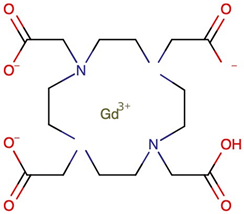
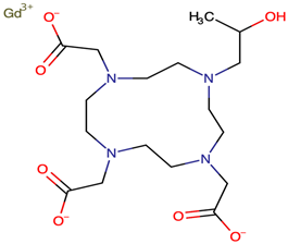
Water solutions of two compounds: gadoteric acid (GA), a gadolinium ionic complex consisting of gadolinium oxide and 1,4,7,10-tetraazacyclododecane-N, N′, N″, N‴ tetraacetic acid (DOTA), presented as a meglumine salt (Clariscan, GE Healthcare AS, Oslo, Norway), and Gadoteridol (GD)/Gd-HP-DO3A (C17H29GdN4O7; ProHance, Bracco Diagnostics, Bengaluru, India), a nonionic macrocyclic compound (Table 1), were evaluated.
Table 1.
Compounds of gadolinium evaluated: chemical properties, therapeutic indications, and safety considerations.
The tested solutions were made by mixing gadoteric acid (GA) solution for injection (Clariscan, 0.5 mmol/mL) and gadoteridol (GD) solution for injection (ProHance, 0.5 mmol/mL) with 35‰ saline solution in a volume-to-volume (v:v) ratio of 1:1 (1 part commercial solution: 1 part saline), yielding a stock solution of 0.25 mmol/mL (250 µmol/mL) for each compound. From this stock solution, volumes of 10 µL, 30 µL, 100 µL, 200 µL, 300 µL, and 400 µL were transferred into individual wells containing a total volume of 2 mL, yielding final test concentrations of 1.25, 3.75, 12.5, 25, 37.5, and 50 µmol/mL, respectively. The pH of each test solution was 8 ± 0.5, measured at the time of preparation and verified at each assessment to confirm that this value remained stable throughout the exposure period, ensuring appropriate physiological conditions for larval maintenance.
2.2. Hatching and Preparing the Larvae for Testing
Artemia eggs were incubated in artificial saline water (30–35%) at a temperature of 22–23 °C for 48 h, under conditions of continuous aeration and illumination. Larvae up to 24 h old (nauplius stage I) were separated using a binocular and placed in the test vessels.
2.3. Assessment of Larval Mortality
Microscopic assessment of larval stage development and anatomical changes was performed using slide preparations. The larvae, known as nauplii, in their early stages (I–III), were transferred to 24-well test plates (10–20 nauplii per well). Test solutions were added to these plates, with a maximum volume of 2 mL. For the control group, larvae were kept under conditions similar to those of the experiment, but the tanks contained only saline solutions. The larvae were not fed, and the experiments were conducted at a constant temperature of approximately 22 ± 1 °C. The effects were assessed at intervals of 24 and 48 h, respectively. The effects were quantified by examining the larvae under a binocular microscope (4× magnification). Larvae that remained at the bottom of the container without showing any specific movement (movement of appendages) were considered dead. Mortality was analyzed using basic descriptive statistics. The calculation was performed using the formula: M (%) = Number of dead organisms/total number of organisms × 100.
2.4. Cytomorphological Observations
Live larvae, which had previously been exposed to the test solutions (for 24–48 h), were collected with a pipette, placed on glass slides and observed under a microscope. A cytomorphological assessment was performed on the exposed organisms, compared with the control, based on the transparency of the larvae, which allows for the visualization under a light microscope of details such as cell size, changes in intercellular connections, and the presence of cytoplasmic inclusions indicating the activation of hydrolytic processes in lysosomes. Cell differentiation processes and the formation of appendages can also be evaluated.
These assessments also allow for the identification of the digestive tract’s contents, as well as observations of the larvae, which will enable the quantification of developmental changes and progression to the next larval stage. During the nauplius stages, these organisms exhibit two areas of interest within their digestive tract relevant for cytotoxicological analysis: the mid-digestive tract (m.d.t.), where absorption and substance exchange occur through the intestinal epithelium, and the posterior or anal region (p.d.t.). The anal region is contractile and plays an important role in evaluating sphincter control and intestinal peristalsis.
The images were acquired and processed using an Optikam camera (Optika, Ponteranica, Italy).
Fluorescence microscopy techniques were used to observe structural changes. The specific stains used were:
- acridine orange (AO) (0.3%) for labeling nuclear and cytoplasmic cellular activity (excitation λ = 450–490 nm);
- neutral red (NR) (1%) for the detection of lysosomal activity (excitation λ = 525 nm; emission λ = 645 nm);
- phalloidin, fluorescein isothiocyanate-labeled (FITC) (excitation λ = 495 nm; emission λ = 513 nm), used to detect cytoskeletal activity.
2.4.1. Identification of Nuclear Structures
For the AO test, 10 µL of acridine orange solution (0.3%) was added to 100 µL saline solutions containing larvae exposed to GA and GD. The samples were kept in the dark for 5–10 min, and the dye was subsequently removed by washing with saline solution.
2.4.2. Lysosomal Activity Assay
The reagent used is neutral red (3-amino-7-dimethylamino-2-methylphenazine hydrochloride), which is a vital dye. The test may help to identify changes in lysosomal function caused by the substances or compounds tested. Lysosomes are vital cellular organelles and can indicate cytotoxicity in cells before morphological changes occur. The protocol is based on the ability to observe the larvae through transparency, using a method that complies with [24].
2.4.3. Preparation of the Solution Neutral Red (NR) and Work Protocol
Neutral red dye (40 µg/mL) was dissolved in phosphate-buffered saline (PBS). A volume of 100 µL was added to Eppendorf tubes containing 10 larvae that had been exposed to the test substances for at least 24 h. Other specimens, from the control vessels, were placed in a similar manner for comparison. The tubes were incubated for 2 h at 28 °C, and then the dye solution was removed. The larvae in each tube were washed with 150 µL of PBS and subsequently analyzed under an epifluorescence microscope (emission λ = 645 nm) in red light.
2.4.4. Analysis of the Distribution and Dynamics of the Cytoskeleton
Phalloidin, Fluorescein isothiocyanate labeled (FITC), Sigma-Aldrich (St. Louis, MO, USA), catalog number P5282, (FITC) (excitation λ = 495 nm; emission λ = 513 nm) was used to assess changes in cytoskeletal formation (F-actin).
2.4.5. Preparation of the Solution (FITC) and Working Protocol
The stock solution was prepared in DMSO (5 mg/mL). The staining solution was prepared in physiological buffers at a concentration of 100 µmol. The larvae were fixed in Davidson’s solution, then washed with PBS and permeabilized with 0.1% Triton X-100 in PBS. For staining, the larvae were placed in tanks with staining solution for 40–50 min; the stain was removed with PBS, and the slides were prepared for evaluation under an epifluorescence microscope in blue light. Photographs were taken with an Optikam camera and analyzed. A quantitative fluorescence intensity analysis was performed across four selected areas within the region of interest corresponding to specific larval structural components.
The fluorescence intensity analysis was performed using OPTIKA Proview professional image analysis software (Version:x64, 2023, Ponteranica, BL, Italy). The intensity averages for these areas were evaluated and statistically analyzed using the following tests: one-way ANOVA and Tukey’s analysis for mean area fluorescence intensity; statistical significance is indicated by an asterisk (** p < 0.005; *** p < 0.001).
2.4.6. Statistics
Statistical analysis was performed on STATPLUS software, StatPlus:mac v8 software (AnalystSoft Inc., Brandon, FL, USA), and consisted of the evaluation of groups by descriptive statistics, comparisons between analyzed groups, one-way ANOVA analysis test. The results were expressed as median values, based on three biological replicates, and differences were considered significant. Probit analysis is a method for analyzing the relationship between a stimulus (the tested concentration) and a binary response. Probit analysis is widely used to interpret analyses in pharmacology, entomology, and pathology (almost exclusively), involving binary (all-or-nothing) responses. The procedure performs a probit regression and calculates the percentiles of the dose–response relationship. The mortality rate at the 50% dose (LD50) was analyzed.
3. Results
3.1. Lethal Effects Observed Following Exposure to Gadolinium Solutions at 24, 48, and 72 h
Following exposure to the GA-treated solutions, 24 h after exposure, the larvae were active in all experimental dishes, whereas exposure to GD caused slow movements along the bottom of the dish in approximately 50% of the specimens.
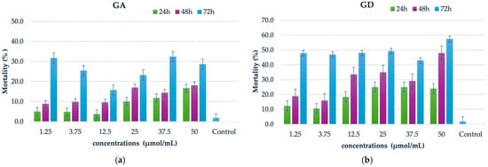
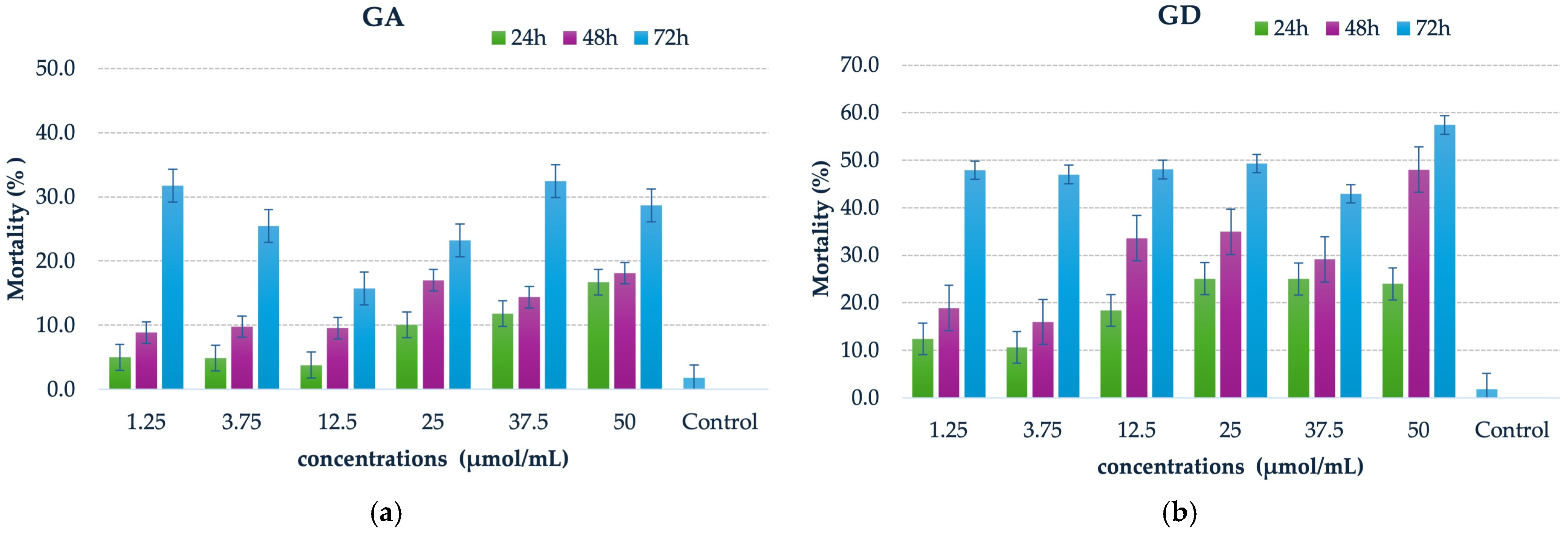
The cytotoxic effects of the contrast agents were moderate; thus, within the first 24 h, the mortality rate among larvae exposed to GA was less than 20%, while that of larvae in GD was slightly over 20%. A gradual increase in mortality was observed at 48 h, correlating with the concentration level. At 72 h, there was a significant increase in the percentage compared with previous time points, but the values did not exceed 35% when exposed to GA solutions (Figure 1a,b). In GD assays, mortality rates can exceed 40%, with the highest rate observed at the maximum test concentration of 50 µmol/mL (Figure 1b). Larvae maintained in the unexposed experimental tanks showed a 100% survival rate during the first 48 h after the start of observations. At 72 h, a mortality rate of 1.8% of the total number of specimens monitored was recorded (Figure 1).
Figure 1.
The larval mortality in the experimental vessels assessed at 24 h, 48 h, and 72 h; the values are averages of the measurements from the samples (triplicate) expressed as a percentage; ± SE; (a) Lethal effects recorded after exposing larvae to gadoteric acid (GA) solutions; (b) Lethal effects recorded after exposing larvae to gadoteridol (GD) solutions; The larvae mortality in the control vessels at 24 h and 48 h was not recorded.
These observations are consistent with the characteristics of GD; thus, given that the effects appeared after 48 h of exposure, we hypothesize that Gadoteridol, being a nonionic macrocycle, may penetrate the digestive tract as the larvae filter water.
The biology of these larvae indicates that, approximately 24 h after hatching, the digestive tract opens, allowing the ingestion of filtered organic particles, including particles of gadolinium compounds. The data reveal a striking trend in mortality at low concentrations when larvae are exposed to GD, compared with GA.
3.2. General Observations on Larval Morphology
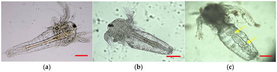
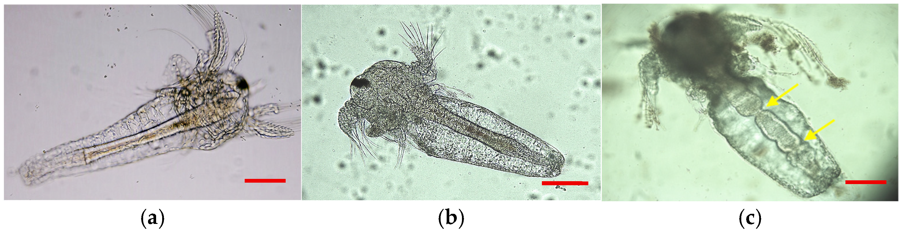
The larvae exhibited visible morphological changes compared with the control (Figure 2). The digestive tract exhibits a significantly altered lumen (Figure 2b,c) compared with the control (Figure 2a), which may affect larval feeding. It can be observed that exposure to gadoteridol (GD) solutions induces constriction of the digestive tract and swelling of the abdominal region (Figure 2c).
Figure 2.
General aspects of larval morphology, metanauplius stage; (a) Larvae with normal body, in saline water, control sample; scale bar = 100 µm; (b) Larvae with swelling body, exposure to GA at 50 µmol/mL; scale bar = 100 µm; (c) Contractions and blockage of the digestive tract, exposure in GD; (sublethal changes), after 48–72 h; white light microscopy (200×), the yellow arrows indicate the constriction point; scale bar = 100 µm.
3.2.1. Cytomorphology—Nuclear Changes
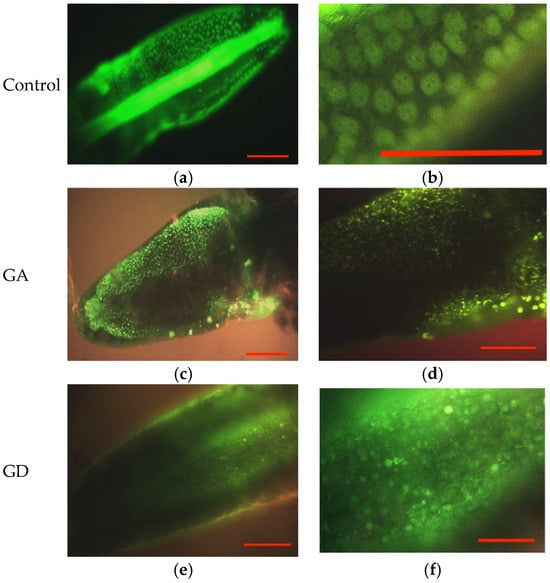
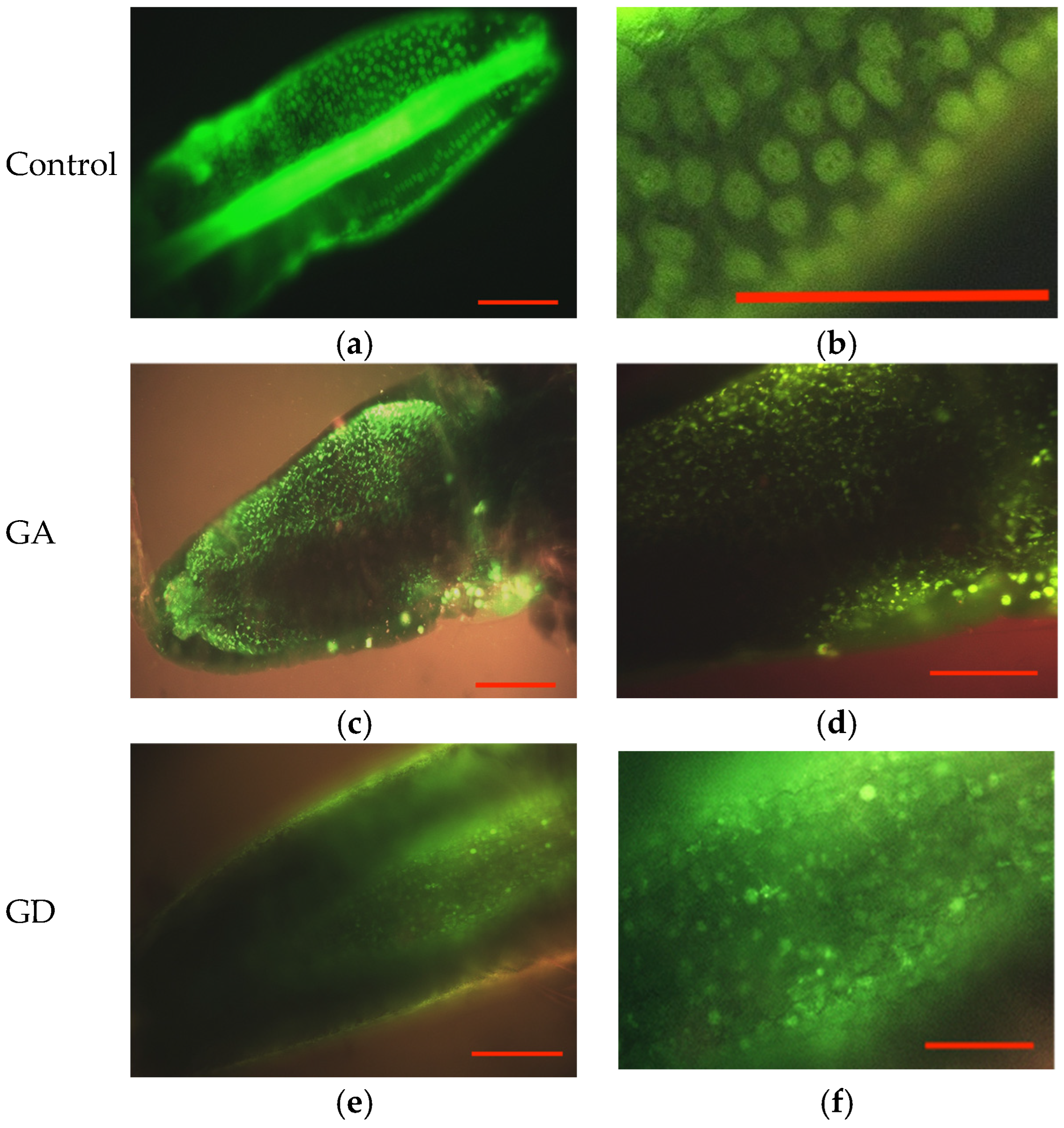
When larvae are exposed to saline solutions without gadolinium, the epithelial cells exhibit normal activity, as evidenced by nuclei with a circular outline and dispersed chromatin (Figure 3a,b), and internally contain 2–3 nucleoli that are distinguishable as intranuclear organelles with low-intensity reddish emissions (Figure 3b).
Figure 3.
Microscopic features of larvae’s in abdominal part of body with details of subcuticular cells and nuclear changes, after 48 h exposure at 50 µmol/mL contrast agents comparative with control sample; (a) larvae from control sample; scale bar = 100 µm; (b) details of the nucleus with loose chromatin (green emission) on larvae from control sample; scale bar = 50 µm; (c) abdominal details from GA larvae exposure; scale bar = 100 µm; (d) cellular details with nuclear karyorrhexis (green emission) after GA exposures; scale bar = 100 µm; (e) abdominal details from GD larvae exposure; scale bar = 100 µm; (f) details with pyknotic nuclei with incipient karyorrhexis after GD larvae exposure; acridine orange (AO) staining; (excitation λ = 495 nm; emission λ = 513 nm); scale bar = 50 µm.
In the gadolinium-enhanced samples, the larvae evaluated after AO staining show a disruption of nuclear structures, manifested by the formation of fragmented nuclei or nuclei with condensed chromatin (pyknotic) in both samples with the contrast agents GA (Figure 3c,d) and GD (Figure 3e,f), suggesting the similarity of the effects induced at the nuclear level.
These observations confirm the existence of direct effects on cellular activity, while the manner in which the nuclear envelope deteriorates and chromatin fragments suggests possible interference with processes related to the integrity of nuclear material and, respectively, with cell division.
The chromatin changes, karyorrhexis, and obvious DNA damage were observed after 48 h. These changes occurred once the Artemia larvae reached stages II–III (metanauplius), when, according to Birndorf (1975) [25], polymerase-type enzyme activity is high, favoring protein synthesis and accelerated development, as dictated by the species’ biology.
3.2.2. Cytomorphology—Lysosomal Assessment and Intracellular Accumulations
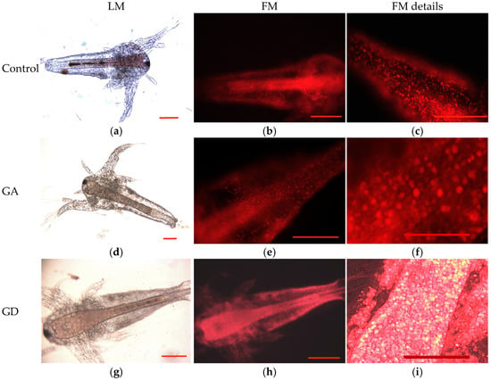
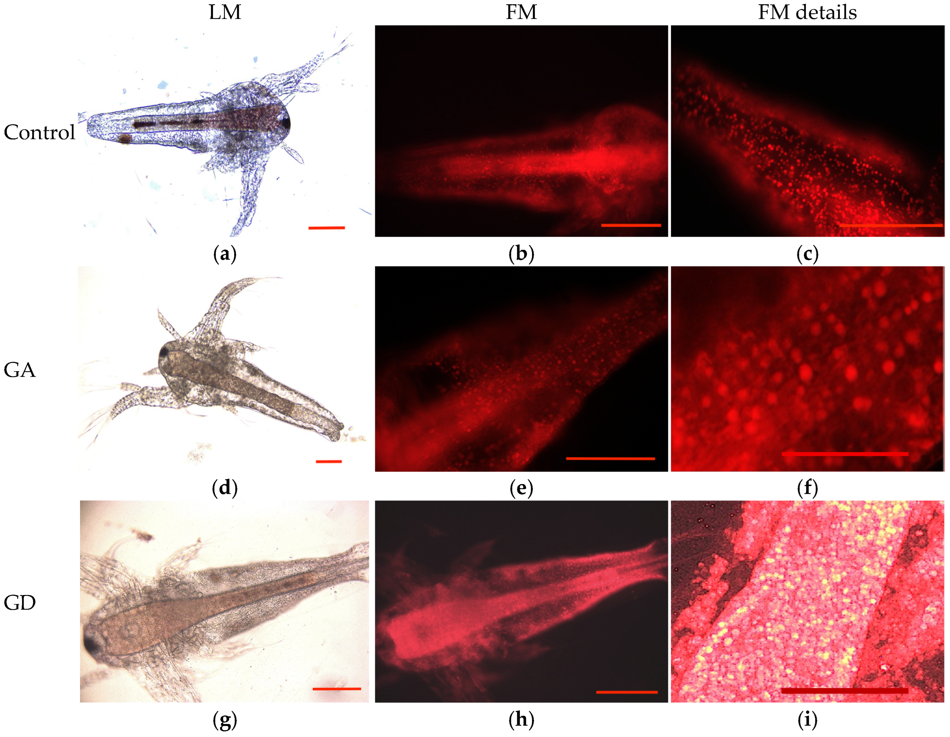
The results reveal a change in fluorescence emission in the GD samples compared with the GA samples, which is more pronounced in the cells lining the digestive tract and limited in the subcuticular epithelial cells (Figure 4c) compared with control (Figure 4b,c) and GA samples (Figure 4b,c). This is explained by the fact that the particles dispersed in the test solutions are taken up through the digestive tract. The processes of endocytosis and the amplification of endolysosome formation are what allow for the exacerbation of fluorescence emissions (Figure 4c).
Figure 4.
Larval body details after GA and GD exposure, exposure at 50 µmol/mL contrast agents concentrations; (a) light microscopy larvae observations, in the control sample; scale bar = 100 µm; (b) fluorescence microscopy assays, neutral red assay, on control sample larvae; scale bar = 100 µm; (c) details with which lysosomal vesicles highlighted in red, on control sample larvae; scale bar = 50 µm; (d) light microscopy larvae observations, in the GA exposure; scale bar = 100 µm; (e) red emissions intensifications after GA exposure; scale bar = 50 µm; (f) details with which lysosomal vesicles highlighted in red, after GA exposure; scale bar = 50 µm; (g) light microscopy larvae observations, in the GD exposure; scale bar = 100 µm; (h) strong emissions in red in the body of larvae after the GD exposure; scale bar = 100 µm; (i) the gut wall of larvae after GD exposure with lysosomal vesicle in red; scale bar = 50 µm; Neutral Red staining (excitation λ = 530 nm, excitation = 645); LM = light microscopy; FM = fluorescence microscopy.
3.2.3. Cytomorphology—The Cellular Cytoskeleton
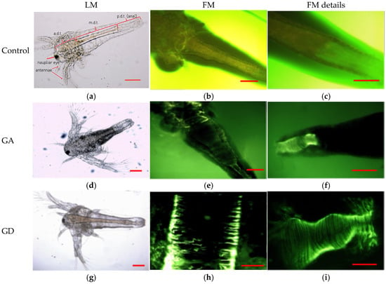
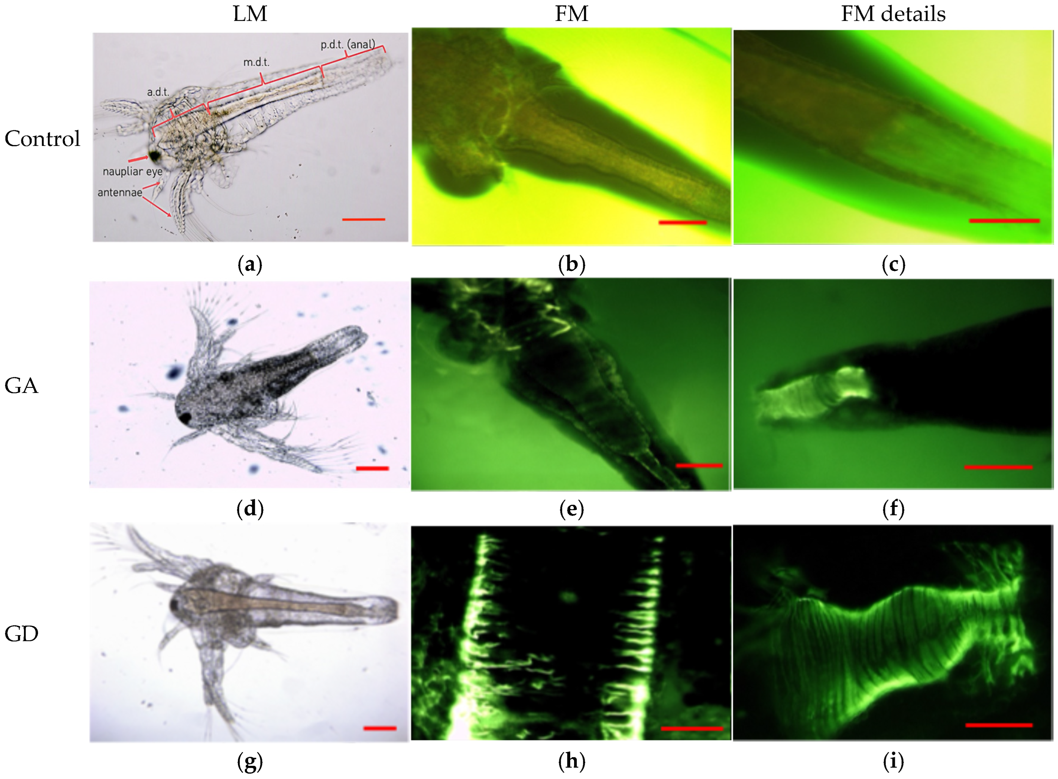
Observations regarding general changes, such as body swelling and contraction of the digestive tract, are verifiable and explained at the intracellular level by FITC labeling of the cytoskeleton (Figure 5).
Figure 5.
Cytoskeleton rearrangements (F-actin evaluation), 48 h exposure in the experimental conditions; (a) unexposed larvae (control samples), and details related to regions of the digestive compartment; a.d.t. = anterior digestive tract; m.d.t = median digestive tract; p.d.t. = posterior digestive tract (anal), light microscopy; scale bar = 100 µm; 100×; (b) details of larvae (control samples) median region, fluorescence microscopy, uniform emissions in FITC dye; scale bar = 50 µm; 400×; (c) larvae terminal region (control samples) fluorescence microscopy, uniform emissions in FITC dye; scale bar = 50 µm; 600×; (d) larvae after GA exposure, m.d.t., modified aspects, light microscopy; scale bar = 100 µm; 100×; (e) details of cytoskeleton reassembles, developing ring-like structures, after GA exposure; scale bar = 50 µm; 400× (f) details of the digestive tract highly pronounced F-actin in the anal region, after GA exposure; scale bar = 50 µm; 600×; (g) larvae after GD exposure, light microscopy; scale bar = 100 µm; 100×; (h) F-actine in the wall of the digestive tract, after GD exposure; scale bar = 20 µm; 1000×; (i) details of the digestive tract highly pronounced F-actin in the anal region, after GD exposure; scale bar = 20 µm; 1000×; FITC staining (excitation λ = 495 nm; emission λ = 513 nm); LM = light microscopy; FM = fluorescence microscopy.
In larvae maintained in saline water (control samples), the median digestive tract (m.d.t.) (Figure 5a) displays a uniform emission along its entire length (Figure 5b). Uniform emissions were also observed in the terminal digestive tract region (p.d.t.), considered the contractile segment responsible for peristalsis and fecal elimination (Figure 5c).
After exposing the larvae to GA (Figure 5d), discrete filaments are observed (Figure 5e) throughout the body, but they are more pronounced in the terminal gut region (Figure 5f), indicating imbalances in polymerization or depolymerization reactions.
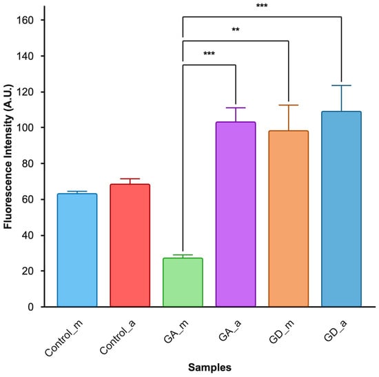
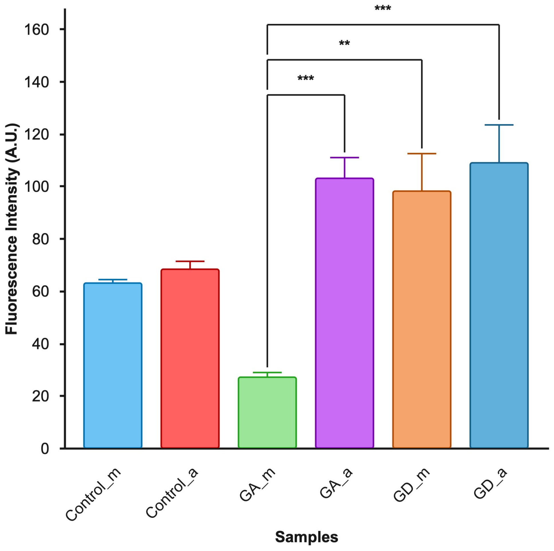
Quantitative assessment of fluorescence intensity (Figure 6) shows a significant reduction in the mid-region (GA_m) of the digestive tract (Figure 6), a phenomenon that can be highlighted in Figure 5e by dotted lines of filaments. In the terminal region of the digestive tract (p.d.t.), circular actin filaments are evident (Figure 5f), with significantly increased intensity (Figure 6); compared with GA_m, GA_a shows very high intensity, with statistically significant differences (** p < 0.005; *** p < 0.001) (Figure 6) compared with GA_m.
Figure 6.
Fluorescence intensity (A.U.), evaluated after FITC dye larvae from control samples in the median digestive tract region evaluation (control_m); larvae evaluation in the anal region (control_a); larvae exposure in GA (GA_m and GA_a); larvae exposure in GD (GD_m and GD_a); ** p < 0.005; *** p < 0.001, one-way ANOVA and Tukey analysis for mean areas fluorescence intensity.
Upon exposure of the larvae to GD (Figure 5g,h,i), a series of constrictions become visible under white light, while fluorescent light reveals clusters of intensely condensed F-actin (Figure 5h,i). Additionally, the formation of transverse bands, clustered in the anal region, with the highest intensity (GD_a) following GD exposure, can be observed (Figure 6). Based on these observations, it can be appreciated that the nonionic compound (GD) intensifies the blocking of actin in the filamentous phase (F-actin), with these processes being visible in the wall of the digestive tract (Figure 5h) and the terminal region (Figure 5i).
Gadolinium can affect the functions related to the polymerization (F-actin) and depolymerization (G-actin) of the molecules that determine the configuration of the cellular cytoskeleton. Given the dynamics of larval development, we assess cytoskeletal changes that are significant in terms of survival prospects.
3.2.4. Comparative Analysis of the Quantified Effects (Behavior, Lethality, and Cellular Changes) for the Two Solutions, GA and GD
An analysis of the swimming behavior of the larvae in the experimental tanks during the first 24 h after exposure revealed significant changes (50%) in the specimens exposed to GD at concentrations of 37 and 50 µmol/mL.
According to our studies, the observed effects also show differences between the two compounds analyzed, mortality after 48 h and 72 h (p < 0.001) in the comparative analysis between GA and GD groups. The lethal effect to 50% (LC50) of the specimens was observed at 48 h (Table 2) at a higher concentration (170 µmol/mL ± 10) of GA compared with the effect LC50 induced by GD (60 (SE ± 0.462).
Table 2.
Comparative assessment of the effects induced on Artemia larvae (48 h after exposure).
The digestive tract contains ingested particles (PP) in specimens exposed to GA solutions and control samples but is empty of contents (TE) in specimens exposed to GD, particularly at concentrations greater than 30 µmol/mL (Table 2). These general observations are also confirmed microscopically, following fluorescence assessments. According to these FITC-labeled assessments for actin filaments, the conformation of the filaments is evident microscopically, the concentration of these filaments (FC), and the very intense emissions indicate agglomeration and possible blockage of the filaments. In samples treated with GA, the emissions are reduced (FL), indicating a different behavior of the compounds regarding interactions specific to the polymerization or depolymerization of G-actin.
Larvae exposed to GA and GD evolved to naupliar stages II and III at a high rate of 90–95%; normal development (ND) of the larvae, as indicated by the stages, was observed in both GA and GD exposure groups. Furthermore, the growth process was also confirmed by the presence of exuviae in the experimental tanks, indicating that larval exposure for 24–48 h does not affect cell division or the physiological activities specific to the molting process.
At the end of the experiment (72 h after exposure), lethal effects were observed in more than 25% of the specimens. Cellular changes visible under light microscopy revealed hypertrophy of the digestive tract epithelium (DTE), likely resulting from direct contact and the stimulation of the cellular response. The qualitative cellular evaluation indicated that GA induced manifestations similar to those observed after GD exposure (Table 2).
Similar findings are also observed through the assessment of nuclear parameters. Both tested solutions exhibit changes in chromatin (CM) conformation and loss of nuclear integrity compared with samples exposed only to saline solutions, suggesting a similar mechanism for the penetration and action of GA and GD compounds on nuclear components.
Cytoplasmic inclusions labeled with NR are present in all samples analyzed, including the control. This result is expected in the control samples, given that in the early larval stages, intracellular lipid stores can persist from the embryonic period, ensuring viability and adaptation to stressors that may occur during the first hours of nauplii life and being utilized under conditions of nutritional deficiency in experimental settings.
However, exposure to high concentrations of GA resulted in a reduction in intracellular lipid stores in the analyzed specimens. This reduction was particularly evident in cells from the subcuticular epithelial layers. The lipid reserves utilized for the physiology of molting would also explain how larval survival is maintained even in the first hours after exposure, with visible molting processes remaining unchanged.
The specimens exhibited in GD show very intense emissions from the digestive tract epithelium, even at high concentrations. These observations can be associated with GD’s interactions with epithelial functions, which are more pronounced for this nonionic compound. The general consequences, such as lack of nutrition and the development of anatomical and morphological changes (the formation of constrictions), are clearly associated with changes that affect molecular structural processes (the formation of condensed F-actin filaments).
4. Discussion
Artemia larvae are used in various testing systems [26], serving as a recognized biological model in the field of nanoecotoxicology, as well as for testing plant extracts or various combinations with pharmaceutical applications, the effects of microplastics [27], and the photosensitivity of certain compounds [28], as well as a screening test for estuarine and marine waters [29].
In addition to these considerations, toxicity and cytomorphological analyses of these organisms make it possible to predict the extent to which effects induced by gadolinium ions and gadolinium-based contrast agents, respectively, will manifest in the aquatic environment [30,31,32]. Heavy metals such as Cd and Hg are highly cytotoxic to aquatic larvae [31], and Gd also exhibits a potential level of toxicity comparable to that of the most well-known toxic heavy metals, as evidenced by its low LC50 and its ability to induce oxidative stress [30,31,32,33].
In the case of GD, a nonionic macrocycle with a lower stability than the GA compound, it can be assumed that interactions with saline solutions may be limited. A comprehensive study conducted by Schijf J and Christy IJ [34] shows changes in the stability of the compounds depending on water salinity and the levels of magnesium or calcium ions, which means that the Gd—DTPA complex, generally considered chemically inert and stable, can be significantly destabilized when river water mixes with seawater in the estuary [34]. In terms of stability, it can be said that nonionic formulations are also less stable than ionic ones. In ionic compounds, the bond between Gd3+ and the negatively charged carboxyl groups is stronger than that with the amides or alcohols in nonionic formulations [35].
According to stability constants and kinetic measurements, the most stable Gd—CA (gadolinium contrast agents) is the ionic macrocyclic chelate Gd—DOTA, while the least stable agents are the nonionic linear chelates gadodiamide and gadoversetamide.
In vivo, data confirmed the low stability of nonionic linear chelates, but no significant difference was observed between macrocyclic agents, whether ionic (Gd—DOTA) or nonionic, such as Gd—HP—DO3A and Gd—BT—DO3A. The stability of Gd—CA appears to be an important factor in the pathogenesis of the serious complication of nephrogenic systemic fibrosis (NSF), observed in exposed patients. Gd-CA, with low stability, releases free Gd3+ ions that deposit in the tissue, attracting fibroblasts, which initiate the fibrosis process [35].
A study characterizing DNA-dependent RNA polymerases in stage II and III nauplius larvae of Artemia salina indicated that the properties of these enzymes were like those of corresponding enzymes from other animal sources. A significant change in the relative amounts of polymerases I and II was recorded immediately after hatching, between 36 and 72 h into development [25]. The authors of this study show that there is a dynamic change in polymerase activity between 36 and 72 h. Specifically, 36 h-old nauplii contained approximately equal amounts of polymerases I and II, whereas polymerase II accounted for more than 80% in 72 h old nauplii [25].
These findings regarding polymerase activity, observed during the 36–72 h time window associated with larval growth and consistent with the results obtained in our experiments, may explain the chromatin degradation and the quantified nuclear phenomena (cariorhexis, pyknotic nuclei showing early signs of cariorhexis) (Figure 3) in the cells of larvae exposed to the contrast agents. Kenger et al. [36] mention the cytotoxicity of gadoteric acid in NIH3T3 fibroblast cells, indicating that gadoteric acid interacts with topoisomerase IIα and IIβ, as well as DNA polymerase β; these details were examined using molecular docking [36]. These nuclear phenomena may explain the increased mortality observed after 48 h, which peaked at 72 h with exposure to GD, exceeding the 50% threshold.
The uptake and interaction of gadolinium ions are still poorly understood. In principle, the uptake and elimination of GD occur via membrane transport mechanisms, such as pinocytosis, exocytosis, or ion channels [37]. Furthermore, bioaccumulation processes in the digestive tract, like those observed in our studies, have also been noted in other crustacean species and sea urchin larvae, where an increase in intracellular autophagosome vesicles has been observed [38].
The presence of abundant lipid vesicles following GD administration may suggest that lipids, after binding to these gadolinium complexes, become trapped within pre-existing vesicles (Figure 4d). These observations may contribute to an understanding of the differences in response and explain the neutral red-labeled lysosomal accumulations in contact with GA and GD. These observations are consistent with our study and demonstrate the importance of model organisms, which help us to understand cellular biology under the influence of these compounds. In the case of Artemia larvae, in a study on lipid profiles, Lopalco et al. [39] noted a high content of phosphatidylcholine in larval stages I and II (12.7–12.9%). Also in this study, the authors indicate that polar lipids represent 30–40%, while neutral lipids represent 60–70% of total lipids [39].
Phalloidin (FITC) staining allowed us to observe differences in the spatial reorganization of F-actin, depending on the type of compound analyzed (Figure 5). Actin filaments are vital for cellular architecture, determining the shape, mobility, vesicular transport, and positioning of organelles. In the nucleus, actin regulates chromatin condensation and DNA repair, while its monomeric form (G-actin) influences transcription and the activity of RNA polymerases. A link between Gd3+ toxicity and Ca2+ channel blockade has been observed in various experimental models, such as sea urchins, Xenopus oocytes [40], and mammalian cell lines [41]. Calcium ions regulate all cellular processes not only by generating electrical signals but also by acting as signaling molecules that can alter the charge and conformation of proteins. Calcium ions can enter and exit the cell nucleus through calcium-dependent channels (DMI1) identified in plant cell nuclei. Calcium can also passively diffuse through nuclear pore complexes (NPCs) in the nuclear membrane [42].
Nuclear calcium regulates G-actin/F-actin dynamics and actin-related nuclear functions, including the structure and condensation of F-actin-dependent chromatin (heterochromatin versus euchromatin) and, consequently, the accessibility of genes to transcription factors and, ultimately, transcription. The nucleus and all cellular organelles (endoplasmic reticulum, Golgi apparatus, and mitochondria) are anchored in specific positions within the cell by actin filaments. Gadolinium ions can disrupt all calcium-related processes in the nucleus and cellular organelles and, by interfering with actin polymerization, can affect cellular polarity and the positioning of all organelles.
Consequently, observations made by exposing Artemia larvae confirm the existence of cellular phenomena that point to the establishment of an absorption pathway for gadolinium compounds. The development of effects such as abdominal swelling of the larvae, changes in cellular morphology, and alterations in cytoskeletal filaments support the previously discussed hypotheses implicating a connection between gadolinium, actin polymerization, and cytoskeletal reorganization, likely through interference with calcium ions.
The identified nuclear changes suggest the activation of apoptosis and the inhibition of cell division, findings that open new large research opportunities. A recent study examines the effects of gadoteric acid on SH-SY5Y neuroblastoma cells and its affinity for the apoptosis inhibitor XIAP. The results show that GA contrast agents induce a genotoxic effect [43] on neuroblastoma cells, which leads to new approaches to neuroblastoma treatment and to understanding the mechanism of apoptosis induced by GA-type contrast agents [44]. The known mechanisms of toxicity of gadolinium-based contrast agents largely depend on the stability of the chelate complex, which determines the release of free gadolinium (Gd3+), blood cell uptake [45], tissue retention, and subsequent toxic effects in the body.
Several studies conducted on experimental test models (Table 3) support the differences also observed in the Artemia study.
Table 3.
Comparative analysis of toxicity effects and cytotoxicity of GA and GD on human cells and animal models.
Although both compounds, gadoteric acid and gadoteridol, are macrocyclic agents and have similar safety profiles, we cannot ignore the toxic effects reported in the tissues examined in laboratory animals (liver, kidney, blood, brain, bone, and other tissues). It is evident (Table 3) that there is a lack of direct, standardized, and comparative studies between these compounds, making it difficult to draw conclusions regarding the advantages of any one of them. Furthermore, the diversity of the experimental models used (species, doses, frequency of administration, follow-up period) makes it difficult to generalize the results.
The safety of these substances has been extensively studied in recent years [50], revealing numerous gaps in our understanding of these substances, the biology of gadolinium, and the mechanisms underlying complications [46,47,49]. Further research is needed to identify ways to prevent or slow the progression of symptoms caused by gadolinium retention or resulting from interactions with other biomolecules [50].
5. Conclusions
Both contrast agents induce low or moderate toxicity at concentrations of 37.5 and 50 µmol/mL. The results obtained from cytomorphological analysis of the larvae indicate that GA and GD interact at the nuclear level, promoting chromatin damage, followed by the onset of the apoptotic phenotype (chromatin fragmentation, pyknotic nuclei) and a decline in larval viability. Significant changes occur in the organization of cytoskeletal proteins, possibly by blocking polymerization processes, which leads to visible internal contraction of the digestive tract and swelling of the larva.
The study highlights how the presence of these compounds in water can be a significant factor disrupting larval growth and viability. This laboratory model may prove useful for monitoring the combined effects of multiple compounds, depending on the affected areas and the degree of pollution (coastal areas, ports, wastewater, etc.), with cellular biomarkers potentially serving as a tool for risk classification within the ecosystem under study.
Gadolinium, which is widely used in medicine and technology, requires an assessment of its environmental impact and, by extension, its impact on human health. Understanding the long-term consequences of this impact on aquatic ecosystems and human health requires studies to monitor and quantify the extent of damage caused by these emerging pollutants in accordance with regional and local environmental and health policies.
Author Contributions
Conceptualization, F.M., G.M. and V.S.; methodology, F.M. and V.S.; software, V.S.; validation, G.M. and I.R.; formal analysis, F.M., A.-M.M., and V.S.; investigation, F.M., G.M., V.S., and A.-M.M.; data curation, I.R. and A.-M.M.; writing—original draft preparation, FM. and V.S.; writing—review and editing, F.M., G.M., A.-M.M., I.M.I., and V.S.; visualization, FM., I.M.I., I.R., and A.-M.M.; supervision, A.-M.M. and I.R. All authors have read and agreed to the published version of the manuscript.
Funding
This research received no external funding.
Institutional Review Board Statement
The animal study protocol was approved by the Bioethics Committee of “OVIDIUS” UNIVERSITY OF CONSTANȚA (Approval Code: 7358 from 19 June 2026).
Data Availability Statement
The original contributions presented in this study are included in the article. Further inquiries can be directed to the corresponding authors.
Conflicts of Interest
The authors declare no conflicts of interest.
References
- Ebrahimi, P.; Barbieri, M. Gadolinium as an Emerging Microcontaminant in Water Resources: Threats and Opportunities. Geosciences 2019, 9, 93. [Google Scholar] [CrossRef] [Scilit]
- Dekker, H.M.; Stroomberg, G.J.; Van der Molen, A.J.; Prokop, M. Review of Strategies to Reduce the Contamination of the Water Environment by Gadolinium-Based Contrast Agents. Insights Imaging 2024, 15, 62. [Google Scholar] [CrossRef] [Scilit] [PubMed]
- Zhang, Z.; Jiang, W.; Gu, T.; Guo, N.; Sun, R.; Zeng, Y.; Han, Y.; Yu, K. Anthropogenic Gadolinium Contaminations in the Marine Environment and Its Ecological Implications. Environ. Pollut. 2024, 359, 124740. [Google Scholar] [CrossRef] [Scilit] [PubMed]
- Bau, M.; Dulski, P. Anthropogenic Origin of Positive Gadolinium Anomalies in River Waters. Earth Planet. Sci. Lett. 1996, 143, 245–255. [Google Scholar] [CrossRef] [Scilit]
- Oluwasola, I.E.; Ahmad, A.L.; Shoparwe, N.F.; Ismail, S. Gadolinium Based Contrast Agents (GBCAs): Uniqueness, Aquatic Toxicity Concerns, and Prospective Remediation. J. Contam. Hydrol. 2022, 250, 104057. [Google Scholar] [CrossRef] [Scilit] [PubMed]
- Černíková, F.; Krejčová, A.; Patočka, J. Quantification of Gadolinium Presence in Flour, Rice, and Carrot Samples Using ICP-MS. J. Food Compos. Anal. 2025, 139, 107106. [Google Scholar] [CrossRef] [Scilit]
- Taheran, M.; Naghdi, M.; Brar, S.K.; Verma, M.; Surampalli, R.Y. Emerging Contaminants: Here Today, There Tomorrow! Environ. Nanotechnol. Monit. Manag. 2018, 10, 122–126. [Google Scholar] [CrossRef] [Scilit]
- Trapasso, G.; Coppola, F.; Queirós, V.; Henriques, B.; Soares, A.M.V.M.; Pereira, E.; Chiesa, S.; Freitas, R. How Ulva lactuca Can Influence the Impacts Induced by the Rare Earth Element Gadolinium in Mytilus galloprovincialis? The Role of Macroalgae in Water Safety towards Marine Wildlife. Ecotoxicol. Environ. Saf. 2021, 215, 112101. [Google Scholar] [CrossRef] [Scilit] [PubMed]
- Liu, Z.; Guo, C.; Tai, P.; Sun, L.; Chen, Z. The Exposure of Gadolinium at Environmental Relevant Levels Induced Genotoxic Effects in Arabidopsis thaliana (L.). Ecotoxicol. Environ. Saf. 2021, 215, 112138. [Google Scholar] [CrossRef] [Scilit] [PubMed]
- Cesarini, G.; Spani, F.; Patricelli, R.; Quattrocchi, C.C.; Colasanti, M.; Scalici, M. Assessing Teratogenic Risks of Gadolinium in Freshwater Environments: Implications for Environmental Health. Ecotoxicol. Environ. Saf. 2024, 278, 116442. [Google Scholar] [CrossRef] [Scilit] [PubMed]
- Revel, M.; Tan, Q.G.; Hursthouse, A.; Heise, S. The Development of a Kinetic Biotic Ligand Model to Predict Acute Toxicity of Gadolinium for Daphnia magna. Ecotoxicol. Environ. Saf. 2025, 289, 117486. [Google Scholar] [CrossRef] [Scilit] [PubMed]
- Secco, S.; Cunha, M.; Libralato, G.; Trifuoggi, M.; Giarra, A.; Soares, A.M.V.M.; Freitas, R.; Scalici, M. Evaluating the Impact of Gadolinium Contamination on the Marine Bivalve Donax trunculus: Implications for Environmental Health. Environ. Toxicol. Pharmacol. 2024, 112, 104580. [Google Scholar] [CrossRef] [Scilit] [PubMed]
- Martino, C.; Chianese, T.; Chiarelli, R.; Roccheri, M.C.; Scudiero, R. Toxicological Impact of Rare Earth Elements (REEs) on the Reproduction and Development of Aquatic Organisms Using Sea Urchins as Biological Models. Int. J. Mol. Sci. 2022, 23, 2876. [Google Scholar] [CrossRef] [Scilit] [PubMed]
- Henderson, I.M.; Benevidez, A.D.; Mowry, C.D.; Watt, J.; Bachand, G.D.; Kirk, M.L.; Dokładny, K.; DeAguero, J.; Escobar, G.P.; Wagner, B. Precipitation of Gadolinium from Magnetic Resonance Imaging Contrast Agents May Be the Brass Tacks of Toxicity. Magn. Reson. Imaging 2025, 119, 110383. [Google Scholar] [CrossRef] [Scilit] [PubMed]
- Manfra, L.; Savorelli, F.; Di Lorenzo, B.; Libralato, G.; Comin, S.; Conti, D.; Floris, B.; Francese, M.; Gallo, M.L.; Gartner, I.; et al. Intercalibration of Ecotoxicity Testing Protocols with Artemia franciscana. Ecol. Indic. 2015, 57, 41–47. [Google Scholar] [CrossRef] [Scilit]
- Morgana, S.; Estévez-Calvar, N.; Gambardella, C.; Faimali, M.; Garaventa, F. A Short-Term Swimming Speed Alteration Test with Nauplii of Artemia franciscana. Ecotoxicol. Environ. Saf. 2018, 147, 558–564. [Google Scholar] [CrossRef] [PubMed]
- Libralato, G.; Prato, E.; Migliore, L.; Cicero, A.M.; Manfra, L. A Review of Toxicity Testing Protocols and Endpoints with Artemia spp. Ecol. Indic. 2016, 69, 35–49. [Google Scholar] [CrossRef] [Scilit]
- Solis, P.; Wright, C.; Anderson, M.; Gupta, M.; Phillipson, J. A Microwell Cytotoxicity Assay Using Artemia salina (Brine Shrimp). Planta Med. 1993, 59, 250–252. [Google Scholar] [CrossRef] [Scilit] [PubMed]
- Xiao, Y.; Xue, R.; You, T.; Li, X.; Pei, F.; Wang, X.; Lei, H. Gadolinium-1,4,7,10-Tetraazacyclododecane-1,4,7,10-Tetraacetic Acid Conjugate of Arabinogalactan as a Potential Liver-Targeting Magnetic Resonance Imaging Contrast Agent. Carbohydr. Res. 2014, 395, 9–14. [Google Scholar] [CrossRef] [Scilit] [PubMed]
- del Poggio, A.; Anello, G.; Calloni, S.F.; Vezzulli, P.; Pereira, C.; Iadanza, A.; Falini, A.; Anzalone, N. Diagnostic Efficacy and Safety of Gadoteridol Compared to Gadobutrol and Gadoteric Acid in a Large Sample of CNS MRI Studies at 1.5 T. J. Neuroradiol. 2022, 49, 73–79. [Google Scholar] [CrossRef] [Scilit] [PubMed]
- Fallenberg, E.M.; Renz, D.M.; Karle, B.; Schwenke, C.; Ingod-Heppner, B.; Reles, A.; Engelken, F.J.; Huppertz, A.; Hamm, B.; Taupitz, M. Intraindividual, Randomized Comparison of the Macrocyclic Contrast Agents Gadobutrol and Gadoterate Meglumine in Breast Magnetic Resonance Imaging. Eur. Radiol. 2015, 25, 837–849. [Google Scholar] [CrossRef] [Scilit] [PubMed]
- Gutierrez, J.E.; Rosenberg, M.; Seemann, J.; Breuer, J.; Haverstock, D.; Agris, J.; Balzer, T.; Anzalone, N. Safety and Efficacy of Gadobutrol for Contrast-Enhanced Magnetic Resonance Imaging of the Central Nervous System: Results from a Multicenter, Double-Blind, Randomized, Comparator Study. Magn. Reson. Insights 2015, 8, MRI-S19794. [Google Scholar] [CrossRef] [Scilit] [PubMed]
- Foshag, K.; Tsiouris, A.J.; Prince, M.; Reichman, M. A Review of Gadolinium-Based Contrast Agents in the Setting of Repeated MRI for High Risk Breast Cancer Screening. Clin. Imaging 2025, 120, 110420. [Google Scholar] [CrossRef] [Scilit] [PubMed]
- Do, M.A.; Dang, H.T.; Doan, N.T.; Pham, H.L.T.; Tran, T.A.; Le, V.C.T.; Young, T.; Le, D.V. Silver Nanoparticle Toxicity on Artemia parthenogenetica Nauplii Hatched on Axenic Tryptic Soy Agar Solid Medium. Sci. Rep. 2023, 13, 6365. [Google Scholar] [CrossRef] [Scilit] [PubMed]
- Birndorf, H.C.; D’Alessio, J.; Bagshaw, J.C. DNA-Dependent RNA-Polymerases from Artemia Embryos: Characterization of Polymerases I and II from Nauplius Larvae. Dev. Biol. 1975, 45, 34–43. [Google Scholar] [CrossRef] [Scilit] [PubMed]
- Azra, M.N.; Noor, M.I.M.; Burlakovs, J.; Abdullah, M.F.; Abd Latif, Z.; Yik Sung, Y. Trends and New Developments in Artemia Research. Animals 2022, 12, 2321. [Google Scholar] [CrossRef] [Scilit] [PubMed]
- Zheng, K.; Wang, W.; Pang, H.; Peng, Z.; Bao, B.; Zhang, D.; Zhang, X. Toxicological Effects of Microplastics and Sulfadiazine on Artemia sinica. Environ. Pollut. 2026, 390, 127455. [Google Scholar] [CrossRef] [Scilit] [PubMed]
- Madkour, K.; Dawood, M.A.O.; Sewilam, H. The Use of Artemia for Aquaculture Industry: An Updated Overview. Ann. Anim. Sci. 2023, 23, 3–10. [Google Scholar] [CrossRef] [Scilit]
- Artoxkit, M. Artemia Toxicity Screening Test for Estuarine and Marine Waters; Standard Operating Procedure; Creasel: Deinze, Belgium, 1990. [Google Scholar]
- Wang, Y.-J.; Chen, C.-Z.; Li, P.; Liu, L.; Chai, Y.; Li, Z.-H. Chronic Toxic Effects of Waterborne Mercury on Silver Carp (Hypophthalmichthys molitrix) Larvae. Water 2022, 14, 1774. [Google Scholar] [CrossRef] [Scilit]
- Wu, Y.-R.; Chen, P.-J. Toxicity Assessments with Medaka (Oryzias latipes) Reveal Elevated Ecological Risks of Lanthanum and Gadolinium in Aquatic Ecosystems. Environ. Pollut. 2025, 383, 126802. [Google Scholar] [CrossRef] [Scilit] [PubMed]
- Fuma, S.; Takeda, H.; Miyamoto, K.; Yanagisawa, K.; Inoue, Y.; Ishii, N.; Sugai, K.; Ishii, C.; Kawabata, Z. Ecological Evaluation of Gadolinium Toxicity Compared with Other Heavy Metals Using an Aquatic Microcosm. Bull. Environ. Contam. Toxicol. 2001, 66, 231–238. [Google Scholar] [CrossRef] [Scilit] [PubMed]
- Ab Hamid, S.; Jumaat, A.H. Toxicity Assessment on Odonata Larvae Survivability in Monitoring Heavy Metal Contaminations. Malays. Appl. Biol. 2023, 52, 47–56. [Google Scholar] [CrossRef] [Scilit]
- Schijf, J.; Christy, I.J. Effect of Mg and Ca on the Stability of the MRI Contrast Agent Gd–DTPA in Seawater. Front. Mar. Sci. 2018, 5, 111. [Google Scholar] [CrossRef] [Scilit]
- Morcos, S.K. Extracellular Gadolinium Contrast Agents: Differences in Stability. Eur. J. Radiol. 2008, 66, 175–179. [Google Scholar] [CrossRef] [Scilit] [PubMed]
- Kenger, İ.H. Gadoteric Acid Induces Dose-Dependent DNA Damage and Enzyme Binding in Fibroblast Cells. Basic Clin. Pharmacol. Toxicol. 2026, 138, e70150. [Google Scholar] [CrossRef] [Scilit] [PubMed]
- Kohl, J.; Schweikert, M.; Klaas, N.; Lemloh, M.-L. Intracellular Bioaccumulation of the Rare Earth Element Gadolinium in Ciliate Cells Resulting in Biogenic Particle Formation and Excretion. Sci. Rep. 2023, 13, 5650. [Google Scholar] [CrossRef] [Scilit] [PubMed]
- Scarciglia, A.; Papi, C.; Romiti, C.; Leone, A.; Di Gregorio, E.; Ferrauto, G. Gadolinium-Based Contrast Agents (GBCAs) for MRI: A Benefit–Risk Balance Analysis from a Chemical, Biomedical, and Environmental Point of View. Glob. Chall. 2025, 9, 2400269. [Google Scholar] [CrossRef] [Scilit] [PubMed]
- Lopalco, P.; Lobasso, S.; Lopes-dos-Santos, R.M.A.; Van Stappen, G.; Corcelli, A. Lipid Profile Changes During the Development of Artemia franciscana, From Cysts to the First Two Naupliar Stages. Front. Physiol. 2019, 9, 1872. [Google Scholar] [CrossRef] [Scilit] [PubMed]
- Rogowska, J.; Olkowska, E.; Ratajczyk, W.; Wolska, L. Gadolinium as a New Emerging Contaminant of Aquatic Environments. Environ. Toxicol. Chem. 2018, 37, 1523–1534. [Google Scholar] [CrossRef] [Scilit] [PubMed]
- Unruh, C.; Van Bavel, N.; Anikovskiy, M.; Prenner, E.J. Benefits and Detriments of Gadolinium from Medical Advances to Health and Ecological Risks. Molecules 2020, 25, 5762. [Google Scholar] [CrossRef] [Scilit] [PubMed]
- Kloc, M.; Halasa, M.; Wosik, J.; Ghobrial, R.M. Gadolinium-Based MRI Contrast Agent Effects on Calcium Signaling and Actin-Dependent Cell Functions. Magn. Med. 2025, 1, 100004. [Google Scholar] [CrossRef] [Scilit]
- Pinto, T.G.; Dedivits, R.A.; Ribeiro, D.A. Do Gadolinium-Based Contrasts Represent a High Risk for Genotoxicity in Mammalian Cells? A Systematic Review. J. Appl. Toxicol. 2025, 45, 1935–1946. [Google Scholar] [CrossRef] [Scilit] [PubMed]
- Bilgin, B.; Husunet, M.T.; Kenger, I.H. Cytogenotoxic Activity of Gadoteric Acid in SH-SY5Y Cells and XIAP Interaction with Molecular Docking. J. Appl. Toxicol. 2025, 45, 2450–2457. [Google Scholar] [CrossRef] [Scilit] [PubMed]
- Ruprecht, N.; Parakkattel, D.; Hofmann, L.; Broekmann, P.; Lüdi, N.; Kempf, C.; Heverhagen, J.T.; von Tengg-Kobligk, H. Uptake of Gadolinium-Based Contrast Agents by Blood Cells During Contrast-Enhanced MRI Examination. Investig. Radiol. 2024, 59, 372–378. [Google Scholar] [CrossRef] [Scilit] [PubMed]
- Coimbra, S.; Rocha, S.; Sousa, N.R.; Catarino, C.; Belo, L.; Bronze-da-Rocha, E.; Valente, M.J.; Santos-Silva, A. Toxicity Mechanisms of Gadolinium and Gadolinium-Based Contrast Agents—A Review. Int. J. Mol. Sci. 2024, 25, 4071. [Google Scholar] [CrossRef] [Scilit] [PubMed]
- Erdoğan, M.A.; Apaydin, M.; Armagan, G.; Taskiran, D. Evaluation of Toxicity of Gadolinium-Based Contrast Agents on Neuronal Cells. Acta Radiol. 2021, 62, 206–214. [Google Scholar] [CrossRef] [Scilit] [PubMed]
- Uosef, A.; Subuddhi, A.; Lu, A.; Ubelaker, H.V.; Karmonik, C.; Wosik, J.; Ghobrial, R.M.; Kloc, M. 7T MRI and Molecular Studies of Dotarem (Gadoterate Meglumine) Retention in Macrophages. J. Magn. Reson. Open 2022, 12–13, 100085. [Google Scholar] [CrossRef] [Scilit]
- Mercantepe, T.; Tümkaya, L.; Çeliker, F.B.; Topal Suzan, Z.; Çinar, S.; Akyildiz, K.; Mercantepe, F.; Yilmaz, A. Effects of Gadolinium-based MRI Contrast Agents on Liver Tissue. J. Magn. Reson. Imaging 2018, 48, 1367–1374. [Google Scholar] [CrossRef] [Scilit] [PubMed]
- Cunningham, A.; Kirk, M.; Hong, E.; Yang, J.; Howard, T.; Brearley, A.; Sáenz-Trevizo, A.; Krawchuck, J.; Watt, J.; Henderson, I.; et al. The Safety of Magnetic Resonance Imaging Contrast Agents. Front. Toxicol. 2024, 6, 1376587. [Google Scholar] [CrossRef] [Scilit] [PubMed]

Disclaimer/Publisher’s Note: The statements, opinions and data contained in all publications are solely those of the individual author(s) and contributor(s) and not of MDPI and/or the editor(s). MDPI and/or the editor(s) disclaim responsibility for any injury to people or property resulting from any ideas, methods, instructions or products referred to in the content. |
© 2026 by the authors. Licensee MDPI, Basel, Switzerland. This article is an open access article distributed under the terms and conditions of the Creative Commons Attribution (CC BY) license.