


Abstract
Antibiotic resistance (ABR) is a significant threat to human and animal health, as well as to the environment. Antibiotic residues, antibiotic-resistant bacteria and antibiotic resistance genes are emerging contaminants of water resources. The present study aimed to assess the prevalence of ABR among waterborne enterococci in an anthropogenic-affected section of the Yantra River (Bulgaria). The susceptibility of 426 strains to 13 antibiotics (ABs) was tested by the disk diffusion method, and the genes encoding resistance by PCR analyses. A total of 39% of isolates were found to be antibiotic-resistant, with 9% mainly being multidrug-resistant to three AB classes. The most common resistance was to erythromycin (19%), tetracycline (18%) and ampicillin (14%), encoded by the ermB, tetM and blaTEM genes. A total of 3% of isolates were ciprofloxacin-resistant and only 1% was resistant to vancomycin or high-level gentamicin. All isolates were susceptible to teicoplanin and linezolid. Spatial variations in ABR levels were found, with the lowest abundance of antibiotic-resistant enterococci occurring in upstream river waters, away from urban areas, and the highest in urban areas. The spread of waterborne antibiotic-resistant enterococci highlights the need for water pollution management, monitoring and control to limit anthropogenic pressures through wastewater discharges and diffuse fecal pollution, and to ensure the ecological well-being of receiving waters.
1. Introduction
Growing antibiotic resistance poses a threat to human and animal health, as well as to the well-being of the environment. Apart from being a global health challenge, antibiotic resistance is becoming an environmental challenge in water resources due to the emergence of contaminants such as antibiotic residues, antibiotic-resistant bacteria, and antibiotic resistance genes (ARG). The spread of ABR in surface waters is closely related to their role as a receiver of antibiotic- and fecal-contaminated waters via routes as wastewater discharges, surface runoff from agricultural lands, livestock farms or unsewered settlements [1,2,3,4].
As natural inhabitants of the intestines of humans and animals, enterococci, together with Escherichia coli, are ubiquitous in wastewater and are used as indicators of fecal pollution of receiving water bodies [5,6,7]. Enterococci inhabit a variety of ecological niches—clinical, farm animals and food, or environmental (plants, soil and water) [8,9,10]. A notable feature is their inherent resistance to cephalosporins, co-trimoxazole, lincomycin, and low levels of penicillin and aminoglycosides [11,12], but they are capable of acquiring ARGs through horizontal gene transfer and thus becoming resistant to amphenicols, tetracyclines, streptogramins, macrolides, glycopeptides, and quinolones. Antibiotic-resistant enterococci are among the leading causes of nosocomial infections worldwide and are considered the fifth most common healthcare-associated pathogen in the United States [13]. They have been identified as causative agents of urinary tract infections, intra-abdominal infections, bacteremia, or endocarditis [14,15,16].
Their ubiquity in the gut, resilience to various environmental stressors, and ability to acquire and transfer antibiotic resistance allow enterococci to persist and survive for extended periods of time in the aquatic environment [17,18]. Enterococcus faecalis is often identified as the most abundant species in natural waters, although E. faecium, E. casseliflavus, E. hirae, and E. mundtii have been found to be dominant or as common as E. faecalis in some aquatic environments [13,19,20]. In a US watershed dominated by forests, with increasing agricultural activity and urban runoff along the river, the prevalent species E. casseliflavus, E. faecalis, and E. hirae showed the highest levels of resistance to lincomycin and tetracycline [13]. In an urban watershed affected by concentrated cattle feeding operations, WWTPs and urban runoff, the predominant species E. mundtii, E. faecalis, and E. faecium are mostly resistant to ciprofloxacin, erythromycin, and tetracycline [21]. In Iranian river and coastal waters, the prevalent species E. faecalis and E. faecium are most commonly resistant to chloramphenicol, ciprofloxacin and tetracycline [22]. Large-scale surveillance of Enterococcus spp. across the One-Health human–animal–environment continuum in Canada revealed diverse antimicrobial resistance encoded by 31 ARGs in E. faecium isolates and 34 ARGs in E. faecium isolates, as well as a much lower rate of multidrug resistance (MAR) in non-clinical isolates compared to clinical isolates [23,24].
Enterococci that are not effectively eliminated during wastewater treatment can spread throughout the river continuum [2,3]. Wastewater treatment plants (WWTP) have been identified as hotspots for the acquisition and spread of ABR, responsible for releasing a significant quantity and diversity of resistant fecal bacteria into the environment [3,4,25,26]. Despite data on the contribution of WWTPs to the spread of ABR in natural waters, there are no legal regulations regarding the microbiological quality of wastewater entering water bodies and the fecal enterobacteria and enterococci content, including those resistant to ABs. Enterococci species are generally not considered to be pathogens of importance outside hospitals and healthcare facilities, so reported studies on antibiotic-resistant enterococci in the environment are not as numerous as those on clinical isolates.
In Bulgaria, there is also little data on the content of antibiotic-resistant fecal bacteria in surface waters and the factors influencing their spread, such as wastewater discharge, urbanization, economic activities, etc. [27,28]. For these reasons, the goal of the present study is to assess the prevalence of ABR among the enterococci population in the waters of the Yantra River (Bulgaria) by determining the phenotypic and genotypic antibiotic resistance patterns of isolates from a river section subjected to significant anthropogenic pressure.
2. Materials and Methods
2.1. Description of the Studied River Section
The Yantra River is a right tributary of the Danube River flowing at 536 km. The study was conducted in a section of the river covering its passage through the city of Veliko Tarnovo and four small settlements located upstream or downstream, as well as the confluence of the right tributary, the Belitsa River. In the selected section, the Yantra River meanders through the urban area of Veliko Tarnovo (about 74,000 inhabitants). A large portion of this area’s wastewater is treated in the municipal WWTP. Its tributary, the Belitsa River, flows into this section, after passing through the city of Debelets (about 3900 inhabitants), whose wastewater is discharged untreated. The ecological status of the Yantra River stretch is directly influenced by discharged, untreated domestic or mixed wastewater and indirectly by surface runoff from adjacent agricultural lands, farms and rural areas.
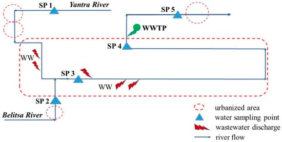
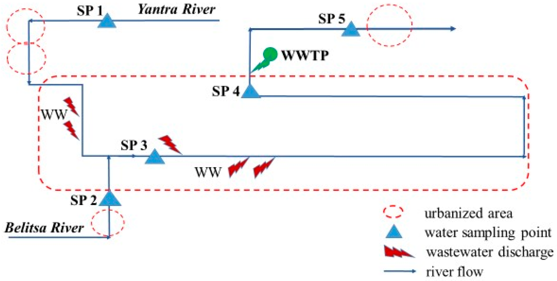
Five water sampling points (SPs) were selected to assess the current state of the river waters and the influence of the tributary and sources of fecal pollution—four of them were located on the Yantra River and one on the Belitsa River (Figure 1).
Figure 1.
Scheme of the studied section of the Yantra River and water sampling points (SPs). SPs located on the Yantra River: SP 1—upstream from two small villages and the town of Veliko Tarnovo (43.076222, 25.516889); SP 3—downstream from wastewater discharge of western industrial zone and confluence of the Belitsa River (bridge in Cholakovtsi residential quarter of Veliko Tarnovo; 43.061739, 25.603784); SP 4—after prolonged meandering through the urban territory of Veliko Tarnovo (bridge in Asenevtsi residential quarter; 43.085281, 25.649081); SP 5—far downstream from the city of Veliko Tarnovo, about 9 km after the inflow from the wastewater treatment plant (WWTP) (bridge upstream from the village of Samovodene; 43.136811, 25.613601). SP located on the Belitsa River: SP 2—downstream from the urban territory of Debelets (north bridge; 43.049500, 25.607300).
SP 1 is located upstream, away from settlements, and can be used as a reference point for water quality. SP 2 was chosen to assess the current state of the Belitsa River waters before its confluence with the Yantra River. SP 3 and SP 4 allow for monitoring of the Yantra River as it passes through the territory of the city of Veliko Tarnovo, as well as the impact of diffuse and point sources of pollution, the latter related to the discharge of domestic and industrial wastewater into the riverbed. The location of SP 5 a few kilometers downstream from the municipal WWTP effluents discharge allows for an assessment of whether there is any impact on river water quality.
2.2. Enumeration and Isolation of Enterococci
Water samples were analyzed for enterococci enumeration in accordance with EN ISO 7899-2:2003 [29] by membrane filtration and subsequent incubation of the membranes on Slanetz–Bartley agar (Merck KGaA, Darmstadt, Germany) at 35 °C for 24 h. Confirmation of enterococcal colonies was performed by transferring the membrane filter to a bile-esculin agar (Merck KGaA, Darmstadt, Germany) plate for 2–4 h. All esculin-metabolizing colonies were counted. Typical colonies were isolated and subcultured on brain heart infusion agar (HiMedia Laboratories Pvt. Ltd., Mumbai, India). Pure cultures were stored at −20 °C.
2.3. Determination of Antibiotic-Resistant Pattern
The antimicrobial susceptibility of enterococci isolates was tested by Kirby Bauer disk diffusion method (DDM) [30] for thirteen ABs from nine classes: beta-lactams (AMP 10 μg, ampicillin; IMI 10 μg, imipenem); aminoglycosides (HLG 30 μg, high-level gentamicin); macrolides (E 15 μg, erythromycin); glycopeptides (VA 5 μg, vancomycin; TEI 30 μg, teicoplanin); quinolones (CIP 5 μg, ciprofloxacin; LE 5 μg, levofloxacin); antifolates (COT 1.25/23.75 μg, trimethoprim/sulfamethoxazole, i.e., co-trimoxazole); tetracyclines (TE 30 μg, tetracycline; TGC 15 μg, tigecycline); amphenicols (C 30 μg, chloramphenicol); oxazolidinones (LZ 30 μg, linezolid).
Each strain was inoculated on the surface of a Mueller Hinton agar plate (HiMedia Laboratories Pvt. Ltd., Mumbai, India) and disks with the tested ABs (HiMedia Laboratories Pvt. Ltd., Mumbai, India) were placed on it. According to the diameter of the zone of inhibition around each AB disk tested, strains were classified as S—sensitive, R—resistant or I—susceptible at increased exposure [11,12]. Resistance to at least one AB from at least three different classes was defined as MAR.
MAR indexing was applied to each individual SP of the river water from which the tested enterococcal strains were isolated. The MAR index value was calculated as a/(b*c), where a is the total antibiotic resistance score of all isolates from the water of a given SP, b is the number of antibiotics tested and c is the number of isolates. An MAR index value exceeding 0.2 suggests high health and environmental hazards associated with the spread of MAR [31].
2.4. PCR Assays for Species Identification and Detection of ARGs
The genomic DNA of thirty enterococci strains with MAR patterns was extracted and purified using the GENE MATRIX Bacterial/Yeast Genomic DNA purification kit (EURx®Molecular Biology Products, Gdansk, Poland).
Species-specific multiplex PCR was performed for the identification of isolates. E. faecalis and E. faecium were specified using the sodA genes [31], E. mundtii using the mu gene, E. casseliflavus/flavescens using the vanC2/3 gene, and E. gallinarum using the vanC1 gene [32].
Qualitative PCR assays were performed to detect selected genes encoding resistance towards beta-lactams (blaTEM), macrolides (ermB), aminoglycosides (aac6′-2″aph), tetracyclines (tetM), glycopeptides (vanB, vanC), quinolones (emeA), amphenicols (cat), sulfonamides (sulI) and trimethoprim (dfrA) [15,32,33,34,35,36,37]. The characteristics of the primers used in PCR amplification of the target ARGs (synthesized by Eurofins Genomics, Ebersberg, Germany) are presented in Table S1 (Supplementary Materials).
The PCR amplifications using the onTaq PCR Master Mix 2x (EURx®Molecular Biology Products, Poland) were performed in a T100 Thermal Cycler (Bio-Rad Laboratories, Inc., Hercules, CA, USA), according to the manufacturer’s instructions of the Master Mix kit. The PCR products were visualized in 1.5% agarose gels in Tris–Acetate–EDTA solution.
2.5. Data Analysis
Chi-square test was used for categorical variables; p < 0.05 was considered statistically significant. Welch’s t-test was performed to assess the significance of differences between the sampling locations on ABR (measured as total number of AB classes to which phenotypic ABR was expressed) as the dependent variable and sampling location as the factor.
3. Results
3.1. Microbiological Characteristics of River Water
The ecological status of the Yantra River, in accordance with the EU Water Framework Directive 2000/60/EC [38], has been assessed as “moderate” for physicochemical and biological quality elements and specific pollutants, and as “good” for pollution by priority substances [39]. However, microbiological parameters are not included in the water quality assessment [38].
Despite the ‘moderate’ ecological quality of the river water, a large number of fecal enterococci was detected in the water sampled from all SPs (Table S2, Supplementary Materials). Water from SP 1 contained the lowest number of enterococci (2.0 ± 0.7 ×10² CFU/100 mL), and an increase of one order of magnitude was observed in water taken from SP 3, SP 4 and SP 5, with the values of 6.5 ± 7.0 ×10³ CFU/100 mL, 2.0 ± 1.9 ×10³ CFU/100 mL, 2.4 ± 2.2 ×10³ CFU/100 mL, respectively. In the waters of the Belitsa River (SP 2), the number of enterococci (1.2 ± 2.4 ×10⁴ CFU/100 mL) was found to be an order of magnitude higher compared to the Yantra River, with the increase compared to the reference SP 1 being two orders of magnitude higher.
The abundance of different Enterococcus species in river water depended on the sampling location. E. fecalis and E. faecium were found to predominate among isolates recovered from SP 3 and SP 4, whose waters were heavily affected by fecal contamination. E. mundtii and E. casselliflavus/flavescens were identified more frequently among yellowish colony-forming isolates from waters in SP 1, SP 2 and SP 5, located in rural or less urbanized areas. Also, a large number of Enterococcus strains remained unspecified.
3.2. Total ABR of Enterococci Isolated from the Tested River Section
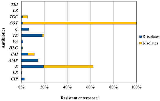
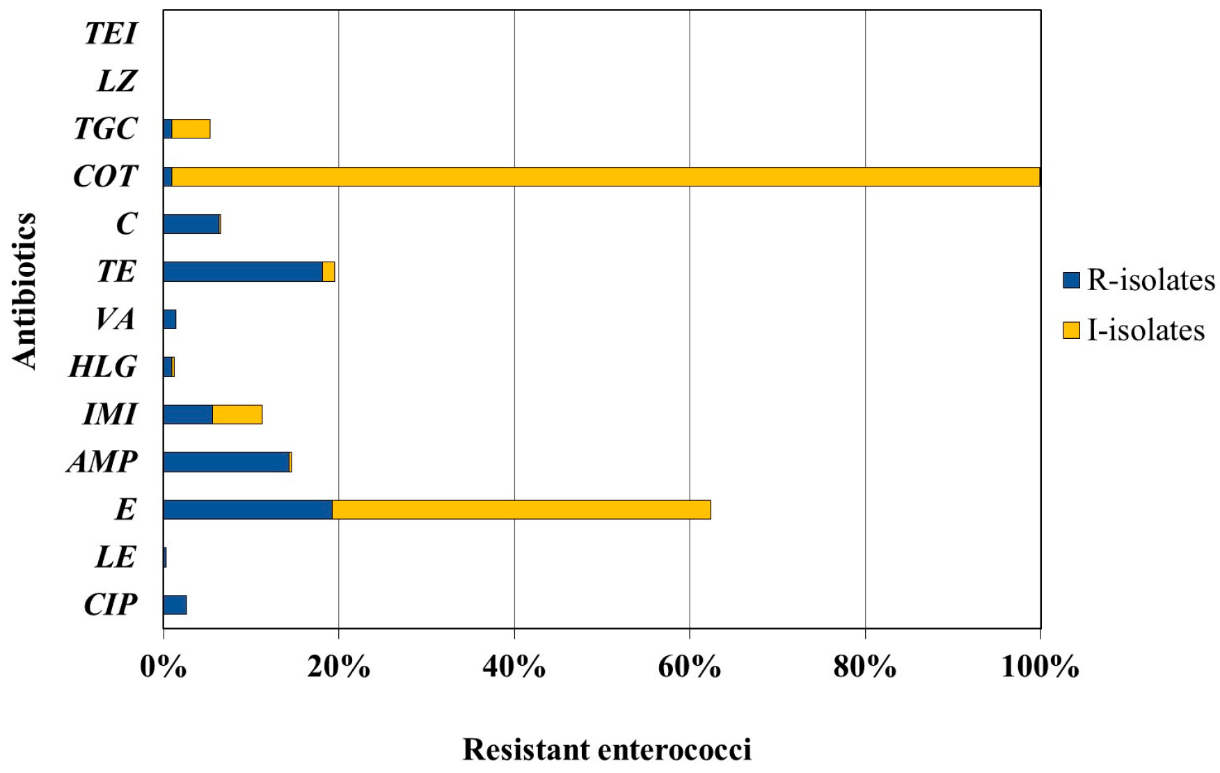
In total, 426 enterococci strains were isolated and their antibiotic resistance was determined. Among the isolates, 39% were found to be resistant to ABs. The most common resistances were to erythromycin (19%), tetracycline (18%), and ampicillin (14%) (Figure 2).
Figure 2.
ABR phenotype of enterococci strains isolated from the selected river stretch (R—resistant; I—susceptible at increased exposure); AMP—ampicillin; C—chloramphenicol; CIP—ciprofloxacin; COT—co-trimoxazole; E—erythromycin; HLG—high-level gentamicin; IMI—imipenem; LE—levofloxacin; LZ—linezolid; TE—tetracycline; TEI—teicoplanin; TGC—tygecycline; VA—vancomycin.
In the present study, 1% of isolates were found to be resistant to high levels of gentamicin, despite the known inherent resistance of enterococci to low levels of aminoglycosides. All tested strains showed intermediate level of resistance to co-trimoxazole, and only 1% of them were completely unsusceptible to its action. Enterococci resistance to the beta-lactams ampicillin and imipenem was 14% and 5%, respectively. Resistance to quinolones was less common—3% of strains were resistant to ciprofloxacin and only 0.3% to levofloxacin. Despite the high tetracycline resistance of the studied Enterococcus population, only 0.7% of the isolates were insensitive to tigecycline, a substance from the same group (which, however, was recently included in a separate class of glycylcyclines). Only 1.4% of the strains were resistant to vancomycin, but all of them were susceptible to teicoplanin. All strains were susceptible to linezolid.
3.3. ABR of Enterococci Depending on River Water Sampling Location
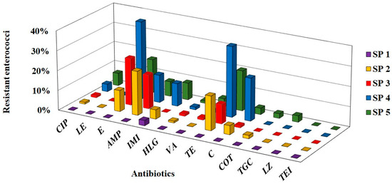
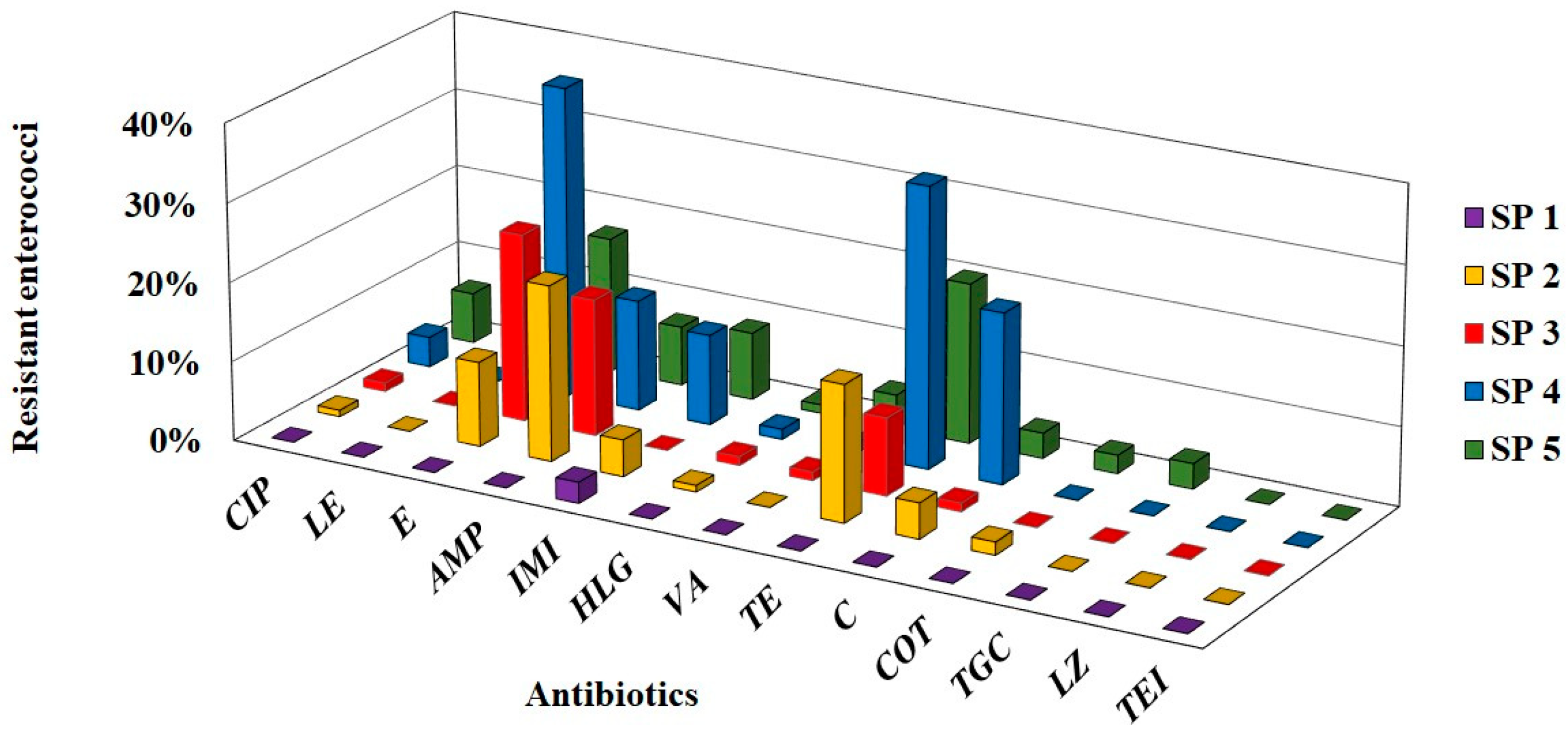
The obtained data (Table 1 and Figure 3) show a diverse, spatially variable level in the ABR of the enterococcal population in the waters of the Yantra River, thus reflecting the influence of the adjacent territories in the watershed.
Table 1.
ABR phenotype of isolated enterococci depending on the location of river water sampling.
Figure 3.
Proportion of R-resistant phenotypes among isolated enterococci depending on river water sampling points (SP). Antibiotics: AMP—ampicillin; C—chloramphenicol; CIP—ciprofloxacin; COT—co-trimoxazole; E—erythromycin; HLG—high-level gentamicin; IMI—imipenem; LE—levofloxacin; LZ—linezolid; TE—tetracycline; TEI—teicoplanin; TGC—tygecycline; VA—vancomycin.
Enterococci isolated from SP 1 were sensitive to all ABs tested, except for one strain resistant to IMI, with some of them demonstrating I-resistance to E. Isolates from the Belitsa River tributary (SP 2) showed a lower level of ABR compared to those isolated from the Yantra River, especially from SP 4 and SP 5, which reveals the influence of specific pollution sources in both river catchments. In contrast to SP 1, isolates from all other SPs were dominated by those resistant to E, TE and b-lactams, although the ABR values varied. The highest ABR levels were observed in SP 2 towards E (11%), TE (17%) and AMP (22%); in SP 3 towards E (24%), TE (10%) and AMP (19%); in SP 4 towards E (39%), TE (35%) and AMP (14%); and in SP 5 towards E (17%), TE (20%) and IMI (9%).
Isolates from SP 2 and SP 3 showed a higher level of resistance to AMP compared to those from the other SPs, which is probably related to the influence of livestock and poultry farming in the Belitsa River basin, and then its confluence into the Yantra River. Isolates from SP 4 had the highest level of resistance to C (22%), while those from SP 5 exhibited the highest level of resistance to CIP (6%). Resistance to COT was rare, found among enterococci from SP 2 and SP 5. Isolates from SP 5 exhibited more diverse ABR profiles than those from the rest SPs, and among them, resistance to ABs of the newer generation, such as VA and TGC, was detected.
The statistical comparison between the resistance of enterococci to individual ABs depending on the sampling point of the river water from which they were isolated showed the greatest differences in terms of resistance to E and TE. The level of resistance to E was statistically different (p < 0.05) in five out of six compared data pairs for the studied SPs, with a similarity only between SP 3 and SP 5. The level of TE-resistance is statistically different (p < 0.05) between four out of six compared combinations of data pairs for SPs, with a similarity of SP 2 separately with SP 3 or SP 5 (Supplementary Materials, Table S3). Insignificant differences were found between all SPs in the level of resistance to HLG, VA and TGC (p > 0.05), associated with the low frequency of enterococci resistant to these ABs.
When comparing all ABR data for each pair of the SPs studied, the greatest statistical similarity was found between enterococci isolated from SP 2 and SP 3, which differed only in the level of resistance to E. The variations in the ABR level in the individual SPs along the river was also verified by the calculated data on the average number of antibiotic classes to which waterborne enterococci expressed phenotypic resistance, with the values being, respectively, for SP 1—0.03 ± 0.16; for SP 2—0.61 ± 0.94; for SP 3—0.55 ± 0.76; for SP 4—1.27 ± 1.35; and for SP 5—0.66 ± 1.12. The statistical comparison between the above ABR values shows significant differences (p < 0.05) between the enterococci from SP 4 and all other SPs, as well as between the reference SP 1 and all other SPs in the river section. Insignificant differences were found between: SP 2 and SP 3 (p, 0.61), SP 2 and SP 5 (p, 0.73), and SP 3 and SP 5 (p, 0.43).
Data on the MAR index of riverine enterococci depending on the SP also show differences in the level of AMR for SP 1 and SP 4 compared to the other SPs in the river section. The calculated MAR index values for R-isolates from the water of the individual SPs were 0.003, 0.07, 0.06, 0.16 and 0.08, respectively, with the lowest value for SP1 and the highest for SP4.
3.4. MAR Phenotype of Enterococci Isolates
Forty strains out of 426 riverine enterococci tested exhibited a multidrug-resistant phenotype. Among the isolates from SP 1, no MAR phenotype was detected. Multidrug resistance was most common among enterococci isolated from SP 4 (25%), while among those from SP 2, SP 3, and SP 5 it was 7%, 3%, and 9%, respectively (Supplementary Materials, Table S4). Although MAR to three AB groups was predominant, resistance to four or five AB groups was found among isolates from the waters of SP 4 and SP 5, accounting for the passage of the Yantra River through the urban area of Veliko Tarnovo or the discharge of treated wastewater from the municipal WWTP. The MAR index of multidrug-resistant enterococci isolated from the individual SPs showed values ranging from 0.36 to 0.41.
Multidrug-resistant enterococci strains showed 16 diverse phenotypic patterns, with the highest level of resistance to E (85%), TE (95%) and C (65%), followed by that to the b-lactams AMP and IMI (38%). As might be expected, the highest rate was found for the E-TE-C phenotypic pattern, which manifested alone in 47.5% of MAR-isolates, and in another 10% of the isolates expressing an extended MAR pattern, including resistance to other AB classes (b-lactams and/or quinolones) (Supplementary Materials, Table S5). The E-TE-b-lact phenotype was expressed overall by 27.5% of isolates (alone in 7.5% of them or included in an extended MAR pattern in 20%). The E-TE-C and E-TE-b-lact patterns were mainly determined among MAR isolates from SP 4. Eight MAR patterns, which included rare resistance to COT, CIP, HLG, VA or TGC, were phenotypically expressed in only one isolate each, seven of which were recovered from SP 5.
3.5. Genotypic ABR of MAR Enterococci
In total, the genomic DNA of 30 enterococci strains expressing MAR phenotype was analyzed for ARGs most commonly found in clinical isolates, such as ermB, tetM, blaTEM, aac(6′)/aph(2″), vanB, etc. (Table 2). The species affiliation of the strains also was determined—15 of them were identified as E. faecium, 13 isolates belonged to E. faecalis, and two to the genus Enterococcus.
Table 2.
Genotypic characteristics of the studied MAR enterococci strains.
The genes encoding macrolide resistance (ermB), tetracycline resistance (tetM) and quinolone resistance (emeA) were common among the tested MAR isolates. The ermB gene was found in all MAR isolates, followed by tetM (77%) and emeA (77%), blaTEM (60%) and aac(6′)-aph(2″) (53%). All tested strains carried the sulI gene for sulfonamide resistance.
Some enterococci isolates harbored any of the tested ARGs without expressing the corresponding ABR phenotype. For example, the aac(6′)/aph(2″) gene was found in five strains that did not exhibit an ABR phenotype. Similarly, the quinolone efflux pump emeA gene was found in 18 strains without phenotypically manifested resistance to ciprofloxacin. The blaTEM gene was detected in 18 isolates, even though six of them were susceptible to ampicillin when analyzed by the disk diffusion method.
For other genes, such as tetM, vanB and cat, an opposite trend of an expressed ABR phenotype without detection of the target ARG was observed, as the resistance to the given AB is likely to be due to it carrying another gene from the same family of ARGs. For example, the vanB gene was not detected, despite the phenotypic resistance to vancomycin of six isolates detected by the disk diffusion method.
The frequency of carriage of the studied ARGs was different among isolates of the two enterococci species. Among E. faecium strains, the genes conferring resistance to beta-lactams, aminoglycosides and amphenicols were more often detected (respectively, 67%, 73% and 47%), while among E. faecalis ones the quinolone efflux gene emeA prevailed (92%). The carriage of the ermB, tetM, sulI and dfrA genes was at a similar frequency regardless of species affiliation. Despite differences in the carriage of individual ARGs among enterococcal isolates of both species, the statistical comparison between E. faecalis and E. faecium populations based on the total carriage of the studied ARGs showed an insignificant difference (p, 0.24). A statistical comparison between E. faecalis and E. faecium of the carriage of each individual ARG showed insignificant differences (p > 0.05), except for the cat gene (p < 0.04).
4. Discussion
Enterococci enter wastewater and environmental waters through feces of human and animals, and fecally contaminated water resources can pose a health risk due to the spread of feces-associated pathogens. Ubiquitous and adaptable, enterococci, along with other important pathogens, are the focus of antimicrobial resistance research in the One Health humans–animals–environment continuum [23,40,41]. In the present study, all enterococci isolated from the studied section of the Yantra River were analyzed to determine their ABR phenotype, and some of them for the encoding ARGs. The ABR study targeted the widely used ABs erythromycin, ampicillin, tetracycline, ciprofloxacin and sulfamethoxazole, but also newer ones, such as vancomycin, teicoplanin, tigecyclin and linezolid.
The abundance of enterococci in the waters of the Yantra River was generally high, indicating that in the studied section they suffered high levels of microbial pollution as a result of the river passing through settlements discharging untreated or incompletely treated sewage water, as well as through rural areas with diffuse fecal contamination. The increased content of enterococci in the water in all SPs compared to SP 1 suggests the possibility of an increased frequency of ABR, both among their population and in the entire aquatic community, due to the possibility of horizontal gene transfer. In the same river section, a high abundance of waterborne Enterobacteriaceae has already been observed, varying depending on the SP [28].
Different Enterococcus species were found in river waters—E. fecalis and E. faecium predominated among isolates from waters in SP 3 and SP 4, heavily affected by fecal pollution, while E. mundtii and E. casselliflavus/flavescens were more common in SP 1, SP 2 and SP 5, located in less urbanized areas. E. faecalis and E. faecium isolates demonstrated higher levels of resistance to the tested ABs, while E. mundtii and E. casselliflavus/flavescens strains were more often susceptible or resistant to one AB class.
Overall, 39% of isolated enterococci were found to be resistant to ABs, including 9% that were primarily multidrug-resistant to three antibiotic classes. The most common resistances were to erythromycin, tetracycline and ampicillin, due to their intensive use. It should be noted that macrolides, beta-lactams and tetracyclines were among the main drugs administered to farm animals, despite the many AB classes used in animal husbandry [42]. Our data are consistent with the trend of high levels of enterococci resistance to erythromycin and tetracycline identified for specific river catchments [21,43] or aquatic environments during large-scale observations within the One Health continuum [23,24,41], despite the differences found in the levels of resistance to ampicillin [41], ampicillin and chloramphenicol [40,44] or ciprofloxacin [23]. In contrast to the high levels of resistance to erythromycin and tetracycline, resistance to beta-lactams varied along the river. Resistance to imipenem was more common among isolates from SP 4 and SP 5, while ampicillin resistance was most common among isolates from the Belitsa River (SP 2) and those from SP 3, with the waters of the latter reflecting the influence of the tributary on the Yantra River. A similar trend of a higher proportion of ampicillin-resistant HPC bacteria in the waters of the Belitsa River (up to 21.8%) compared to the Yantra River (up to 10%) has already been reported for the same river section, as well as a high level of ampicillin-resistant Enterobacteriaceae isolates (36%) [28].
Resistance to ciprofloxacin was lower compared to that reported for other river bodies [21,23,43], with the highest value for isolates from SP 5. Despite the inherent resistance to gentamicin, very few enterococci strains were resistant to high levels of gentamicin. Resistance to co-trimoxazole followed a similar trend, with only 1% of strains being fully resistant and the rest exhibiting an I-phenotype.
In the present study, all enterococci isolates were susceptible to the newer ABs teicoplanin and linezolid, and three isolates were resistant to tigecycline. Susceptibility to vancomycin was also high, with only six strains of vancomycin-resistant enterococci (VRE) being recovered—one strain each from the water of SP 3 and SP 4 and four others from SP 5. These results are consistent with previously reported data on the low numbers of VRE isolates from surface waters [4,22] or epilithic biofilms [44], despite some opposing data [45]. There are data on vanA-harboring E. faecium that are phenotypically resistant to vancomycin and teicoplanin isolated from a Polish river [4], and VRE isolates expressing the vanB phenotype were reported for the Ebro River [26]. Three out of the six phenotypic VRE isolates were identified as E. faecalis, one as E. faecium, and two remained unidentified, and neither the vanB nor the vanC gene was detected. Although our study failed to determine which genes encoded vancomycin resistance, PCR assays were able to detect the vanC2/3 gene in E. casseliflavus/flavescens strains phenotypically susceptible to vancomycin—one isolated from SP 1, seven from SP 2, and 19 from SP 5. This corresponds to the finding of an inability of DDM to recognize enterococcal strains with reduced susceptibility to vancomycin [46,47]. However, E. casseliflavus and E. gallinarum with reduced susceptibility to vancomycin carrying vanC genes are often detected in environmental water [1,17,48]. Waterborne VRE strains screened using 4 μg/mL vancomycin-supplemented agar harboring the vanC1 and vanC2/3 genes have been identified as E. casseliflavus/gallinarum, and the vanC2/3 gene was also found in a small proportion of E. faecium or E. faecalis isolates [48].
The ABR patterns of river enterococci differed significantly from those established for clinical, food and environmental isolates in Bulgaria [10,16]; however, the observed similarities with wastewater enterococci [49] suggest a contribution of untreated wastewater entering the river to the exhibited ABR. E. faecalis and E. faecium isolates from the treated wastewater of urban WWTP (discharged into the studied river section) were also most frequently resistant to erythromycin (31%) and tetracycline (37%), but showed a lower level of resistance to ampicillin (4%) compared to riverine isolates [49].
Comparing the MAR phenotypes of riverine isolates and those from wastewater [49], some similarities in the prevailing MAR patterns were also found. Among riverine enterococci, the E-TE-C phenotype predominated found in 47.5% of isolates, followed by the E-TE-b-lact and E-TE-C-b-lact patterns (each in 7.5% of them), while among wastewater isolates, the E-TE-C and E-TE-b-lact-CIP-COT phenotypes predominated, each found in 21.4% of isolates, followed by the E-TE-b-lact and E-TE-CIP phenotypes, each in 14.3% of isolates. Also, the incidence of MAR isolates in the Yantra River water and wastewater amounted to 9% and 11.2%, respectively. However, the most prevalent ABR phenotype among clinical isolates from Bulgarian hospitals was the Amp-CIP-HLG phenotype (79%), and a high level of multidrug resistance (39%) was found [16]. In contrast, very low levels of ABR and pathogenicity were determined in food and environmental enterococci isolated from Bulgarian habitats, such as garden snail intestine, cow milk, yogurt, goat and cow feta cheese, street food, and human breast milk [10].
It should be noted that the prevalence of ABR among enterococci population in the water of the Yantra River, assessed by genes encoding resistance to clinically relevant ABs, reveals a significant anthropogenic impact. Minor differences in the carriage of the tested ARGs were found between E. faecalis and E. faecium isolates, with the most common ABR being encoded by the genes ermB for resistance to macrolides, tetM for resistance to tetracyclines and blaTEM for resistance to beta-lactams. The tetM gene was found to be dominant among non-clinical enterococci in Bulgaria, being detected in 31% of isolates, with the location linked to the Tn6009 transposon and the possession of genes for horizontal gene transfer [50]. Some of the genes, such as aac(6′)/aph(2″) and emeA, were detected in strains without an expressed ABR phenotype, which may be related to regulatory mechanisms, point mutations or other causes [16]. The reverse trend of an expressed ABR phenotype in the absence of any tested ARG, such as tetM, vanB and cat, suggests the presence of another gene conferring resistance to the respective AB.
Spatial Variations in ABR
Numerous studies have identified changes in the ABR level and pattern along the river under the influence of factors in the catchment area, such as landscape cover, agricultural activities, point or diffuse sources of fecal pollution [41,44,51]. The present data indicate that the waters of the Yantra River have undergone changes in the density and the level of ABR in the enterococcal population along the river course as a result of fecal pollution occurring downstream from SP 1. Enterococci isolates from individual SPs expressed a higher level of ABR and more diverse phenotypic profiles compared to those from SP 1. The spatial variations in ABR found in the studied river section support previously reported data on changing enterococcal population abundance and antibiotic resistance diversity along the river [26,27,52], with increasing gradients from upstream to downstream [1,13,51].
The dynamics of ABR in the studied river section suggests an influence of urbanization, land use and economic activities on water quality through emerging pollutants, such as antibiotic-resistant bacteria and AB residues. The extremely low level of antibiotic-resistant enterococci in the water of SP 1, located in a rural area with no nearby settlements, increased significantly in SP 3 and SP 4, located in highly urbanized areas, but also in the less urbanized areas of SP 2 and SP 5. Although enterococci isolated from all river water sampling sites were most frequently resistant to erythromycin and tetracycline, the ABR values varied significantly along the river, being statistically different for most of the SPs compared. Isolates from the individual SPs differed in the level of resistance to other ABs, with the highest resistance to chloramphenicol (22%) being found among isolates from SP 4, the highest resistance to ampicillin observed in those from SP 2 and SP 3 (22% and 19%, respectively), and the highest resistance to ciprofloxacin observed in SP 5 (6%). Isolates from SP 5 expressed resistance to vancomycin and tygecycline, which was not recorded among wastewater isolates from upstream WWTP [49], suggesting a stronger influence of the adjacent territories compared to wastewater discharge.
The highest levels of E. faecalis and E. faecium resistant to tetracycline, erythromycin, ampicillin and chloramphenicol were found in the urban area of Veliko Tarnovo. In the rural areas, influenced by agricultural runoffs or unsewered settlements, environmental enterococci such as E. mundtii and E. casseliflavus/flavescens with low ABR levels were often detected, but also E. faecium and E. faecium, which exhibited higher and more diverse ABR levels. The specific physico-chemical, hydraulic and biological characteristics of the river body and the water pollution sources contribute to the significant spatial variations in the populations of antibiotic-resistant bacteria detected in the studied river stretch. It should be noted that the spatial variations in the ABR level of enterococci recorded along the Yantra River reveal locally altered water quality due to the point or diffuse sources of pollution. The continuous impact of multiple sources of fecal pollution in the studied river section is probably the reason for the lack of significant differences in the ABR level and phenotypic ABR patterns of riverine enterococci and those from wastewater.
The present ABR data on riverine enterococci support our previous findings on the ABR levels of heterotrophic bacteria and Enterobacteriaceae in the same section of the Yantra River [28], and showed that all these parameters well describe the dynamics of the aquatic microbiome under the influence of humans and their activities. The ABR data on enterococci, together with those on aquatic HPC bacteria and Enterobacteriaceae, can be used to indicate changes in water quality in a given river catchment as a result of organic and/or fecal pollution, urbanization, and human activities. These parameters of the distribution of ABR in the waters of the Yantra River reveal the negative anthropogenic impact on the water body and that monitoring and control of these indicators is particularly important in the context of the One Health continuum and for sustainable water resources management.
5. Conclusions
This study assessed the prevalence of ABR among the waterborne enterococci population, taking into account the influence of the rural–urban landscape in the Yantra River section. Urbanization and human activities in the river’s catchment area have inevitably affected the river water quality, causing an increased content of total and antibiotic-resistant enterococci. Enterococci resistant to antibiotics (39%), including those with multiple resistance (9%), were found in the waters of the Yantra River, revealing the anthropogenic impact in the studied river section.
The prevalence of waterborne antibiotic-resistant enterococci highlights the need to monitor, control and manage surface water pollution, including the effectiveness of wastewater treatment, to limit anthropogenic impact through the discharge of incoming untreated or incompletely treated wastewater and to ensure the ecological well-being of the receiving water. Knowledge of the occurrence, abundance and fate of antibiotic-resistant bacteria and ARGs in natural waters will contribute to a better understanding and management of the health and environmental hazards caused by these emergent pollutants.
Supplementary Materials
The following supporting information can be downloaded at: https://www.mdpi.com/article/10.3390/w18101149/s1, Table S1. Primer sets used in PCR analyses; Table S2. Microbiological characteristics of the river water; Table S3. Statistical comparison by chi-square test between pairs of data for enterococci resistance to a given antibiotic; Table S4. Phenotypic ABR of the isolated enterococci depending on sampling point; Table S5. Incidence of MAR phenotypes among enterococci isolated from the individual SPs.
Author Contributions
Conceptualization, Z.T. and H.N.; methodology, Z.T.; investigation, R.B., T.C.K. and Z.T.; data curation, Z.T.; writing—original draft preparation, Z.T.; writing—review and editing, H.N.; project administration, Z.T.; funding acquisition, Z.T. All authors have read and agreed to the published version of the manuscript.
Funding
This research was funded by: (1) National Science Fund, Republic of Bulgaria, grant No. KΠ-06-H31/20 “Antibiotic resistant bacteria and antibiotic resistance genes in Bulgarian natural waters and waters under anthropogenic impact”; (2) Research, Innovation and Digitalization for Smart Transformation Program 2021–2027, grant No. BG 16RFPR002-1.014-0015. Center of Competence “Clean Technologies for a Sustainable Environment—Water, Waste, Energy for a Circular Economy” (Clean & Circle).
Data Availability Statement
Data is contained within the article or Supplementary Materials. Further inquiries can be directed to the corresponding author.
Conflicts of Interest
The authors declare no conflicts of interest. The funders had no role in the design of the study; in the collection, analyses, or interpretation of data; in the writing of the manuscript; or in the decision to publish the results.
Abbreviations
The following abbreviations are used in this manuscript:
| AB | Antibiotic |
| ABR | Antibiotic resistance |
| AMP | Ampicillin |
| ARG | Antibiotic resistance gene |
| C | Chloramphenicol |
| CIP | Ciprofloxacin |
| CFU | Colony form unit |
| CLSI | Clinical and Laboratory Standards Institute |
| COT | Co-trimoxazole (trimethoprim/sulfamethoxazole) |
| E | Erythromycin |
| EUCAST | European Committee on Antimicrobial Susceptibility Testing |
| HLG | High level gentamycin |
| HPC | Heterotrophic plate count |
| IMI | Imipenem |
| LE | Levofloxacin |
| LZ | Linezolid |
| MAR | Multidrug antibiotic resistance |
| p | Probability |
| SP | Sampling point |
| TE | Tetracycline |
| TEI | Teicoplanin |
| TGC | Tygecycline |
| VA | Vancomycin |
| WHO | World Health Organization |
| WW | Waste water |
| WWTP | Waste water treatment plant |
References
- Gotkowska-Płachta, A. The prevalence of virulent and multidrug-resistant enterococci in river water and in treated and untreated municipal and hospital wastewater. Int. J. Environ. Res. Public Health 2021, 18, 563. [Google Scholar] [CrossRef] [Scilit]
- Park, J.-H.; Bae, K.-S.; Kang, J.; Park, E.-R.; Yoon, J.-K. Comprehensive study of antibiotic resistance in Enterococcus spp.: Comparison of influents and effluents of wastewater treatment plants. Antibiotics 2024, 13, 1072. [Google Scholar] [CrossRef] [Scilit]
- Gotkowska-Płachta, A.; Gołas, I. The importance of enterococci in the monitoring of fecal pollution in river water in forests and urban areas. Water 2023, 15, 3708. [Google Scholar] [CrossRef] [Scilit]
- Hricová, K.; Röderová, M.; Fryčák, P.; Pauk, V.; Kurka, O.; Mezerová, K.; Štosová, T.; Bardoň, J.; Milde, D.; Kučová, P.; et al. Prevalence of vancomycin-resistant enterococci and antimicrobial residues in wastewater and surface water. Life 2021, 11, 1403. [Google Scholar] [CrossRef] [Scilit]
- Byappanahalli, M.N.; Nevers, M.B.; Korajkic, A.; Staley, Z.R.; Harwood, V.J. Enterococci in the environment. Microbio. Mol. Biol. Rev. 2012, 76, 685. [Google Scholar] [CrossRef] [Scilit] [PubMed]
- Davis, B.C.; Keenum, I.; Calarco, J.; Liguori, K.; Milligan, E.; Pruden, A.; Harwood, V.J. Towards the standardization of Enterococcus culture methods for waterborne antibiotic resistance monitoring: A critical review of trends across studies. Water Res. X 2022, 17, 100161. [Google Scholar] [CrossRef] [Scilit] [PubMed]
- Suzuki, Y.; Kanda, N.; Furukawa, T. Abundance of Enterococcus species, Enterococcus faecalis and Enterococcus faecium, essential indicators of fecal pollution, in river water. J. Environ. Sci. Health Part A 2012, 47, 1500–1505. [Google Scholar] [CrossRef] [Scilit]
- Krawczyk, B.; Wityk, P.; Gałecka, M.; Michalik, M. The many faces of Enterococcus spp.—Commensal, probiotic and opportunistic pathogen. Microorganisms 2021, 9, 1900. [Google Scholar] [CrossRef] [Scilit] [PubMed]
- Micallef, S.A.; Goldstein, R.E.R.; George, A.; Ewing, A.; Tall, B.D.; Boyer, M.S.; Joseph, S.W.; Sapkota, A.R. Diversity, distribution and antibiotic resistance of Enterococcus spp. recovered from tomatoes, leaves, water and soil on U.S. Mid-Atlantic farms. Food Microbiol. 2013, 36, 465–474. [Google Scholar] [CrossRef] [Scilit]
- Pandova, M.; Kizheva, Y.; Tsenova, M.; Rusinova, M.; Borisova, T.; Hristova, P. Pathogenic potential and antibiotic susceptibility: A comprehensive study of enterococci from different ecological settings. Pathogens 2024, 13, 36. [Google Scholar] [CrossRef] [Scilit]
- Clinical and Laboratory Standards Institute. CLSI M100 Performance Standards for Antimicrobial Susceptibility Testing, 27th ed.; CLSI: Berwyn, PA, USA, 2017; Available online: https://clsi.org/shop/standards/m100/ (accessed on 6 May 2026).
- European Committee on Antimicrobial Susceptibility Testing. Intrinsic Resistance and Unusual Phenotypes Version 3.3, 2021. Available online: https://www.eucast.org/fileadmin/src/media/PDFs/EUCAST_files/Expert_Rules/2021/Intrinsic_Resistance_and_Unusual_Phenotypes_Tables_v3.3_20211018.pdf (accessed on 10 April 2026).
- Cho, S.; Hiott, L.M.; McDonald, J.M.; Barrett, J.B.; McMillan, E.A.; House, S.L.; Adams, E.S.; Frye, J.G.; Jackson, C.R. Diversity and antimicrobial resistance of Enterococcus from the Upper Oconee Watershed, Georgia. J. Appl. Microbiol. 2020, 128, 1221–1233. [Google Scholar] [CrossRef] [Scilit]
- Diab, M.; Salem, D.; El-Shenawy, A.; El-Far, A.; Abdelghany, A.; Awad, A.R.; El Defrawy, I.; Shemis, M. Detection of high level aminoglycoside resistance genes among clinical isolates of Enterococcus species. Egypt. J. Med. Hum. Genet. 2019, 20, 28. [Google Scholar] [CrossRef] [Scilit]
- Jia, W.; Li, G.; Wang, W. Prevalence and antimicrobial resistance of Enterococcus species: A hospital-based study in China. Int. J. Environ. Res. Public Health 2014, 11, 3424–3442. [Google Scholar] [CrossRef] [Scilit] [PubMed]
- Yordanova, R.; Yaneva, Z.; Gencheva, D.; Beev, G. Antimicrobial resistance distribution and quorum-sensing regulation of enterococcal strains, isolated from hospitalized patients. Appl. Sci. 2022, 12, 8735. [Google Scholar] [CrossRef] [Scilit]
- Cho, S.; Jackson, C.R.; Frye, J.G. The prevalence and antimicrobial resistance phenotypes of Salmonella, Escherichia coli and Enterococcus spp. in surface water. Lett. Appl. Microbiol. 2020, 71, 3–25. [Google Scholar] [CrossRef] [Scilit]
- Lleò, M.M.; Bonato, B.; Benedetti, D.; Canepari, P. Survival of enterococcal species in aquatic environments. FEMS Microb. Ecol. 2005, 54, 189–196. [Google Scholar] [CrossRef] [Scilit] [PubMed]
- Monticelli, L.S.; Decembrini, F.; Bergamasco, A.; Caruso, G. Water quality assessment of transitional and coastal marine Sicilian waters (Italy): Ecological and epidemiological significance of multiple antimicrobial resistant Enterococcus spp. Estuar. Coast. Shelf Sci. 2019, 217, 173–184. [Google Scholar] [CrossRef] [Scilit]
- Tamai, S.; Suzuki, Y. Diversity of fecal indicator enterococci among different hosts: Importance to water contamination source tracking. Microorganisms 2023, 11, 2981. [Google Scholar] [CrossRef] [Scilit] [PubMed]
- Ibekwe, A.M.; Obayiuwana, A.C.; Murinda, S.E. Enterococcus species and their antimicrobial resistance in an urban watershed affected by different anthropogenic sources. Water 2024, 16, 116. [Google Scholar] [CrossRef] [Scilit]
- Alipour, M.; Hajiesmaili, R.; Talebjannat, M.; Yahyapour, Y. Identification and antimicrobial resistance of Enterococcus spp. isolated from the river and coastal waters in northern Iran. Hindawi. Sci. World J. 2014, 2014, 287458. [Google Scholar] [CrossRef] [Scilit]
- Zaheer, R.; Cook, S.R.; Barbieri, R.; Goji, N.; Cameron, A.; Petkau, A.; Polo, R.O.; Tymensen, L.; Stamm, C.; Song, J.; et al. Surveillance of Enterococcus spp. reveals distinct species and antimicrobial resistance diversity across a One-Health continuum. Sci. Rep. 2020, 10, 3937. [Google Scholar] [CrossRef] [Scilit]
- Zaidi, S.-e.-Z.; Zaheer, R.; Poulin-Laprade, D.; Scott, A.; Rehman, M.A.; Diarra, M.; Topp, E.; Domselaar, G.V.; Zovoilis, A.; McAllister, T.A. Comparative genomic analysis of enterococci across sectors of the One Health continuum. Microorganisms 2023, 11, 727. [Google Scholar] [CrossRef] [Scilit]
- Varela, A.R.; Ferro, G.; Vredenburg, J.; Yanık, M.; Vieira, L.; Rizzo, L.; Lameiras, C.; Manaia, C.M. Vancomycin resistant enterococci: From the hospital effluent to the urban wastewater treatment plant. Sci. Total Environ. 2013, 450–451, 155–161. [Google Scholar] [CrossRef] [Scilit]
- Pérez-Etayo, L.; González, D.; Leiva, J.; Vitas, A.I. Multidrug-resistant bacteria isolated from different aquatic environments in the north of Spain and south of France. Microorganisms 2020, 8, 1425. [Google Scholar] [CrossRef] [Scilit] [PubMed]
- Donchev, D.; Ivanov, I.N.; Stoikov, I.; Ivanova, M. Metagenomic investigation of the short-term temporal and spatial dynamics of the bacterial microbiome and the resistome downstream of a wastewater treatment plant in the Iskar river in Bulgaria. Microorganisms 2024, 12, 1250. [Google Scholar] [CrossRef] [Scilit]
- Tsvetanova, Z.; Najdenski, H. Antimicrobial resistance of heterotrophic bacteria and Enterobacteriaceae inhabiting an anthropogenic-affected river stretch in Bulgaria. Processes 2023, 11, 2792. [Google Scholar] [CrossRef] [Scilit]
- EN ISO 7899-2:2003; Water Quality—Detection and Enumeration of Intestinal Enterococci—Part 2: Membrane Filtration Method. ISO: Geneva, Switzerland, 2003.
- Bauer, A.W.; Kirby, W.M.; Sherris, J.C.; Turck, M. Antibiotic susceptibility testing by a standardized single disk method. Am. J. Clin. Pathol. 1966, 45, 493–496. [Google Scholar] [CrossRef] [Scilit]
- Krumperman, P.H. Multiple antibiotic resistance indexing of Escherichia coli to identify high-risk sources of fecal contamination of foods. Appl. Environ. Microbiol. 1983, 46, 165–170. [Google Scholar] [CrossRef] [Scilit]
- Jackson, C.R.; Fedorka-Cray, P.J.; Barrett, J.B. Use of a genus- and species-specific multiplex PCR for identification of enterococci. J. Clin. Microbiol. 2004, 42, 3558–3565. [Google Scholar] [CrossRef] [Scilit]
- Dutka-Malen, S.; Evers, S.; Courvalin, P. Detection of glycopeptide resistance genotypes and identification to the species level of clinically relevant enterococci by PCR. J. Clin. Microbiol. 1995, 33, 24–27. [Google Scholar] [CrossRef] [Scilit] [PubMed]
- Böckelmann, U.; Dörries, H.; Ayuso-Gabella, M.N.; Salgot de Marçay, M.; Tandoi, V.; Levantesi, C.; Masciopinto, C.; Van Houtte, E.; Szewzyk, U.; Wintgens, T.; et al. Quantitative PCR monitoring of antibiotic resistance genes and bacterial pathogens in three European artificial groundwater recharge systems. Appl. Environ. Microbiol. 2009, 75, 154–163. [Google Scholar] [CrossRef] [Scilit]
- Destiani, R.; Templeton, M.R. The antibiotic resistance of heterotrophic bacteria in tap waters in London. Water Supply 2019, 19, 179–190. [Google Scholar] [CrossRef] [Scilit]
- Pei, R.; Kim, S.C.; Carlson, K.H.; Pruden, A. Effect of river landscape on the sediment concentrations of antibiotics and corresponding antibiotic resistance genes (ARG). Water Res. 2006, 4, 2427–2435. [Google Scholar] [CrossRef] [Scilit] [PubMed]
- Xi, C.; Zhang, Y.; Marrs, C.F.; Ye, W.; Simon, C.; Foxman, B.; Nriagu, J. Prevalence of antibiotic resistance in drinking water treatment and distribution systems. Appl. Environ. Microbiol. 2009, 75, 5714–5718. [Google Scholar] [CrossRef] [Scilit] [PubMed]
- Commission of the European Community. Directive 2000/60/EC of the European Parliament and of the Council of 23 October 2000 Establishing a Framework for Community Action in the Field of Water Policy. Available online: http://data.europa.eu/eli/dir/2000/60/oj (accessed on 6 May 2026).
- Ministry of Environment and Water (MOEW). Regional Environment and Water Inspectorate Veliko Tarnovo. Annual Report on the State of the Environment for 2020. Available online: https://www.riosvt.org/wp-content/uploads/2025/05/RIOSV-VT-reg.doklad_2024-1.pdf (accessed on 6 May 2026). (In Bulgarian)
- Hasanpour, F.; Khademi, F.; Ghalehbin, B.M.; Kheljan, M.N.; Jannati, E.; Iranpour, S.; Arzanlou, M. A comprehensive One Health investigation of erythromycin and quinupristin/dalfopristin resistant Enterococcus spp. in Iran. J. Water Health 2025, 23, 439–449. [Google Scholar] [CrossRef] [Scilit]
- Leclercq, R.; Oberlé, K.; Galopin, S.; Cattoir, V.; Budzinski, H.; Petit, F. Changes in enterococcal populations and related antibiotic resistance along a medical center-wastewater treatment plant-river continuum. Appl. Environ. Microbiol. 2013, 79, 2428–2434. [Google Scholar] [CrossRef] [Scilit] [PubMed]
- Czatzkowska, M.; Wolak, I.; Harnisz, M.; Korzeniewska, E.E. Impact of anthropogenic activities on the dissemination of ARGs in the environment—A review. Int. J. Environ. Res. Public Health 2022, 19, 12853. [Google Scholar] [CrossRef] [Scilit]
- Łuczkiewicz, A.; Jankowska, K.; Kurlenda, J.; Olańczuk-Neyman, K. Identification and antimicrobial resistance of Enterococcus spp. isolated from surface water. Water Sci. Technol. 2010, 62, 466–473. [Google Scholar] [CrossRef] [Scilit]
- Barbosa-Vasconcelos, A.; Mendes, Â.; Martins, F.; Lopes, E.; Machado, A.; Bordalo, A.A.; Vaz-Pires, P.; Vieira, N.; da Costa, P.M.; Bessa, L.J. River water analysis using a multiparametric approach: Portuguese river as a case study. J. Water Health 2018, 16, 991–1006. [Google Scholar] [CrossRef] [Scilit]
- Molale, L.G.; Bezuidenhout, C.C. Antibiotic resistance, efflux pump genes and virulence determinants in Enterococcus spp. from surface water systems. Environ. Sci. Pollut. Res. 2016, 23, 21501–21510. [Google Scholar] [CrossRef] [Scilit]
- Adhikari, L. High-level aminoglycosides resistance and reduced susceptibility to vancomycin in nosocomial enterococci. J. Glob. Infect. Dis. 2010, 2, 231–235. [Google Scholar] [CrossRef] [Scilit] [PubMed]
- Monticelli, J.; Knezevich, A.; Luzzati, R.; Di Bella, S. Clinical management of non-faecium non-faecalis vancomycin-resistant enterococci infection. Focus on Enterococcus gallinarum and Enterococcus casseliflavus/flavescens. J. Inf. Chemoth. 2018, 24, 237–246. [Google Scholar] [CrossRef] [Scilit]
- Nishiyama, M.; Iguchi, A.; Suzuki, Y. Identification of Enterococcus faecium and Enterococcus faecalis as vanC-type vancomycin-resistant enterococci (VRE) from sewage and river water in the provincial city of Miyazaki, Japan. J. Environ. Sci. Health Part A 2014, 50, 16–25. [Google Scholar] [CrossRef] [Scilit]
- Tsvetanova, Z.; Boshnakov, R. Antimicrobial resistance of waste water microbiome in an urban waste water treatment plant. Water 2025, 17, 39. [Google Scholar] [CrossRef] [Scilit]
- Pandova, M.; Kizheva, Y.; Hristova, P. Relationship between CRISPR-Cas systems and acquisition of tetracycline resistance in non-clinical Enterococcus populations in Bulgaria. Antibiotics 2025, 14, 145. [Google Scholar] [CrossRef] [Scilit] [PubMed]
- Lata, P.; Ram, S.; Agrawal, M.; Shanker, R. Enterococci in river Ganga surface waters: Propensity of species distribution, dissemination of antimicrobial-resistance and virulence-markers among species along landscape. BMC Microbiol. 2009, 9, 140. [Google Scholar] [CrossRef] [Scilit] [PubMed]
- Solaiman, S.; Patterson, R.; Davey, K.; Katz, Y.; Payne-Sturges, D.; Sapkota, A.R.; Micallef, S.A. Effects of season and water type on the distribution and antimicrobial resistance of Enterococcus faecalis and Ent. faecium from surface and reclaimed water. J. Appl. Microbiol. 2022, 133, 477–487. [Google Scholar] [CrossRef] [Scilit]

Disclaimer/Publisher’s Note: The statements, opinions and data contained in all publications are solely those of the individual author(s) and contributor(s) and not of MDPI and/or the editor(s). MDPI and/or the editor(s) disclaim responsibility for any injury to people or property resulting from any ideas, methods, instructions or products referred to in the content. |
© 2026 by the authors. Licensee MDPI, Basel, Switzerland. This article is an open access article distributed under the terms and conditions of the Creative Commons Attribution (CC BY) license.