


Abstract
This study investigated the effects of a high-fat diet (HFD), particulate matter (PM) exposure, and resistance exercise training on circulating lipid profiles, adipokines, inflammatory responses, neurotrophic factors, and blood–brain barrier (BBB) permeability. Forty-eight 10-week-old male C57BL/6 mice were randomly assigned to four groups (n = 12 per group): normal diet (ND), HFD, HFD with PM exposure (HFD + PM), and HFD with PM exposure plus exercise training (HFD + PM + EX). ND and HFD were administered for 16 weeks, whereas PM exposure and exercise training interventions were initiated after 8 weeks of dietary treatment and continued for an additional 8 weeks. PM was administered via tail vein injection three times per week, and resistance exercise training consisted of a ladder-climbing exercise performed five times per week. The results indicated that body weight, total cholesterol (TC), triglycerides (TGs), low-density lipoprotein cholesterol (LDL-C), leptin, interleukin-6 (IL-6), tumor necrosis factor-α (TNF-α), S100 calcium-binding protein B (S100B), and neuron-specific enolase (NSE) levels were significantly higher in the HFD group than in the ND group (p < 0.05), whereas adiponectin and brain-derived neurotrophic factor (BDNF) levels were significantly lower (p < 0.05). In addition, the HFD + PM group exhibited significantly lower BDNF and vascular endothelial growth factor (VEGF) levels (p < 0.05) and significantly higher S100B and NSE levels (p < 0.05) than the HFD group. In contrast, the HFD + PM + EX group showed significantly lower TG, LDL-C, leptin, and IL-6 levels than the HFD group (p < 0.05). Moreover, compared with the HFD + PM group, the HFD + PM + EX group demonstrated significantly lower TG, LDL-C, leptin, S100B, and NSE levels (p < 0.05) and significantly higher high-density lipoprotein cholesterol (HDL-C), adiponectin, BDNF, and VEGF levels (p < 0.05). Collectively, these findings suggest that an HFD may contribute to dyslipidemia, heightened inflammatory responses, downregulation of neurotrophic factors, and increased BBB permeability and that concurrent PM exposure under HFD conditions may exacerbate adverse alterations in neurotrophic factors and BBB permeability. The results indicate that an HFD induces metabolic and neurovascular alterations, whereas concurrent PM exposure under HFD conditions is associated with additional changes in neurotrophic factors and BBB-related markers. Resistance exercise training attenuated these changes.
1. Introduction
Particulate matter (PM) is a major environmental pollutant with well-documented adverse effects on human health. In addition to its established associations with cardiovascular and metabolic disorders, increasing evidence indicates that PM exposure is linked to impairments in central nervous system (CNS) function [1,2]. Previous experimental and epidemiological studies have suggested that prolonged PM exposure promotes systemic oxidative stress and chronic low-grade inflammation, which may disrupt vascular homeostasis and compromise blood–brain barrier (BBB) integrity, contributing to neurovascular dysfunction [3,4]. Importantly, these adverse effects appear to be more pronounced in individuals with pre-existing metabolic abnormalities, such as obesity and dyslipidemia [5,6].
A high-fat diet (HFD) is widely used in experimental animal models to induce obesity and metabolic disorders and is known to promote insulin resistance, dyslipidemia, and chronic inflammatory responses [7,8]. In addition to peripheral metabolic disturbances, HFD-induced metabolic stress has been shown to negatively influence brain health by reducing neurotrophic support, impairing vascular regulation, and increasing BBB permeability [9,10]. Therefore, metabolic dysregulation associated with an HFD may increase the vulnerability of the neurovascular system to environmental stressors, including PM exposure [11,12]. However, whether PM exposure exacerbates neurovascular dysfunction under HFD conditions remains unclear.
HFD-induced metabolic dysregulation is closely associated with systemic inflammation and alterations in adipose tissue-derived signaling, which may contribute to downstream changes in neurotrophic factor regulation and BBB integrity. In addition, environmental stressors, such as PM exposure, have been suggested to exacerbate neurovascular vulnerability, particularly under metabolically compromised conditions [5,6,11,12]. Accordingly, the present study simultaneously assessed the lipid profiles, adipokines, inflammatory markers, neurotrophic factors, and BBB-related markers to capture the interconnected metabolic, inflammatory, and neurovascular responses to combined dietary and environmental challenges.
Neurotrophic factors, including brain-derived neurotrophic factor (BDNF) and vascular endothelial growth factor (VEGF), play essential roles in neuronal plasticity, cerebrovascular regulation, and maintenance of BBB stability [13,14]. In contrast, circulating markers such as S100 calcium-binding protein B (S100B) and neuron-specific enolase (NSE) are commonly used as peripheral indicators of BBB disruption and neurovascular injury [15,16]. Despite their physiological relevance, studies simultaneously evaluating metabolic parameters, inflammatory responses, neurotrophic factors, and BBB-related markers under combined HFD and PM exposure are limited.
Exercise training is a well-established non-pharmacological strategy for improving metabolic health and attenuating systemic inflammation [17,18]. In particular, resistance exercise contributes to the regulation of lipid metabolism and adipokine secretion through the maintenance and enhancement of skeletal muscle mass and has been shown to improve vascular function [19,20]. Emerging evidence suggests that exercise may exert protective effects on the CNS by enhancing the availability of neurotrophic factors and supporting BBB integrity [21,22]. Experimental studies have demonstrated increases in BDNF and VEGF following resistance exercise interventions, and resistance exercise has been proposed as a potential strategy to preserve BBB function and mitigate cerebrovascular dysfunction [21,22]. Nevertheless, the extent to which resistance exercise modulates neurovascular alterations under combined HFD and PM exposure remains unclear.
Therefore, the present study aimed to comprehensively investigate the effects of HFD, PM exposure, and resistance exercise training on metabolic parameters, adipokines, inflammatory responses, neurotrophic factors, and BBB permeability-related markers. We hypothesized that HFD would induce metabolic and neurovascular dysfunction, that PM exposure under HFD conditions would be primarily associated with additional alterations in neurotrophic factors and BBB-related markers, and that resistance exercise training would attenuate these adverse changes.
2. Methods
2.1. Animals and Housing Conditions
Forty-eight male C57BL/6 mice were obtained from a licensed commercial breeder (Samtako, Osan, Gyeonggi-do, Republic of Korea) and housed in groups of four per cage. All animals were maintained in a controlled laboratory animal facility equipped with a high-efficiency particulate air (HEPA) filtration system and a positive-pressure ventilation system to minimize the inflow of external contaminants. Environmental conditions were maintained at a constant temperature of 22 ± 2 °C and relative humidity of 55% ± 5%, under a 12 h light/12 h dark cycle. All the experimental procedures were conducted in accordance with the guidelines approved by the National Research Foundation of Korea (NRF-2019R1F1A1064296).
2.2. Experimental Design and Dietary Intervention
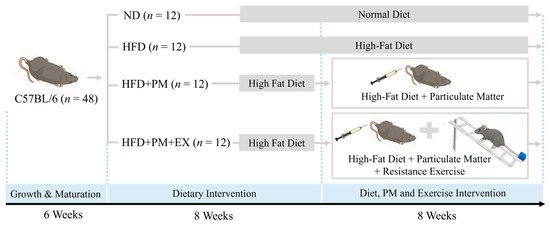
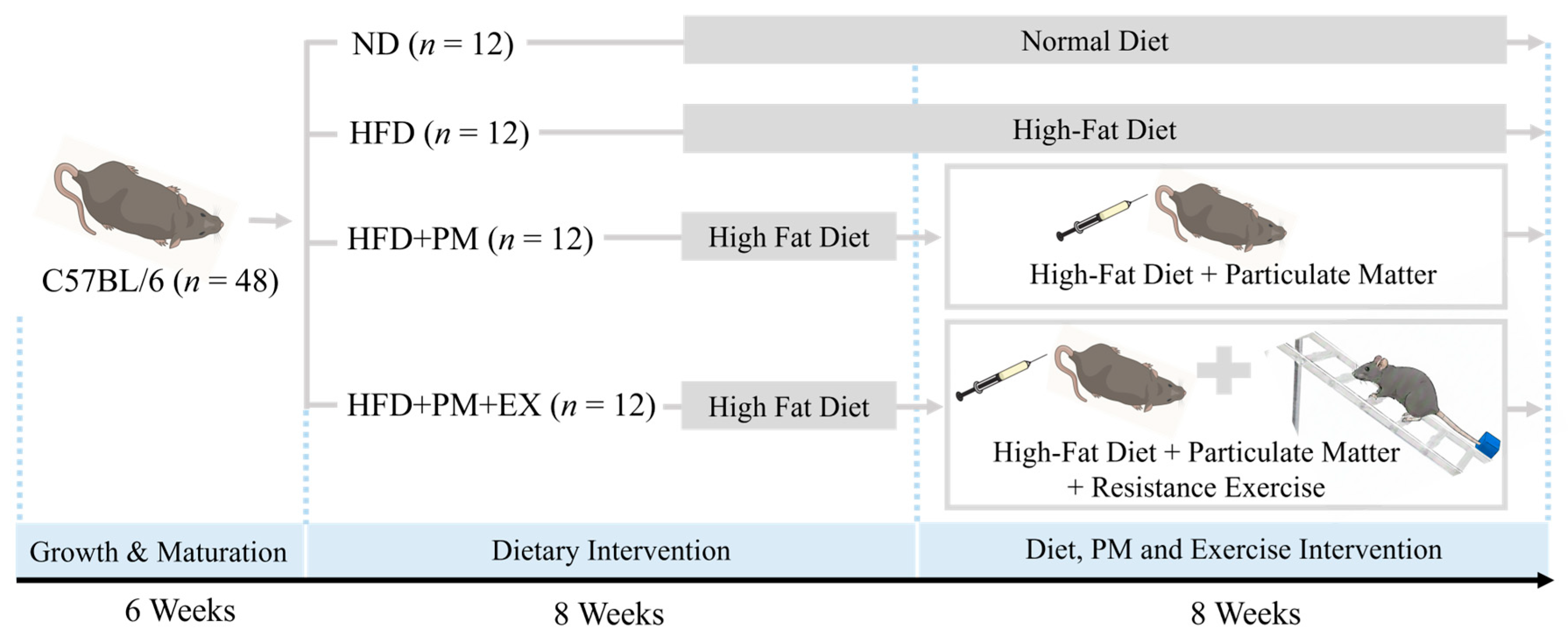
Male C57BL/6 mice were introduced at 4 weeks of age and maintained until 10 weeks of age to allow normal growth and maturation before experimental interventions. At 10 weeks of age, the animals were randomly allocated to four experimental groups according to diet, PM exposure, and exercise training status (n = 12 per group): normal diet (ND), high-fat diet (HFD), high-fat diet with PM exposure (HFD + PM), and high-fat diet with PM exposure combined with resistance exercise training (HFD + PM + EX).
Dietary interventions were continued for a total duration of 16 weeks. The ND consisted of 69.4% carbohydrates, 24.3% proteins, and 6.3% fats, whereas the HFD consisted of 35.0% carbohydrates, 20.0% proteins, and 45.0% fats. All the animals had ad libitum access to food and water throughout the experimental period. PM exposure and resistance exercise training interventions were initiated after the first eight weeks of dietary treatment and were subsequently conducted concurrently for an additional eight weeks (Figure 1).
Figure 1.
Experimental design and timeline of the study.
2.3. PM Exposure Protocol
The PM used in this study was a certified reference material with physicochemical characteristics comparable to those of ambient PM10, specifically fine dust (PM10-like; ERM-CZ120). The material was obtained from European Reference Materials and supplied by Sigma-Aldrich (St. Louis, MO, USA). PM exposure was performed three times per week over an 8-week period. Based on a previously described protocol [23], the PM suspension was prepared by dissolving the particles in sterile saline at a concentration of 15 μg per 200 μL. The suspension was administered via tail vein injection at a final dosage of 0.5 μg per gram of body weight. Mice in the ND group received an equivalent volume (200 μL) of sterile saline via tail vein injection following the same schedule.
2.4. Resistance Exercise Training Protocol
The resistance exercise was implemented using a ladder-climbing paradigm adapted from previously validated experimental models [24]. The exercise apparatus consisted of a vertically oriented ladder inclined at 80°, measuring 110 cm in height and 18 cm in width, with rungs evenly spaced at 2 cm intervals. Before the initiation of the formal training program, the mice completed a 3 d acclimation period to familiarize them with the climbing task. During this phase, the animals climbed the ladder without external loading, completing three sessions per day. To facilitate progressive motor learning, mice were initially placed 35 cm below the top platform, followed by placement at the midpoint and subsequently at the bottom of the ladder in later trials. After the first climb in each session, a rest period of 60 s was provided in a chamber located at the top of the ladder.
Following familiarization, resistance exercise training was performed over an 8-week period, five days per week. The training intensity was set relative to each animal’s capacity and corresponded to 50% of the normalized maximal load in accordance with established protocols [24]. Each training session consisted of 15 ladder climbs with a standardized rest interval of 1 min between successive climbs. The maximal carrying capacity was determined by progressively attaching weights to the base of the tail, starting at 75% of the animal’s body weight, and identifying the heaviest load that could be successfully transported to the top of the ladder. This maximal-load assessment was repeated at 2-week intervals to allow adjustment of the training load and maintenance of the relative exercise intensity throughout the intervention period.
2.5. Sample Collection and Biochemical Analyses
Blood collection was performed following deep anesthesia induced by ethyl ether. Approximately 5 mL of whole blood was collected from the inferior vena cava and immediately processed for serum isolation. Samples were centrifuged at 10,000× g for 10 min at 4 °C, after which the supernatant serum was aliquoted and preserved at −80 °C until biochemical analyses were conducted.
Serum lipid profiles, including triglyceride (TG), total cholesterol (TC), and high-density lipoprotein cholesterol (HDL-C) levels, were determined using commercially available enzymatic assay kits (TG: AM157S-K; TC: AM202-K; HDL-C: AM203-K; Asan Pharmaceutical Co., Seoul, Korea). Low-density lipoprotein cholesterol (LDL-C) concentrations were estimated using the Friedewald formula: LDL-C = TC − (HDL-C + TG/5).
Circulating levels of inflammatory cytokines interleukin-6 (IL-6; DY406, R&D Systems, Minneapolis, MN, USA) and tumor necrosis factor-α (TNF-α; DY410, R&D Systems), adipokines leptin (MOB00B, R&D Systems) and adiponectin (MRP300, R&D Systems), neurotrophic factors BDNF (DY248, R&D Systems) and VEGF (MMV00, R&D Systems), and BBB-related markers S100B (CSB-EL020643MO, CUSABIO, Wuhan, China) and NSE (CSB-E07962m, CUSABIO) were measured using enzyme-linked immunosorbent assay (ELISA) kits in accordance with the manufacturers’ protocols. For ELISA, serum samples (100 μL) were loaded into microplate wells and incubated under controlled conditions. Following sequential washing, detection antibodies and horseradish peroxidase-conjugated streptavidin were applied. Color development was achieved using a TMB substrate solution (R&D Systems, Minneapolis, MN, USA) and terminated with a stop solution. The optical density was measured at 450 nm using a microplate reader (Infinite M200, Tecan Austria GmbH, Grödig, Austria).
2.6. Statistical Analysis
Statistical analyses were conducted using SPSS Statistics software (version 29.0; IBM Corp., Armonk, NY, USA). All quantitative data are expressed as mean ± standard deviation (SD). Differences among the experimental groups were evaluated using one-way analysis of variance (ANOVA). Statistical significance was defined a priori as a two-sided p-value of <0.05.
3. Results
3.1. Effects on Body Weight
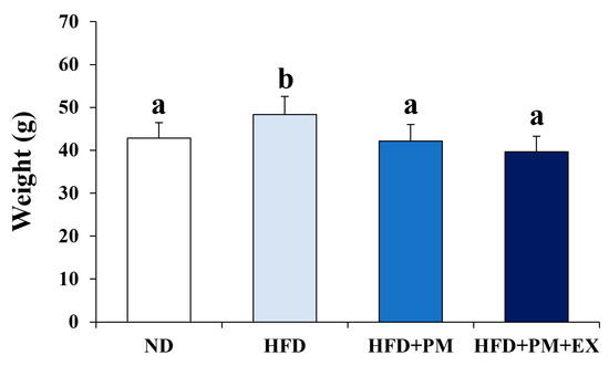
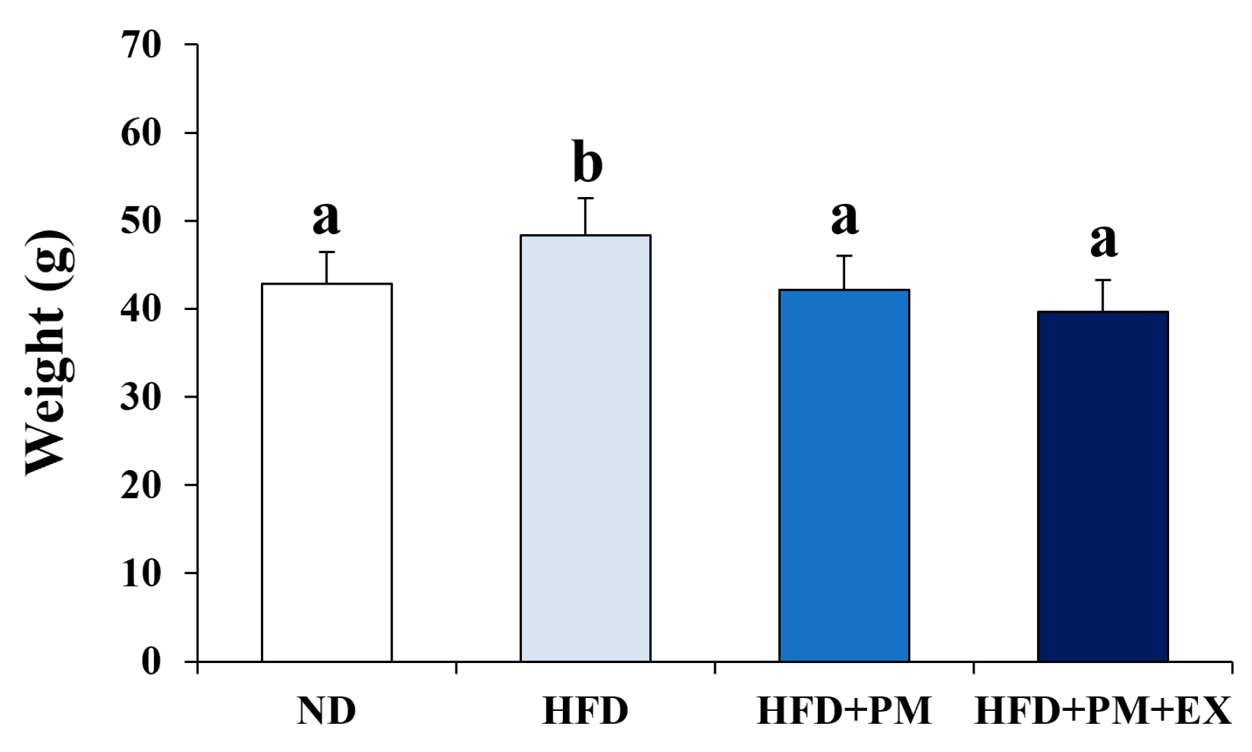
Figure 2 illustrates the effects of the HFD, PM exposure, and resistance exercise training on the final body weight. One-way ANOVA revealed significant differences in final body weight among the four groups (F(3, 44) = 11.161, p < 0.001). Final body weights were 42.83 ± 3.61 g in the ND group, 48.38 ± 4.17 g in the HFD group, 42.15 ± 3.85 g in the HFD + PM group, and 39.67 ± 3.59 g in the HFD + PM + EX group.
Figure 2.
Final body weight across experimental groups following dietary intervention, PM exposure, and resistance exercise training. Data are presented as mean ± SD. Different letters above the bars indicate statistically significant differences between groups (p < 0.05).
Bonferroni-adjusted post hoc comparisons revealed that the HFD group exhibited significantly higher body weight compared with the ND group (p = 0.005), as well as compared with the HFD + PM group (p = 0.001) and the HFD + PM + EX group (p < 0.001). In contrast, no significant differences in body weight were observed between the ND and HFD + PM groups (p = 1.000), between the ND and HFD + PM + EX groups (p = 0.288), or between the HFD + PM and HFD + PM + EX groups (p = 0.707).
3.2. Effects on Lipid Profiles
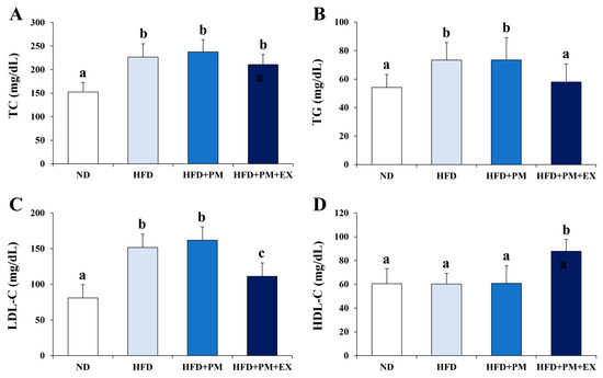
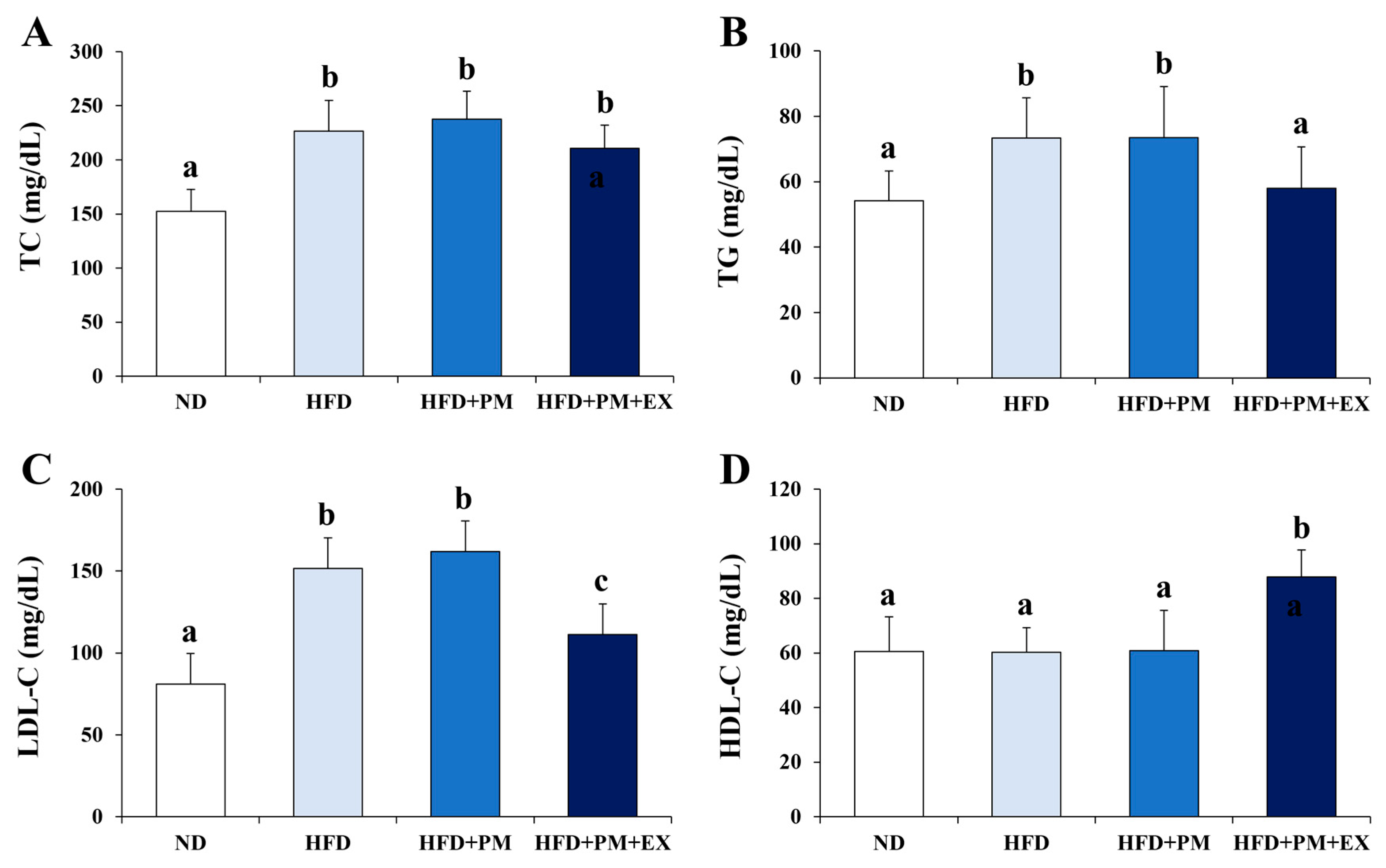
The effects of the HFD, PM exposure, and resistance exercise training on serum lipid profiles are presented in Figure 3. One-way ANOVA revealed significant group differences in TC, TG, LDL-C, and HDL-C levels (TC: F(3, 44) = 29.276; TG: F(3, 44) = 7.780; LDL-C: F(3, 44) = 24.108; HDL-C: F(3, 44) = 16.034; all p < 0.001).
Figure 3.
Serum lipid profile parameters ((A) TC, (B) TG, (C) LDL-C, (D) HDL-C) measured after HFD, PM exposure, and resistance exercise training. Values represent mean ± SD. Different letters above the bars indicate statistically significant differences between groups (p < 0.05).
Post hoc analysis demonstrated that TC, TG, and LDL-C levels were significantly higher in both the HFD and HFD + PM groups compared with the ND group (all p < 0.01), whereas no significant differences in HDL-C levels were observed between these groups (p > 0.05). In contrast, the HFD + PM + EX group exhibited significantly lower TG and LDL-C levels compared with both the HFD and HFD + PM groups (p < 0.05), along with significantly higher HDL-C levels compared with all other groups (p < 0.001). TC levels in the HFD + PM + EX group remained significantly higher than those in the ND group (p < 0.001), but did not differ significantly from those in the HFD or HFD + PM groups (p > 0.05).
3.3. Adipokine Responses
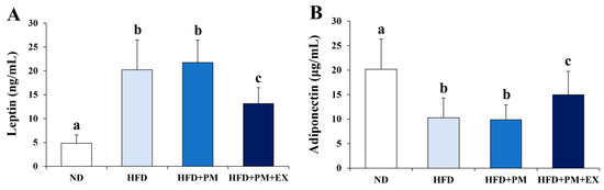
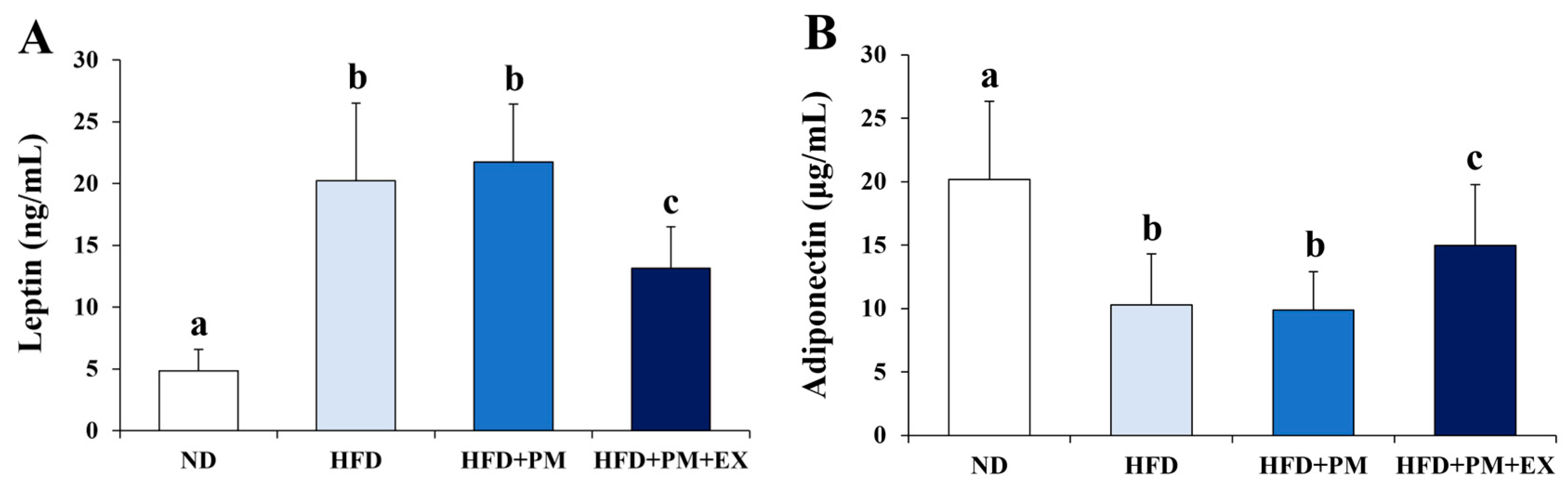
The responses of the serum leptin and adiponectin levels to the HFD, PM exposure, and resistance exercise training are presented in Figure 4. One-way ANOVA revealed significant group differences in leptin and adiponectin levels (leptin: F(3, 44) = 38.076, p < 0.001; adiponectin: F(3, 44) = 13.002, p < 0.001).
Figure 4.
Circulating adipokine concentrations, including leptin (A) and adiponectin (B), in response to dietary condition, PM exposure, and exercise intervention. Results are expressed as mean ± SD. Different letters above the bars indicate statistically significant differences between groups (p < 0.05).
Serum leptin levels were significantly elevated in the HFD and HFD + PM groups compared with the ND group (both p < 0.001). Although leptin levels in the HFD + PM + EX group were significantly lower than those in the HFD and HFD + PM groups (p = 0.001 and p < 0.001, respectively), they remained significantly higher than those in the ND group (p < 0.001). No significant difference in leptin level was observed between the HFD and HFD + PM groups (p = 1.000).
Serum adiponectin levels were significantly reduced in the HFD and HFD + PM groups compared with the ND group (both p < 0.001). Although adiponectin levels in the HFD + PM + EX group showed an increasing trend compared with the HFD and HFD + PM groups, these differences did not reach statistical significance (p = 0.101 and p = 0.060, respectively). No significant difference was observed between the ND and HFD + PM + EX groups (p = 0.052).
3.4. Inflammatory Marker Responses
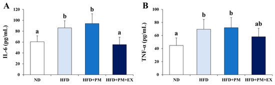
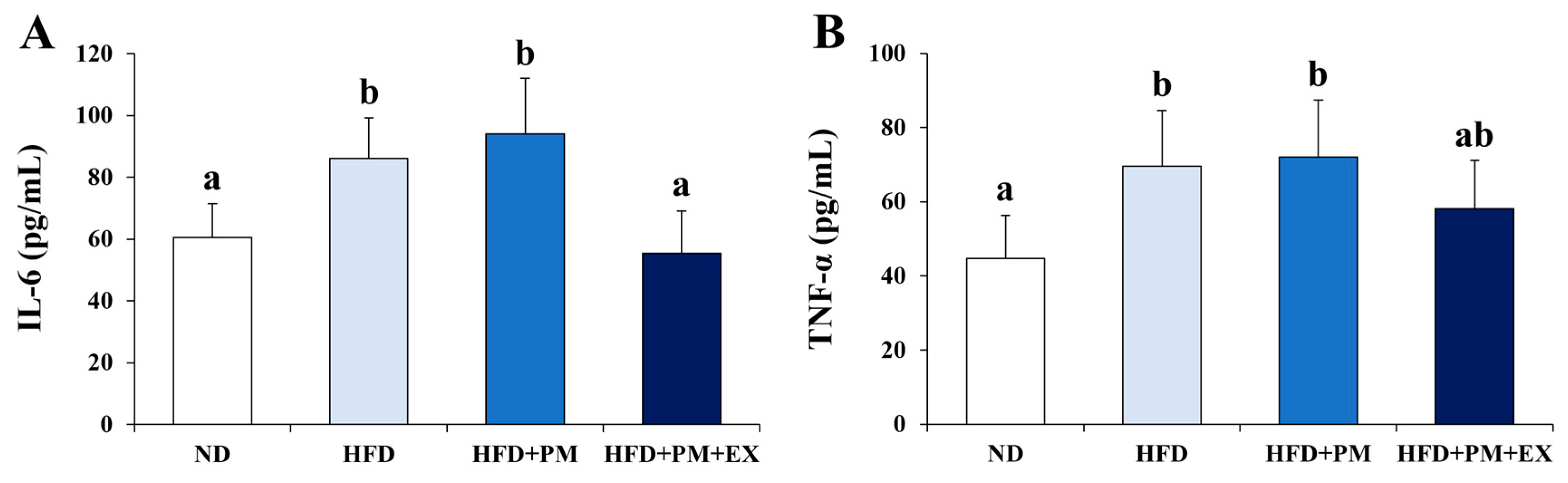
Figure 5 presents the effects of the HFD, PM exposure, and resistance exercise training on the circulating inflammatory markers. One-way ANOVA revealed significant group differences in both IL-6 and TNF-α levels (IL-6: F(3, 44) = 21.284, p < 0.001; TNF-α: F(3, 44) = 9.902, p < 0.001).
Figure 5.
Serum inflammatory marker levels of IL-6 (A) and TNF-α (B) following HFD, PM exposure, and resistance exercise training. Data are shown as mean ± SD. Different letters above the bars indicate statistically significant differences between groups (p < 0.05).
Post hoc analysis indicated that IL-6 levels were significantly higher in the HFD and HFD + PM groups compared with the ND group (both p < 0.001). The HFD + PM group exhibited the highest IL-6 levels among all groups; however, no significant difference was observed between the HFD and HFD + PM groups (p = 1.000). In contrast, IL-6 levels in the HFD + PM + EX group were significantly lower than those in both the HFD and HFD + PM groups (both p < 0.001) and were comparable to those in the ND group (p = 1.000).
TNF-α levels were also significantly elevated in the HFD and HFD + PM groups compared with the ND group (both p < 0.001). No significant difference in TNF-α levels was observed between the HFD and HFD + PM groups (p = 1.000). Although TNF-α levels in the HFD + PM + EX group tended to be lower than those in the HFD (p = 0.281) and HFD + PM (p = 0.103) groups, these differences were not statistically significant. No significant difference was observed between the ND and HFD + PM + EX groups (p = 0.130).
3.5. Neurotrophic Factor Responses
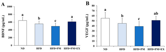
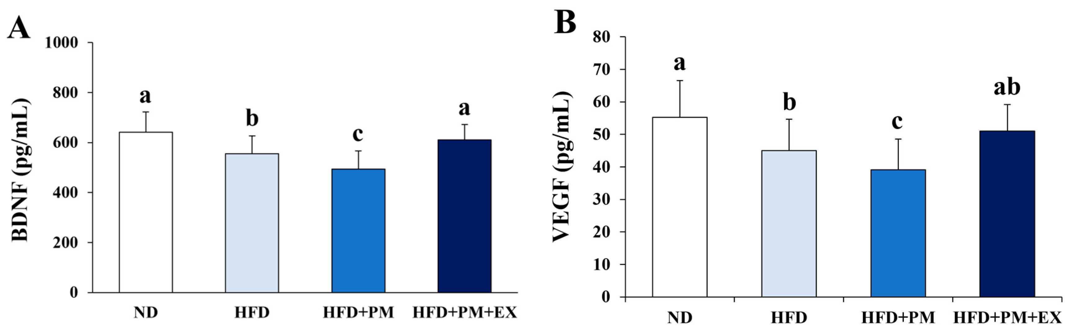
The effects of the HFD, PM exposure, and resistance exercise training on circulating neurotrophic factors are presented in Figure 6. One-way ANOVA revealed significant group differences in BDNF and VEGF levels (BDNF: F(3, 44) = 9.923, p < 0.001; VEGF: F(3, 44) = 6.304, p = 0.001).
Figure 6.
Changes in circulating neurotrophic factors, including BDNF (A) and VEGF (B), across experimental conditions. All values are expressed as mean ± SD. Different letters above the bars indicate statistically significant differences between groups (p < 0.05).
Serum BDNF levels were significantly reduced in the HFD and HFD + PM groups compared with the ND group (p = 0.032 and p < 0.001, respectively). In contrast, the HFD + PM + EX group exhibited significantly higher BDNF levels compared with the HFD + PM group (p = 0.001), with no significant difference compared with the ND group (p = 1.000). No significant difference in BDNF level was observed between the HFD and HFD + PM groups (p = 0.230).
For VEGF, the HFD + PM group showed significantly lower levels compared with the ND group (p = 0.001). VEGF levels in the HFD + PM + EX group were significantly higher than those in the HFD + PM group (p = 0.027). No significant differences were observed between the ND and HFD groups (p = 0.077), between the ND and HFD + PM + EX groups (p = 1.000), or between the HFD and HFD + PM + EX groups (p > 0.05).
3.6. BBB Permeability–Related Markers
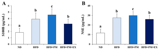
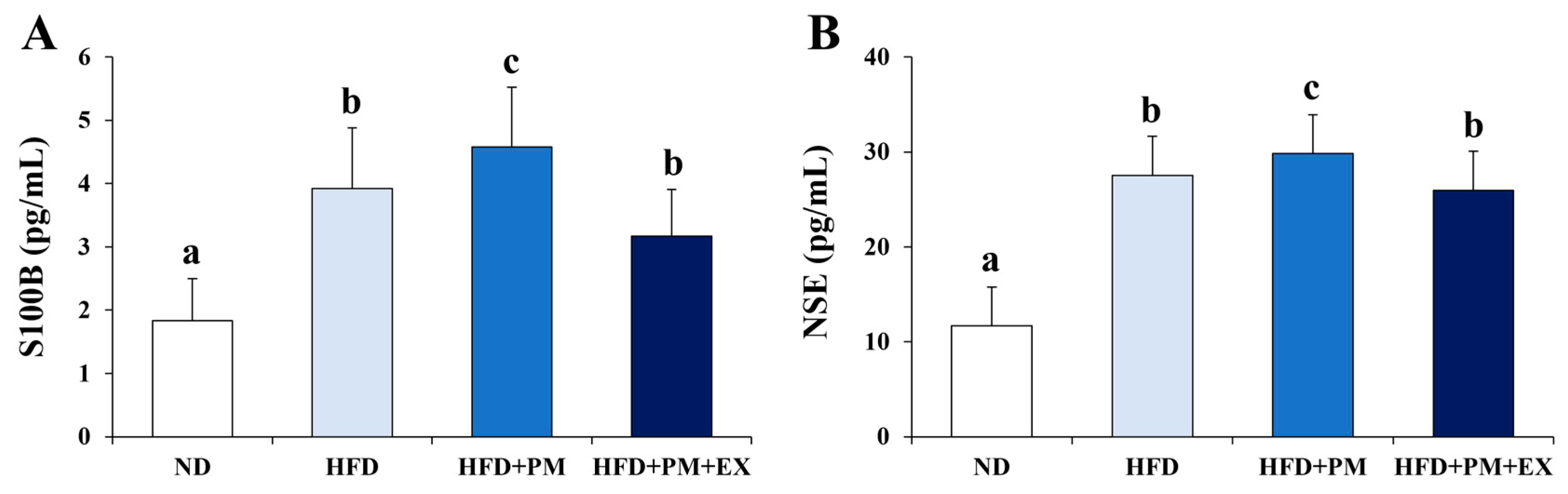
Figure 7 shows the changes in BBB permeability-related markers after the HFD, PM exposure, and resistance exercise training. One-way ANOVA revealed significant group differences in S100B and NSE levels (S100B: F(3, 44) = 23.680, p < 0.001; NSE: F(3, 44) = 35.716, p < 0.001).
Figure 7.
Serum levels of BBB permeability-related markers S100B (A) and NSE (B) measured following HFD, PM exposure, and resistance exercise training. Results are presented as mean ± SD. Different letters above the bars indicate statistically significant differences between groups (p < 0.05).
Post hoc analysis demonstrated that S100B levels were significantly higher in the HFD and HFD + PM groups compared with the ND group (both p < 0.001). Furthermore, S100B levels were significantly higher in the HFD + PM group than in the HFD group (p = 0.001). In contrast, S100B levels in the HFD + PM + EX group were significantly lower than those in the HFD + PM group (p = 0.001), although they remained significantly higher than those in the ND group (p = 0.002).
Similarly, NSE levels were significantly elevated in the HFD and HFD + PM groups compared with the ND group (both p < 0.001). No significant differences in NSE levels were observed between the HFD and HFD + PM groups (p = 1.000), or between the HFD + PM + EX group and either the HFD or HFD + PM groups (p > 0.05). However, NSE levels in the HFD + PM + EX group remained significantly higher than those in the ND group (p < 0.001).
4. Discussion
The regulation of food intake involves complex interactions between peripheral and central pathways. Although genetic factors play a critical role in body weight homeostasis, diet and regular exercise are important environmental determinants of body weight regulation [25]. Consistent with this concept, the present study demonstrated that the HFD significantly increased body weight compared to the ND, whereas resistance exercise training effectively attenuated HFD-induced weight gain. Several studies have suggested that exposure to high PM concentrations increases the risk of obesity [26,27]. However, in the present study, the body weight was unexpectedly lower in the HFD + PM group than in the HFD group. This finding is consistent with a previous study that reported significant weight loss in mice exposed to fine PM (PM2.5) for 40 d [28]. However, given that food intake, energy expenditure, and physical activity levels were not assessed in the present study, the mechanisms underlying the lower body weight observed in the HFD + PM group could not be determined. Therefore, future studies incorporating quantitative assessments of these factors are needed to clarify the mechanisms underlying the observed body weight changes.
An HFD is well recognized as a major contributor to adiposity and dyslipidemia in animal models of obesity [29]. In the present study, mice in the HFD group exhibited significantly higher serum levels of TC, TG, and LDL-C than mice in the ND group, which is consistent with previous findings from HFD-based experimental models [29,30]. These alterations represent characteristic metabolic features commonly observed in obesity and metabolic disorders, indicating that the HFD model employed in this study successfully induced metabolic disturbances, including dyslipidemia. Although reports suggest that PM exposure may increase the risk of dyslipidemia [31,32], no significant differences in lipid profiles were observed between the HFD and HFD + PM groups. This finding suggests that under the present experimental conditions, PM exposure had limited additional effects on HFD-induced dyslipidemia. Consistent with this observation, Zhou et al. reported that the metabolic risk associated with short-term PM10 exposure is relatively limited [33]. Notably, the HFD + PM + EX group exhibited significantly lower TC, TG, and LDL-C levels and significantly higher HDL-C levels than the HFD and HFD + PM groups. These findings suggest that resistance exercise training may improve lipid composition by enhancing skeletal muscle metabolic function, even with PM exposure. Supporting this interpretation, Zeng et al. reported that PM exposure is associated with an increased incidence of dyslipidemia, whereas regular physical activity may exert protective effects against PM-related dyslipidemia [34].
Adipokines are key bioactive molecules secreted by adipose tissue and play essential roles in energy homeostasis, insulin sensitivity, and inflammatory regulation [35]. Among them, leptin and adiponectin are widely recognized as representative adipokines that reflect the pathophysiology of obesity and metabolic diseases [35,36]. In this study, serum leptin and adiponectin levels were analyzed to assess adipokine responses to HFD, PM exposure, and resistance exercise training. The HFD significantly increased leptin levels and decreased adiponectin levels, whereas resistance exercise training markedly attenuated HFD-induced leptin elevation and adiponectin reduction. These findings are consistent with those of previous studies, demonstrating that exercise training can oppositely regulate leptin and adiponectin secretion by reducing fat mass and improving adipose tissue endocrine function [37,38]. Bouassida et al. reported that most exercise training studies that induced improvements in physical fitness and body composition were associated with decreased leptin and increased adiponectin levels [37]. Similar to the lipid profile results, no significant differences in adipokine levels were observed between the HFD and HFD + PM groups, suggesting that under the present experimental conditions, PM exposure had limited additional effects on HFD-induced adipokine dysregulation. In support of this interpretation, Chen et al. reported no significant associations between short-term PM2.5 exposure and adipokine levels [39].
Chronic overnutrition, including HFD consumption, is an independent factor contributing to increased systemic inflammation [40], and changes in circulating pro-inflammatory cytokines such as IL-6 and TNF-α have been proposed as biomarkers of PM-induced inflammatory responses [41]. Conversely, dietary regulation and regular exercise are considered effective strategies for mitigating systemic inflammation [42]. In the present study, serum IL-6 and TNF-α levels were significantly elevated in the HFD group compared with the ND group, indicating enhanced systemic inflammation. These findings support those of previous animal studies reporting significant increases in circulating cytokine levels following HFD consumption [43,44]. Specifically, van der Heijden et al. demonstrated that a 45% kcal fat HFD significantly increased TNF-α levels in male C57BL/6J mice [43], whereas Kim et al. reported significant increases in IL-6 and TNF-α following a 60% kcal fat HFD [44]. In contrast, regular exercise has been shown to attenuate systemic inflammation [45,46]. In the present study, the HFD + PM + EX group exhibited significantly lower IL-6 levels than the HFD and HFD + PM groups. Mazur-Bialy et al. reported that regular exercise reduced HFD-induced increases in IL-6 levels, and Ma et al. demonstrated significant reductions in IL-6 levels after ladder-climbing resistance exercise training. Notably, no significant differences in the inflammatory markers were observed between the HFD and HFD + PM groups. This finding suggests that under the present experimental conditions, PM-induced inflammatory responses may occur preferentially at tissue-specific or immune cell-specific levels, rather than being consistently reflected in systemic circulating markers such as IL-6 and TNF-α. Supporting this interpretation, Miller et al. reported that although PM exposure can amplify inflammatory cytokine responses and tissue-level inflammation, it does not necessarily correlate with circulating biomarkers of low-grade inflammation [47].
Excessive HFD consumption exacerbates obesity and obesity-related metabolic disorders and is closely associated with CNS dysfunction [40]. In contrast, regular exercise upregulates the expression of neurotrophic factors and improves BBB function [48]. In the present study, serum levels of BDNF, VEGF, S100B, and NSE were analyzed to evaluate the changes in neurotrophic factors and BBB permeability in response to an HFD, PM exposure, and resistance exercise training. Compared with the ND group, the HFD group exhibited significantly lower BDNF and VEGF levels and significantly higher S100B and NSE levels. Moreover, the HFD + PM group showed further reductions in neurotrophic factor levels and increased levels of BBB permeability markers relative to the HFD group. These findings suggest that an HFD may induce the downregulation of neurotrophic factors and impair BBB function and that concurrent PM exposure may exacerbate these effects through the cumulative burden of metabolic and environmental stressors. Lobato et al. proposed that combined exposure to an HFD and PM amplifies physiological damage, including oxidative stress, metabolic dysfunction, and transcriptional dysregulation, compared with single exposures [11]. Suwannasual et al. suggested that HFD consumption may exacerbate BBB integrity alterations induced by air pollutant exposure [49], further supporting the present findings. Notably, the HFD + PM + EX group exhibited significantly higher BDNF and VEGF levels and significantly lower S100B and NSE levels than the HFD + PM group. These results suggest that resistance exercise training may have protective effects against HFD- and PM-related alterations in neurotrophic factors and BBB-related markers. Previous studies reported that regular exercise is associated with increased neurotrophic factor expression and improved neurovascular function, which may contribute to the maintenance of BBB integrity and limit the release of BBB-related injury markers into circulation [48,50,51]. In addition, our previous study indicated that exercise training attenuates PM-induced increases in BBB-related injury markers and reductions in neurotrophic factors, further supporting the potentially beneficial role of exercise under PM exposure [52]. These findings are consistent with previous observations.
In the present study, PM exposure was modeled by systemic administration via tail vein injection to ensure controlled and reproducible exposure to PM-like particles. This approach was adopted to investigate the systemic and neurovascular effects of circulating PM, rather than to directly replicate real-world inhalational exposure. Accordingly, the present findings should be interpreted within the context of the systemic PM burden and its potential impact on neurovascular-related markers.
The strengths of this study include its integrative experimental design, which simultaneously considers metabolic (HFD), environmental (PM exposure), and behavioral (resistance exercise training) factors, allowing comprehensive evaluation of their combined effects on body weight, lipid metabolism, adipokines, inflammatory responses, neurotrophic factors, and BBB-related markers. However, this study had several limitations. First, BBB permeability was indirectly assessed using circulating biomarkers such as S100B and NSE, without direct tissue-level analyses of tight junction protein expression or BBB structural integrity. Second, changes in neurotrophic factors were evaluated only at the serum level, limiting the ability to distinguish whether these alterations were driven by changes in central production or BBB permeability. Third, PM exposure was modeled using systemic administration via tail vein injection, which does not fully replicate real-world inhalational exposure and may influence systemic distribution patterns and neurovascular outcomes. Fourth, although standardized, the resistance exercise protocol is inherently difficult to precisely quantify in terms of mechanical load, which may limit the interpretation of exercise dose–response relationships. Fifth, the absence of a non-PM exercise control group (HFD + EX) limits the ability to distinguish PM-specific exercise effects from general exercise-induced adaptations under HFD conditions. Sixth, food intake, energy expenditure, and physical activity levels were not assessed, which constrains the interpretation of the mechanisms underlying the observed body weight changes. Seventh, LDL-C levels were estimated using the Friedewald equation, which has been widely used in rodent studies [53,54] but may have limited accuracy in mice, particularly under conditions of elevated triglycerides. Therefore, LDL-C results should be interpreted with caution. Eighth, only male C57BL/6 mice were included in this study, and potential sex-specific differences in metabolic, inflammatory, and neurotrophic responses could not be evaluated. Finally, statistical analyses were based on a one-way ANOVA without correction for multiple comparisons across different biomarker categories. Correlation analyses were not performed, which may have increased the risk of type I error and limited the ability to quantitatively assess the relationships among the measured variables. Despite these limitations, the present study provides experimental evidence that resistance exercise training can exert beneficial effects on both metabolic health and CNS homeostasis under combined HFD and PM stress conditions. It provide a valuable foundation for future mechanistic and clinical investigations.
5. Conclusions
This study comprehensively investigated the effects of HFD, PM exposure, and resistance exercise training on metabolic health and CNS-related markers. These findings indicate that HFD is associated with not only dyslipidemia and increased inflammatory responses but also reduced circulating levels of neurotrophic factors (BDNF and VEGF) and elevated markers related to BBB permeability (S100B and NSE). Under HFD conditions, PM exposure was associated with additional alterations in neurotrophic factors and BBB-related markers. However, under the present experimental conditions, no significant additional effects of PM were observed on systemic metabolic markers, including lipid profiles, adipokines, and inflammatory markers. Notably, resistance exercise training was associated with the attenuation of HFD- and PM-related metabolic disturbances and inflammatory responses, along with improvements in circulating neurotrophic factors and BBB-related markers. Collectively, these findings suggest that resistance exercise may represent a promising non-pharmacological strategy to support metabolic health and CNS homeostasis under combined metabolic and environmental stress.
Supplementary Materials
The following supporting information can be downloaded at: https://www.mdpi.com/article/10.3390/atmos17020203/s1.
Author Contributions
Conceptualization, S.-Y.C. and H.-T.R.; methodology, S.-Y.C. and H.-T.R.; software, S.-Y.C. and H.-T.R.; validation, S.-Y.C. and H.-T.R.; formal analysis, S.-Y.C. and H.-T.R.; investigation, S.-Y.C.; data curation, S.-Y.C.; writing—original draft preparation, S.-Y.C. and H.-T.R.; writing—review and editing, H.-T.R.; visualization, S.-Y.C. and H.-T.R.; supervision, H.-T.R.; funding acquisition, H.-T.R. All authors have read and agreed to the published version of the manuscript.
Funding
This work has been supported by a National Research Foundation of Korea (NRF) grant funded by the Korea government (MSIT) (No. 2021R1F1A1063415).
Institutional Review Board Statement
All animal experimental procedures were approved by the National Research Foundation of Korea (NRF-2019R1F1A1064296).
Informed Consent Statement
Not applicable.
Data Availability Statement
The data presented in this study are included in this article and the Supplementary Materials. Additional data are available from the corresponding author upon reasonable request.
Conflicts of Interest
The authors declare no conflicts of interest.
References
- Arias-Pérez, R.D.; Taborda, N.A.; Gómez, D.M.; Narvaez, J.F.; Porras, J.; Hernandez, J.C. Inflammatory effects of particulate matter air pollution. Environ. Sci. Pollut. Res. Int. 2020, 27, 42390–42404. [Google Scholar] [CrossRef] [Scilit] [PubMed]
- Hussain, R.; Graham, U.; Elder, A.; Nedergaard, M. Air pollution, glymphatic impairment, and Alzheimer’s disease. Trends Neurosci. 2023, 46, 901–911. [Google Scholar] [CrossRef] [Scilit]
- Shang, M.; Tang, M.; Xue, Y. Neurodevelopmental toxicity induced by airborne particulate matter. J. Appl. Toxicol. 2023, 43, 167–185. [Google Scholar] [CrossRef] [Scilit]
- Brooks, A.W.; Sandri, B.J.; Nixon, J.P.; Nurkiewicz, T.R.; Barach, P.; Trembley, J.H.; Butterick, T.A. Neuroinflammation and Brain Health Risks in Veterans Exposed to Burn Pit Toxins. Int. J. Mol. Sci. 2024, 25, 9759. [Google Scholar] [CrossRef] [Scilit]
- Keller, Z.A.; Eggers, K.M.; Nixon, J.P.; Butterick, T.A. The Impact of Air Pollution and Obesity on Cognitive Decline and Risk of Alzheimer’s Disease. Int. J. Mol. Sci. 2025, 27, 92. [Google Scholar] [CrossRef] [Scilit]
- Chen, Z.; Liu, P.; Xia, X.; Wang, L.; Li, X. The underlying mechanism of PM2.5-induced ischemic stroke. Environ. Pollut. 2022, 310, 119827. [Google Scholar] [CrossRef] [Scilit]
- Martínez-Hernández, G.B.; Jiménez-Ferrer, E.; González-Cortazar, M.; Alejandro, Z.; Monterrosas-Brisson, N.; Herrera-Ruiz, M. Salvia elegans Vahl Counteracting Metabolic Syndrome and Depression in Mice on a High-Fat Diet. Molecules 2024, 29, 4070. [Google Scholar] [CrossRef] [Scilit]
- Kumari, D.; Gautam, J.; Sharma, V.; Gupta, S.K.; Sarkar, S.; Jana, P.; Singhal, V.; Babele, P.; Kamboj, P.; Bajpai, S.; et al. Effect of herbal extracts and saroglitazar on high-fat diet-induced obesity, insulin resistance, dyslipidemia, and hepatic lipidome in C57BL/6J mice. Heliyon 2023, 9, e22051. [Google Scholar] [CrossRef] [Scilit] [PubMed]
- Hajiluian, G.; Nameni, G.; Shahabi, P.; Mesgari-Abbasi, M.; Sadigh-Eteghad, S.; Farhangi, M.A. Vitamin D administration, cognitive function, BBB permeability, and neuroinflammatory factors in high-fat diet-induced obese rats. Int. J. Obes. 2017, 41, 639–644. [Google Scholar] [CrossRef] [Scilit]
- Wang, H.; Wang, B.; Yin, H.; Zhang, G.; Yu, L.; Kong, X.; Yuan, H.; Fang, X.; Liu, Q.; Liu, C.; et al. Reduced neurotrophic factor level is the early event before functional neuronal deficiency in high-fat diet-induced obese mice. Metab. Brain Dis. 2017, 32, 247–257. [Google Scholar] [CrossRef] [Scilit] [PubMed]
- Lobato, S.; Salomón-Soto, V.M.; Espinosa-Méndez, C.M.; Herrera-Moreno, M.N.; García-Solano, B.; Pérez-González, E.; Comba-Marcó-Del-Pont, F.; Montesano-Villamil, M.; Mora-Ramírez, M.A.; Mancilla-Simbro, C.; et al. Molecular Pathways Linking High-Fat Diet and PM2.5 Exposure to Metabolically Abnormal Obesity: A Systematic Review and Meta-Analysis. Biomolecules 2024, 14, 1607. [Google Scholar] [CrossRef] [Scilit]
- Costa-Beber, L.C.; Goettems-Fiorin, P.B.; Dos Santos, J.B.; Friske, P.T.; Frizzo, M.N.; Heck, T.G.; Ludwig, M.S. Air pollution combined with high-fat feeding aggravates metabolic and cardiovascular diseases: A dangerous, oxidative, and immune-inflammatory association. Life Sci. 2023, 317, 121468. [Google Scholar] [CrossRef] [Scilit]
- Toader, C.; Serban, M.; Munteanu, O.; Covache-Busuioc, R.A.; Enyedi, M.; Ciurea, A.V.; Tataru, C.P. From Synaptic Plasticity to Neurodegeneration: BDNF as a Transformative Target in Medicine. Int. J. Mol. Sci. 2025, 26, 4271. [Google Scholar] [CrossRef] [Scilit]
- Wang, J.; Meng, F.; Wang, L.; Li, Z. Vascular endothelial growth factor: A key factor in the onset and treatment of depression. Front. Cell. Neurosci. 2025, 19, 1645437. [Google Scholar] [CrossRef] [Scilit]
- Marchi, N.; Rasmussen, P.; Kapural, M.; Fazio, V.; Kight, K.; Mayberg, M.R.; Kanner, A.; Ayumar, B.; Albensi, B.; Cavaglia, M.; et al. Peripheral markers of brain damage and blood–brain barrier dysfunction. Restor. Neurol. Neurosci. 2003, 21, 109–121. [Google Scholar] [CrossRef] [PubMed]
- Hong, S.J.; De Souza, B.J.; Penberthy, K.K.; Hwang, L.; Procaccini, D.E.; Kheir, J.N.; Bembea, M.M. Plasma brain-related biomarkers and potential therapeutic targets in pediatric ECMO. Neurotherapeutics 2025, 22, e00521. [Google Scholar] [CrossRef] [Scilit] [PubMed]
- Geng, L.; Liao, B.; Jin, L.; Huang, Z.; Triggle, C.R.; Ding, H.; Zhang, J.; Huang, Y.; Lin, Z.; Xu, A. Exercise Alleviates Obesity-Induced Metabolic Dysfunction via Enhancing FGF21 Sensitivity in Adipose Tissues. Cell Rep. 2019, 26, 2738–2752.e4. [Google Scholar] [CrossRef] [Scilit] [PubMed]
- Soltani, N.; Marandi, S.M.; Kazemi, M.; Esmaeil, N. The Exercise Training Modulatory Effects on the Obesity-Induced Immunometabolic Dysfunctions. Diabetes Metab. Syndr. Obes. 2020, 13, 785–810. [Google Scholar] [CrossRef] [Scilit]
- Bagheri, N.; Bagheri, R.; Mesinovic, J.; Ghobadi, H.; Scott, D.; Kargarfard, M.; Dutheil, F. Effects of Resistance Training on Muscular Adaptations and Inflammatory Markers in Overweight and Obese Men. Med. Sci. Sports Exerc. 2025, 57, 600–612. [Google Scholar] [CrossRef] [Scilit]
- Kolahdouzi, S.; Baghadam, M.; Kani-Golzar, F.A.; Saeidi, A.; Jabbour, G.; Ayadi, A.; De Sousa, M.; Zouita, A.; Abderrahmane, A.B.; Zouhal, H. Progressive circuit resistance training improves inflammatory biomarkers and insulin resistance in obese men. Physiol. Behav. 2019, 205, 15–21. [Google Scholar] [CrossRef] [Scilit]
- Azevedo, C.V.; Otaviano, S.F.S.D.; Campos, H.C.; Figueiredo, E.V.; Pereira, A.R.; Alves, L.; Braga, M.; Gimenes, C.; Boldarine, V.T.; Oyama, L.M.; et al. Distinct effects of long-term resistance exercise on cognitive deficits, neurotrophins, and amyloid pathology in male and female APP/PS1 mice. J. Alzheimers Dis. 2025, 108, 1790–1806. [Google Scholar] [CrossRef] [Scilit]
- Sun, Z.X.; Xie, F.; Zhao, Y.; Wang, X.; Sun, Z.W.; Qian, L.J.; Feng, H. Exercise and the blood-brain barrier: Mechanistic insights and therapeutic implications. Neurobiol. Dis. 2025, 217, 107166. [Google Scholar] [CrossRef] [Scilit]
- Bai, N.; van Eeden, S.F. Systemic and vascular effects of circulating diesel exhaust particulate matter. Inhal. Toxicol. 2013, 25, 725–734. [Google Scholar] [CrossRef] [Scilit]
- Sanches, I.C.; Conti, F.F.; Sartori, M.; Irigoyen, M.C.; De Angelis, K. Standardization of resistance exercise training: Effects in diabetic ovariectomized rats. Int. J. Sports Med. 2014, 35, 323–329. [Google Scholar] [CrossRef] [Scilit] [PubMed]
- Faccioli, N.; Poitou, C.; Clément, K.; Dubern, B. Current Treatments for Patients with Genetic Obesity. J. Clin. Res. Pediatr. Endocrinol. 2023, 15, 108–119. [Google Scholar] [CrossRef] [Scilit]
- Lin, L.; Li, T.; Sun, M.; Liang, Q.; Ma, Y.; Wang, F.; Duan, J.; Sun, Z. Global association between atmospheric particulate matter and obesity: A systematic review and meta-analysis. Environ. Res. 2022, 209, 112785. [Google Scholar] [CrossRef] [Scilit] [PubMed]
- Yang, S.; Hong, F.; Li, S.; Han, X.; Li, J.; Wang, X.; Chen, L.; Zhang, X.; Tan, X.; Xu, J.; et al. The association between chemical constituents of ambient fine particulate matter and obesity in adults: A large population-based cohort study. Environ. Res. 2023, 231, 116228. [Google Scholar] [CrossRef] [Scilit] [PubMed]
- Dai, S.; Wang, Z.; Yang, Y.; Du, P.; Li, X. PM2.5 induced weight loss of mice through altering the intestinal microenvironment: Mucus barrier, gut microbiota, and metabolic profiling. J. Hazard. Mater. 2022, 431, 128653. [Google Scholar] [CrossRef] [Scilit]
- Kwon, E.Y.; Choi, M.S. Dietary Eriodictyol Alleviates Adiposity, Hepatic Steatosis, Insulin Resistance, and Inflammation in Diet-Induced Obese Mice. Int. J. Mol. Sci. 2019, 20, 1227. [Google Scholar] [CrossRef] [Scilit]
- Jing, L.; Zhang, Y.; Fan, S.; Gu, M.; Guan, Y.; Lu, X.; Huang, C.; Zhou, Z. Preventive and ameliorating effects of citrus D-limonene on dyslipidemia and hyperglycemia in mice with high-fat diet-induced obesity. Eur. J. Pharmacol. 2013, 715, 46–55. [Google Scholar] [CrossRef] [Scilit] [PubMed]
- Zhang, Z.; Su, Y.; Jing, R.; Qi, J.; Qi, X.; Xie, Z.; Cui, B. Acute and lag effects of ambient fine particulate matter on the incidence of dyslipidemia in Chengdu, China: A time-series study. Environ. Sci. Pollut. Res. Int. 2022, 29, 37919–37929. [Google Scholar] [CrossRef] [Scilit] [PubMed]
- Sun, M.; Li, T.; Sun, Q.; Ren, X.; Sun, Z.; Duan, J. Associations of long-term particulate matter exposure with cardiometabolic diseases: A systematic review and meta-analysis. Sci. Total Environ. 2023, 903, 166010. [Google Scholar] [CrossRef] [Scilit] [PubMed]
- Zhou, C.; Cui, G.Y.; Tang, Y.H.; Zhang, W.Y.; Zou, X.L. Long-term and short-term exposure to outdoor air pollution and its association with glycolipid metabolic disorders. World J. Diabetes 2025, 16, 110028. [Google Scholar] [CrossRef] [Scilit]
- Zeng, Y.Q.; Chang, L.Y.; Guo, C.; Lin, C.; Bo, Y.; Wong, M.C.S.; Tam, T.; Lau, A.K.H. Chronic fine particulate matter exposure, habitual exercise, and dyslipidemia: A longitudinal cohort study. Environ. Epidemiol. 2022, 6, e190. [Google Scholar] [CrossRef] [Scilit]
- Choe, S.S.; Huh, J.Y.; Hwang, I.J.; Kim, J.I.; Kim, J.B. Adipose Tissue Remodeling: Its Role in Energy Metabolism and Metabolic Disorders. Front. Endocrinol. 2016, 7, 30. [Google Scholar] [CrossRef] [Scilit] [PubMed]
- Engin, A. Adiponectin Resistance in Obesity: Adiponectin Leptin/Insulin Interaction. Adv. Exp. Med. Biol. 2024, 1460, 431–462. [Google Scholar]
- Bouassida, A.; Chamari, K.; Zaouali, M.; Feki, Y.; Zbidi, A.; Tabka, Z. Review on leptin and adiponectin responses and adaptations to acute and chronic exercise. Br. J. Sports Med. 2010, 44, 620–630. [Google Scholar] [CrossRef] [Scilit]
- Lin, H.; Hu, M.; Yan, Y.; Zhang, H. The effect of exercise on adiponectin and leptin levels in overweight or obese subjects: A meta-analysis of randomized controlled trials. Sport Sci. Health 2017, 13, 303–314. [Google Scholar] [CrossRef] [Scilit]
- Chen, W.; Han, Y.; Wang, Y.; Chen, X.; Qiu, X.; Li, W.; Yao, Y.; Zhu, T. Associations between changes in adipokines and exposure to fine and ultrafine particulate matter in ambient air in Beijing residents with and without pre-diabetes. BMJ Open Diabetes Res. Care 2020, 8, e001215. [Google Scholar] [CrossRef] [Scilit]
- Duan, Y.; Zeng, L.; Zheng, C.; Song, B.; Li, F.; Kong, X.; Xu, K. Inflammatory Links Between High Fat Diets and Diseases. Front. Immunol. 2018, 9, 2649. [Google Scholar] [CrossRef] [Scilit]
- Ghozikali, M.G.; Ansarin, K.; Naddafi, K.; Nabizadeh, R.; Yaghmaeian, K.; Jaafari, J.; Dehghanzadeh, R.; Atafar, Z.; Faraji, M.; Mohammadi, A.; et al. Status of TNF-alpha and IL-6 as pro-inflammatory cytokines in exhaled breath condensate of late adolescents with asthma and healthy in the dust storm and non-dust storm conditions. Sci. Total Environ. 2022, 838, 155536. [Google Scholar] [CrossRef] [Scilit]
- Celik, O.; Yildiz, B.O. Obesity and physical exercise. Minerva Endocrinol. 2021, 46, 131–144. [Google Scholar] [CrossRef]
- van der Heijden, R.A.; Sheedfar, F.; Morrison, M.C.; Hommelberg, P.P.; Kor, D.; Kloosterhuis, N.J.; Gruben, N.; Youssef, S.A.; de Bruin, A.; Hofker, M.H.; et al. High-fat diet induced obesity primes inflammation in adipose tissue prior to liver in C57BL/6j mice. Aging 2015, 7, 256–268. [Google Scholar] [CrossRef] [Scilit]
- Kim, K.A.; Gu, W.; Lee, I.A.; Joh, E.H.; Kim, D.H. High fat diet-induced gut microbiota exacerbates inflammation and obesity in mice via the TLR4 signaling pathway. PLoS ONE 2012, 7, e47713. [Google Scholar] [CrossRef] [Scilit]
- Mazur-Bialy, A.I.; Bilski, J.; Wojcik, D.; Brzozowski, B.; Surmiak, M.; Hubalewska-Mazgaj, M.; Chmura, A.; Magierowski, M.; Magierowska, K.; Mach, T.; et al. Beneficial Effect of Voluntary Exercise on Experimental Colitis in Mice Fed a High-Fat Diet: The Role of Irisin, Adiponectin and Proinflammatory Biomarkers. Nutrients 2017, 9, 410. [Google Scholar] [CrossRef] [Scilit] [PubMed]
- Ma, X.; Gao, H.; Wang, Z.; Zhu, D.; Dai, W.; Wu, M.; Guo, Y.; Zhao, L.; Xiao, W. Beneficial Effects of Different Types of Exercise on Diabetic Cardiomyopathy. Biomolecules 2025, 15, 1223. [Google Scholar] [CrossRef] [Scilit] [PubMed]
- Miller, G.E.; Passarelli, V.; Chen, E.; Kloog, I.; Wright, R.J.; Amini, H. Ambient PM2.5 and specific sources increase inflammatory cytokine responses to stimulators and reduce sensitivity to inhibitors. Environ. Res. 2024, 252, 118964. [Google Scholar] [CrossRef] [Scilit] [PubMed]
- Roh, H.T.; So, W.Y. The effects of aerobic exercise training on oxidant-antioxidant balance, neurotrophic factor levels, and blood-brain barrier function in obese and non-obese men. J. Sport Health Sci. 2017, 6, 447–453. [Google Scholar] [CrossRef] [Scilit]
- Suwannasual, U.; Lucero, J.; McDonald, J.D.; Lund, A.K. Exposure to traffic-generated air pollutants mediates alterations in brain microvascular integrity in wildtype mice on a high-fat diet. Environ. Res. 2018, 160, 449–461. [Google Scholar] [CrossRef] [Scilit]
- Kraemer, R.R.; Kraemer, B.R. The effects of peripheral hormone responses to exercise on adult hippocampal neurogenesis. Front. Endocrinol. 2023, 14, 1202349. [Google Scholar] [CrossRef] [Scilit]
- Zhu, Z.; Xu, J.; Jin, Y.; Wang, L.; Li, X. Effects of Aerobic Exercise on Markers of Brain Injury in Methamphetamine-Dependent Individuals: A Randomized Controlled Trial. Brain Sci. 2022, 12, 1521. [Google Scholar] [CrossRef] [Scilit]
- Cho, S.Y.; Roh, H.T. Combined Effects of Particulate Matter Exposure and Exercise Training on Neuroplasticity-Related Growth Factors and Blood–Brain Barrier Integrity. Atmosphere 2025, 16, 220. [Google Scholar] [CrossRef] [Scilit]
- Bae, J.Y. Aerobic Exercise Increases Meteorin-Like Protein in Muscle and Adipose Tissue of Chronic High-Fat Diet-Induced Obese Mice. Biomed. Res. Int. 2018, 2018, 6283932. [Google Scholar] [CrossRef] [Scilit] [PubMed]
- Ko, K.; Woo, J.; Bae, J.Y.; Roh, H.T.; Lee, Y.H.; Shin, K.O. Exercise Training Improves Intramuscular Triglyceride Lipolysis Sensitivity in High-Fat Diet-Induced Obese Mice. Lipids Health Dis. 2018, 17, 81. [Google Scholar] [CrossRef] [Scilit] [PubMed]
Disclaimer/Publisher’s Note: The statements, opinions and data contained in all publications are solely those of the individual author(s) and contributor(s) and not of MDPI and/or the editor(s). MDPI and/or the editor(s) disclaim responsibility for any injury to people or property resulting from any ideas, methods, instructions or products referred to in the content. |
© 2026 by the authors. Licensee MDPI, Basel, Switzerland. This article is an open access article distributed under the terms and conditions of the Creative Commons Attribution (CC BY) license.