


Abstract
Genome stability is the cornerstone of cellular health, and imbalances can cause a number of outcomes, including aging, cancer, and other pathologies. DNA damage is a strong driver of both cellular senescence and mitochondrial dysfunction, two other key hallmarks of aging. Both nuclear and mitochondrial genome instability have been shown to drive aging in the hematopoietic system, which then propagates to non-lymphoid tissues, enhancing morbidity and mortality. The loss of TFAM, a key regulator of mitochondrial DNA replication and nucleoid stability, in T cells has been shown to cause mitochondrial dysfunction, leading to premature immune aging and eventual systemic aging. We sought to investigate whether the loss of TFAM in all immune cells would have a comparable or stronger effect on both the immune system and parenchyma. To address this, we attempted to generate Vav-iCre+/−; Tfamfl/fl mice, which are deficient in TFAM in all immune cells. However, this genotype was unrecoverable as no mutant pups were born, suggesting embryonic lethality. Conversely, we generated mice lacking SIRT6, a nuclear DNA repair enzyme that also regulates mitochondrial homeostasis, in all immune cells and found them to be viable and born at expected Mendelian frequencies. Our findings demonstrate the necessity of mitochondrial genome maintenance and homeostasis repair in immunity.
1. Introduction
Genome integrity is essential for cellular and physiological function; consequently, organisms have evolved robust repair pathways and responses to genotoxic insults [1]. Genomic instability can lead not only to transformation, which drives carcinogenesis, but also other cell fates such as apoptosis and cellular senescence [2]. A wealth of evidence from animal studies and human clinical studies demonstrates that DNA damage is a potent driver of the aging process [2,3,4]. In fact, genomic instability is one of the twelve hallmarks of aging, which represent key molecular perturbations that play a causal role in both chronological and biological aging [5,6]. Many in vitro and in vivo studies rely on treatments with supraphysiological doses of DNA-damaging agents or the deletion of critical DNA repair enzymes to instigate genome instability to investigate the impact on cellular function and physiological outcomes. While considerable focus is given to the damage and repair that occurs within the nuclear genome, the integrity of the mitochondrial genome is also critical for maintaining homeostasis [7,8,9].
Mitochondrial DNA (mtDNA) damage can accelerate biological aging by impairing the replication or transcription of indispensable genes involved in respiration and other processes, ultimately giving rise to mitochondrial dysfunction [10,11,12]. Unlike the nuclear genome, mtDNA integrity can be maintained through the degradation of damaged mitochondrial genomes via mitophagy [12,13]. mtDNA is organized into DNA–protein structures called nucleoids that reside within the mitochondrial matrix [14]. Each nucleoid typically contains a single mitochondrial genome bound by several proteins that regulate mitochondrial replication, transcription, repair, and packaging [15]. One of these proteins is Mitochondrial Transcription Factor A (TFAM), which is a key mitochondrial protein that is encoded within the nuclear genome. Beyond its role in transcription-primed mtDNA replication, the formation of mitochondrial DNA nucleoid structures, it also plays a role in fission-mediated mtDNA segregation [16,17,18,19]. The constitutive loss of Tfam is lethal; however haploinsufficiency (Tfam+/−) leads to enlarged nucleoids and mtDNA loss and leakage into the cytoplasm, which can stimulate cGAS-STING signaling [19,20]. The tissue-specific deletion of Tfam has been used to study the role of mitochondrial dysfunction in aging and age-related chronic diseases [21,22,23,24,25,26,27]. Notably, the deletion of Tfam in T cells causes CD4-Cre+/−; Tfamfl/fl mice to rapidly develop numerous features of immunosenescence [24], the progressive age-related decline in immune function [28,29]. Furthermore, CD4-Cre+/−; Tfamfl/fl mice eventually develop premature aging in non-lymphoid organs well after the onset of immunosenescence. This phenomenon is driven by both cell autonomous and cell non-autonomous effects, which result in premature senescence in the solid organs that shorten both the healthspan and lifespan in CD4-Cre+/−; Tfamfl/fl mice [24]. Even the other immune cell lineages can be affected by the T-cell-specific loss of Tfam. For example, the remodeling of the bone marrow niche and enhanced granulopoiesis, which contributed to age-related phenotypes, were observed in CD4-Cre+/−; Tfamfl/fl mice [27].
Previously, it has also been shown that the loss of the DNA repair gene Ercc1 in hematopoietic cells of mice (Vav-iCre+/−; Ercc1−/fl mice), disrupting multiple DNA repair pathways and leading to the accumulation of spontaneously occurring endogenous DNA damage, caused similar effects [30]. This led to interest in whether the effect of mitochondrial dysfunction broadly across all immune cells (via loss of Tfam) would yield a similar or stronger phenotype than that observed in CD4-Cre+/−; Tfamfl/fl mice [24]. Thus, Tfam conditional floxed mice were crossed with Vav-iCre mice that express a codon-improved variant of the cre recombinase (iCre) expressed under the control of the murine vav1 oncogene promoter that induces recombination in hematopoietic stem cells and their progenitors [31]. While useful for studying immune biology, Vav-iCre mice are not without limitations, such as potential leaky expression in non-target tissues (further discussed in the Results and Conclusions Sections) [32]. We hypothesized that Vav-iCre+/−; Tfamfl/fl mice would have an accelerated or stronger phenotype than CD4-Cre+/−; Tfamfl/fl mice [24], but we were surprised at the inability to recover a Vav-iCre+/−; Tfamfl/fl mouse from multiple breedings. To further investigate the role of DNA repair deficits in murine immunology, we deleted the DNA repair factor SIRT6, which is localized to the nucleus but plays a role in regulating mitochondrial homeostasis [33,34,35], in all murine immune cells (Vav-iCre+/−; Sirt6fl/fl mice). Interestingly, Vav-iCre+/−; Sirt6fl/fl mice are viable and born at near Mendelian frequencies. These results highlight the importance of TFAM and mtDNA stability in homeostasis and immune function.
2. Methods
2.1. Animal Studies
All animal studies were approved by the Columbia University Institutional Animal Care and Use Committee in accordance with institutional, local, state, and federal regulations. All animal breeding studies were carried out from May 2023 to November 2025. Tfamfl/fl (#026123), Sirt6fl/fl, (#017334), CD4-Cre+/− (#017336), and Vav-iCre+/− (#008610) mouse strains were all obtained from The Jackson Laboratory (Bar Harbor, ME) and maintained on a C57BL/6J background. All mice were housed in a specific pathogen-free condition barrier facility with routine serological analysis of sentinel mouse cages. Sirt6fl/fl mice were originally provided on a mixed background and backcrossed onto the C57BL/6J strain using speed congenics, and genetic background was confirmed via genetic monitoring services provided by TransnetYX. All immune-specific mutant mice (X-Cre+/−; Yfl/fl; X = CD4-Cre or Vav-iCre; Y = Sirt6, or Tfam) used in the experimental cohorts were generated by crossing homozygous floxed (Sirt6fl/fl, Tfamfl/fl) mice with the individual compound heterozygous cre mice (X-Cre+/−; Y+/fl; X = CD4-Cre or Vav-iCre; Y = Sirt6, or Tfam) with the same genetic background (C57BL/6J). For Vav-iCre breedings, only females carrying the Vav-iCre allele were used for breeding to limit potential germline leakage that has been previously reported in male Vav-iCre mice. All offspring from crosses were given ear tags with unique identifiers, and ear punches were used for genotyping services provided by TransnetYX. Post-natal mortality of pups was monitored daily by animal laboratory technicians.
2.2. Immunoblotting
Tissues were isolated from two-month-old mice, and protein lysates were produced using T-PER buffer (Thermo-Fisher, Waltham, MA, USA) according to manufacturer’s specification. Polyacrylamide (4–12%) gel electrophoresis was performed before protein was transferred to PVDF membrane and blocked for 1 h in 10% milk TBST at room temperature. Membranes were incubated overnight at 4 °C in primary antibodies: anti-SIRT6 (Cell Signaling Technology, Danvers, MA, USA, catalog #12486, 1:500 dilution in TBST) and anti-Vinculin (Abcam, Waltham, MA, USA, catalog #ab129002, 1:2000 dilution in TBST). Following three 5 min washes in TBST, membranes were incubated for 2 h at room temperature with a goat anti-rabbit HRP-conjugated secondary antibody (Millipore Sigma, Burlington, MA, USA, catalog #A0545, 1:2000 dilution in TBST). Membranes were then washed three additional times for 5 min in TBST, and protein detection was performed by chemiluminescence detection using ECL reagent (Pierce, Rockford, IL, USA).
2.3. Statistical Analysis
Genotyping data (expected versus observed) was plotted in GraphPad Prism version 10.6.0, and statistical analysis was performed using Chi-squared (χ2) analysis.
3. Results
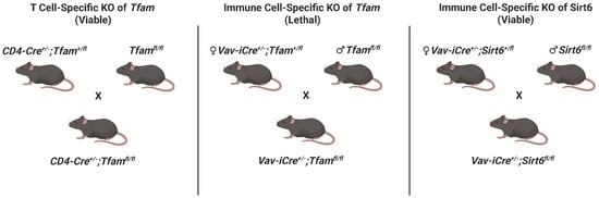
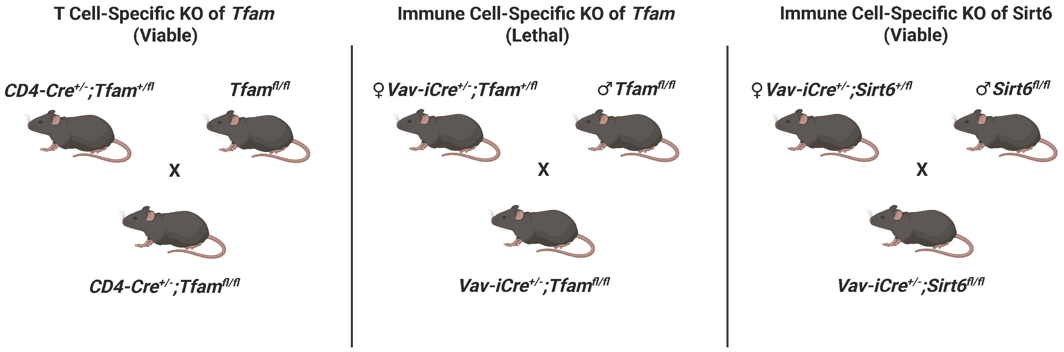
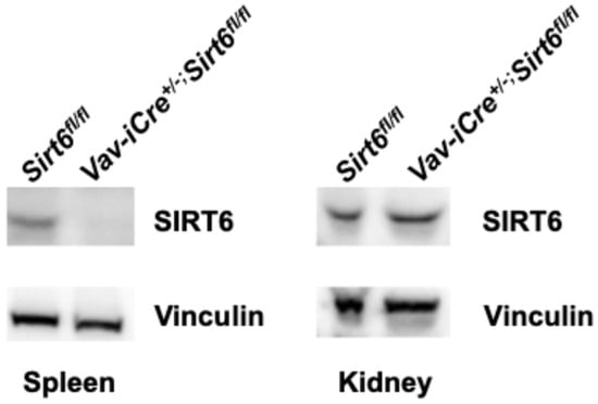
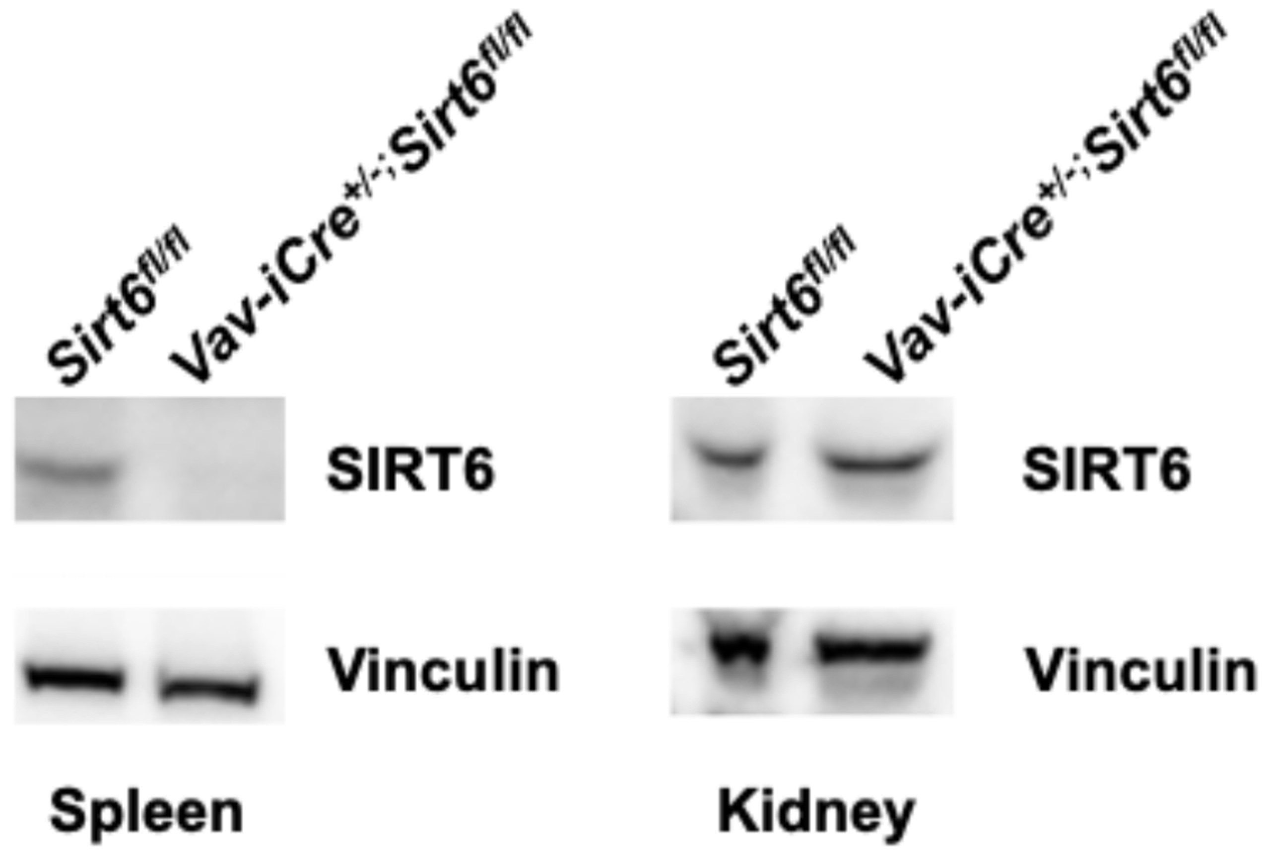
In order to study the impact of loss of TFAM across immune cells, we sought to generate Vav-iCre+/−; Tfamfl/fl mice. Because of prior reports of aberrant cre recombinase expression in the testis of Vav-iCre mice, only female Vav-iCre breeders were used in mating pairs (Figure 1, Table S1). Unfortunately, after examining the offspring in several litters (n = 26) resulting from crosses of female Vav-iCre+/−; Tfam+/fl mice with male Tfamfl/fl breeders, no Vav-iCre+/−; Tfamfl/fl mice were present despite an expectation of a 25% frequency (0 observed versus ~41 expected Vav-iCre+/−; Tfamfl/fl offspring; p < 0.001) (Table 1). All other relevant genotypes (Vav-iCre+/−; Tfam+/fl, Tfam+/fl, Tfamfl/fl mice) were born at near Mendelian frequencies. Furthermore, we observed no post-natal lethality in pups from the litters, suggesting embryonic lethality. This stands in stark contrast to CD4-Cre+/−; Tfamfl/fl mice, which are viable and born at expected frequencies (25 observed versus ~26 expected CD4-Cre+/−; Tfamfl/fl offspring) (Table 1). Meanwhile, a similar breeding strategy was employed to create Vav-iCre+/−; Sirt6fl/fl mice to limit off-target effects (Figure 1). Vav-iCre+/−; Sirt6fl/fl mice were born at a ~30.7% frequency across multiple litters (20 observed versus ~16 expected Vav-iCre+/−; Sirt6fl/fl offspring) (Table 1). Likewise, no post-natal mortality was observed in pups from the Vav-iCre; Sirt6 breedings. An immunoblot analysis of spleens from Vav-iCre+/−; Sirt6fl/fl mice confirmed a loss of SIRT6, whereas the renal levels of SIRT6 protein in the kidneys of these mice were comparable to those in the Sirt6fl/fl control mice (Figure 2).
Figure 1.
Breeding schematic for crosses. Individual breeding schemes show crosses to generate CD4-Cre+/−; Tfamfl/fl, Vav-iCre+/−; Tfamfl/fl, and Vav-iCre+/−; Sirt6fl/fl mutant mice and their status (lethal/viable). Please note that Vav-iCre breeding schemes assign cre alleles to female mice to reduce potential off-target germline effects. The figure was created with BioRender.
Table 1.
Mendelian frequency of immune-specific mutants. Statistical significance determined using Chi-squared analysis. *** p < 0.001; ns = not significant.
Figure 2.
Targeted loss of SIRT6 in the spleens of Vav-iCre+/−; Sirt6fl/fl mice. Immunoblots analysis of spleen and kidney tissues from two-month-old Vav-iCre+/−; Sirt6fl/fl mice and Sirt6fl/fl control mice, assessing SIRT6 and Vinculin protein abundance.
4. Discussion
Prior studies demonstrated that T-cell-specific knockouts of Tfam developed multiple features of immunosenescence before the development of secondary senescence in peripheral tissues [24]. CD4-Cre+/−; Tfamfl/fl mice develop multimorbidity as a result of both the loss of protective immune functions and the adoption of pathogenic immune changes [36]. Similar changes were discovered in Vav-iCre+/−; Ercc1−/fl mice, which lack Ercc1 in multiple immune lineages [30]. This led to the investigation of the broad deletion of Tfam in all immune cells rather than just T cells as the rationale for generating Vav-iCre+/−; Tfamfl/fl mice. While other immune-specific deletions of Tfam are viable [24,37,38,39,40], our findings raise issue with deleting Tfam throughout the immune system (Vav-iCre+/−; Tfamfl/fl mice). Our inability to generate this mouse suggests embryonic lethality, although this possibility requires further investigation. Together with prior reports, our findings underscore the importance of mtDNA homeostasis in the immune system. Furthermore, there is a paucity of studies related to the immune-specific deletion of mitochondrial maintenance genes let alone mtDNA repair genes, thus leaving much biology to be explored. Future investigations into the lethality of Vav-iCre+/−; Tfamfl/fl mice should make use of timed pregnancies to determine on which day of gestation embryonic lethality occurs and if this coincides with key developments of fetal hematopoiesis.
While useful, Vav-iCre mice are less than ideal due to reports of cre expression in testis that do not express Vav, potentially due to transgene insertion effects [31,41,42]. Beyond this, another study crossed a stop-floxed-enhanced Yellow Fluorescent Protein reporter into Vav-iCre+/− mice (Vav-iCre+/−; Rosa26+/LSL-EYFP) and found recombination (EYFP+ cells) both in hematopoietic stem cells and in Lin− CD45− CD31− CD51− Sca-1− bone cells that lack hematopoietic surface markers [32]. Previously, studies showed that Vav-iCre+/−; Ercc1−/fl mice demonstrated a loss of Ercc1 in lymphoid tissues but not in parenchyma tissues, such as the aorta, kidney, or liver [30]. Our immunoblotting data from Vav-iCre+/−; Sirt6fl/fl mice yielded no evidence of an off-target loss of SIRT6 in the kidneys. While this data argues against significant “leaky” germline cre expression in Vav-iCre+/−; Sirt6fl/fl mice, more comprehensive, multi-tissue analyses are necessary to definitively exclude this possibility.
The use of other broader immune-specific cre lines—like hCD2-iCre, which drives the cre recombinase from the human CD2 promoter—may be a useful alternative [32,43]. However, their expression is limited to the lymphocyte lineage and not myeloid cells and still suffers from potential off-target expression in the testis [32]. Some of these limitations to potentially targeting Tfam and other mitochondrial maintenance genes across a wide swath of immune cells could be overcome by the development of more precise hematopoietic stem-cell-specific cre lines or the use of inducible cre (ex. tamoxifen-inducible CreERT2 models) that could be employed post-development in mature mice. Even if some off-target cre expression was still present, the reconstitution of immune systems in recipient mice after immunoablation with hematopoietic stem cells from donor Tfam immune-deficient mice could be employed as an alternative. These findings highlight the critical nature of mitochondrial genome integrity in the immune system and the need for better tools to study the immune-wide deletion of DNA repair genes to better understand their role in immune function and limiting immunosenescence.
Supplementary Materials
The following supporting information can be downloaded at: https://www.mdpi.com/article/10.3390/genes17030255/s1, Table S1: Breeding details.
Author Contributions
Conceptualization by M.J.Y.; methodology, investigation, formal analysis, writing—original draft preparation, and editing by R.G., E.S., and M.J.Y.; supervision by M.J.Y.; funding acquisition by M.J.Y. The order of co-first authorship was chosen alphabetically. All authors contributed to this article and approved the submitted version. All authors have read and agreed to the published version of the manuscript.
Funding
This work was supported by the Irene Diamond Fund/American Federation of Aging Research Supplemental Award and by an NIH/NIA grant (R21AG087361) to M.J.Y.
Institutional Review Board Statement
The animal protocol used for this experiment was approved by the Columbia University Institutional Animal Care and Use Committee (Protocol # AC-AABV1653, latest date of approval: 8 September 2025).
Informed Consent Statement
Not applicable.
Data Availability Statement
The original contributions presented in this study are included in the article/Supplementary Material. Further inquiries can be directed to the corresponding author.
Conflicts of Interest
The authors declare no competing interest.
References
- Friedberg, E.C. DNA Repair and Mutagenesis, 2nd ed.; ASM Press: Washington, DC, USA, 2006; 1118p. [Google Scholar]
- Yousefzadeh, M.; Henpita, C.; Vyas, R.; Soto-Palma, C.; Robbins, P.; Niedernhofer, L. DNA damage-how and why we age? eLife 2021, 10, e62852. [Google Scholar] [CrossRef] [Scilit]
- Niedernhofer, L.J.; Gurkar, A.U.; Wang, Y.; Vijg, J.; Hoeijmakers, J.H.J.; Robbins, P.D. Nuclear Genomic Instability and Aging. Annu. Rev. Biochem. 2018, 87, 295–322. [Google Scholar] [CrossRef] [Scilit]
- Schumacher, B.; Pothof, J.; Vijg, J.; Hoeijmakers, J.H.J. The central role of DNA damage in the ageing process. Nature 2021, 592, 695–703. [Google Scholar] [CrossRef] [Scilit] [PubMed]
- Lopez-Otin, C.; Blasco, M.A.; Partridge, L.; Serrano, M.; Kroemer, G. The hallmarks of aging. Cell 2013, 153, 1194–1217. [Google Scholar] [CrossRef] [Scilit]
- Lopez-Otin, C.; Blasco, M.A.; Partridge, L.; Serrano, M.; Kroemer, G. Hallmarks of aging: An expanding universe. Cell 2023, 186, 243–278. [Google Scholar] [CrossRef] [Scilit]
- Nadalutti, C.A.; Ayala-Pena, S.; Santos, J.H. Mitochondrial DNA damage as driver of cellular outcomes. Am. J. Physiol. Cell Physiol. 2022, 322, C136–C150. [Google Scholar] [CrossRef] [Scilit] [PubMed]
- Newman, L.E.; Shadel, G.S. Mitochondrial DNA Release in Innate Immune Signaling. Annu. Rev. Biochem. 2023, 92, 299–332. [Google Scholar] [CrossRef] [Scilit] [PubMed]
- Wu, Z.; Sainz, A.G.; Shadel, G.S. Mitochondrial DNA: Cellular genotoxic stress sentinel. Trends Biochem. Sci. 2021, 46, 812–821. [Google Scholar] [CrossRef] [Scilit]
- Pinto, M.; Moraes, C.T. Mechanisms linking mtDNA damage and aging. Free Radic. Biol. Med. 2015, 85, 250–258. [Google Scholar] [CrossRef] [Scilit]
- Gorman, G.S.; Chinnery, P.F.; DiMauro, S.; Hirano, M.; Koga, Y.; McFarland, R.; Suomalainen, A.; Thorburn, D.R.; Zeviani, M.; Turnbull, D.M. Mitochondrial diseases. Nat. Rev. Dis. Primers 2016, 2, 16080. [Google Scholar] [CrossRef] [Scilit]
- Liao, S.; Chen, L.; Song, Z.; He, H. The fate of damaged mitochondrial DNA in the cell. Biochim. Biophys. Acta Mol. Cell Res. 2022, 1869, 119233. [Google Scholar] [CrossRef] [Scilit]
- Picca, A.; Faitg, J.; Auwerx, J.; Ferrucci, L.; D’Amico, D. Mitophagy in human health, ageing and disease. Nat. Metab. 2023, 5, 2047–2061. [Google Scholar] [CrossRef] [Scilit]
- Liu, H.; Zhuang, H.; Feng, D. Quality control of mitochondrial nucleoids. Trends Cell Biol. 2025, 35, 922–932. [Google Scholar] [CrossRef] [Scilit]
- Bogenhagen, D.F.; Wang, Y.; Shen, E.L.; Kobayashi, R. Protein components of mitochondrial DNA nucleoids in higher eukaryotes. Mol. Cell Proteom. 2003, 2, 1205–1216. [Google Scholar] [CrossRef] [Scilit]
- Kasashima, K.; Sumitani, M.; Endo, H. Human mitochondrial transcription factor A is required for the segregation of mitochondrial DNA in cultured cells. Exp. Cell Res. 2011, 317, 210–220. [Google Scholar] [CrossRef] [Scilit]
- Bonawitz, N.D.; Clayton, D.A.; Shadel, G.S. Initiation and beyond: Multiple functions of the human mitochondrial transcription machinery. Mol. Cell 2006, 24, 813–825. [Google Scholar] [CrossRef] [Scilit]
- Parisi, M.A.; Clayton, D.A. Similarity of human mitochondrial transcription factor 1 to high mobility group proteins. Science 1991, 252, 965–969. [Google Scholar] [CrossRef] [Scilit]
- West, A.P.; Khoury-Hanold, W.; Staron, M.; Tal, M.C.; Pineda, C.M.; Lang, S.M.; Bestwick, M.; Duguay, B.A.; Raimundo, N.; MacDuff, D.A.; et al. Mitochondrial DNA stress primes the antiviral innate immune response. Nature 2015, 520, 553–557. [Google Scholar] [CrossRef] [Scilit] [PubMed]
- Newman, L.E.; Weiser Novak, S.; Rojas, G.R.; Tadepalle, N.; Schiavon, C.R.; Grotjahn, D.A.; Towers, C.G.; Tremblay, M.E.; Donnelly, M.P.; Ghosh, S.; et al. Mitochondrial DNA replication stress triggers a pro-inflammatory endosomal pathway of nucleoid disposal. Nat. Cell Biol. 2024, 26, 194–206. [Google Scholar] [CrossRef] [Scilit] [PubMed]
- Silva, J.P.; Kohler, M.; Graff, C.; Oldfors, A.; Magnuson, M.A.; Berggren, P.O.; Larsson, N.G. Impaired insulin secretion and beta-cell loss in tissue-specific knockout mice with mitochondrial diabetes. Nat. Genet. 2000, 26, 336–340. [Google Scholar] [CrossRef] [Scilit] [PubMed]
- Vernochet, C.; Mourier, A.; Bezy, O.; Macotela, Y.; Boucher, J.; Rardin, M.J.; An, D.; Lee, K.Y.; Ilkayeva, O.R.; Zingaretti, C.M.; et al. Adipose-specific deletion of TFAM increases mitochondrial oxidation and protects mice against obesity and insulin resistance. Cell Metab. 2012, 16, 765–776. [Google Scholar] [CrossRef] [Scilit]
- Viader, A.; Sasaki, Y.; Kim, S.; Strickland, A.; Workman, C.S.; Yang, K.; Gross, R.W.; Milbrandt, J. Aberrant Schwann cell lipid metabolism linked to mitochondrial deficits leads to axon degeneration and neuropathy. Neuron 2013, 77, 886–898. [Google Scholar] [CrossRef] [Scilit] [PubMed]
- Desdin-Mico, G.; Soto-Heredero, G.; Aranda, J.F.; Oller, J.; Carrasco, E.; Gabande-Rodriguez, E.; Blanco, E.M.; Alfranca, A.; Cusso, L.; Desco, M.; et al. T cells with dysfunctional mitochondria induce multimorbidity and premature senescence. Science 2020, 368, 1371–1376. [Google Scholar] [CrossRef] [Scilit] [PubMed]
- Del Rey, M.J.; Merono, C.; Municio, C.; Usategui, A.; Mittelbrunn, M.; Garcia-Consuegra, I.; Criado, G.; Pablos, J.L. TFAM-deficient mouse skin fibroblasts—An ex vivo model of mitochondrial dysfunction. Dis. Model. Mech. 2021, 14, dmm048995. [Google Scholar] [CrossRef] [Scilit] [PubMed]
- Oller, J.; Gabande-Rodriguez, E.; Ruiz-Rodriguez, M.J.; Desdin-Mico, G.; Aranda, J.F.; Rodrigues-Diez, R.; Ballesteros-Martinez, C.; Blanco, E.M.; Roldan-Montero, R.; Acuna, P.; et al. Extracellular Tuning of Mitochondrial Respiration Leads to Aortic Aneurysm. Circulation 2021, 143, 2091–2109. [Google Scholar] [CrossRef] [Scilit]
- Gabandé-Rodríguez, E.; Soto-Heredero, G.; Carrasco, E.; Anerillas, C.; Escrig-Larena, J.I.; Delgado-Pulido, S.; Francos-Quijorna, I.; Gómez de las Heras, M.M.; Fernández-Almeida, Á.; Blanco, E.M.; et al. Cytotoxic CD4+ T cells in the bone marrow compromise healthy ageing by enhancing granulopoiesis. bioRxiv 2024. [Google Scholar] [CrossRef] [Scilit]
- Wrona, M.V.; Ghosh, R.; Coll, K.; Chun, C.; Yousefzadeh, M.J. The 3 I’s of immunity and aging: Immunosenescence, inflammaging, and immune resilience. Front. Aging 2024, 5, 1490302. [Google Scholar] [CrossRef] [Scilit]
- Andersson, R.; Mejia-Ramirez, E.; Florian, M.C. Haematopoietic ageing in health and lifespan. Nat. Cell Biol. 2025, 27, 1398–1410. [Google Scholar] [CrossRef] [Scilit]
- Yousefzadeh, M.J.; Flores, R.R.; Zhu, Y.; Schmiechen, Z.C.; Brooks, R.W.; Trussoni, C.E.; Cui, Y.; Angelini, L.; Lee, K.A.; McGowan, S.J.; et al. An aged immune system drives senescence and ageing of solid organs. Nature 2021, 594, 100–105. [Google Scholar] [CrossRef] [Scilit]
- de Boer, J.; Williams, A.; Skavdis, G.; Harker, N.; Coles, M.; Tolaini, M.; Norton, T.; Williams, K.; Roderick, K.; Potocnik, A.J.; et al. Transgenic mice with hematopoietic and lymphoid specific expression of Cre. Eur. J. Immunol. 2003, 33, 314–325. [Google Scholar] [CrossRef] [Scilit]
- Siegemund, S.; Shepherd, J.; Xiao, C.; Sauer, K. hCD2-iCre and Vav-iCre mediated gene recombination patterns in murine hematopoietic cells. PLoS ONE 2015, 10, e0124661. [Google Scholar] [CrossRef] [Scilit]
- Onn, L.; Portillo, M.; Ilic, S.; Cleitman, G.; Stein, D.; Kaluski, S.; Shirat, I.; Slobodnik, Z.; Einav, M.; Erdel, F.; et al. SIRT6 is a DNA double-strand break sensor. eLife 2020, 9, e51636. [Google Scholar] [CrossRef] [Scilit]
- Hou, T.; Tian, Y.; Cao, Z.; Zhang, J.; Feng, T.; Tao, W.; Sun, H.; Wen, H.; Lu, X.; Zhu, Q.; et al. Cytoplasmic SIRT6-mediated ACSL5 deacetylation impedes nonalcoholic fatty liver disease by facilitating hepatic fatty acid oxidation. Mol. Cell 2022, 82, 4099–4115.e4099. [Google Scholar] [CrossRef] [Scilit]
- Smirnov, D.; Eremenko, E.; Stein, D.; Kaluski, S.; Jasinska, W.; Cosentino, C.; Martinez-Pastor, B.; Brotman, Y.; Mostoslavsky, R.; Khrameeva, E.; et al. SIRT6 is a key regulator of mitochondrial function in the brain. Cell Death Dis. 2023, 14, 35. [Google Scholar] [CrossRef] [Scilit]
- Delgado-Pulido, S.; Yousefzadeh, M.J.; Mittelbrunn, M. Aging reshapes the adaptive immune system from healer to saboteur. Nat. Aging 2025, 5, 1393–1403. [Google Scholar] [CrossRef] [Scilit] [PubMed]
- Fu, Z.; Ye, J.; Dean, J.W.; Bostick, J.W.; Weinberg, S.E.; Xiong, L.; Oliff, K.N.; Chen, Z.E.; Avram, D.; Chandel, N.S.; et al. Requirement of Mitochondrial Transcription Factor A in Tissue-Resident Regulatory T Cell Maintenance and Function. Cell Rep. 2019, 28, 159–171.e154. [Google Scholar] [CrossRef] [Scilit]
- Lu, T.; Zhang, Z.; Bi, Z.; Lan, T.; Zeng, H.; Liu, Y.; Mo, F.; Yang, J.; Chen, S.; He, X.; et al. TFAM deficiency in dendritic cells leads to mitochondrial dysfunction and enhanced antitumor immunity through cGAS-STING pathway. J. Immunother. Cancer 2023, 11, e005430. [Google Scholar] [CrossRef] [Scilit]
- Yazicioglu, Y.F.; Marin, E.; Sandhu, C.; Galiani, S.; Raza, I.G.A.; Ali, M.; Kronsteiner, B.; Compeer, E.B.; Attar, M.; Dunachie, S.J.; et al. Dynamic mitochondrial transcription and translation in B cells control germinal center entry and lymphomagenesis. Nat. Immunol. 2023, 24, 991–1006. [Google Scholar] [CrossRef] [Scilit] [PubMed]
- Nguyen, J.; Van, C.; Karpurapu, M.; Kim, J.; Lee, T.J.; Yoo, J.Y.; Pabla, N.; Christman, J.W.; Chung, S. Myeloid-specific TFAM deficiency drives mitochondrial DNA stress and exacerbates allergic airway inflammation. bioRxiv 2025. [Google Scholar] [CrossRef] [Scilit]
- Georgiades, P.; Ogilvy, S.; Duval, H.; Licence, D.R.; Charnock-Jones, D.S.; Smith, S.K.; Print, C.G. VavCre transgenic mice: A tool for mutagenesis in hematopoietic and endothelial lineages. Genesis 2002, 34, 251–256. [Google Scholar] [CrossRef] [Scilit] [PubMed]
- Goodwin, L.O.; Splinter, E.; Davis, T.L.; Urban, R.; He, H.; Braun, R.E.; Chesler, E.J.; Kumar, V.; van Min, M.; Ndukum, J.; et al. Large-scale discovery of mouse transgenic integration sites reveals frequent structural variation and insertional mutagenesis. Genome Res. 2019, 29, 494–505. [Google Scholar] [CrossRef] [Scilit] [PubMed]
- Wilson, T.J.; Cowdery, H.E.; Xu, D.; Kola, I.; Hertzog, P.J. A human CD2 minigene directs CRE-mediated recombination in T cells in vivo. Genesis 2002, 33, 181–184. [Google Scholar] [CrossRef] [Scilit] [PubMed]

Disclaimer/Publisher’s Note: The statements, opinions and data contained in all publications are solely those of the individual author(s) and contributor(s) and not of MDPI and/or the editor(s). MDPI and/or the editor(s) disclaim responsibility for any injury to people or property resulting from any ideas, methods, instructions or products referred to in the content. |
© 2026 by the authors. Licensee MDPI, Basel, Switzerland. This article is an open access article distributed under the terms and conditions of the Creative Commons Attribution (CC BY) license.