


Abstract
Background: Airway basal cells (BCs) maintain epithelial homeostasis and contribute to epithelial regeneration after injury. How enhancer of zeste homolog 2 (EZH2), a key epigenetic regulator, controls the transcriptional programs, especially the coding and non-coding transcriptional consequences underlying human airway basal cell states, remains poorly understood. Methods: Here, we performed rRNA-depleted total RNA sequencing of donor-matched control and shEZH2 primary human airway basal cells from four independent donors and integrated coding and non-coding transcriptomic analyses with basal-cell state inference and multi-criteria candidate prioritization. Results: EZH2 depletion broadly remodeled the BCs transcriptome. GSEA showed prominent negative enrichment of nucleosome organization, chromosome segregation, DNA replication, and cell-cycle programs, accompanied by positive enrichment of extracellular matrix- and signaling-related processes. Reference-guided analysis further revealed a marked reduction in the transcriptional similarity to the Cycling Basal reference state, accompanied by increased similarity to Basal and Suprabasal reference states, indicating a shift in the basal-cell transcriptional landscape away from a Cycling Basal-like program. In parallel, differentially expressed lncRNAs were associated with distinct transcriptional programs, while multi-criteria integration prioritized eight consensus lncRNA–PCG relationships for further investigation. Conclusions: These findings define coordinated coding and non-coding transcriptional remodeling following EZH2 knockdown in primary human airway basal cells and nominate candidate lncRNA–PCG regulatory relationships that may contribute to human airway basal-cell state regulation, providing a framework for understanding epigenetic regulation of basal-cell states and its potential relevance to airway epithelial remodeling in respiratory disease.
1. Introduction
Airway basal cells (BCs) form the principal stem and progenitor compartment of the pseudostratified conducting airway epithelium, supporting epithelial maintenance and regeneration through self-renewal and differentiation into secretory, multiciliated, and other specialized epithelial lineages [1,2,3]. Recent single-cell and spatial atlases have substantially expanded this classical view by revealing marked molecular heterogeneity within the human airway epithelium and by resolving basal, cycling basal, suprabasal, secretory, glandular, and transitional epithelial populations across anatomical locations and disease contexts [1,2]. These observations emphasize that basal-cell behavior is governed not only by lineage identity but also by dynamic transcriptional programs associated with proliferation, differentiation, metabolism, and environmental responses. Functional studies further demonstrate that human airway BCs retain substantial regenerative potential, including long-term epithelial reconstitution after transplantation [3]. Dysregulation of basal-cell proliferation, differentiation, and cell-state programs has also been implicated in pathological epithelial remodeling in chronic respiratory diseases, including chronic obstructive pulmonary disease (COPD) and pulmonary fibrosis [4,5]. Together, these findings highlight the regulation of basal-cell transcriptional states as an important component of airway epithelial homeostasis, regeneration, and disease-associated remodeling.
Epigenetic mechanisms provide a means by which progenitor cells maintain transcriptional identity while remaining responsive to regenerative and differentiation cues. Enhancer of zeste homolog 2 (EZH2) is the catalytic component of Polycomb repressive complex 2 (PRC2), which establishes repressive histone H3 lysine 27 methylation and contributes to chromatin-based control of developmental and cell-fate programs. The importance of this pathway in lung epithelial specification was initially demonstrated in a mouse model with epithelial Ezh2 deletion, which caused inappropriate expansion of the basal-cell lineage during lung development [6]. More recent work has extended this relationship to adult airway biology. A study using lung tissues from patients with COPD, mouse lung organoids, and mouse models showed that dysregulated PRC2 activity in chronic obstructive pulmonary disease was associated with reduced progenitor self-renewal and altered epithelial differentiation, indicating that Polycomb-dependent regulation remains relevant beyond development [7]. EZH2 has also been implicated in transcriptional responses of injured lung epithelium and tissue remodeling using human alveolar epithelial cell models and mouse models of pulmonary fibrosis [8]. These studies support a role for EZH2/PRC2 in regulation of basal-state-associated transcriptional programs. However, the transcriptome-wide consequences of directly reducing EZH2 in donor-derived primary human airway basal cells remain incompletely defined, particularly across both coding and non-coding transcriptional programs.
The transcriptional consequences of EZH2 depletion may extend beyond protein-coding genes. Long non-coding RNAs (lncRNAs) are increasingly recognized as context-dependent regulators of transcription, RNA processing, chromatin organization, and cellular identity [9,10,11]. Their regulatory mechanisms are diverse: lncRNAs may influence nearby or more distant genes through diverse mechanisms involving transcriptional activity, RNA–protein interactions, RNA–RNA interactions, and chromatin organization [10,11]. This diversity is particularly relevant to Polycomb biology. PRC2 interacts extensively with RNA, including lncRNAs and nascent transcripts, and recent structural and biochemical studies have shown that RNA can modulate PRC2 recruitment, catalytic activity, and chromatin association in mechanistically distinct ways [12,13,14]. Expression correlation has been used to identify potential functional associations between lncRNAs and protein-coding genes (PCGs) based on coordinated expression patterns [15]. Genomic proximity provides another basis for identifying candidate cis-associated relationships, as some lncRNA loci can influence the expression of neighboring genes [16]. However, neither feature alone establishes a direct lncRNA-mediated regulatory mechanism. Integrating differential expression, coexpression, genomic proximity, and sequence-based evidence may therefore help prioritize candidate lncRNA–PCG associations for subsequent functional investigation. How the lncRNA transcriptome responds to EZH2 depletion in primary human airway BCs, and whether coding and non-coding changes converge on common biological programs, remain largely unexplored.
Here, we characterized the coding and non-coding transcriptional consequences of EZH2 depletion in donor-matched primary human airway BCs using rRNA-depleted total RNA sequencing. We integrated PCG and lncRNA differential-expression analysis, functional enrichment, and reference-guided basal epithelial state inference to define the major transcriptional programs associated with EZH2 knockdown. We further combined genomic proximity and expression correlation to identify lncRNA–PCG relationships and applied a stepwise computational prioritization framework incorporating expression association, sequence complementarity, genomic proximity, and complementary ranking approaches to refine these associations. This study aimed to define how EZH2 depletion reshapes coding and non-coding transcriptional programs associated with human airway basal-cell states, providing a basis for further understanding epigenetic regulation of airway epithelial homeostasis and remodeling.
2. Materials and Methods
2.1. Primary Human Airway Epithelial Cell Isolation and Culture
Primary human airway epithelial cells were isolated from non-tumor lung tissues obtained from four independent donors undergoing lung resection at West China Hospital of Sichuan University under a protocol approved by the Ethics Committee of 2025-797. The donors included three males (aged 66, 55, and 58 years) and one female (aged 68 years), all undergoing surgery for lung cancer. Written informed consent was obtained from all donors. Airway tissues were dissected free of surrounding connective tissue, blood vessels and digested with Accutase (SCR005, Sigma-Aldrich, St. Louis, MO, USA) and Pronase (5 mg/mL; 10165921001, Sigma-Aldrich) for 1 h at 37 °C with gentle agitation. Following neutralization with fetal bovine serum (FBS), dissociated cells were filtered through a 70-μm cell strainer and collected by centrifugation at 300× g for 5 min. To reduce fibroblast contamination, freshly isolated cells were subjected to differential adhesion on uncoated plates for 1–2 h at 37 °C. The non-adherent epithelial-enriched fraction was subsequently transferred to PureCol-coated plates (5005, Advanced BioMatrix, Carlsbad, CA, USA) and maintained in conditionally reprogrammed culture medium containing DMEM/F12 (3:1; 11995-065 and 11765-054, Gibco, Waltham, MA, USA), 7.5% FBS, 25 ng/mL hydrocortisone, 25 ng/mL EGF, 5 μg/mL insulin, 250 ng/mL amphotericin B, 10 μg/mL gentamicin, 1 nM cholera toxin, 5 μM Y-27632, and 1% penicillin–streptomycin. Cells were cultured at 37 °C in 5% CO2, with medium replacement every 48 h, and passages 1–3 were used for subsequent experiments.
2.2. Lentiviral Knockdown of EZH2 and Experimental Design
EZH2 was knocked down by lentiviral transduction with a validated MISSION® shRNA targeting EZH2 (TRCN0000040077; target sequence, 5′-CCCAACATAGATGGACCAAAT-3′; Sigma-Aldrich) in the pLKO.1 vector. Lentiviral particles were produced in HEK293T cells by co-transfection of the shRNA construct and packaging plasmids using polyethylenimine, as previously described [17]. Viral supernatants were collected 48 h after transfection, concentrated, and used to transduce primary airway epithelial cells. Puromycin selection (2 μg/mL) was initiated 48 h after transduction and continued for 2 days. Cells transduced in parallel with lentivirus carrying the empty pLKO.1 vector served as a control. A donor-matched experimental design was used, with cells from each of the four donors subjected in parallel to control and shEZH2 treatment. This design yielded four paired biological replicates per condition and eight samples in total for RNA sequencing. For RT-qPCR validation, cDNA was synthesized using HiScript III RT SuperMix for qPCR (Vazyme, Nanjing, China. Cat. No. R323-01). qPCR was performed on a CFX96 Real-Time PCR Detection System (Bio-Rad, Hercules, CA, USA) with initial denaturation at 95 °C for 5 min, followed by 40 cycles at 95 °C for 10 s and 60 °C for 30 s, followed by melting-curve analysis. GAPDH was used as the reference gene. Primer sequences were as follows: EZH2, 5′-GAGTGGTGCTGAAGCCTCAA-3′ (forward) and 5′-GTATCCACATCCTCAGCGGG-3′ (reverse); GAPDH, 5′-GTCAGTGGTGGACCTGACCT-3′ (forward) and 5′-CCCTGTTGCTGTAGCCAAAT-3′ (reverse). Relative EZH2 expression was normalized to GAPDH, and differences were assessed using a paired Student’s t-test.
Western blotting was performed to assess EZH2 protein abundance and histone H3 lysine 27 trimethylation (H3K27me3) levels in donor-matched control and shEZH2 samples from four independent donors (n = 4 pairs). Total protein was extracted, separated by SDS-PAGE, and transferred onto PVDF membranes. Membranes were incubated with primary antibodies against EZH2 and H3K27me3, followed by appropriate secondary antibodies. Protein signals were detected using chemiluminescence, and band intensities were quantified by densitometry. Differences between control and shEZH2 samples were assessed using paired Student’s t-tests.
2.3. RNA Extraction, Library Preparation and Sequencing
Following puromycin selection, control and shEZH2-treated cells were passaged twice and harvested at approximately 90% confluence. Total RNA was extracted from control and shEZH2-treated cells using TRIzol reagent (Invitrogen, Carlsbad, CA, USA). RNA concentration and purity were assessed using a NanoDrop ND-1000 spectrophotometer (Thermo Fisher Scientific, Wilmington, DE, USA), and RNA integrity was evaluated using an Agilent 2100 Bioanalyzer (Agilent Technologies, Santa Clara, CA, USA). Samples with an RNA integrity number (RIN) > 7.0 were used for library preparation. To enable transcriptome-wide profiling of both protein-coding and long non-coding RNAs, approximately 2 μg of total RNA per sample was subjected to ribosomal RNA depletion using the Ribo-Zero Gold Kit (Illumina, San Diego, CA, USA). The rRNA-depleted RNA was fragmented and used to generate strand-specific cDNA libraries incorporating dUTP during second-strand synthesis. Following adapter ligation, size selection, second-strand degradation, and limited-cycle PCR amplification, libraries with an average insert size of approximately 300 ± 50 bp were obtained. Libraries were sequenced on an Illumina NovaSeq 6000 platform using 150-bp paired-end sequencing by LC-Bio Technology Co., Ltd. (Hangzhou, China).
2.4. RNA-Seq Processing and Expression Quantification
Raw sequencing reads were subjected to adapter trimming and quality filtering before downstream analysis using fastp (version 0.23.2). Reads containing adapter contamination, excessive ambiguous bases, or low-quality sequence were removed. Then, the clean reads were aligned to the human reference genome (GRCh38, Ensembl release 106) using STAR (2.7.10a) [18] to assess genome-level mapping quality, with Ensembl release 106 used for reference genome annotation throughout the analysis. In parallel, transcript abundance was independently quantified from the clean paired-end reads using kallisto (v0.44.0) [19] against the corresponding transcriptome index (--rf-stranded), with sequence-bias correction enabled (--bias). Transcript-level estimates were summarized to the gene level using tximport (1.32.0) [20], with gene-level counts generated using the lengthScaledTPM (transcripts per million) option. Gene biotypes were defined according to the Ensembl annotation. For expression filtering, PCGs with TPM ≥ 0.1 in at least two biological replicates within either experimental condition were retained. Given the generally lower abundance and condition-specific expression of lncRNAs, those with TPM > 0 in at least two biological replicates within either condition were retained for the primary analysis. TPM values were used for expression filtering, while the corresponding filtered gene-level length-scaled estimated counts were used for differential-expression analysis. The resulting PCG and lncRNA datasets were used for subsequent transcriptomic and lncRNA–PCG association analyses. For sensitivity analyses, the retained PCG and lncRNA count matrices were rounded and further filtered separately using edgeR (v4.4.2) filterByExpr (group = treatment; min.count = 10; min.total.count = 15; large.n = 10; min.prop = 0.7). These filtered gene sets were used for differential-expression and candidate-prioritization sensitivity analyses; the retained lncRNA IDs were also used to subset the TPM matrix for entropy sensitivity analyses.
2.5. Global Transcriptomic Characteristics and Sample-Level Similarity
Global transcriptional characteristics were evaluated separately for PCGs and lncRNAs using the filtered expression matrices. To characterize the distribution of transcriptional output, genes were ranked by TPM within each sample, and cumulative expression fractions were examined in relation to the corresponding cumulative proportions of genes. Transcriptome-wide expression complexity was further quantified using normalized Shannon entropy based on detectably expressed genes [21,22], with differences between control and shEZH2 conditions evaluated using two-sided paired Student’s t-tests in accordance with the donor-matched design. Global sample distributions were examined using log2(TPM + 1)-transformed expression values. Principal component analysis (PCA) was performed separately for PCGs and lncRNAs to visualize sample-level expression patterns. Pairwise sample similarity was additionally evaluated by Pearson correlation across the corresponding filtered gene sets, with correlation matrices visualized by hierarchical clustering. Individual biological replicates were retained in graphical summaries where applicable.
2.6. Differential Expression and Functional Enrichment Analysis of PCGs
Differential expression analysis of PCGs was performed using the quasi-likelihood framework in edgeR with gene-level length-scaled estimated counts [23]. TMM normalization was applied, and a donor-matched design was incorporated to account for inter-donor variation, with shEZH2 versus control as the primary contrast. PCGs with a Benjamini–Hochberg FDR < 0.05 and |log2 fold change| > 1 were considered differentially expressed. Gene set enrichment analysis (GSEA) was performed using PCGs ranked by log2 fold change against GO (Gene Ontology) Biological Process (BP), Cellular Component (CC), and Molecular Function (MF), and KEGG (Kyoto Encyclopedia of Genes and Genomes) gene sets. Pathways with BH-adjusted p < 0.05 were considered significant, with positive and negative normalized enrichment scores (NESs) indicating enrichment toward genes upregulated and downregulated following EZH2 knockdown, respectively. Selected signaling pathways relevant to basal epithelial regulation were further examined from the GSEA results.
2.7. Airway Epithelial Cell-Type Deconvolution and Functional Characterization
Airway epithelial cell-type profiles were inferred using MuSiC (v1.0.0) [24] with the healthy human airway single-cell atlas reported by Deprez et al. as the reference dataset (GEO: GSE143868) [25]. The original cell annotations and low-dimensional coordinates provided with the dataset were retained for reference visualization. Because the present study focused on basal epithelial states, nasal-derived cells and nasal-specific epithelial populations were excluded, and three basal-lineage populations—Basal, Cycling Basal, and Suprabasal—were retained for downstream analysis. MuSiC deconvolution was performed using the filtered PCG raw count matrix from the RNA-seq dataset together with raw single-cell counts and donor information from the reference dataset. The music_prop() function was applied with the corresponding bulk expression matrix (bulk.mtx), single-cell reference (sc.sce), cell-type annotations (clusters), and donor identifiers (samples). Genes shared between them were included without restriction to a predefined marker panel (markers = NULL) and all other parameters were kept at their default settings. Weighted MuSiC estimates were obtained for the three basal-lineage populations and are hereafter reported as MuSiC-inferred relative representation (%), reflecting their relative transcriptional representation within the basal epithelial reference framework rather than absolute cellular proportions. To further assess the inferred airway epithelial composition, we performed an additional MuSiC analysis using an expanded epithelial reference. To reduce potential instability associated with sparsely represented populations, only epithelial cell types represented by at least 100 reference cells count were retained, including Basal, Cycling Basal, Suprabasal, Secretory, SMG Goblet, Deuterosomal, Multiciliated, and Serous cells. As a sensitivity analysis, cell-cycle-associated genes annotated to GO:0007049, including descendant terms, were excluded using org.Hs.eg.db (v3.20.0), and the expanded-reference analysis was repeated with the remaining shared genes. Differences between control and shEZH2 conditions were evaluated using two-sided paired Student’s t-tests.
To examine the basal-state-associated functions affected by EZH2 knockdown, positive markers of Basal, Cycling Basal, and Suprabasal populations were identified from the reference single-cell dataset using the Wilcoxon rank-sum test. Positive markers were defined by a detection rate ≥10%, average log2 fold change ≥0.25, and adjusted p < 0.05. Markers for each basal state were intersected with DEGs between control and shEZH2 samples, and the overlapping genes were further separated into upregulated and downregulated sets according to their expression changes following EZH2 knockdown. GO (BP, CC, and MF) and KEGG over-representation analyses were performed separately for the upregulated and downregulated gene sets using clusterProfiler (v4.14.6) [26,27]. The enrichment background comprised all genes with annotations available in the corresponding databases, following the default settings of clusterProfiler. Terms with BH-adjusted p < 0.05 were considered significant, and up to the top 20 significant terms in each category were displayed after removing redundant GO terms and disease-related KEGG pathways.
2.8. Differential lncRNA Analysis, Target Prediction, and Functional Enrichment
Differential expression analysis of lncRNAs was performed using the same edgeR quasi-likelihood framework and donor-matched design described above. LncRNAs with FDR < 0.05 and |log2 fold change| > 1 were considered differentially expressed (DE-lncRNAs). To characterize the potential functions of DE-lncRNAs, lncRNA–PCG associations were identified by Pearson correlation across the eight samples using log2(TPM + 1) transformed expression values, with |r| ≥ 0.90 and p < 0.05 as the selection criteria. The resulting associations were further evaluated using LncTar (v 1.0) [28]. One representative transcript per gene was selected from the Ensembl release 106 annotation by prioritizing a compatible transcript biotype, the basic tag, a lower transcript_support_level (missing values ranked last), and longer spliced length, with remaining ties resolved by transcript ID. The corresponding spliced sequences were extracted from the GRCh38 genome using the annotated exon coordinates. Pairs with normalized binding free energy (ndG) < −0.10 were retained. For functional enrichment, the retained PCGs were separated into upregulated and downregulated sets based on their expression changes following EZH2 knockdown and analyzed separately using clusterProfiler as described above. All PCGs tested for differential expression and successfully mapped to Entrez Gene IDs were set as the background. Up to the top 20 significant terms in each category were displayed.
2.9. Multi-Method Prioritization and Characterization of Candidate lncRNA–PCG Pairs
LncRNA–PCG associations supported by both expression correlation and LncTar analysis were further prioritized by genomic proximity and multi-criteria ranking. Associations between DE-lncRNAs and PCGs located on the same chromosome within 100 kb were retained, with genomic distance defined as the minimum distance between gene intervals and overlapping genes assigned a distance of zero. The retained associations were then independently ranked using Pareto stratification combined with Robust Rank Aggregation (Pareto–RRA) [29], Technique for Order Preference by Similarity to Ideal Solution (TOPSIS) [30], and Preference Ranking Organization Method for Enrichment Evaluation II (PROMETHEE II) [31], integrating differential-expression magnitude and significance, expression association, genomic distance, and correlation robustness. The top 10 associations from each ranking method were compared, and those shared by all three lists were defined as consensus candidates. This approach was intended to identify a focused set of candidates while reducing reliance on any single ranking method, rather than providing independent biological validation. To assess the prioritized associations, treatment- and donor-adjusted correlations and leave-one-donor-pair-out (LODO) analyses were performed. Benjamini–Hochberg correction was applied separately to the original and adjusted correlations across their respective complete testing sets. For genomic characterization of the prioritized pairs, strand-specific RNA-seq coverage tracks were generated from STAR-aligned reads using deepTools (v 3.5.6) [32]. Coverage signals were normalized to counts per million (CPM) mapped reads and displayed together with the corresponding gene models to visualize local transcriptional activity, exon structure, genomic position, and transcriptional orientation of each lncRNA–PCG pair.
3. Results
3.1. EZH2 Knockdown Reshapes Global Coding and Non-Coding Expression Patterns
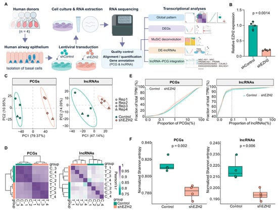
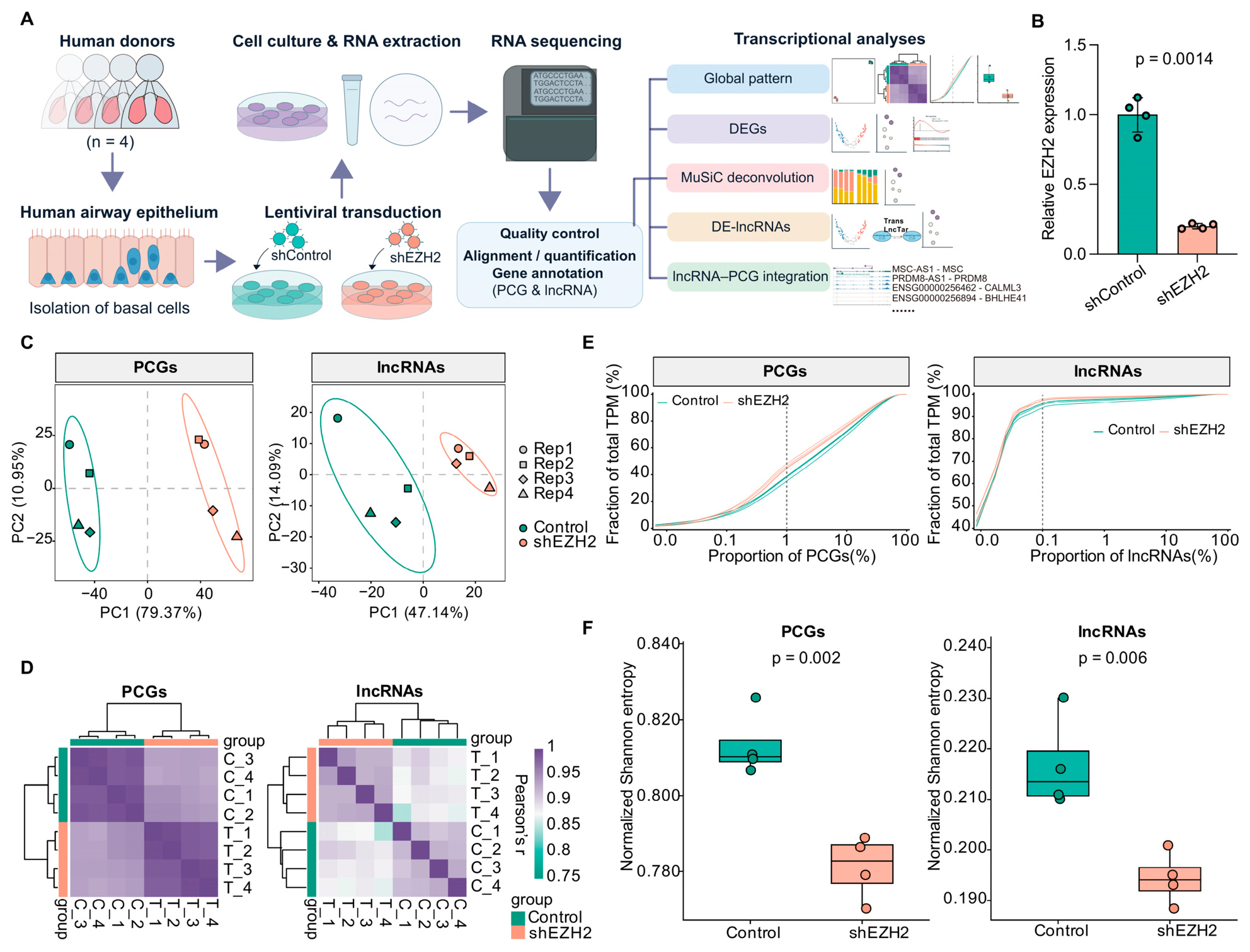
To characterize the global transcriptional consequences of EZH2 knockdown in primary airway basal cells, control and shEZH2 cells from four independent human were subjected to RNA sequencing and integrated analysis of coding and non-coding transcripts (Figure 1A). EZH2 knockdown was confirmed by RT-qPCR, with significantly reduced EZH2 expression in shEZH2 cells relative to matched controls (p = 0.0014; Figure 1B). Western blot analysis further showed reduced EZH2 protein levels and decreased H3K27me3 in the shEZH2 group (Figure S1). RNA sequencing generated 26.46–32.25 million paired-end reads per sample, with STAR alignment rates of 95.63–96.41% across the eight libraries (Supplementary Table S1). After expression filtering, 15,483 PCGs and 11,769 lncRNAs were retained for downstream analyses. Principal component analysis (PCA) separated control and shEZH2 samples based on both PCG and lncRNA expression profiles, with PC1 explaining 79.37% and 47.14% of the variance, respectively (Figure 1C). Sample-wise expression correlations were generally higher within than between conditions for both transcript classes (Figure 1D), and hierarchical clustering similarly grouped samples according to treatment condition (Figure S2A,B). Transcript abundance was markedly more concentrated among lncRNAs than PCGs, with just 0.1% of lncRNAs accounting for over 90% of total lncRNA abundance, compared with 1% of PCGs contributing 50% of total PCG abundance (Figure 1E). EZH2 knockdown further increased expression concentration in both transcript classes, accompanied by significantly reduced normalized Shannon entropy for PCGs (p = 0.002) and lncRNAs (p = 0.006) (Figure 1F). To assess whether highly abundant lncRNAs disproportionately influenced this result, we identified the top 0.1% most abundant lncRNAs (13 unique transcripts) and recalculated entropy after excluding these transcripts, applying stricter count-based filtering, or combining both approaches. Mean normalized entropy remained lower in shEZH2 samples under all three settings (p = 0.0253, 0.0184, and 0.0253, respectively) (Figure S2C). These findings indicate broad remodeling of both coding and non-coding expression profiles following EZH2 knockdown, including a less even distribution of transcript abundance.
Figure 1.
Experimental design and global transcriptomic characteristics. (A) Schematic of the experimental design and analysis workflow, including primary airway basal cell culture, lentiviral transduction, RNA sequencing, and downstream transcriptomic analyses. (B) qPCR validation of EZH2 knockdown in four paired samples. Statistical significance was assessed using a paired two-tailed Student’s t-test. (C) PCA plots of PCG and lncRNA expression profiles across the eight samples. (D) Sample correlation heatmaps based on PCG and lncRNA expression profiles and the heatmap color scales indicate pairwise Pearson correlation coefficients (r). (E) Cumulative abundance distributions of PCGs and lncRNAs, with transcripts ranked by TPM. (F) Shannon entropy of PCG and lncRNA expression distributions. Statistical significance was assessed using a paired two-tailed Student’s t-test.
3.2. EZH2 Knockdown Suppresses Proliferative and Chromatin-Associated PCG Programs
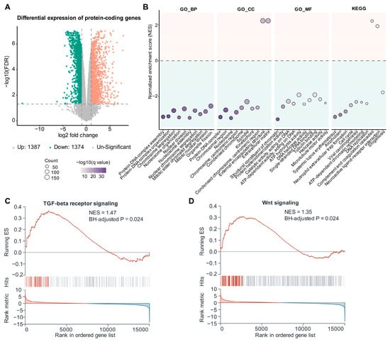
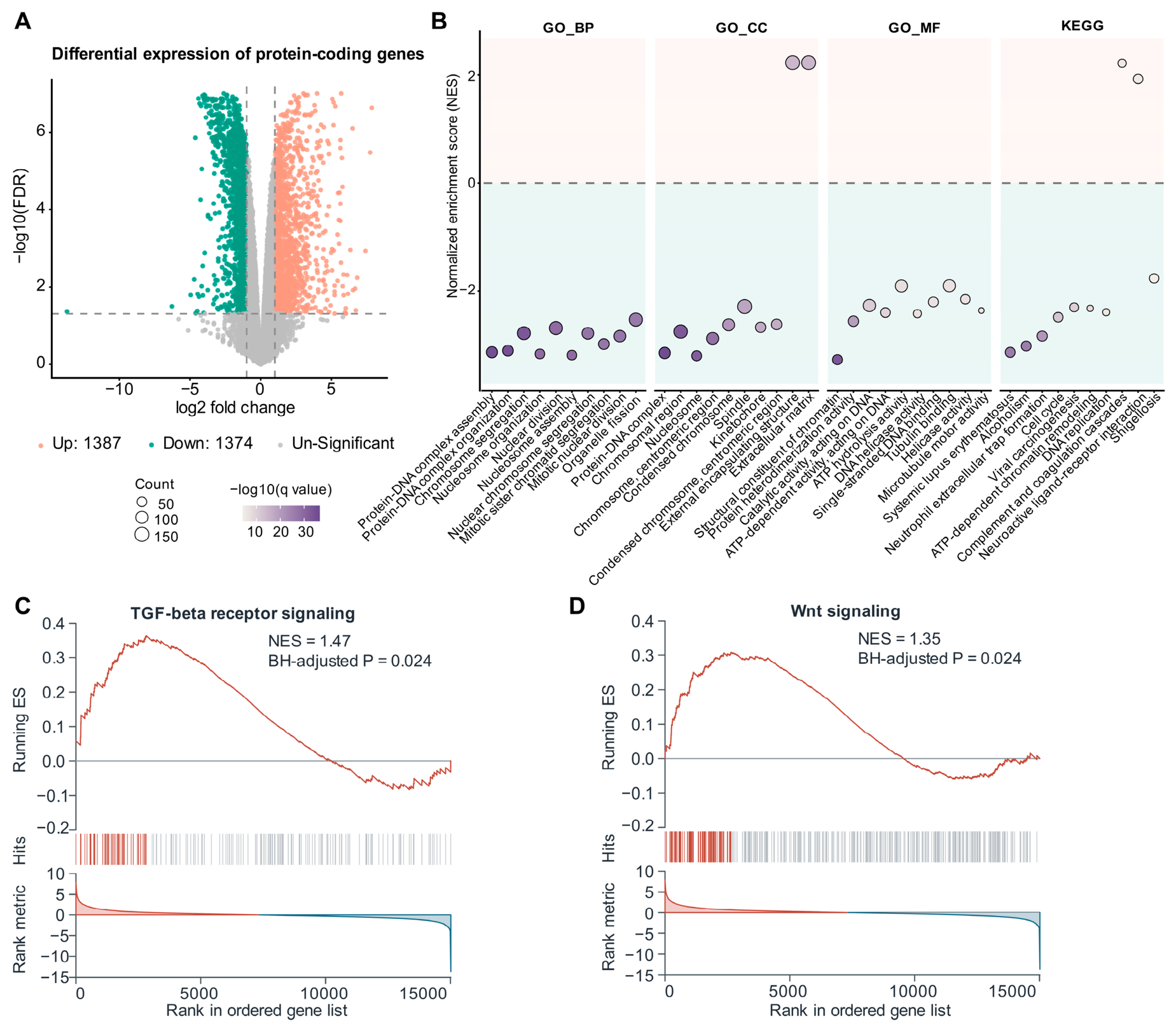
To define the protein-coding transcriptional changes associated with EZH2 knockdown, differential-expression and gene set enrichment analyses were performed across the 15,483 retained PCGs. A total of 2761 PCGs were differentially expressed, including 1387 upregulated and 1374 downregulated genes (Figure 2A and Supplementary Table S2). Hierarchical clustering of the differentially expressed PCGs separated control and shEZH2 samples according to treatment condition (Figure S3A). GSEA revealed significant negative enrichment of pathways involved in chromatin and nucleosome organization, chromosome segregation and nuclear division, DNA replication, and cell-cycle progression following EZH2 knockdown (Figure 2B and Supplementary Table S2). Positive enrichment was observed in a more restricted set of pathways, including extracellular matrix and external encapsulating structure organization, complement and coagulation cascades, and neuroactive ligand–receptor interaction. Given the established involvement of TGF-β, WNT, BMP, EGF, FGF, and Notch signaling in airway basal cell fate and differentiation potential, these pathways were further examined by GSEA. TGF-β and WNT signaling showed significant positive enrichment (Figure 2C,D), whereas the enrichment profiles of BMP, EGF, FGF, and Notch signaling are presented in Figure S3B–E. Collectively, EZH2 knockdown elicited a transcriptional response characterized by negative enrichment of chromatin-, DNA replication-, and cell-cycle-associated programs, accompanied by more selective positive enrichment of extracellular and signaling-related processes.
Figure 2.
Differential expression and functional enrichment of PCGs. (A) Volcano plot of PCG differential expression between shEZH2 and control cells. Differential expression was defined as FDR < 0.05 and |log2FC| > 1. (B) GSEA of GO-BP, GO-CC, GO-MF, and KEGG gene sets. The top 10 significant terms in each category are shown. Positive and negative NESs indicate enrichment toward shEZH2 and control, respectively. Dot size represents gene count, and color represents −log10(BH-adjusted p value). BH-adjusted p < 0.05 was considered significant. (C,D) GSEA enrichment plots for TGF-β receptor signaling (C) and WNT signaling (D).
3.3. EZH2 Knockdown Alters Transcriptional Similarity to the Features of Basal Epithelial States
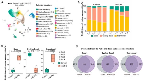
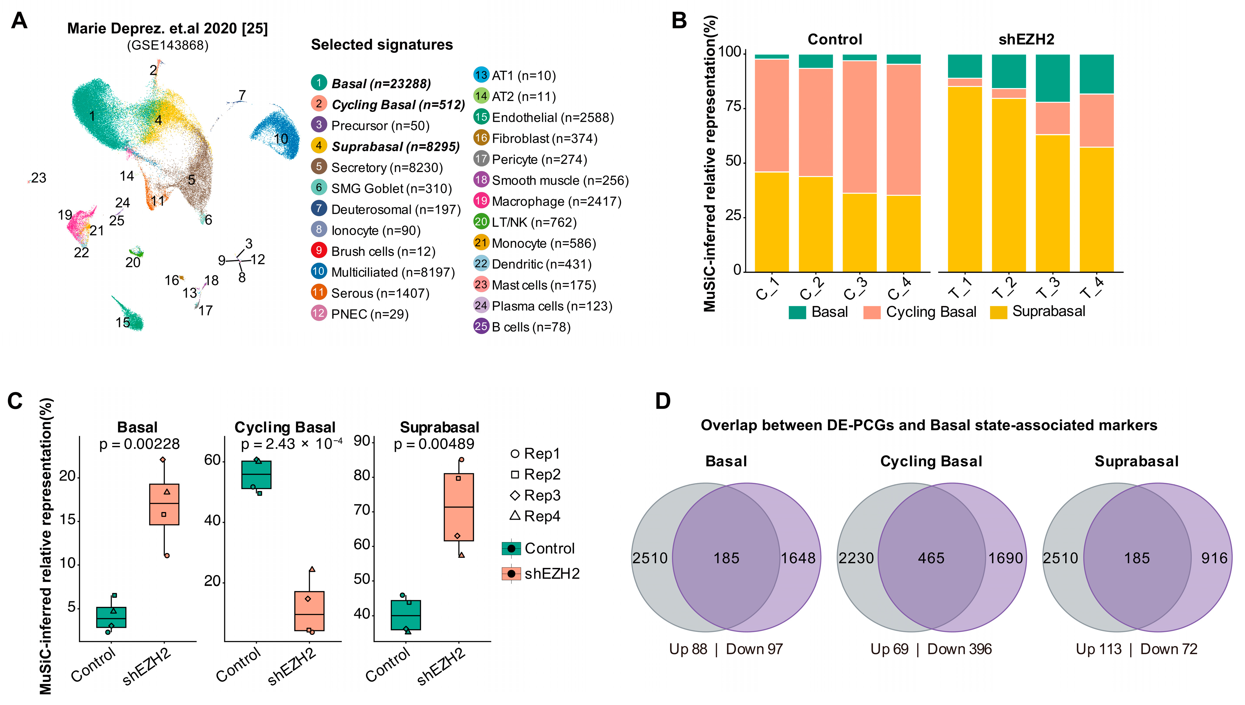
To assess the effects of EZH2 knockdown on airway basal epithelial transcriptional states, we performed MuSiC-based deconvolution using the healthy human airway single-cell atlas of Deprez et al., focusing on Basal, Cycling Basal, and Suprabasal transcriptional signatures (Figure 3A). Following EZH2 knockdown, the inferred contribution of Cycling Basal cells decreased markedly from 55.5% to 11.9% (p = 2.43 × 10⁻⁴), whereas Suprabasal and Basal cells increased from 40.3% to 71.3% (p = 0.00489) and from 4.2% to 16.8% (p = 0.00228), respectively (Figure 3B,C and Supplementary Table S3).
Figure 3.
MuSiC-inferred basal cell-state representation and overlap with state-associated markers. (A) Airway epithelial single-cell reference from Deprez et al., with the cell signatures retained for MuSiC analysis indicated. (B) MuSiC-inferred relative representation of Basal, Cycling Basal, and Suprabasal states across individual samples. (C) Comparison of the inferred relative representation of the three basal cell states between control and shEZH2 samples. Statistical significance was assessed using paired two-tailed Student’s t-tests. (D) Overlap between DEGs and positive markers associated with Basal, Cycling Basal, and Suprabasal states. The analysis included 2695 of the 2761 DE-PCGs that were successfully mapped to Entrez Gene IDs. Overlapping genes are separated according to their direction of differential expression. The Venn diagrams are not drawn to scale.
To further assess the robustness of these findings, we expanded the reference to eight airway epithelial populations. The same pattern persisted, with reduced Cycling Basal representation (54.03% to 10.04%, p = 3.635 × 10⁻⁵) and increased Basal (5.05% to 16.11%, p = 0.010395) and Suprabasal representation (39.85% to 57.89%, p = 0.030436). Serous representation also increased (0% to 4.93%, p = 0.015456), whereas Secretory representation remained statistically unchanged (p = 0.146492), and Multiciliated representation was zero across all samples (Figure S4A,B and Supplementary Table S3). Excluding cell-cycle genes yielded consistent results, with decreased Cycling Basal (58.99% to 18.16%, p = 5.970 × 10⁻⁵) and increased Basal (1.87% to 10.34%, p = 0.0057) and Suprabasal representation (38.22% to 59.39%, p = 0.0021) (Figure S4C,D and Supplementary Table S3). Thus, the inferred transcriptional shifts were robust to reference expansion and cell-cycle gene exclusion.
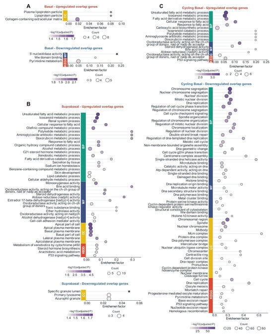
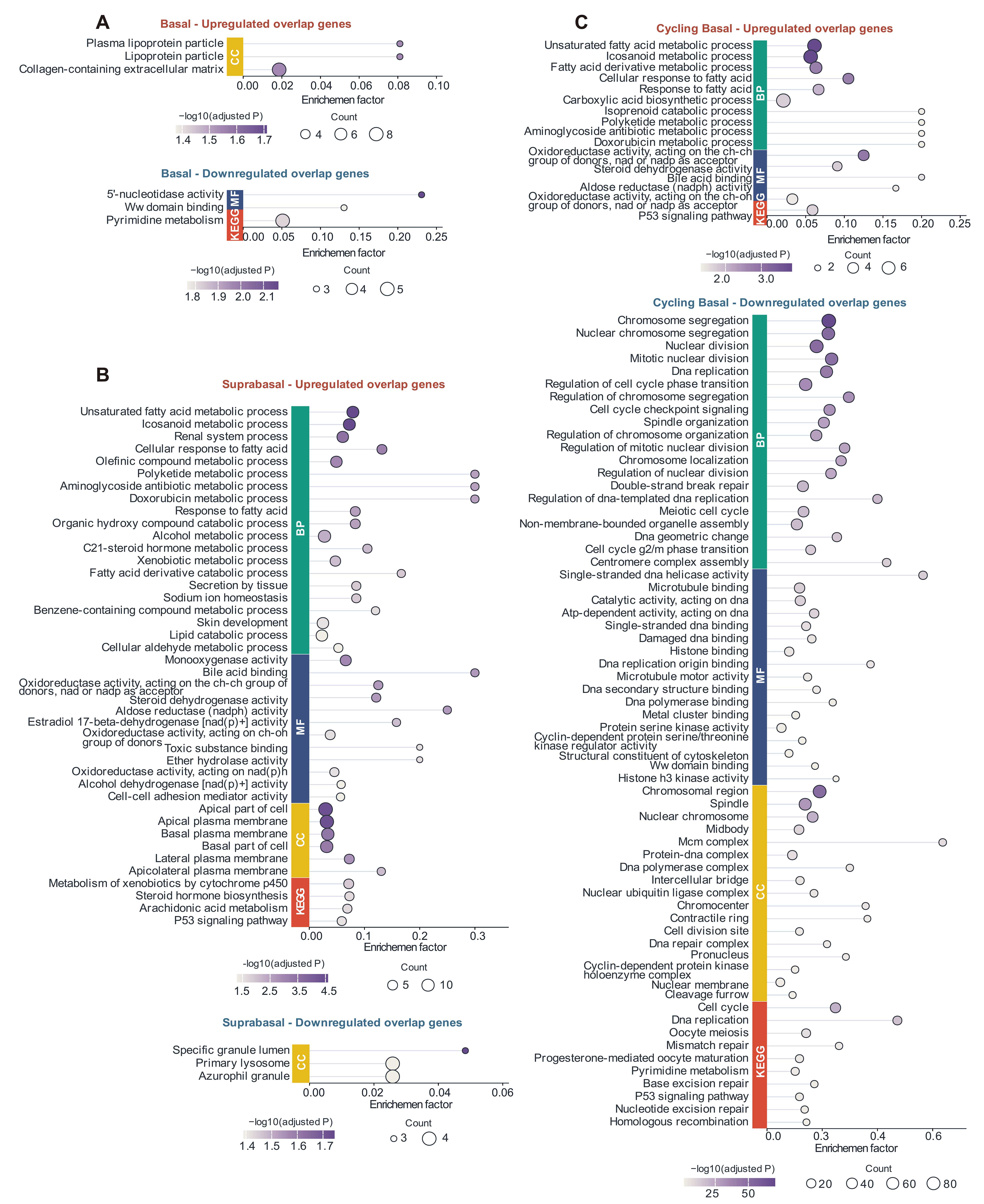
To characterize the transcriptional programs associated with these reference-defined states, DEGs were intersected with positive markers of each reference-defined cell state (Figure 3D). Basal and Suprabasal signatures each overlapped with 185 differentially expressed PCGs, comprising 88 upregulated and 97 downregulated genes for Basal and 113 upregulated and 72 downregulated genes for Suprabasal. The largest overlap was observed for Cycling Basal, with 465 differentially expressed PCGs, of which 396 (85.2%) were downregulated following EZH2 knockdown. Functional enrichment revealed distinct patterns among the state-associated DEGs (Figure 4; Supplementary Table S4). Basal-associated genes showed relatively limited enrichment, whereas upregulated Suprabasal-associated genes were mainly enriched in lipid and fatty-acid metabolism and related metabolic processes (Figure 4A,B). Interestingly, Cycling Basal-associated genes showed a strongly asymmetric functional pattern: the smaller upregulated subset was mainly enriched in fatty-acid metabolic and oxidoreductase-related processes, whereas the much larger downregulated subset was strongly enriched in chromosome segregation, DNA replication, and cell-cycle-related programs (Figure 4C). These findings suggest that EZH2 knockdown shifts basal epithelial transcriptional profiles away from proliferation-associated programs toward non-cycling basal and suprabasal signatures, without necessarily reflecting changes in cellular identity or actual cell-type proportions.
Figure 4.
Functional enrichment of DEGs overlapping with basal state-associated markers. Functional enrichment of upregulated and downregulated DEGs overlapping with Basal (A), Suprabasal (B), and Cycling Basal (C) state-associated markers. Up to the top 20 significant terms or pathways are shown for each category; all are displayed when fewer than 20 were identified. Terms with BH-adjusted p < 0.05 were considered significant. The x-axis indicates enrichment factor, dot size represents gene count, and color represents −log10(adjusted p value).
3.4. Differential lncRNAs Are Associated with Distinct Transcriptional Programs Following EZH2 Knockdown
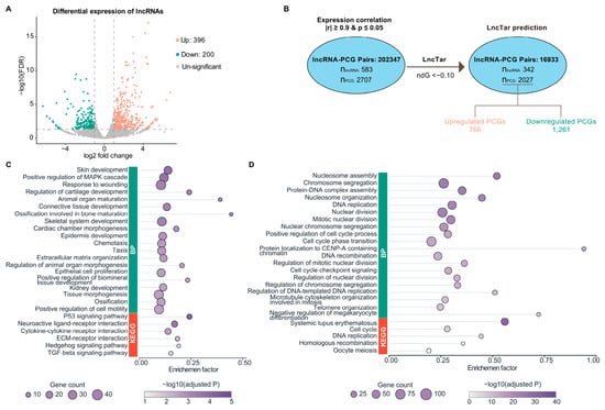
To characterize the non-coding transcriptional response to EZH2 knockdown and its association with PCG changes, differential lncRNA expression and lncRNA–PCG expression associations were analyzed. Among the 11,769 analyzed lncRNAs, 596 were differentially expressed (DE-lncRNAs), including 396 upregulated and 200 downregulated lncRNAs (Figure 5A and Supplementary Table S5). Hierarchical clustering of the DE-lncRNAs separated control and shEZH2 samples according to treatment condition (Figure S5A). Correlation analysis between the 596 DE-lncRNAs and 2761 DEGs across the eight samples identified 202,347 expression associations meeting the predefined criteria of |r| ≥ 0.90 and p < 0.05, involving 583 lncRNAs and 2707 PCGs. Subsequent sequence-complementarity filtering using LncTar retained 16,933 candidate associations involving 342 lncRNAs and 2027 PCGs (Figure 5B), of which 766 PCGs were upregulated and 1261 were downregulated following EZH2 knockdown.
Figure 5.
Differential lncRNA expression and functional enrichment of lncRNA-associated PCGs. (A) Volcano plot of differentially expressed lncRNAs between shEZH2 and control cells. Differential expression was defined as FDR < 0.05 and |log2FC| > 1 with marked dashed lines. (B) Selection of lncRNA–PCG candidate relationships by expression correlation (|r| ≥ 0.9, p ≤ 0.05), followed by LncTar prediction using ndG < −0.10. (C,D) BP and KEGG enrichment of lncRNA-associated upregulated (C) and downregulated (D) PCGs. Up to the 20 most significant terms or pathways are shown; all are displayed when fewer than 20 were identified. Terms with BH-adjusted p < 0.05 were considered significant. The x-axis indicates enrichment factor, dot size represents gene count, and color represents −log10(adjusted p value).
To infer the biological functions potentially associated with differentially expressed lncRNAs, these lncRNA-associated PCGs were stratified according to their direction of differential expression and subjected to functional enrichment analysis. Upregulated PCGs were enriched in extracellular matrix organization, response to wounding, skin development, and positive regulation of the MAPK cascade, with significant pathway enrichment also involving TGF-β signaling, Hedgehog signaling, ECM–receptor interaction, cytokine–cytokine receptor interaction, and p53 signaling (Figure 5C and Figure S5B). In contrast, downregulated PCGs were prominently enriched in nucleosome assembly, chromosome segregation, DNA replication, nuclear division, and cell-cycle-related processes, with cell cycle, DNA replication, and homologous recombination also represented among the significant pathways (Figure 5D and Figure S5C). These contrasting functional patterns suggest a potential dual role for the altered lncRNA landscape following EZH2 knockdown, potentially promoting extracellular and signaling-related remodeling while restraining proliferation, DNA replication, and chromatin-associated processes.
3.5. Multi-Criteria Prioritization Identifies Consensus lncRNA–PCG Candidates
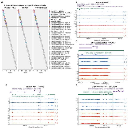
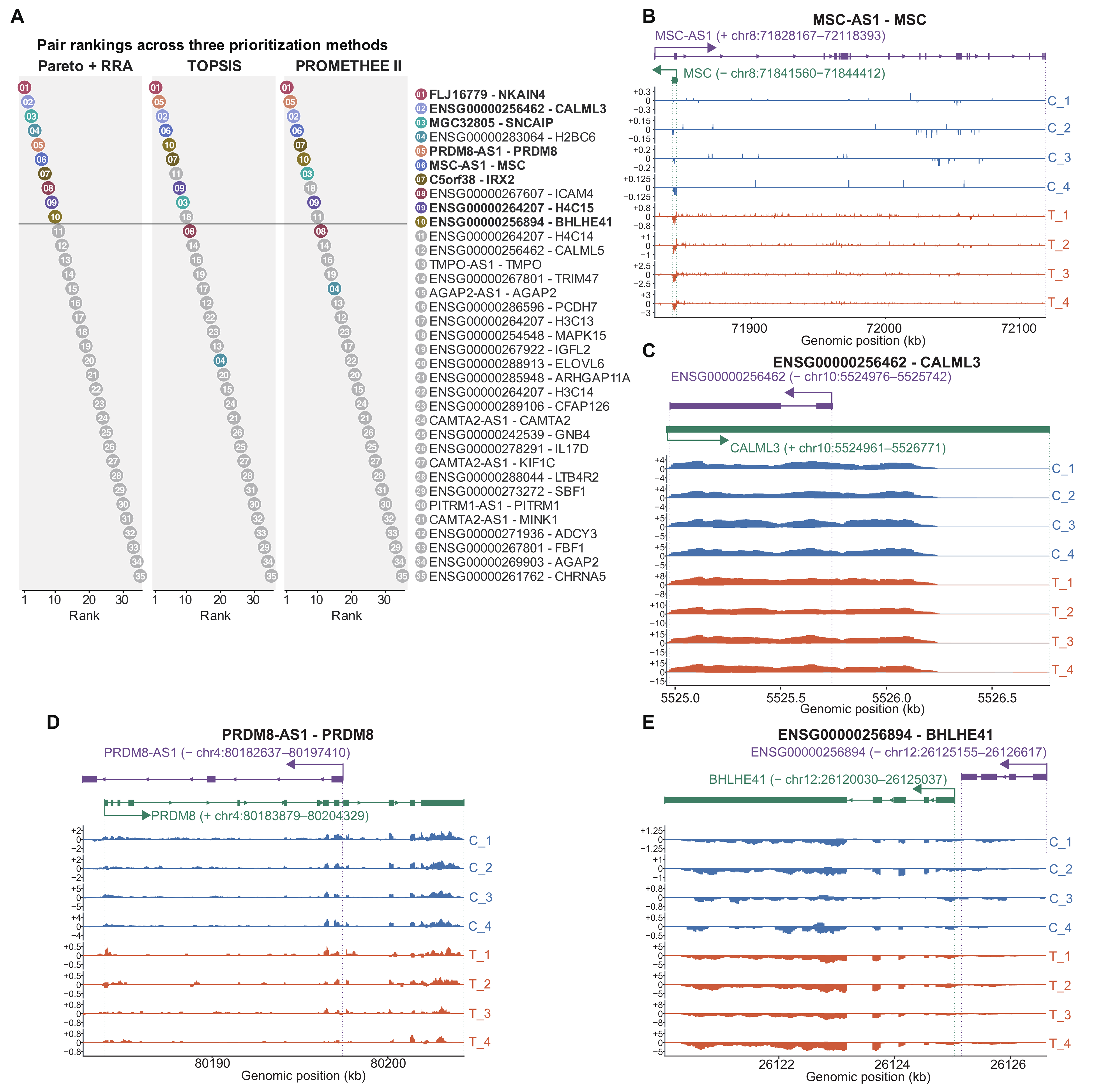
To further prioritize lncRNA–PCG candidates for investigation, the 16,933 associations supported by expression correlation and LncTar were refined according to genomic proximity, yielding 35 associations located within 100 kb (Figure 6A and Supplementary Table S6). These candidates were evaluated and ranked using Pareto–RRA, TOPSIS, and PROMETHEE II. The three ranking methods showed high overall concordance, with Spearman correlation coefficients of 0.909 between Pareto–RRA and TOPSIS, 0.934 between Pareto–RRA and PROMETHEE II, and 0.989 between TOPSIS and PROMETHEE II. (Supplementary Table S6) Intersection of the top 10 candidates identified by each method yielded eight consensus candidates: FLJ16779-NKAIN4, ENSG00000256462-CALML3, MGC32805-SNCAIP, PRDM8-AS1-PRDM8, MSC-AS1-MSC, C5orf38-IRX2, ENSG00000264207-H4C15, and ENSG00000256894-BHLHE41. The eight prioritized pairs showed strong pooled correlations (r = 0.937–0.989), all significant after BH correction. After adjustment for treatment and donor effects, correlations remained positive (r = 0.366–0.996), but none remained significant after multiple-testing correction. Across LODO analyses, pooled correlations ranged from 0.919 to 0.996, with a maximum within-pair range of 0.067 (Supplementary Table S7). These results indicate limited sensitivity of the pooled correlations to individual donor pairs, without establishing treatment-independent associations. Under stricter count-based filtering, MGC32805 was excluded because of insufficient count support, whereas both genes in the remaining seven original pairs remained differentially expressed. Re-ranking eligible pairs within the original 35-pair cis candidate pool retained all seven in the three-method Top 10 intersection (Supplementary Table S7). All eight pairs were retained for subsequent genomic characterization, including MGC32805–SNCAIP as an exploratory candidate whose biological relevance remains to be assessed despite its limited count support.
Figure 6.
Multi-method prioritization and genomic characterization of candidate lncRNA–PCG relationships. (A) Rankings of 35 candidate lncRNA–PCG relationships using Pareto–RRA, TOPSIS, and PROMETHEE II. Candidates are independently ordered within each method; identical colors and numbers denote the same highlighted candidates across methods, while the remaining candidates are shown in gray. (B–E) Genomic organization and RNA-seq coverage profiles of MSC-AS1-MSC (B), ENSG00000256462-CALML3 (C), PRDM8-AS1-PRDM8 (D), and ENSG00000256894-BHLHE41 (E). Gene models indicate genomic organization and transcriptional orientation. Strand-specific RNA-seq coverage is shown for four control (C1–C4) and four shEZH2 (T1–T4) samples, with positive- and negative-strand signals displayed above and below the zero baseline, respectively. Blue and orange coverage tracks represent control and shEZH2 samples, respectively.
The eight consensus candidates showed coordinated transcriptional changes following EZH2 knockdown. PRDM8-AS1-PRDM8 and ENSG00000264207-H4C15 were concordantly downregulated of both the lncRNA and corresponding PCG, whereas the other six showed concordant upregulation. Expression relationships across the eight RNA-seq samples are shown in Figure S6E. The candidates also showed close genomic relationships: three lncRNA–PCG pairs were located within 500 bp of each other (ENSG00000264207-H4C15: 112 bp; ENSG00000256894-BHLHE41: 118 bp; C5orf38-IRX2: 454 bp), and the other five were loci that overlapped. Locus-level RNA-seq coverage profiles of representative candidates further illustrated their genomic organization and treatment-associated expression patterns (Figure 6B–E and S6A–D). Together, these findings identify a set of EZH2-responsive lncRNA–PCG relationships that might provide candidates for further investigation of how coding and non-coding transcriptional programs are coordinated following EZH2 depletion.
4. Discussion
EZH2 has an established role in restricting epithelial lineage programs during lung development, but its contribution to the transcriptional configuration of adult human airway basal cells, particularly in coordinating coding and non-coding transcription, is less well defined [6,33]. Here, using primary human airway basal epithelial cells, we found EZH2 knockdown is accompanied by coordinated changes in protein-coding and non-coding RNA expression, a pronounced attenuation of proliferation- and chromatin-associated gene programs, and a shift in transcriptional similarity away from Cycling Basal toward non-cycling Basal and Suprabasal reference states. Together, our findings extend the role of EZH2 from developmental lung epithelial lineage regulation to the transcriptional organization of adult human airway basal cells and implicate non-coding transcription as an additional component of the response to EZH2 depletion.
The most prominent PCG-level consequence of EZH2 depletion was a coordinated attenuation of proliferative and chromatin-associated programs. Cell-cycle progression, DNA replication, chromosome segregation, DNA repair, and nucleosome-related functions were broadly suppressed. These interconnected processes are central to the proliferative capacity of airway basal cells, which serve as self-renewing progenitors and expand in response to epithelial injury [34]. Their concordant suppression was consistent with the marked decrease in MuSiC-inferred Cycling Basal representation, together indicating a shift away from a proliferation-dominant basal-cell state. This interpretation is also supported by evidence from adult lung organoids, in which EZH2 loss reduced self-renewal and substantially altered epithelial progenitor populations [7]. Importantly, the transcriptional response was not limited to reduced proliferation. Upregulated PCGs preferentially involved extracellular matrix organization, cell–matrix interactions, and signaling-related processes, while TGF-β and WNT pathways showed positive enrichment. The extracellular matrix is an active component of the airway basal-cell niche and contributes to the regulation of basal-cell proliferation, stemness, and differentiation [35]. TGF-β and WNT signaling are likewise closely linked to airway basal-cell behavior: TGF-β/SMAD signaling can restrain basal-cell proliferation, whereas the effects of WNT signaling depend on cellular and temporal context and can extend from proliferative responses to altered epithelial differentiation [36]. Thus, the concurrent decrease in Cycling Basal representation and increase in non-cycling Basal and Suprabasal representation may reflect a broader redistribution from replication- and chromatin-dominant programs toward extracellular, signaling, and non-cycling epithelial features after EZH2 depletion.
Selective positive enrichment of extracellular matrix, TGF-beta, and WNT-related programs suggests accompanying changes in extracellular and signaling responses. TGF-beta activated through integrin alpha-v-beta-8 can inhibit proliferation in human bronchial epithelium [37]. This direction is consistent with the attenuated proliferative program observed here, although transcriptional enrichment alone does not establish increased TGF-β pathway activity or causality. WNT signaling presents a more complex relationship. During airway regeneration, WNT/β-catenin activity is required for basal-cell proliferation after injury, whereas WNT ligands derived from basal cells at later stages promote early progenitor differentiation toward ciliated cells, demonstrating distinct stage- and source-dependent functions [38]. Consistent with this context dependence, experimental activation of WNT/β-catenin signaling can induce airway basal-cell hyperproliferation and loss of ciliated-cell differentiation [39], while persistent WNT stimulation in primary human airway basal cells can redirect differentiation toward a squamous epithelial program [36]. Notably, in the latter model, TGF-β suppressed proliferation without preventing WNT-associated squamous differentiation, indicating that proliferative activity and epithelial-state remodeling need not change in parallel. Thus, the positive enrichment of WNT-related genes despite reduced Cycling Basal representation is not necessarily contradictory. Rather than indicating increased WNT-driven proliferation, the concurrent TGF-β and WNT signatures may reflect broader signaling reorganization accompanying the shift away from a proliferation-dominant transcriptional state after EZH2 depletion.
The broad lncRNA response to EZH2 depletion indicates that transcriptional remodeling in airway basal cells extends beyond protein-coding genes. EZH2 knockdown in embryonic stem cells derepresses a subset of H3K27me3-associated lncRNAs, providing direct evidence that lncRNA expression can be subject to EZH2-dependent chromatin regulation [40]. EZH2 depletion also produces bidirectional changes in lncRNA expression in neuroblastoma cells [41]. In the human airway epithelium, perturbation of the HOTAIR–EZH2 interaction alters H3K27me3 and cigarette-smoke-induced inflammatory responses in primary epithelial cells [42]. LncRNAs can also influence bronchial epithelial state independently of EZH2; TINCR depletion in primary human bronchial epithelial cells alters differentiation, wound-response, and extracellular-matrix programs and promotes a basal-like phenotype [43]. These observations support a role for lncRNAs in airway epithelial biology, whereas the broader non-coding response to EZH2 depletion in primary human airway basal cells remains poorly characterized. In our study, EZH2 depletion was accompanied by broad lncRNA remodeling, and PCGs associated with these lncRNAs partially converged with the independently defined protein-coding response. The decreased component was preferentially associated with chromatin, DNA replication, and proliferative functions, consistent with the attenuation of cell-cycle and chromatin-associated programs described above. The increased component was associated with extracellular matrix, wound-response, and signaling processes, including TGF-β and Hedgehog pathways. The recurrence of these functional themes across the two analytical layers links the lncRNA response to the broader transcriptional reorganization following EZH2 depletion. Their incomplete overlap is also informative, suggesting that the lncRNA-associated analysis captures features not apparent from differential PCG expression alone. The shift from proliferative and chromatin-associated programs toward extracellular and signaling-related programs is therefore reflected at both coding and non-coding levels.
Identifying specific lncRNA–PCG relationships within this broad transcriptional response requires evidence beyond expression correlation, as coordinated changes may simply reflect parallel responses to EZH2 depletion. Integrating complementary computational features prioritized eight pairs with concordant expression changes. Seven retained their consensus prioritization after stricter count-based filtering and re-ranking, supporting the consistency of their selection, whereas MGC32805 was excluded because of limited count support. Nevertheless, low abundance does not necessarily preclude biological function, as some lncRNAs exert local regulatory effects at low copy numbers [44]. We therefore retained all eight pairs as exploratory associations. Several of these directional changes are biologically informative in the context of the broader transcriptional phenotype. Among these candidates, ENSG00000256462 and CALML3 were both upregulated after EZH2 depletion. CALML3 is preferentially expressed in differentiating suprabasal keratinocytes in human epidermis and is inversely associated with Ki-67 [45], making its increased expression directionally consistent with the reduced Cycling Basal and increased Suprabasal representation observed here. In contrast, PRDM8-AS1 and PRDM8 were both downregulated. PRDM8 has been implicated in differentiation and transcriptional regulation [46], and has been reported to possess histone H3K9 methyltransferase activity and can mediate transcriptional repression [47]; their concordant decrease therefore occurred alongside the broader attenuation of chromatin-associated programs after EZH2 depletion. MSC-AS1 and MSC were both upregulated. MSC is a transcriptional regulator implicated in differentiation-related processes in other biological contexts [48]. MSC-AS1 has also been associated with cellular proliferation [49], and linked to WNT/β-catenin signaling and proliferation through a miR-3924/WNT5A-dependent mechanism in renal carcinoma cells [50]. Its increase is directionally compatible with the WNT-related enrichment observed here, although the reduced proliferative program in our airway basal cells differs from its reported pro-proliferative activity in that tumor context, and whether these functions involve MSC remains unknown. ENSG00000256894 and BHLHE41 were concurrently upregulated; BHLHE41 can repress cell-cycle regulators in other cellular contexts [51], a direction compatible with the attenuated proliferative program observed here. MGC32805–SNCAIP has also been reported as a positively correlated lncRNA–PCG pair in a transcriptomic study of medulloblastoma, where both genes were upregulated [52]. This observation provides additional context for the association identified here, although its functional significance in airway basal cells remains unclear. The remaining three candidates have more limited or less directly relevant functional evidence. Whereas previous in vivo studies have shown that EZH2 restricts basal-lineage acquisition during lung development, our findings in primary human airway BCs show that EZH2 depletion is accompanied by broad attenuation of proliferative transcriptional programs and altered resemblance to reference-defined basal epithelial states. Given the importance of basal-cell state regulation for epithelial repair and remodeling, these findings provide a basis for investigating whether EZH2-associated transcriptional regulation contributes to dysregulated epithelial responses in respiratory disease. How the prioritized lncRNA–PCG candidates participate in these processes—as drivers, modulators, or consequences of the transcriptional response—remains to be determined.
Several limitations should be considered. The small samples and single shRNA limit causal inference and generalizability. Independent EZH2 perturbation and rescue, direct proliferation and air-liquid interface differentiation assays, and H3-normalized H3K27me3 measurements are needed to validate the inferred phenotypes. Chromatin profiling and targeted lncRNA perturbation would further test direct EZH2 regulation and candidate lncRNA-PCG relationships. Collectively, these findings identify coordinated coding and non-coding transcriptional responses to EZH2 depletion in primary human airway basal cells, while leaving their causal links to epithelial fate unresolved. Future studies incorporating differentiation-resolved profiling, targeted perturbation of selected candidates, and disease-relevant epithelial models are needed to clarify candidate functions and their relevance to airway epithelial remodeling.
5. Conclusions
This study characterizes coding and non-coding transcriptomic alterations in EZH2 knockdown of primary human airway basal epithelial cells. We identified coordinated changes in protein-coding genes and lncRNAs, accompanied by altered transcriptional similarity to reference-defined basal epithelial states, extending the known role of EZH2 in epithelial proliferation and differentiation. Although limited by the single-shRNA design and the lack of statistically significant lncRNA–PCG associations after adjustment for treatment, sample effects, and multiple testing, these findings provide a foundation for investigating lncRNA-associated transcriptional regulation in human airway basal cells.
Supplementary Materials
The following supporting information can be downloaded at: https://www.mdpi.com/article/10.3390/genes17111479/s1, Figure S1: Western blot analysis of EZH2 and H3K27me3 protein levels in primary human airway basal cells; Figure S2: Unsupervised clustering of PCG and lncRNA expression profiles; Figure S3: Differential PCG expression patterns and GSEA of additional selected signaling pathways; Figure S4: Expanded MuSiC-inferred airway epithelial representation; Figure S5: Expression patterns of differentially expressed lncRNAs and additional functional enrichment of lncRNA-associated PCGs; Figure S6: Genomic organization, RNA-seq coverage, and expression relationships of priori-tized lncRNA–PCG candidates; Table S1: RNA-seq summary and quality metrics; Table S2: DEGs and functional enrichment results; Table S3: MuSiC-inferred relative representation; Table S4: Functions of DEGs associated with Basal, Suprabasal, and Cy-cling Basal states; Table S5: DE-lncRNAs and functional enrichment; Table S6: Multi-method prioritization and final lncRNA–PCG candidates; Table S7: Sensitivity analysis of the prioritized lncRNA–PCG candidates.
Author Contributions
Conceptualization, X.T. and F.L.; methodology, S.H., L.C. (Lu Chen) and L.C. (Ling Chen); software, S.H.; validation, S.H., L.C. (Lu Chen) and L.C. (Ling Chen); formal analysis, S.H.; investigation, L.C. (Ling Chen), L.Z. and C.L.; resources, L.Z. and C.L.; data curation, S.H.; writing—original draft preparation, S.H., L.C. (Lu Chen) and L.C. (Ling Chen); writing—review and editing, S.H., X.T., L.C. (Lu Chen) and L.C. (Ling Chen); visualization, S.H., X.T., L.C. (Lu Chen); supervision, X.T. and F.L.; project administration, X.T. and F.L.; funding acquisition, S.H., X.T. and F.L. All authors have read and agreed to the published version of the manuscript.
Funding
This research was funded by the Noncommunicable Chronic Diseases—National Science and Technology Major Project (No.2025ZD0551807) and the Postdoctoral Research Fund of West China Hospital, Sichuan University (No. 2024HXBH060).
Institutional Review Board Statement
The study was conducted in accordance with the Declaration of Helsinki and approved by the Biomedical Ethics Review Committee, West China Hospital of Sichuan University (approval No. 2025-797; approval date: 13 June 2025).
Informed Consent Statement
Written informed consent for participation was obtained from all donors involved in the study.
Data Availability Statement
The data presented in this study was submitted in National Genomics Data Center (https://ngdc.cncb.ac.cn/gsa-human) under accession number HRA021349 (https://ngdc.cncb.ac.cn/gsa-human/s/SKehVIPG accessed on 28 September 2026), and are available from the corresponding author upon reasonable request, subject to applicable ethical and privacy considerations. Analysis scripts are available from the corresponding author upon reasonable request.
Conflicts of Interest
The authors declare no conflicts of interest.
References
- Sikkema, L.; Ramírez-Suástegui, C.; Strobl, D.C.; Gillett, T.E.; Zappia, L.; Madissoon, E.; Markov, N.S.; Zaragosi, L.-E.; Ji, Y.; Ansari, M.; et al. An Integrated Cell Atlas of the Lung in Health and Disease. Nat. Med. 2023, 29, 1563–1577. [Google Scholar] [CrossRef] [Scilit] [PubMed]
- Madissoon, E.; Oliver, A.J.; Kleshchevnikov, V.; Wilbrey-Clark, A.; Polanski, K.; Richoz, N.; Ribeiro Orsi, A.; Mamanova, L.; Bolt, L.; Elmentaite, R.; et al. A Spatially Resolved Atlas of the Human Lung Characterizes a Gland-Associated Immune Niche. Nat. Genet. 2023, 55, 66–77. [Google Scholar] [CrossRef] [Scilit] [PubMed]
- Ma, L.; Thapa, B.R.; Le Suer, J.A.; Tilston-Lünel, A.; Herriges, M.J.; Berical, A.; Beermann, M.L.; Wang, F.; Bawa, P.S.; Kohn, A.; et al. Airway Stem Cell Reconstitution by the Transplantation of Primary or Pluripotent Stem Cell-Derived Basal Cells. Cell Stem Cell 2023, 30, 1199–1216.e7. [Google Scholar] [CrossRef] [Scilit] [PubMed]
- Jaeger, B.; Schupp, J.C.; Plappert, L.; Terwolbeck, O.; Artysh, N.; Kayser, G.; Engelhard, P.; Adams, T.S.; Zweigerdt, R.; Kempf, H.; et al. Airway Basal Cells Show a Dedifferentiated KRT17highPhenotype and Promote Fibrosis in Idiopathic Pulmonary Fibrosis. Nat. Commun. 2022, 13, 5637. [Google Scholar] [CrossRef] [Scilit] [PubMed]
- Stoleriu, M.G.; Ansari, M.; Strunz, M.; Schamberger, A.; Heydarian, M.; Ding, Y.; Voss, C.; Schneider, J.J.; Gerckens, M.; Burgstaller, G.; et al. COPD Basal Cells Are Primed towards Secretory to Multiciliated Cell Imbalance Driving Increased Resilience to Environmental Stressors. Thorax 2024, 79, 524–537. [Google Scholar] [CrossRef] [Scilit] [PubMed]
- Snitow, M.E.; Li, S.; Morley, M.P.; Rathi, K.; Lu, M.M.; Kadzik, R.S.; Stewart, K.M.; Morrisey, E.E. Ezh2 Represses the Basal Cell Lineage during Lung Endoderm Development. Development 2015, 142, 108–117. [Google Scholar] [CrossRef] [Scilit] [PubMed]
- Byrd, A.L.; Qu, X.; Lukyanchuk, A.; Liu, J.; Chen, F.; Naughton, K.J.; DuCote, T.J.; Song, X.; Bowman, H.C.; Zhao, Y.; et al. Dysregulated Polycomb Repressive Complex 2 Contributes to Chronic Obstructive Pulmonary Disease by Rewiring Stem Cell Fate. Stem Cell Rep. 2023, 18, 289–304. [Google Scholar] [CrossRef] [Scilit] [PubMed]
- Le, H.Q.; Hill, M.A.; Kollak, I.; Keck, M.; Schroeder, V.; Wirth, J.; Skronska-Wasek, W.; Schruf, E.; Strobel, B.; Stahl, H.; et al. An EZH2-Dependent Transcriptional Complex Promotes Aberrant Epithelial Remodelling after Injury. EMBO Rep. 2021, 22, e52785. [Google Scholar] [CrossRef] [Scilit] [PubMed]
- Statello, L.; Guo, C.-J.; Chen, L.-L.; Huarte, M. Gene Regulation by Long Non-Coding RNAs and Its Biological Functions. Nat. Rev. Mol. Cell Biol. 2021, 22, 96–118, Erratum in Nat. Rev. Mol. Cell Biol. 2021, 22, 159. [Google Scholar] [CrossRef] [Scilit] [PubMed]
- Mattick, J.S.; Amaral, P.P.; Carninci, P.; Carpenter, S.; Chang, H.Y.; Chen, L.-L.; Chen, R.; Dean, C.; Dinger, M.E.; Fitzgerald, K.A.; et al. Long Non-Coding RNAs: Definitions, Functions, Challenges and Recommendations. Nat. Rev. Mol. Cell Biol. 2023, 24, 430–447. [Google Scholar] [CrossRef] [Scilit] [PubMed]
- Ferrer, J.; Dimitrova, N. Transcription Regulation by Long Non-Coding RNAs: Mechanisms and Disease Relevance. Nat. Rev. Mol. Cell Biol. 2024, 25, 396–415. [Google Scholar] [CrossRef] [Scilit] [PubMed]
- Aguilar, R.; Rosenberg, M.; Levy, V.; Lee, J.T. An Evolving Landscape of PRC2-RNA Interactions in Chromatin Regulation. Nat. Rev. Mol. Cell Biol. 2025, 26, 631–642. [Google Scholar] [CrossRef] [Scilit] [PubMed]
- Song, J.; Gooding, A.R.; Hemphill, W.O.; Love, B.D.; Robertson, A.; Yao, L.; Zon, L.I.; North, T.E.; Kasinath, V.; Cech, T.R. Structural Basis for Inactivation of PRC2 by G-Quadruplex RNA. Science 2023, 381, 1331–1337. [Google Scholar] [CrossRef] [Scilit] [PubMed]
- Kasinath, V.; Beck, C.; Sauer, P.; Poepsel, S.; Kosmatka, J.; Faini, M.; Toso, D.; Aebersold, R.; Nogales, E. JARID2 and AEBP2 Regulate PRC2 in the Presence of H2AK119ub1 and Other Histone Modifications. Science 2021, 371, eabc3393. [Google Scholar] [CrossRef] [Scilit] [PubMed]
- Liao, Q.; Liu, C.; Yuan, X.; Kang, S.; Miao, R.; Xiao, H.; Zhao, G.; Luo, H.; Bu, D.; Zhao, H.; et al. Large-Scale Prediction of Long Non-Coding RNA Functions in a Coding-Non-Coding Gene Co-Expression Network. Nucleic Acids Res. 2011, 39, 3864–3878. [Google Scholar] [CrossRef] [Scilit] [PubMed]
- Engreitz, J.M.; Haines, J.E.; Perez, E.M.; Munson, G.; Chen, J.; Kane, M.; McDonel, P.E.; Guttman, M.; Lander, E.S. Local Regulation of Gene Expression by lncRNA Promoters, Transcription and Splicing. Nature 2016, 539, 452–455. [Google Scholar] [CrossRef] [Scilit] [PubMed]
- Chen, L.; Zhang, M.; Fang, L.; Yang, X.; Cao, N.; Xu, L.; Shi, L.; Cao, Y. Coordinated Regulation of the Ribosome and Proteasome by PRMT1 in the Maintenance of Neural Stemness in Cancer Cells and Neural Stem Cells. J. Biol. Chem. 2021, 297, 101275. [Google Scholar] [CrossRef] [Scilit] [PubMed]
- Dobin, A.; Davis, C.A.; Schlesinger, F.; Drenkow, J.; Zaleski, C.; Jha, S.; Batut, P.; Chaisson, M.; Gingeras, T.R. STAR: Ultrafast Universal RNA-Seq Aligner. Bioinformatics 2013, 29, 15–21. [Google Scholar] [CrossRef] [Scilit] [PubMed]
- Bray, N.L.; Pimentel, H.; Melsted, P.; Pachter, L. Near-Optimal Probabilistic RNA-Seq Quantification. Nat. Biotechnol. 2016, 34, 525–527, Erratum in Nat. Biotechnol. 2016, 34, 888. [Google Scholar] [CrossRef] [Scilit] [PubMed]
- Soneson, C.; Love, M.I.; Robinson, M.D. Differential Analyses for RNA-Seq: Transcript-Level Estimates Improve Gene-Level Inferences. F1000Res 2015, 4, 1521. [Google Scholar] [CrossRef] [Scilit]
- Shannon, C.E. A Mathematical Theory of Communication. Bell Syst. Tech. J. 1948, 27, 379–423. [Google Scholar] [CrossRef] [Scilit]
- Jones, E.F.; Haldar, A.; Oza, V.H.; Lasseigne, B.N. Quantifying Transcriptome Diversity: A Review. Brief. Funct. Genom. 2024, 23, 83–94. [Google Scholar] [CrossRef] [Scilit] [PubMed]
- Chen, Y.; Chen, L.; Lun, A.T.L.; Baldoni, P.L.; Smyth, G.K. EdgeR v4: Powerful Differential Analysis of Sequencing Data with Expanded Functionality and Improved Support for Small Counts and Larger Datasets. Nucleic Acids Res. 2025, 53, gkaf018. [Google Scholar] [CrossRef] [Scilit] [PubMed]
- Wang, X.; Park, J.; Susztak, K.; Zhang, N.R.; Li, M. Bulk Tissue Cell Type Deconvolution with Multi-Subject Single-Cell Expression Reference. Nat. Commun. 2019, 10, 380. [Google Scholar] [CrossRef] [Scilit] [PubMed]
- Deprez, M.; Zaragosi, L.-E.; Truchi, M.; Becavin, C.; Ruiz García, S.; Arguel, M.-J.; Plaisant, M.; Magnone, V.; Lebrigand, K.; Abelanet, S.; et al. A Single-Cell Atlas of the Human Healthy Airways. Am. J. Respir. Crit. Care Med. 2020, 202, 1636–1645. [Google Scholar] [CrossRef] [Scilit] [PubMed]
- Wu, T.; Hu, E.; Xu, S.; Chen, M.; Guo, P.; Dai, Z.; Feng, T.; Zhou, L.; Tang, W.; Zhan, L.; et al. ClusterProfiler 4.0: A Universal Enrichment Tool for Interpreting Omics Data. Innovation 2021, 2, 100141. [Google Scholar] [CrossRef] [Scilit] [PubMed]
- Yu, G. Thirteen Years of clusterProfiler. Innovation 2024, 5, 100722. [Google Scholar] [CrossRef] [Scilit] [PubMed]
- Li, J.; Ma, W.; Zeng, P.; Wang, J.; Geng, B.; Yang, J.; Cui, Q. LncTar: A Tool for Predicting the RNA Targets of Long Noncoding RNAs. Brief. Bioinform. 2015, 16, 806–812. [Google Scholar] [CrossRef] [Scilit] [PubMed]
- Kolde, R.; Laur, S.; Adler, P.; Vilo, J. Robust Rank Aggregation for Gene List Integration and Meta-Analysis. Bioinformatics 2012, 28, 573–580. [Google Scholar] [CrossRef] [Scilit] [PubMed]
- Hwang, C.-L.; Yoon, K. Multiple Attribute Decision Making: Methods and Applications A State-of-the-Art Survey. In Lecture Notes in Economics and Mathematical Systems; Springer: Berlin/Heidelberg, Germany, 1981. [Google Scholar]
- Brans, J.P.; Vincke, P. A Preference Ranking Organisation Method: (The PROMETHEE Method for Multiple Criteria Decision-Making). Manag. Sci. 1985, 31, 647–656. [Google Scholar]
- Ramírez, F.; Ryan, D.P.; Grüning, B.; Bhardwaj, V.; Kilpert, F.; Richter, A.S.; Heyne, S.; Dündar, F.; Manke, T. DeepTools2: A next Generation Web Server for Deep-Sequencing Data Analysis. Nucleic Acids Res. 2016, 44, W160–W165. [Google Scholar] [CrossRef] [Scilit] [PubMed]
- Galvis, L.A.; Holik, A.Z.; Short, K.M.; Pasquet, J.; Lun, A.T.L.; Blewitt, M.E.; Smyth, I.M.; Ritchie, M.E.; Asselin-Labat, M.-L. Repression of Igf1 Expression by Ezh2 Prevents Basal Cell Differentiation in the Developing Lung. Development 2015, 142, 1458–1469. [Google Scholar] [CrossRef] [Scilit] [PubMed]
- Rock, J.R.; Randell, S.H.; Hogan, B.L.M. Airway Basal Stem Cells: A Perspective on Their Roles in Epithelial Homeostasis and Remodeling. Dis. Model. Mech. 2010, 3, 545–556. [Google Scholar] [CrossRef] [Scilit] [PubMed]
- Busch, S.M.; Lorenzana, Z.; Ryan, A.L. Implications for Extracellular Matrix Interactions with Human Lung Basal Stem Cells in Lung Development, Disease, and Airway Modeling. Front. Pharmacol. 2021, 12, 645858. [Google Scholar] [CrossRef] [Scilit] [PubMed]
- Zhang, Y.; Black, K.E.; Phung, T.-K.N.; Thundivalappil, S.R.; Lin, T.; Wang, W.; Xu, J.; Zhang, C.; Hariri, L.P.; Lapey, A.; et al. Human Airway Basal Cells Undergo Reversible Squamous Differentiation and Reshape Innate Immunity. Am. J. Respir. Cell Mol. Biol. 2023, 68, 664–678. [Google Scholar] [CrossRef] [Scilit] [PubMed]
- Fjellbirkeland, L.; Cambier, S.; Broaddus, V.C.; Hill, A.; Brunetta, P.; Dolganov, G.; Jablons, D.; Nishimura, S.L. Integrin Alphavbeta8-Mediated Activation of Transforming Growth Factor-Beta Inhibits Human Airway Epithelial Proliferation in Intact Bronchial Tissue. Am. J. Pathol. 2003, 163, 533–542. [Google Scholar] [CrossRef] [Scilit] [PubMed]
- Aros, C.J.; Vijayaraj, P.; Pantoja, C.J.; Bisht, B.; Meneses, L.K.; Sandlin, J.M.; Tse, J.A.; Chen, M.W.; Purkayastha, A.; Shia, D.W.; et al. Distinct Spatiotemporally Dynamic Wnt-Secreting Niches Regulate Proximal Airway Regeneration and Aging. Cell Stem Cell 2020, 27, 413–429.e4. [Google Scholar] [CrossRef] [Scilit] [PubMed]
- Aros, C.J.; Paul, M.K.; Pantoja, C.J.; Bisht, B.; Meneses, L.K.; Vijayaraj, P.; Sandlin, J.M.; France, B.; Tse, J.A.; Chen, M.W.; et al. High-Throughput Drug Screening Identifies a Potent Wnt Inhibitor That Promotes Airway Basal Stem Cell Homeostasis. Cell Rep. 2020, 30, 2055–2064.e5. [Google Scholar] [CrossRef] [Scilit] [PubMed]
- Wu, S.C.; Kallin, E.M.; Zhang, Y. Role of H3K27 Methylation in the Regulation of lncRNA Expression. Cell Res. 2010, 20, 1109–1116. [Google Scholar] [CrossRef] [Scilit] [PubMed]
- Ye, M.; Xie, L.; Zhang, J.; Liu, B.; Liu, X.; He, J.; Ma, D.; Dong, K. Determination of Long Non-Coding RNAs Associated with EZH2 in Neuroblastoma by RIP-Seq, RNA-Seq and ChIP-Seq. Oncol. Lett. 2020, 20, 1. [Google Scholar] [CrossRef] [Scilit] [PubMed]
- Zheng, X.; Villegas, P.P.; Nawijn, M.C.; Heijink, I.H. LncRNA HOTAIR Contributes to Cigarette Smoke-Induced pro-Inflammatory Responses in Human Airway Epithelial Cells. Am. J. Physiol. Lung Cell. Mol. Physiol. 2026, 330, L446–L456. [Google Scholar] [CrossRef] [Scilit] [PubMed]
- Omote, N.; Sakamoto, K.; Li, Q.; Schupp, J.C.; Adams, T.; Ahangari, F.; Chioccioli, M.; DeIuliis, G.; Hashimoto, N.; Hasegawa, Y.; et al. Long Noncoding RNA TINCR Is a Novel Regulator of Human Bronchial Epithelial Cell Differentiation State. Physiol. Rep. 2021, 9, e14727. [Google Scholar] [CrossRef] [Scilit] [PubMed]
- Dimitrova, N.; Zamudio, J.R.; Jong, R.M.; Soukup, D.; Resnick, R.; Sarma, K.; Ward, A.J.; Raj, A.; Lee, J.T.; Sharp, P.A.; et al. LincRNA-P21 Activates P21 in Cis to Promote Polycomb Target Gene Expression and to Enforce the G1/S Checkpoint. Mol. Cell 2014, 54, 777–790. [Google Scholar] [CrossRef] [Scilit] [PubMed]
- Bennett, R.D.; Pittelkow, M.R.; Strehler, E.E. Immunolocalization of the Tumor-Sensitive Calmodulin-like Protein CALML3 in Normal Human Skin and Hyperproliferative Skin Disorders. PLoS ONE 2013, 8, e62347. [Google Scholar] [CrossRef] [Scilit] [PubMed]
- Ross, S.E.; McCord, A.E.; Jung, C.; Atan, D.; Mok, S.I.; Hemberg, M.; Kim, T.-K.; Salogiannis, J.; Hu, L.; Cohen, S.; et al. Bhlhb5 and Prdm8 Form a Repressor Complex Involved in Neuronal Circuit Assembly. Neuron 2012, 73, 292–303. [Google Scholar] [CrossRef] [Scilit] [PubMed]
- Eom, G.H.; Kim, K.; Kim, S.-M.; Kee, H.J.; Kim, J.-Y.; Jin, H.M.; Kim, J.-R.; Kim, J.H.; Choe, N.; Kim, K.-B.; et al. Histone Methyltransferase PRDM8 Regulates Mouse Testis Steroidogenesis. Biochem. Biophys. Res. Commun. 2009, 388, 131–136. [Google Scholar] [CrossRef] [Scilit] [PubMed]
- Wu, C.; Chen, Z.; Dardalhon, V.; Xiao, S.; Thalhamer, T.; Liao, M.; Madi, A.; Franca, R.F.; Han, T.; Oukka, M.; et al. The Transcription Factor Musculin Promotes the Unidirectional Development of Peripheral Treg Cells by Suppressing the TH2 Transcriptional Program. Nat. Immunol. 2017, 18, 344–353. [Google Scholar] [CrossRef] [Scilit] [PubMed]
- Tian, T.; Luo, B.; Shen, G.; Ji, G. LncRNA MSC-AS1, as an Oncogene in Melanoma, Promotes the Proliferation and Glutaminolysis by Regulating the miR-330-3p/ YAP1 Axis. Anti-Cancer Drugs 2022, 33, 1012–1023. [Google Scholar] [CrossRef] [Scilit] [PubMed]
- Hu, Z.; Li, L.; Cheng, P.; Liu, Q.; Zheng, X.; Peng, F.; Zhang, Q. LncRNA MSC-AS1 Activates Wnt/β-Catenin Signaling Pathway to Modulate Cell Proliferation and Migration in Kidney Renal Clear Cell Carcinoma via miR-3924/WNT5A. J. Cell. Biochem. 2020, 121, 4085–4093. [Google Scholar] [CrossRef] [Scilit] [PubMed]
- Kreslavsky, T.; Vilagos, B.; Tagoh, H.; Poliakova, D.K.; Schwickert, T.A.; Wöhner, M.; Jaritz, M.; Weiss, S.; Taneja, R.; Rossner, M.J.; et al. Essential Role for the Transcription Factor Bhlhe41 in Regulating the Development, Self-Renewal and BCR Repertoire of B-1a Cells. Nat. Immunol. 2017, 18, 442–455. [Google Scholar] [CrossRef] [Scilit] [PubMed]
- Martínez de Estíbariz, I.; Illarregi, U.; Sinnett, D.; Gutiérrez-Camino, A.; Lopez-Lopez, E.; Zaldumbide, L.; García-Ariza, L.Z.M.; Alaña, L.; Callado, L.F.; Bilbao-Aldaiturriaga, N.; et al. Global lncRNA Expression Profiles in Medulloblastoma Reveal Crucial lncRNA-Oncogene Interactions in Sonic Hedgehog and Group 4. Neurooncol. Adv. 2025, 7, vdaf194. [Google Scholar] [CrossRef] [Scilit] [PubMed]
Disclaimer/Publisher’s Note: The statements, opinions and data contained in all publications are solely those of the individual author(s) and contributor(s) and not of MDPI and/or the editor(s). MDPI and/or the editor(s) disclaim responsibility for any injury to people or property resulting from any ideas, methods, instructions or products referred to in the content. |
© 2026 by the authors. Licensee MDPI, Basel, Switzerland. This article is an open access article distributed under the terms and conditions of the Creative Commons Attribution (CC BY) license.