


Abstract
The porcine male germline is the route by which a validated genome edit becomes a heritable trait, and where most large-animal editing programmes slow down. Cloning and zygote microinjection remain the standard routes to an edited founder, but cloning is inefficient and zygote editing often yields mosaicism that breeding must later resolve. Spermatogonial stem cells (SSCs) offer a different order of operations: the edit is completed and verified in culture, and the germline is restored by transplantation into a recipient whose own spermatogenesis has been ablated, a design often called a surrogate sire. This review follows the three links of that chain. Enrichment by differential plating, light-scatter sorting, STA-PUT or SSEA4 selection now delivers highly enriched undifferentiated spermatogonia, and defined growth factors, Sertoli cell feeders, extracellular matrix, small molecules or bioreactors sustain expansion for weeks to months, although precise single-nucleotide replacement remains inefficient. NANOS2 knockout is the only recipient strategy shown in pigs to combine male-specific germline ablation, an intact somatic niche and fertility of the genotypes needed to propagate the line. We add a cross-link analysis of where the chain fails and a roadmap with explicit pass criteria, centred on donor-derived sperm as a fraction of total sperm.
1. Introduction
Pigs occupy an unusual position in genome engineering. They are livestock, so genetic improvement must be delivered at population scale through breeding, and they are also biomedical models whose organ size, anatomy and metabolism are close enough to humans’ to support xenotransplantation research and disease modelling [1]. Both roles depend on the same bottleneck: a genome edit is only useful if it reaches the next generation reliably. In mice, this problem is solved by the availability of germline-competent embryonic stem cells and by efficient zygote editing followed by a few generations of breeding. Neither shortcut translates well to pigs. Porcine embryonic stem cells with convincing germline competence have not been established, and pluripotent states that grow in culture contribute mainly to somatic lineages [2], which leaves somatic cell nuclear transfer (SCNT) and zygote microinjection as the practical routes to an edited founder [3]. SCNT requires a somatic edit followed by cloning, a sequence that is long, inefficient, and burdened by placental and developmental abnormalities arising from incomplete nuclear reprogramming [3]. Zygote microinjection or electroporation avoids cloning but not mosaicism: editing events that occur after the first cleavage are distributed unequally among blastomeres, so an edited piglet may carry several alleles and remains a mosaic in its germline [4]. The consequence is familiar to anyone who has worked with large animals. The edit can be made, but proving that every gamete carries it, and then fixing it in a population, is slow.
The contrast with rodents is a recurring theme in germ cell biology, and it is not only a matter of scale [5]. Species differ in the timing of germline specification, in the factors that maintain spermatogonia, and in the immunology of the testis, and these differences are why methods developed in mice have had to be rebuilt rather than transferred [6]. The male germline offers a different order of operations. Spermatogonial stem cells (SSCs) are the only adult stem cells that both self-renew and produce gametes, and they can be manipulated in culture before any animal is generated [6]. If an edit is introduced into SSCs and verified in the resulting cell population, and then the cells are transplanted into a recipient testis whose own germ cells have been removed, the recipient can produce sperm carrying the donor genotype and transmit it by natural mating. Because the recipient contributes the somatic environment but not the gametes, such an animal is often called a surrogate sire [7]. The attraction is that the edit is characterised before it enters an animal, and that a single edited donor line can be propagated as needed rather than re-derived for each founder. The difficulty is that each link in the chain has its own failure mode, and the chain is only as strong as its weakest link.
Three questions organise the field, and this review follows them in order. First, can porcine SSCs be obtained in sufficient number and purity to support editing and selection? Porcine SSCs are rare in the testis, and their in vitro expansion has lagged well behind mouse and even cattle systems [6]. Second, can they be edited with enough precision that the intended allele, rather than a mixture of indels, is what reaches the germline? Third, can a recipient be prepared that is truly empty of endogenous germ cells, healthy, and able to propagate the sterile genotype? The third question turns out to have a solution that is qualitatively better than the alternatives, and it explains why NANOS2 has become the reference target in pigs.
We have tried to keep two disciplines throughout. Where a claim rests on a single laboratory or a small number of animals, we say so rather than smoothing it into a general statement. Where a mechanism has been demonstrated in mice but not in pigs, we mark the boundary instead of assuming conservation. That convention matters in this field because a negative result can be as informative as a success: PRAMEF12 knockout produces complete germline ablation and infertility in mice [8], whereas the same lesion introduced into pigs did not ablate the germline in preliminary unpublished observations from the authors’ laboratory, (unpublished data)wherever they appear. Studies, covering 1984 to March 2026, were included when they reported original data on porcine SSC isolation, culture, gene delivery, editing, recipient preparation or germline transmission. Figure 1 summarises the chain and the evidence status of each step.
Figure 1.
The porcine male germline editing chain and its evidence status. Each step has been demonstrated in pigs except the last, for which no edited allele has been carried from a cultured SSC to offspring and the donor-derived fraction of sperm has not been reported. Red labels mark the bottleneck at each interface. The measurement proposed for the final step is donor-derived sperm as a fraction of total sperm, reported per animal.
2. Obtaining Porcine Spermatogonial Stem Cells
2.1. Enrichment from Testis Tissue
An editing experiment begins with a cell population, and in pigs that population is difficult to obtain in a defined state. Undifferentiated spermatogonia, the compartment that contains SSCs, make up only a small fraction of testicular cells, and the somatic cells that surround them are the same cells that support their growth in culture. The classical solution exploits differences in adhesion: differential plating removes Sertoli cells and peritubular myoid cells that attach quickly, raising the spermatogonial fraction to roughly 40 to 60% [9]. That preparation is sufficient for some applications but not for clonal work.
Further enrichment can be achieved without antibodies. Porcine undifferentiated spermatogonia form a distinct population by forward and side light scatter, and sorting this “germ cell gate” yields populations in which more than 92% of cells are UCH-L1 positive with viability above 95% [9]. The approach works across a wide donor range, from 1 to 14 weeks of age, and does not require a transgenic reporter line or a validated surface marker, which is a practical advantage in a species where reagent coverage is uneven. Where finer separation by developmental stage is needed, velocity sedimentation at unit gravity (STA-PUT) has been adapted to separate spermatogonia, spermatocytes and spermatids from piglet and adult boar testes, as well as from mice [10]. Cell-surface markers provide a complementary route: stage-specific embryonic antigen 4 (SSEA4) marks porcine undifferentiated spermatogonia, and SSEA4 selection enriches SSC activity about 21-fold relative to the negative fraction, with the selected cells colonising recipient seminiferous tubules after transplantation [11]. The general lesson is that enrichment is no longer the limiting step for bulk experiments. It becomes limiting when the experiment requires a defined founder clone, because none of the available markers is a pure SSC marker; each enriches a population rather than identifying the stem cell.
2.2. Culture Systems
Prolonged culture is the harder problem. Mouse SSCs can be expanded clonally for years, and the contrast with pigs is stark [6]. Primary studies have established long-term culture of mouse SSCs [12] and propagation of human SSCs in vitro [13]; porcine SSCs remain substantially harder to maintain [6]. Progress in pigs has come from several directions at once, and the resulting systems differ in what they optimise. Replacing fetal bovine serum with 5% Knockout Serum Replacement sustains porcine SSCs for about 30 days with morphology, alkaline phosphatase activity and marker expression comparable to serum-containing controls, and with marginally larger and more numerous colonies [14]. Growth factor combinations have been optimised in parallel: glial cell line-derived neurotrophic factor (GDNF), leukaemia inhibitory factor, fibroblast growth factor 2 (FGF2) and insulin-like growth factor 1, on poly-L-lysine and laminin, maintain proliferation and an undifferentiated phenotype for about 28 days [15]. In testicular tissue fragments, GDNF and basic FGF were the most effective factors for preserving tubular architecture and germ cell numbers, and both induced the meiotic markers STRA8 and c-KIT, indicating that differentiation can be initiated in situ [16].
Somatic cells contribute to these cultures in ways that are partly understood. Sertoli cells, peritubular myoid cells and porcine fetal fibroblasts support spermatogonial proliferation to a similar degree, and Sertoli-cell-derived extracellular vesicles are taken up by spermatogonia; blocking vesicle release reduces proliferation, which points to paracrine signalling through extracellular vesicles as one component of feeder-layer support [17]. A more informative comparison is between Sertoli cells and testicular endothelial cells. Sertoli cell feeders support grape-like colony formation, maintain stemness markers, and sustain expansion for more than 100 days, and the cultured cells colonise busulfan-treated mouse testes after xenotransplantation. Testicular endothelial cells support only short-term proliferation (less than 60 days), with loose aggregates and appreciable apoptosis. Secretome analysis associates the difference with adhesion molecules, anti-stress proteins and self-renewal factors in the Sertoli cell condition, against a profile dominated by secretory differentiation, matrix remodelling and metabolism in the endothelial condition [18]. Feeder identity therefore matters, and it matters in a way that can be rationalised rather than merely observed.
Feeder-free and matrix-based systems broaden the options. A peptide-coated two-dimensional substrate combined with small molecules (CHIR99021, Repsox, vitamin C, folate and a chemically defined lipid concentrate) extended culture by at least three months relative to previous methods, and the cells proliferated and self-renewed after transplantation into infertile mouse testes, although they did not enter meiosis [19]. The GSK-3β inhibitor SB216763 at 2.5 μM increases proliferation, upregulates PCNA and BCL2, downregulates BAX and caspase-3, maintains DAZL, UCHL1, NANOS2 and OCT4 expression, and preserves a high proliferation rate and undifferentiated state over 55 days [20]. Matrix properties also matter. Gelatin scaffolds formed at a dissolution temperature of 40 °C present nanoscale pores of 5 to 6 μm that support adhesion and colony formation better than coarser structures [21], and extracellular matrix prepared from porcine tissue promotes larger, better-defined colonies, increases expression of PGP9.5, Thy-1, PLZF, GFRA1, NANOG and VASA, and suppresses overgrowth of feeder cells [22]. Scale-up has been addressed with stirred suspension bioreactors, in which proliferation exceeds static culture and oxygen tension provides a control input: 10% O2 maintains the undifferentiated state better than ambient oxygen while reducing proliferation, an effect associated with Wnt/β-catenin signalling and improved gas and nutrient exchange [23]. Dynamic organotypic culture has also been explored; an agarose gel and polydimethylsiloxane chamber system modestly improved maintenance of tubular integrity and spermatogenic differentiation relative to static culture [24].
The mechanisms that keep porcine SSCs undifferentiated are becoming visible, which is what allows culture media to be designed rather than screened. ZBTB16 (PLZF) marks a subpopulation of immature spermatogonia in the prepubertal porcine testis. Knocking it down reduces self-renewal genes and increases differentiation genes, and CUT&Tag profiling links ZBTB16 to GDNF and mTOR signalling, placing it at the intersection of the two pathways most strongly implicated in SSC maintenance [25]. Retinoic acid-induced differentiation of porcine prospermatogonia provided a transcriptomic map of the transition, and identified a pig-specific long non-coding RNA, lncRNA-106504875, that positively regulates proliferation through ZBTB16 [26]. These are still descriptions of association rather than tests of necessity in vivo, and the gap between a knockdown phenotype in culture and a requirement in the animal should be kept in view.
Taken together, the field has moved from serum-dependent short-term survival to a multi-parameter system in which defined factors, feeder identity, matrix, small molecules and bioreactor format can each be varied deliberately, with the best conditions sustaining expansion beyond 100 days [18]. That is a substantial change, and it is enough to support the editing experiments discussed below. It is not, however, equivalent to a validated expansion system. Three properties remain largely unmeasured. The first is genomic and epigenetic stability over prolonged passage; long-term culture is exactly the setting in which copy-number change and methylation drift accumulate, and porcine SSC cultures have not been characterised at the resolution now routine for mouse and human cells. The second is clonality. Most reported protocols expand a population, and population-level marker expression can conceal the loss of the stem cells that matter. The third is predictive value for transplantation. Reports of colonisation after xenotransplantation into busulfan-treated mice are encouraging, but the mouse testis is a permissive surrogate, and it is the porcine recipient that determines whether a culture protocol is fit for breeding [6]. A practical consequence follows: culture conditions should be judged by the fraction of donor-derived sperm produced after transplantation into a germline-ablated pig, not only by colony morphology or marker expression in vitro. Several values quoted in this section come from single laboratories without independent replication, including the 21-fold SSEA4 enrichment [11], expansion beyond 100 days on Sertoli cell feeders [18], and 56% transfection with the nanowire injector [27]; they should be read as reported points rather than established benchmarks.
3. Editing Porcine Spermatogonial Stem Cells
3.1. Delivery
Getting a nuclease or a repair template into porcine spermatogonia is a different problem from getting it into a zygote. These cells divide slowly, are sensitive to membrane disruption, and must remain competent for engraftment after the manipulation. The methods that have been carried furthest divide into viral and non-viral routes, and the division maps onto a trade-off between efficiency and control.
Adeno-associated virus and lentivirus were the first vectors shown to modify the pig germline through SSCs. Both delivered EGFP into porcine germline stem cells, and after transplantation into recipient boars, the transgene was detected in semen, with the highest proportion of positive ejaculates reaching 54.8% in the adeno-associated virus group and 25% in the lentivirus group across the animals tested. Sperm from these boars produced EGFP-positive embryos by in vitro fertilisation, and semen collection in some animals continued for up to five years, so the demonstration covers both germline transmission and persistence [28]. A second study using lentiviral transduction with laminin-based purification detected transgenic sperm in two of six recipient boars and obtained EGFP-positive embryos by intracytoplasmic sperm injection [29]. These results established the principle that an edited SSC can rebuild spermatogenesis and pass its genotype on. They also established the objections: lentiviral and adeno-associated vectors integrate at unpredictable positions, with the insertional risk that implies, and the immune response to viral antigens and the packaging limit constrain the constructs that can be delivered.
Non-viral delivery has improved substantially. Systematic optimisation of electroporation in porcine SSCs identified 100 V, 250 μF and a single pulse with 1 μg of plasmid as the condition that best balances transfection efficiency (about 7.5%) against viability, and the optimum was independent of plasmid size. Cells transfected under these conditions retained the capacity to complete spermatogenesis after transplantation into immunodeficient mouse testes and produced EGFP-positive blastocysts by in vitro fertilisation [30]. Because electroporation still damages a fragile cell, a preconditioning step has been tested: exposing cells to far-infrared radiation for 10 min before electroporation raised transfection efficiency, reporter intensity and viability, and allowed stronger pulse settings (200 V, 500 μF) without a significant increase in cell death, an effect attributed to membrane fluidity, accelerated membrane repair and mitochondrial biogenesis [31]. Nanoscale delivery provides a third route. An electrically responsive gold nanowire injector with a diameter below 300 nm penetrates the nuclear envelope non-invasively and releases plasmid in response to a −0.8 V stimulus; transfection efficiency reached 56%, compared with 1.2 to 8.3% for Lipofectamine, jetPEI and conventional electroporation, a 6.7- to 46.7-fold improvement, and the method was used to integrate human bone morphogenetic protein 2 into the porcine SSC genome [27]. These comparisons come from a single laboratory and small numbers of replicates, and independent replication is still lacking.
3.2. Site-Specific Editing
TALENs provided the first demonstration of targeted modification in porcine spermatogonia. Nucleofection of TALENs against the DMD locus produced indels through non-homologous end joining in up to 18% of transfected cells [9]. The result was limited in practice by its costs: the protocol required 20 to 25 μg of plasmid, TALEN activity was higher at 30 °C than at 37 °C, and recovery of nucleofected cells fell to about 18% at the highest dose. Attempts to add a single-stranded oligodeoxynucleotide template and obtain homology-directed repair with TALENs were unsuccessful [9], which is the point at which the field turned to CRISPR.
CRISPR/Cas9 ribonucleoproteins (RNPs) raised both efficiency and recoverability. Pre-assembling Cas9 with guide RNA and delivering the complex by nucleofection produced indels in 20 to 35% of cells depending on the locus, roughly doubling cell recovery relative to the TALEN protocol because the editing reagents act immediately and do not require transcription or translation [32]. Beyond efficiency, RNP delivery made homology-directed repair feasible in these cells. Co-delivering single-stranded oligodeoxynucleotide templates produced 10 to 40% homology-directed repair at HNF1A, ROSA26 and INS, including the introduction of restriction sites and of a single-nucleotide variant associated with human diabetes (INS C94Y) [32]. Homology-mediated end joining with a circular donor carrying short homology arms (48 bp) integrated a UbC-EGFP cassette of about 3 kb into the ROSA26 safe harbour, with roughly 8% of spermatogonia expressing EGFP after 11 days in culture [32]. One pattern recurs across species: insertional edits are more efficient than single-nucleotide substitutions. In mouse germline stem cells, lipofection of Cas9:gRNA RNPs with a BFP template produced almost undetectable homology-directed gene conversion (below 0.7%), indicating that non-homologous end joining dominates double-strand break repair in spermatogonia [33]. The asymmetry between insertion and substitution is therefore not a peculiarity of pig cells, and it is the central obstacle to modelling point mutations.
3.3. From an Edited Cell to an Edited Gamete
Editing efficiency in culture is not the endpoint. The question that matters for breeding is whether an edited SSC can repopulate a recipient testis and produce sperm carrying the intended allele. Two experimental designs address this. In xenotransplantation, porcine SSCs electroporated with an EGFP plasmid were transplanted into busulfan-treated immunodeficient mice; eight weeks later, pig-derived transgenic sperm were recovered from the epididymis and identified by a mitochondrial marker, and these sperm produced EGFP-positive blastocysts by in vitro fertilisation [30]. Xenotransplantation is useful because it tests whether the transplanted cells retain spermatogenic potential, but the mouse testis is more permissive than a pig testis, so a positive result is necessary rather than sufficient. In allotransplantation, modified porcine SSCs returned to busulfan-treated recipient boars that produced transgenic sperm that recurred over a follow-up extending to about five years [28]. The persistence is notable, and it raises a point about immunology that is often simplified: in these large-animal studies, donor SSCs established spermatogenesis in immunocompetent recipients without matching or immunosuppression, whereas mouse allogeneic transplantation usually requires an immune-compatible or immunodeficient recipient. Testicular immune privilege is frequently invoked to explain the difference, but the mechanism has not been established, and the observation should be treated as empirical rather than explained [1,28].
Three problems limit what SSC editing can currently deliver. The first is repair-pathway bias. Homology-directed repair operates at useful rates for insertions but poorly for single-nucleotide replacement, and the deficiency is conserved enough between mouse and pig to suggest a property of spermatogonia rather than of the protocol [32,33]. Base and prime editing, which avoid double-strand breaks, are the obvious candidates here; they have not yet been established in porcine SSCs, and until they are, the field cannot routinely generate the isogenic point-mutant lines that disease modelling requires. The second is selection. An editing experiment in a population yields a mixture of alleles, and obtaining a clone carrying only the intended genotype requires either single-cell cloning, which is not yet routine for porcine SSCs, or enrichment strategies that do not exist for most loci. Without that step, an apparently edited culture can transmit an unintended allele to offspring, and the mosaicism problem that zygote editing was supposed to avoid reappears at a different stage. The third is the standard of evidence used to claim germline competence. Most published claims rest on marker expression, colony morphology or colonisation of mouse testes. Quantitative reporting of donor-derived sperm as a fraction of total sperm, with genotyping of individual sperm or offspring, is what would allow protocols and laboratories to be compared. This is not a demand for more data for its own sake; it is the measurement that determines whether a culture and editing pipeline is ready for breeding use [1].
4. Preparing the Recipient Male
Transplantation only produces offspring of donor origin if the recipient testis has no endogenous germ cells capable of competing. In rodents, this requirement is met by naturally occurring germ cell-deficient strains such as W/Wv mice, but comparable mutations are not available in pigs, and recipients must be manufactured. Recipient preparation therefore became the decisive technical problem in large-animal SSC transplantation, and the standard against which a method is judged is demanding: complete and durable ablation of endogenous germ cells, preservation of the somatic niche, acceptable effects on animal health, and a way to propagate the recipient genotype [7].
4.1. Busulfan and Irradiation
Busulfan, a DNA alkylating agent that kills dividing cells, has been the default chemical approach. In prepubertal boars treated four times with 0, 5, 10 or 15 mg/kg, testis weight fell dose-dependently, and systemic toxicity was limiting: mortality was 25% at 10 mg/kg and 50% at 15 mg/kg [34]. Prenatal treatment avoided the adult toxicity problem only partly. Treating sows with 7.5 mg/kg on gestational days 98 and 108 reduced gonocyte numbers in the offspring, and at 16 weeks of age, fewer than 8% of seminiferous tubule cross-sections contained meiotic germ cells, against 100% in controls. The effect was transient: by 32 weeks, spermatogenesis had recovered in 72% of tubule cross-sections. Treated piglets were lighter at birth and grew more slowly, and bilateral cataracts appeared in some litters before resolving by five months [34].
Local administration reduces systemic exposure. Injection into the cavum vaginale produced dose-dependent germ cell loss, with complete loss at 3.0 mg/kg in Landrace-Large crossbred pigs and 2.0 mg/kg in Seghers pigs, and no severe disease or weight loss was reported [35]. Direct intratesticular injection into Duroc boars achieved complete ablation of endogenous SSCs at 3 mg/kg while leaving Sertoli cell structure intact, and haematological parameters returned to normal by 18 weeks [36]. Local busulfan is the most practical chemical option available, but it does not guarantee an empty niche. Surviving endogenous SSCs can re-establish spermatogenesis over time, and in mice, busulfan-treated wild-type recipients have produced offspring derived from endogenous rather than donor germ cells after transplantation [37]. The implication is procedural: a negative result from busulfan-based transplantation can reflect incomplete ablation rather than a failure of the donor cells, and the origin of offspring has to be genotyped rather than assumed.
Testicular irradiation is the other classical option. In goats irradiated at 1, 5 or 9.5 weeks of age with 2 Gy on three consecutive days, recovery at six months was 60% of tubules in animals treated at 5 or 9.5 weeks but only 29% in those treated at 1 week [34]. The concern with irradiation is not only incomplete ablation but damage to the somatic compartment: work in mice showed that the radiation-induced block to spermatogonial differentiation arises from damage to the somatic environment rather than to the germ cells themselves [38]. Since the somatic environment is precisely what must remain intact for donor cells to engraft, this mechanism is a fundamental objection to irradiation, not a dose-tuning problem. Here, Gy denotes the gray, the SI unit of absorbed dose, and 1 Gy equals 1 J/kg.
4.2. NANOS2 Knockout
NANOS2 provided the conceptual break. In mice, NANOS2 is expressed in male germ cells, and its loss eliminates male germ cells without affecting female fertility [39]. NANOS2 maintains the self-renewing state of spermatogonial stem cells, acts downstream of GDNF signalling, and is required for their maintenance [40,41]. The expression pattern is therefore close to ideal for a recipient: removing it should empty the germline while leaving the somatic testis untouched.
Pigs follow the mouse phenotype. CRISPR/Cas9 editing of pig zygotes produced boars with biallelic frameshift mutations in NANOS2. True knockouts had intact seminiferous tubule architecture with no germ cells and no sperm in the ejaculate, while serum testosterone was normal, indicating that the hypothalamic–pituitary–gonadal axis was unaffected. Knockout sows cycled and were fertile, and heterozygous boars produced sperm [42]. The two fertile genotypes are what make the strategy scalable: heterozygous males mated to homozygous knockout females generate null offspring at the expected frequency, so the sterile genotype can be propagated by ordinary breeding instead of repeated cloning or embryo injection. Mosaicism is a real feature of the founder generation; in the same study, one third of biallelically edited piglets carried three different alleles [42]. Because the fertile genotypes provide a breeding route, however, this complication can be resolved by selection or by producing non-mosaic animals through SCNT rather than being managed indefinitely.
The decisive experiment combined the two techniques. Undifferentiated spermatogonia enriched to about 70% ZBTB16-positive cells were transplanted into NANOS2 knockout boars by ultrasound-guided rete testis injection. Sperm with normal morphology and motility appeared in the ejaculate of one boar at about 100 days after transplantation and persisted for more than 200 days, and genotyping confirmed that the sperm carried the donor NANOS2 allele rather than the recipient mutation [43]. In parallel mouse experiments, allogeneic SSCs transplanted into CD1 NANOS2 knockout recipients without immunosuppression produced offspring by natural mating, and 100% of those offspring carried the donor marker. Donor-derived sperm were also obtained in NANOS2 knockout goats produced by SCNT [43]. The same study documented the conserved phenotype in pigs, goats and cattle, which is the basis for describing NANOS2 knockout as a cross-species route to a recipient rather than a pig-specific trick. In that study, six cloned NANOS2 knockout boars were transplanted; two showed round cells in the ejaculate and one produced motile sperm [43]. The mouse experiments used six Nanos2 knockout recipients aged 21 to 43 days at transplantation [43].
The remaining limitations are quantitative rather than conceptual. Sperm output from transplanted boars is far below the concentration needed for natural mating, and two causes have been identified. First, transplantation at the optimal prepubertal age is critical; in mice, recipients transplanted between 21 and 28 days of age recovered fertility, while older recipients did not, and pig recipients are typically transplanted after puberty has begun [43]. Second, the donor cell suspension is not distributed efficiently through the seminiferous tubules in pigs, which reflects the difficulty of accessing the rete testis in this species [43,44]. A separate concern is health: some cloned NANOS2 knockout boars developed degenerative joint disease. NANOS2 knockout mice and goats do not show equivalent pathology, so the effect is likely to be associated with the cloning procedure rather than with NANOS2 loss, but it has not been formally excluded [43]. The joint disease was recorded in that cloned cohort and has not been separated experimentally from the cloning procedure [43]; no controlled comparison isolating this effect is available.
4.3. DAZL and NANOS3
DAZL, an RNA-binding protein expressed in germ cells, is required for germ cell development in both sexes in mice [45,46]. In pigs, DAZL knockout testes contained germ cells at one week but none by ten weeks, so ablation is complete. Transplantation of germ cells from various donors into 18 DAZL knockout recipients aged 10 to 13 weeks produced sperm in 89% of recipients (16 of 18), and genotyping showed that the sperm were exclusively donor-derived. The outcome was nonetheless far from adequate for breeding: peak sperm concentrations ranged from 5 × 10⁴ to 7.3 × 10⁶ per mL, against 1 × 10⁹ to 9 × 10⁹ per mL in wild-type boars; motile sperm were found in only 4 of 18 recipients; and only about 22% of seminiferous tubule cross-sections contained germ cells, mostly near the injection site [47]. Two structural problems compound this. DAZL knockout females are infertile, so the genotype cannot be propagated by mating and must be regenerated by SCNT or embryo editing [45,47]. Germ cell tumours occur at increased frequency in both DAZL knockout mice and pigs, which is a further safety concern when recipients are kept for breeding [47].
NANOS3 is another Nanos family member with a different expression pattern: it acts in migrating primordial germ cells and is required in both sexes [39,48]. In pigs, triple guide RNA editing of the initiation codon and zinc-finger domain produced knockout piglets with no germ cells in testes or ovaries, confirming that NANOS3 is required for germ cell formation in both sexes [49]. Non-mosaic knockout pigs generated by SCNT have been used as transplantation recipients; wild-type spermatogonia injected into the rete testis produced a small but significant number of mature sperm six months later [50]. As with DAZL, knockout females are infertile, so the line cannot be expanded by simple mating [49,50]. Reported expression of NANOS3 outside the germline, together with the possibility of non-germline phenotypes after knockout, adds uncertainty that NANOS2 does not carry [48].
4.4. PRAMEF12: A Negative Result Worth Keeping
Cross-species conservation cannot be assumed, and the clearest example in this field is a failure. Pramef12 knockout male mice show a Sertoli cell-only phenotype with complete infertility [8], a phenotype superficially similar to NANOS2 loss and therefore an obvious candidate for recipient production. The same lesion has been introduced into pigs, and the preliminary unpublished observations from the authors’ laboratory do not reproduce it: knockout boars examined at 6 and 13 months had normal testicular histology with orderly spermatogenesis and mature sperm in the epididymis, and computer-assisted semen analysis at 16 months showed reduced progressive and total motility without germline ablation. These observations are unpublished, come from one laboratory and rest on small numbers of animals, so they are presented as a caution rather than as a defined phenotype. The requirement they illustrate, that every candidate must be validated in the target species before it is used to design a breeding scheme, does not depend on this dataset alone, since species differences in SSC culture and testicular immunology point the same way [1,6]. In current evidence, PRAMEF12 is not a recipient target in pigs.
The ranking is qualitative. Every strategy was assessed against the same five criteria: completeness of germline ablation, preservation of the somatic niche, fertility of the genotypes required to propagate the line, scalability through breeding, and availability of in vivo pig data. Each criterion was assigned a high, moderate or low rating from the cited reports. The aim is to identify which strategy currently satisfies all criteria, not to compare methods on a single numerical scale. The ranking in Table 1 and Table 2 reflects the current evidence base rather than the demonstrated ceiling of each approach: most strategies rest on one or two reports, often with small numbers of animals, and the counts are given in the tables. On the criteria that determine whether a recipient strategy can be deployed, NANOS2 is currently alone (Table 1 and Table 2). It is the only approach that combines male-specific germline ablation, a preserved somatic niche, normal endocrine function, and fertility of the genotypes needed to propagate the line. DAZL and NANOS3 achieve ablation but sterilise females, which converts a breeding problem into a cloning problem and removes the main practical advantage of the surrogate-sire design [47,50]. Busulfan and irradiation are applicable to any genotype and remain useful for experiments where the recipient does not need to be propagated, but neither is a scalable basis for production: toxicity limits dosing, ablation is incomplete or reversible, and irradiation damages the somatic support that donor cells require [34,38].
Table 1.
Chemical and physical strategies for preparing a germline-ablated porcine recipient male.
Table 2.
Gene-edited strategies for preparing a germline-ablated porcine recipient male.
The comparison also clarifies where the remaining work lies. The bottleneck for NANOS2 recipients is no longer the genotype; it is transplantation itself. Donor cell delivery, dispersal through the tubules, the age window for transplantation, and the fraction of sperm that are donor-derived are the variables that determine whether a NANOS2 boar is a research tool or a production animal [7,43,44].
5. Routes to Stable Germline Transmission
5.1. Transplantation of Edited SSCs
The route with the clearest evidence base is transplantation of edited SSCs. Its logic is that the edit is completed, characterised and, where possible, clonally resolved in culture, after which the recipient animal supplies only the somatic environment. The expected advantage is that a single validated donor line can be propagated indefinitely: one edit, transmitted continuously [1]. The technical core is delivery of donor cells into the seminiferous tubules. The first mouse experiments injected cells into accessible regions of the tubules and produced donor-derived spermatogenesis, but efficiency was low and the approach did not transfer easily to other species [51,52]. The improvement came from recognising that the rete testis connects all seminiferous tubules: a microneedle inserted through the efferent duct allows a cell suspension to fill most of the testis [53]. In livestock, the anatomy works against this method. The mouse efferent duct can be externalised and freed of fat, whereas in farm animals it is embedded in connective tissue and is difficult to identify and cannulate [54,55]. Pig transplantation therefore uses ultrasound-guided puncture of the rete testis, with imaging used to place the needle and to confirm perfusion [43]. The procedure is technically demanding, but it is the method that has produced donor-derived sperm in a germline-ablated pig. Reviews of the domestic-animal literature reach the same conclusion, that transplantation efficiency in livestock is limited by recipient preparation and by cell delivery rather than by the availability of donor cells [56].
The composition of the injected suspension matters almost as much as the route. Early transplants used single-cell suspensions of whole testis, in which SSCs are rare and must compete for engraftment after injection [51,52]. Antibody-based and differential-plating enrichment methods were introduced to increase the SSC fraction of the inoculum [57,58], and combining enrichment with accurate rete testis injection produced donor-derived spermatogenesis in NANOS2 knockout pigs and goats [43]. The unresolved variables are the optimal age of the recipient, which in mice is prepubertal [43], and the ability to expand donor SSCs sufficiently for large-scale use, which remains the culture problem described in Section 2.
Evidence for transmission through this route comes almost entirely from viral or non-viral reporter constructs rather than from targeted edits. Adeno-associated virus and lentivirus delivered EGFP through porcine SSCs to sperm, with positive ejaculates in up to 54.8% of collections in the adeno-associated virus group (17 recipients tested), and produced EGFP-positive embryos by in vitro fertilisation [28]. Lentiviral transduction with laminin purification yielded transgenic sperm in two of six boars and EGFP-positive embryos after intracytoplasmic sperm injection [29]. Non-viral delivery achieved comparable downstream outcomes: electroporated SSCs completed spermatogenesis in immunodeficient mouse testes and produced EGFP-positive blastocysts [30], nanowire injection raised transfection efficiency to 56% and integrated a human BMP2 construct [27], and CRISPR/Cas9 RNPs reached 10 to 40% homology-directed repair at three loci [32]. The chain from edited SSC to donor-derived gamete is therefore demonstrated end to end in pigs, but for reporter or model constructs. A targeted, heritable, agriculturally relevant allele has not yet been carried through the same chain in a pig, and closing that gap is the natural next milestone rather than an already achieved result.
Low sperm output does not by itself exclude breeding use. IVF and ICSI require fewer sperm than natural mating or artificial insemination, and both have already produced embryos from transplanted boars: IVF yielded EGFP-positive embryos from AAV-transduced SSCs [28], and ICSI yielded EGFP-positive embryos after lentiviral transduction [29]. For NANOS2 knockout recipients, the limiting variables are the donor-derived fraction and motility rather than concentration alone, and no offspring have yet been reported from this configuration [43]. IVF or ICSI would therefore be the practical first route for testing germline transmission from these boars, whereas natural mating and AI would require a substantial increase in donor-derived sperm output.
Two further constraints deserve emphasis because they affect breeding design rather than laboratory practice. The first is clonal selection. Complex edits, such as the combination of several alleles into an elite background, require the donor line to carry a defined genotype before transplantation, and the absence of routine single-cell cloning for porcine SSCs limits that step [47,59]. The second is the donor-derived fraction of sperm. Reports typically document the presence of donor-derived sperm and their genotype, not the proportion of the ejaculate they represent. That proportion determines how many females must be inseminated per pregnancy and therefore whether the approach is commercially meaningful [7,43].
5.2. Sperm- and Testis-Mediated Gene Transfer
A second family of methods bypasses SSCs entirely and attempts to introduce DNA at the level of spermatozoa or the testis. Sperm-mediated gene transfer (SMGT) exploits the capacity of sperm to bind and internalise exogenous DNA; the DNA is co-incubated with sperm, and fertilisation then carries it into the oocyte [60]. Testis-mediated gene transfer (TMGT) injects DNA directly into the testis and relies on endogenous spermatogenesis to incorporate it into germ cells [61]. Both are attractive on operational grounds: they require neither embryo manipulation nor cell culture.
Reported outcomes vary widely. Linker-based SMGT, in which a monoclonal antibody tethers DNA to the sperm surface, has been reported to generate transgenic pigs and mice with germline transmission [62]. Co-incubation of sperm with pseudotyped lentivirus followed by artificial insemination produced six transgenic piglets from 59 births (10.17%), and transgene was detected in the offspring of positive boars, confirming germline transmission in that experiment [63]. Factors governing DNA uptake in pigs have been examined systematically; semen composition, incubation conditions and DNA concentration all influence binding, but even under optimised conditions the proportion of transgenic embryos remains below what production would require [64]. Attempts to improve delivery have borrowed tools from other species, including liposome-mediated transfer in cattle [65], membrane-destabilising agents such as streptolysin O in cattle [66,67], dimethyl sulfoxide treatment, which rendered 61.9% of pig sperm GFP-positive in one porcine study [64] and improved transgene expression in mouse embryos in another [68], and nanoparticle or metal–organic framework carriers such as ZIF-8 and bacterial magnetic particles in mice [69,70]. None of these has been validated at the level of offspring in pigs.
TMGT has been carried furthest in goats. Injection of a linearised EGFP plasmid into the testicular interstitium followed by electroporation produced transgenic sperm and, after natural mating, one transgenic offspring; the same study also reported that only about 0.83% of sperm contained the fluorescent protein and that the transgenic offspring did not express EGFP, indicating random integration and epigenetic silencing [71]. A systematic comparison of SMGT and TMGT concluded that neither matches SSC transplantation or embryo editing in efficiency, reproducibility or safety [61]. The underlying difficulty is mechanistic rather than incremental. Exogenous DNA reaches germ cells without targeting, integrates at unpredictable sites, and in many cases remains episomal, so stable Mendelian transmission and expression cannot be assumed [60,64]. Combined with poor reproducibility between laboratories and the risk of transgene silencing, this places SMGT and TMGT in the category of exploratory tools for pigs rather than candidate production strategies [61,72].
5.3. Embryo Complementation
Embryo complementation approaches germline replacement from the opposite direction. Instead of editing the germ cells of the animal that will breed, it constructs a host embryo whose germline cannot form, then lets donor pluripotent cells fill the vacant niche. If the donor cells are the sole source of germ cells, every offspring carries the donor genome, a property sometimes described as absolute transmission [73]. The method originated in immunology. Blastocyst complementation was developed to test gene function in lymphocyte development by injecting pluripotent cells from a donor embryo into blastocysts lacking the gene of interest, so that the injected cells filled the developmental vacancy [74]. Early chimaera experiments showed that complementation could generate modified animals but did not guarantee germline transmission, and the decisive change was to use hosts whose germline maintenance genes, such as Tsc22d3, Nanos3 or Prdm14, had been deleted [75,76]. In the “goGermline” configuration, donor embryonic stem cells were injected into Tsc22d3 knockout blastocysts; because the host germ cells cannot complete spermatogenesis, all offspring of the resulting males carried the donor genome [75]. The approach has since been extended across species. Rat embryonic stem cells injected into Tsc22d3 knockout mouse blastocysts produced spermatozoa entirely of rat origin [77]. Using Prdm14 knockout rats as germline-deficient hosts, mouse embryonic stem cells produced functional mouse spermatids, and round spermatid injection generated healthy mouse offspring, which is the clearest demonstration that interspecies germline complementation can be functional [78]. A triple-guide CRISPR strategy against Nanos3 produced germline-ablated mouse blastocysts in which spermatozoa were fully derived from injected embryonic stem cells [79].
Transfer to livestock is at an earlier stage, and the distinction between species matters. In sheep, NANOS2 knockout rams lack germ cells while the somatic testis is intact, and knockout ewes and heterozygous rams are fertile. Aggregating compacted morulae, rather than earlier cleavage stages, produced 97% blastocyst chimerisation, and a subset of germline chimeras had normal numbers of prospermatogonia, indicating full restoration of the germline [80]. This study also identified the stage at which complementation works in ruminants, which differs from the eight-cell stage commonly used in mice, and it is the most advanced demonstration in a livestock species. In cattle, complementation between NANOS3-null, germ-cell-ablated embryos and wild-type blastomeres generated oocytes carrying an intact NANOS3 allele, but the physiological function and true origin of those gametes were not established [81]. In pigs, blastocyst complementation has been used to generate functional organs, particularly a pancreas in apancreatic cloned pigs, and chimeric boars transmitted a lethal phenotype in Mendelian fashion, which demonstrates that complementation can incorporate donor cells into the porcine germline [82,83]. Host resources for germline complementation are also available, since NANOS2 and NANOS3 knockout pigs have been produced [42,50].
What is missing in pigs is the donor cell. Germline complementation requires pluripotent cells that colonise the germline efficiently, which is the property that mouse embryonic stem cells provide and that porcine pluripotent lines have not convincingly demonstrated [2]. Porcine expanded potential stem cells have been derived and share features with the human equivalent [84], but their contribution to the germline has not been assessed systematically. Chimaerism is a second issue. Donor cells integrate into mesoderm, endoderm and ectoderm lineages as well as the germline, so the resulting animals are chimeras of variable composition, which complicates both phenotype interpretation and regulatory review [76,85]. The choice of ablation gene interacts with this problem: a host lesion in a gene that is not germ-cell-specific can produce developmental or health abnormalities in the offspring, whereas a germ-cell-specific lesion such as NANOS2 confines the effect to the germline [73,80]. For pigs, the realistic assessment is that complementation is validated in principle but blocked at the pluripotent cell stage, and that the block is not a matter of optimisation alone.
The three routes are not interchangeable, and their differences are large enough to affect breeding decisions rather than only experimental design (Table 3). Transplantation of edited SSCs is the only route that has produced donor-derived sperm in a germline-ablated pig and the only one in which the edit is verified in cells before an animal is generated; its weaknesses are the transplantation procedure, the dependence on a knockout recipient, and the low donor-derived sperm fraction. SMGT and TMGT are simple and inexpensive but fail on reproducibility, integration control and offspring-level evidence. Embryo complementation offers the strongest genetic guarantee, since all offspring derive from donor cells, and it has been demonstrated in rodents and sheep; in pigs it awaits pluripotent cells with germline competence. Somatic cell nuclear transfer remains the reference method when a complex, multi-allele genotype must be generated in one step, and it is the method that the other routes are trying to replace rather than complement [3,86].
Table 3.
Routes to germline transmission of an edited allele in pigs. Entries in the efficiency column are not like-for-like. They differ in species and in the unit of measurement and are listed for completeness rather than for ranking.
6. Discussion and Conclusions
6.1. Four Bottlenecks That Recur Across the Chain
Editing versus expansion: Delivering a nuclease into porcine SSCs costs viability, and the edits that matter most for disease modelling and breeding are the least efficient to obtain [9,30]. Insertions are repaired relatively well, single-nucleotide substitutions poorly, and the pattern is shared with mouse germline stem cells, so it appears to reflect the repair biology of spermatogonia rather than a protocol detail [32,33]. One editing round is therefore usually followed by a selection problem with no general solution. Base and prime editing are the plausible ways out, since they avoid double-strand breaks, but neither has been established in porcine SSCs, and claims about routine point-mutant production in pigs remain premature.
Culture versus breeding requirement: Expansion beyond 100 days is now achievable with Sertoli cell feeders [18], a feeder-free small-molecule formulation [19], or extracellular matrix support [22]. What is missing is the information needed to trust them: genomic and epigenetic stability over prolonged passage, evidence of clonality rather than population-level marker expression, and a quantitative link between in vitro characteristics and the fraction of sperm derived from donor cells after transplantation. Assays that stop at colony morphology or marker expression cannot distinguish a culture that will breed from one that will not.
Recipient and transplant: NANOS2 knockout removes the genotype problem and allows a sterile recipient line to be propagated by ordinary breeding [42], but not the delivery problem. Donor cells must be placed in the rete testis, distributed through the tubules, and delivered within an age window that is well defined in mice but not in pigs [43,44]. The published outcome, donor-derived sperm appearing about 100 days after transplantation and persisting for more than 200 days in one boar, demonstrates feasibility rather than yield [43]. Because the donor-derived fraction was not reported, the number of inseminations needed per pregnancy remains unknown, and this is the most consequential open question for breeding [7].
Traceability: A breeding claim of this kind depends on knowing the origin of every allele in the offspring, which requires records running from the donor cell line through the editing event and its verification to the genotyping of sperm and offspring. Off-target assessment, karyotype and freedom from unintended alleles should be established before transplantation rather than inferred afterwards.
6.2. A Roadmap with Measurable Endpoints
Progress is best organised around one integrated demonstration rather than further optimisation of individual steps, because the uncertainties lie at the interfaces. The criteria below are proposed by this review as indicative minimum standards. They are meant to be tightened or replaced by the field, but a claim that the chain has been completed should not be accepted unless all of them are met.
In the near term, one to three years, the objective is a complete quantitative demonstration in pigs. The pass criteria cover the cell line, the transplantation and the outcome. The cell line must carry the intended allele confirmed by sequencing, with a normal karyotype and an off-target assessment against the donor line. At least three NANOS2 knockout recipients must be transplanted with a recorded age, cell dose, injection route and volume, and donor-derived sperm must be detected in at least two of them. The donor-derived fraction must be reported per animal, counted on at least 200 sperm per ejaculate by donor-specific polymorphisms or a reporter, with median and range. At least one live offspring must be confirmed as donor-derived at five or more independent loci, and the number of matings or inseminations required per pregnancy must be given.
In the middle term, three to five years, the questions become comparative and should be reported as effect sizes rather than as activities. Three comparisons matter most. The first is how the donor-derived sperm fraction depends on recipient age (at least three age groups), cell dose (at least two doses) and injection route. The second is how NANOS2 recipients compare with busulfan-treated recipients in colonisation efficiency and purity of germline origin, with at least five animals per group. The third is whether two or more donor lines transplanted into the same recipient differ in colonisation fitness, reported as the share of donor-derived sperm per line, and how surrogate sires combine with genomic selection and sex-sorted semen, reported as offspring per donor line per year [7].
In the longer term, two developments would change capability rather than efficiency, and each has a testable endpoint. The first is an inducible or reversible ablation system that can be switched off and on at two or more defined time points while restoring donor-derived spermatogenesis in at least one recipient. The second is base or prime editing in porcine SSCs that generates a heritable point mutation at an endogenous locus, with efficiency reported at clone level and transmission confirmed in offspring.
6.3. Boundaries of the Evidence
Several claims that are easy to make are not supported by this evidence base. Within it, no targeted, heritable allele has yet been carried from an edited porcine SSC through transplantation to offspring, and the end-to-end demonstrations in pigs use reporter constructs [28,30,32]. Presence and genotype of donor-derived sperm have been documented in NANOS2 knockout boars, but not their proportion, and no offspring have been reported from that configuration [43]. Germline complementation in pigs has been shown to incorporate donor cells, but not to produce offspring whose germline derives entirely from donor pluripotent cells [83], because porcine pluripotent lines with the required germline competence do not yet exist [2,84]. Base and prime editing have not been demonstrated in porcine SSCs, genomic and epigenetic stability of long-term cultures is largely unmeasured, donor-derived sperm fractions are reported in only a minority of the transplantation studies surveyed here, and the long-term health of germline-ablated recipients has been described only in small numbers of animals [43]. Each of these corresponds to a decision a breeding programme would have to make.
Species extrapolation is a second boundary. The unpublished PRAMEF12 observations, in which a mouse phenotype of complete germline ablation was not reproduced in pigs [8], are the clearest case but not an isolated one: porcine SSC culture requires different conditions from mouse culture, and transplantation immunology differs between rodents and pigs [1,6]. Where a mechanism is established in one species and assumed in another, that assumption should be stated. Welfare and regulation lie outside the biology and will shape adoption. Busulfan and irradiation cause systemic or local toxicity [34], and NANOS2 knockout avoids that harm but creates permanently sterile animals that must be managed as such, so the design of recipient management deserves the same attention as the molecular strategy [7]. Embryo complementation raises different issues, since it produces animals with a chimeric composition that varies between individuals and tissues [85]. Edited alleles, recipient genotypes, transplantation procedures and offspring each generate records a regulator may require; the ability to show that an offspring derives from a characterised donor line is a practical advantage of the SSC route and should be built in from the outset.
6.4. Implementation Status, Costs and Application in Pig Breeding
Only two of the routes discussed here are used routinely in pig transgenesis, namely SCNT and zygote editing [3,4]. Editing of SSCs followed by transplantation has provided in vivo proof of donor-derived sperm in a germline-ablated boar, but no offspring have been reported and it remains a laboratory procedure [43]. SMGT and TMGT are exploratory [61], and embryo complementation in pigs has produced chimeric animals but not germline replacement [82,83]. For breeding decisions, this distinction matters more than the theoretical efficiency of each method, because only the first two currently generate animals at a scale that a breeding programme can use.
Published monetary cost estimates are not available for these routes in pigs, and we have not converted resource use into currency. What can be compared are the inputs that drive cost. SCNT requires an edited somatic cell line, oocyte recovery, embryo transfer and a cloning programme, and it carries the developmental losses described above [3]. Zygote editing requires embryo production and transfer but avoids cloning [4]. SSC editing requires a recipient line, cell culture, editing and transplantation, and current transplantation efficiency is low: six cloned boars were transplanted and one produced sperm [43]. We therefore present these resource metrics rather than speculative monetary figures, and we note the absence of a published cost model as a gap that a breeding programme must resolve with its own data.
In pig breeding, the most direct application of the SSC route is the dissemination of an edited genotype from a single elite donor through a surrogate sire, which would avoid repeating cloning or embryo editing for every founder. The same platform would support the introgression of disease-resistance alleles and the production of porcine models for human disease. Realising this requires integration with genomic selection, sex-sorted semen and conventional artificial insemination, and none of these applications have yet been demonstrated at breeding scale.
6.5. Conclusions
The claim the field can currently make is bounded. It can prepare editable porcine SSCs, edit them, and show that edited SSCs survive transplantation and generate donor-derived sperm. Enrichment and culture now support editing and selection over weeks to months, CRISPR/Cas9 RNPs reach 10 to 40% homology-directed repair at the loci tested while single-nucleotide replacement remains inefficient, and NANOS2 knockout provides a recipient that is male-specifically germline-ablated, somatically intact, and propagable by breeding. Alternatives are informative but not equivalent: DAZL and NANOS3 sterilise females and cannot be scaled by mating, and PRAMEF12 shows that mouse phenotypes cannot be assumed. What the field cannot yet claim is that a targeted allele of breeding significance has been transmitted to offspring through this route in pigs, or how large a share of the ejaculate carries the donor genotype. Closing that gap requires one integrated experiment with defined measurements rather than a further round of method development.
Author Contributions
Conceptualization, X.Z. (Xin Zhao), M.Z. and X.Z. (Xianyu Zhang); investigation, X.Z. (Xianyu Zhang), Y.S., Y.L., B.L. and X.Z. (Xin Zhao); data curation, X.Z. (Xianyu Zhang) and Y.S.; writing—original draft preparation, X.Z. (Xianyu Zhang), M.Z. and Y.S.; writing—review and editing, X.Z. (Xin Zhao) and M.Z.; visualization, X.Z. (Xin Zhao) and Y.S.; funding acquisition, X.Z. (Xin Zhao), M.Z. and X.Z. (Xianyu Zhang). All authors have read and agreed to the published version of the manuscript.
Funding
This work was supported by the Zhaoqing Science and Technology Innovation Guidance Project (Grant No. 241226090091099), and the funding for the establishment of the Guangdong Provincial Doctoral Workstation at Zhaoqing Animal Disease Control Center.
Institutional Review Board Statement
Not applicable.
Informed Consent Statement
Not applicable.
Data Availability Statement
No new data were created or analyzed in this study. Data sharing is not applicable to this article.
Acknowledgments
The authors are deeply grateful to the editor and reviewers for their expeditious and constructive remarks, which have enhanced this manuscript in numerous ways. We also extend our heartfelt appreciation to our families, peers, and institutional heads for their steadfast backing and thought-provoking dialogues throughout the project. Any omission of relevant publications due to page restrictions is sincerely regretted.
Conflicts of Interest
The authors declare no conflicts of interest.
Abbreviations
The following abbreviations are used in this manuscript:
| AAV | adeno-associated virus |
| AI | artificial insemination |
| Cas9 | CRISPR-associated protein 9 |
| CRISPR | clustered regularly interspaced short palindromic repeats |
| EGFP | enhanced green fluorescent protein |
| EPSC | expanded potential stem cell |
| FACS | fluorescence-activated cell sorting |
| FGF2 | fibroblast growth factor 2 |
| GDNF | glial cell line-derived neurotrophic factor |
| Gy | gray |
| HDR | homology-directed repair |
| HMEJ | homology-mediated end joining |
| ICSI | intracytoplasmic sperm injection |
| IVF | in vitro fertilisation |
| NHEJ | non-homologous end joining |
| RNP | ribonucleoprotein |
| SCNT | somatic cell nuclear transfer |
| SMGT | sperm-mediated gene transfer |
| SSC | spermatogonial stem cell |
| SSCT | spermatogonial stem cell transplantation |
| SSEA4 | stage-specific embryonic antigen 4 |
| TMGT | testis-mediated gene transfer |
References
- Peckham, G.; Lara, N.D.L.E.M.; Dobrinski, I. Male Germ Cell Technologies in Large Animals. J. Reprod. Dev. 2026, 72, 493–501. [Google Scholar] [CrossRef] [Scilit] [PubMed]
- Wu, J.; Platero-Luengo, A.; Sakurai, M.; Sugawara, A.; Gil, M.A.; Yamauchi, T.; Suzuki, K.; Bogliotti, Y.S.; Cuello, C.; Morales Valencia, M.; et al. Interspecies Chimerism with Mammalian Pluripotent Stem Cells. Cell 2017, 168, 473–486.E15. [Google Scholar] [CrossRef] [Scilit] [PubMed]
- Lai, L.; Kolber-Simonds, D.; Park, K.W.; Cheong, H.T.; Greenstein, J.L.; Im, G.S.; Samuel, M.; Bonk, A.; Rieke, A.; Day, B.N.; et al. Production of alpha-1,3-galactosyltransferase Knockout Pigs by Nuclear Transfer Cloning. Science 2002, 295, 1089–1092. [Google Scholar] [CrossRef] [Scilit] [PubMed]
- Hauschild, J.; Petersen, B.; Santiago, Y.; Queisser, A.L.; Carnwath, J.W.; Lucas-Hahn, A.; Zhang, L.; Meng, X.; Gregory, P.D.; Schwinzer, R.; et al. Efficient Generation of a Biallelic Knockout in Pigs Using Zinc-Finger Nucleases. Proc. Natl. Acad. Sci. USA 2011, 108, 12013–12017. [Google Scholar] [CrossRef] [Scilit] [PubMed]
- González, R.; Dobrinski, I. Beyond the Mouse Monopoly: Studying the Male Germ Line in Domestic Animal Models. ILAR J. 2015, 56, 83–98. [Google Scholar] [CrossRef] [Scilit] [PubMed]
- Schlag, F.; Dion, C.; Sepulveda-Rincon, L.P.; Schlatt, S.; Sharma, S. Spermatogonia: A Unique Stem Cell Orchestrating Species-Specific Transition from Pluripotency to Sperm Production. Hum. Reprod. Update 2025, 31, 478–496. [Google Scholar] [CrossRef] [Scilit] [PubMed]
- Latham, B.E.; Oatley, J.M. Considerations for Recipient Male Selection in a Livestock Surrogate Sire Breeding Strategy. J. Reprod. Dev. 2026, 72, 121–127. [Google Scholar] [CrossRef] [Scilit] [PubMed]
- Wang, Z.; Xu, X.; Li, J.L.; Palmer, C.; Maric, D.; Dean, J. Sertoli Cell-Only Phenotype and scRNA-seq Define PRAMEF12 as a Factor Essential for Spermatogenesis in Mice. Nat. Commun. 2019, 10, 5196. [Google Scholar] [CrossRef] [Scilit] [PubMed]
- Tang, L.; Bondareva, A.; González, R.; Rodriguez-Sosa, J.R.; Carlson, D.F.; Webster, D.; Fahrenkrug, S.; Dobrinski, I. TALEN-mediated Gene Targeting in Porcine Spermatogonia. Mol. Reprod. Dev. 2018, 85, 250–261. [Google Scholar] [CrossRef] [Scilit] [PubMed]
- Li, B.; Zhao, X.; Jin, T.; Wu, Z.; Yang, H. Efficient Isolation and Purification of Spermatogonia, Spermatocytes, and Spermatids from Mice, Piglets, and Adult Boars Using an Optimized STA-PUT Method. Theriogenology 2024, 213, 97–108. [Google Scholar] [CrossRef] [Scilit] [PubMed]
- Zhang, P.; Li, F.; Zhang, L.; Lei, P.; Zheng, Y.; Zeng, W. Stage-Specific Embryonic Antigen 4 Is a Membrane Marker for Enrichment of Porcine Spermatogonial Stem Cells. Andrology 2020, 8, 1923–1934. [Google Scholar] [CrossRef] [Scilit] [PubMed]
- Kanatsu-Shinohara, M.; Ogonuki, N.; Inoue, K.; Miki, H.; Ogura, A.; Toyokuni, S.; Shinohara, T. Long-Term Proliferation in Culture and Germline Transmission of Mouse Male Germline Stem Cells. Biol. Reprod. 2003, 69, 612–616. [Google Scholar] [CrossRef] [Scilit] [PubMed]
- Sadri-Ardekani, H.; Mizrak, S.C.; van Daalen, S.K.; Korver, C.M.; Roepers-Gajadien, H.L.; Koruji, M.; Hovingh, S.; de Reijke, T.M.; de la Rosette, J.J.; van der Veen, F.; et al. Propagation of Human Spermatogonial Stem Cells In Vitro. JAMA 2009, 302, 2127–2134. [Google Scholar] [CrossRef] [Scilit] [PubMed]
- Das, A.; Bhuyan, D.; Lalmalsawma, T.; Das, P.P.; Koushik, S.; Chauhan, M.S.; Bhuyan, M. Propagation of Porcine Spermatogonial Stem Cells in Serum-Free Culture Conditions Using Knockout Serum Replacement. Reprod. Domest. Anim. 2023, 58, 219–229. [Google Scholar] [CrossRef] [Scilit] [PubMed]
- Zhao, X.; Wan, W.; Li, B.; Zhang, X.; Zhang, M.; Wu, Z.; Yang, H. Isolation and In Vitro Expansion of Porcine Spermatogonial Stem Cells. Reprod. Domest. Anim. 2022, 57, 210–220. [Google Scholar] [CrossRef] [Scilit] [PubMed]
- Ibtisham, F.; Cham, T.C.; Fayaz, M.A.; Honaramooz, A. Effects of Growth Factors on In Vitro Culture of Neonatal Piglet Testicular Tissue Fragments. Cells 2023, 12, 2234. [Google Scholar] [CrossRef] [Scilit] [PubMed]
- Thiageswaran, S.; Steele, H.; Voigt, A.L.; Dobrinski, I. A Role for Exchange of Extracellular Vesicles in Porcine Spermatogonial Co-Culture. Int. J. Mol. Sci. 2022, 23, 4535. [Google Scholar] [CrossRef] [Scilit] [PubMed]
- You, X.; Wu, Z.; Shi, L.; Zheng, J.; Pang, W.; Zeng, W.; Dong, W.; Zheng, Y. Sertoli Cells, but Not Testicular Endothelial Cells, Contribute to Optimized Culture of Spermatogonia in Pigs. Reproduction 2026, 171, xaag047. [Google Scholar] [CrossRef] [Scilit] [PubMed]
- Zhang, Y.; de Lima, C.B.; Zhou, Z.; Shen, Q.; Zhu, Z.; Hua, J. Peptide-Coating 2D and Small Chemical Molecules Prolong the Passage of Porcine Spermatogonia Stem Cells. Reprod. Domest. Anim. 2022, 57, 200–209. [Google Scholar] [CrossRef] [Scilit] [PubMed]
- Zhou, B.; Zhang, Z.; Duan, A.; Ji, J.; Li, M.; Chen, R.; Zhu, X.; Shang, J.; Yang, X.; Liang, X. SB216763 Enhances Proliferation and Sustains the Undifferentiated State of Porcine Spermatogonial Cells in Prolonged In Vitro Culture. Theriogenology 2026, 254, 117816. [Google Scholar] [CrossRef] [Scilit] [PubMed]
- Lee, R.; Park, H.J.; Lee, W.Y.; Choi, Y.; Song, H. Nanoscale Level Gelatin-Based Scaffolds Enhance Colony Formation of Porcine Testicular Germ Cells. Theriogenology 2023, 202, 125–135. [Google Scholar] [CrossRef] [Scilit] [PubMed]
- Kim, D.; Han, M.G.; Jeon, Y.; Maeng, H.; Choi, Y.; Hong, K.; Do, J.T.; Song, H. Effect of Extracellular Matrix Derived from Porcine Tissue on Stemness of Porcine Spermatogonial Stem Cells. Int. J. Mol. Sci. 2025, 26, 9937. [Google Scholar] [CrossRef] [Scilit] [PubMed]
- Sakib, S.; Voigt, A.; de Lima E Martins Lara, N.; Su, L.; Ungrin, M.; Rancourt, D.; Dobrinski, I. The Proliferation of Pre-Pubertal Porcine Spermatogonia in Stirred Suspension Bioreactors Is Partially Mediated by the Wnt/β-Catenin Pathway. Int. J. Mol. Sci. 2021, 22, 13549. [Google Scholar] [CrossRef] [Scilit] [PubMed]
- Kanbar, M.; de Michele, F.; Poels, J.; Van Loo, S.; Giudice, M.G.; Gilet, T.; Wyns, C. Microfluidic and Static Organotypic Culture Systems to Support Ex Vivo Spermatogenesis from Prepubertal Porcine Testicular Tissue: A Comparative Study. Front. Physiol. 2022, 13, 884122. [Google Scholar] [CrossRef] [Scilit] [PubMed]
- Cui, Y.; Liu, W.; You, X.; Li, W.; Wu, R.; Zeng, W.; Pang, W.; Wang, P.; Zheng, Y. The Regulatory Repertoire of ZBTB16 in Porcine Immature Spermatogonia. Theriogenology 2025, 236, 21–32. [Google Scholar] [CrossRef] [Scilit] [PubMed]
- You, X.; Li, T.; Cui, Y.; Liu, W.; Cheng, Z.; Zeng, W.; Wang, P.; Zheng, Y. Retinoic Acid-Induced Differentiation of Porcine Prospermatogonia In Vitro. Theriogenology 2023, 198, 344–355. [Google Scholar] [CrossRef] [Scilit] [PubMed]
- Park, K.; Kim, M.S.; Kang, M.; Kang, T.; Kim, B.; Lee, S.T. Successful Genetic Modification of Porcine Spermatogonial Stem Cells via an Electrically Responsive Au Nanowire Injector. Biomaterials 2019, 193, 22–29. [Google Scholar] [CrossRef] [Scilit] [PubMed]
- Zeng, W.; Tang, L.; Bondareva, A.; Honaramooz, A.; Tanco, V.; Dores, C.; Megee, S.; Modelski, M.; Rodriguez-Sosa, J.R.; Paczkowski, M.; et al. Viral Transduction of Male Germline Stem Cells Results in Transgene Transmission after Germ Cell Transplantation in Pigs. Biol. Reprod. 2013, 88, 27. [Google Scholar] [CrossRef] [Scilit] [PubMed]
- Kim, B.G.; Kim, Y.H.; Lee, Y.A.; Kim, B.J.; Kim, K.J.; Jung, S.E.; Chung, H.J.; Hwang, S.; Choi, S.H.; Kim, M.J.; et al. Production of Transgenic Spermatozoa by Lentiviral Transduction and Transplantation of Porcine Spermatogonial Stem Cells. Tissue Eng. Regen. Med. 2014, 11, 458–466. [Google Scholar] [CrossRef] [Scilit]
- Kim, M.S.; Park, M.H.; Park, J.E.; Yun, J.I.; Choi, J.H.; Lee, E.; Lee, S.T. Establishment of an Electroporation-Mediated Gene Delivery System in Porcine Spermatogonial Stem Cells. Vitr. Cell. Dev. Biol. Anim. 2019, 55, 177–188. [Google Scholar] [CrossRef] [Scilit] [PubMed]
- Jeon, Y.R.; Kim, M.S.; Park, M.H.; Lee, S.T. Enhancement of the Transfection Efficiency of Porcine Spermatogonial Stem Cells by Far-Infrared Radiation-Based Electroporation. Vitr. Cell. Dev. Biol. Anim. 2024, 60, 965–968. [Google Scholar] [CrossRef] [Scilit] [PubMed]
- Webster, D.; Bondareva, A.; Solin, S.; Goldsmith, T.; Su, L.; Lara, N.L.M.; Carlson, D.F.; Dobrinski, I. Targeted Gene Editing in Porcine Spermatogonia. Front. Genet. 2021, 11, 627673. [Google Scholar] [CrossRef] [Scilit] [PubMed]
- Obermeier, M.; Vadolas, J.; Verhulst, S.; Goossens, E.; Baert, Y. Lipofection of Non-Integrative CRISPR/Cas9 Ribonucleoproteins in Male Germline Stem Cells: A Simple and Effective Knockout Tool for Germline Genome Engineering. Front. Cell Dev. Biol. 2022, 10, 891173. [Google Scholar] [CrossRef] [Scilit] [PubMed]
- Honaramooz, A.; Behboodi, E.; Hausler, C.L.; Blash, S.; Ayres, S.; Azuma, C.; Echelard, Y.; Dobrinski, I. Depletion of Endogenous Germ Cells in Male Pigs and Goats in Preparation for Germ Cell Transplantation. J. Androl. 2005, 26, 698–705. [Google Scholar] [CrossRef] [Scilit] [PubMed]
- Lin, Z.; Bao, J.; Kong, Q.; Bai, Y.; Luo, F.; Songyang, Z.; Wu, Y.; Huang, J. Effective Production of Recipient Male Pigs for Spermatogonial Stem Cell Transplantation by Intratesticular Injection with Busulfan. Theriogenology 2017, 89, 365–373. [Google Scholar] [CrossRef] [Scilit] [PubMed]
- Zhang, M.; Zhao, X.; Li, H.; Cai, G.; Yang, H.; Wu, Z. Intratesticular Injection of Busulfan for Producing Recipient Male Pigs for Spermatogonial Stem Cell Transplantation. Livest. Sci. 2021, 245, 104448. [Google Scholar] [CrossRef] [Scilit]
- Brinster, C.J.; Ryu, B.Y.; Avarbock, M.R.; Karagenc, L.; Brinster, R.L.; Orwig, K.E. Restoration of Fertility by Germ Cell Transplantation Requires Effective Recipient Preparation. Biol. Reprod. 2003, 69, 412–420. [Google Scholar] [CrossRef] [Scilit] [PubMed]
- Zhang, Z.; Shao, S.; Meistrich, M.L. The Radiation-Induced Block in Spermatogonial Differentiation Is Due to Damage to the Somatic Environment, Not the Germ Cells. J. Cell. Physiol. 2007, 211, 149–158. [Google Scholar] [CrossRef] [Scilit] [PubMed]
- Tsuda, M.; Sasaoka, Y.; Kiso, M.; Abe, K.; Haraguchi, S.; Kobayashi, S.; Saga, Y. Conserved Role of Nanos Proteins in Germ Cell Development. Science 2003, 301, 1239–1241. [Google Scholar] [CrossRef] [Scilit] [PubMed]
- Sada, A.; Suzuki, A.; Suzuki, H.; Saga, Y. The RNA-binding Protein NANOS2 Is Required to Maintain Murine Spermatogonial Stem Cells. Science 2009, 325, 1394–1398. [Google Scholar] [CrossRef] [Scilit] [PubMed]
- Sada, A.; Hasegawa, K.; Pin, P.H.; Saga, Y. NANOS2 Acts Downstream of Glial Cell Line-Derived Neurotrophic Factor Signaling to Suppress Differentiation of Spermatogonial Stem Cells. Stem Cells 2012, 30, 280–291. [Google Scholar] [CrossRef] [Scilit] [PubMed]
- Park, K.E.; Kaucher, A.V.; Powell, A.; Waqas, M.S.; Sandmaier, S.E.S.; Oatley, M.J.; Park, C.H.; Tibary, A.; Donovan, D.M.; Blomberg, L.A.; et al. Generation of Germline Ablated Male Pigs by CRISPR/Cas9 Editing of the NANOS2 Gene. Sci. Rep. 2017, 7, 40176. [Google Scholar] [CrossRef] [Scilit] [PubMed]
- Ciccarelli, M.; Giassetti, M.I.; Miao, D.; Oatley, M.J.; Robbins, C.; Lopez-Biladeau, B.; Waqas, M.S.; Tibary, A.; Whitelaw, B.; Lillico, S.; et al. Donor-Derived Spermatogenesis Following Stem Cell Transplantation in Sterile NANOS2 Knockout Males. Proc. Natl. Acad. Sci. USA 2020, 117, 24195–24204. [Google Scholar] [CrossRef] [Scilit] [PubMed]
- Honaramooz, A.; Megee, S.O.; Dobrinski, I. Germ Cell Transplantation in Pigs. Biol. Reprod. 2002, 66, 21–28. [Google Scholar] [CrossRef] [Scilit] [PubMed]
- Ruggiu, M.; Speed, R.; Taggart, M.; McKay, S.J.; Kilanowski, F.; Saunders, P.; Dorin, J.; Cooke, H.J. The Mouse Dazla Gene Encodes a Cytoplasmic Protein Essential for Gametogenesis. Nature 1997, 389, 73–77. [Google Scholar] [CrossRef] [Scilit] [PubMed]
- Lin, Y.; Page, D.C. Dazl Deficiency Leads to Embryonic Arrest of Germ Cell Development in XY C57BL/6 Mice. Dev. Biol. 2005, 288, 309–316. [Google Scholar] [CrossRef] [Scilit] [PubMed]
- Lara, N.L.M.; Goldsmith, T.; Rodriguez-Villamil, P.; Ongaratto, F.; Solin, S.; Webster, D.; Ganbaatar, U.; Hodgson, S.; Corbiere, S.M.A.S.; Bondareva, A.; et al. DAZL Knockout Pigs as Recipients for Spermatogonial Stem Cell Transplantation. Cells 2023, 12, 2582. [Google Scholar] [CrossRef] [Scilit] [PubMed]
- Suzuki, A.; Tsuda, M.; Saga, Y. Functional Redundancy Among Nanos Proteins and a Distinct Role of Nanos2 during Male Germ Cell Development. Development 2007, 134, 77–83. [Google Scholar] [CrossRef] [Scilit] [PubMed]
- Kogasaka, Y.; Murakami, S.; Yamashita, S.; Kimura, D.; Furumoto, Y.; Iguchi, K.; Sendai, Y. Generation of Germ Cell-Deficient Pigs by NANOS3 Knockout. J. Reprod. Dev. 2022, 68, 361–368. [Google Scholar] [CrossRef] [Scilit] [PubMed]
- Wang, J.; Ren, J.; Wang, Q.; Li, C.; Han, Z.; Chen, T.; Sun, K.; Feng, G.; Zhang, Y.; Han, J.; et al. Nanos3 Knockout Pigs to Model Transplantation and Reconstruction of the Germline. Cell Prolif. 2023, 56, e13463. [Google Scholar] [CrossRef] [Scilit] [PubMed]
- Brinster, R.L.; Zimmermann, J.W. Spermatogenesis Following Male Germ-Cell Transplantation. Proc. Natl. Acad. Sci. USA 1994, 91, 11298–11302. [Google Scholar] [CrossRef] [Scilit] [PubMed]
- Brinster, R.L.; Avarbock, M.R. Germline Transmission of Donor Haplotype Following Spermatogonial Transplantation. Proc. Natl. Acad. Sci. USA 1994, 91, 11303–11307. [Google Scholar] [CrossRef] [Scilit] [PubMed]
- Ogawa, T.; Arechaga, J.M.; Avarbock, M.R.; Brinster, R.L. Transplantation of Testis Germinal Cells into Mouse Seminiferous Tubules. Int. J. Dev. Biol. 1997, 41, 111–122. [Google Scholar] [CrossRef] [Scilit]
- Hess, R.A. Efferent Ductules: Structure and Function. In Encyclopedia of Reproduction; Skinner, M.K., Ed.; Elsevier: Amsterdam, The Netherlands, 2018; pp. 270–278. [Google Scholar]
- Carvalho, R.P.R.; Silva, K.D.P.; Ramirez-Lopez, C.J.; Guimarães-Ervilha, L.O.; Lima, T.I.; Teixeira-Soares, C.M.; de Azevedo Viana, A.G.; Ribeiro, I.M.; Morais-Santos, M.; Vergara, J.C.M.; et al. Regional Variations in the Morphology of the Efferent Ductules: A Histological Perspective in Rats, Bulls, Stallions, and Boars. BMC Vet. Res. 2025, 21, 682. [Google Scholar] [CrossRef] [Scilit] [PubMed]
- Giassetti, M.I.; Ciccarelli, M.; Oatley, J.M. Spermatogonial Stem Cell Transplantation: Insights and Outlook for Domestic Animals. Annu. Rev. Anim. Biosci. 2019, 7, 385–401. [Google Scholar] [CrossRef] [Scilit] [PubMed]
- Kubota, H.; Avarbock, M.R.; Brinster, R.L. Spermatogonial Stem Cells Share Some, but Not All, Phenotypic and Functional Characteristics with Other Stem Cells. Proc. Natl. Acad. Sci. USA 2003, 100, 6487–6492. [Google Scholar] [CrossRef] [Scilit] [PubMed]
- Oatley, M.J.; Kaucher, A.V.; Yang, Q.E.; Waqas, M.S.; Oatley, J.M. Conditions for Long-Term Culture of Cattle Undifferentiated Spermatogonia. Biol. Reprod. 2016, 95, 14. [Google Scholar] [CrossRef] [Scilit] [PubMed]
- Zheng, Y.; Feng, T.; Zhang, P.; Lei, P.; Li, F.; Zeng, W. Establishment of Cell Lines with Porcine Spermatogonial Stem Cell Properties. J. Anim. Sci. Biotechnol. 2020, 11, 33. [Google Scholar] [CrossRef] [Scilit] [PubMed]
- Lavitrano, M.; Forni, M.; Bacci, M.L.; Di Stefano, C.; Varzi, V.; Wang, H.; Seren, E. Sperm Mediated Gene Transfer in Pig: Selection of Donor Boars and Optimization of DNA Uptake. Mol. Reprod. Dev. 2003, 64, 284–291. [Google Scholar] [CrossRef] [Scilit] [PubMed]
- Dehghan, Z.; Darya, G.; Mehdinejadiani, S.; Derakhshanfar, A. Comparison of Two Methods of Sperm- and Testis-Mediated Gene Transfer in Production of Transgenic Animals: A Systematic Review. Anim. Genet. 2024, 55, 328–343. [Google Scholar] [CrossRef] [Scilit] [PubMed]
- Chang, K.; Qian, J.; Jiang, M.; Liu, Y.H.; Wu, M.C.; Chen, C.D.; Lai, C.K.; Lo, H.L.; Hsiao, C.T.; Brown, L.; et al. Effective Generation of Transgenic Pigs and Mice by Linker Based Sperm-Mediated Gene Transfer. BMC Biotechnol. 2002, 2, 5. [Google Scholar] [CrossRef] [Scilit] [PubMed]
- Zhang, Y.; Xi, Q.; Ding, J.; Cai, W.; Meng, F.; Zhou, J.; Li, H.; Jiang, Q.; Shu, G.; Wang, S.; et al. Production of Transgenic Pigs Mediated by Pseudotyped Lentivirus and Sperm. PLoS ONE 2012, 7, e35335. [Google Scholar] [CrossRef] [Scilit] [PubMed]
- García-Vázquez, F.A.; Ruiz, S.; Grullón, L.A.; de Ondiz, A.; Gutiérrez-Adán, A.; Gadea, J. Factors Affecting Porcine Sperm Mediated Gene Transfer. Res. Vet. Sci. 2011, 91, 446–453. [Google Scholar] [CrossRef] [Scilit] [PubMed]
- Rubessa, M.; Lotti, S.N.; Kandel, M.E.; Popescu, G.; Wheeler, M.B. SLIM Microscopy Allows for Visualization of DNA-containing Liposomes Designed for Sperm-Mediated Gene Transfer in Cattle. Mol. Biol. Rep. 2019, 46, 695–703. [Google Scholar] [CrossRef] [Scilit] [PubMed]
- Sánchez-Villalba, E.; Arias, M.E.; Zambrano, F.; Loren, P.; Felmer, R. Improved Exogenous DNA Uptake in Bovine Spermatozoa and Gene Expression in Embryos Using Membrane Destabilizing Agents in ICSI-SMGT. Zygote 2018, 26, 104–109. [Google Scholar] [CrossRef] [Scilit] [PubMed]
- Sánchez-Villalba, E.; Arias, M.E.; Loren, P.; Fuentes, F.; Pereyra-Bonnet, F.; Salamone, D.; Felmer, R. Improved Expression of Green Fluorescent Protein in Cattle Embryos Produced by ICSI-mediated Gene Transfer with Spermatozoa Treated with Streptolysin-O. Anim. Reprod. Sci. 2018, 196, 130–137. [Google Scholar] [CrossRef] [Scilit] [PubMed]
- Kurd, S.; Hosseini, S.; Fathi, F.; Jajarmi, V.; Salehi, M. Dimethyl Sulphoxide and Electrolyte-Free Medium Improve Exogenous DNA Uptake in Mouse Sperm and Subsequently Gene Expression in the Embryo. Zygote 2018, 26, 403–407. [Google Scholar] [CrossRef] [Scilit] [PubMed]
- Wang, C.; Sun, G.; Wang, Y.; Kong, N.; Chi, Y.; Yang, L.; Lv, X.; Fan, H.; Liu, K.; Li, Q.; et al. Bacterial Magnetic Particles Improve Testes-Mediated Transgene Efficiency in Mice. Drug Deliv. 2017, 24, 651–659. [Google Scholar] [CrossRef] [Scilit] [PubMed]
- Sameni, M.; Moradbeigi, P.; Hosseini, S.; Ghaderian, S.M.H.; Jajarmi, V.; Miladipour, A.H.; Basati, H.; Abbasi, M.; Salehi, M. ZIF-8 Nanoparticle: A Valuable Tool for Improving Gene Delivery in Sperm-Mediated Gene Transfer. Biol. Proced. Online 2024, 26, 4. [Google Scholar] [CrossRef] [Scilit] [PubMed]
- Pramod, R.K.; Mitra, A. Intratesticular Injection Followed by Electroporation Allows Gene Transfer in Caprine Spermatogenic Cells. Sci. Rep. 2018, 8, 3169. [Google Scholar] [CrossRef] [Scilit] [PubMed]
- Hamze, J.G.; Cambra, J.M.; Navarro-Serna, S.; Martinez-Serrano, C.A. Navigating Gene Editing in Porcine Embryos: Methods, Challenges, and Future Perspectives. Genomics 2025, 117, 111014. [Google Scholar] [CrossRef] [Scilit] [PubMed]
- Oback, B.; Cossey, D.A. Chimaeras, Complementation, and Controlling the Male Germline. Trends Biotechnol. 2023, 41, 1237–1247. [Google Scholar] [CrossRef] [Scilit] [PubMed]
- Chen, J.; Lansford, R.; Stewart, V.; Young, F.; Alt, F.W. RAG-2-deficient Blastocyst Complementation: An Assay of Gene Function in Lymphocyte Development. Proc. Natl. Acad. Sci. USA 1993, 90, 4528–4532. [Google Scholar] [CrossRef] [Scilit] [PubMed]
- Koentgen, F.; Lin, J.; Katidou, M.; Chang, I.; Khan, M.; Watts, J.; Mombaerts, P. Exclusive Transmission of the Embryonic Stem Cell-Derived Genome through the Mouse Germline. Genesis 2016, 54, 326–333. [Google Scholar] [CrossRef] [Scilit] [PubMed]
- Bradley, A.; Evans, M.; Kaufman, M.H.; Robertson, E. Formation of Germ-Line Chimaeras from Embryo-Derived Teratocarcinoma Cell Lines. Nature 1984, 309, 255–256. [Google Scholar] [CrossRef] [Scilit] [PubMed]
- Zwick, J.; Tarnowska-Sengül, M.; Ghosh, A.; Bundschuh, N.; Gjonlleshaj, P.; Hinte, L.C.; Trautmann, C.L.; Noé, F.; Qabrati, X.; Domenig, S.A.; et al. Exclusive Generation of Rat Spermatozoa in Sterile Mice Utilizing Blastocyst Complementation with Pluripotent Stem Cells. Stem Cell Rep. 2022, 17, 1942–1958. [Google Scholar] [CrossRef] [Scilit] [PubMed]
- Kobayashi, T.; Goto, T.; Oikawa, M.; Sanbo, M.; Yoshida, F.; Terada, R.; Niizeki, N.; Kajitani, N.; Kazuki, K.; Kazuki, Y.; et al. Blastocyst Complementation Using Prdm14-deficient Rats Enables Efficient Germline Transmission and Generation of Functional Mouse Spermatids in Rats. Nat. Commun. 2021, 12, 1328. [Google Scholar] [CrossRef] [Scilit] [PubMed]
- Miura, K.; Matoba, S.; Hirose, M.; Ogura, A. Generation of Chimeric Mice with Spermatozoa Fully Derived from Embryonic Stem Cells Using a Triple-Target CRISPR Method for Nanos3. Biol. Reprod. 2021, 104, 223–233. [Google Scholar] [CrossRef] [Scilit] [PubMed]
- McLean, Z.L.; Fermin, L.M.; Appleby, S.J.; Wei, J.; Meng, F.; Maclean, P.H.; Perry, B.J.; Brophy, B.; Turner, P.; Forrester-Gauntlett, B.; et al. Morula Complementation Restores Male Germline in NANOS2 Null Sheep. PNAS Nexus 2025, 4, pgaf200. [Google Scholar] [CrossRef] [Scilit] [PubMed]
- Ideta, A.; Yamashita, S.; Seki-Soma, M.; Yamaguchi, R.; Chiba, S.; Komaki, H.; Ito, T.; Konishi, M.; Aoyagi, Y.; Sendai, Y. Generation of Exogenous Germ Cells in the Ovaries of Sterile NANOS3-null Beef Cattle. Sci. Rep. 2016, 6, 24983. [Google Scholar] [CrossRef] [Scilit] [PubMed]
- Matsunari, H.; Watanabe, M.; Umeyama, K.; Nakano, K.; Nagaya, M.; Nakauchi, H.; Nagashima, H. Transmission of Lethal Phenotype in a Mendelian Fashion by Genetically Modified Pigs That Underwent Blastocyst Complementation. Reprod. Abstr. 2014, 1, P318. [Google Scholar] [CrossRef] [Scilit]
- Matsunari, H.; Nagashima, H.; Watanabe, M.; Umeyama, K.; Nakano, K.; Nagaya, M.; Kobayashi, T.; Yamaguchi, T.; Sumazaki, R.; Herzenberg, L.A.; et al. Blastocyst Complementation Generates Exogenic Pancreas In Vivo in Apancreatic Cloned Pigs. Proc. Natl. Acad. Sci. USA 2013, 110, 4557–4562. [Google Scholar] [CrossRef] [Scilit] [PubMed]
- Gao, X.; Nowak-Imialek, M.; Chen, X.; Chen, D.; Herrmann, D.; Ruan, D.; Chen, A.C.H.; Eckersley-Maslin, M.A.; Ahmad, S.; Lee, Y.L.; et al. Establishment of Porcine and Human Expanded Potential Stem Cells. Nat. Cell Biol. 2019, 21, 687–699. [Google Scholar] [CrossRef] [Scilit] [PubMed]
- McLean, Z.; Oback, B.; Laible, G. Embryo-Mediated Genome Editing for Accelerated Genetic Improvement of Livestock. Front. Agric. Sci. Eng. 2020, 7, 148–160. [Google Scholar] [CrossRef] [Scilit]
- Tan, W.; Carlson, D.F.; Lancto, C.A.; Garbe, J.R.; Webster, D.A.; Hackett, P.B.; Fahrenkrug, S.C. Efficient Nonmeiotic Allele Introgression in Livestock Using Custom Endonucleases. Proc. Natl. Acad. Sci. USA 2013, 110, 16526–16531. [Google Scholar] [CrossRef] [Scilit] [PubMed]
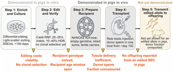
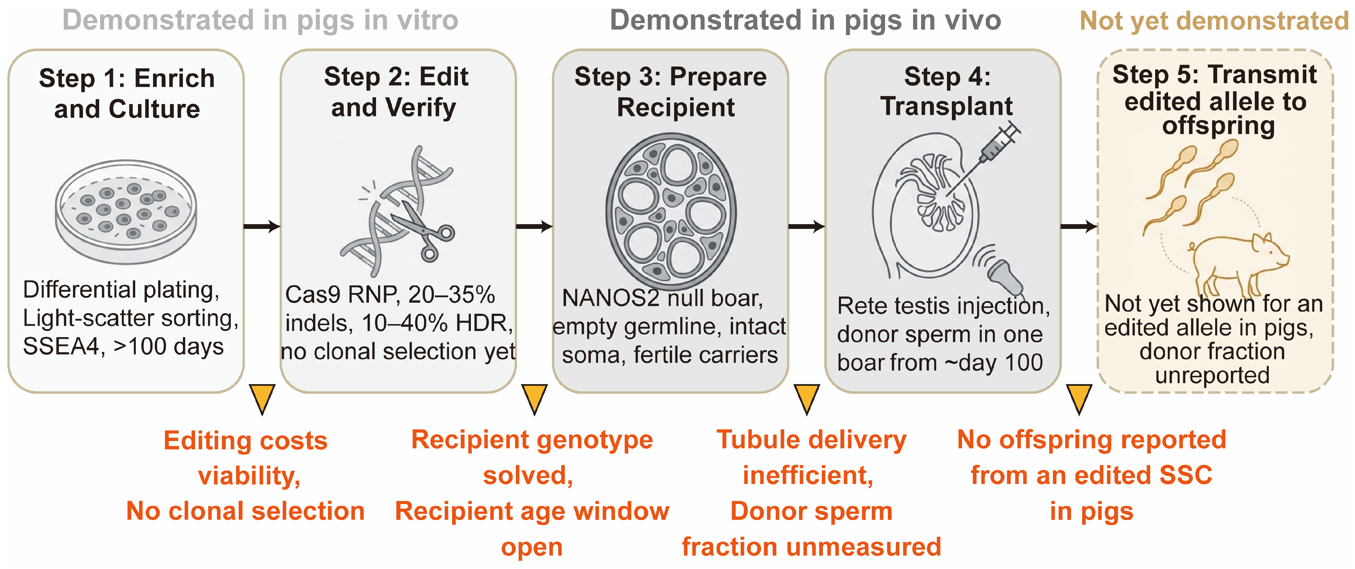

Disclaimer/Publisher’s Note: The statements, opinions and data contained in all publications are solely those of the individual author(s) and contributor(s) and not of MDPI and/or the editor(s). MDPI and/or the editor(s) disclaim responsibility for any injury to people or property resulting from any ideas, methods, instructions or products referred to in the content. |
© 2026 by the authors. Licensee MDPI, Basel, Switzerland. This article is an open access article distributed under the terms and conditions of the Creative Commons Attribution (CC BY) license.