


Abstract
Background/Objectives: The release from bud dormancy and subsequent break are critical processes for perennial woody plants to acclimatize to seasonal variations. This study aimed to elucidate the hormonal and transcriptional mechanisms governing walnut bud development. Methods: We utilized the early-fruiting, thin-shelled, and stress-tolerant cultivar ‘Jinboxiang 1’, collecting bud samples at four distinct developmental stages: Dormancy, Bud swelling, Bud burst, and Leaf unfolding. By integrating hormone metabolome and transcriptome analyses with weighted gene co-expression network analysis (WGCNA), key regulatory elements were systematically identified. Results: A total of seventy-seven hormones and related metabolites spanning eight classes were identified, with cytokinins and auxins being predominant. Transcriptomic profiling revealed 2935, 1963, and 3519 differentially expressed genes across the three successive transitions, respectively. Early bud swelling was linked to pathways involving hormone signaling, MAPK cascades, hypoxia responses, and defense mechanisms. Bud burst was associated with processes related to cutin, suberin, wax, fatty acids, carbohydrates, and secondary metabolism, whereas leaf unfolding predominantly involved photosynthetic activity and photosystem assembly. A core set of 276 genes was enriched in pathways related to jasmonate metabolism, ethylene signaling, α-linolenic acid metabolism, and MAPK signaling. WGCNA revealed 16 co-expression modules, among which the blue module was progressively activated during break and harbored JrGAI, a gene encoding a DELLA protein and a known repressor of gibberellin signaling. Heterologous overexpression of JrGAI in poplar reduced plant height and stem diameter and delayed bud break after dormancy release, supporting JrGAI as a candidate DELLA-associated negative regulator of bud development in walnut. Conclusions: These results reveal a stage-specific regulatory network underlying walnut bud development and demonstrate that JrGAI functions as a DELLA-associated negative regulator of both vegetative growth and bud break in walnut.
1. Introduction
Bud dormancy is a crucial adaptive mechanism evolved by perennial woody plants in temperate and boreal regions to endure seasonal variations in light, temperature, and water availability [1]. The perception of decreasing photoperiods and temperatures initiates a cessation of growth and the establishment of dormancy, thereby preventing premature growth resumption. During winter, cumulative chilling helps fulfill the chilling requirement and release buds from endodormancy, restoring their capacity for growth. Subsequent exposure to favorable forcing conditions, particularly warmer temperatures, promotes bud growth and leads to visible bud break in spring [2,3]. For economically significant forest tree species, this phenological process is pivotal as it influences annual growth patterns, survival under stress, and directly affects spring bud break timing, late frost vulnerability, vegetative-reproductive development, and ultimately, fruit yield and quality [4]. In the context of global climate change, rising winter temperatures and increased fluctuations in spring temperatures elevate the risk of inadequate chilling accumulation and advanced phenology in economically important woody tree species. Female flowers that develop prematurely are particularly susceptible to late spring frost, resulting in flower abortion, significantly reduced fruit set, and substantial economic losses for forestry industries [5]. Therefore, elucidating the molecular mechanisms governing dormancy release and bud break in trees is of considerable theoretical and practical importance for understanding seasonal adaptation, enhancing tree phenology and stress tolerance, and stabilizing fruit yield and quality.
Bud dormancy is traditionally categorized into three phases: paradormancy, endodormancy, and ecodormancy. The establishment and release of dormancy are co-regulated by external environmental factors and internal physiological conditions [6]. Paradormancy involves temporary growth inhibition mediated by signals from adjacent organs or tissues [7]. Endodormancy is characterized by intrinsic physiological processes that prevent growth resumption, even under favorable external conditions [8]. Ecodormancy refers to growth suppression induced by adverse external factors such as low temperature, drought, or nutrient deficiency, without intrinsic repression; growth can recommence once environmental conditions improve [9]. Evidence indicates that environmental signals, including photoperiod and temperature, precisely modulate meristematic activity by influencing the biosynthesis, metabolism, transport, and signal transduction of endogenous phytohormones, thereby facilitating transitions between bud growth and dormancy [10,11]. Endogenous phytohormones play a central role in signaling during dormancy cycles. Key hormones such as abscisic acid (ABA), gibberellin (GA), and ethylene (ETH) collaboratively regulate the processes of dormancy establishment, maintenance, and release through complex interaction networks. ABA primarily sustains dormancy by inhibiting meristem activity and bud break while enhancing stress tolerance; in contrast, GA acts as a growth-promoting signal that contributes to cell elongation and the release from dormancy [9]. In poplar, the PtoPHYB2-PtoHY5a-PtoFT2 module initiates winter dormancy and modulates bud break by adjusting gibberellin levels, thereby coordinating seasonal growth [12]. Winter chilling strongly induces FT expression in dormant poplar buds, facilitates the recruitment of GA-inducible 1,3-beta-glucanases, reopens signaling pathways, and restores communication between meristems and surrounding tissues to promote dormancy release [13]. Additionally, FT1 in poplar suppresses callose deposition, while short-term warming inhibits cold-activated FT1 and gibberellin pathways, obstructing plasmodesmata opening and hindering dormancy release [14].
Gibberellins orchestrate various growth and developmental processes, with the GA signaling cascade serving as a crucial regulatory pathway for organ development [13,15]. GA biosynthesis predominantly occurs in young apical tissues. The binding of GA to the GID1 receptor leads to the formation of a stable complex that recognizes the N-terminal domain of DELLA proteins. Subsequently, the SCFSLY1/GID2 ubiquitin-ligase complex mediates the degradation of DELLA proteins, alleviating their repression on growth-related transcription factors and regulatory proteins, thus influencing multiple developmental programs [16,17,18]. DELLA proteins, part of the GRAS protein family, are vital transcriptional regulators that integrate environmental signals and diverse hormone signaling pathways [19]. They are believed to play essential roles in seasonal growth cycles and bud break in woody plants [20]. For instance, the knockdown of CsDELLA5Hap2 significantly inhibits axillary bud sprouting in the early-sprouting tea cultivar LJ43 [21]. The tree peony DELLA protein PsRGL1 interacts with PsWRKY71 to modulate the expression of the cyclin gene PsCYCD6.1, thereby regulating cell proliferation and bud break [22].
Walnut (Juglans regia L.) is an economically significant woody oil-bearing species, with its spring bud break timing critically influencing sensitivity to early-spring frost, new shoot growth, fruit yield, and overall tree development. However, in walnut, stage-specific changes in endogenous phytohormone levels during bud break remain poorly characterized. The relationships between hormone abundance and gene expression, as well as the role of DELLA-associated signaling in bud-break progression, are also unclear. The functions of candidate genes involved in these processes require experimental validation. This study employs ‘Jinboxiang 1,’ an early-fruiting, thin-shelled, and stress-resistant walnut cultivar, to conduct hormone metabolome and transcriptome profiling across four consecutive developmental stages: Dormancy, Bud swelling, Bud burst, and Leaf unfolding. We characterize hormonal dynamics and transcriptomic reprogramming throughout these developmental transitions. Weighted Gene Co-expression Network Analysis (WGCNA) is used to identify hub genes associated with bud break progression, and heterologous expression in poplar is utilized to validate the functional role of the candidate gene JrGAI in vegetative growth and bud break. Our findings provide a theoretical framework for unraveling hormone-transcription regulatory networks and inform genetic improvement strategies for bud break phenology in walnut.
2. Materials and Methods
2.1. Plant Materials and Sample Collection
Plant samples of the cultivar ‘Jinboxiang 1’ were sourced from the Walnut Germplasm Nursery at the Pomology Institute, Shanxi Agricultural University, situated in the hilly areas of the Loess Plateau (112°32′ E, 37°32′ N; altitude: 820–900 m). The site exhibits a warm-temperate continental climate, characterized by a mean annual temperature of 10.6 °C, an annual sunlight duration of 2300 h, and annual precipitation ranging from 400 to 600 mm. Twelve- to sixteen-year-old disease-free trees demonstrating uniform vigor and normal fruiting patterns under conventional orchard management were selected for sample collection. Based on external morphological characteristics, bud development was categorized into four distinct stages: Dormancy (JBX-1, sampled on 11 March), bud scales are tightly closed, with no significant bud expansion observed; Bud swelling (JBX-2, sampled on 17 March), noticeable enlargement of the bud occurs, with scales loosened but still closed; Bud burst (JBX-3, sampled on 24 March), the bud scales open, revealing green tissues or young leaf apices; Leaf unfolding (JBX-4, sampled on March 31), young leaves emerge and expand. Three biological replicates were prepared for each developmental stage, with each replicate comprising pooled buds from five individual trees. Fresh samples were rapidly snap-frozen in liquid nitrogen and subsequently stored at −80 °C for hormone metabolome and RNA sequencing analyses.
2.2. Hormone Metabolome Analysis
Frozen bud tissues were homogenized using a tissue grinder (30 Hz, 1 min). An accurately weighed 50 mg of the resulting powder was combined with 10 μL of an internal standard mixture (100 ng/mL) and 1 mL of extraction solvent (methanol/water/formic acid in a 15:4:1 ratio, v/v/v). The mixture was vortexed for 10 min at low temperature, followed by centrifugation at 12,000 r/min for 5 min at 4 °C. The supernatant was collected, evaporated to dryness, reconstituted in 100 μL of 80% methanol (v/v), and filtered through a 0.22 μm membrane filter before analysis. Data acquisition was carried out using Ultra-Performance Liquid Chromatography (UPLC; ExionLC™ AD, Sciex, Framingham, MA, USA) coupled with Tandem Mass Spectrometry (MS/MS; QTRAP® 6500+, Sciex). Chromatographic separation was performed on a Waters ACQUITY UPLC HSS T3 C18 column (100 mm × 2.1 mm i.d., 1.8 µm) at 40 °C with a flow rate of 0.35 mL/min. Mobile phase A was water containing 0.04% acetic acid, and mobile phase B was acetonitrile containing 0.04% acetic acid. The gradient program was as follows: 5% B (0–1 min), 5–95% B (1–8 min), 95% B (8–9 min), 95–5% B (9.1–12 min). The injection volume was 2 μL. Mass detection was conducted using an ESI Turbo Ion-Spray interface in both positive and negative ionization modes; source temperature, 550 °C; ion spray voltage, 5500 V (positive) and −4500 V (negative); curtain gas, 35 psi. Scheduled MRM mode was applied, and DP and CE values were optimized individually for each MRM transition. Qualitative identification was performed against a database of authentic standards, while quantification utilized multiple-reaction monitoring (MRM) mode in triple-quadrupole mass spectrometry. Representative UPLC-MRM chromatograms of target phytohormones are provided in Figure S1. All 77 phytohormone-related compounds were annotated based on authentic reference standards. Hormone concentrations were determined from standard curves generated through serial dilutions of reference compounds. Quantification was performed using external standard calibration curves. Quality control samples were incorporated during detection to monitor system stability. Metabolites exhibiting more than a two-fold change and p < 0.05 were designated as differentially accumulated.
2.3. Transcriptome Sequencing Analysis
Total RNA was extracted from bud samples, with RNA integrity, purity, and concentration assessed via agarose gel electrophoresis, NanoPhotometer spectrophotometry, Qubit 4.0 fluorometry, and Qsep400 bioanalyzer. The RIN values of RNA samples ranged from 6.3 to 7.7, and ≥1 μg total RNA was used as the starting material for library preparation. Libraries were constructed using the NEBNext Ultra™ RNA Library Prep Kit for Illumina (New England Biolabs, Ipswich, MA, USA). Briefly, polyA-tailed mRNA was enriched with oligo (dT) magnetic beads and randomly fragmented using divalent cations in NEB Fragmentation Buffer. First-strand cDNA was synthesized from fragmented mRNA templates with random hexamer primers using M-MuLV reverse transcriptase. The RNA template was degraded by RNase H, and second-strand cDNA was synthesized by DNA polymerase I with dNTPs as substrates. After purification, double-stranded cDNA was subjected to end repair, A-tailing and adapter ligation. Fragments of approximately 200 bp were enriched with AMPure XP beads, followed by PCR amplification and further purification of PCR products using AMPure XP beads to obtain the final libraries. High-quality RNA was utilized for library construction, with qualified libraries (paired-end, 150 bp read length) sequenced on the Illumina NovaSeq 6000 platform (Illumina, San Diego, CA, USA). Raw sequencing reads were processed to eliminate adapters, low-quality sequences, and reads with excessive N content, yielding clean reads. Raw reads per sample ranged from 45,185,808 to 55,403,528, and clean reads after filtering ranged from 44,175,790 to 54,552,644. Q20, Q30, and GC content metrics were computed to assess sequencing quality. Clean reads were aligned to the walnut reference genome (Juglans_regia.Walnut_2.0.DNA.toplevel.fa; Ensembl Plants release 57) [23] using HISAT2 software (v2.2.1) [24]. The corresponding GFF3 annotation file was Juglans_regia. Walnut_2.0.57.chr.gff3. The featureCounts tool (v2.0.3) was employed to normalize mapped read counts based on transcript length, with gene expression levels reported as FPKM [25]. DESeq2 (v1.22.1) was utilized for differential expression analysis between adjacent developmental stages (JBX-1 vs. JBX-2; JBX-2 vs. JBX-3; JBX-3 vs. JBX-4) [26]. Genes with an adjusted p-value (padj) < 0.05 and |log2 (fold change)| ≥ 1 were classified as differentially expressed genes (DEGs). Gene Ontology (GO) and Kyoto Encyclopedia of Genes and Genomes (KEGG) databases were employed for functional annotation and enrichment analysis of DEGs.
2.4. Cloning of JrGAI, Vector Construction, and Poplar Genetic Transformation
Total RNA was extracted from walnut buds using the RNAprep Pure Plant Kit (TIANGEN, DP441, Beijing, China) according to the manufacturer’s instructions. First-strand cDNA was synthesized using the FastKing RT Kit (TIANGEN, KR116, Beijing, China). The JrGAI coding sequence was amplified from the cDNA using gene-specific primers (Table S1) and KOD FX DNA polymerase (TOYOBO, KFX-101, Osaka, Japan). The amplicon was separated by agarose gel electrophoresis, purified using the DNA Gel Extraction Kit (Simgen, 2001050, Hangzhou, China), and ligated into the pMD™19-T vector (Takara, 6013, Kusatsu, Japan). The ligation product was introduced into E. coli TOP10 competent cells by the freeze–thaw transformation method. Positive colonies were screened by colony PCR, and the insert sequence was verified by Sanger sequencing performed by Tsingke Biotechnology (Beijing, China).
The conserved domains of the JrGAI protein were predicted using the NCBI Conserved Domain Database [27]. The verified JrGAI coding sequence was inserted into the pART-CAM-FLAG-GFP plant expression vector using the Uniclone One Step Seamless Cloning Kit (Genesand Biotech, SC612, Beijing, China), following the manufacturer’s instructions. The pART-CAM-FLAG-GFP backbone was obtained from Shaanxi Boride Biotechnology Co., Ltd. (Yangling, Shaanxi, China) and was derived from the pART-CAM binary vector described previously by Gleave [28]. The resulting construct was verified by full insert sequencing.
The verified construct was introduced into Agrobacterium tumefaciens GV3101 by freeze–thaw method. Briefly, 1 μg plasmid DNA was mixed with competent GV3101 cells, incubated on ice for 30 min, frozen in liquid nitrogen for 5 min, heat-shocked at 37 °C for 5 min, followed by addition of LB medium and shaking recovery at 28 °C. Transformants were selected on LB medium containing 50 mg/L rifampicin and 50 mg/L kanamycin, and the presence of the construct was confirmed by colony PCR.
Genetic transformation of 84K poplar (Populus alba × P. glandulosa) was performed using a leaf-disc method [29]. In brief, the 3rd–5th fully expanded leaves were excised from robust 4–5-week-old in vitro-grown plantlets of 84K poplar, and three cuts along the main vein were made with a sharp scalpel. The wounded leaves were immersed in an A. tumefaciens suspension adjusted to OD600 = 0.6 for 10 min with gentle shaking three times (1 min each time). After infection, the leaves were taken out and blotted dry with sterile filter paper to remove excess bacterial suspension. Subsequently, the leaf explants were transferred onto shoot induction medium and co-cultivated in darkness at 25 °C for 3 d. Explants were then transferred to shoot induction selection medium supplemented with kanamycin (20 mg/L) and cefotaxime (400 mg/L), and cultured at 25 °C under a 16 h light/8 h dark photoperiod. The explants were subcultured onto fresh selection medium every 3 d for the first two rounds, followed by subculture every 10 d until resistant shoots regenerated. Regenerated shoots reaching 1–2 cm in length were excised and transferred to rooting medium containing kanamycin (50 mg/L) and cefotaxime (400 mg/L) for further selection. Plantlets with normal root development were subjected to molecular validation.
Putative transgenic plants were screened using genomic DNA extracted from young leaves with the Plant Genomic DNA Kit (TIANGEN, DP305, Beijing, China). The presence of the transgene was assessed by PCR using a forward primer designed from the JrGAI CDS region and a universal reverse primer derived from the vector backbone (Table S1). Wild-type genomic DNA and nuclease-free water were used as negative controls, and A. tumefaciens suspension harboring the recombinant plasmid served as the positive control. Transgene expression was further assessed by RT-qPCR. The primers for RT-qPCR are listed in Table S1, with Actin used as the reference gene. The relative expression levels of JrGAI in different transgenic lines were calculated using the 2−ΔΔCt method.
2.5. Induction of Poplar Bud Dormancy, Cold Treatment, and Evaluation of Bud Break
One-month-old tissue-cultured poplar plantlets were transplanted into soil pots and cultured for an additional two months in a BSG-400 growth chamber (Boxun, Shanghai, China) at 25 °C with a photoperiod of 16 h light/8 h dark and 60% light intensity. Subsequently, wild-type and transgenic plants at this stage were subjected to short-day conditions (8 h light/16 h dark, 25 °C) for eight weeks to induce apical bud formation and dormancy. Dormancy induction, chilling accumulation, and bud break assays were all performed in the BSG-400 growth chamber under controlled conditions. Following bud establishment, the plants underwent cold treatment at 4 °C for five weeks to promote chilling accumulation. Plants were then transferred to long-day conditions (16 h light/8 h dark, 25 °C, 60% light intensity) to trigger bud break. The status of apical buds was recorded daily following the transfer to long-day conditions until nearly all apical buds had germinated. The development of the apical buds was classified according to established phenological grading criteria into five grades: grade 0 (fully dormant), grade 1 (swollen bud scales), grade 2 (visible green bud tip), grade 3 (leaf protrusion), grade 4 (leaf expansion), and grade 5 (separated young leaves) [30]. Break grades were documented for individual plants at each time point, with mean values calculated for each genotype. Six biological replicates were sampled for wild-type and each of the four highest-expressing overexpression lines under every experimental condition.
2.6. Statistical Analysis
All statistical analyses were conducted with SPSS 27. Student’s t-test was applied for comparisons between two groups. A difference was regarded as statistically significant at p < 0.05. Data are presented as mean ± standard deviation (SD).
3. Results
3.1. Dynamic Changes in Phytohormones During Walnut Bud Break
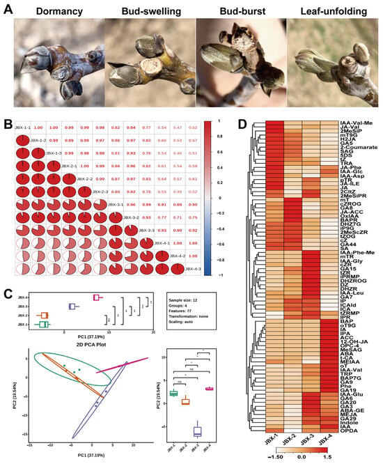
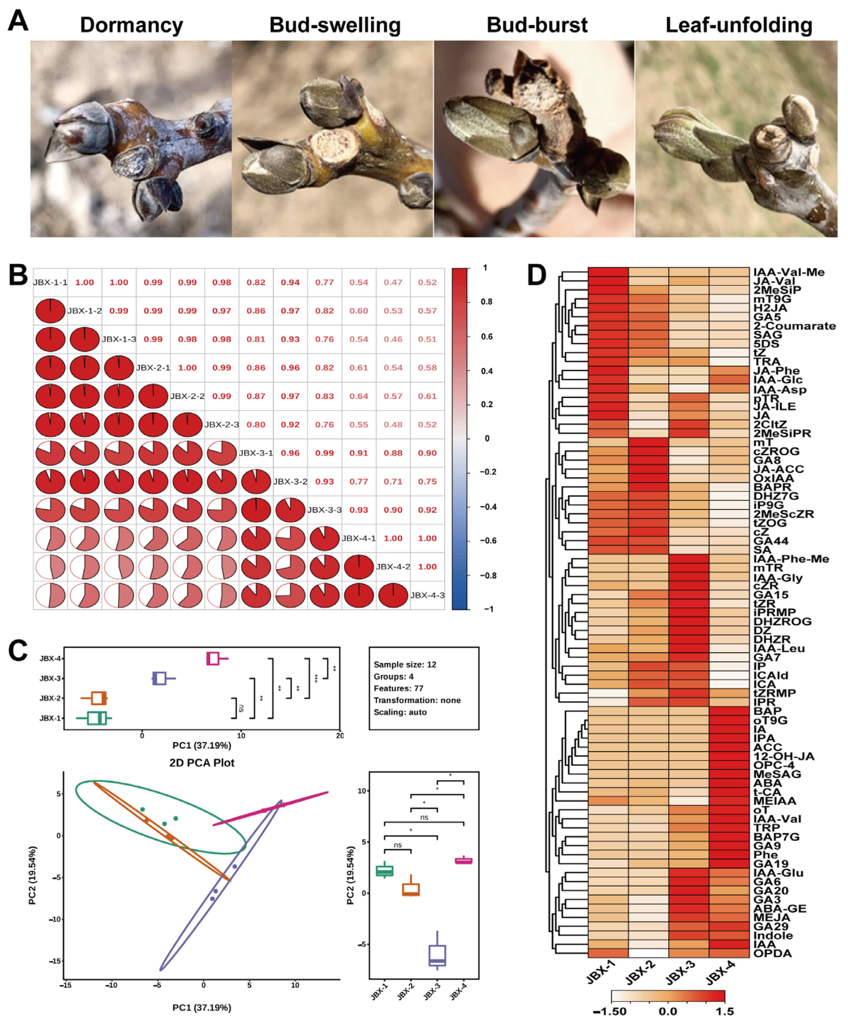
Phytohormones play pivotal roles in various plant physiological processes. To elucidate the changes in endogenous hormone levels associated with the transition of walnut buds from dormancy to break, buds of the cultivar ‘Jinboxiang 1’ were sampled at four consecutive developmental stages: JBX-1, JBX-2, JBX-3, and JBX-4 (Figure 1A). Correlation analysis of samples showed strong consistency among biological replicates at each stage, indicating reliable hormone profiling (Figure 1B). Principal-component analysis (PCA) demonstrated distinct clustering of samples according to their developmental stages, highlighting significant metabolic reorganization of hormone profiles during dormancy release and leaf unfolding (Figure 1C). A total of 77 phytohormones and hormone-related metabolites across eight primary categories were identified: 28 cytokinins, 18 auxins, 11 gibberellins, 10 jasmonates, six salicylic acid derivatives, two forms of abscisic acid, one ethylene-related compound, and one strigolactone. Cytokinins represented the predominant category, followed by auxins and gibberellins (Figure 1D, Table S2).
Figure 1.
Phenotypes and hormone-metabolome characteristics of walnut buds across different developmental stages. (A) Four developmental phases: Dormancy, Bud swelling, Bud burst, and Leaf unfolding, corresponding to sample codes JBX-1, JBX-2, JBX-3, and JBX-4. (B) Correlation analysis of hormone profiles among samples, with color intensity indicative of correlation coefficient magnitude. (C) PCA of hormone metabolomes across stages, with each dot representing a biological replicate and colors denoting developmental stages. Statistical significance: * p < 0.05, ** p < 0.01, *** p < 0.001; ns, not significant (p ≥ 0.05). (D) Heatmap illustrating hormone concentrations in buds at various developmental stages.
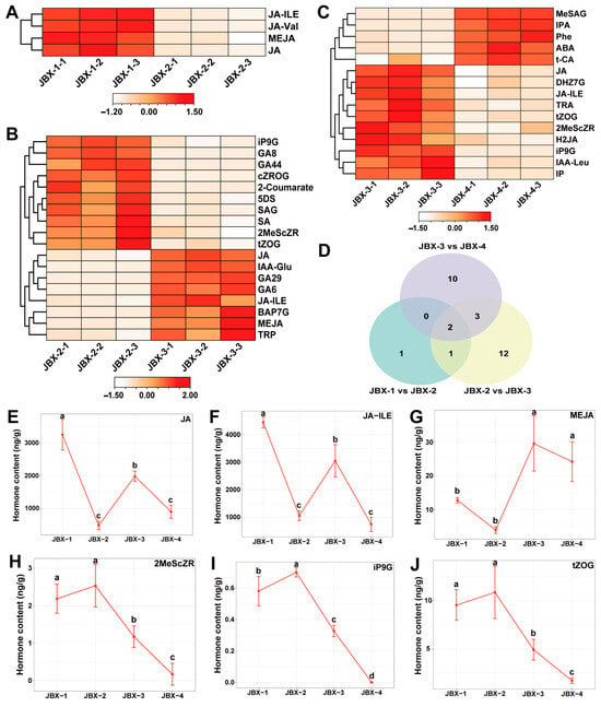
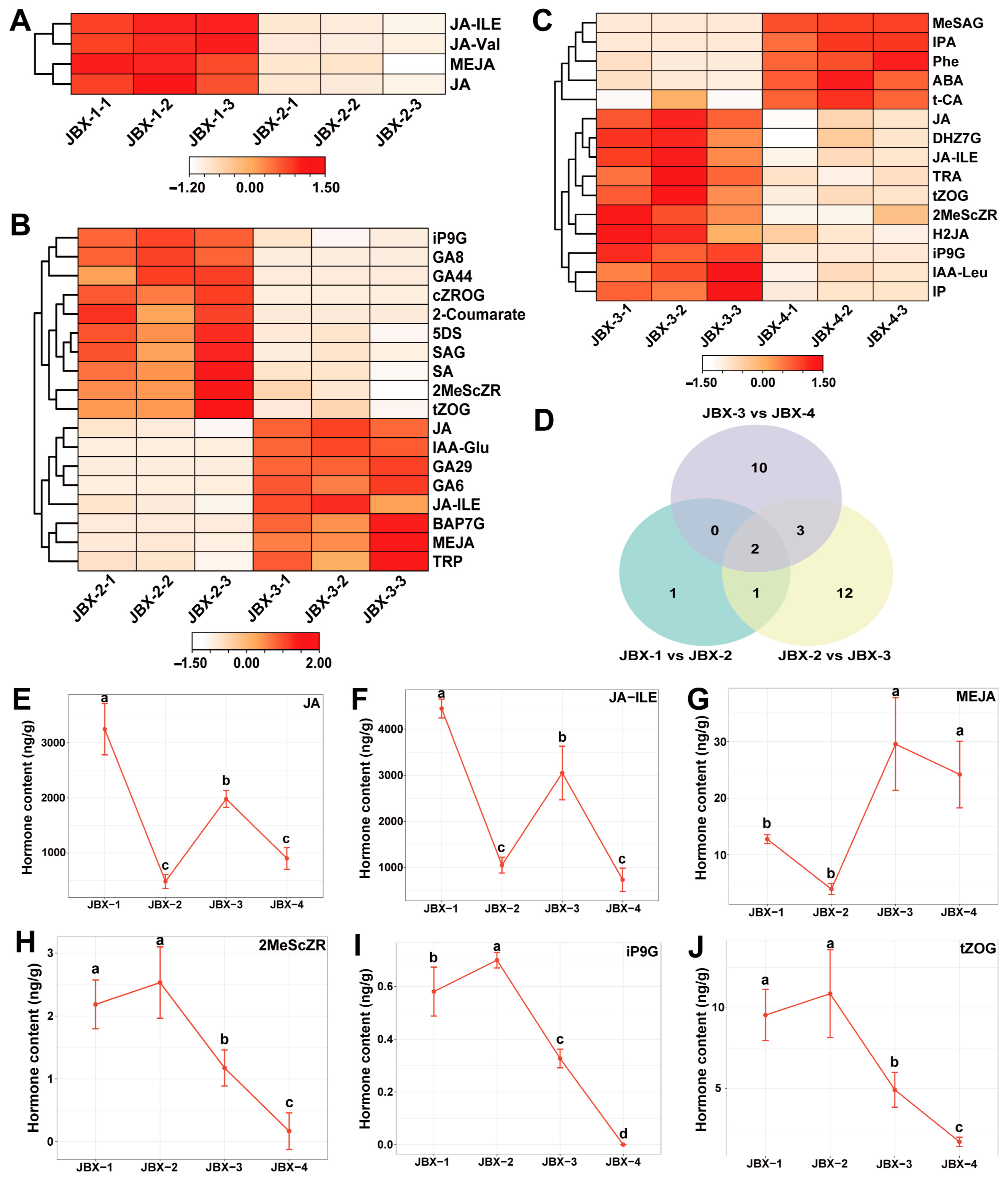
Comparative analyses between adjacent stages revealed a significant reduction in four jasmonate-related compounds during the transition from Dormancy to Bud swelling, indicating that early bud swelling is linked to changes in jasmonate metabolism (Figure 2A, Dataset S1). During the transition from Bud swelling to Bud burst, eighteen hormones exhibited differential accumulation, with ten down-regulated (four cytokinins, three salicylates, two gibberellins, and one strigolactone) and eight up-regulated (three jasmonates, two auxins, two gibberellins, and one cytokinin) (Figure 2B, Dataset S1). This suggests that the processes of bud-scale opening and green-tissue emergence are governed by the coordinated regulation of multiple hormones rather than alterations in a single hormone. Fifteen hormones showed significant changes in abundance during the Bud burst to Leaf unfolding transition, with ten down-regulated (five cytokinins, three jasmonates, and two auxins) and five up-regulated (three salicylates, one auxin, and one abscisic acid-related metabolite) (Figure 2C, Dataset S1). These metabolic alterations may reflect the transition of bud meristems towards rapid leaf growth and environmental adaptability. Notably, both jasmonic acid (JA) and jasmonoyl-isoleucine (JA-Ile) demonstrated differential accumulation across all three comparisons, suggesting that jasmonic acid metabolism and signaling are crucial throughout the phases of dormancy release, bud swelling, and leaf unfolding in walnut buds (Figure 2D). The dynamics of these shared hormones across developmental stages are visualized in the line plots (Figure 2E–J), with statistical differences assessed by one-way ANOVA.
Figure 2.
Differentially accumulated hormones during walnut-bud break stages. (A) Categories and patterns of changes in hormones between Dormancy and Bud swelling. (B) Categories and changes between Bud swelling and Bud burst. (C) Categories and changes between Bud burst and Leaf unfolding. (D) Venn diagram summarizing differentially accumulated hormones identified across developmental transitions. (E–J) Line plots of shared hormone concentrations from panel (D). One-way ANOVA, with different lowercase letters denoting significant differences between groups.
3.2. Transcriptomic Dynamics During Walnut Bud Break
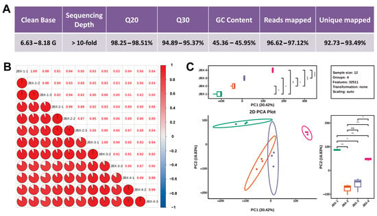
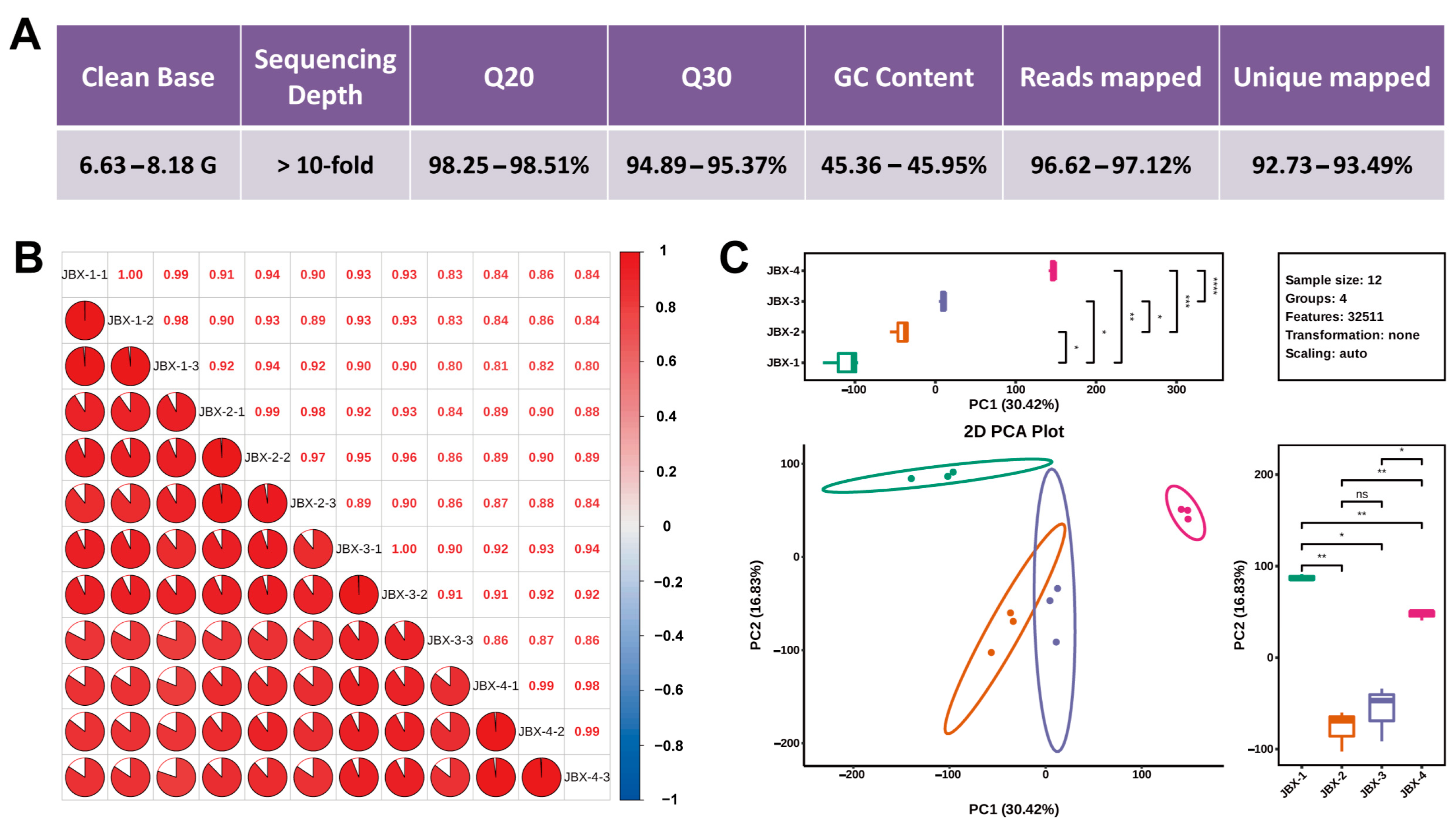
RNA sequencing (RNA-seq) was conducted on bud samples JBX-1 to JBX-4 to further explore the gene expression reprogramming underlying bud break. Each sample generated clean data ranging from 6.63 to 8.18 Gb, with sequencing depths exceeding 10×. Q20 and Q30 values were above 98.25% and 94.89%, respectively, while GC content varied between 45.36% and 45.95%, confirming the high quality of the sequencing reads (Figure 3A). The overall mapping rates of clean reads against the walnut reference genome were between 96.62% and 97.12%, with unique mapping rates of 92.73% to 93.49%, indicating good compatibility for subsequent expression analyses (Figure 3A, Table S3). Correlation analyses showed high consistency in expression profiles among biological replicates within each stage (Figure 3B). PCA illustrated distinct separation among samples from different developmental stages, with replicates clustering tightly within each stage (Figure 3C). Collectively, the transcriptomic datasets exhibited high reproducibility and marked transcriptional divergence across developmental phases.
Figure 3.
Quality assessment of transcriptome sequencing for walnut bud samples at distinct developmental stages. (A) Statistics of sequencing data for individual samples. (B) Correlation matrix of gene expression profiles across samples, with a color gradient indicating correlation coefficients. (C) PCA based on gene expression abundance, with each dot representing a biological replicate and colors denoting developmental stages. Statistical significance: * p < 0.05, ** p < 0.01, *** p < 0.001, **** p < 0.0001; ns, not significant (p ≥ 0.05).
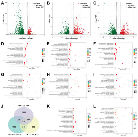
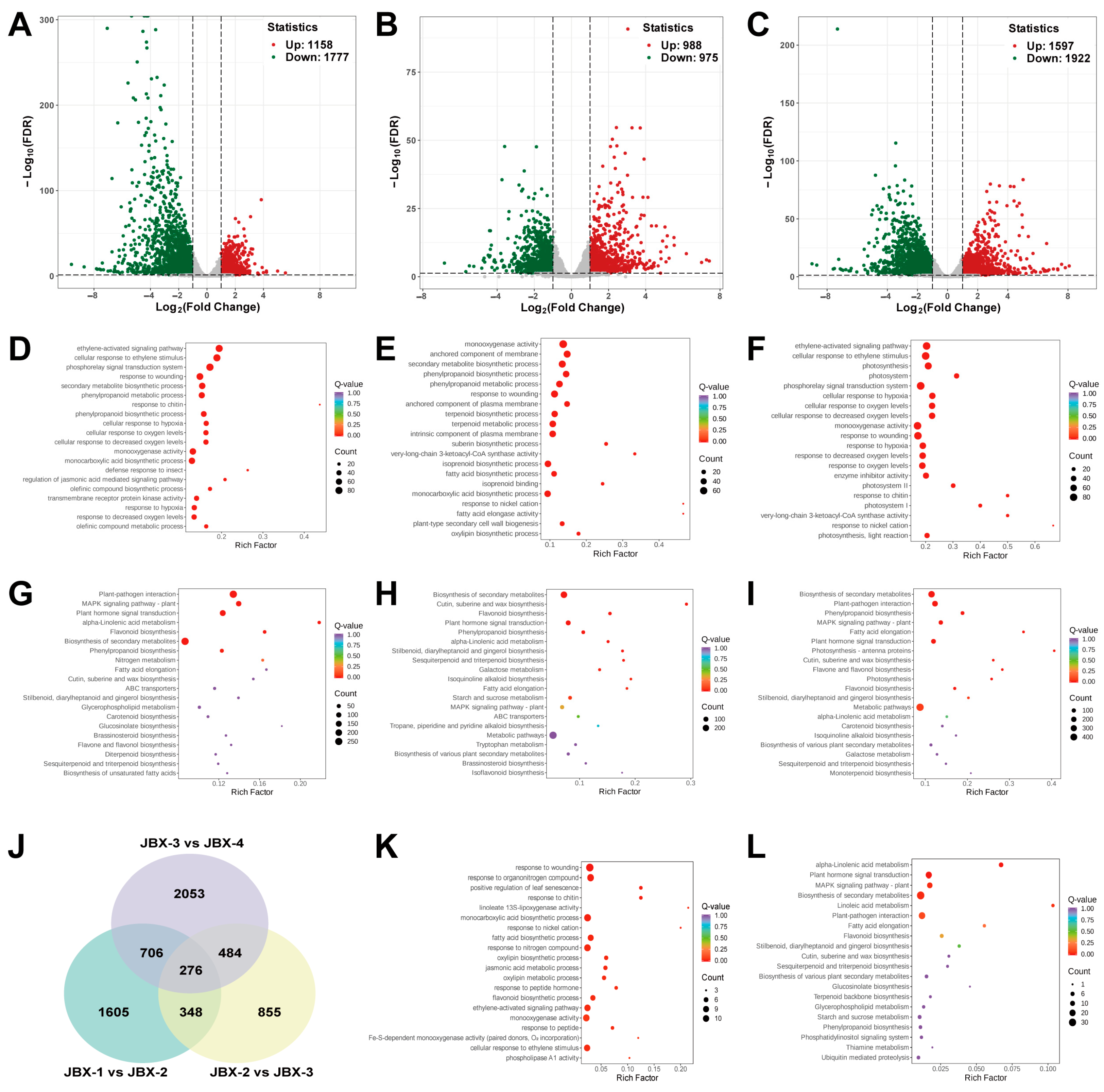
Differential expression analysis identified 2935 DEGs between JBX-1 and JBX-2 (1158 up-regulated, 1777 down-regulated) (Figure 4A); 1963 DEGs between JBX-2 and JBX-3 (988 up-regulated, 975 down-regulated) (Figure 4B); and 3519 DEGs between JBX-3 and JBX-4 (1597 up-regulated, 1922 down-regulated) (Figure 4C). The highest number of DEGs was observed in the comparison between JBX-3 and JBX-4, indicating that the most extensive transcriptional reprogramming occurs post-bud burst during leaf unfolding. Both up-regulated and down-regulated genes comprised significant proportions in each comparison, suggesting that walnut bud break entails not only the activation of growth-related genes but also the concomitant repression or remodeling of pathways associated with dormancy maintenance, defense, storage, and primary metabolism.
Figure 4.
Statistics and functional enrichment of differentially expressed genes (DEGs) during walnut bud break. (A–C) Summary of DEGs identified between adjacent developmental stages. (D–F) GO enrichment analyses of DEGs for the three pairwise comparisons. (G–I) KEGG enrichment analyses of DEGs for the three pairwise comparisons. (J) Venn diagram illustrating DEGs shared among the three comparisons. (K) GO enrichment analysis of the 276 shared DEGs. (L) KEGG enrichment analysis of the 276 shared DEGs.
3.3. Stage-Specific Shifts in Biological Processes and Metabolic Pathways During Walnut Bud Break
In the transition from dormancy to bud swelling, GO enrichment analyses identified significant terms related to ethylene response and signal transduction, such as ethylene-activated signaling pathway, cellular response to ethylene stimulus, and phosphorelay signal transduction system (Figure 4D). Additionally, multiple hypoxia-responsive terms, including cellular response to hypoxia, cellular response to oxygen levels, and response to decreased oxygen levels, were notably enriched, indicating a substantial reorganization of internal oxygen status and oxygen-sensing mechanisms initiated upon bud swelling. GO enrichment also revealed strong associations with defense mechanisms and jasmonate signaling, including terms like response to wounding, response to chitin, defense response to insect, and regulation of jasmonic acid-mediated signaling pathway, alongside secondary metabolism-related entries such as secondary metabolite biosynthetic process, phenylpropanoid metabolic process, and monooxygenase activity. KEGG pathway analysis further underscored enrichments in plant-pathogen interaction, MAPK signaling pathway-plant, plant hormone signal transduction, alpha-Linolenic acid metabolism, flavonoid biosynthesis, and biosynthesis of secondary metabolites (Figure 4G). Together with the observed decrease in jasmonate levels from metabolomic analyses, these findings suggest a coordinated modulation of jasmonate metabolism, ethylene signaling, MAPK pathways, and defense mechanisms at the onset of bud swelling.
For the transition from bud swelling to bud burst, DEGs were predominantly associated with secondary metabolic processes, including secondary metabolite biosynthetic process, phenylpropanoid biosynthetic process, and terpenoid biosynthetic process (Figure 4E). Significant enrichment was also noted for terms related to lipid biosynthesis and elongation, such as suberin biosynthetic process and fatty acid elongation. Additionally, membrane-component terms, including anchored component of membrane and intrinsic component of plasma membrane, were enriched. The enrichment of plant-type secondary cell wall biogenesis highlights concurrent modifications of plasma membranes, cell walls, and protective epidermal structures during the transition from bud swelling to bud burst. KEGG enrichment analyses identified pathways involved in biosynthesis of secondary metabolites, cutin, suberin and wax biosynthesis, flavonoid biosynthesis, and others (Figure 4H). Thus, the bud burst phase is characterized by extensive remodeling of lipid and secondary metabolism, cutin and wax biosynthesis, cell wall restructuring, and carbohydrate metabolism.
During the transition from bud burst to leaf unfolding, alongside the previously mentioned ethylene signaling, phosphorelay transduction, hypoxia response, and defense response terms, GO entries related to photosynthesis, such as photosynthesis, photosystem I, and photosystem II, exhibited significant enrichment (Figure 4F). KEGG analysis further indicated enrichments in photosynthesis, photosynthesis-antenna proteins, and other related pathways (Figure 4I). These findings indicate a rapid activation of transcriptional programs for light-harvesting complexes and photosystem machinery upon leaf unfolding, signifying the shift in young leaves from heterotrophy and limited photosynthesis reliant on internal reserves towards autotrophic functionality. Concurrently, pathways related to epidermal lipids, secondary metabolism, and hormone signaling remain dynamically regulated to synchronize leaf maturation, photoprotection, and environmental adaptation.
Venn diagram analysis delineated 1605, 855, and 2053 stage-specific DEGs for the respective transitions, with 276 DEGs common across all three stages (Figure 4J). GO enrichment of these shared DEGs emphasized processes related to hormone and stress responses, including ethylene-activated signaling pathway, jasmonic acid metabolic process, and response to wounding. Furthermore, metabolic processes such as oxylipin biosynthetic process and fatty acid biosynthetic process were enriched (Figure 4K). KEGG enrichment revealed pathways associated with alpha-Linolenic acid metabolism, plant hormone signal transduction, and biosynthesis of secondary metabolites (Figure 4L). Collectively, these results illustrate that genes implicated in hormone signaling, secondary metabolism, and stress responses are consistently regulated throughout the various developmental transitions of walnut bud break.
3.4. WGCNA Identifies Co-Expression Modules and Candidate Hub Genes Associated with Bud Break
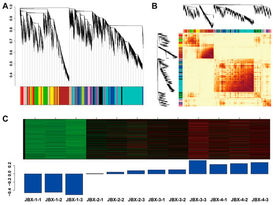
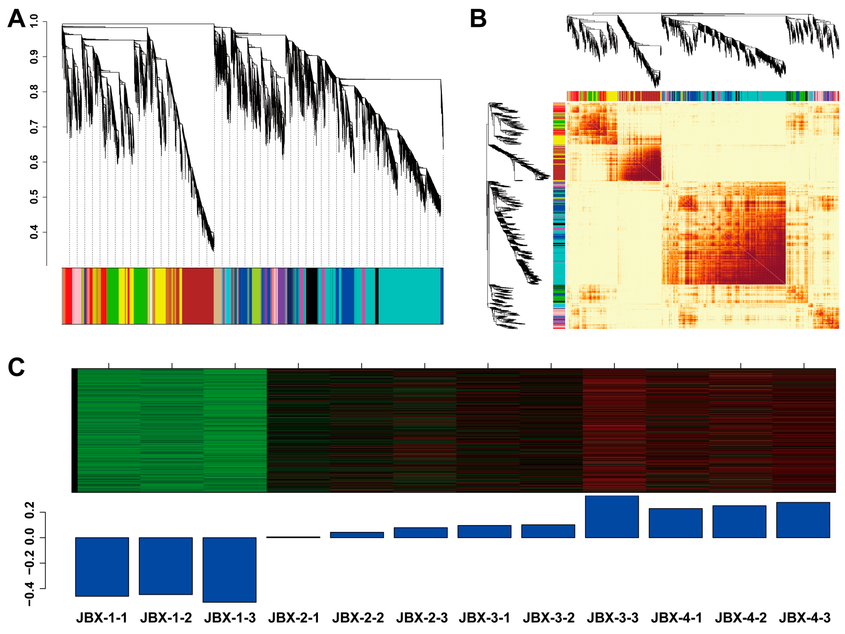
WGCNA was conducted on transcriptome datasets from four developmental stages to identify key regulatory genes involved in walnut bud development. Following the construction of a scale-free network, hierarchical clustering, and dynamic tree cutting, sixteen stable gene co-expression modules were identified, each designated by distinct colors (Figure 5A). The topological overlap matrix heatmap revealed strong intra-module co-expression and effective inter-module separation, indicating the high reliability and cohesion of the constructed network (Figure 5B). Expression profiling of module eigengenes demonstrated that the ‘blue’ module exhibited a progressive increase in expression throughout bud-break stages, from Dormancy to Bud swelling, Bud burst, and Leaf unfolding, suggesting its significant role in dormancy release, bud growth, and young leaf development. The gene JrGAI (Jr01_29220) was identified as a hub gene within the core network of the ‘blue’ module (Dataset S2). As GAI encodes a DELLA repressor involved in gibberellin signaling closely linked to cell elongation, dormancy release, and bud break, JrGAI was selected for subsequent functional validation.
Figure 5.
Construction of the gene co-expression network and identification of relevant modules for walnut bud break. (A) Clustering dendrogram representing the weighted gene co-expression network derived from four developmental stages. Each branch corresponds to a gene, with colors indicating distinct co-expression modules identified through dynamic tree cutting. (B) TOM-based heatmap illustrating the gene co-expression network, where color intensity reflects the topological overlap between gene pairs. (C) Global expression patterns of genes in the blue module: the upper panel displays a clustering heatmap of module genes; the lower panel shows the dynamic expression of the module eigengene across experimental samples.
3.5. JrGAI Encodes a Canonical DELLA-Family Protein
The full-length coding sequence of JrGAI was amplified via PCR using cDNA from walnut buds as a template. This open reading frame spans 1650 bp, encoding a polypeptide of 549 amino acids (Table S4). Conserved-domain analysis revealed a typical N-terminal DELLA domain and a C-terminal GRAS domain in JrGAI. The DELLA domain is responsible for GID1-mediated gibberellin perception and subsequent protein degradation, while the GRAS domain facilitates transcriptional regulation and various protein–protein interactions. These structural features confirm that JrGAI is part of the canonical DELLA subfamily of GRAS proteins and likely serves as a negative regulator of gibberellin signaling.
3.6. Overexpression of JrGAI Suppresses Vegetative Growth and Delays Break of Dormant Buds in Poplar
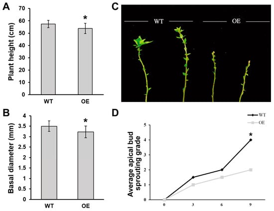
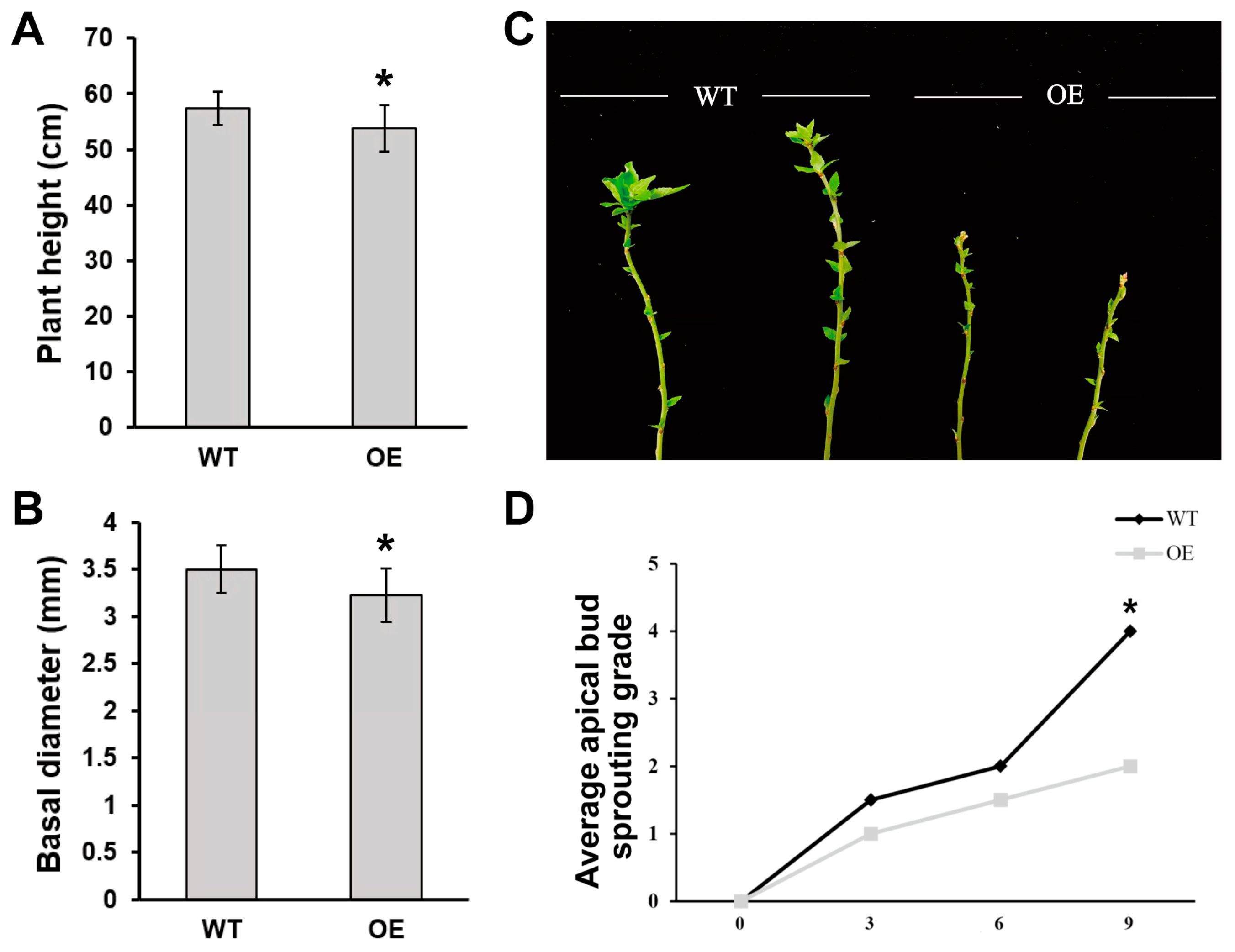
To elucidate the biological functions of JrGAI, its coding region was cloned into the pART-CAM-FLAG-GFP overexpression vector. Transgenic poplar lines expressing JrGAI were generated through Agrobacterium-mediated transformation (Figures S2 and S3). Under identical growth conditions, JrGAI-overexpressing plants exhibited noticeable growth retardation. Quantitative analyses revealed a significant reduction in plant height and basal stem diameter in transgenic lines compared to wild-type controls (Figure 6A,B). These observations align with the known function of DELLA proteins in inhibiting gibberellin-dependent cell division and elongation, confirming that heterologous expression of JrGAI suppresses vegetative growth in poplar.
Figure 6.
Overexpression of JrGAI represses vegetative growth and delays bud break in poplar. (A,B) Measurement of plant height and basal stem diameter for wild-type and JrGAI-overexpressing poplars. (C) Phenotypes of apical bud break in cold-chilled wild-type and JrGAI-overexpressing plants under permissive growth conditions. (D) Dynamic changes in apical bud break scores for wild-type and JrGAI-overexpressing genotypes. Data from the four independent OE lines, with six biological replicates per line, were pooled as a single OE group for comparison with wild type. Student’s t-test: * p < 0.05.
To assess the role of JrGAI in bud break, wild-type and transgenic poplar plants were subjected to short-day treatment to induce apical bud formation and dormancy. Following a period of cold treatment, plants were transitioned to long-day conditions to stimulate bud break, and the apical buds’ developmental status was continuously monitored using a 0–5 grading system (Figure 6C). Initial measurements indicated only minor differences in break scores between wild-type and JrGAI-overexpressing plants. However, from day 3 post-transfer, the mean break scores of wild-type apical buds consistently exceeded those of transgenic plants, indicating earlier bud swelling and burst in the wild type. This genotypic divergence became more pronounced as treatment progressed; by day 9, wild-type plants demonstrated significantly higher break scores compared to JrGAI-overexpressing lines (Figure 6D). These findings suggest that JrGAI overexpression delays apical bud break following dormancy release. In conjunction with its inhibitory effects on plant height and basal diameter, these results support the conclusion that JrGAI negatively regulates vegetative growth and bud break in poplar by reinforcing DELLA-dependent growth repression.
4. Discussion
4.1. Stage-Specific Hormonal Coordination in Walnut Bud Break
Bud break in perennial woody plants is a multifaceted developmental process rather than a straightforward hormonal response, involving intricate interactions among various phytohormones [31]. Our investigation identified 77 phytohormones and related metabolites across four developmental stages of walnut buds, with cytokinins and auxins exhibiting the greatest diversity. Notable alterations in hormone categories were observed at specific developmental transitions, underscoring the stage-dependent hormonal regulation during the progression from dormancy to bud swelling, bud burst, and leaf unfolding. The transition from bud swelling to bud burst was particularly marked by differential accumulation of cytokinins, auxins, gibberellins, jasmonates, salicylates, and strigolactones, indicating that bud scale opening is a critical phase for hormonal network reconfiguration [11]. These coordinated shifts in multiple hormone pools suggest that bud-scale opening represents a key node for hormone network reprogramming, rather than demonstrating direct causal links between bud-scale separation and hormone signaling. Cytokinins and auxins work synergistically to regulate meristem activity and cell division, while gibberellins facilitate cell elongation and tissue expansion. Strigolactones play a broad role in axillary bud outgrowth, tillering, and branching across various plant species [32]. Divergent abundance trends among molecular variants within the same hormone class may reflect differences in biosynthesis, activation, conjugate-mediated inactivation, and degradation pathways [11]. In walnut, the divergent accumulation of hormone variants was accompanied by altered expression of hormone metabolic and signaling genes, indicating dynamic regulation of active hormone pools and their signaling pathways. In the transition from bud burst to leaf unfolding, a decline in multiple cytokinin, jasmonate, and auxin-related compounds was observed, concomitant with an increase in certain salicylate, auxin, and abscisic acid-related metabolites. This metabolomic reorganization likely represents the physiological shift from rapid bud outgrowth to leaf expansion, maturation of photosynthetic organs, and environmental defense mechanisms following the emergence of young leaves.
4.2. Reprogramming of Ethylene, Hypoxia, and Defense Signaling During Early Dormancy Release
DEGs associated with the transition from dormancy to bud swelling showed significant enrichment in pathways related to ethylene signaling, including ethylene-activated signaling pathway, cellular response to ethylene stimulus, and phosphorelay signal transduction system. This indicates that ethylene-responsive transcription is a crucial molecular feature at the onset of bud swelling. Ethylene plays a vital role in tissue expansion, cell wall remodeling, and the plant’s response to mechanical stimuli and stress, while also interacting extensively with abscisic acid, jasmonate, and gibberellin signaling pathways [33]. Consequently, transcriptional changes related to ethylene during this phase are likely linked to processes such as water uptake, loosening of bud scales, and tissue expansion. Additionally, a substantial number of DEGs were enriched for hypoxia-responsive terms, including cellular response to hypoxia and response to hypoxia, highlighting the restricted gas exchange in dormant buds due to multiple bud scales. The reactivation of metabolism elevates respiratory demands and may lead to transient oxygen imbalances. As buds swell and scale structures change, there is a dynamic shift in internal oxygen status [34]. This transcriptomic enrichment is consistent with prior physiological evidence that bud scales restrict gas diffusion and that bud reactivation is accompanied by rising respiratory oxygen consumption. The enrichment of hypoxia-related genes likely reflects metabolic adaptations associated with the transition from low-metabolic dormancy to a high-respiration growth state. Furthermore, the enrichment of terms such as response to wounding, response to chitin, plant-pathogen interaction, and MAPK signaling pathway-plant indicates a reprogramming of defense signaling during early break. The progressive loosening of bud scales exposes vulnerable internal tissues to external conditions, necessitating the pre-activation of pathogen recognition and defense metabolic pathways [35].
4.3. Coordinated Metabolic Remodeling During Bud Burst
The transition from bud swelling to bud burst featured significant enrichment of functional categories related to cutin, suberin and wax biosynthesis, fatty acid elongation, suberin biosynthetic process, and very-long-chain 3-ketoacyl-CoA synthase activity, indicating a remodeling of fatty acid elongation and lipid barrier metabolism. KCS enzymes are pivotal regulators of very-long-chain fatty acid biosynthesis; their products serve as precursors for the formation of epidermal wax and the assembly of aliphatic domains in suberin [36]. The synthesis of cutin and wax contributes to the cuticle of aerial organs, while suberin is deposited in periderm, endodermis, and specific specialized tissues. Our findings suggest a coordinated restructuring of epidermal wax biosynthesis and localized suberin deposition during bud burst, enhancing water loss tolerance, physical protection, and pathogen barrier capabilities for the newly developed tissues [36]. This interpretation is based on transcript enrichment; cutin, suberin, and wax contents were not quantified here. Enrichment of processes related to plant-type secondary cell wall biogenesis, phenylpropanoid biosynthetic process, and phenylpropanoid biosynthesis indicates concurrent restructuring of cell wall architecture and phenylpropanoid metabolism. The phenylpropanoid pathway is responsible for producing lignin, flavonoids, and various phenolic derivatives, which contribute to cell wall integrity, antioxidant defense, and biotic resistance. The rapid expansion of buds relies on a dynamic balance between cell wall loosening and new wall synthesis; differential expression of genes in these pathways may cater to the dual needs for rapid tissue expansion and cell wall reinforcement. Moreover, a significant enrichment of terms associated with starch and sucrose metabolism and galactose metabolism reflects the mobilization of carbon sources and reprogramming of sugar metabolism during bud burst. Prior to the establishment of a fully functional photosynthetic apparatus, the growth of dormant buds is largely dependent on the reserve carbohydrates supplied by the mother tree; these shifts in sugar metabolism form crucial metabolic foundations for the transition from bud swelling to bud burst [35].
4.4. Photosynthetic Capacity as a Defining Characteristic of the Leaf-Unfolding Stage
The transition from bud burst to leaf unfolding is marked by the greatest number of DEGs, indicating extensive transcriptional reprogramming during this phase. There is significant enrichment in processes related to photosynthesis, including photosystem I, photosystem II, light reactions of photosynthesis, and photosynthesis-antenna proteins, underscoring photosystem biogenesis as the key biological event during leaf unfolding. Additionally, pathways such as flavonoid biosynthesis, flavone and flavonol biosynthesis, and phenylpropanoid biosynthesis also show significant enrichment. The roles of flavones and flavonols in absorbing ultraviolet radiation and scavenging reactive oxygen species suggest their involvement in photoprotection as young leaves encounter high-light environments [37]. The ongoing enrichment of cutin, suberin, and wax biosynthesis indicates the maturation of leaf epidermal barriers. Consequently, the leaf-unfolding stage is characterized not only by enhanced photosynthesis but also by the coordinated development of photosynthetic capacity, antioxidant defenses, and protective epidermal structures.
4.5. Shared DEGs Form Persistent-Response Networks During Walnut Bud Break
The 276 DEGs identified across the three developmental transitions are continuously regulated during walnut bud break, rather than being restricted to a single morphological change. Functional enrichment analysis reveals a focus on processes such as jasmonic acid metabolism, ethylene-activated signaling pathways, alpha-linolenic acid metabolism, MAPK signaling pathways in plants, plant hormone signal transduction, and plant-pathogen interactions. This finding suggests that hormonal signaling, lipid oxidation, and defense responses form a shared regulatory framework during the successive developmental transitions of bud break [11,35].
4.6. JrGAI Functions as a Repressor of Bud Break via DELLA-Mediated Gibberellin Signaling
WGCNA classified co-expressed genes into 16 modules, with the ‘blue’ module showing progressive upregulation during walnut bud break, identifying JrGAI as the central hub gene. Sequence analysis confirmed that JrGAI contains canonical DELLA and GRAS domains, designating it as a DELLA-family repressor in gibberellin signaling. According to the established GA-GID1-DELLA regulatory framework, DELLA proteins accumulate under low GA conditions, inhibiting cell elongation and growth through interactions with transcription regulators like PIFs. Increased GA levels facilitate the formation of the GA-GID1 complex, leading to the ubiquitination and degradation of DELLA proteins, thereby alleviating growth repression [38]. Our findings indicate that poplar lines overexpressing JrGAI exhibit significantly reduced height and basal stem diameter, aligning with the known DELLA-mediated suppression of vegetative growth. Importantly, following dormancy induction and cold-chilling, JrGAI-overexpressing lines showed delayed bud break compared to wild-type, suggesting that JrGAI not only inhibits continuous vegetative growth but also postpones the resumption of growth in dormant buds. Integrating these findings, we propose that JrGAI maintains a strong DELLA-dependent repression of growth, limiting meristematic cell division, elongation, and leaf primordium expansion, thus delaying developmental transitions from bud swelling to bud burst and leaf unfolding. We selected 84K poplar for this heterologous functional assay because it has a highly efficient and well-established genetic transformation system, which facilitates rapid phenotypic characterization. Poplar and walnut are both woody perennial species, and they share broadly conserved regulatory pathways governing bud dormancy and stress responses. Nevertheless, we acknowledge that functional conservation of this gene may not be complete, and its native roles in walnut require further investigation.
However, the increase in JrGAI transcript abundance during walnut bud break, while overexpression inhibits bud break, suggests a negative feedback mechanism within GA signaling. Enhanced GA signaling during bud break promotes the degradation of DELLA proteins while concurrently inducing DELLA transcription to mitigate excessive growth responses. Thus, mRNA levels may not accurately reflect JrGAI protein accumulation [39]. Future studies should focus on assessing the abundance of JrGAI protein, its ubiquitination status, and its responsiveness to GA treatments at the protein level.
5. Conclusions
This study integrates hormone metabolomics, transcriptomics, WGCNA, and heterologous genetic transformation to systematically investigate the molecular changes accompanying the transition of walnut buds from dormancy through bud swelling, bud burst, to leaf unfolding. Walnut bud break exhibits distinct stage-specific regulatory features: early dormancy release is primarily influenced by ethylene, jasmonate, MAPK signaling, hypoxia, and defense mechanisms; the transition from bud swelling to bud burst involves extensive remodelling of lipid, cell wall, secondary metabolism, and carbohydrate pathways; and leaf unfolding is distinguished by rapid establishment of photosystems. The gibberellin-signaling repressor JrGAI was identified through WGCNA. The heterologous overexpression of JrGAI significantly reduced plant height and basal stem diameter and delayed apical bud break following cold-chilling treatment in poplar, confirming JrGAI as a negative regulator of vegetative growth and bud break in poplar. This research provides valuable insights into the coordinated hormone-transcription regulatory network governing walnut bud break and offers candidate genes for molecular breeding aimed at optimizing bud break phenology and enhancing late frost tolerance.
Supplementary Materials
The following supporting information can be downloaded at: https://www.mdpi.com/article/10.3390/genes17111383/s1. Figure S1: Representative UPLC-MRM chromatograms for phytohormone detection. (A) MRM detection of multimodal maps-N; (B) MRM detection of multimodal maps-P; Figure S2: DNA-based molecular detection of transgenic plants; Figure S3: Relative expression analysis of JrGAI in transgenic poplar lines. Relative expression was calculated using wild-type as a reference. Student’s t-test: * p < 0.05; Table S1: List of primers utilized in this study; Table S2: Phytohormone contents in different samples; Table S3: Summary of sequencing and mapping information for transcriptome data; Table S4: Coding region of JrGAI and its encoded amino acid sequence; Dataset S1: Differentially accumulated hormones in walnut buds at different developmental stages; Dataset S2: Hub Genes list of blue module.
Author Contributions
Conceptualization, F.L.; methodology, F.L.; validation, X.W.; formal analysis, X.W.; investigation, X.W.; data curation, X.W.; writing—original draft preparation, X.W.; writing—review and editing, F.L.; visualization, X.W.; funding acquisition, F.L. All authors have read and agreed to the published version of the manuscript.
Funding
This research was funded by the Project for the Construction of Modern Agricultural Industrial Technology System in Shanxi Province, grant number 2026CYJSTX07-23; the Shanxi Special Guidance Program for the Transformation of Scientific and Technological Achievements, grant number 202304021301047.
Institutional Review Board Statement
Not applicable.
Informed Consent Statement
Not applicable.
Data Availability Statement
The original contributions presented in this study are included in the article. The raw RNA-seq data have been deposited in the National Genomics Data Center (NGDC) under BioProject accession PRJCA077776. Further inquiries can be directed to the corresponding author.
Conflicts of Interest
The authors declare no conflicts of interest.
Abbreviations
The following abbreviations are used in this manuscript:
| WGCNA | Weighted gene co-expression network analysis |
| ABA | Abscisic acid |
| GA | Gibberellin |
| ETH | Ethylene |
| DEGs | Differentially expressed genes |
| GO | Gene Ontology |
| KEGG | Kyoto Encyclopedia of Genes and Genomes |
| PCA | Principal component analysis |
| JA | Jasmonic acid |
| JA-Ile | Jasmonoyl-isoleucine |
| RNA-seq | RNA sequencing |
References
- Hu, Z.; Wu, Z.; Zhu, Q.; Ma, M.; Li, Y.; Dai, X.; Han, S.; Xiang, S.; Yang, S.; Luo, J.; et al. Multilayer regulatory landscape and new regulators identification for bud dormancy release and bud break in Populus. Plant Cell Environ. 2024, 47, 3181–3197. [Google Scholar] [CrossRef] [Scilit]
- Singh, R.K.; Svystun, T.; AlDahmash, B.; Jönsson, A.M.; Bhalerao, R.P. Photoperiod- and temperature-mediated control of phenology in trees—A molecular perspective. New Phytol. 2017, 213, 511–524. [Google Scholar] [CrossRef] [Scilit]
- Maurya, J.P.; Miskolczi, P.C.; Mishra, S.; Singh, R.K.; Bhalerao, R.P. A genetic framework for regulation and seasonal adaptation of shoot architecture in hybrid aspen. Proc. Natl. Acad. Sci. USA 2020, 117, 11523–11530. [Google Scholar] [CrossRef] [Scilit]
- Sabir, I.A.; Hu, X.; Khan, I.; Qin, Y. Regulatory Mechanisms of Bud Dormancy: Environmental, Hormonal, and Genetic Perspectives. Int. J. Mol. Sci. 2025, 26, 2517. [Google Scholar] [CrossRef] [Scilit]
- Rodrigo, J. Spring frosts in deciduous fruit trees—Morphological damage and flower hardiness. Sci. Hortic. 2000, 85, 155–173. [Google Scholar] [CrossRef] [Scilit]
- Lang, G.A.; Early, J.D.; Martin, G.C.; Darnell, R.L. Endo-, Para, and Ecodormancy: Physiological Terminology and Classification for Dormancy Research. HortScience 1987, 22, 701. [Google Scholar] [CrossRef] [Scilit]
- Wei, J.; Yang, Q.; Ni, J.; Gao, Y.; Tang, Y.; Bai, S.; Teng, Y. Early defoliation induces auxin redistribution, promoting paradormancy release in pear buds. Plant Physiol. 2022, 190, 2739–2756, Correction in Plant Physiol. 2023, 191, 2066. [Google Scholar] [CrossRef] [Scilit]
- Jewaria, P.K.; Hänninen, H.; Li, X.; Bhalerao, R.P.; Zhang, R. A hundred years after: Endodormancy and the chilling requirement in subtropical trees. New Phytol. 2021, 231, 565–570. [Google Scholar] [CrossRef] [Scilit]
- Huang, C.; Zhang, R.; Ye, C.; Xu, Y.; Li, Y.; Liang, K.; Wang, Y.; Jin, Q. Deciphering the ecodormancy and survival strategies of tropical water lily in northern subtropical environments. BMC Plant Biol. 2025, 25, 1422. [Google Scholar] [CrossRef] [Scilit]
- Cooke, J.E.K.; Eriksson, M.E.; Junttila, O. The dynamic nature of bud dormancy in trees: Environmental control and molecular mechanisms. Plant Cell Environ. 2012, 35, 1707–1728. [Google Scholar] [CrossRef] [Scilit]
- Liu, J.; Sherif, S.M. Hormonal Orchestration of Bud Dormancy Cycle in Deciduous Woody Perennials. Front. Plant Sci. 2019, 10, 1136. [Google Scholar] [CrossRef] [Scilit]
- Gao, Y.; Chen, Z.; Feng, Q.; Long, T.; Ding, J.; Shu, P.; Deng, H.; Yu, P.; Tan, W.; Liu, S.; et al. ELONGATED HYPOCOTYL 5a modulates FLOWERING LOCUS T2 and gibberellin levels to control dormancy and bud break in poplar. Plant Cell 2024, 36, 1963–1984. [Google Scholar] [CrossRef] [Scilit]
- Rinne, P.L.; Welling, A.; Vahala, J.; Ripel, L.; Ruonala, R.; Kangasjärvi, J.; van der Schoot, C. Chilling of dormant buds hyperinduces FLOWERING LOCUS T and recruits GA-inducible 1,3-beta-glucanases to reopen signal conduits and release dormancy in Populus. Plant Cell 2011, 23, 130–146. [Google Scholar] [CrossRef] [Scilit]
- Pandey, S.K.; Moraes, T.S.; Nair, A.; Aryal, B.; Azeez, A.; Miskolczi, P.; Maucort, G.; Cordelières, F.P.; Brocard, L.; Davis, G.V.; et al. Variable temperature processing by plasmodesmata regulates robust bud dormancy release. Nat. Commun. 2026, 17, 348. [Google Scholar] [CrossRef] [Scilit]
- Su, H.; Wan, C.; Fu, X.; Hu, J.; Tao, Y.; Luo, K.; Xu, C. Locally biosynthesized gibberellins in Populus stems are involved in the regulation of wood development. For. Res. 2025, 5, e005. [Google Scholar] [CrossRef] [Scilit]
- McGinnis, K.M.; Thomas, S.G.; Soule, J.D.; Strader, L.C.; Zale, J.M.; Sun, T.-p.; Steber, C.M. The Arabidopsis SLEEPY1 gene encodes a putative F-box subunit of an SCF E3 ubiquitin ligase. Plant Cell 2003, 15, 1120–1130. [Google Scholar] [CrossRef] [Scilit]
- Ueguchi-Tanaka, M.; Ashikari, M.; Nakajima, M.; Itoh, H.; Katoh, E.; Kobayashi, M.; Chow, T.-y.; Hsing, Y.-i.C.; Kitano, H.; Yamaguchi, I.; et al. GIBBERELLIN INSENSITIVE DWARF1 encodes a soluble receptor for gibberellin. Nature 2005, 437, 693–698. [Google Scholar] [CrossRef] [Scilit]
- Huang, X.; Tian, H.; Park, J.; Oh, D.-H.; Hu, J.; Zentella, R.; Qiao, H.; Dassanayake, M.; Sun, T.-P. The master growth regulator DELLA binding to histone H2A is essential for DELLA-mediated global transcription regulation. Nat. Plants 2023, 9, 1291–1305. [Google Scholar] [CrossRef] [Scilit]
- Wang, J.D.; Liu, Q.Q.; Li, Q.F. DELLA family proteins function beyond the GA pathway. Trends Plant Sci. 2025, 30, 352–355. [Google Scholar] [CrossRef] [Scilit]
- Hernández-García, J.; Serrano-Mislata, A.; Lozano-Quiles, M.; Úrbez, C.; Nohales, M.A.; Blanco-Touriñán, N.; Peng, H.; Ledesma-Amaro, R.; Blázquez, M.A. DELLA proteins recruit the Mediator complex subunit MED15 to coactivate transcription in land plants. Proc. Natl. Acad. Sci. USA 2024, 121, e2319163121. [Google Scholar] [CrossRef] [Scilit]
- Huang, C.; Tang, J.; Sun, W.; Huang, M.; Chen, Y.; Zhang, Y.; Li, H.; Chen, W.; Wang, L.; Li, N.; et al. Gibberellin modulates the post-transcriptional regulation of CsHDZ34 to positively regulate bud break in tea plants. New Phytol. 2026, 249, 1850–1870. [Google Scholar] [CrossRef] [Scilit]
- Gao, L.; Niu, D.; Cai, W.; Cui, M.; Yuan, Y.; Liu, C.; Gai, S.; Zhang, Y. PsWRKY71 acts as a GA signaling component to promote bud break in tree peony (Paeonia suffruticosa). New Phytol. 2026, 251, 3332–3346. [Google Scholar] [CrossRef] [Scilit]
- Marrano, A.; Britton, M.; Zaini, P.A.; Zimin, A.V.; Workman, R.E.; Puiu, D.; Bianco, L.; Pierro, E.A.D.; Allen, B.J.; Chakraborty, S.; et al. High-quality chromosome-scale assembly of the walnut (Juglans regia L.) reference genome. GigaScience 2020, 9, giaa050. [Google Scholar] [CrossRef] [Scilit]
- Kim, D.; Langmead, B.; Salzberg, S.L. HISAT: A fast spliced aligner with low memory requirements. Nat. Methods 2015, 12, 357–360. [Google Scholar] [CrossRef] [Scilit]
- Liao, Y.; Smyth, G.K.; Shi, W. featureCounts: An efficient general purpose program for assigning sequence reads to genomic features. Bioinformatics 2014, 30, 923–930. [Google Scholar] [CrossRef] [Scilit]
- Love, M.I.; Huber, W.; Anders, S. Moderated estimation of fold change and dispersion for RNA-seq data with DESeq2. Genome Biol. 2014, 15, 550. [Google Scholar] [CrossRef] [Scilit]
- Wang, J.; Chitsaz, F.; Derbyshire, M.K.; Gonzales, N.R.; Gwadz, M.; Lu, S.; Marchler, G.H.; Song, J.S.; Thanki, N.; Yamashita, R.A.; et al. The conserved domain database in 2023. Nucleic Acids Res. 2023, 51, D384–D388. [Google Scholar] [CrossRef] [Scilit]
- Gleave, A.P. A versatile binary vector system with a T-DNA organisational structure conducive to efficient integration of cloned DNA into the plant genome. Plant Mol. Biol. 1992, 20, 1203–1207. [Google Scholar] [CrossRef] [Scilit]
- Zhou, B.; Xu, N.; Yang, Z.; Sun, X.; Sun, Y.; Ji, Z.; Liu, L.-J. The Class I HD-ZIP transcription factor PagHB7a functions as a positive regulator of salt tolerance in Populus. For. Res. 2025, 5, e030. [Google Scholar] [CrossRef] [Scilit]
- Pletsers, A.; Caffarra, A.; Kelleher, C.T.; Donnelly, A. Chilling temperature and photoperiod influence the timing of bud burst in juvenile Betula pubescens Ehrh. and Populus tremula L. trees. Ann. For. Sci. 2015, 72, 941–953. [Google Scholar] [CrossRef] [Scilit]
- Ding, J.; Wang, K.; Pandey, S.; Perales, M.; Allona, I.; Khan, M.R.I.; Busov, V.B.; Bhalerao, R.P. Molecular advances in bud dormancy in trees. J. Exp. Bot. 2024, 75, 6063–6075. [Google Scholar] [CrossRef] [Scilit]
- Zhang, Y.; Zhu, Y.; Wang, C.; Ge, H.; Guo, K.; Zhou, X.; Li, Y.; Zhao, Q.; Sun, Z.; Tian, Y.; et al. Chilling-responsive strigolactone signaling orchestrates bud break and chilling tolerance in apple. Plant Cell 2026, 38, koag139. [Google Scholar] [CrossRef] [Scilit]
- Vanstraelen, M.; Benková, E. Hormonal interactions in the regulation of plant development. Annu. Rev. Cell Dev. Biol. 2012, 28, 463–487. [Google Scholar] [CrossRef] [Scilit]
- Meitha, K.; Konnerup, D.; Colmer, T.D.; Considine, J.A.; Foyer, C.H.; Considine, M.J. Spatio-temporal relief from hypoxia and production of reactive oxygen species during bud burst in grapevine (Vitis vinifera). Ann. Bot. 2015, 116, 703–711. [Google Scholar] [CrossRef] [Scilit]
- Beauvieux, R.; Wenden, B.; Dirlewanger, E. Bud Dormancy in Perennial Fruit Tree Species: A Pivotal Role for Oxidative Cues. Front. Plant Sci. 2018, 9, 657. [Google Scholar] [CrossRef] [Scilit]
- Yeats, T.H.; Rose, J.K. The formation and function of plant cuticles. Plant Physiol. 2013, 163, 5–20. [Google Scholar] [CrossRef] [Scilit]
- Agati, G.; Azzarello, E.; Pollastri, S.; Tattini, M. Flavonoids as antioxidants in plants: Location and functional significance. Plant Sci. 2012, 196, 67–76. [Google Scholar] [CrossRef] [Scilit]
- Li, K.; Yu, R.; Fan, L.-M.; Wei, N.; Chen, H.; Deng, X.W. DELLA-mediated PIF degradation contributes to coordination of light and gibberellin signalling in Arabidopsis. Nat. Commun. 2016, 7, 11868. [Google Scholar] [CrossRef] [Scilit]
- Zentella, R.; Zhang, Z.L.; Park, M.; Thomas, S.G.; Endo, A.; Murase, K.; Fleet, C.M.; Jikumaru, Y.; Nambara, E.; Kamiya, Y.; et al. Global analysis of della direct targets in early gibberellin signaling in Arabidopsis. Plant Cell 2007, 19, 3037–3057. [Google Scholar] [CrossRef] [Scilit]
Disclaimer/Publisher’s Note: The statements, opinions and data contained in all publications are solely those of the individual author(s) and contributor(s) and not of MDPI and/or the editor(s). MDPI and/or the editor(s) disclaim responsibility for any injury to people or property resulting from any ideas, methods, instructions or products referred to in the content. |
© 2026 by the authors. Licensee MDPI, Basel, Switzerland. This article is an open access article distributed under the terms and conditions of the Creative Commons Attribution (CC BY) license.