


Abstract
The macrophage immunometabolism regulator (MACIR) gene, also known as C5orf30, has emerged as a potential regulator of immune responses and inflammatory processes. Increasing evidence suggests that MACIR gene expression is associated with macrophage function, cellular metabolism, and inflammatory signalling, potentially contributing to the maintenance of immune homeostasis. Dysregulation of MACIR gene expression has been associated with pathological inflammatory responses and may play a role in the development and progression of autoimmune and chronic inflammatory diseases, particularly rheumatoid arthritis. However, the molecular mechanisms underlying the biological functions of MACIR remain incompletely understood. This review summarises current knowledge regarding the structure, expression, and regulation of the MACIR gene, with particular emphasis on its potential role in immune cell function and inflammatory signalling. The involvement of MACIR in rheumatoid arthritis and other immune-mediated diseases is discussed, together with emerging evidence linking MACIR to immunometabolism and disease-associated cellular responses. Furthermore, the potential of MACIR as a biomarker and therapeutic target is considered. A better understanding of the mechanisms regulating MACIR gene expression may provide new insights into the pathogenesis of inflammatory and autoimmune diseases and contribute to the development of novel diagnostic and therapeutic strategies.
1. Introduction
The human immune system relies on a delicate equilibrium between proinflammatory and anti-inflammatory responses. While inflammation is an essential physiological process required for pathogen clearance and tissue repair, it must be precisely controlled and terminated in a timely manner. Proinflammatory mechanisms facilitate the rapid elimination of noxious stimuli, whereas anti-inflammatory pathways limit collateral tissue damage and promote resolution. A disruption of this regulatory balance leads to pathology: unrestrained inflammation culminates in autoimmune disorders and chronic inflammatory diseases, whereas excessive immunosuppression compromises host defence, increasing susceptibility to severe infections and malignancies [1].
Immune cells, particularly macrophages and lymphocytes, do not operate in isolation; their functional state is governed by an intricate network of genetic activators and inhibitors. Identifying these key regulators of immune homeostasis is critical for understanding disease aetiology and developing targeted therapeutics. These regulatory genes maintain systemic balance by modulating both immunological cascades and non-immunological tissue responses [2].
At the molecular level, this equilibrium relies on the dynamic interplay between pro- and anti-inflammatory cytokines. At the genomic level, intracellular signalling pathways dictate cell fate and functional polarisation.
Importantly, the cessation of an immune response is not merely a passive decay of activation signals but an active, highly orchestrated physiological programme. Specialised pro-resolving mediators, such as lipoxins, resolvins, and protectins, actively terminate neutrophil recruitment, stimulate non-phlogistic macrophage efferocytosis, and promote tissue regeneration without inducing generalised immunosuppression [3].
Disruptions in these regulatory networks directly drive disease pathogenesis. Uncontrolled proinflammatory signalling culminates in chronic autoimmune disorders (e.g., rheumatoid arthritis [RA] or inflammatory bowel disease), which are currently managed with targeted biologics such as tumour necrosis factor-α (TNF-α) inhibitors. Conversely, malignant tumours exploit anti-inflammatory pathways via immune checkpoint molecules (e.g., programmed cell death protein 1/programmed death-ligand 1 and cytotoxic T-lymphocyte-associated protein 4) to evade immunosurveillance, highlighting the therapeutic potential of recalibrating immune homeostasis in modern oncology [4].
The gene encoding macrophage immunometabolism regulator (MACIR) was originally identified in the human genome as an uncharacterised open reading frame designated chromosome 5 open reading frame 30 (C5orf30). High-resolution genomic mapping placed the locus in chromosomal region 5q31.1, a critical genomic hotspot enriched with key immune-related genes and cytokine clusters. Early transcriptomic analyses demonstrated that the expression of this gene is dynamically regulated by classical proinflammatory mediators, particularly interleukin (IL)-1β and TNF-α. As functional studies subsequently revealed its pivotal role in modulating macrophage metabolic reprogramming and immune responses, the locus was renamed MACIR [5].
Structurally, MACIR exhibits no significant sequence homology to other known human proteins; however, it is highly conserved across vertebrate genomes.
Functionally, MACIR serves as a key bridge between cellular metabolism and immune resolution. In M2 macrophages, it suppresses glycolytic flux, thereby promoting the resolution of inflammation and driving tissue repair programmes. Furthermore, clinical and functional studies have strongly linked MACIR to the pathogenesis of RA [6]. In rheumatoid synovial tissue, MACIR acts as an endogenous negative regulator of destructive joint pathology by dampening the aggressive inflammatory phenotype of synovial fibroblasts and infiltrating macrophages [5].
Although research on MACIR has expanded significantly in recent years, current empirical evidence remains fragmented across various subfields and lacks comprehensive synthesis. While initial studies have shed light on its molecular interactions and metabolic influence, the full spectrum of its biological activities, evolutionary conservation, and structural dynamics remains incompletely understood.
The primary objective of this review is to consolidate the current state of knowledge concerning MACIR, with emphasis on its physiological role in immune response regulation and its mechanistic involvement in disease pathogenesis. Moreover, this review evaluates the functional contribution of MACIR to chronic inflammatory pathologies, such as RA [7] and periodontitis [8].
2. Molecular Characteristics of MACIR
2.1. Gene Structure and Expression Profile
C5orf30 is now known as the MACIR gene. The majority of studies have focused on the variant rs26232, located in the first intron of the MACIR gene on chromosome 5, which has been linked to increased susceptibility to RA, as well as disease severity and radiological damage [5,9,10]. This association has been confirmed by several studies, including a genome-wide association study comprising 12,277 RA cases and 28,975 controls [5].
MACIR (C5orf30) also appears as part of a fusion gene, the CCNH–C5orf30 fusion, in patients with hepatocellular carcinoma (HCC), where it has been observed as circulating cell-free RNA. This finding may suggest a potential role as a diagnostic biomarker [11].
CCNH, as part of the TFIIH and RNA polymerase complexes, is a crucial element in the regulation of transcription and cell division. Fusion with C5orf30 results in a truncated cyclin H protein, potentially impairing the function of TFIIH and RNA polymerase [12].
2.2. Protein Structure and Intracellular Localization
The MACIR protein consists of 206 amino acids and has been identified only in vertebrates. Further structural analysis is difficult because of the lack of substantial α-helical or β-sheet content in its secondary structure; thus, interactions between MACIR and other proteins are crucial for elucidating its function [13].
Studies published in bioRxiv (2019) and Proteomics (2020) have shown that MACIR interacts with transport receptors such as transportin 1 (TNPO1). TNPO1 recognises nuclear localisation signals (NLSs) and transports substrates to the nucleus through nuclear pore complexes. Immunocytochemistry and confocal microscopy have demonstrated that MACIR is localised not only in the cytoplasm but also in the nucleus of human embryonic kidney (HEK293T) cells [13].
In addition, immunoprecipitation of FLAG-tagged MACIR with anti-FLAG M2 demonstrated co-precipitation of TNPO1, UNC119A, and UNC119B in HEK293T cells [13].
UNC119 is essential for T-cell function [14]. UNC119A interacts with some myristoylated members of the SRC family kinases [15]. UNC119B transports lipid-modified proteins (via N-myristoylation) to their cellular destinations [16].
These interactions may suggest a potential contribution of MACIR to immune cell activation.
2.3. Regulation of MACIR Expression
MACIR is involved in modulating macrophage immunometabolism. Its blockade activates c-Jun N-terminal kinase (JNK), leading to increased glycolysis. Additionally, MACIR inhibition limits the release of anti-inflammatory mediators, resulting in impaired wound healing [8,13].
JNK occurs in three main isoforms, JNK1, JNK2, and JNK3, which belong to the mitogen-activated protein kinase family. This family is involved in phosphorylation reactions necessary to initiate nuclear responses and also targets cytosolic and cell membrane substrates. In the nuclear compartment, c-Jun plays a central role in gene expression [17].
Studies involving patients with periodontitis have analysed the function of human gingival fibroblasts under the influence of certain inflammatory cytokines. The results showed that stimulation of human gingival fibroblasts with IL-1β and TNF-α reduces MACIR protein expression. MACIR acts as a negative regulator; hence, its downregulation amplifies inflammation [8].
Additionally, C5orf30 (MACIR) promotes IL-10 expression in the synovial fibroblasts of patients with RA [12]. This effect, combined with the inhibition of TNF-α and IL-1β, may have an anti-inflammatory effect.
MACIR is linked to many biochemical processes. Gene set enrichment analysis has demonstrated its association with amino acid metabolism through glycine, serine, and threonine pathways, as well as with protein production and processing. Additionally, it has been identified as a prognostic marker in triphenyl phosphate-treated prostate cancer, associated with decreased survival [18].
3. Biological Functions of MACIR in the Immune System
3.1. MACIR and Macrophage Biology
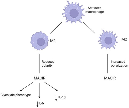
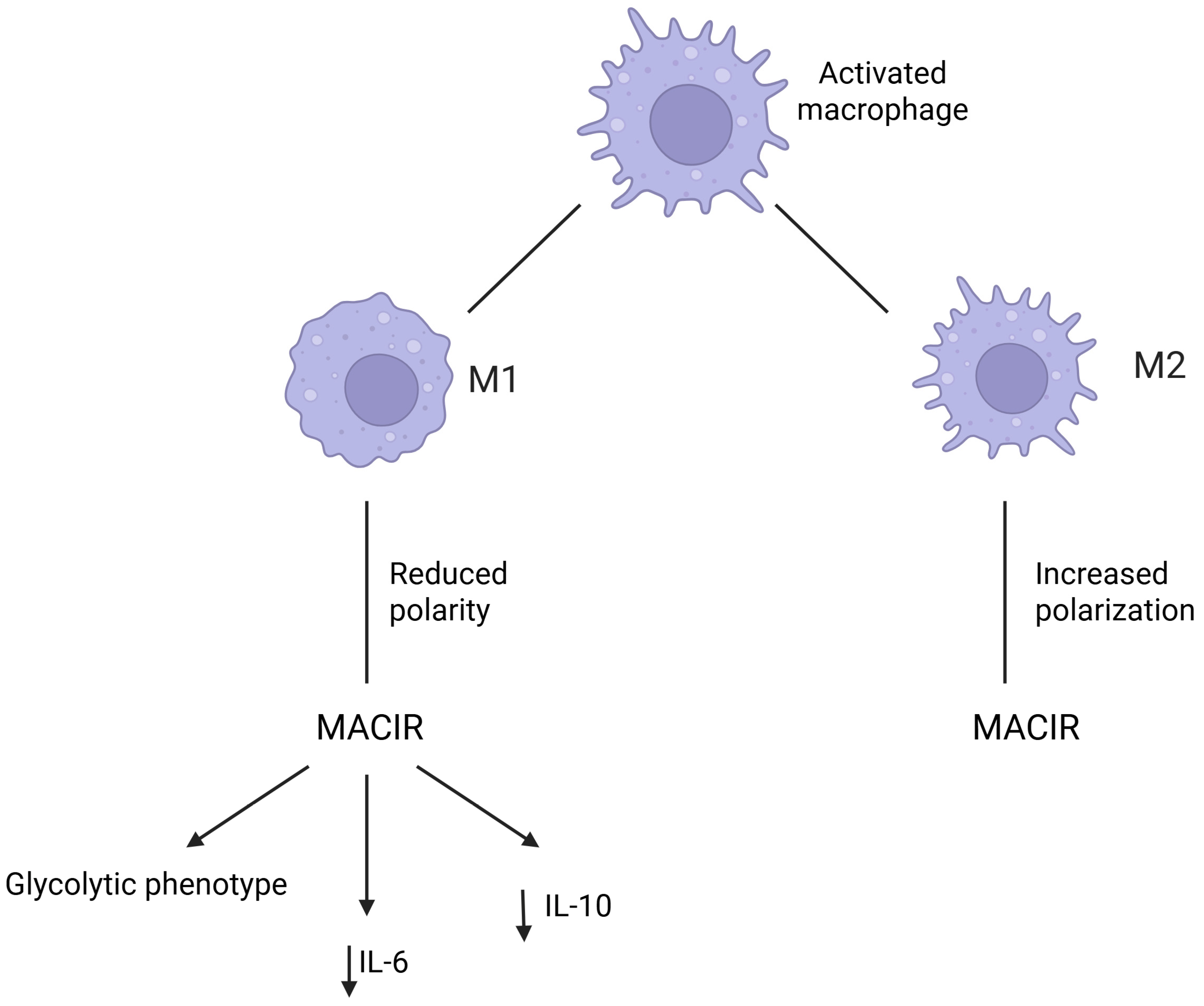
MACIR is highly expressed in macrophages. Activated macrophages can polarise into two main phenotypes: pro-inflammatory M1 macrophages, which are effector cells involved in the direct response against pathogens and cancer cells, and anti-inflammatory M2 macrophages, which are involved in tissue regeneration, remodelling, and immune regulation [19,20]. It should be noted that the M1/M2 dichotomy is an oversimplification of the complexity of macrophage phenotypes in vivo; however, it remains useful in in vitro experiments [21,22]. In a study by Dorris et al., MACIR expression was shown to be associated with the M2 phenotype. Polarisation of macrophages towards the M1 phenotype reduced MACIR expression, whereas polarisation towards the M2 phenotype increased its expression. Moreover, MACIR knockdown in macrophages resulted in downregulation of M2 markers and increased phagocytosis, highlighting the role of MACIR in limiting inflammation. MACIR also affects the expression of key cytokines in macrophages. Silencing of this gene resulted in reduced expression of multifunctional IL-6 and anti-inflammatory IL-10. In addition, Dorris et al. investigated the role of MACIR in immunometabolism. Silencing of MACIR promoted a glycolytic phenotype, which is characteristic of pro-inflammatory M1 macrophages (Figure 1) [23,24]. The glycolytic switch in inflammatory cells allows rapid adenosine triphosphate generation, which is essential for sustaining inflammation. Moreover, the products of glycolysis sustain inflammation through multiple signalling pathways, epigenetic alterations, and post-transcriptional and post-translational modifications [25,26]. The increase in glycolysis following MACIR silencing may be associated with the aforementioned reduction in IL-10 expression because this cytokine is known to inhibit glycolysis in macrophages by suppressing mechanistic target of rapamycin signalling. IL-10 also reduces glucose uptake by macrophages by inhibiting the translocation of glucose transporter 1 to the plasma membrane [27].
Figure 1.
The role of MACIR in macrophage polarization and metabolism. MACIR expression is increased in M2 macrophages and decreased in M1 macrophages. Silencing MACIR reduces IL-6 and IL-10 expression and promotes a glycolytic, pro-inflammatory macrophage phenotype. Created in BioRender. Plewa, P. (2026) https://app.biorender.com/citation/6ac756a85d96553ee67510ac (accessed on 8 October 2026).
3.2. Protein–Protein Interactions
Protein interaction studies have provided important insights into the molecular function of MACIR, which remains a relatively poorly characterised protein. Affinity proteomics identified 63 significant candidate MACIR-interacting proteins, with enrichment of proteins associated with the nuclear and mitochondrial compartments [28]. Among these candidates, interactions with the nuclear transport receptor TNPO1 and the intracellular trafficking proteins UNC119A and UNC119B were independently validated by immunoprecipitation [28]. These findings suggest that MACIR may participate in intracellular protein trafficking and processes involving different subcellular compartments.
However, the functional significance of most proteins identified in the MACIR interactome remains unknown. The majority of candidate interactions have not been independently validated or functionally characterised, and the original interactome was generated using an affinity proteomics approach with heterologous MACIR expression [28]. Therefore, the available interaction network should be considered a framework for identifying potential MACIR-dependent mechanisms rather than a fully established signalling pathway. Among the currently validated partners, the interaction with TNPO1 is particularly relevant because it provides a potential mechanism linking the MACIR interactome to its intracellular localisation, as discussed below.
3.3. Nuclear Transport and Cellular Signaling
Among the validated MACIR-interacting proteins, TNPO1 suggests a potential mechanism for regulating MACIR nuclear localisation [28]. TNPO1, also known as karyopherin-β2, is a nuclear import receptor that recognises non-classical proline-tyrosine NLSs (PY-NLSs) in cargo proteins [29,30,31]. In MACIR, mutational analysis supports the involvement of a putative non-classical PY-NLS spanning amino acids 98–117 in TNPO1 binding [28]. This provides experimental support for a mechanism in which TNPO1 recognises MACIR through this sequence and may regulate its nucleocytoplasmic trafficking.
The downstream significance of this interaction remains unclear. Although the MACIR–TNPO1 interaction and the involvement of the PY-NLS region are experimentally supported, a specific nuclear signalling pathway controlled by MACIR has not yet been defined. Thus, TNPO1-dependent recognition represents a plausible mechanism regulating MACIR localisation, but further studies are required to determine how nuclear transport contributes to its immunoregulatory functions.
4. MACIR in Autoimmune and Inflammatory Diseases
4.1. Rheumatoid Arthritis
RA is the autoimmune disease in which MACIR, formerly known as C5orf30, has been most extensively studied. Functional studies consistently support a protective role of MACIR in limiting tissue-destructive responses within the rheumatoid synovium, particularly through its effects on synovial fibroblasts and macrophages [5,23]. Genetic evidence also implicates the genomic region containing MACIR in both RA susceptibility and structural disease severity [9,32]. However, more recent findings indicate that the genetic association at this locus should not be interpreted as direct evidence of altered MACIR function because the disease-associated variant may also regulate neighbouring genes, particularly PAM [10,33].
Initial interest in C5orf30 arose from genetic studies identifying rs26232, located within its first intron, as a susceptibility variant for RA [32]. Its clinical relevance was subsequently strengthened by an allele–dose association with radiographic joint damage across three independent Northern European RA cohorts. The C allele was associated with greater structural damage, whereas each copy of the T allele was associated with lower radiographic severity, independently of established prognostic markers [9]. These observations implicated the C5orf30 genomic region in both RA susceptibility and joint destruction, providing the rationale for subsequent functional investigation of MACIR.
Functional evidence has demonstrated that MACIR is highly expressed in RA synovial tissue, particularly in synovial fibroblasts and macrophages, whereas considerably lower expression is observed in osteoarthritis and non-inflammatory control synovium [5]. Importantly, its expression is dynamically regulated by the inflammatory microenvironment. Stimulation of human fibroblasts with TNF-α and IL-1β reduces both MACIR mRNA and protein expression, indicating that pro-inflammatory cytokines actively suppress this regulator [8]. Moreover, within RA synovial tissue, MACIR expression is inversely associated with clinical disease activity and synovial TNF expression [23]. Rather than indicating a simple relationship between MACIR abundance and inflammation, these findings suggest that its expression may reflect competing inflammatory and compensatory signals within the rheumatoid joint.
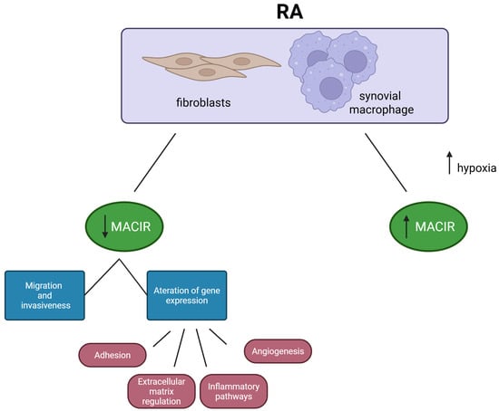
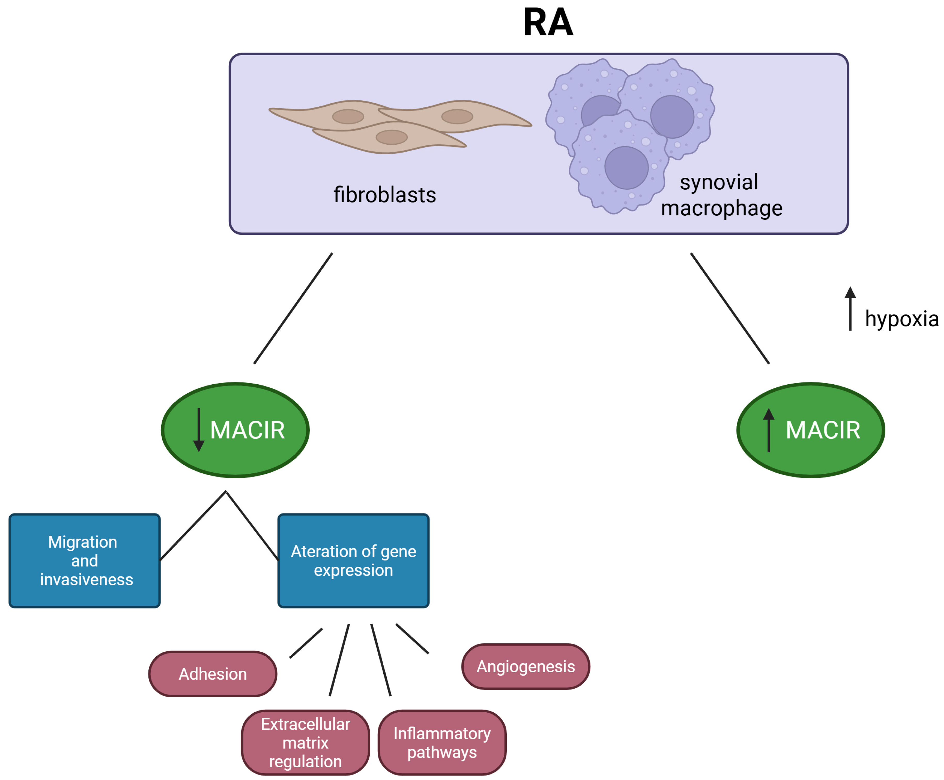
The functional consequences of MACIR loss further support a protective role. Silencing C5orf30 in RA synovial fibroblasts increases their migration and invasiveness and alters the expression of genes involved in adhesion, angiogenesis, extracellular matrix regulation, and inflammatory pathways (Figure 2) [5]. These processes contribute directly to the aggressive phenotype of synovial fibroblasts responsible for pannus formation and progressive tissue destruction. Consistently, experimental inhibition of C5orf30 in collagen-induced arthritis markedly increases joint inflammation and cartilage damage [5]. Thus, MACIR appears to restrain fibroblast-mediated tissue destruction rather than promote the inflammatory phenotype characteristic of RA.
Figure 2.
Regulation of MACIR expression and function in RA. MACIR expression is increased in RA synovial fibroblasts and macrophages, and its loss enhances fibroblast migration, invasiveness, and inflammatory activation. Created in BioRender. Plewa, P. (2026) https://app.biorender.com/citation/6ac75da31b08b703de23bed1 (accessed on 8 October 2026).
A complementary regulatory function has been identified in macrophages. MACIR expression is highest in macrophages and synovial fibroblasts, and its abundance differs between inflammatory and inflammation-resolving macrophage phenotypes [23]. MACIR inhibition impairs wound-healing and repair-associated macrophage functions, reduces signalling involved in inflammatory resolution, and decreases the secretion of anti-inflammatory mediators. It also alters macrophage immunometabolism, including the regulation of glycolytic activity, thereby linking metabolic state to inflammatory function [23]. Together with the fibroblast data, these observations suggest that MACIR may protect the rheumatoid joint through coordinated regulation of both stromal cell invasiveness and macrophage-mediated resolution.
However, the genetic relationship between rs26232 and MACIR is more complex. RA synovial fibroblasts carrying the CC risk genotype are more invasive and express higher levels of pathogenic mediators, including intercellular adhesion molecule 1, matrix metalloproteinase 14, and C-X-C motif chemokine ligand 10, than cells carrying the CT genotype [10]. Nevertheless, the rs26232 genotype is not associated with either total MACIR/C5orf30 mRNA expression or the expression of its individual transcript variants [10]. Therefore, at least in resting synovial fibroblasts, rs26232 does not behave as a conventional expression quantitative trait locus for MACIR. This observation argues against a simple model in which the risk allele directly promotes severe RA by reducing MACIR expression.
Recent evidence provides an alternative explanation for this genetic association. The rs26232 C allele has been linked to reduced expression of peptidylglycine alpha-amidating monooxygenase (PAM) in several tissues, including RA synovial fibroblasts [33]. PAM expression in the synovium is inversely associated with inflammatory activity, whereas experimental inhibition of PAM increases fibroblast invasiveness. PAM-dependent amidated peptides, particularly adrenomedullin and pro-adrenomedullin N-terminal peptide, reduce IL-6 and IL-8 production, increase transforming growth factor-β, and limit fibroblast invasiveness. Inhibition of peptide amidation in fibroblast–macrophage co-cultures also favours pro-inflammatory MerTK-negative macrophage phenotypes [33]. These findings identify PAM as a plausible functional mediator of the rs26232 association and demonstrate that the biological effect of this locus cannot be attributed solely to MACIR on the basis of the variant’s intronic location.
Overall, current evidence therefore supports two related but distinct conclusions. First, MACIR itself functions as an endogenous regulator that limits the tissue-destructive phenotype of synovial fibroblasts and supports macrophage programmes associated with inflammatory resolution [5,23]. Second, although rs26232 initially directed attention towards MACIR, its association with RA severity may involve regulatory effects on other genes within the locus, particularly PAM [10,33]. Distinguishing these functional and genetic effects is essential to avoid overinterpretation of non-coding association signals. Further studies are required to determine whether MACIR expression or activity independently predicts structural disease progression and to clarify how MACIR- and PAM-dependent mechanisms interact within the rheumatoid synovial microenvironment.
4.2. Psoriasis and Inflammatory Skin Diseases
In contrast to RA, direct disease-specific evidence linking MACIR to psoriasis or other inflammatory skin diseases is currently lacking. Therefore, its potential involvement should be regarded as mechanistically plausible rather than established. This hypothesis is supported mainly by the convergence between the known role of MACIR in macrophage immunometabolism and the contribution of metabolically activated macrophages to chronic cutaneous inflammation [21,34,35,36,37].
Macrophages are an important component of the psoriatic inflammatory environment. Depletion of macrophages with clodronate liposomes inhibits imiquimod-induced psoriasiform disease in mice, and adoptive transfer of competent macrophages restores it, indicating that their accumulation is not merely secondary to the inflammatory process [38]. Selective ablation of the CD169+ skin macrophage subset likewise attenuates disease, producing milder lesions, lower pro-inflammatory cytokine levels and a reduced lesional Th17 fraction [39]. In human disease, single-cell profiling of lesional skin identifies inflammatory macrophage populations, including TREM2+ cells with elevated antigen-presentation signatures, that interact with IL-17-producing CD8+ tissue-resident memory T cells within plaques [40]. Macrophage-derived mediators such as TNF, IL-1β and IL-23 amplify local inflammation and sustain the IL-23/IL-17 axis in keratinocyte–macrophage circuits [34], and macrophage-intrinsic lipid handling contributes directly to this amplification, since deletion of fatty acid-binding protein 5 in skin macrophages reduces IL-1β/IL-17A signalling and alleviates psoriasiform inflammation [41]. Importantly, the inflammatory activity of macrophages is closely linked to their metabolic state. Experimental psoriasiform inflammation is associated with enhanced glycolysis, whereas interventions that reduce macrophage glycolytic activity are accompanied by lower inflammatory cytokine production and improvement in skin lesions [36]. More recent evidence further indicates that promoting oxidative phosphorylation while suppressing glycolysis can shift macrophages towards a less inflammatory phenotype and attenuate psoriasis-like inflammation [37]. Epidermal-derived lactate has additionally been shown to rewire dermal macrophage metabolism and to sustain psoriatic disease burden, which is reduced by blocking lactate transport [42], consistent with the broader view of psoriasis as a metabolically regulated inflammatory skin disease [35].
These observations are particularly relevant to MACIR because this protein restrains macrophage glycolysis and supports functions associated with inflammatory resolution and tissue repair [23]. Thus, reduced MACIR activity could theoretically favour the persistence of metabolically activated, pro-inflammatory macrophages in psoriatic skin, whereas preserved MACIR function might facilitate the transition towards a resolving phenotype. However, this proposed relationship remains indirect: the psoriasis studies described above evaluated macrophage abundance, polarisation, or metabolic pathways independently of MACIR. Moreover, findings obtained predominantly in experimental psoriasis-like models cannot be directly extrapolated to the heterogeneous macrophage populations present in human disease. Overall, MACIR represents a biologically plausible but currently unconfirmed regulator of psoriatic inflammation. Its relevance will require direct assessment of MACIR expression in lesional and non-lesional skin, identification of MACIR-expressing myeloid populations, and functional studies to determine whether its modulation alters macrophage metabolism or disease severity in psoriasis.
4.3. Inflammatory Bowel Diseases (IBDs)
Direct evidence implicating MACIR in IBDs is currently lacking, and neither disease-specific alterations in MACIR expression nor the functional effects of its modulation have been established in Crohn’s disease (CD) or ulcerative colitis. Nevertheless, intestinal macrophages are critically involved in mucosal homeostasis and exhibit substantial functional reprogramming in both forms of IBD, providing a potential mechanistic context for MACIR involvement [43]. Importantly, macrophage dysfunction in IBD is closely associated with metabolic reprogramming. CD14+ intestinal macrophages from patients with CD exhibit enhanced glycolytic activity that contributes to inflammatory signalling [44]. Recent evidence from ulcerative colitis further demonstrates increased PKM2-dependent glycolysis in macrophages within damaged intestinal regions, while its inhibition promotes reparative macrophage differentiation, improves epithelial barrier function, and attenuates colitis progression [45].
Recent work has broadened this picture from individual pathways to a framework in which intestinal macrophages act as metabolic sensors whose reprogramming stabilises either inflammatory persistence or mucosal repair [46]. In ulcerative colitis, glucose metabolic reprogramming drives M1-like polarisation and determines macrophage fate through mitochondrial reactive oxygen species [47], and multi-omic analyses of patient tissue have identified macrophage-associated immunometabolic regulators, including CYBB, INPP5D and NCF4, whose expression tracks with macrophage infiltration and cytokine signalling [48]. Conversely, interventions that restore oxidative metabolism can reverse the inflammatory programme, as the microbiota-derived metabolite 4-hydroxyphenylacetic acid stabilises SIRT1, promotes oxidative metabolic remodelling in macrophages and attenuates experimental Crohn’s-like colitis in a myeloid SIRT1-dependent manner [49].
These findings are consistent with the established function of MACIR in limiting macrophage glycolysis and promoting inflammation-resolving and tissue-repair responses [23]. Thus, MACIR could theoretically contribute to intestinal homeostasis by restraining persistent metabolic activation of macrophages and supporting their transition towards reparative functions. However, this interpretation remains hypothesis-generating, as the current evidence demonstrates convergence between MACIR-regulated pathways and IBD-associated macrophage dysfunction rather than a direct role for MACIR in intestinal disease. Disease-specific studies examining MACIR expression and function in intestinal myeloid populations are therefore required.
4.4. Other Autoimmune Disorders
The potential involvement of MACIR in autoimmune diseases beyond RA remains considerably less well defined.
In systemic lupus erythematosus (SLE), no direct association between MACIR expression or genetic variation and disease development has yet been established. Nevertheless, macrophage dysfunction and metabolic reprogramming are increasingly recognised as components of SLE pathogenesis. Alterations in macrophage immunometabolism can influence the balance between pro-inflammatory and regulatory phenotypes, and modulation of these metabolic programmes has been shown to attenuate inflammatory macrophage responses in SLE [50]. More recent work has localised this dependency to the inflamed kidney: glycolytic activation of renal macrophages and tubular epithelial cells promotes pyroptosis and ferroptosis in lupus nephritis, and pharmacological inhibition of glycolysis reduces both cell death and renal injury [51]. Reprogramming of macrophage lipid metabolism has comparable effects, as annexin A1 signalling inhibits mTOR/FABP4 activity, enhances fatty acid oxidation and shifts monocyte-derived macrophages towards an antifibrotic phenotype that limits renal fibrosis [52]. Given that MACIR regulates macrophage metabolism and promotes inflammation-resolving functions [23], its involvement in SLE is biologically plausible, although it is currently supported only by indirect mechanistic evidence.
Evidence in multiple sclerosis (MS) is similarly preliminary. The RA-associated C5orf30 variant rs26232 showed a nominal association with MS in a study investigating shared genetic susceptibility across autoimmune diseases [53]. However, the modest statistical significance and limited sample size indicate that this finding should be interpreted as preliminary evidence of potential pleiotropy rather than a confirmed MS-specific association. A mechanistic rationale is provided by experimental neuroinflammation studies showing that modulation of macrophage metabolism can reduce pro-inflammatory macrophage activity and disease severity in experimental autoimmune encephalomyelitis [54]. Metabolic control of myeloid phenotype in the central nervous system has since been reviewed systematically, with glycolytic programming of M1 microglia and infiltrating macrophages identified as a modifiable amplifier of chronic neuroinflammation and of remyelination failure [55]. These observations are compatible with the immunometabolic function of MACIR but do not establish its direct involvement in MS.
Among other potential autoimmune disorders, primary biliary cholangitis (PBC) provides more direct genetic support. A large genome-wide meta-analysis identified the 5q21 locus containing C5orf30, with rs526231 reaching genome-wide significance for PBC susceptibility [56]. However, the association of this genomic region does not establish MACIR as the causal gene, and its functional involvement in hepatic autoimmunity remains unconfirmed. Subsequent genetic work on PBC has concentrated on other susceptibility loci and functional variants rather than on 5q21, and no functional follow-up of C5orf30 in hepatic autoimmunity has been reported [57].
Outside the classical autoimmune spectrum, periodontitis currently provides the only direct human evidence that MACIR expression is altered in an inflammatory disease other than RA. MACIR mRNA expression in gingival tissue from patients with periodontitis is significantly lower than in tissue from healthy controls (p = 0.009), and stimulation of primary human gingival fibroblasts with IL-1β or TNF-α decreases both MACIR mRNA and protein [8]. This reproduces, in a second barrier tissue and a second chronic inflammatory disease, the pattern described in the rheumatoid synovium, in which MACIR is suppressed by pro-inflammatory cytokines and its loss is associated with a tissue-destructive phenotype. It therefore supports the interpretation of MACIR as a general cytokine-responsive brake on fibroblast and macrophage activation rather than an RA-restricted regulator, although expression data alone cannot establish causality.
Overall, the evidence for MACIR outside RA remains heterogeneous. In SLE and MS, its possible involvement is primarily supported by overlap between known MACIR-regulated pathways and disease-associated macrophage dysfunction, whereas PBC provides a stronger genetic rationale but lacks functional validation, and periodontitis supplies the only direct human expression data [8]. Thus, these disorders should currently be regarded as potential contexts for MACIR activity rather than diseases in which its pathogenic role has been established. Table 1 summarises the disease-specific evidence discussed in this section, together with its type and current strength.
Table 1.
MACIR in autoimmune and inflammatory diseases: type, content and current strength of the available evidence.
5. MACIR and Cancer-Associated Inflammation
5.1. Links Between Chronic Inflammation and Cancer
Chronic inflammation is a significant factor in cancer development because prolonged activation of immune cells leads to the production of mediators such as TNF-α, IL-6, IL-1β, and transforming growth factor-β, which can promote cell proliferation, angiogenesis, epithelial–mesenchymal transition, and tumour progression [58]. Under the influence of pro-inflammatory cytokines (e.g., TNF-α or IL-1β), MACIR gene expression is downregulated. MACIR acts as a negative regulator of tissue damage and inflammation. Under conditions of elevated TNF-α and IL-1β concentrations, decreased MACIR expression reduces its physiological inhibitory effect, which may exacerbate the autoaggressive and pro-invasive phenotype of stromal cells and amplify the inflammatory cascade in the tissue [23].
The tumour microenvironment comprises tumour cells, stromal cells (including fibroblasts), and numerous populations of immune cells that, through mutual communication, regulate tumour growth, invasion, and the immune response against the tumour [59,60,61]. In chronic inflammation, the cytokine and chemokine environment promotes the recruitment of myeloid cells, including monocytes and macrophages, which can then adopt functional phenotypes that support tumour progression [62,63]. MACIR is involved in regulating the immunometabolism and plasticity of these cells; its dysregulated expression affects fatty acid metabolism and glycolysis in monocytes and macrophages, influencing their survival in an adverse tissue microenvironment [8,32].
Tumour-associated macrophages are among the most abundant populations of immune cells in many solid tumours and exhibit significant functional heterogeneity. Tumour-associated macrophages can promote angiogenesis, extracellular matrix remodelling, invasion, and metastasis and can inhibit the antitumour activity of lymphocytes [64]. MACIR turnover rates vary significantly depending on the activation status of macrophages; a decrease in MACIR levels promotes the silencing of mechanisms that protect against tissue damage and may induce a phenotype that promotes extracellular matrix remodelling [5].
At the same time, macrophages retain high plasticity and, under appropriate conditions, can exhibit anticancer activity; therefore, the simplified classification into M1 and M2 macrophages does not fully reflect their heterogeneity within the tumour microenvironment. Understanding the role of immunometabolic proteins such as MACIR allows for a more accurate analysis of the continuous spectrum of macrophage polarisation in tumour regions characterised by hypoxia and chronic inflammation [10,65].
5.2. Potential Role of MACIR in Tumor Immunity
MACIR, also known as C5orf30, is a highly conserved protein involved in immunometabolic regulation and the response to tissue damage. Its expression is highest in cells of the myeloid lineage (monocytes and macrophages) and in fibroblasts located in tissues affected by chronic inflammation [23,66]. In vitro and in vivo studies have shown that inhibition (knockdown or knockout) of the MACIR/C5orf30 gene substantially alters the immunological phenotype and metabolic profile of macrophages. MACIR protein deficiency reduces the reparative potential of these cells, limits the secretion of key anti-inflammatory mediators (including IL-10), and may promote the persistence of an unbalanced pro-invasive cascade [5].
Studies have reported that inhibition of C5orf30 homologue expression leads to excessive and uncontrolled recruitment of macrophages to sites of tissue damage. This suggests that MACIR acts as a physiological ‘brake’ on the inflammatory response, protecting tissues from autoaggression by controlling myeloid cell chemotaxis [9]. At the cellular level, MACIR regulates key metabolic pathways, including fatty acid metabolism and the efficiency of oxidative phosphorylation, which directly determine the ability of macrophages to suppress inflammation [28]. Although these relationships have been clearly defined in inflammatory and autoimmune diseases, the direct role of MACIR in the function of tumour-associated macrophages within the tumour microenvironment remains relatively poorly understood. Most mechanistic analyses to date have focused on models of RA or periodontitis, creating a significant research gap in the context of immuno-oncology.
The metabolic state of TAMs is associated with their immunoregulatory functions and effects on tumor growth and antitumor immunity. However, the specific contribution of MACIR to these metabolic processes in tumor-associated macrophages has not been established. Further studies are needed to determine whether MACIR-dependent metabolic regulation directly influences macrophage-mediated tumor immunity [67].
5.3. MACIR as a Diagnostic and Prognostic Biomarker
Because of the limited number of direct clinical studies focused exclusively on the native MACIR protein, a single measurement of its expression does not currently constitute a standalone diagnostic test in oncology [68]. However, specific genetic alterations and transcriptional aberrations involving the C5orf30/MACIR locus show considerable potential. A specific CCNH–C5orf30 fusion transcript has been identified in the plasma of patients with HCC; its absence in the healthy control group suggests that abnormal chimeric RNA molecules derived from this locus could be used as sensitive liquid biopsy markers [12,69,70]. A subsequent study analyzed serum fusion transcripts in 61 patients with HCC and 75 individuals with non-HCC conditions. The CCNH–C5orf30 fusion transcript was detected in 55.7% of HCC samples. A machine-learning model combining two fusion transcripts with serum alpha-fetoprotein (AFP) achieved 95% accuracy in the testing cohort. These findings support further investigation of fusion transcripts as components of multimarker approaches for HCC detection; however, the reported accuracy cannot be attributed to CCNH–C5orf30 alone [71].
MACIR may have prognostic significance in cancer because its activity is associated with DNA repair processes that influence cancer cell survival and response to treatment. In ovarian cancer, high activity of the MACIR–FIRRM–FIGNL1 complex was associated with poorer patient prognosis, suggesting a possible link between this pathway and disease aggressiveness [72,73]. C5orf30 has also been identified as a potential marker associated with survival in gene expression analyses of prostate cancer; however, these findings require further clinical validation [18].
The CCNH–C5orf30 fusion transcript can be detected in circulating RNA from patients with HCC, and its levels decreased following anticancer treatment [71]. These results suggest a potential role for C5orf30 in treatment monitoring; however, they pertain to the fusion transcript rather than normal MACIR expression, so MACIR cannot currently be considered a validated marker of treatment response [12].
5.4. Current Evidence and Future Directions
Research to date indicates that MACIR is a protein with a relatively poorly understood molecular function; its 206-amino acid sequence shows no significant similarity to previously characterised proteins, and structural analysis suggests that it is disordered.
Analysis of the MACIR interactome has revealed interactions with proteins from various cellular compartments, including components associated with the cell membrane, nucleus, and mitochondria, suggesting that this regulator may have multifaceted functions [28].
More recent studies have provided direct evidence of the role of MACIR in maintaining genomic stability through its interaction with the FIGNL1–FIRRM complex, which regulates RAD51 filaments during homologous recombination. A study published in 2026 directly identified MACIR/C5orf30 as an adaptor for the FIGNL1–FIRRM complex. The authors showed that MACIR binds FIRRM and facilitates RAD51 filament disassembly, thereby contributing to DNA damage repair and genome stability. Loss of MACIR led to the accumulation of DNA damage and genomic instability and was associated with inflammatory signaling [74].
Loss of MACIR led to impaired RAD51 filament disassembly, increased accumulation of DNA damage, and genomic instability, suggesting that its function extends beyond regulation of the macrophage response [75,76].
In ovarian cancer cell models, inhibition of this pathway increased the sensitivity of cancer cells to platinum compounds, providing the first experimental evidence that MACIR could form part of a strategy to sensitise cancer cells to chemotherapy [74,77]. In HCC, the CCNH–C5orf30 fusion transcript was detected in patients’ circulating RNA but not in the serum of healthy individuals in the analysed cohort [12]. In a subsequent study involving 61 patients with HCC, the CCNH–C5orf30 transcript was detected in 55.7% of samples, and a model utilising the levels of several fusion transcripts achieved 95% accuracy in the test cohort when combined with α-fetoprotein levels [78]. These data suggest that the role of C5orf30/MACIR in cancer may extend beyond immune regulation to include processes related to genome stability and the potential use of its molecular products in diagnostics. A major gap in current research is whether the function of MACIR in DNA repair is preserved across different types of cancer or whether its significance depends on tumour-specific abnormalities in DNA repair pathways.
Studies determining whether MACIR affects the efficacy of immunotherapy and interactions between cancer cells and the immune system are also lacking. Future studies should assess the potential of MACIR as a therapeutic target and determine whether its inhibition can increase tumour sensitivity to existing treatments [74,79,80]. Table 2 summarizes the role of MACIE in cancer-related inflammation.
Table 2.
Role of MACIR in cancer-associated inflammation, tumor progression, and potential clinical applications.
6. Discussion and Future Directions
Current evidence indicates that MACIR is an important regulator at the interface between immunometabolism, inflammation, and tissue repair. Its expression is particularly associated with macrophage and synovial fibroblast functions, while experimental studies suggest that reduced MACIR activity promotes glycolytic reprogramming, alters cytokine production, and favours tissue-destructive phenotypes. These findings are most consistently supported in RA, where MACIR appears to limit fibroblast invasiveness, macrophage dysfunction, and joint tissue damage.
However, the relationship between MACIR and disease-associated genetic variation requires careful interpretation. Although rs26232 was initially associated with RA susceptibility and radiographic severity, this variant is not consistently associated with MACIR transcript abundance. Evidence of effects on the neighbouring PAM gene suggests that the clinical associations of this locus may involve regulatory effects beyond MACIR itself. Thus, genetic association alone is insufficient to establish MACIR as the causal mediator.
Beyond RA, the role of MACIR in psoriasis, IBDs, SLE, MS, and other autoimmune disorders remains largely hypothetical. In these conditions, the proposed involvement of MACIR is mainly supported by the known importance of macrophage immunometabolism or by preliminary genetic associations rather than direct functional evidence.
Emerging findings also suggest that the functions associated with MACIR may extend beyond immune regulation. Interactions involving TNPO1 indicate a potential role in intracellular and nuclear transport, while studies of the MACIR protein suggest links to DNA repair and genome stability. Nevertheless, the downstream mechanisms and their relevance to disease remain insufficiently defined.
Future studies should therefore combine single-cell and spatial transcriptomics with CRISPR-based gene perturbation, proteomics, metabolomics, and epigenomic analyses to define the cell-specific functions of MACIR. Particular attention should be given to the relationship between MACIR and cellular metabolism, inflammatory signalling, and the PAM locus. Direct functional studies are also needed in diseases beyond RA, particularly psoriasis, IBD, and systemic autoimmune diseases, for which evidence is currently limited.
In cancer, future research should determine the role of MACIR in tumour-associated macrophages, tumour–stroma interactions, DNA repair, and responses to chemotherapy and immunotherapy. The diagnostic and prognostic potential of MACIR-associated transcripts, including the CCNH–C5orf30 fusion, should be validated in larger, independent clinical cohorts.
Overall, MACIR represents a promising but incompletely characterised regulator of immune and tissue responses. Defining its cell-specific functions and distinguishing direct MACIR-dependent effects from those mediated by neighbouring genes will be essential for determining its potential as a biomarker or therapeutic target.
Author Contributions
Conceptualization, A.P.; writing—original draft preparation, P.P., K.F., A.J., H.O., J.S., I.K. and A.P.; writing—review and editing, P.P., K.F., A.J., H.O., J.S., I.K. and A.P.; supervision, A.P. All authors have read and agreed to the published version of the manuscript.
Funding
This research received no external funding.
Institutional Review Board Statement
Not applicable.
Informed Consent Statement
Not applicable.
Data Availability Statement
No new data were created or analyzed in this study. Data sharing is not applicable to this article.
Conflicts of Interest
The authors declare no conflicts of interest.
Abbreviations
The following abbreviations are used in this manuscript:
| C5orf30 | Chromosome 5 Open Reading Frame 30 |
| CD | Crohn’s disease |
| EMT | Epithelial–mesenchymal transition |
| HEK293T | Human Embryonic Kidney |
| GLUT1 | Glucose transporter 1 |
| GSEA | Gene Set Enrichment Analysis |
| IBD | Inflammatory bowel diseases |
| JNK | cJun NH2-terminal kinase |
| MACIR | Macrophage Immunometabolism Regulator |
| MAPK | Mitogen-activated protein kinase |
| MS | Multiple sclerosis |
| NLS | Nuclear localization signal |
| PBC | Primary biliary cholangitis |
| PY-NLS | Proline-tyrosine nuclear localization signals |
| RA | Rheumatoid arthritis |
| SLE | Systemic lupus erythematosus |
| SPM | Specialized pro-resolving mediator |
| TAM | Tumor-associated macrophage |
| TME | Tumor microenvironment |
| TNPO1 | Transport receptor transportin-1 |
| TPP | Triphenyl phosphate |
| UC | Ulcerative colitis |
References
- Chen, L.; Deng, H.; Cui, H.; Fang, J.; Zuo, Z.; Deng, J.; Li, Y.; Wang, X.; Zhao, L. Inflammatory responses and inflammation-associated diseases in organs. Oncotarget 2018, 9, 7204–7218. [Google Scholar] [CrossRef] [Scilit] [PubMed]
- Arneth, B. Molecular Mechanisms of Immune Regulation: A Review. Cells 2025, 14, 283. [Google Scholar] [CrossRef] [Scilit] [PubMed]
- Perez-Hernandez, J.; Chiurchiù, V.; Perruche, S.; You, S. Regulation of T-Cell Immune Responses by Pro-Resolving Lipid Mediators. Front. Immunol. 2021, 12, 768133. [Google Scholar] [CrossRef] [Scilit] [PubMed]
- Song, X.; Liang, H.; Nan, F.; Chen, W.; Li, J.; He, L.; Cun, Y.; Li, Z.; Zhang, W.; Zhang, D. Autoimmune Diseases: Molecular Pathogenesis and Therapeutic Targets. MedComm 2025, 6, e70262. [Google Scholar] [CrossRef] [Scilit] [PubMed]
- Muthana, M.; Hawtree, S.; Wilshaw, A.; Linehan, E.; Roberts, H.; Khetan, S.; Adeleke, G.; Wright, F.; Akil, M.; Fearon, U.; et al. C5orf30 is a negative regulator of tissue damage in rheumatoid arthritis. Proc. Natl. Acad. Sci. USA 2015, 112, 11618–11623. [Google Scholar] [CrossRef] [Scilit] [PubMed]
- Ali, A.M.; Vino, S. Genetic markers as therapeutic target in rheumatoid arthritis: A game changer in clinical therapy? Rheumatol. Int. 2016, 36, 1601–1607. [Google Scholar] [CrossRef] [Scilit] [PubMed]
- Onuora, S. Rheumatoid arthritis: C5orf30 regulates severity of tissue destruction in RA. Nat. Rev. Rheumatol. 2015, 11, 622. [Google Scholar] [CrossRef] [Scilit] [PubMed]
- Serwin, K.; Kozak, M.; Mazurek-Mochol, M.; Piotrowska, K.; Czerewaty, M.; Pawlik, A. Human macrophage immunometabolism regulator (MACIR) in patients with periodontitis. Immunobiology 2023, 228, 152760. [Google Scholar] [CrossRef] [Scilit] [PubMed]
- Teare, M.D.; Knevel, R.; Morgan, M.D.; Kleszcz, A.; Emery, P.; Moore, D.J.; Conaghan, P.; Huizinga, T.W.; Morgan, A.W.; van der Helm-van Mil, A.H.; et al. Allele-dose association of the C5orf30 rs26232 variant with joint damage in rheumatoid arthritis. Arthritis Rheum. 2013, 65, 2555–2561. [Google Scholar] [CrossRef] [Scilit] [PubMed]
- Dorris, E.R.; Linehan, E.; Trenkmann, M.; Veale, D.J.; Fearon, U.; Wilson, A.G. Association of the Rheumatoid Arthritis Severity Variant rs26232 with the Invasive Activity of Synovial Fibroblasts. Cells 2019, 8, 1300. [Google Scholar] [CrossRef] [Scilit] [PubMed]
- Yu, Y.P.; Tsung, A.; Liu, S.; Nalesnick, M.; Geller, D.; Michalopoulos, G.; Luo, J.H. Detection of fusion transcripts in the serum samples of patients with hepatocellular carcinoma. Oncotarget 2019, 10, 3352–3360. [Google Scholar] [CrossRef] [Scilit]
- Yu, Y.P.; Liu, P.; Nelson, J.; Hamilton, R.L.; Bhargava, R.; Michalopoulos, G.; Chen, Q.; Zhang, J.; Ma, D.; Pennathur, A.; et al. Identification of recurrent fusion genes across multiple cancer types. Sci. Rep. 2019, 9, 1074. [Google Scholar] [CrossRef] [Scilit] [PubMed]
- McGauran, G.; Dorris, E.; Borza, R.; Morgan, N.; Shields, D.C.; Matallanas, D.; Wilson, A.G.; O’Connell, D.J. The nuclear transport receptor TNPO1 binds macrophage immunometabolism regulator MACIR via a PY-NLS motif. bioRxiv 2019. Epub ahead of printing. [Google Scholar] [CrossRef] [Scilit]
- Gorska, M.M.; Stafford, S.J.; Cen, O.; Sur, S.; Alam, R. Unc119, a novel activator of Lck/Fyn, is essential for T cell activation. J. Exp. Med. 2004, 199, 369–379. [Google Scholar] [CrossRef] [Scilit] [PubMed]
- Stephen, L.A.; ElMaghloob, Y.; McIlwraith, M.J.; Yelland, T.; Castro Sanchez, P.; Roda-Navarro, P.; Ismail, S. The Ciliary Machinery Is Repurposed for T Cell Immune Synapse Trafficking of LCK. Dev. Cell 2018, 47, 122–132.e4. [Google Scholar] [CrossRef] [Scilit] [PubMed]
- Mittal, A.; Buscaglia, P.; Srivastava, D.; Artemyev, N.O.; Sebag, J.A. Inhibition of Unc119b improves insulin sensitivity through potentiation of Rac1 activation in skeletal muscle and brown adipose tissue. Mol. Metab. 2025, 100, 102230. [Google Scholar] [CrossRef] [Scilit] [PubMed]
- Biggi, S.; Buccarello, L.; Sclip, A.; Lippiello, P.; Tonna, N.; Rumio, C.; Di Marino, D.; Miniaci, M.C.; Borsello, T. Evidence of Presynaptic Localization and Function of the c-Jun N-terminal Kinase. Neural Plast. 2017, 2017, 6468356. [Google Scholar] [CrossRef] [Scilit] [PubMed]
- Cai, L.; Du, G. Transcriptomic and functional analysis of triphenyl phosphate exposure in prostate cancer progression. Discov. Oncol. 2025, 16, 1497. [Google Scholar] [CrossRef] [Scilit] [PubMed]
- Italiani, P.; Boraschi, D. From Monocytes to M1/M2 Macrophages: Phenotypical vs. Functional Differentiation. Front. Immunol. 2014, 5, 514. [Google Scholar] [CrossRef] [Scilit] [PubMed]
- He, W.; Xu, J.; Li, X. Macrophage polarization in inflammatory regulation: Molecular mechanisms, therapeutic targets, and translational challenges. Cell. Mol. Life Sci. 2026, 83, 164. [Google Scholar] [CrossRef] [Scilit] [PubMed]
- Pantazi, P.; Clements, T.; Venø, M.; Abrahams, V.M.; Holder, B. Distinct non-coding RNA cargo of extracellular vesicles from M1 and M2 human primary macrophages. J. Extracell. Vesicles 2022, 11, e12293. [Google Scholar] [CrossRef] [Scilit] [PubMed]
- Strizova, Z.; Benesova, I.; Bartolini, R.; Novysedlak, R.; Cecrdlova, E.; Foley, L.K.; Striz, I. M1/M2 macrophages and their overlaps—Myth or reality? Clin. Sci. 2023, 137, 1067–1093. [Google Scholar] [CrossRef] [Scilit] [PubMed]
- Dorris, E.R.; Tazzyman, S.J.; Moylett, J.; Ramamoorthi, N.; Hackney, J.; Townsend, M.; Muthana, M.; Lewis, M.J.; Pitzalis, C.; Wilson, A.G. The Autoimmune Susceptibility Gene C5orf30 Regulates Macrophage-Mediated Resolution of Inflammation. J. Immunol. 2019, 202, 1069–1078. [Google Scholar] [CrossRef] [Scilit] [PubMed]
- Chen, S.; Saeed, A.F.U.H.; Liu, Q.; Jiang, Q.; Xu, H.; Xiao, G.G.; Rao, L.; Duo, Y. Macrophages in immunoregulation and therapeutics. Signal Transduct. Target. Ther. 2023, 8, 207. [Google Scholar] [CrossRef] [Scilit] [PubMed]
- Soto-Heredero, G.; Gómez de Las Heras, M.M.; Gabandé-Rodríguez, E.; Oller, J.; Mittelbrunn, M. Glycolysis—A key player in the inflammatory response. FEBS J. 2020, 287, 3350–3369. [Google Scholar] [CrossRef] [Scilit] [PubMed]
- Namgaladze, D.; Brüne, B. Rapid glycolytic activation accompanying innate immune responses: Mechanisms and function. Front. Immunol. 2023, 14, 1180488. [Google Scholar] [CrossRef] [Scilit] [PubMed]
- Ip, W.K.E.; Hoshi, N.; Shouval, D.S.; Snapper, S.; Medzhitov, R. Anti-inflammatory effect of IL-10 mediated by metabolic reprogramming of macrophages. Science 2017, 356, 513–519. [Google Scholar] [CrossRef] [Scilit] [PubMed]
- McGauran, G.; Dorris, E.; Borza, R.; Morgan, N.; Shields, D.C.; Matallanas, D.; Wilson, A.G.; O’Connell, D.J. Resolving the Interactome of the Human Macrophage Immunometabolism Regulator (MACIR) with Enhanced Membrane Protein Preparation and Affinity Proteomics. Proteomics 2020, 20, e2000062. [Google Scholar] [CrossRef] [Scilit] [PubMed]
- Lee, B.J.; Cansizoglu, A.E.; Süel, K.E.; Louis, T.H.; Zhang, Z.; Chook, Y.M. Rules for nuclear localization sequence recognition by karyopherin beta 2. Cell 2006, 126, 543–558. [Google Scholar] [CrossRef] [Scilit] [PubMed]
- Twyffels, L.; Gueydan, C.; Kruys, V. Transportin-1 and Transportin-2: Protein nuclear import and beyond. FEBS Lett. 2014, 588, 1857–1868. [Google Scholar] [CrossRef] [Scilit] [PubMed]
- Yang, Y.; Guo, L.; Chen, L.; Gong, B.; Jia, D.; Sun, Q. Nuclear transport proteins: Structure, function, and disease relevance. Signal Transduct. Target. Ther. 2023, 8, 425. [Google Scholar] [CrossRef] [Scilit] [PubMed]
- Stahl, E.A.; Raychaudhuri, S.; Remmers, E.F.; Xie, G.; Eyre, S.; Thomson, B.P.; Li, Y.; Kurreeman, F.A.; Zhernakova, A.; Hinks, A.; et al. Genome-wide association study meta-analysis identifies seven new rheumatoid arthritis risk loci. Nat. Genet. 2010, 42, 508–514. [Google Scholar] [CrossRef] [Scilit] [PubMed]
- Sheridan, K.J.; Dorris, E.R.; Pimenta, M.I.; Falkov, J.; Fisher, M.; Pledger, S.; Devey, H.; Muthana, M.; Shields, D.C.; Mains, R.E.; et al. Tissue Damage in Rheumatoid Arthritis Is Genetically Linked to Low Peptidylglycine Alpha-Amidating Monooxygenase Activity in Synovial Fibroblasts. Arthritis Rheumatol. 2026, 78, 1837–1849. [Google Scholar] [CrossRef] [Scilit] [PubMed]
- Liu, Q.; Wu, Y.; Tang, J.; Kuang, Y. Macrophages in Psoriasis: From Functional Heterogeneity to Metabolic Reprogramming and Novel Therapeutics. Exp. Dermatol. 2026, 35, e70275. [Google Scholar] [CrossRef] [Scilit] [PubMed]
- Hong, D.; Xiong, H.; Lu, S.; Ma, J.; Shi, Z. Metabolic regulation of the immune cell in psoriasis: Mechanisms and interventions. Curr. Opin. Immunol. 2025, 96, 102614. [Google Scholar] [CrossRef] [Scilit] [PubMed]
- Wang, Y.; Qi, C.; Feng, F.; Hu, X.; Zhao, N.; Zhao, J.; Di, T.; Meng, Y.; Yang, D.; Zhu, H.; et al. Resveratrol Ameliorates Imiquimod-Induced Psoriasis-Like Mouse Model via Reducing Macrophage Infiltration and Inhibiting Glycolysis. J. Inflamm. Res. 2023, 16, 3823–3836. [Google Scholar] [CrossRef] [Scilit] [PubMed]
- Wang, B.; Li, D.; Pan, C.; Wang, Y.; Tan, Y.; Yang, Y.; Liu, H.; Yao, Z.; Zhang, H.; Wang, Z. Platelet-Derived Extracellular Vesicles Alleviate Psoriatic Inflammation via Mitochondrial Transfer to Macrophages. Exp. Dermatol. 2025, 34, e70152. [Google Scholar] [CrossRef] [Scilit] [PubMed]
- Tan, R.Z.; Zhong, X.; Han, R.Y.; Xie, K.H.; Jia, J.; Yang, Y.; Cheng, M.; Yang, C.Y.; Lan, H.Y.; Wang, L. Macrophages mediate psoriasis via Mincle-dependent mechanism in mice. Cell Death Discov. 2023, 9, 140. [Google Scholar] [CrossRef] [Scilit] [PubMed]
- Li, M.; Yu, W.; Liu, Z.; Liu, S. CD169+ Skin Macrophages Function as a Specialized Subpopulation in Promoting Psoriasis-like Skin Disease in Mice. Int. J. Mol. Sci. 2024, 25, 5705. [Google Scholar] [CrossRef] [Scilit] [PubMed]
- Chen, Q.; Wei, L.; Liu, Y.; Li, Y.; Jia, X.; Li, Y.; Wu, W.; Du, X. Single-cell RNA sequencing reveals the interaction of TREM2+ macrophages and Tc17 lymphocytes in scalp psoriasis. Immunol. Res. 2026, 74, 116. [Google Scholar] [CrossRef] [Scilit] [PubMed]
- Yu, J.; Hao, J.; Yorek, M.S.; Jiang, X.; Avellino, A.; Liu, S.; Han, X.; Shilyansky, J.; Wang, Z.; Wang, Y.; et al. FABP5 in skin macrophages mediates saturated fat-induced IL-1β signaling in psoriatic inflammation. Cell Rep. 2025, 44, 116254. [Google Scholar] [CrossRef] [Scilit] [PubMed]
- Ayyangar, U.; Karkhanis, A.; Tay, H.; Afandi, A.F.B.; Bhattacharjee, O.; Ks, L.; Lee, S.H.; Chan, J.; Raghavan, S. Metabolic rewiring of macrophages by epidermal-derived lactate promotes sterile inflammation in the murine skin. EMBO J. 2024, 43, 1113–1134. [Google Scholar] [CrossRef] [Scilit] [PubMed]
- Dharmasiri, S.; Garrido-Martin, E.M.; Harris, R.J.; Bateman, A.C.; Collins, J.E.; Cummings, J.R.F.; Sanchez-Elsner, T. Human Intestinal Macrophages Are Involved in the Pathology of Both Ulcerative Colitis and Crohn Disease. Inflamm. Bowel Dis. 2021, 27, 1641–1652. [Google Scholar] [CrossRef] [Scilit] [PubMed]
- Zeng, Z.; Cheng, S.; Li, X.; Liu, H.; Lin, J.; Liang, Z.; Liu, X.; Cao, C.; Li, S.; He, X.; et al. Glycolytic Activation of CD14+ Intestinal Macrophages Contributes to the Inflammatory Responses via Exosomal Membrane Tumor Necrosis Factor in Crohn’s Disease. Inflamm. Bowel Dis. 2024, 30, 90–102. [Google Scholar] [CrossRef] [Scilit] [PubMed]
- Zhang, D.; Tao, P.; Li, J.; Zhou, X.; Qu, H.; Li, Y.; Chen, H.; Yuan, H.; Huang, J.; Ji, Z.; et al. Targeting PKM2-dependent glycolysis reprogrammes monocytes into Cadm1+ macrophages to promote mucosal repair and attenuate colitis progression. Gut 2026. Epub ahead of printing. [Google Scholar] [CrossRef] [Scilit] [PubMed]
- Guan, G.; Sun, X.; Chen, Y.; Guo, Q.; Dong, Y. Macrophage-Orchestrated Metabolic Sensing Drives IBD Pathogenesis: A Framework for Targeted Therapy. Int. J. Biol. Sci. 2026, 22, 5754–5779. [Google Scholar] [CrossRef] [Scilit] [PubMed]
- Zhang, Y.; Yang, D.; Jiang, J.; Wang, D.; Ji, F.; Sun, Z. The plasticity and metabolic reprogramming of macrophage in ulcerative colitis. Front. Immunol. 2026, 17, 1903462. [Google Scholar] [CrossRef] [Scilit] [PubMed]
- Zhang, R.; Zhang, H.; Wang, Z.; Wang, Y.; Jia, Z. Multi-omics analysis identifies macrophage immunometabolic signatures in ulcerative colitis. Front. Cell. Infect. Microbiol. 2026, 16, 1858403. [Google Scholar] [CrossRef] [Scilit] [PubMed]
- Cheng, C.; Cheng, S.; Jiang, W.; Fu, S.; Shang, Y.; Tang, X.; Zhao, J.; Wang, H. Microbiota-derived 4-HPAA alleviates Crohn’s disease by stabilizing SIRT1 and reprogramming macrophage immunometabolism. Apoptosis 2026, 31, 215. [Google Scholar] [CrossRef] [Scilit] [PubMed]
- Lai, J.H.; Wu, D.W.; Huang, C.Y.; Hung, L.F.; Wu, C.H.; Ho, L.J. USP18 induction regulates immunometabolism to attenuate M1 signal-polarized macrophages and enhance IL-4-polarized macrophages in systemic lupus erythematosus. Clin. Immunol. 2024, 265, 110285. [Google Scholar] [CrossRef] [Scilit] [PubMed]
- Jativa, S.; Torrico, S.; Muñoz, A.; Torres Salido, M.T.; Hotter, G. Cells’ survival pattern of kidney macrophages and epithelial cells in lupus nephritis is influenced by glycolysis. Lupus Sci. Med. 2026, 13, e001933. [Google Scholar] [CrossRef] [Scilit] [PubMed]
- Tao, J.; Cheng, Q.; Zhang, P.; Yu, G.; Chen, Q.; Yang, M.; Wu, Q.; Fang, H.; Wu, H.; Song, X.; et al. ANXA1-mediated mTOR/FABP4 Inhibition Drives Antifibrotic Macrophage Reprogramming in Lupus Nephritis. Int. J. Biol. Sci. 2026, 22, 2343–2360. [Google Scholar] [CrossRef] [Scilit] [PubMed]
- Restrepo, N.A.; Butkiewicz, M.; McGrath, J.A.; Crawford, D.C. Shared Genetic Etiology of Autoimmune Diseases in Patients from a Biorepository Linked to De-identified Electronic Health Records. Front. Genet. 2016, 7, 185. [Google Scholar] [CrossRef] [Scilit] [PubMed]
- Cai, F.; Liu, S.; Lei, Y.; Jin, S.; Guo, Z.; Zhu, D.; Guo, X.; Zhao, H.; Niu, X.; Xi, Y.; et al. Epigallocatechin-3 gallate regulates macrophage subtypes and immunometabolism to ameliorate experimental autoimmune encephalomyelitis. Cell. Immunol. 2021, 368, 104421. [Google Scholar] [CrossRef] [Scilit] [PubMed]
- Badawi, G.A.; El-Sayed, R.M.; Fawzy, M.N. Immunometabolic reprogramming in multiple sclerosis: From pathogenic amplifier to therapeutic target in neuroinflammation and remyelination. Inflammopharmacology 2026, 34, 5843–5872. [Google Scholar] [CrossRef] [Scilit] [PubMed]
- Cordell, H.J.; Han, Y.; Mells, G.F.; Li, Y.; Hirschfield, G.M.; Greene, C.S.; Xie, G.; Juran, B.D.; Zhu, D.; Qian, D.C.; et al. International genome-wide meta-analysis identifies new primary biliary cirrhosis risk loci and targetable pathogenic pathways. Nat. Commun. 2015, 6, 8019. [Google Scholar] [CrossRef] [Scilit] [PubMed]
- Zhang, M.; Wang, C.; Huang, Y.; Wang, L.; Dai, Y.; Jiang, Y.; Han, C.; Gong, Y.; Li, L.; Tian, Y.; et al. The Genomics/Genetics of Primary Biliary Cholangitis: The Case for a Functional SNP rs10893900 in ETS1/FLI1 and Review of the Literature. Clin. Rev. Allergy Immunol. 2025, 68, 96. [Google Scholar] [CrossRef] [Scilit] [PubMed]
- Salmaninejad, A.; Layeghi, S.M.; Falakian, Z.; Foroushani, P.S.; Golestani, S.; Pourghazi, F.; Kobravi, S.; Yousefi, M. The role of tumor-associated macrophages in cancer development and their significance as prognostic markers. Clin. Exp. Med. 2026, 26, 155. [Google Scholar] [CrossRef] [Scilit] [PubMed]
- Sica, A.; Allavena, P.; Mantovani, A. Cancer related inflammation: The macrophage connection. Cancer Lett. 2008, 267, 204–215. [Google Scholar] [CrossRef] [Scilit] [PubMed]
- Allavena, P.; Sica, A.; Solinas, G.; Porta, C.; Mantovani, A. The inflammatory micro-environment in tumor progression: The role of tumor-associated macrophages. Crit. Rev. Oncol. Hematol. 2008, 66, 1–9. [Google Scholar] [CrossRef] [Scilit] [PubMed]
- de Visser, K.E.; Joyce, J.A. The evolving tumor microenvironment: From cancer initiation to metastatic outgrowth. Cancer Cell 2023, 41, 374–403. [Google Scholar] [CrossRef] [Scilit] [PubMed]
- Petty, A.J.; Yang, Y. Tumor-associated macrophages: Implications in cancer immunotherapy. Immunotherapy 2017, 9, 289–302. [Google Scholar] [CrossRef] [Scilit] [PubMed]
- Pan, Y.; Yu, Y.; Wang, X.; Zhang, T. Tumor-Associated Macrophages in Tumor Immunity. Front. Immunol. 2020, 11, 583084, Erratum in Front. Immunol. 2021, 12, 775758. https://doi.org/10.3389/fimmu.2021.775758. [Google Scholar] [CrossRef] [Scilit] [PubMed]
- Wang, J.; Li, D.; Cang, H.; Guo, B. Crosstalk between cancer and immune cells: Role of tumor-associated macrophages in the tumor microenvironment. Cancer Med. 2019, 8, 4709–4721. [Google Scholar] [CrossRef] [Scilit] [PubMed]
- Kim, J.; Lee, H.; Jang, Y.S. Macrophage Immune Responses in Viral Infections: Functional Plasticity and Therapeutic Targeting. J. Microbiol. Biotechnol. 2026, 36, e2602035. [Google Scholar] [CrossRef] [Scilit] [PubMed]
- Binnewies, M.; Roberts, E.W.; Kersten, K.; Chan, V.; Fearon, D.F.; Merad, M.; Coussens, L.M.; Gabrilovich, D.I.; Ostrand-Rosenberg, S.; Hedrick, C.C.; et al. Understanding the tumor immune microenvironment (TIME) for effective therapy. Nat. Med. 2018, 24, 541–550. [Google Scholar] [CrossRef] [Scilit] [PubMed]
- Dussold, C.; Zilinger, K.; Turunen, J.; Heimberger, A.B.; Miska, J. Modulation of macrophage metabolism as an emerging immunotherapy strategy for cancer. J. Clin. Investig. 2024, 134, e175445. [Google Scholar] [CrossRef] [Scilit] [PubMed]
- Abdel-Latif, R.T.; Wadie, W.; Abdel-Mottaleb, Y.; Abdallah, D.M.; El-Maraghy, N.N.; El-Abhar, H.S. Reposition of the anti-inflammatory drug diacerein in an in-vivo colorectal cancer model. Saudi Pharm. J. 2022, 30, 72–90. [Google Scholar] [CrossRef] [Scilit] [PubMed]
- Trevisan França de Lima, L.; Broszczak, D.; Zhang, X.; Bridle, K.; Crawford, D.; Punyadeera, C. The use of minimally invasive biomarkers for the diagnosis and prognosis of hepatocellular carcinoma. Biochim. Biophys. Acta Rev. Cancer 2020, 1874, 188451. [Google Scholar] [CrossRef] [Scilit] [PubMed]
- Pallozzi, M.; Di Tommaso, N.; Maccauro, V.; Santopaolo, F.; Gasbarrini, A.; Ponziani, F.R.; Pompili, M. Non-Invasive Biomarkers for Immunotherapy in Patients with Hepatocellular Carcinoma: Current Knowledge and Future Perspectives. Cancers 2022, 14, 4631. [Google Scholar] [CrossRef] [Scilit] [PubMed]
- Yu, Y.P.; Liu, S.; Geller, D.; Luo, J.H. Serum Fusion Transcripts to Assess the Risk of Hepatocellular Carcinoma and the Impact of Cancer Treatment through Machine Learning. Am. J. Pathol. 2024, 194, 1262–1271. [Google Scholar] [CrossRef] [Scilit] [PubMed]
- Yuan, J.; Chen, J. FIGNL1-containing protein complex is required for efficient homologous recombination repair. Proc. Natl. Acad. Sci. USA 2013, 110, 10640–10645. [Google Scholar] [CrossRef] [Scilit] [PubMed]
- Dungl, D.A.; Maginn, E.N.; Stronach, E.A. Preventing Damage Limitation: Targeting DNA-PKcs and DNA Double-Strand Break Repair Pathways for Ovarian Cancer Therapy. Front. Oncol. 2015, 5, 240. [Google Scholar] [CrossRef] [Scilit] [PubMed]
- Zhou, T.; Pang, M.; Liang, X.; Zhou, Z.; Yang, H.; Cui, Y.; Yu, S.; Liang, L.; Wang, P.; Chen, Y.; et al. A regulatory adaptor for RAD51’s AAA+ unfoldase promotes homologous recombination-mediated DNA repair and genome stability. Proc. Natl. Acad. Sci. USA 2026, 123, e2531632123. [Google Scholar] [CrossRef] [Scilit] [PubMed]
- Tsaridou, S.; van Vugt, M.A.T.M. FIRRM and FIGNL1: Partners in the regulation of homologous recombination. Trends Genet. 2024, 40, 467–470. [Google Scholar] [CrossRef] [Scilit] [PubMed]
- Stok, C.; Tsaridou, S.; van den Tempel, N.; Everts, M.; Wierenga, E.; Bakker, F.J.; Kok, Y.; Alves, I.T.; Jae, L.T.; Raas, M.W.D.; et al. FIRRM/C1orf112 is synthetic lethal with PICH and mediates RAD51 dynamics. Cell Rep. 2023, 42, 112668. [Google Scholar] [CrossRef] [Scilit] [PubMed]
- Zainu, A.; Dupaigne, P.; Bouchouika, S.; Cau, J.; Clément, J.A.J.; Auffret, P.; Ropars, V.; Charbonnier, J.B.; de Massy, B.; Mercier, R.; et al. FIGNL1-FIRRM is essential for meiotic recombination and prevents DNA damage-independent RAD51 and DMC1 loading. Nat. Commun. 2024, 15, 7015. [Google Scholar] [CrossRef] [Scilit] [PubMed]
- Liu, L.; Li, M.; Qin, Y.; Liu, L.; Xiao, Y. Serum follistatin like 1 in children with obesity and metabolic-associated fatty liver disease. BMC Endocr. Disord. 2024, 24, 165. [Google Scholar] [CrossRef] [Scilit] [PubMed]
- Zhou, Z.; Yang, H.; Liang, X.; Zhou, T.; Zhang, T.; Yang, Y.; Wang, J.; Wang, W. C1orf112 teams up with FIGNL1 to facilitate RAD51 filament disassembly and DNA interstrand cross-link repair. Cell Rep. 2023, 42, 112907. [Google Scholar] [CrossRef] [Scilit] [PubMed]
- Paniagua, R.T.; Chang, A.; Mariano, M.M.; Stein, E.A.; Wang, Q.; Lindstrom, T.M.; Sharpe, O.; Roscow, C.; Ho, P.P.; Lee, D.M.; et al. c-Fms-mediated differentiation and priming of monocyte lineage cells play a central role in autoimmune arthritis. Arthritis Res. Ther. 2010, 12, R32. [Google Scholar] [CrossRef] [Scilit] [PubMed]

Disclaimer/Publisher’s Note: The statements, opinions and data contained in all publications are solely those of the individual author(s) and contributor(s) and not of MDPI and/or the editor(s). MDPI and/or the editor(s) disclaim responsibility for any injury to people or property resulting from any ideas, methods, instructions or products referred to in the content. |
© 2026 by the authors. Licensee MDPI, Basel, Switzerland. This article is an open access article distributed under the terms and conditions of the Creative Commons Attribution (CC BY) license.