Quantitative Analysis of Arsenic- and Sucrose-Induced Liver Collagen Remodeling Using Machine Learning on Second-Harmonic Generation Microscopy Images
 , , ,
, , ,  , ,
, ,  , and
, and
Abstract
1. Introduction
- Experimental parameters: Wavelength and polarization strongly affect SHG intensity. While longer wavelengths (>1000 nm) improve penetration depth, intratissue scattering may reduce the usable signal [19]. Polarization dependence is significant; While parallel polarization may maximize the signal, elliptical and circular polarizations permit orientation-insensitive SHG generation [18,20].
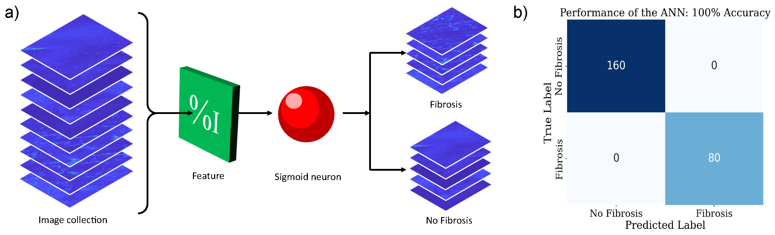
- Computational tools: Computational approaches such as Monte Carlo modeling and machine learning are useful for quantifying SHG images, as they enable modeling of light–tissue interactions and support automated fiber analysis, noise reduction, and image classification for localized tissue health assessment [12,16,24,25].
2. Materials and Methods
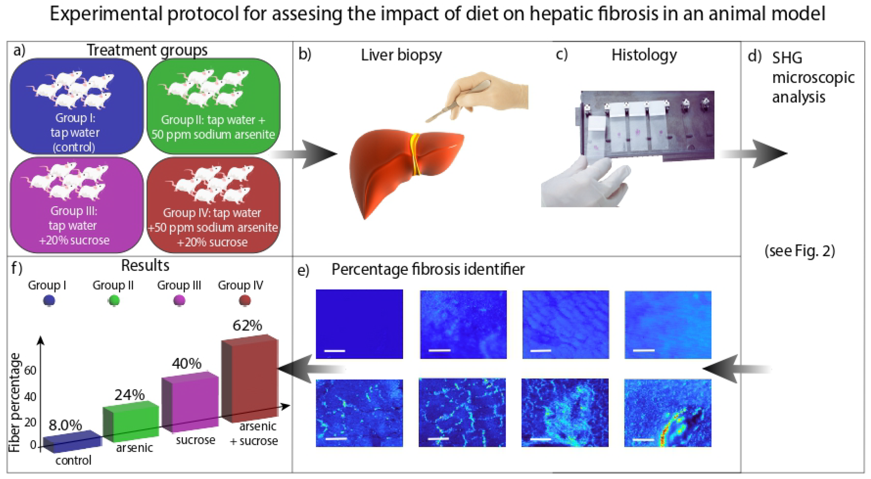
2.1. Biological Experimental Design
2.1.1. Metabolic Syndrome Model in Rat
2.1.2. Histology and Oil Red O Staining
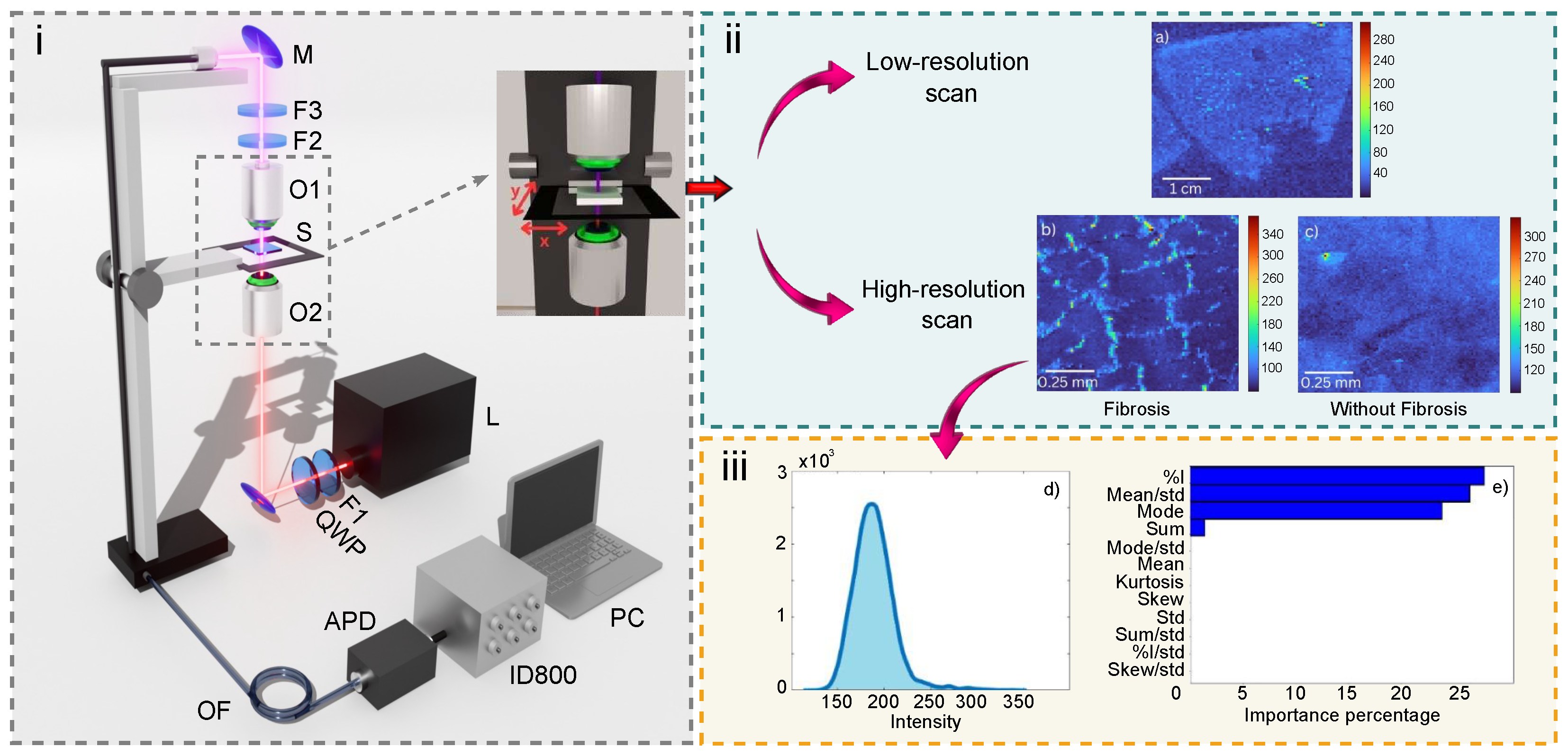
2.2. Experiment
2.3. Machine Learning
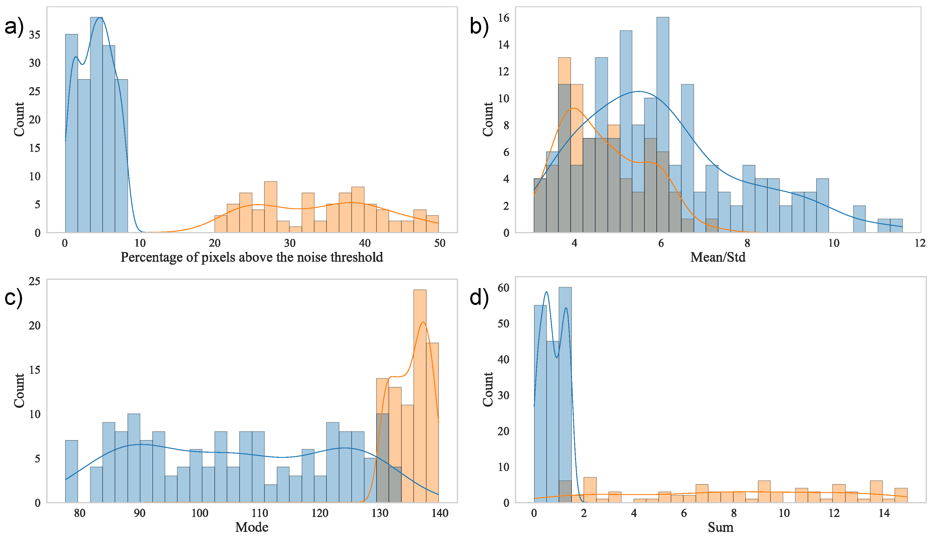
2.3.1. Feature Selection
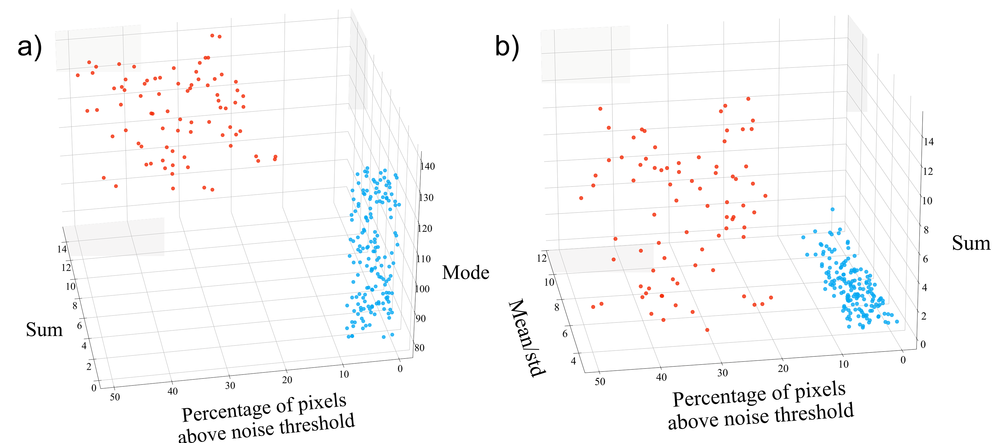
2.3.2. Unsupervised Learning
2.3.3. Supervised Machine Learning Image Classification
3. Results
4. Discussion
5. Conclusions
Author Contributions
Funding
Institutional Review Board Statement
Informed Consent Statement
Data Availability Statement
Conflicts of Interest
Abbreviations
| NAFLD | Non-alcoholic fatty liver disease |
| SHG | Second-harmonic generation |
| BMI | Body mass index |
References
- Roehlen, N.; Crouchet, E.; Baumert, T.F. Liver fibrosis: Mechanistic concepts and therapeutic perspectives. Cells 2020, 9, 875. [Google Scholar] [CrossRef] [Scilit] [PubMed]
- Pérez, B.G.; De León Escobedo, R.; García, J.A.; Flores, R.S. Enfermedad de Hígado Graso no Alcohólico. Un Enfoque Multidisciplinario; Editorial Fontamara S.A. de C.V.: Mexico City, Mexico, 2024. [Google Scholar]
- Kumar, S.; Duan, Q.; Wu, R.; Harris, E.N.; Su, Q. Pathophysiological communication between hepatocytes and non-parenchymal cells in liver injury from NAFLD to liver fibrosis. Adv. Drug Deliv. Rev. 2021, 176, 113869. [Google Scholar] [CrossRef] [Scilit] [PubMed]
- EFSA CONTAM Panel (EFSA Panel on Contaminants in the Food Chain); Schrenk, D.; Bignami, M.; Bodin, L.; Chipman, J.K.; del Mazo, J.; Grasl-Kraupp, B.; Hogstrand, C.; Hoogenboom, L.; Leblanc, J.-C.; et al. Update of the risk assessment of inorganic arsenic in food. EFSA J. 2024, 22, e8488. [Google Scholar] [CrossRef] [Scilit] [PubMed]
- Samal, A.C.; Kar, S.; Bhattacharya, P.; Santra, S.C. Assessment of potential health risk through arsenic flow in food chain—A study in gangetic delta of West Bengal. In Management and Sustainable Development of Coastal Zone Environments; Springer: Dordrecht, The Netherlands, 2010; pp. 259–269. [Google Scholar] [CrossRef] [Scilit]
- Zhuang, F.; Huang, J.; Li, H.; Peng, X.; Xia, L.; Zhou, L.; Zhang, T.; Liu, Z.; He, Q.; Luo, F. Biogeochemical behavior and pollution control of arsenic in mining areas: A review. Front. Microbiol. 2023, 14, 1043024. [Google Scholar] [CrossRef] [Scilit]
- Drahota, P.; Filippi, M. Secondary arsenic minerals in the environment: A review. Environ. Int. 2009, 35, 1243–1255. [Google Scholar] [CrossRef] [Scilit]
- Pánico, P.; Velasco, M.; Salazar, A.M.; Picones, A.; Ortiz-Huidobro, R.I.; Guerrero-Palomo, G.; Salgado-Bernabé, M.E.; Ostrosky-Wegman, P.; Hiriart, M. Is arsenic exposure a risk factor for metabolic syndrome? A review of the potential mechanisms. Front. Endocrinol. 2022, 13, 878280. [Google Scholar] [CrossRef] [Scilit]
- Panico, P.; Velasco, M.; Salazar, A.M.; Ostrosky-Wegman, P.; Hiriart, M. The effects of sucrose and arsenic on muscular insulin signaling pathways differ between the gastrocnemius and quadriceps muscles. Front. Endocrinol. 2023, 14, 1165415. [Google Scholar] [CrossRef] [Scilit]
- Aghigh, A.; Bancelin, S.; Rivard, M.; Pinsard, M.; Ibrahim, H.; Légaré, F. Second harmonic generation microscopy: A powerful tool for bio-imaging. Biophys. Rev. 2023, 15, 43–70. [Google Scholar] [CrossRef] [Scilit]
- Keikhosravi, A.; Bredfeldt, J.S.; Sagar, M.A.K.; Eliceiri, K.W. Second-harmonic generation imaging of cancer. In Methods in Cell Biology; Elsevier: Amsterdam, The Netherlands, 2014; Volume 123, pp. 531–546. [Google Scholar] [CrossRef] [Scilit]
- Duboisset, J.; Aït-Belkacem, D.; Roche, M.; Rigneault, H.; Brasselet, S. Generic model of the molecular orientational distribution probed by polarization-resolved second-harmonic generation. Phys. Rev. A 2012, 85, 043829. [Google Scholar] [CrossRef] [Scilit]
- Mostaço-Guidolin, L.B.; Osei, E.T.; Ullah, J.; Hajimohammadi, S.; Fouadi, M.; Li, X.; Li, V.; Shaheen, F.; Yang, C.X.; Chu, F.; et al. Defective Fibrillar Collagen Organization by Fibroblasts Contributes to Airway Remodeling in Asthma. Am. J. Respir. Crit. Care Med. 2019, 200, 431–443. [Google Scholar] [CrossRef] [Scilit]
- Han, M.; Giese, G.; Bille, J. Second harmonic generation imaging of collagen fibrils in cornea and sclera. Opt. Express 2005, 13, 5791–5797. [Google Scholar] [CrossRef] [Scilit] [PubMed]
- Lo, W.; Chen, W.; Hsueh, C.; Ghazaryan, A.A.; Chen, S.; Ma, D.H.; Dong, C.; Tan, H. Fast Fourier transform-based analysis of second-harmonic generation image in keratoconic cornea. Investig. Ophthalmol. Vis. Sci. 2012, 53, 3501–3507. [Google Scholar] [CrossRef] [Scilit] [PubMed]
- Liu, S.; Sinclair, M.B.; Saravi, S.; Keeler, G.A.; Yang, Y.; Reno, J.; Peake, G.M.; Setzpfandt, F.; Staude, I.; Pertsch, T.; et al. Resonantly enhanced second-harmonic generation using III–V semiconductor all-dielectric metasurfaces. Nano Lett. 2016, 16, 5426–5432. [Google Scholar] [CrossRef] [Scilit] [PubMed]
- Sivaguru, M.; Durgam, S.; Ambekar, R.; Luedtke, D.; Fried, G.; Stewart, A.; Toussaint, K.C. Quantitative analysis of collagen fiber organization in injured tendons using Fourier transform-second harmonic generation imaging. Opt. Express 2010, 24, 24983. [Google Scholar] [CrossRef] [Scilit]
- Rao, R.A.R.; Mehta, M.R.; Toussaint, K.C., Jr. Fourier transform-second-harmonic generation imaging of biological tissues. Opt. Express 2009, 17, 14534. [Google Scholar] [CrossRef] [Scilit]
- Bueno, J.M.; Ávila, F.J.; Artal, P. Comparison of second harmonic microscopy images of collagen-based ocular tissues with 800 and 1045 nm. Biomed. Opt. Express 2008, 8, 5065–5074. [Google Scholar] [CrossRef] [Scilit]
- Williams, R.M.; Zipfel, W.R.; Webb, W.W. Interpreting Second-Harmonic Generation Images of Collagen I Fibrils. Biophys. J. 2005, 88, 1377–1386. [Google Scholar] [CrossRef] [Scilit]
- Brown, E.; McKee, T.; diTomaso, E.; Pluen, A.; Seed, B.; Boucher, Y.; Jain, R.K. Dynamic imaging of collagen and its modulation in tumors in vivo using second-harmonic generation. Nat. Med. 2003, 9, 796–800. [Google Scholar] [CrossRef] [Scilit]
- Falzon, G.; Pearson, S.; Murison, R. Analysis of collagen fibre shape changes in breast cancer. Phys. Med. Biol. 2008, 53, 6641. [Google Scholar] [CrossRef] [Scilit]
- LaComb, R.; Nadiarnykh, O.; Carey, S.; Campagnola, P.J. Alterations of the extracellular matrix in ovarian cancer studied by Second Harmonic Generation imaging microscopy. BMC Cancer 2010, 10, 94. [Google Scholar] [CrossRef] [Scilit]
- Kistenev, Y.V.; Nikolaev, V.V.; Kurochkina, O.S.; Borisov, A.V.; Vrazhnov, D.A.; Sandykova, E.A. Application of multiphoton imaging and machine learning to lymphedema tissue analysis. Biomed. Opt. Express 2019, 10, 3353–3368. [Google Scholar] [CrossRef] [Scilit] [PubMed]
- Kafian-Attari, I.; Nippolainen, E.; Semenov, D.; Hauta-Kasari, M.; Töyräs, J.; Afara, I.O. Tissue optical properties combined with machine learning enables estimation of articular cartilage composition and functional integrity. Biomed. Opt. Express 2020, 11, 6480–6494. [Google Scholar] [CrossRef] [Scilit] [PubMed]
- Fox, M. Quantum Optics. An Introduction; Oxford University Press: Oxford, UK, 2006. [Google Scholar]
- Sil, S.; Gautam, R.; Umapathy, S. Chapter 6—Applications of Raman and Infrared Microscopy to Materials and Biology. In Molecular and Laser Spectroscopy; Gupta, V.P., Ed.; Elsevier: Amsterdam, The Netherlands, 2018; pp. 117–146. [Google Scholar] [CrossRef] [Scilit]
- Shen, Y.R. Surface second harmonic generation: A new technique for surface studies. Annu. Rev. Mater. Sci. 1986, 16, 69–86. [Google Scholar] [CrossRef]
- Lan, X. Laser Technology; CRC Press: Boca Raton, FL, USA, 1991. [Google Scholar]
- Pavone, F.S.; Campagnola, P.J. Second Harmonic Generation Imaging; CRC Press: Boca Raton, FL, USA, 1991. [Google Scholar]
- Reider, G.A. Photonics. An Introduction; Springer: Berlin/Heidelberg, Germany, 2012. [Google Scholar]
- Theodossiou, T.A.; Thrasivoulou, C.; Ekwobi, C.; Becker, D.L. Second harmonic generation confocal microscopy of collagen type I from rat tendon cryosections. Biophys. J. 2006, 91, 4665–4677. [Google Scholar] [CrossRef] [Scilit]
- Schindelin, J.; Arganda-Carreras, I.; Frise, E.; Kaynig, V.; Longair, M.; Pietzsch, T.; Preibisch, S.; Rueden, C.; Saalfeld, S.; Schmid, B.; et al. Fiji: An open-source platform for biological-image analysis. Nat. Methods 2012, 9, 676–682. [Google Scholar] [CrossRef] [Scilit]
- Neuer, M.J. Unsupervised learning. In Machine Learning for Engineers: Introduction to Physics-Informed, Explainable Learning Methods for AI in Engineering Applications; Springer: Berlin/Heidelberg, Germany, 2024; pp. 141–172. [Google Scholar] [CrossRef] [Scilit]
- Ahmed, M.; Seraj, R.; Islam, S.M.S. The k-means algorithm: A comprehensive survey and performance evaluation. Electronics 2020, 9, 1295. [Google Scholar] [CrossRef] [Scilit]
- Kingma, D.P. Adam: A method for stochastic optimization. arXiv 2014, arXiv:1412.6980. [Google Scholar]
- Ruby, U.; Yendapalli, V. Binary cross entropy with deep learning technique for image classification. Int. J. Adv. Trends Comput. Sci. Eng. 2020, 9, 5393–5397. [Google Scholar] [CrossRef] [Scilit]
- Wong, T.-T.; Yeh, P.-Y. Reliable accuracy estimates from k-fold cross validation. IEEE Trans. Knowl. Data Eng. 2019, 32, 1586–1594. [Google Scholar] [CrossRef] [Scilit]
- Wu, X.N.; Wang, M.Z.; Zhang, N.; Zhang, W.; Dong, J.; Ke, M.Y.; Xiang, J.; Ma, F.; Xue, F.; Hou, J.; et al. Sex-determining region Y gene promotes liver fibrosis and accounts for sexual dimorphism in its pathophysiology. J. Hepatol. 2024, 80, 928–940. [Google Scholar] [CrossRef] [Scilit]
- Aristu-Zabalza, P.; Andrés-Rozas, M.; Boyer-Díaz, Z.; Al-Adra, D.P.; Maya-Miles, D.; Guixé-Muntet, S.; Fernández-Iglesias, A.; Gracia-Sancho, J. Sex-specific differences in preclinical models of advanced chronic liver disease and portal hypertension. Biol. Sex Differ. 2025, 16, 39. [Google Scholar] [CrossRef] [Scilit] [PubMed]
- Eisa, M.A.; Mansour, A.M.; Salama, S.A.; Elsadek, B.E.; Ashour, A.A.; Abdelghany, T.M. Estrogen/estrogen receptor activation protects against DEN-induced liver fibrosis in female rats via modulating TLR-4/NF-κβ signaling. Eur. J. Pharmacol. 2023, 960, 176165. [Google Scholar] [CrossRef] [Scilit] [PubMed]
- Wang, Y.; Cela, E.; Gagnon, S.; Sweezey, N.B. Estrogen aggravates inflammation in Pseudomonas aeruginosa pneumonia in cystic fibrosis mice. Respir. Res. 2010, 11, 166. [Google Scholar] [CrossRef] [Scilit] [PubMed]
- Ortiz-Huidobro, R.I.; Larqué, C.; Velasco, M.; Chávez-Maldonado, J.P.; Sabido, J.; Sanchez-Zamora, Y.I.; Hiriart, M. Sexual dimorphism in the molecular mechanisms of insulin resistance during a critical developmental window in Wistar rats. Cell Commun. Signal. 2022, 20, 154. [Google Scholar] [CrossRef] [Scilit]
- Ortenzi, V.H.; Oliveira, A.C.D.; Vasconcelos, R.P.; Neves, M.B.; Teixeira, A.J.; Oliveira, K.A.; Ferreira, A.C.F.; Takiya, C.M.; Fortunato, R. S High-fat diet elicits sex-based differences in liver inflammatory cytokines and redox homeostasis. Appl. Physiol. Nutr. Metab. 2024, 49, 1083–1092. [Google Scholar] [CrossRef] [Scilit]
- Ruoppolo, M.; Caterino, M.; Albano, L.; Pecce, R.; Di Girolamo, M.G.; Crisci, D.; Costanzo, M.; Milella, L.; Franconi, F.; Campesi, I. Targeted metabolomic profiling in rat tissues reveals sex differences. Sci. Rep. 2018, 8, 4663. [Google Scholar] [CrossRef] [Scilit]
- Schmitz, C.R.R.; Eichwald, T.; Flores, M.V.B.; Varela, K.G.; Mantovani, A.; Steffani, J.A.; Glaser, V.; de Carvalho, D.; Remor, A.P. Sex differences in subacute manganese intoxication: Oxidative parameters and metal deposition in peripheral organs of adult Wistar rats. Regul. Toxicol. Pharmacol. 2019, 104, 98–107. [Google Scholar] [CrossRef] [Scilit]
- Shoulders, M.D.; Raines, R.T. Collagen structure and stability. Annu. Rev. Biochem. 2009, 78, 929–958. [Google Scholar] [CrossRef] [Scilit]
- Alizadeh, M.; Krouglov, S.; Barzda, V. Polarimetric second-harmonic generation microscopy of partially oriented fibers I: Digital modeling. Biophys. J. 2023, 122, 3924–3936. [Google Scholar] [CrossRef] [Scilit]
- Campagnola, P. Second harmonic generation imaging microscopy: Applications to diseases diagnostics. Anal. Chem. 2011, 83, 3224–3231. [Google Scholar] [CrossRef] [Scilit]
- Mirsanaye, K.; Uribe Castaño, L.; Kamaliddin, Y.; Golaraei, A.; Augulis, R.; Kontenis, L.; Done, S.J.; Žurauskas, E.; Stambolic, V.; Wilson, B.C.; et al. Machine learning-enabled cancer diagnostics with widefield polarimetric second-harmonic generation microscopy. Sci. Rep. 2022, 12, 10290. [Google Scholar] [CrossRef] [Scilit]
- Mijderwijk, H.J.; Steiger, H.J. Predictive analytics in clinical practice: Advantages and disadvantages. In Machine Learning in Clinical Neuroscience: Foundations and Applications; Springer International Publishing: Cham, Switzerland, 2021; pp. 263–268. [Google Scholar] [CrossRef] [Scilit]



| Group | Percentage of Images with Fibers |
|---|---|
| Control group | 8.0 ± 3.0% |
| Arsenic group | 24 ± 5.7% |
| Sucrose group | 40 ± 5.9% |
| Arsenic–sucrose group | 62 ± 18% |
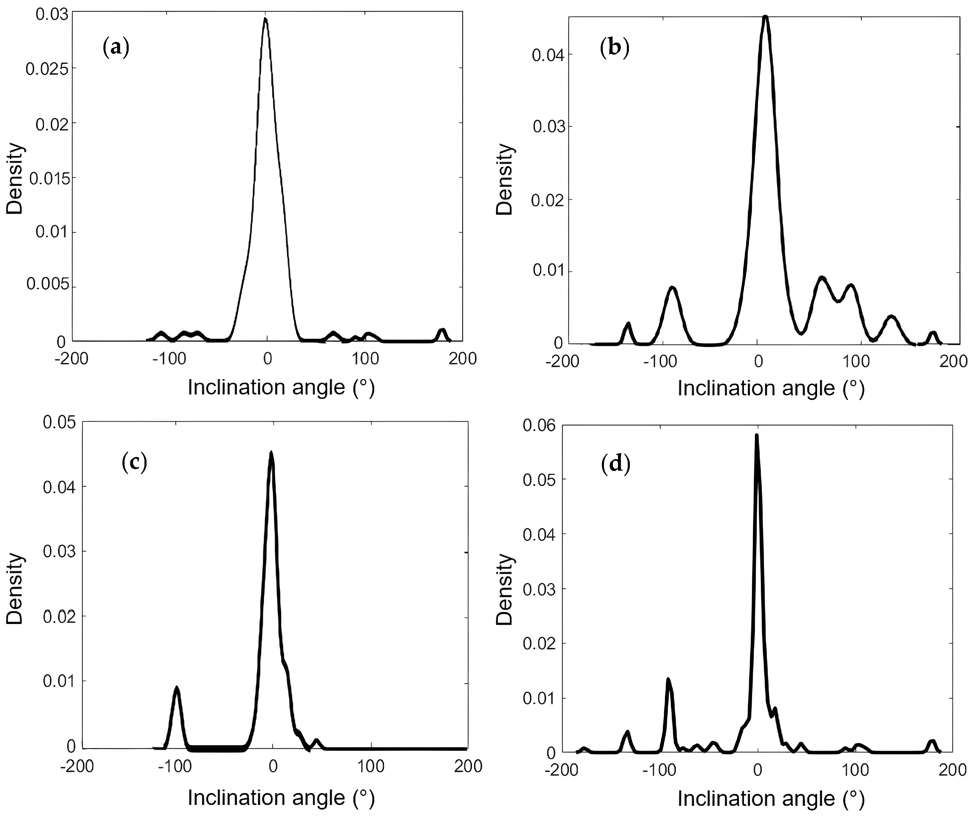
| Group | Distribution Width (°) |
|---|---|
| Control group | 26 |
| Arsenic group | 24 |
| Sucrose group | 16 |
| Arsenic–sucrose group | 2.8 |
Disclaimer/Publisher’s Note: The statements, opinions and data contained in all publications are solely those of the individual author(s) and contributor(s) and not of MDPI and/or the editor(s). MDPI and/or the editor(s) disclaim responsibility for any injury to people or property resulting from any ideas, methods, instructions or products referred to in the content. |
© 2026 by the authors. Licensee MDPI, Basel, Switzerland. This article is an open access article distributed under the terms and conditions of the Creative Commons Attribution (CC BY) license.
Share and Cite
Maldonado-Terrón, M.; Guerrero-Lara, J.C.; Felipe-Elizarraras, R.; Frausto-Avila, C.M.; Manriquez-Amavizca, J.P.; Velasco, M.; Borja, Z.I.; Cruz-Ramírez, H.; Rivera, A.L.; Hiriart, M.; et al. Quantitative Analysis of Arsenic- and Sucrose-Induced Liver Collagen Remodeling Using Machine Learning on Second-Harmonic Generation Microscopy Images. Cells 2026, 15, 214. https://doi.org/10.3390/cells15030214
Maldonado-Terrón M, Guerrero-Lara JC, Felipe-Elizarraras R, Frausto-Avila CM, Manriquez-Amavizca JP, Velasco M, Borja ZI, Cruz-Ramírez H, Rivera AL, Hiriart M, et al. Quantitative Analysis of Arsenic- and Sucrose-Induced Liver Collagen Remodeling Using Machine Learning on Second-Harmonic Generation Microscopy Images. Cells. 2026; 15(3):214. https://doi.org/10.3390/cells15030214
Chicago/Turabian StyleMaldonado-Terrón, Mónica, Julio César Guerrero-Lara, Rodrigo Felipe-Elizarraras, C. Mateo Frausto-Avila, Jose Pablo Manriquez-Amavizca, Myrian Velasco, Zeferino Ibarra Borja, Héctor Cruz-Ramírez, Ana Leonor Rivera, Marcia Hiriart, and et al. 2026. "Quantitative Analysis of Arsenic- and Sucrose-Induced Liver Collagen Remodeling Using Machine Learning on Second-Harmonic Generation Microscopy Images" Cells 15, no. 3: 214. https://doi.org/10.3390/cells15030214
APA StyleMaldonado-Terrón, M., Guerrero-Lara, J. C., Felipe-Elizarraras, R., Frausto-Avila, C. M., Manriquez-Amavizca, J. P., Velasco, M., Borja, Z. I., Cruz-Ramírez, H., Rivera, A. L., Hiriart, M., Quiroz-Juárez, M. A., & U’Ren, A. B. (2026). Quantitative Analysis of Arsenic- and Sucrose-Induced Liver Collagen Remodeling Using Machine Learning on Second-Harmonic Generation Microscopy Images. Cells, 15(3), 214. https://doi.org/10.3390/cells15030214