


Highlights
What are the main findings?
- T cells and monocytes/macrophages contribute to hepatic inflammation and immune dysregulation in AIH.
- Monocytes/macrophages may influence T-cell responses through costimulatory signals and inflammatory cytokines.
What are the implications of the main findings?
- T-cell–monocyte/macrophage interactions may offer potential therapeutic targets and biomarkers.
- Further functional and longitudinal studies are needed to clarify these interactions in human AIH.
Abstract
Autoimmune hepatitis (AIH) is a chronic immune-mediated liver disease in which persistent inflammation and relapse remain challenges despite immunosuppressive treatment. This review critically examines the roles of effector T cells (Teffs), regulatory T cells (Tregs), and monocytes/macrophages, with particular emphasis on their interactions in the hepatic inflammatory microenvironment. Teffs contribute to inflammatory responses and hepatocyte injury, whereas heterogeneous Treg alterations indicate that increased regulatory cell abundance does not necessarily ensure effective immune control. Monocytes and macrophages may modulate T-cell responses through antigen presentation, costimulation, and cytokine production, potentially reinforcing hepatic inflammation. Importantly, this review distinguishes observed changes in immune cell populations from functionally demonstrated cellular interactions and evaluates the strength of evidence supporting these mechanisms in human AIH versus experimental models. Although selected cytokine-dependent pathways have been functionally demonstrated in human AIH, the causal contributions of specific monocyte/macrophage subsets and many proposed cellular interactions remain uncertain. We propose that defining functionally relevant immune cell interactions, rather than relying solely on changes in cell abundance or activation markers, is essential for understanding persistent inflammation and relapse. Future studies integrating longitudinal immune profiling with cell-specific functional perturbation may help identify causal pathways and guide the development of more selective therapeutic strategies.
1. Introduction
Autoimmune hepatitis (AIH) is a chronic immune-mediated liver disease characterized by persistent or recurrent hepatic inflammation that can progress to fibrosis and cirrhosis [1]. Typical findings include elevated serum immunoglobulin G (IgG), circulating autoantibodies [1,2], and interface hepatitis with portal lymphoplasmacytic inflammation [3]. AIH has traditionally been classified into two types based on characteristic autoantibody profiles. Type 1 AIH, the more common form, is characterized by antinuclear antibodies (ANA) and/or smooth muscle antibodies (SMA) and can occur at any age. Type 2 AIH is associated with anti-liver kidney microsomal type 1 (anti-LKM1) and/or anti-liver cytosol type 1 (anti-LC1) antibodies and occurs predominantly in children and adolescents [4]. Although immunosuppression controls disease activity in many patients, prolonged treatment and relapse after treatment withdrawal remain major clinical challenges [1,5]. Understanding how hepatic immune responses persist despite treatment may help identify more selective approaches to disease control.
Hepatic immune homeostasis depends on coordinated interactions among antigen-presenting cells, effector T cells (Teffs), and regulatory populations [6]. In AIH, autoreactive T-cell responses and insufficient immune regulation are accompanied by changes in monocytes and macrophages. Human studies have demonstrated antigen-specific T-cell responses and increased intrahepatic regulatory T-cell (Treg) abundance, although the latter does not establish effective suppression within the liver [7,8,9]. Macrophage-associated inflammatory changes have also been observed in human AIH, with experimental studies supporting roles in inflammatory polarization and monocyte recruitment [10,11].
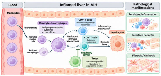
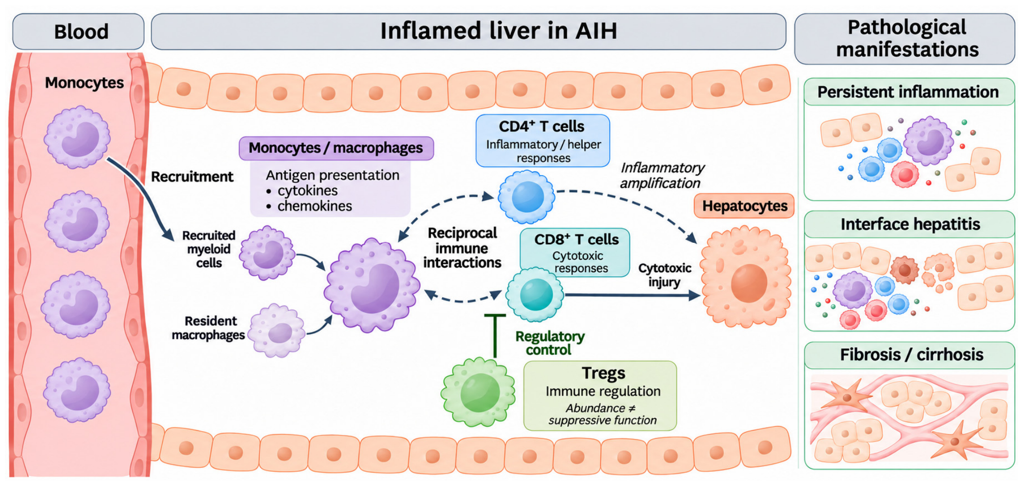
Monocytes and macrophages can influence T-cell responses through antigen presentation, costimulation, and cytokine production, while signals from activated T cells can modify innate immune responses [12]. Recent functional work in human AIH has established selected cytokine-mediated connections among T cells, myeloid cells, and hepatocytes [13]. However, these findings do not yet define the contribution of each monocyte or macrophage subset or establish that all proposed interactions sustain disease in patients. The distinction between observed cellular changes and functionally demonstrated interactions therefore provides the organizing framework for this review. Figure 1 summarizes the cellular populations and proposed inflammatory connections discussed below.
Figure 1.
Overview of immune cell networks and hepatic injury in AIH. The schematic summarizes monocyte recruitment, resident macrophages, reciprocal myeloid–T-cell interactions, Treg-mediated regulation, and T-cell-associated hepatocyte injury. Persistent inflammation, interface hepatitis, and fibrosis/cirrhosis are shown as related pathological manifestations rather than an obligatory sequence. Dashed links represent proposed interactions across study contexts; with pathway-specific evidence is discussed in the main text (Figure 3). Increased Treg abundance does not necessarily indicate effective suppression. AIH, autoimmune hepatitis; Treg, regulatory T cell.
The aim of this review is to critically evaluate the roles of Teffs, Tregs, and monocytes/macrophages in AIH, with particular emphasis on their interactions in the hepatic inflammatory microenvironment. Specifically, we seek to distinguish established findings in human AIH from mechanisms proposed based on experimental models, identify gaps in the current understanding of T-cell–monocyte/macrophage interactions, and discuss their potential implications for therapeutic strategies. By integrating the available evidence, this review aims to provide a clearer understanding of how these immune cell populations may contribute to persistent hepatic inflammation and to highlight priorities for future mechanistic and translational research.
2. Material and Methods
Literature Search and Evidence Selection
This narrative review integrates mechanistic, experimental, and clinical evidence concerning the roles and interactions of T cells, monocytes, and macrophages in AIH. Targeted PubMed searches were conducted from 10 September to 28 October 2026, with the final search performed on 28 October 2026. Search terms included combinations of “autoimmune hepatitis” or “AIH” with “T cells”, “CD4 T cells”, “CD8 T cells”, “Regulatory T cells”, “Tregs”, “monocytes”, “macrophages”, “Kupffer cells”, “cytokines”, “costimulatory signaling”, and “immune cell interactions”. Additional relevant publications were identified by screening the reference lists of key original studies and review articles.
Studies involving patients with AIH, in vitro and ex vivo investigations, observational clinical studies, and relevant experimental animal models were considered. Priority was given to studies investigating immune cell phenotypes, functional responses, cellular interactions, and their associations with disease activity or therapeutic responses in AIH. Experimental studies using concanavalin A-induced hepatitis and spontaneous AIH models were considered when they provided relevant mechanistic insights. Studies unrelated to the immunopathogenesis of AIH or lacking relevant mechanistic or clinical evidence were excluded. Findings from other liver diseases and post-transplant supporting evidence were distinguished from findings obtained in patients with native-liver AIH. The principal findings, study contexts, and limitations of selected studies are summarized in Table 1.
Table 1.
Selected studies of T cells and monocytes/macrophages in AIH.
3. Effector T Cells in AIH
3.1. Autoantigens and Antigen-Specific T-Cell Responses
Established AIH-associated autoantigens include cytochrome P450 2D6 (CYP2D6), formiminotransferase cyclodeaminase (FTCD), and O-phosphoseryl-tRNA:selenocysteinyl-tRNA synthase (SepSecS), the target of soluble liver antigen/liver pancreas antigen (SLA/LP) antibodies [1]. CYP2D6 and SepSecS provide the most detailed examples of antigen-specific T-cell recognition. In type 2 AIH, CYP2D6-reactive CD4+ T cells recognize multiple antigenic regions, and the magnitude of these responses is associated with disease activity [8]. HLA-A2-restricted CYP2D6-specific CD8+ T-cell responses have also been characterized [21]. For SepSecS, epitope-mapping studies have identified CD4+ T-cell recognition under distinct HLA class II restrictions [9,22].
Clonal studies extend these observations beyond the detection of autoreactive cells. Kramer et al. isolated functionally responsive SepSecS-specific CD4+ T-cell clones from patients with AIH, including patients without detectable anti-SLA antibodies, and demonstrated presentation of soluble SepSecS by antigen-specific B-cell clones to cognate T cells [23]. Together, these studies establish antigen recognition and selected antigen-dependent cellular responses in human AIH. A recent review emphasizes that antigen recognition and sustained tissue injury require distinct functional evidence and that heterogeneous antigen-specific responses may converge on shared inflammatory processes [24]. They leave open how frequently these responses produce hepatocyte injury in vivo and how they interact with cytokine-driven effector mechanisms.
3.2. T-Cell Effector Function and Hepatocyte Injury
CD4+ effector T cells can sustain inflammation through cytokine production and helper activity. Human AIH studies have identified inflammatory CD4+ populations producing IFN-γ and TNF [13,25]. Th17-associated inflammation provides an additional pathway: hepatic IL-17-related responses are increased in AIH, and experimental assays show that IL-17 can induce IL-6 expression in hepatocytes [26]. Intrahepatic CD6highCD4+ T cells also exhibit pro-inflammatory activity, with ALCAM–CD6 implicated in their hepatic recruitment [25]. A distinct helper function is evident in autoreactive PD-1+CXCR5− memory CD4+ T cells, which support B-cell differentiation through IL-21 and display a peripheral helper T-cell-like transcriptional profile [9,27]. These functions connect autoreactivity to both local inflammation and humoral immune responses.
CD8+ T cells provide a more direct route to hepatocyte injury. CYP2D6-reactive CD8+ T cells in type 2 AIH produce IFN-γ and exhibit cytotoxicity against peptide-loaded targets, demonstrating antigen-specific effector potential [21]. Hepatic CD69+CD103+CD8+ T cells also express granzyme B and IFN-γ [28]. In addition, functional studies in human AIH support IL-15-dependent auto-aggressive CD8+ T-cell activity enhanced by TNF [13]. Antigen-specific cytotoxicity and cytokine-driven activation may therefore coexist in AIH. However, CYP2D6-specific cytotoxic responses have been demonstrated in type 2 AIH, whereas the subtype-specific contributions of cytokine-driven activation and their relative roles in hepatocyte injury remain incompletely defined.
3.3. Intrahepatic T-Cell Phenotypes and Chronic Activation
The intrahepatic location and differentiation state of T cells may help explain the persistence of inflammation. CD69+CD103+CD8+ tissue-resident memory (TRM)-phenotype cells are enriched in AIH liver, and their abundance is associated with inflammatory activity and fibrosis [28]. More recent paired blood–liver analyses identified intrahepatic enrichment of CD8+ T cells expressing CD69, CD103, and CXCR6, with residency-associated populations linked to fibrosis [29]. T-cell receptor (TCR) analyses in autoimmune liver disease have also identified shared clonotypes between circulating autoreactive CD4+ populations and liver tissue [27]. These findings connect peripheral and hepatic immune compartments, although marker expression and clonotype sharing alone cannot establish long-term tissue retention or the direction of trafficking.
Persistent activation is accompanied by inhibitory receptor expression. Untreated AIH liver contains activated T cells with increased PD-1 expression [30], while circulating liver-autoreactive CD4+ cells can co-express PD-1, TIGIT, and HLA-DR [27]. Such phenotypes require functional interpretation: autoreactive CD4+ cells can acquire exhaustion-associated features [31], yet cytokine-producing and helper functions remain detectable in relevant populations [9,27,29]. In complementary studies of chronic liver disease, TIGIT signaling restrained CD8+ T-cell-mediated hepatocyte cytotoxicity, indicating that inhibitory receptors can actively limit tissue injury [32].
4. Tregs and Insufficient Immune Control
4.1. Treg Frequency and Distribution in AIH
Reports of circulating Treg abundance in AIH differ across studies. Early analyses identified reduced peripheral CD4+CD25+ T-cell frequencies, particularly at diagnosis [33]. Later studies using CD25highCD127lowFOXP3+ phenotyping and FOXP3 demethylation analysis found no reduction compared with healthy controls and higher frequencies in active disease than in remission [14]. Differences in identification methods, disease activity, treatment exposure, and cohort composition complicate comparisons. Peripheral Treg deficiency is therefore not a uniform finding, and frequency measurements should be interpreted in their clinical and methodological context.
Intrahepatic Treg accumulation has been observed more consistently. Histological studies found increased FOXP3+ cells in inflamed AIH liver [14,15], and recent flow-cytometric and histological analyses confirmed increased hepatic Treg abundance and associations with inflammatory activity [7]. Tregs must also be considered relative to the effector populations they regulate: higher intrahepatic Treg-to-Teff ratios have been associated with biochemical remission [15]. These observations establish altered distribution and abundance, while the effectiveness of suppression requires separate functional assessment.
4.2. Treg Suppressive Function and Immune Dysregulation
Functional studies identify heterogeneous regulatory abnormalities. In an early study, reduced Treg numbers and expansion coexisted with preserved suppression of CD4+ T-cell IFN-γ production [33]. A subsequent study reported impaired control of CD8+ T-cell proliferation and cytokine responses, with improvement during remission [34], whereas Peiseler et al. found preserved suppressive activity under their assay conditions [14]. More recently, Kwon et al. demonstrated impaired suppression in co-cultures using peripheral blood-derived Tregs and Teffs from patients with treatment-naïve AIH [7]. The hepatic Treg accumulation described in the same study was assessed separately and does not demonstrate dysfunction of isolated liver-derived Tregs. Differences in responder cells, readouts, and sampling compartments therefore matter when comparing apparently conflicting results.
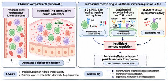
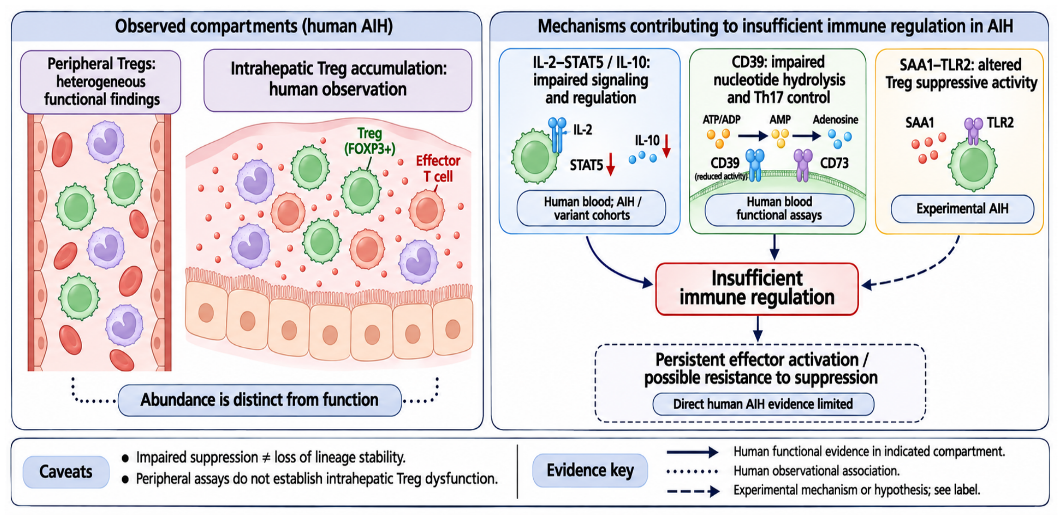
Mechanistic studies suggest that insufficient regulation can arise through several pathways. In type 1 AIH and the AIH–sclerosing cholangitis variant, reduced IL-2 responsiveness and STAT5 activation were associated with low IL-10 production and impaired suppression [16], suggesting abnormalities in Treg-associated signaling and suppressive function. Peripheral CD39+ Tregs showed defective nucleotide hydrolysis and control of Th17 responses [35], indicating impairment of a specific suppressive mechanism rather than generalized Treg dysfunction. In another study of AIH/autoimmune sclerosing cholangitis, activated effector cells acquired regulatory properties under Treg-polarizing conditions but lost suppressive activity after inflammatory challenge [17]; these induced cells should be distinguished from naturally occurring hepatic Tregs. Experimental AIH work further showed that hepatic serum amyloid A1 impaired intrahepatic Treg function through TLR2 [36], supporting a role for local inflammatory signals in modifying regulatory activity. However, these findings do not necessarily demonstrate a loss of Treg lineage stability, which requires separate assessment. Together, these findings implicate both cellular regulatory defects and the inflammatory environment, without establishing a single mechanism shared by all patients. These alterations in Treg distribution and proposed mechanisms of insufficient immune regulation are summarized in Figure 2.
Figure 2.
Treg abundance and mechanisms associated with insufficient immune regulation in AIH. Intrahepatic Treg accumulation is distinguished from heterogeneous peripheral functional findings. Proposed mechanisms include impaired IL-2–STAT5/IL-10 signaling, CD39 dysfunction, and experimental SAA1–TLR2-mediated alterations in suppressive activity. Red downward arrows indicate reduced STAT5 signaling and IL-10 levels. Solid arrows indicate human functional evidence in the specified compartment, dotted connectors indicate observational findings, and dashed arrows indicate experimental mechanisms or hypotheses. Effector-cell resistance remains a possibility with limited direct human evidence; peripheral dysfunction does not establish intrahepatic dysfunction, and impaired suppression does not establish loss of lineage stability. Treg, regulatory T cell; SAA1, serum amyloid A1.
4.3. Mechanisms Underlying Insufficient Treg-Mediated Immune Regulation
Taken together, these findings suggest that insufficient immune regulation in AIH may reflect at least three distinct, potentially overlapping mechanisms: intrinsic abnormalities in Treg suppressive function, inflammation-induced alterations in regulatory activity, and relative insufficiency of suppression in the presence of persistent Teff activation [16,17,35,36]. Importantly, impaired suppressive activity observed in peripheral blood-derived Tregs cannot be assumed to represent the functional state of intrahepatic Tregs, particularly given differences in local cytokine exposure and cellular interactions [7,14,15]. Moreover, persistent Teff activation may contribute to insufficient immune control through increased resistance to Treg-mediated suppression or an inflammatory burden that exceeds the available regulatory capacity. However, direct evidence of Teff resistance in human AIH remains limited. Thus, increased intrahepatic Treg abundance, impaired peripheral suppressive activity, and ongoing Teff responses should be interpreted as distinct observations rather than equivalent evidence of intrinsic Treg dysfunction. Paired functional analyses of circulating and intrahepatic Tregs, together with assessments of Teff susceptibility to suppression, are needed to clarify these mechanisms.
5. Monocytes and Macrophages in AIH
5.1. Monocyte and Macrophage Alterations in AIH
Although AIH is commonly characterized by T-cell-mediated liver injury, monocytes and macrophages are increasingly recognized as important components of the inflammatory response. The hepatic monocyte–macrophage compartment comprises heterogenous populations, including liver-resident Kupffer cells and recruited monocytes that can differentiate into macrophages following liver injury. Spatial proteogenomic studies of human and murine livers have identified distinct macrophage populations and highlighted the influence of local tissue niches on their cellular identities. In particular BMP9/10-ALK1 signaling contributes to Kupffer cell development and maintenance, while monocyte-derived cells can acquire Kupffer cell-like phenotypes in experimental models [37].
In patients with AIH, single-cell RNA sequencing of peripheral blood mononuclear cells (PBMCs) has identified altered monocyte transcriptional programs involving antigen processing and presentation, interferon-associated signaling, and innate immune responses [19]. A subsequent analysis by Chi et al. [20] further explored immune cell composition and inflammatory signaling using the same PBMC dataset originally reported by Abe et al. [19]. Therefore, these findings represent a secondary analysis rather than independent validation in a separate patient cohort. In the liver, histological studies have reported increased CD68+ macrophage-associated cells and MAC387+ infiltrating myeloid cells within inflamed tissue [38]. However, CD68 and MAC387 staining alone cannot reliably distinguish liver-resident Kupffer cells from recruited monocytes or monocyte-derived macrophages.
More recently, integrated transcriptomic analyses of the human autoimmune liver disease dataset, including AIH, have identified heterogeneous macrophage populations, including Kupffer cell-like and lipid-associated macrophage populations [39]. In human AIH datasets, Kupffer cell-annotated populations exhibited an efferocytosis-associated transcriptional signature involving MRC1, MERTK, and AXL [39]. However, these transcriptional features do not directly establish efferocytic activity or developmental origin. Thus, although emerging evidence highlights macrophage heterogeneity in AIH, the origins and distinct functional contributions of these populations in human disease remain incompletely defined.
5.2. Inflammatory Activation and Monocyte/Macrophage Recruitment
Alongside changes in their abundance and distribution, monocytes and macrophages may contribute to hepatic inflammation through the production of inflammatory mediators. In human AIH liver, CD68+ macrophage-associated cells express TNF-α and IL-6, and hepatic CCL2 expression is increased [38]. RIPK3 (RIP3)-associated inflammatory signaling has been proposed as one potential contributor to these inflammatory responses. RIP3 expression has been detected in hepatic macrophages and circulating monocytes from patients with AIH [40]. In experimental studies, pharmacological inhibition of RIP3 reduced hepatic IL-6 and CCL2 expression in a DSS-ConA mouse model, while RIP3 knockdown decreased the expression of these mediators in murin macrophage cells [38].
Beyond inflammatory mediator production, monocytes and macrophages may also respond to cytokine signals associated with AIH and contribute to the recruitment of additional inflammatory cells. Elevated serum IFN-γ levels have been reported in patients with active AIH compared with patients in remission and healthy controls [41]. In this context, IFN-γ-responsive CD169+ monocytes and macrophages have emerged as populations of interest. IFN-γ stimulation of CD169 expression in human CD14+ monocytes in vitro [10,42]. In a ConA-induced mouse model of acute hepatitis, IFN-γ neutralization reduces hepatic CD169+ macrophages accumulation, while IFN-γ stimulation enhances CCL12 production in murine bone marrow-derived macrophages. Furthermore, depletion of CD169-expressing cells or neutralization of CCL12 reduces inflammatory infiltration and liver injury [10].
Together, these findings suggest that inflammatory mediator production and chemokine-associated recruitment may contribute to monocyte and macrophage responses during hepatic inflammation. However, functional evidence has primarily been obtained from murine models and macrophage culture, and the acute ConA model does not fully recapitulate chronic human AIH. Furthermore, CCL12 lacks a direct human ortholog, and depletion of CD169-expressing cells does not establish a direct functional role for the CD169 molecule itself. Whether these mechanisms operate similarly in patients with AIH remains to be determined.
5.3. Association of Monocyte and Macrophage Markers with Disease Activity in AIH
The inflammatory changes described above have also motivated investigations into the association between monocyte- and macrophage-related markers and disease activity of AIH. In human AIH liver tissue, the abundance of CD169+ macrophage-associated cells correlates with histological inflammation and biochemical indices, including aminotransferases and serum IgG [10]. Circulating soluble CD163 (sCD163), a marker associated with macrophage activation, shows positive correlations with ALT and IgG levels [43]. Serum sCD163 levels are higher in patients with acute AIH than in complete biochemical responders and are also higher in incomplete responders than in complete responders. Furthermore, sCD163 levels decrease during prednisolone treatment in a small prospective cohort [43].
Despite these associations, it remains unclear whether CD169 and sCD163 provide incremental predictive value beyond established indicators such as ALT, AST, and IgG, as sufficient validation is currently lacking. Correlations with biochemical measures and treatment-associated changes do not establish the ability of these markers to independently predict subsequent treatment outcomes or relapse. Further longitudinal studies, multivariable analyses, and independent validation are needed to determine whether CD169 and sCD163 provide additional information beyond conventional assessments.
6. Monocyte/Macrophage–T-Cell Interactions in the AIH Liver
6.1. Monocyte-Associated Costimulatory Signaling and Treg-Mediated Regulation
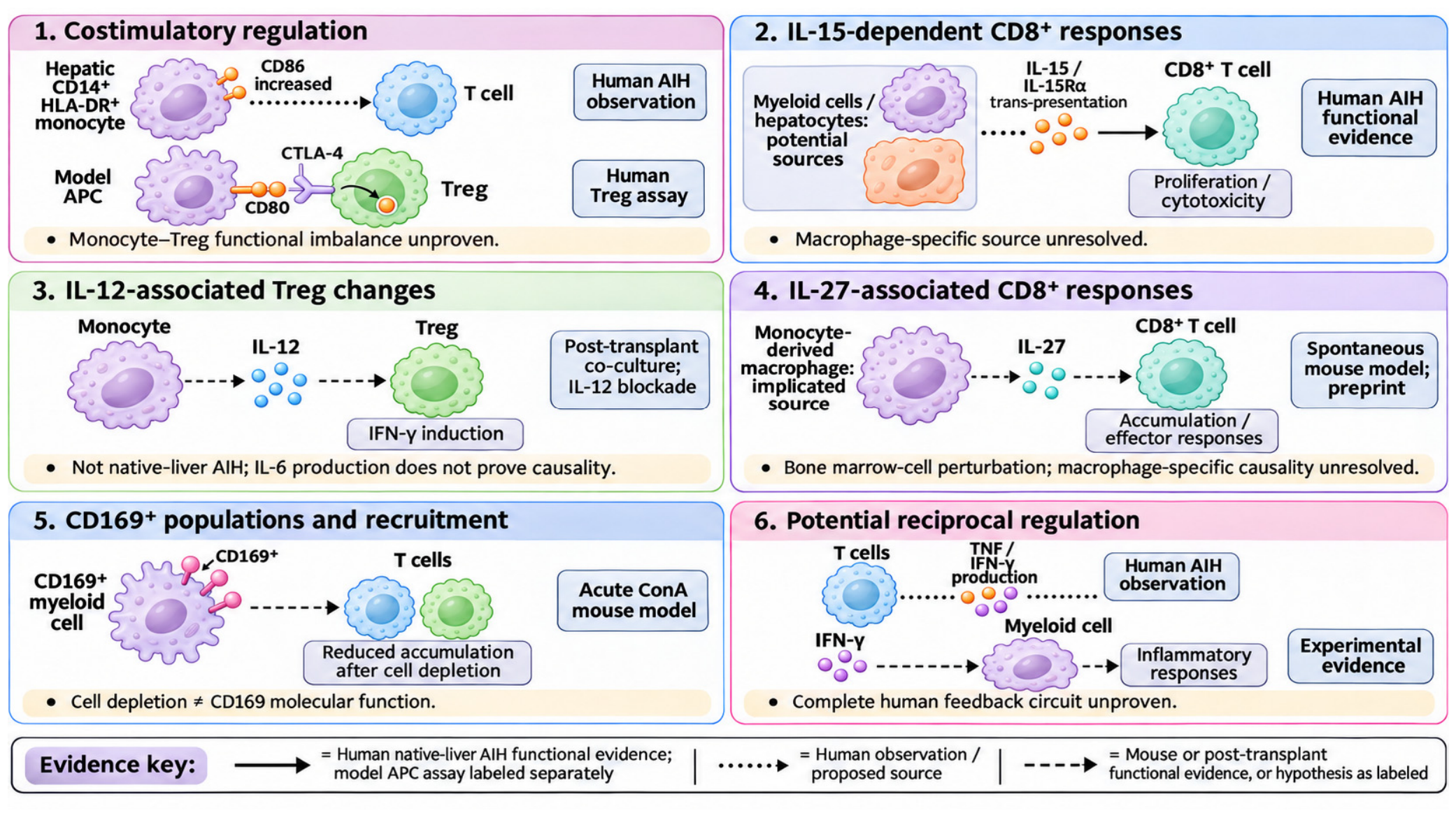
Costimulatory interactions between antigen-presenting cells and T cells are mediated by CD80/CD86 engagement of CD28 and regulated by CTLA-4-dependent removal of these ligands by Tregs [44,45]. In explanted livers from patients with advanced AIH, CD14+HLA-DR+ monocytes exhibited increased CD86 expression intensity, whereas the frequency of CD80+ monocytes did not increase significantly [18]. Notably, intrahepatic Tregs showed increased CTLA-4 and PD-1 coexpression while retaining CTLA-4-dependent CD80 trans-endocytosis capacity in functional assays using model antigen-presenting cells [18]. However, preserved CD80 removal does not necessarily indicate equivalent regulation of CD86, as these ligands differ in their interactions with CTLA-4 and subsequent intracellular trafficking [46]. In a complementary mouse model lacking endogenous CD80 and CD86, selective CD28 stimulation induced chronic AIH-like liver inflammation accompanied by increased accumulation of IFN-γ-producing CD8+ T cells [47]. Although this model supports the potential contribution of CD28 signaling to hepatic inflammation, enhanced CD28 signaling mediated by hepatic monocytes has not been directly demonstrated in human AIH. Together, these findings suggest that altered costimulatory ligand availability may influence intrahepatic T-cell responses, although the functional consequences of increased monocyte CD86 expression remain unclear.
6.2. Monocyte- and Macrophage-Associated Cytokine Signaling and T-Cell Responses
Beyond costimulatory signaling, cytokines associated with monocytes and macrophages may influence both effector and regulatory T-cell responses during hepatic inflammation. In human AIH, integrative analyses identified myeloid cells and hepatocytes as potential sources of IL-15, while functional experiments demonstrated that IL-15 promotes the proliferation and cytotoxicity of liver-derived auto-aggressive CD8+ T cells [13]. Consistent with a potential contribution from hepatic macrophages, macrophages and neutrophils were identified as major IL-15-expressing immune cell populations in an experimental AIH mouse model [48]. However, given the multiple cellular sources of IL-15 and its regulation through IL-15Rα-mediated trans-presentation [49], these findings do not establish the specific contribution of macrophage-derived IL-15 to CD8+ T-cell responses in human AIH.
While IL-15-associated findings highlight the enhancement of CD8+ T-cell effector responses, experimental studies have also investigated mechanisms that may sustain these responses during chronic hepatic inflammation. In a recent non-peer-reviewed preprint using a spontaneous mouse model of chronic autoimmune hepatitis, TLR9-dependent accumulation of inflammatory monocyte-derived macrophages was associated with IL-27 expression and the expansion of PD-1+TOX+ CD8+ T cells that retained effector function. Disruption of IL-27 or its receptor in bone marrow-derived cells reduced CD8+ T-cell accumulation and effector responses [50]. These findings support a role for IL-27 signaling in maintaining CD8+ T-cell responses in this experimental model, although a macrophage-specific causal contribution and its relevance to human AIH remain to be established.
6.3. CD169+ Macrophages and Intrahepatic T-Cell Accumulation
Beyond costimulatory and cytokine-mediated signaling, CD169+ macrophage populations may influence intrahepatic T-cell accumulation and subset composition. In a ConA-induced mouse model of acute hepatitis, bone marrow-derived Ly6ChiCD169+ macrophages expanded, while depletion of CD169-expressing cells reduced hepatic T-cell infiltration and was associated with lower proportions of IFN-γ-producing Th1 and CD8+ T cells and a higher proportion of Tregs. The proportion of activated CD8+ T cells did not change significantly, and although CD169+ macrophages exhibited increased CXCL10 expression, its direct contribution to T-cell recruitment was not functionally established [10]. Together, these findings suggest that CD169-expressing macrophage populations may influence T-cell accumulation and subset composition rather than directly promoting T-cell activation. However, these results derive from an acute ConA model and do not establish the molecular function of CD169 itself in chronic human AIH.
Taken together, these findings suggest that monocytes and macrophages may influence intrahepatic T-cell responses through distinct but potentially interconnected mechanisms involving costimulatory regulation, cytokine-mediated modulation of effector and regulatory T-cell responses, and immune cell accumulation. The supporting evidence varies across these pathways, ranging from observations and functional studies in human AIH to findings from post-transplant autoimmune-like hepatitis and experimental mouse models. Importantly, these interactions may also operate in the opposite direction. In human AIH, intrahepatic CD4+ and CD8+ T-cell populations have been shown to produce inflammatory cytokines, including TNF and IFN-γ [13,51], while experimental studies indicate that IFN-γ signaling can promote inflammatory monocyte and macrophage responses [10,50]. These observations suggest a potential reciprocal inflammatory network in which T-cell-associated cytokines influence monocyte and macrophage activation, which may in turn shape T-cell responses. To define these interactions in human AIH, spatial transcriptomics and multiplex tissue imaging could be combined with patient-derived monocyte/macrophage–T-cell co-cultures or liver-slice cultures and targeted cytokine blockade to distinguish cellular sources, signaling directionality, and functional contributions of individual monocyte and macrophage populations.
Table 2 integrates these findings by organizing the reported monocyte/macrophage populations according to their associated pathways, proposed functions, study contexts, and supporting functional evidence, while highlighting unresolved cellular identities and key limitations.
Table 2.
Monocyte and macrophage populations, associated signaling pathways, and functional evidence in AIH.
Figure 3.
Monocyte/Macrophage–T-cell interactions in AIH: pathways and supporting evidence. (1) Costimulatory regulation, (2) IL-15-dependent CD8+ T-cell responses, (3) IL-12-associated Treg changes, (4) IL-27-associated CD8+ T-cell responses, (5) CD169+ cell-associated recruitment, and (6) potential reciprocal regulation. Solid arrows indicate human native-liver AIH functional evidence, with the model antigen-presenting-cell assay labeled separately; dotted connectors indicate human observations or proposed cellular sources; dashed arrows indicate experimental or post-transplant evidence, or hypotheses as labeled. Cytokine activity does not establish source-specific causality, and the depicted pathways do not constitute a fully validated network in human AIH. APC, antigen-presenting cell; ConA, concanavalin A; Treg, regulatory T cell.
7. Therapeutic Implications
7.1. Immune Changes Associated with Treatment Response
Current AIH management uses corticosteroid-based induction with a steroid-sparing agent; the 2025 EASL guideline recognizes mycophenolate mofetil (MMF) as a first-line alternative to azathioprine [1]. The 3-year follow-up of the CAMARO trial further showed that MMF was associated with a faster biochemical response than azathioprine, while subsequent outcomes were comparable among patients who tolerated their initially assigned treatment [52]. Treatment-associated immune changes do not necessarily represent restoration of tolerance. For example, tissue analyses found a disproportionate reduction in intrahepatic Tregs relative to Teffs during treatment [15], while circulating activated T-cell frequencies and sCD163 levels vary with disease activity or treatment response [5,43]. These observations may reflect direct drug effects, reduced inflammatory stimulation, or both. Their principal therapeutic implication is that biochemical improvement should not be assumed to establish recovery of the regulatory–effector balance.
7.2. Targeting Inflammatory Cytokine and Monocyte/Macrophage Pathways
The mechanistic rationale for targeting TNF and IL-15 is discussed in Section 5.2. Clinical translation is most advanced for TNF inhibition. In a steroid-free, open-label phase IIa infliximab trial, 12 patients were enrolled and 2/12 achieved complete biochemical remission, defined by normalization of ALT, AST, and IgG, at 24 weeks. Azathioprine was introduced after the week 2 visit, and the small, non-randomized design limits attribution of benefit to infliximab [13]. Observational studies also report responses in difficult-to-treat AIH [53,54], including patients with inadequate responses to conventional therapies and those receiving infliximab for concomitant extrahepatic autoimmune diseases. Infectious complications, including infections reported in seven of 11 patients in an early rescue-treatment cohort [53], and reports of infliximab-induced autoimmune-like hepatitis further complicate assessment of benefit and risk [53,55]. TNF inhibition therefore remains a selected investigational or rescue approach rather than an established replacement for standard induction; its use should be restricted to carefully selected difficult-to-treat patients under close monitoring, rather than being considered a routine treatment option for AIH. The cited AIH studies provide mechanistic, rather than clinical interventional, support for IL-15 targeting [13]. Although these findings provide a basis for IL-15 inhibition, its therapeutic application in AIH remains at the preclinical stage.
Monocyte/Macrophage-directed treatment remains at an earlier stage. Most therapeutic strategies targeting monocytes and macrophages are currently at the preclinical stage. RIPK3-associated IL-6 production provides a rationale for modulating macrophage inflammatory signaling [40], while CCL12 neutralization reduced inflammatory infiltration and liver injury in the ConA model discussed in Section 4.2 [10]. These findings support preclinical target evaluation but do not establish efficacy or safety in human AIH. In particular, the murine CCL12–CCR2 mechanism should be distinguished from the human CCL2–CCR2 pathway when considering translation. Further preclinical studies and functional validation in human AIH are needed before these strategies can be evaluated in clinical trials.
7.3. Treg-Directed Strategies for Immune Tolerance
Treg-directed approaches aim to improve immune regulation through cytokine support or cell transfer. In two patients with refractory AIH, low-dose IL-2 transiently increased circulating Tregs, while AST and IgG normalized in one patient [56]. An open-label study across 11 autoimmune diseases also demonstrated Treg expansion, but its mixed-disease design does not establish AIH-specific efficacy [57]. For adoptive therapy, a proof-of-concept study infused autologous GMP-grade Tregs into four patients with AIH and demonstrated hepatic localization of approximately 22–44% of transferred cells for up to 72 h [58]. These studies establish biological activity and feasibility. Whether expanded or transferred Tregs retain suppressive function within the inflamed liver and provide sustained clinical benefit remains the central therapeutic question. The inflammatory hepatic microenvironment, characterized by elevated TNF, IL-6, and SAA1, may impair Treg stability and suppressive function, potentially promoting effector-like phenotypes [36,59]. Strategies under investigation to address these challenges include antigen-specific Tregs, approaches to maintain FOXP3 expression and epigenetic stability, and modulation of inflammatory pathways that contribute to Treg dysfunction [59,60]. However, their ability to preserve Treg function and improve long-term clinical outcomes in AIH remains to be established. Candidate immune biomarkers and therapeutic targets, together with their biological rationale, developmental stage, and limitations, are summarized in Table 3.
Table 3.
Candidate immune biomarkers and therapeutic targets in AIH.
8. Outstanding Questions in Human AIH
8.1. Bridging Peripheral and Intrahepatic Immunity
A central sampling problem is whether blood-based immune measurements represent the cells and interactions responsible for hepatic inflammation. Shared CD4+ TCR clonotypes provide one link between blood and liver [27], whereas paired analyses demonstrate enrichment of residency-associated CD8+ populations within the liver [29]. Future studies should therefore combine matched blood–liver profiling with spatial localization and protein-level phenotyping. For monocytes/macrophages, absolute counts and markers that distinguish circulating monocytes from hepatic macrophage subsets would complement frequency measurements. Sampling site, biopsy size, and tissue dissociation should be recorded because they can affect apparent cell composition. The aim is to determine which circulating signatures reflect intrahepatic processes and which remain restricted to the peripheral compartment.
8.2. Longitudinal Immune Dynamics and Relapse Prediction
Relapse prediction requires separating immune correlates of active disease from abnormalities that persist during remission. Imbert et al. associated elevated activated peripheral helper CD4+ and/or CD8+ T-cell frequencies with relapse after immunosuppression withdrawal, but the withdrawal subgroup comprised only 18 patients [5]. The evidence supports candidate biomarkers rather than a validated withdrawal rule. Biochemical remission can also coexist with residual histological activity [61]. Longitudinal studies should follow the same patients through active disease, treatment, remission, and, where clinically appropriate, withdrawal or relapse. Analyses should account for treatment exposure and duration, integrate ALT, AST, IgG, and available histology, and test whether immune measures improve prediction beyond these established indicators. Prespecified thresholds and independent validation would be needed before clinical use.
8.3. Establishing Causality in Cellular Networks
Functional validation in human AIH could combine patient-derived cell assays with tissue-based approaches. Multiparameter flow cytometry of paired blood and liver samples has already been used to characterize compartment-specific T-cell phenotypes in AIH [29]. Extending such analyses to candidate myeloid populations, followed by flow sorting, could support functional testing in autologous co-cultures. These assays could measure T-cell proliferation, cytokine production, cytotoxicity, and Treg-mediated suppression. Building on functional studies in human AIH [13], selective cytokine neutralization or receptor blockade could test pathway dependence, whereas source-specific perturbation would be needed to identify the cell population providing the relevant signal.
Human liver organoid–autologous T-cell co-cultures have been used to model immune-mediated drug-induced liver injury [62], while optimized precision-cut human liver slices can retain tissue architecture and selected cellular and functional features during short-term culture [63]. These platforms could be adapted to investigate AIH, although their suitability for reproducing AIH-specific cellular interactions requires validation. Spatial profiling, as applied in human AIH [13], could guide the selection of candidate interactions for perturbation experiments. Limited tissue availability, cell recovery, and culture-associated changes remain practical constraints.
9. Conclusions
AIH involves antigen-specific effector responses, heterogeneous regulatory abnormalities, and inflammatory changes in monocytes and macrophages. However, the available evidence suggests that these immune abnormalities should not be interpreted as independent processes, but rather as potentially interconnected components of hepatic inflammation whose causal relationships remain incompletely established. Functional human studies have established selected cytokine-dependent pathways connecting T-cell activity with hepatocyte injury [13], but the contribution of individual monocyte/macrophage subsets and many proposed costimulatory or recruitment interactions remains incompletely defined. In particular, evidence of increased Treg abundance does not necessarily indicate effective immune regulation, while phenotypic changes in monocytes/macrophages do not establish their direct contribution to pathogenic T-cell activation. Treg abundance, peripheral suppressive assays, and hepatic regulatory activity must likewise be distinguished when interpreting immune control. We therefore propose that the pathogenic relevance of T-cell–monocyte/macrophage networks should be evaluated according to the strength of functional evidence, rather than inferred solely from changes in immune cell frequency markers, or predicted cellular interactions. The next priority is to determine which cellular connections persist during remission and causally influence relapse or treatment response. Integrating paired blood–liver immune profiling, longitudinal sampling, and cell-specific functional perturbation may help distinguish disease-associated immune changes from mechanisms that actively sustain hepatic inflammation. This distinction will be necessary to select therapeutic targets and to establish whether targeted intervention can provide durable disease control.
Author Contributions
Conceptualization, M.H.K., G.H., K.Y.T. and P.S.S.; investigation, M.H.K. and G.H.; visualization, M.H.K. and G.H.; writing—original draft preparation, M.H.K. and G.H.; writing—review and editing, M.H.K., G.H., K.Y.T. and P.S.S.; supervision, K.Y.T. and P.S.S. All authors have read and agreed to the published version of the manuscript.
Funding
This work was supported by the National Research Foundation of Korea (NRF), funded by the Korean government (Ministry of Science and ICT, grant no. RS-2026-25517156 to K.Y.T. and RS-2026-25504267); the Korean Association for the Study of the Liver (KASL to K.Y.T.).
Institutional Review Board Statement
Not applicable. This narrative review reports no new studies involving human participants or animals.
Informed Consent Statement
Not applicable.
Data Availability Statement
No new datasets were generated or analyzed for this narrative review. The evidence discussed is available in the cited publications.
Acknowledgments
Generative AI was used to assist with language editing, manuscript restructuring, reference organization, and schematic figure preparation (ChatGPT and Codex, GPT-6 Astra, OpenAI, https://chatgpt.com; Claude, Claude Sonnet 5, Anthropic, https://claude.ai; accessed 17 November 2026). The authors reviewed and edited the AI-assisted material, verified the factual accuracy of all text, references, and figures, and take full responsibility for the content of the manuscript.
Conflicts of Interest
The authors declare no conflicts of interest.
Abbreviations
| AIH | Autoimmune Hepatitis |
| ALCAM | Activated Leukocyte Cell Adhesion Molecule |
| ConA | Concanavalin A |
| CTLA-4 | Cytotoxic T-Lymphocyte-associated protein 4 |
| CYP2D6 | Cytochrome P450 2D6 |
| HLA | Human Leukocyte Antigen |
| IFN-g | Interferon-gamma |
| IgG | Immunoglobulin G |
| PBMCs | Peripheral blood mononuclear cells |
| RIPK3 | Receptor-interacting serine/threonine-protein kinase 3 |
| SAA1 | Serum Amyloid A1 |
| sCD163 | Soluble CD163 |
| SepSecS | Selenocysteine synthase |
| SIGLEC1 | Sialic acid-binding immunoglobulin-like lectin 1 |
| SLA | Soluble Liver Antigen |
| STAT5 | Signal transducer and activator of transcription 5 |
| Teffs | Effector T cells |
| Tfh | T follicular helper cells |
| Tfr | T follicular regulatory cells |
| Th | T helper cells |
| Tregs | Regulatory T cells |
| TRM | Tissue-resident memory T cells |
References
- European Association for the Study of the Liver. EASL Clinical Practice Guidelines on the management of autoimmune hepatitis. J. Hepatol. 2025, 83, 453–501. [Google Scholar] [CrossRef] [Scilit] [PubMed]
- Kim, N.; Kim, S.; Choi, J.R.; Park, Y. Evaluation of autoimmune liver disease antibodies in hepatitis patients. PLoS ONE 2024, 19, e0307285. [Google Scholar] [CrossRef] [Scilit] [PubMed]
- Ahn, S.; Jeong, S.H.; Cho, E.J.; Lee, K.; Kim, G.; Kim, H. Comparison of four histological scoring systems for autoimmune hepatitis to improve diagnostic sensitivity. Clin. Mol. Hepatol. 2024, 30, 37–48. [Google Scholar] [CrossRef] [Scilit] [PubMed]
- Terziroli Beretta-Piccoli, B.; Mieli-Vergani, G.; Vergani, D. Autoimmmune hepatitis. Cell Mol. Immunol. 2022, 19, 158–176. [Google Scholar] [CrossRef] [Scilit] [PubMed]
- Imbert, A.; Gavlovsky, P.J.; Judor, J.P.; Bardou-Jacquet, E.; Elkrief, L.; Lannes, A.; Silvain, C.; Schnee, M.; Tanne, F.; Chevalier, C.; et al. T cell immuno-phenotyping: A source of predictive biomarkers for autoimmune hepatitis relapse. Sci. Rep. 2024, 14, 24448. [Google Scholar] [CrossRef] [Scilit] [PubMed]
- Ronca, V.; Gerussi, A.; Collins, P.; Parente, A.; Oo, Y.H.; Invernizzi, P. The liver as a central “hub” of the immune system: Pathophysiological implications. Physiol. Rev. 2025, 105, 493–539. [Google Scholar] [CrossRef] [Scilit] [PubMed]
- Kwon, M.H.; Jang, E.J.; Tak, K.Y.; Cho, H.S.; Han, J.W.; Han, E.S.; Kim, Y.; Lee, H.; Jang, J.W.; Lee, S.H.; et al. Expanded but dysfunctional regulatory T cells in treatment-naive autoimmune hepatitis. Hepatol. Int. 2026, 20, 163–173. [Google Scholar] [CrossRef] [Scilit] [PubMed]
- Ma, Y.; Bogdanos, D.P.; Hussain, M.J.; Underhill, J.; Bansal, S.; Longhi, M.S.; Cheeseman, P.; Mieli-Vergani, G.; Vergani, D. Polyclonal T-cell responses to cytochrome P450IID6 are associated with disease activity in autoimmune hepatitis type 2. Gastroenterology 2006, 130, 868–882. [Google Scholar] [CrossRef] [Scilit] [PubMed]
- Renand, A.; Cervera-Marzal, I.; Gil, L.; Dong, C.; Garcia, A.; Kervagoret, E.; Auble, H.; Habes, S.; Chevalier, C.; Vavasseur, F.; et al. Integrative molecular profiling of autoreactive CD4 T cells in autoimmune hepatitis. J. Hepatol. 2020, 73, 1379–1390. [Google Scholar] [CrossRef] [Scilit] [PubMed]
- Lin, B.; Zhang, H.; Zhu, P.; Chen, J.; Li, D.; Zhou, J.; Zhang, T.; Chen, Q.; Tang, C.; Song, X.; et al. Bone marrow-derived CD169(+) macrophages promote autoimmune hepatitis by recruiting CCR2(+) monocytes via secreting CCL12. Exp. Mol. Med. 2025, 57, 2930–2942. [Google Scholar] [CrossRef] [Scilit] [PubMed]
- Lin, J.; Lin, H.W.; Wang, Y.X.; Fang, Y.; Jiang, H.M.; Li, T.; Huang, J.; Zhang, H.D.; Chen, D.Z.; Chen, Y.P. FGF4 ameliorates the liver inflammation by reducing M1 macrophage polarization in experimental autoimmune hepatitis. J. Transl. Med. 2024, 22, 717. [Google Scholar] [CrossRef] [Scilit] [PubMed]
- Warrick, K.A.; Vallez, C.N.; Meibers, H.E.; Pasare, C. Bidirectional Communication Between the Innate and Adaptive Immune Systems. Annu. Rev. Immunol. 2025, 43, 489–514. [Google Scholar] [CrossRef] [Scilit] [PubMed]
- Xu, Y.; Weltzsch, J.P.; Kilian, C.; Steglich, B.; Weiler-Normann, C.; Dudek, M.; Fackler, J.; Wehmeyer, M.H.; Tintelnot, J.; Liebig, L.A.; et al. Integrative omics and phase IIa clinical trial identify TNF as key node in autoimmune hepatitis. J. Hepatol. 2026, 85, 71–90. [Google Scholar] [CrossRef] [Scilit] [PubMed]
- Peiseler, M.; Sebode, M.; Franke, B.; Wortmann, F.; Schwinge, D.; Quaas, A.; Baron, U.; Olek, S.; Wiegard, C.; Lohse, A.W.; et al. FOXP3+ regulatory T cells in autoimmune hepatitis are fully functional and not reduced in frequency. J. Hepatol. 2012, 57, 125–132. [Google Scholar] [CrossRef] [Scilit] [PubMed]
- Taubert, R.; Hardtke-Wolenski, M.; Noyan, F.; Wilms, A.; Baumann, A.K.; Schlue, J.; Olek, S.; Falk, C.S.; Manns, M.P.; Jaeckel, E. Intrahepatic regulatory T cells in autoimmune hepatitis are associated with treatment response and depleted with current therapies. J. Hepatol. 2014, 61, 1106–1114. [Google Scholar] [CrossRef] [Scilit] [PubMed]
- Liberal, R.; Grant, C.R.; Holder, B.S.; Cardone, J.; Martinez-Llordella, M.; Ma, Y.; Heneghan, M.A.; Mieli-Vergani, G.; Vergani, D.; Longhi, M.S. In autoimmune hepatitis type 1 or the autoimmune hepatitis-sclerosing cholangitis variant defective regulatory T-cell responsiveness to IL-2 results in low IL-10 production and impaired suppression. Hepatology 2015, 62, 863–875. [Google Scholar] [CrossRef] [Scilit] [PubMed]
- Liberal, R.; Grant, C.R.; Yuksel, M.; Graham, J.; Kalbasi, A.; Ma, Y.; Heneghan, M.A.; Mieli-Vergani, G.; Vergani, D.; Longhi, M.S. Regulatory T-cell conditioning endows activated effector T cells with suppressor function in autoimmune hepatitis/autoimmune sclerosing cholangitis. Hepatology 2017, 66, 1570–1584. [Google Scholar] [CrossRef] [Scilit] [PubMed]
- Halliday, N.; Kennedy, A.; Williams, C.; Soskic, B.; Hinze, C.; Pinzani, M.; Thorburn, D.; Sansom, D.M. An imbalance between CD80 and CD86 levels and CD4 regulatory T cell number and transendocytosis function exists in the liver in autoimmune hepatitis. Clin. Exp. Immunol. 2026, 220, uxag013. [Google Scholar] [CrossRef] [Scilit] [PubMed]
- Abe, K.; Abe, N.; Sugaya, T.; Takahata, Y.; Fujita, M.; Hayashi, M.; Takahashi, A.; Ohira, H. Characteristics of peripheral blood mononuclear cells and potential related molecular mechanisms in patients with autoimmune hepatitis: A single-cell RNA sequencing analysis. Med. Mol. Morphol. 2024, 57, 110–123. [Google Scholar] [CrossRef] [Scilit] [PubMed]
- Chi, G.; Che, Y.; Pei, J.; Li, X.; Wang, J. Decoding the distinct immune landscape and possible regulatory mechanisms of autoimmune hepatitis through integrated single-cell and bulk RNA sequencing. PLoS ONE 2025, 20, e0335605. [Google Scholar] [CrossRef] [Scilit] [PubMed]
- Longhi, M.S.; Hussain, M.J.; Bogdanos, D.P.; Quaglia, A.; Mieli-Vergani, G.; Ma, Y.; Vergani, D. Cytochrome P450IID6-specific CD8 T cell immune responses mirror disease activity in autoimmune hepatitis type 2. Hepatology 2007, 46, 472–484. [Google Scholar] [CrossRef] [Scilit] [PubMed]
- Guinebretiere, T.; Garcia, A.; Dong, C.; Bernier, L.; Huchet, V.; Gavlovsky, P.J.; Gil, L.; Ado, S.; David, L.; Chevalier, C.; et al. Mapping of class II HLA restriction of SepSecS-specific CD4 T-cell epitopes in autoimmune hepatitis. JHEP Rep. 2026, 8, 101952. [Google Scholar] [CrossRef] [Scilit] [PubMed]
- Kramer, M.; Mele, F.; Jovic, S.; Fernandez, B.M.; Jarrossay, D.; Low, J.S.; Sokollik, C.; Filipowicz Sinnreich, M.; Ferrari-Lacraz, S.; Mieli-Vergani, G.; et al. Clonal analysis of SepSecS-specific B and T cells in autoimmune hepatitis. J. Clin. Investig. 2025, 135, e183776. [Google Scholar] [CrossRef] [Scilit] [PubMed]
- Xu, Z.; Liu, Y.; Zou, H.; Guo, S. Antigen specificity and inflammatory amplification in autoimmune hepatitis. Front. Immunol. 2026, 17, 1996626. [Google Scholar] [CrossRef] [Scilit] [PubMed]
- Qian, Q.; Cui, N.; Huang, B.; Zhao, Y.; Liu, Q.; Hu, M.; Li, B.; Wang, Q.; Miao, Q.; You, Z.; et al. Intrahepatic activated leukocyte cell adhesion molecule induces CD6(high)CD4(+) T cell infiltration in autoimmune hepatitis. Front. Immunol. 2022, 13, 967944. [Google Scholar] [CrossRef] [Scilit] [PubMed]
- Zhao, L.; Tang, Y.; You, Z.; Wang, Q.; Liang, S.; Han, X.; Qiu, D.; Wei, J.; Liu, Y.; Shen, L.; et al. Interleukin-17 contributes to the pathogenesis of autoimmune hepatitis through inducing hepatic interleukin-6 expression. PLoS ONE 2011, 6, e18909. [Google Scholar] [CrossRef] [Scilit] [PubMed]
- Cardon, A.; Guinebretiere, T.; Dong, C.; Gil, L.; Ado, S.; Gavlovsky, P.J.; Braud, M.; Danger, R.; Schultheiss, C.; Domene, A.; et al. Single cell profiling of circulating autoreactive CD4 T cells from patients with autoimmune liver diseases suggests tissue imprinting. Nat. Commun. 2025, 16, 1161. [Google Scholar] [CrossRef] [Scilit] [PubMed]
- You, Z.; Li, Y.; Wang, Q.; Zhao, Z.; Li, Y.; Qian, Q.; Li, B.; Zhang, J.; Huang, B.; Liang, J.; et al. The Clinical Significance of Hepatic CD69(+) CD103(+) CD8(+) Resident-Memory T Cells in Autoimmune Hepatitis. Hepatology 2021, 74, 847–863. [Google Scholar] [PubMed]
- Stary, V.; Losek, S.; Mairinger, M.; Prager, G.; Santol, J.; Starlinger, P.P.; Reiberger, T.; Stattermayer, A.F.; Stary, G. Intrahepatic Tissue-Resident CD8(+) T-Cell Programmes Associate with Fibrosis and Local Immune Remodelling in Autoimmune Hepatitis. Liver Int. 2026, 46, e70897. [Google Scholar] [CrossRef] [Scilit] [PubMed]
- Macek Jilkova, Z.; Hilleret, M.N.; Gerster, T.; Sturm, N.; Mercey-Ressejac, M.; Zarski, J.P.; Leroy, V.; Marche, P.N.; Costentin, C.; Decaens, T. Increased Intrahepatic Expression of Immune Checkpoint Molecules in Autoimmune Liver Disease. Cells 2021, 10, 2671. [Google Scholar] [CrossRef] [Scilit] [PubMed]
- Saggau, C.; Bacher, P.; Esser, D.; Rasa, M.; Meise, S.; Mohr, N.; Kohlstedt, N.; Hutloff, A.; Schacht, S.S.; Dargvainiene, J.; et al. Autoantigen-specific CD4(+) T cells acquire an exhausted phenotype and persist in human antigen-specific autoimmune diseases. Immunity 2024, 57, 2416–2432 e2418. [Google Scholar] [CrossRef] [Scilit] [PubMed]
- Bozward, A.G.; Davies, S.P.; Fiancette, R.; Wootton, G.E.; Faustini, S.; Kwok, H.F.; Richardson, N.; Morris, S.M.; Kayani, K.; Middleton, G.; et al. The dual role of TIGIT in regulatory and effector T cells in chronic liver disease. JHEP Rep. 2025, 7, 101405. [Google Scholar] [CrossRef] [Scilit] [PubMed]
- Longhi, M.S.; Ma, Y.; Bogdanos, D.P.; Cheeseman, P.; Mieli-Vergani, G.; Vergani, D. Impairment of CD4(+)CD25(+) regulatory T-cells in autoimmune liver disease. J. Hepatol. 2004, 41, 31–37. [Google Scholar] [CrossRef] [Scilit] [PubMed]
- Longhi, M.S.; Ma, Y.; Mitry, R.R.; Bogdanos, D.P.; Heneghan, M.; Cheeseman, P.; Mieli-Vergani, G.; Vergani, D. Effect of CD4+ CD25+ regulatory T-cells on CD8 T-cell function in patients with autoimmune hepatitis. J. Autoimmun. 2005, 25, 63–71. [Google Scholar] [CrossRef] [Scilit] [PubMed]
- Grant, C.R.; Liberal, R.; Holder, B.S.; Cardone, J.; Ma, Y.; Robson, S.C.; Mieli-Vergani, G.; Vergani, D.; Longhi, M.S. Dysfunctional CD39(POS) regulatory T cells and aberrant control of T-helper type 17 cells in autoimmune hepatitis. Hepatology 2014, 59, 1007–1015. [Google Scholar] [CrossRef] [Scilit] [PubMed]
- Wang, H.; Wang, S.; Lei, Y.; Chen, Y.; Huang, Z.; Nie, S.; Han, P.; Xia, Y.; Feng, X.; Yu, J.; et al. Serum amyloid A1-induced intrahepatic regulatory T-cell dysfunction drives autoimmune hepatitis progression. Hepatology 2026, 83, 1049–1065. [Google Scholar] [CrossRef] [Scilit] [PubMed]
- Guilliams, M.; Bonnardel, J.; Haest, B.; Vanderborght, B.; Wagner, C.; Remmerie, A.; Bujko, A.; Martens, L.; Thone, T.; Browaeys, R.; et al. Spatial proteogenomics reveals distinct and evolutionarily conserved hepatic macrophage niches. Cell 2022, 185, 379–396 e338. [Google Scholar] [CrossRef] [Scilit] [PubMed]
- Zhang, H.; Liu, M.; Zhong, W.; Zheng, Y.; Li, Y.; Guo, L.; Zhang, Y.; Ran, Y.; Zhao, J.; Zhou, L.; et al. Leaky Gut Driven by Dysbiosis Augments Activation and Accumulation of Liver Macrophages via RIP3 Signaling Pathway in Autoimmune Hepatitis. Front. Immunol. 2021, 12, 624360. [Google Scholar] [CrossRef] [Scilit] [PubMed]
- Zhao, P.; Zhao, L.; Zhang, H.; Pu, X.; Qian, J.; Zhao, Y.; You, Z.; Ma, X. Single-Cell Transcriptomic Analysis Reveals Distinct Pathogenic Subtypes in Hepatocyte- and Cholangiocyte-Directed Autoimmune Liver Injury. Int. J. Mol. Sci. 2026, 27, 9323. [Google Scholar] [CrossRef] [Scilit] [PubMed]
- Zhang, J.; Guo, L.; Liu, M.; Jing, Y.; Zhou, S.; Li, H.; Li, Y.; Zhao, J.; Zhao, X.; Karunaratna, N.; et al. Receptor-interacting protein kinase 3 mediates macrophage/monocyte activation in autoimmune hepatitis and regulates interleukin-6 production. United Eur. Gastroenterol. J. 2018, 6, 719–728. [Google Scholar] [CrossRef] [Scilit] [PubMed]
- Liang, M.; Liwen, Z.; Yun, Z.; Yanbo, D.; Jianping, C. Serum Levels of IL-33 and Correlation with IL-4, IL-17A, and Hypergammaglobulinemia in Patients with Autoimmune Hepatitis. Mediat. Inflamm. 2018, 2018, 7964654. [Google Scholar] [CrossRef] [Scilit] [PubMed]
- Wang, D.; Ling, J.; Tan, R.; Wang, H.; Qu, Y.; Li, X.; Lin, J.; Zhang, Q.; Hu, Q.; Liu, Z.; et al. CD169(+) classical monocyte as an important participant in Graves’ ophthalmopathy through CXCL12-CXCR4 axis. iScience 2024, 27, 109213. [Google Scholar] [CrossRef] [Scilit] [PubMed]
- Gronbaek, H.; Kreutzfeldt, M.; Kazankov, K.; Jessen, N.; Sandahl, T.; Hamilton-Dutoit, S.; Vilstrup, H.; Moller, H.J. Single-centre experience of the macrophage activation marker soluble (s)CD163-associations with disease activity and treatment response in patients with autoimmune hepatitis. Aliment. Pharmacol. Ther. 2016, 44, 1062–1070. [Google Scholar] [CrossRef] [Scilit] [PubMed]
- Esensten, J.H.; Helou, Y.A.; Chopra, G.; Weiss, A.; Bluestone, J.A. CD28 Costimulation: From Mechanism to Therapy. Immunity 2016, 44, 973–988. [Google Scholar] [CrossRef] [Scilit] [PubMed]
- Qureshi, O.S.; Zheng, Y.; Nakamura, K.; Attridge, K.; Manzotti, C.; Schmidt, E.M.; Baker, J.; Jeffery, L.E.; Kaur, S.; Briggs, Z.; et al. Trans-endocytosis of CD80 and CD86: A molecular basis for the cell-extrinsic function of CTLA-4. Science 2011, 332, 600–603. [Google Scholar] [CrossRef] [Scilit] [PubMed]
- Kennedy, A.; Waters, E.; Rowshanravan, B.; Hinze, C.; Williams, C.; Janman, D.; Fox, T.A.; Booth, C.; Pesenacker, A.M.; Halliday, N.; et al. Differences in CD80 and CD86 transendocytosis reveal CD86 as a key target for CTLA-4 immune regulation. Nat. Immunol. 2022, 23, 1365–1378. [Google Scholar] [CrossRef] [Scilit] [PubMed]
- Corse, E.; Gottschalk, R.A.; Park, J.S.; Sepulveda, M.A.; Loke, P.; Sullivan, T.J.; Johnson, L.K.; Allison, J.P. Cutting edge: Chronic inflammatory liver disease in mice expressing a CD28-specific ligand. J. Immunol. 2013, 190, 526–530. [Google Scholar] [CrossRef] [Scilit] [PubMed]
- Fujimori, S.; Chu, P.S.; Teratani, T.; Harada, Y.; Suzuki, T.; Amiya, T.; Taniki, N.; Kasuga, R.; Mikami, Y.; Koda, Y.; et al. IL-15-producing splenic B cells play pathogenic roles in the development of autoimmune hepatitis. JHEP Rep. 2023, 5, 100757. [Google Scholar] [CrossRef] [Scilit] [PubMed]
- Lee, H.; Park, S.H.; Shin, E.C. IL-15 in T-Cell Responses and Immunopathogenesis. Immune Netw. 2024, 24, e11. [Google Scholar] [CrossRef] [Scilit] [PubMed]
- Hao, K.; Baptista, S.G.; Beaumond, A.S.V.; Schafer, D.M.; Gao, K.M.; Kaja, P.L.; Chiang, K.; Nundel, K.; Kumar, S.V.; Sherman, M.S.; et al. Macrophage-derived IL-27 sustains autoreactive CD8(+) cytotoxic T cells in autoimmune hepatitis. bioRxiv 2026. [Google Scholar] [CrossRef] [Scilit] [PubMed]
- Bovensiepen, C.S.; Schakat, M.; Sebode, M.; Zenouzi, R.; Hartl, J.; Peiseler, M.; Li, J.; Henze, L.; Woestemeier, A.; Schramm, C.; et al. TNF-Producing Th1 Cells Are Selectively Expanded in Liver Infiltrates of Patients with Autoimmune Hepatitis. J. Immunol. 2019, 203, 3148–3156. [Google Scholar] [CrossRef] [Scilit] [PubMed]
- Slooter, C.D.; Snijders, R.J.; Stoelinga, A.E.C.; Gerrits, K.M.A.; Gevers, T.J.G.; Pape, S.; Biewenga, M.; van der Meer, A.J.; Tushuizen, M.E.; Beuers, U.H.W.; et al. Azathioprine versus mycophenolate mofetil in treatment-naive autoimmune hepatitis: 3-year follow-up of the CAMARO-trial. JHEP Rep. 2026, 102058. [Google Scholar] [CrossRef] [Scilit] [PubMed]
- Weiler-Normann, C.; Schramm, C.; Quaas, A.; Wiegard, C.; Glaubke, C.; Pannicke, N.; Moller, S.; Lohse, A.W. Infliximab as a rescue treatment in difficult-to-treat autoimmune hepatitis. J. Hepatol. 2013, 58, 529–534. [Google Scholar] [CrossRef] [Scilit] [PubMed]
- Efe, C.; Lytvyak, E.; Eskazan, T.; Liberal, R.; Androutsakos, T.; Turan Gokce, D.; Terziroli Beretta-Piccoli, B.; Janik, M.; Bernsmeier, C.; Arvaniti, P.; et al. Efficacy and safety of infliximab in patients with autoimmune hepatitis. Hepatology 2025, 81, 1660–1670. [Google Scholar] [CrossRef] [Scilit] [PubMed]
- Jenkins, A.; Austin, A.; Hughes, K.; Sadowski, B.; Torres, D. Infliximab-induced autoimmune hepatitis. BMJ Case Rep. 2021, 14, e239944. [Google Scholar] [CrossRef] [Scilit] [PubMed]
- Lim, T.Y.; Martinez-Llordella, M.; Kodela, E.; Gray, E.; Heneghan, M.A.; Sanchez-Fueyo, A. Low-Dose Interleukin-2 for Refractory Autoimmune Hepatitis. Hepatology 2018, 68, 1649–1652. [Google Scholar] [CrossRef] [Scilit] [PubMed]
- Rosenzwajg, M.; Lorenzon, R.; Cacoub, P.; Pham, H.P.; Pitoiset, F.; El Soufi, K.; RIbet, C.; Bernard, C.; Aractingi, S.; Banneville, B.; et al. Immunological and clinical effects of low-dose interleukin-2 across 11 autoimmune diseases in a single, open clinical trial. Ann. Rheum. Dis. 2019, 78, 209–217. [Google Scholar] [CrossRef] [Scilit] [PubMed]
- Oo, Y.H.; Ackrill, S.; Cole, R.; Jenkins, L.; Anderson, P.; Jeffery, H.C.; Jones, N.; Jeffery, L.E.; Lutz, P.; Wawman, R.E.; et al. Liver homing of clinical grade Tregs after therapeutic infusion in patients with autoimmune hepatitis. JHEP Rep. 2019, 1, 286–296. [Google Scholar] [CrossRef] [Scilit] [PubMed]
- Fleming, M.; Sanchez-Fueyo, A.; Safinia, N. Regulatory T cell therapy in autoimmune liver disease and transplantation. JHEP Rep. 2025, 7, 101394. [Google Scholar] [CrossRef] [Scilit] [PubMed]
- Longhi, M.S.; Zhang, L.; Mieli-Vergani, G.; Vergani, D. Can we cure autoimmune hepatitis? Curr. Opin. Immunol. 2025, 96, 102609. [Google Scholar] [CrossRef] [Scilit] [PubMed]
- Derben, F.C.; Engel, B.; Zachou, K.; Hartl, J.; Hartleben, B.; Bantel, H.; Schramm, C.; Dalekos, G.N.; Manns, M.P.; Jaeckel, E.; et al. CK-18 cell death markers improve the prediction of histological remission in autoimmune hepatitis during biochemical remission. Liver Int. 2021, 41, 123–127. [Google Scholar] [CrossRef] [Scilit] [PubMed]
- Soussi, F.E.A.; Brusilovsky, M.; Buck, E.; Bacon, W.C.; Dadgar, S.; Fullerton, A.; Durban, V.M.; Barrile, R.; Helmrath, M.A.; Takebe, T.; et al. Autologous Organoid-T Cell Co-Culture Platform for Modeling of Immune-Mediated Drug-Induced Liver Injury. Adv. Sci. 2025, 12, e08584. [Google Scholar] [CrossRef] [Scilit] [PubMed]
- Feng, H.; Esposito, C.; Benjak, A.; Matter, M.S.; Cieri, M.; De Carlo, C.; Sedlaczek, P.; Haak, F.; Marinucci, M.; Hess, G.F.; et al. Optimized Ex Vivo Human Liver Slice Culture Maintains Extended Viability and Function for Hepatotoxicity Testing. Adv. Sci. 2026, 13, e23976. [Google Scholar] [CrossRef] [Scilit] [PubMed]

Disclaimer/Publisher’s Note: The statements, opinions and data contained in all publications are solely those of the individual author(s) and contributor(s) and not of MDPI and/or the editor(s). MDPI and/or the editor(s) disclaim responsibility for any injury to people or property resulting from any ideas, methods, instructions or products referred to in the content. |
© 2026 by the authors. Licensee MDPI, Basel, Switzerland. This article is an open access article distributed under the terms and conditions of the Creative Commons Attribution (CC BY) license.