


Abstract
Over the last decade, chimeric antigen receptor (CAR) T cell therapies have demonstrated notable success for the treatment of some aggressive hematological diseases such as lymphoma. However, the same success could not be replicated for solid tumors. This missing efficacy towards solid tumors is frequently linked to the extensive heterogeneity of tumor antigens and rather hostile and complex immunosuppressive tumor microenvironment. Unlike blood cancers where easy circulation facilitates strong interaction with CAR T therapy, such interaction in the case of solid tumors is hampered by poor infiltration, physical barriers in the tumor microenvironment, and limited expansion to solid tumor sites. As well as these intrinsic characteristics of solid tumors, the complex manufacturing process, high production costs, and challenges in quality control and risk management of ex vivo CAR T are behind the increasing role of in vivo CAR T cell therapy. This emerging approach is based on the use of in vivo engineering, using lipid nanoparticles and viral victor platforms. Early results suggest that the use of both platforms is likely to address a number of important limitations associated with ex vivo CAR T. Data furnished by clinical, pharmaceutical, and academic research have put in focus the central role of the tumor microenvironment in solid tumor resistance to most existing therapeutic modalities, including cell therapy. The same research findings underline the need for more innovation to enhance the therapeutic performance of chimeric antigen receptor (CAR) T cell therapies. Such innovation implies more versatile CAR constructs, and more efficient, simpler, and more economic production of engineered T cells, which is likely to be realized through wider application of the in vivo approach.
1. Introduction
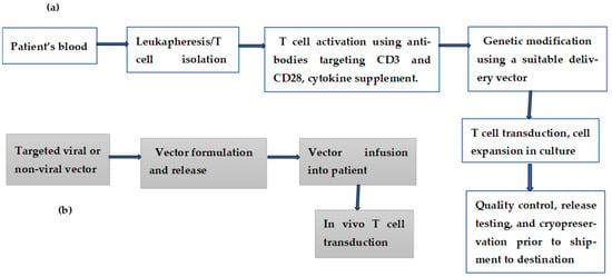
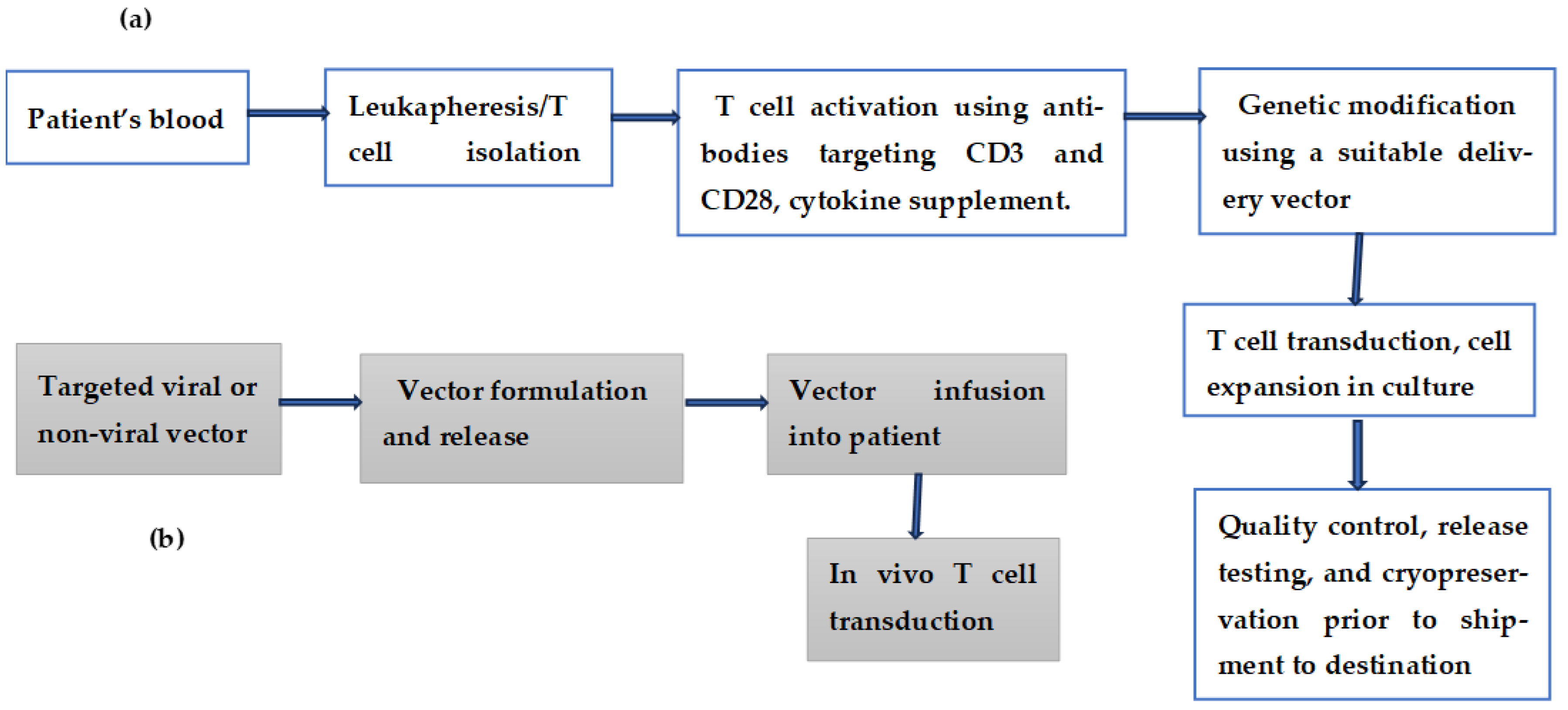
The introduction of chimeric antigen receptor (CAR) T cell therapy into the landscape of oncology represented a major therapeutic leap in the treatment of some aggressive forms of hematological tumors [1,2]. However, clinical data accumulated over the last decade demonstrate that the ex vivo approach has a number of limitations, including complex manufacturing processes, high production costs, and challenges in quality control and risk management. These limitations rendered this therapy out of reach for many cancer patients. To address some of these limitations, the manufacturing of chimeric antigen T cells and associated medicinal products has shifted from ex vivo to in vivo methods [3,4]. In this emerging approach, targeted delivery systems such as lentiviral vectors and lipid nanoparticles are used to introduce CAR-encoding genetic material into endogenous T cells. In recent years, non-viral vectors, particularly lipid nanoparticles (LNPs), have emerged as the platforms of choice for in vivo delivery of genetic material. This role has been consolidated by certain characteristics of these platforms, including efficient nucleic acid encapsulation, targeted delivery, and controlled release [5]. Clinical validation, stability, and an encouraging safety profile have also contributed to the emergence of this non-viral targeted delivery platform. A typical LNP platform is composed of four main components: (a) ionizable lipids responsible for the encapsulation and delivery of the genetic material; (b) polyethylene glycol lipids, which determine the size and stability of the vector; (c) a phospholipid component that contributes to better fusion with the host cells; and (d) a cholesterol component to improve the stability and the fluidity of the LNPs. As well as these major components, there are certain LNPs that contain additional minor components to enhance targeting and to improve their capability for efficient delivery of their load [6,7,8]. A schematic comparison between ex vivo and in vivo CAR T cell therapy is given in Figure 1a,b, while Table 1 gives the main limitations of the two approaches.
Figure 1.
A brief description of the various steps for T cell genetic modification: (a) ex vivo and (b) in vivo cell therapy.
Table 1.
Some known limitations of ex vivo and in vivo CAR T cell therapy.
The Shift from Ex Vivo to In Vivo CAR T Cell Therapy
In vivo CAR T cell engineering, in which CAR T cells are generated directly within the patient, offers the opportunity to bypass most limitations associated with its ex vivo counterpart. This emerging technique offers a shorter treatment timeline, a significant reduction in overall costs, broader clinical accessibility, and future large-scale implementation. That said, this highly promising technique is still undergoing various phases of optimization. One of the optimization areas is focused on improving the efficiency and accuracy of the delivery vectors. A simple comparison between the parameters of viral and non-viral vectors is given in Table 2. Such a delivery vector must selectively find and modify the target T cells without affecting other healthy tissues or other non-targeted cells [15,16]. Off-target sequestration of non-targeted cells or plasma proteins can trap the delivery vector, leading to a reduction in the effective therapeutic dose. The same vector can be neutralized by pre-existing immunity, patient antibodies or innate immune responses, preventing these vectors from reaching their selected target [14]. With the continuous optimization of gene delivery systems, gene editing technologies and advancements in CAR structure, an enhanced and safer role for in vivo CAR T therapy in solid cancer treatment is gaining additional momentum [17,18,19,20]. For example, the current literature describes five generations of CARs. All five generations share the same basic structure, which includes an extracellular antigen-binding domain, a hinge region, a transmembrane region, and the intracellular signaling domain. The main difference between these generations is within the intracellular signaling region. The first generation has the CD3ζ signaling molecule; the second generation has a single CD3ζ and CD28 or 4-1BB as a co-stimulatory domain; the third generation has a single CD3ζ and two different co-stimulatory domains, CD28 and 4-1BB; and the fourth generation has a single CD3ζ as well as a gene sequence encoding cytokines and other molecules. Once activated, CAR T cells release immunostimulatory cytokines, including IL-12 and IL-15. The fifth generation is a universal CAR, containing three domains: conventional T cell receptor activation signals, co-stimulation signals, and cytokine signals [21,22].
Table 2.
A comparison of the various parameters of viral and non-viral vectors used in CAR T and CAR-NK cell therapy.
2. Discussion
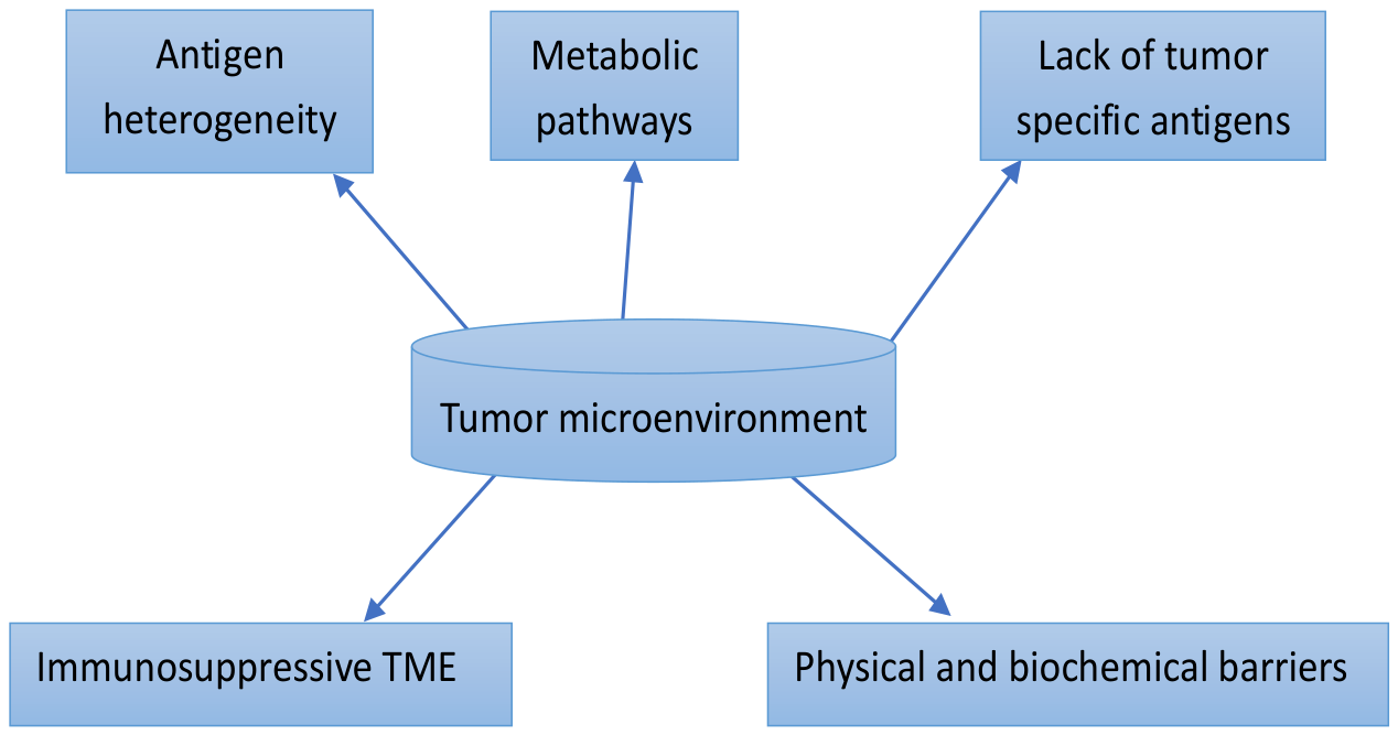
One of the challenges facing the in vivo approach is to contribute to a more detailed analysis of the tumor microenvironment of some solid tumors. Such a microenvironment is extensively heterogenous, composed of many components, each of which can represent a driver of resistance to CAR T cell therapy. Figure 2 shows a number of these components, including metabolic pathways. Identification of the main components within such an environment, and the obstacles to CAR T therapy created by such a composition, are indispensable for the design of various clinical trials and subsequent therapeutic modalities. Metabolic alterations within the tumor microenvironment are considered one of the key drivers of resistance to existing therapies. The in vivo CAR T approach, which addresses most limitations of the ex vivo approach, can contribute to a better understanding of this key driver of resistance.
Figure 2.
The main drivers of resistance to CAR T cell therapy within the solid tumor microenvironment. The physical barriers include dense extracellular matrix (ECM) and growth-induced solid stress. The biochemical barriers include hypoxia, acidity due to the accumulation of some metabolic waste products, and immunosuppressive cells such as cancer-associated fibroblasts (CAFs), regulatory T cells (Tregs), and tumor-associated macrophages (TAMs).
2.1. Metabolic Alterations Within Tumor Microenvironment
There is strong evidence that the tumor microenvironment (TME) exerts significant metabolic effects, which have a significant impact on the activities of the invading immune cells. Natural killer (NK) cells and macrophages are the first to be influenced by these events. Effective anti-tumor activities are closely linked to interactions between NK cells and macrophages [31]. A number of investigations have shown that certain metabolic events, including glucose depletion, lipid metabolism, hypoxia, and lactate accumulation, can influence and even reshape NK cell activities and macrophage polarization in the TME [32,33]. A number of studies have also suggested that the metabolic pathways involving MYC, hypoxia-inducible factor 1α (HIF1α), AMP-activated protein kinase (AMPK) and mammalian target of rapamycin (mTOR) are activated upon antigen recognition and that they are required for directing the consequences of T cell receptor engagement [34,35]. A number of solid tumors are known to harbor hypoxic areas due to rapid tumor growth and structural abnormalities of tumor vasculature [36]. To withstand the cellular stress caused by hypoxia, cells respond with activation of the hypoxia-inducible factor 1α (HIF1α) pathway. This metabolic pathway is a known regulator of gene expression in response to oxygen starvation; the upregulation of this pathway induces glycolytic genes, resulting in much higher levels of lactate [37,38]. According to the same investigations, the heterogenous world of tumors is composed of fibroblasts, immune cells, endothelial cells and extracellular matrix; this world engages in nutrient competition, metabolite exchange and paracrine signaling, where competition and cooperation among these components can influence tumor growth, immune evasion, and resistance to therapy. Recent advances in metabolic research underline the organizational principles of amino acid and lipid metabolism within the tumor microenvironment; such revelations assisted in clarifying certain mechanisms by which cancer and nonmalignant cancer cells participate in reciprocal nutrient manipulation, focusing on glutamine, arginine, tryptophan, branched-chain amino acids and lipids [39]. These findings suggest that targeting a single metabolic pathway within the TME is not the ideal strategy to understand the central role of various metabolic alterations within the TME. Most existing investigations on various metabolic pathways within the TME have been conducted using the ex vivo CAR T approach. Given the known characteristics of in vivo CAR T cell therapy, a major contribution to our understanding of the various metabolic alterations within the TME is to be expected.
2.2. Improving Target Specificity of CAR T Cell Therapy in Solid Tumors
CAR T cell therapy has demonstrated efficacy in the treatment of hematologic malignancies [40,41]. The failure of the same technique to realize similar success against solid tumors is generally attributed to lack of specific, safe target proteins shared by all tumor cells. The lack of truly solid-tumor-specific targets has been and still represents one of the challenges on the path to a more effective cell therapy for these fatal cancers. The last five years have witnessed a number of research initiatives that may lead to a substantial improvement in the target specificity of CAR T cell therapy. In one of these initiatives, a research group developed an approach to T cell therapy based on a dual-receptor system called Tmod™ [42]. This approach exploits a well-known genetic mutation known as homozygous deletion [43,44]. Although such deletions are rare, in various forms of cancer, the loss of heterozygosity is rather common, affecting about 20% of genes in such diseases [45]. This loss of genetic material (heterozygosity) in tumor cells represents an opportunity for distinguishing tumorous from normal cells. This process is carried out by targeting these genetic losses with a cellular NOT logic gate (some sort of a biological switch inside a living cell) designed to mediate the attack of the engineered immune cells on cancer cells that lack expression of a specific antigen, while sparing normal cells that possess the same antigen [46,47,48]. An allogeneic logic-gated Tmod™ CAR T cell construct was used in an ongoing medical trial listed in Table 3 (NCT06682793). It is recognized that epithelial growth factor receptor (EGFR) plays a critical role in oncogenesis across several types of cancer and is often upregulated [49,50]. The trial under consideration uses A2B395, an allogeneic, logic-gated, EGFR-targeted, Tmod CAR T therapy. The construct used in this trial contains two CARs: an activator targeting EGFR and a blocker targeting human leukocyte antigen (HLA)-A*02. The activator recognizes EGFR on both tumor and normal cells, while the blocker inhibits CAR T activity against normal cells with HLA expression, thus reducing the risk of on-target, off-tumor toxicity.
Table 3.
Some clinical trials to test the safety and maximum dose tolerability of CAR T for treating different forms of solid tumors. It is relevant to note that the final results from one of these trials will not be available before the year 2039.
2.3. Dual-Target CAR T Therapy
This strategy uses CAR T cells capable of expressing two distinct antigen recognition domains, allowing simultaneous targeting of two different antigens on tumor cells. In this innovative approach, the use of either ex vivo or in vivo CAR T therapy does not influence the acquired data. This strategy has been introduced to address a number of limitations associated with the single-target strategies [51,52]. These limitations include antigen loss/mutation, off-tumor damage to healthy tissues that happen to express tumor-associated antigens, limited CAR T cell persistence, and functional exhaustion [53,54]. A representative example of a phase I clinical trial describing a dual-target strategy is given in Table 3. The investigation describes the application of CAR T cells targeting EGFR epitope 806 and IL13Rα2 in recurrent glioblastoma. The main difference between this trial and those listed in the same table is the double rather than single targeting by CARs. Six patients with recurrent glioblastoma were treated with CART-EGFR-IL13Rα2 cells a year ago; some interim results of this trial were reported [55]. Some of these results are highly informative and worth careful consideration: The authors reported that three patients received a dose of 1 × 10⁷ cells; the other three patients received 2.5 × 10⁷ cells. In both groups, the administration of CART-EGFR-IL13Rα2 cells was associated with early-onset neurotoxicity, most consistent with immune effector cell-associated neurotoxicity syndrome (ICANS). This adverse event was managed with a high dose of dexamethasone and anakinra (anti-recombinant human interleukin-1 receptor antagonist, IL1R). One patient receiving high-dose treatment demonstrated dose-limiting toxicity, including grade 3 anorexia, generalized muscle weakness and fatigue. The authors also reported that a reduction in tumor size was observed for all six patients at early magnetic resonance imaging timepoints; however, none of the treated patients met the criteria for the objective response rate (ORR).
2.4. Armored T Cells Secreting Cytokines
The use of armored T cells secreting cytokines or other specific immune-boosting molecules is one of the current strategies to overcome the interference of some components of the TME. These molecules are released directly into the TME, a local delivery that helps the survival of T cells, and could recruit other immune cells to enhance the efficacy of CAR T therapy. Two of the clinical trials listed in Table 4 were designed to investigate the effect of interleukin-15-armored GPC3-CAR T cells for the treatment of solid tumors [56,57]. The first trial, NCT02905188, started in 2021 and ended in 2023. The patients within this cohort received GPC3-CAR T cells, which were safe but produced no objective anti-tumor responses and reached peak expansion within two weeks. In the second trial, NCT02932956, patients received GPC3-CAR T cells that co-expressed IL-15, which mediated increased cell expansion and induced a disease control rate of 66% and anti-tumor response rate of 33%. The authors also reported that infusion of 15 CAR T cells was associated with increased incidence of cytokine release syndrome. These results are preliminary, since the trial started in 2021 and is expected to be completed in 2037. Armored T cells co-expressing chemokine receptors, chemokines, or extracellular matrix-degrading enzymes have been linked to improved trafficking and infiltration to solid-tumor sites [58,59,60,61]. In a relatively recent study, the authors generated FAP/IL-15 CAR T. The authors hypothesized that the first component targets fibroblast activation protein (FAP), a protein within the TEM known to contribute to the immunosuppressive barrier that hinders the infiltration of the therapy to targeted sites, while the IL-15 component of the load can contribute to the activation of CAR T cells, reducing their fast exhaustion, and increases the proportion of T stem cell memory [62]. To underline the role of interleukin-15 (IL-15) in enhancing the anti-tumor properties of CAR T cells, another study compared the performance of CAR T cells with and without IL-15 co-expression [56]. The authors reported the first evaluation in humans of the effect of IL-15 co-expression on Glypican-3 (GPC3)-CAR T cells. It is worth noting that GPC3 is a cell surface protein hardly expressed in normal tissues, yet it is heavily expressed in liver cancer, specifically in hepatocellular carcinoma [63]. The authors reported that a group of solid-tumor patients received GPC3-CAR T cells at safe doses but produced no measurable anti-tumor response. A second group of patients received GPC3-CAR T cells that co-expressed IL15. Analysis of this group of patients revealed increased cell expansion, induced disease control, and a higher anti-tumor response rate.
Table 4.
Some phase I clinical trials sponsored by Baylor College of Medicine to determine the highest safe dose of modified T cells, including those co-expressing IL-15, the duration of the dose within the patient’s body, efficacy and the likely irAEs associated with the administration of this therapy in some solid-tumor patients.
Effective anti-tumor activities are known to be closely linked to interactions as well as cross talk between natural killer (NK) cells and macrophages within the tumor microenvironment [31]. Recent preclinical and clinical studies have yielded promising results on the use of CAR-NK to treat some forms of solid tumors; the same studies demonstrated limited in vivo efficacy of this approach. To enhance the expansion and persistence of CAR-NK cells, various groups have developed IL-15 cytokine-armored CAR-NK therapeutics targeting various forms of solid tumors [64]. Although IL-15 has been extensively used to improve CAR-NK cell persistence and effectiveness, the balance between the effectiveness of this approach and its safety remains an important limitation that needs to be addressed. An attempt to improve the balance between safety and effectiveness is given in Ref. [65]. The authors conducted an in vitro investigation where IL-15 fused with full-length IL-15Ra was systematically compared with secretory IL-15 in a B7H3-targeting CAR-NK cell system. The authors concluded that in a head-to-head comparative study, signaling-intact membrane-bound IL-15 fused with full-length IL-15Ra significantly improved the therapeutic efficacy and safety of CAR-NK cells as compared to secretory IL-15. The same authors also observed that CAR-NK cells with signaling IL-15 quickly eliminated intraperitoneal ovarian cancer; however, the treated mice experienced severe consequences, including dysregulated CAR-NK cell expansion, intense inflammatory responses, and irreversible organ damage. On the contrary, CAR-NK cells carrying membrane-bound IL15 showed moderate cell proliferation and strong tumor killing activity without observable adverse effects.
The conclusions in the above study were based on cell line analysis and tests in animal models; therefore, these conclusions can be considered a useful example of a proof of concept. Whether such conclusions will coincide with those of future investigations in humans is difficult to predict. That said, attempts to find a balance between the safety and efficacy of interleukin-armored CAR T and CAR-NK cells are still ongoing; therefore, this and other studies, particularly on humans, are to be encouraged.
3. Conclusions and Future Perspectives
Advances in targeted delivery systems such as lentiviral vectors and lipid nanoparticles are responsible for the unusually fast emergence of in vivo CAR T cell therapy. The use of these platforms for in vivo genetic modification of T cells will facilitate the design and performance of large clinical trials, particularly those investigating solid tumors and their microenvironment. The most recent preclinical results suggest that the optimization and consolidation of this approach is likely to give fresh momentum to the search for effective therapies to treat fatal solid tumors. However, this tantalizing objective has a number of hurdles on its way: The safety of viral vector-based therapy can be challenged by a number of the host’s innate immune responses. These responses can result in transgene silencing, vector clearance, and loss of transduced cells. The risks related to off-target genetic modification and efficiency still need to be optimized. Preclinical data generated by this approach indicate that most therapeutic strategies designed to overcome the heterogeneity and immune suppressiveness of the solid-tumor microenvironment are bound to be enhanced by more use of the in vivo approach. Multiple targeting of tumor antigens and the use of armored T cells secreting cytokines or other specific immune-boosting molecules are two representative strategies that are likely to benefit most from the application of the in vivo approach. Present-day comparisons between the roles of the well-tested ex vivo with that of the emerging in vivo CAR T cell therapy are rather immature. A rational comparison between the two approaches would benefit from additional clinical data, preferably generated by phase III trials.
Author Contributions
Conceptualization, M.H.; writing—original draft preparation, M.H.; writing—review and editing, P.T. and M.A.; P.T. and M.A. contributed equally to the reviewing of the text. All authors have read and agreed to the published version of the manuscript.
Funding
This research received no external funding.
Institutional Review Board Statement
Not applicable.
Informed Consent Statement
Not applicable.
Data Availability Statement
No new data were created or analyzed in this study. Data sharing is not applicable to this article.
Conflicts of Interest
The authors declare no conflicts of interest.
Abbreviations
| CAR T | Chimeric antigen receptor |
| CRS | Cytokine release syndrome |
| TME | Tumor microenvironment |
| ECM | Extracellular matrix |
| ADCs | Antibody–drug conjugate |
| FAP | Fibroblast activation protein |
| TGF-β | Transforming growth factor-beta |
| BCL | B-cell lymphoma |
| GPC3 | Glypican-3 protein |
| IL-15 | Interleukin 15 |
| EGFR epitope 806 | 16-amino-acid region within epidermal growth factor receptor |
| IL13Rα2 | Interleukin-13 receptor subunit alpha-2 |
| IL-1R | Human interleukin-1 receptor antagonist |
| ORR | Objective response rate |
| ICANS | Immune effector cell-associated neurotoxicity syndrome |
| rGBM | Recurrent glioblastoma |
| NSCLC | Non-small cell lung cancer |
| LYL273 | Experimental CAR T cell therapy |
| irAEs | Immune-related adverse events |
| HLA | Human leukocyte antigen |
| PDL1 | Cell death ligand-L1 |
| CTLA-4 | Cytotoxic T-lymphocyte antigen-4 |
References
- Dana, H.; Chalbatani, G.M.; Jalali, S.A.; Mirzaei, H.R.; Grupp, S.A.; Suarez, E.R.; Rapôso, C.; Webste, T.J. CAR-T cells: Early successes in blood cancer and challenges in solid tumors. Acta Pharm. Sin. B 2021, 11, 1129–1147. [Google Scholar] [CrossRef] [Scilit] [PubMed]
- Aljabali, A.A.A.; Obeid, M.A.; Alqudah, A.; Hamzat, Y.; Alqudah, A.; Alzoubi, L.; Gammoh, O.; Qnais, E.; Mishra, V.; Mishra, Y.; et al. CAR T-Cell Therapy in Hematological Malignancies: Mechanisms, Innovations, and Challenges. Cancers 2026, 18, 3254. [Google Scholar] [CrossRef] [Scilit] [PubMed]
- Upadhyay, S.; Cho, S.; Upmanyu, K.; Gabr, M.T. CAR T cell therapy beyond cancer: Current status: Challenges and future prospects. Signal Transduct. Target. Ther. 2026, 11, 319. [Google Scholar] [CrossRef] [Scilit] [PubMed]
- Rosa, R.; Liu, J.; Lu, C.; Abou-el-Enein, M.; Murad, J.P.; Priceman, S.J. Current state of CAR-T cell therapies for solid tumors. Med 2026, 7, 101028. [Google Scholar] [CrossRef] [Scilit] [PubMed]
- Siddiqui, A.; Purwar, A.; Purwar, R.; Kumar, S. Next-generation CAR-T: In vivo engineering with lipid nanoparticles. J. Clin. Med. Surg. 2025, 5, 120. [Google Scholar] [CrossRef] [Scilit]
- Xu, S.; Hu, Z.; Song, F.; Xu, Y.; Han, X. Lipid nanoparticles: Composition, formulation, and application. Mol. Ther. Methods Clin. Dev. 2025, 33, 101463. [Google Scholar] [CrossRef] [Scilit] [PubMed]
- Cheng, Q.; Wei, T.; Farbiak, L.; Johnson, L.T.; Dilliard, S.A.; Siegwart, D.J. Selective organ targeting (SORT) nanoparticles for tissue-specific mRNA delivery and CRISPR–Cas gene editing. Nat. Nano technol. 2020, 15, 313–320. [Google Scholar] [CrossRef] [Scilit] [PubMed]
- Shan, X.; Zhao, Z.; Lai, P.; Liu, Y.; Li, B.; Ke, Y.; Jiang, H.; Zhou, Y.; Li, W.; Wang, Q.; et al. RNA nano therapeutics with fibrosis overexpression and retention for MASH treatment. Nat. Commun. 2024, 15, 7263. [Google Scholar] [CrossRef] [Scilit] [PubMed]
- Ayala, C.M.; Khericha, M.; Harris, C.M.; Puig-Saus, C.; Chen, Y.Y. CAR-T cell manufacturing: Major process parameters and next-generation strategies. J. Exp. Med. 2024, 221, e2023095. [Google Scholar] [CrossRef] [Scilit] [PubMed]
- Alfar, H.R.; Chen, C.; Lachacz, E.; Tang, W.; Zhang, Y. Clinical evidence of immunogenicity of CAR-T cell therapies and its implication in the clinical development of CAR-T drug products. Front. Immunol. 2025, 16, 1512494. [Google Scholar] [CrossRef] [Scilit] [PubMed]
- Short, L.; Holt, R.A.; Cullis, P.R.; Evgin, L. Direct in vivo CAR T cell engineering. Trends Pharmacol. Sci. 2024, 45, 406–418. [Google Scholar] [CrossRef] [Scilit] [PubMed]
- Zhang, Y.; Qin, D.; Shou, A.C.; Liu, Y.; Wang, Y.; Zhou, L. Exploring CAR-T Cell Therapy Side Effects: Mechanisms and Management Strategies. J. Clin. Med. 2023, 12, 6124. [Google Scholar] [CrossRef] [Scilit] [PubMed]
- Majzner, R.G.; Mackall, C.L. Tumor antigen escapes from CAR T cell therapy. Cancer Discov. 2018, 8, 1219–1226. [Google Scholar] [CrossRef] [Scilit] [PubMed]
- Liu, Z.; Song, S. Reprogramming innate immunity through viral interference: A double-edged strategy for enhancing and containing gene therapies. Mol. Ther. Nucleic Acids 2026, 37, 102911. [Google Scholar] [CrossRef] [Scilit] [PubMed]
- Zugasti, I.; Espinosa-Aroca, L.; Klaudyna, F.; Mulens-Arias, V.; Diaz-Beya, M.; Juan, M.; Urbano-Ispizua, A.; Esteve, J.; Velasco-Hernandez, T.; Menéndez, P. CAR-T cell therapy for cancer: Current challenges and future directions. Signal Transduct. Target Ther. 2025, 10, 210. [Google Scholar] [CrossRef] [Scilit] [PubMed]
- Amatya, S.; Green, J.; Chu, C.-S.; Hilterbrand, A.; Burch, R.; Baldeviano, C.; Rebar, E.; Fry, T.J.; Ruzo, A.; Elpek, K.G.; et al. Enabling Potent T Cell Delivery and Mitigating Key Off-Target Concerns of In Vivo CAR T Lentiviral Vectors with the Targeted Paramyxovirus Fusogen System. Blood 2024, 144, 2047–2048. [Google Scholar]
- Gaj, T.; Sirk, S.J.; Shui, S.L.; Liu, J. Genome-Editing Technologies: Principles and Applications. Cold Spring Harb. Perspect. Biol. 2016, 8, a023754. [Google Scholar] [CrossRef] [Scilit] [PubMed]
- Liu, D.; Cao, D.; Han, R. Recent advances in therapeutic gene-editing technologies. Mol. Ther. 2025, 33, 2619–2644. [Google Scholar] [CrossRef] [Scilit] [PubMed]
- Ma, Y.; Dong, S.; Wu, A.; Jeong, S.D.; Lee, A.S.; Jiang, W.; Kim, B.Y.S. Engineering challenges and translational opportunities in emerging gene delivery platforms. Nat. Biomed. Eng. 2026, 10, 1060–1083. [Google Scholar] [CrossRef] [Scilit] [PubMed]
- Wang, F.; Liang, L.; Wang, C.; Zhang, C.; Ye, Y.; Dou, L.; Shi, L.; Zhou, D.; Sun, X. Non-Viral Gene Delivery Systems: Current Advances and Therapeutic Applications. Chem. Asian J. 2025, 20, e00684. [Google Scholar] [CrossRef] [Scilit] [PubMed]
- Zheng, Z.; Li, S.; Liu, M.; Chen, C.; Zhang, L.; Zhou, D. Fine-Tuning through Generations: Advances Structure and Production of CAR-T Therapy. Cancers 2023, 15, 3476. [Google Scholar] [CrossRef] [Scilit] [PubMed]
- Kondo, T.; Umehara, C.; Kagoya, Y. Computational optimization of chimeric antigen receptor. Mol. Ther. 2025, 33, 4680–4681. [Google Scholar] [CrossRef] [Scilit] [PubMed]
- Wang, W.; Li, W.; Ma, N.; Steinhoff, G. Non-viral gene delivery methods. Curr. Pharm. Biotechnol. 2013, 14, 46–60. [Google Scholar] [CrossRef] [Scilit]
- Halagali, P.; Seenivasan, R.; Sreedevi, A.; Ligade, V.S.; Bugadannavar, P.; Sharma, H.; Rai, S.N.; Tippavajhala, V.K. Advances in gene transfer technologies: Comparing viral and non-viral vectors for therapeutic applications. 3 Biotech 2026, 16, 381. [Google Scholar] [CrossRef] [Scilit] [PubMed]
- Baker, A.H.; Kritz, A.; Work, L.M.; Nicklin, S.A. Cell-selective viral gene delivery vectors for the vasculature. Exp. Physiol. 2005, 90, 27–31. [Google Scholar] [CrossRef] [Scilit] [PubMed]
- Honrath, S.; Burger, M.; Leroux, J.-C. Hurdles to healing: Overcoming cellular barriers for viral and nonviral gene therapy. Int. J. Pharm. 2025, 674, 125470. [Google Scholar] [CrossRef] [Scilit] [PubMed]
- Zu, H.; Gao, D. Non-viral Vectors in Gene Therapy: Recent Development, Challenges, and Prospects. AAPS J. 2021, 23, 78. [Google Scholar] [CrossRef] [Scilit] [PubMed]
- Ren, S.; Wang, M.; Wang, C.; Wang, Y.; Sun, C.; Zeng, Z.; Cui, H.-; Zhao, X. Application of non-viral vectors in drug delivery and gene therapy. Polymers 2021, 13, 3307. [Google Scholar] [CrossRef] [Scilit] [PubMed]
- Madani, F.; Izadi, E.; Motamedi, M.; Ganjalikhani Hakemi, M.; Saltanatpour, Z.; Webster, T.J.; Mohammadi, S.F. Toward Safe and Effective Gene Therapy: Non-Viral Nanostructured Delivery Systems. Int. J. Nanomed. 2026, 15, 598766. [Google Scholar] [CrossRef] [Scilit] [PubMed]
- Hauck, E.S.; Hecker, J.G. non-viral delivery of RNA gene therapy to the central nervous system. Pharmaceutics 2022, 14, 165. [Google Scholar] [CrossRef] [Scilit] [PubMed]
- Keane, C.; Mayock, S.; Corkish, C.; Rogerson, E.; de Aguiar, C.F.; Gao, Y.; Kenny, S.; Brennan, L.; Cloonan, S.M.; Finlay, D.K. Macrophages tune NK cell IFNγ production through direct lipid transfer. Nat. Commun. 2026, 17, 9527. [Google Scholar] [CrossRef] [Scilit] [PubMed]
- Wang, B.; Zhao, Q.; Zhang, Y.; Liu, Z.; Zheng, Z.; Liu, S.; Meng, L.; Xin, Y.; Jiang, X. Targeting hypoxia in the tumor microenvironment: A potential strategy to improve cancer immunotherapy. J. Exp. Clin. Cancer Res. 2021, 40, 24. [Google Scholar] [CrossRef] [Scilit] [PubMed]
- Pollizzi, K.N.; Powell, J.D. Integrating canonical and metabolic signaling programs in the regulation of T cell responses. Nat. Rev. Immunol. 2014, 14, 435–446. [Google Scholar] [CrossRef] [Scilit] [PubMed]
- Trejo-Solís, C.; Castillo-Rodríguez, R.A.; Serrano-García, N.; Silva-Adaya, D.; Vargas-Cruz, S.; Chávez-Cortéz, E.G.; Gallardo-Pérez, J.C.; Zavala-Vega, S.; Cruz-Salgado, A.; Magaña-Maldonado, R. Metabolic Roles of HIF1, c-MYC, and p53 in Glioma Cells. Metabolites 2024, 14, 249. [Google Scholar] [CrossRef] [Scilit] [PubMed]
- Yang, K.; Chi, H. mTOR and metabolic pathways in T cell quiescence and functional activation. Semin. Immunol. 2012, 24, 421–428. [Google Scholar] [CrossRef] [Scilit] [PubMed]
- Kim, J.W.; Tchernyshyov, I.; Semenza, G.L.; Dang, C.V. HIF-1-mediated expression of pyruvate dehydrogenase kinase: A metabolic switch required for cellular adaptation to hypoxia. Cell Metab. 2006, 3, 177–185. [Google Scholar] [CrossRef] [Scilit] [PubMed]
- de Goede, K.E.; Driessen, A.J.M.; den Bossche, J.V. Metabolic Cancer-Macrophage Crosstalk in the Tumor Microenvironment. Biology 2020, 9, 380. [Google Scholar] [CrossRef] [Scilit] [PubMed]
- Jain, R.K. Normalization of tumor vasculature: An emerging concept in antiangiogenic therapy. Science 2005, 307, 5867. [Google Scholar] [CrossRef] [Scilit] [PubMed]
- Sung, Y.; Kim, D.K.; Kim, J.S.; Kim, S.-J.; Kim, J.H.; Han, J.M. Metabolic networks in the tumor microenvironment: Roles of amino acid and lipid metabolism pathways in cancer progression and therapy. Exp. Mol. Med. 2026, 58, 1128–1148. [Google Scholar] [CrossRef] [Scilit] [PubMed]
- Zhang, X.; Zhu, L.; Zhang, H.; Chen, S.; Xiao, Y. CAR-T Cell Therapy in Hematological Malignancies: Current Opportunities and Challenges. Front. Immunol. 2022, 13, 927153. [Google Scholar] [CrossRef] [Scilit] [PubMed]
- Hernández-Idarraga, K.J.; Arias-Rozo, A.J.; Arango-Rodrigue, M.L.; Sossa, C.L.; Becerra-Bayona, S.M. CAR T-cells in hematologic malignancies: Advances, challenges, and future directions. iScience 2026, 29, 115213. [Google Scholar] [CrossRef] [Scilit] [PubMed]
- Manry, D.; Bolanos, K.; DiAndreth, B.; Mock, J.Y.; Kamb, A. Robust In Vitro Pharmacology of Tmod, a Synthetic Dual-Signal Integrator for Cancer Cell Therapy. Front. Immunol. 2022, 13, 826747. [Google Scholar] [CrossRef] [Scilit] [PubMed]
- Cheng, J.; Demeulemeester, J.; Wedge, D.C.; Vollan, H.K.M.; Pitt, J.J.; Russnes, H.G.; Pandey, B.P.; Gro Nilsen, G.; Nord, S.; Bignell, G.R.; et al. Pan-cancer analysis of homozygous deletions in primary tumours uncovers rare tumor suppressors. Nat. Commun. 2017, 8, 1221. [Google Scholar] [CrossRef] [Scilit] [PubMed]
- Schmitz-Abe, K.; Sanchez-Schmitz, G.; Doan, R.N.; Hill, R.S.; Chahrour, M.H.; Mehta, B.K.; Servattalab, S.; Ataman, B.; Lam, A.-T.N.; Morrow, E.M.; et al. Homozygous deletions implicate non-coding epigenetic marks in Autism spectrum disorder. Sci. Rep. 2020, 10, 14045. [Google Scholar] [CrossRef] [Scilit] [PubMed]
- Beroukhim, R.; Mermel, C.H.; Porter, D.; Wei, G.; Raychaudhuri, S.; Donovan, J.; Barretina, J.; Boehm, J.S.; Dobson, J.; Urashima, M.; et al. The Landscape of Somatic Copy-Number Alteration Across Human Cancers. Nature 2010, 463, 899–905. [Google Scholar] [CrossRef] [Scilit] [PubMed]
- Dannenfelser, R.; Allen, G.M.; VanderSluis, B.; Koegel, A.K.; Levinson, S.; Stark, S.R.; Yao, V.; Tadych, A.; Troyanskaya, O.G.; Lim, W.A. Discriminatory Power of Combinatorial Antigen Recognition in Cancer T Cell Therapies. Cell Syst. 2020, 11, 215–228.e215. [Google Scholar] [CrossRef] [Scilit] [PubMed]
- Emanuelson, C.; Bardhan, A.; Deiters, A. DNA Computing: NOT Logic Gates See the Light DNA Computing: NOT Logic Gates See the Light. ACS Synth. Biol. 2021, 10, 1682–1689. [Google Scholar] [PubMed]
- Di Andreth, B.; Hamburger, A.E.; Xu, H.; Kamb, A. The Tmod cellular logic gate as a solution for tumor-selective immunotherapy. Clin. Immunol. 2022, 241, 1091030. [Google Scholar] [CrossRef] [Scilit] [PubMed]
- Chen, X.; Wang, Z. Targeting EGFR Endocytosis and Signaling for Cancer Drug Delivery and Cancer Treatment. Cancers 2026, 18, 2451. [Google Scholar] [CrossRef] [Scilit] [PubMed]
- Katreddy, R.R.; Bollu, L.R.; Su, F.; Xian, N.; Srivastava, S.; Thomas, R.; Dai, Y.; Wu, B.; Xu, Y.; Rea, M.A.; et al. Targeted reduction of the EGFR protein, but not inhibition of its kinase activity, induces mitophagy and death of cancer cells through activation of mTORC2 and Akt. Oncogenesis 2018, 7, 5. [Google Scholar] [CrossRef] [Scilit] [PubMed]
- Chen, Z.; Liu, Y.; Chen, N.; Xing, H.; Tian, Z.; Tang, K.; Rao, Q.; Xu, Y.; Wang, Y.; Wang, M.; et al. Loop CD20/CD19 CAR-T cells eradicate B-cell malignancies efficiently. Sci. China Life Sci. 2023, 66, 754–770. [Google Scholar] [CrossRef] [Scilit] [PubMed]
- Wei, G.; Zhang, Y.; Zhao, H.; Wang, Y.; Liu, Y.; Liang, B.; Wang, X.; Xu, H.; Cui, J.; Wu, W.; et al. CD19/CD22 Dual-Targeted CAR T-cell Therapy for Re-lapsed/Refractory Aggressive B-cell Lymphoma: A Safety and Efficacy Study. Cancer Immunol. Res. 2021, 9, 1061–1070. [Google Scholar] [CrossRef] [Scilit] [PubMed]
- Wang, Z.; Wang, M.; Wang, M.; Zhou, R.; Deng, X.; Ouyang, X.; Chu, M.; Wei, X.; Yang, L.; Liu, J.; et al. From molecular design to clinical translation: Dual-targeted CAR-T strategies in cancer immunotherapy. Int. J. Biol. Sci. 2025, 21, 2676–2691. [Google Scholar] [CrossRef] [Scilit] [PubMed]
- Gómez-Melero, S.; Hassouneh, F.; Vallejo-Bermúdez, I.M.; Agüera-Morales, E.; Solana, R.; Caballero-Villarraso, J. Tandem CAR-T cell therapy: Recent advances and current challenges. Front. Immunol. 2025, 16, 1546172. [Google Scholar] [CrossRef] [Scilit] [PubMed]
- Bagley, S.J.; Desai, A.S.; Fraietta, J.A.; Silverbush, D.; Chafamo, D.; Freeburg, N.F.; Gopikrishna, G.K.; Rech, A.J.; Nabavizadeh, A.; Bagley, L.J.; et al. Intracerebroventricular bivalent CAR T cells targeting EGFR and IL-13Rα2 in recurrent glioblastoma: A phase 1 trial. Nat. Med. 2025, 31, 2778–2787. [Google Scholar] [CrossRef] [Scilit] [PubMed]
- Steffin, D.; Ghatwai, N.; Montalbano, A.; Rathi, P.; Courtney, A.N.; Arnett, A.B.; Fleurence, J.; Sweidan, R.; Wang, T.; Zhang, H.; et al. Interleukin-15-armoured GPC3 CAR T cells for patients with solid cancers. Nature 2025, 637, 940–946. [Google Scholar] [CrossRef] [Scilit] [PubMed]
- Li, Y.-R.; Zhu, Y.; Yang, L. IL-15 in CAR engineering: Striking an efficacy–safety balance. Trends Mol. Med. 2025, 31, 977–981. [Google Scholar] [CrossRef] [Scilit] [PubMed]
- Escobar, G.; Berger, T.R.; Maus, M.V. CAR-T cells in solid tumors: Challenges and Breakthroughs. Cell Rep. Med. 2025, 6, 102353. [Google Scholar] [CrossRef] [Scilit] [PubMed]
- Yang, D.D.; Macmorland, W.; Arnold, J.N. Current strategies for armoring chimeric antigen receptor T-cells to overcome barriers of the solid tumor microenvironment. Front. Immunol. 2025, 16, 1643941. [Google Scholar] [CrossRef] [Scilit] [PubMed]
- Rafiq, S.; Hackett, C.S.; Brentjens, R.J. Engineering strategies to overcome the current roadblocks in CAR T cell therapy. Nat. Rev. Clin. Oncol. 2020, 17, 147–167. [Google Scholar] [CrossRef] [Scilit] [PubMed]
- Lindo, L.; Wilkinson, L.H.; Hay, K.A. Befriending the hostile tumor microenvironment in CAR T-cell therapy. Front. Immunol. 2020, 11, 618387. [Google Scholar] [CrossRef] [Scilit] [PubMed]
- Pang, Y.; Chen, L.; Sun, Q.; He, N.; Lai, Z.; Wang, X.; Xie, Z.; Yang, W.; Wang, W. A new strategy for CAR-T therapy in solid tumors: IL-15-autocrine signaling augments tumor stroma depletion and promotes a TSCM subset in the TME. Cell Death Dis. 2025, 17, 39. [Google Scholar] [CrossRef] [Scilit] [PubMed]
- Kandil, D.H.; Cooper, K. Glypican-3: A novel diagnostic marker for hepatocellular carcinoma and more. Adv. Anat. Pathol. 2009, 16, 125–129. [Google Scholar] [PubMed]
- Baughan, S.L.; Folsom, T.; Johnson, M.; Kreuger, J.; Webber, B.R.; Moriarity, B.S. Perspective: IL-15 cytokine- armored NK cells as ready-to- use immunotherapy for diverse malignancies: Therapeutic potential and toxicity risks. Front. Immunol. 2025, 16, 1704404. [Google Scholar] [CrossRef] [Scilit] [PubMed]
- Xu, X.; Cao, P.; Wang, M.; Wan, Y.; Sun, S.; Chen, Y.; Liu, Y.; Su, T.; Gao, G.; Liu, X.; et al. Signaling intact membrane-bound IL-15 enables potent anti-tumor activity and safety of CAR-NK cells. Front. Immunol. 2025, 16, 1658580. [Google Scholar] [CrossRef] [Scilit] [PubMed]

Disclaimer/Publisher’s Note: The statements, opinions and data contained in all publications are solely those of the individual author(s) and contributor(s) and not of MDPI and/or the editor(s). MDPI and/or the editor(s) disclaim responsibility for any injury to people or property resulting from any ideas, methods, instructions or products referred to in the content. |
© 2026 by the authors. Licensee MDPI, Basel, Switzerland. This article is an open access article distributed under the terms and conditions of the Creative Commons Attribution (CC BY) license.