


Highlights
What are the main findings?
- Repetitive exposure to organic dust, or its components including LPS, may promote sustained activation of macrophages and lymphocytes.
- Unresolved inflammation associated with repetitive organic dust exposure drives the structural changes in the airways that may lead to chronic bronchitis.
What is the implication of the main finding?
- The review provides a mechanistic explanation for organic dust-associated chronic bronchitis.
- Defining the efficacy of cellular and molecular targeting agents is essential for prevention or relief of prolonged airway inflammation associated with organic dust exposure.
- Chronic exposure to organic dust in swine production facilities results in activation of macrophages and lymphocytes in lungs via multiple signaling pathways including NF-κB, MAPK, and PKC pathways.
- Activation of these signaling pathways results in hypersecretion of pro- and anti-inflammatory cytokines, release of reactive oxygen and nitrogen species, and changes in airway pathophysiology.
- Understanding the role of alveolar macrophages and lymphocytes and the involved signaling pathways provides insight into the mechanism of organic dust-induced chronic bronchitis.
Abstract
In livestock production settings, repetitive exposure to organic dust (OD) is a risk factor for chronic bronchitis (CB). Macrophages and lymphocytes cooperate to defend the lungs against inhaled dust, but when chronically activated, their actions contribute to chronic inflammation, oxidative stress, and tissue remodeling. Despite the strong body of literature examining the effects of OD exposure on leukocytes and lungs, the contribution of macrophages and lymphocytes to organic dust-associated chronic bronchitis is not fully understood. This review examines the role of macrophages and lymphocytes in organic dust-associated chronic bronchitis. To identify the body of literature addressing this goal, we conducted Boolean searches with keywords (e.g., bronchitis, leukocytes, organic dust) across four major databases: PubMed, Science Direct, Scopus, and Web of Science. From a total of 25,488 hits from all time, 18 research articles (from 2015 to 2025) provided contextual connection for explaining organic dust-associated chronic bronchitis. The results showed continuous or repetitive exposure to OD or its components (e.g., lipopolysaccharide) causes prolonged activation of macrophages and lymphocytes, which leads to overexpression of pro- and anti-inflammatory cytokines and chemokines via Toll-like receptor (TLR)4, TLR2, nuclear factor kappa B, protein kinase C, Extracellular Signal-Regulated Kinase (ERK), and Mitogen-Activated Protein Kinase (MAPK) pathways. Unresolved lung inflammation drives pathologic changes including mucus hypersecretion, airway remodeling, and onset of chronic bronchitis symptoms. Addition of micronutrients or other anti-inflammatory treatment helps to mitigate OD-induced lung inflammation. Defining the cellular and molecular contributions to organic dust-associated chronic bronchitis is essential for the development of targeted therapies that prevent or relieve prolonged airway inflammation.
1. Introduction
Disease can be caused by seven primary ultimate causes: genetics, environment, by-products of defense, lack of maintenance, disruption of homeostasis, stochastic events during development, and fitness conflicts between relatives [1]. Any specific disease may result from multiple ultimate causes. Chronic bronchitis (CB) is a disease that is caused by a combination of genetics, environment, and as a by-product of defense, specifically inflammation. CB, a long-term inflammatory (by-product of defense) airway disease, arises from repetitive exposure to noxious substances (environment). The prolonged, unresolved inflammation can result in decreased lung function, respiratory complications, and increased morbidity in severe cases. Genetics may play a role as some individuals will be more susceptible to immune hypersensitivity, and its impacts are also age-specific, with aging (senescent) individuals much more likely to display adverse disease than younger (non-senescent) individuals. In addition to chronic bronchitis, other respiratory complications observed in the agricultural farming environment are hypersensitivity pneumonitis, asthma, allergic and non-allergic rhinitis, and chronic cough with phlegm [2,3,4]. These diseases share the same ultimate causality with chronic bronchitis. The development of chronic bronchitis in the agricultural farming environment, especially in individuals exposed to livestock production facilities, is associated with repetitive exposure to organic dust (OD) present in those facilities [2,5].
Over the past several decades, there has been increasing evidence to suggest that exposure to organic dust and livestock production settings including swine buildings can contribute to the onset of pulmonary symptoms and disease including increase in bronchial responsiveness, decrease in forced expiratory volume, chronic cough, decline in lung function, and chronic bronchitis in humans [6,7,8] Livestock and dairy farmers developed diagnostic markers indicating possible bronchial obstruction such as chronic cough, chronic bronchitis, wheeze, and dyspnea with reduced FEV1 [9,10]. Similarly, poultry farmers, who worked for long periods (>2 h daily), developed similar symptoms, which were detected upon pulmonary function test, with a risk factor being exposure to organic dust present in the building [11]. Upon measurement, principal contaminants found in the farms were organic dust and endotoxin, where both dust and endotoxin were associated with COPD in a dose-dependent manner, especially in swine, cattle, and poultry farmers [11,12].
The United States (U.S.) is the third-largest global producer of pork, and the hog production market has increased by 40% over the last fifty years; harvesting over 120 million head in 2021 in the U.S. alone (with Iowa, North Carolina, and Minnesota being the three largest producers, respectively) [13]. In 2021, 67,000 farms averaging 1,080 hogs per farm were reported in the U.S. [13]. Livestock farmers are at greater risk of developing respiratory complications, especially the swine farmers, due to high demand and an increased number of pigs, large-scale production, and an increase in working hours in the confined building facilities for 8 h per day [14]. As a result, swine barn workers suffer from respiratory health issues such as impaired airflow, and lung inflammation due to inhalation of barn air containing pollutants, including OD [14]. Organic dust, present in livestock production facilities, consists of particulate matter (PM) ranging from inhalable (PM10) to respirable (PM2.5) dust and PM of organic origins such as bacteria, viruses, fungi, fecal matter, pollen, etc. [15,16]. In acute exposures, OD caused immediate changes, including airway damage, prior to returning to normal function after the causative agent is removed/cleared. Whereas repetitive or chronic inhalation of OD can result in permanent loss of cell function and disease [15,16,17,18]. For instance, studies of swine farmers reported that workers spent an average of 6.6 ± 1.8 h per day in production buildings and worked at those sites for 10.5 ± 7.5 years and thus are at increased risk of developing increased airway responsiveness, airflow obstruction, chronic cough, and phlegm compared to shorter daily contacts, including a higher prevalence of CB (21.94% vs. 13.25%) in those swine farmers [19,20,21].
Chronic bronchitis is defined by persistent cough and sputum production lasting at least 3 months for 2 consecutive years, and is one of two common clinical phenomena of chronic obstructive pulmonary disease (COPD) [22]. In addition to persistent productive cough, CB is characterized by mucus hypersecretion, chronic inflammation, obstruction of small airways, airway remodeling, alteration of airway surface tension, decreased lung function, shortness of breath, fatigue, and wheezing in severe cases [23,24]. Unlike emphysema, one of the two main subtypes of COPD, CB can occur independently in cases where affected individuals have a normal breathing test [22]. The prevalence of CB is 14–74% of all patients who have developed COPD [24]. In a study conducted from 2007 to 2012, among 213 million ever-employed adults, 52% of adults employed in industries considered to have workplace dust, vapors, or fumes exposure associated with CB and emphysema, whereas, people working in farming, fishing, and forestry occupations were one of the occupations to have higher prevalence of developing CB and emphysema [25]. The primary cause of CB is cigarette smoking; however, working within environments with a higher risk of exposure to dust, vapors, and fumes also leads to CB upon long-term exposure at a rate of 4.6% in about 213 million ever-employed adults (crude prevalence and with the rate of 2.3% in age-related prevalence) [25,26]. Among farming occupations, prevalence of CB is higher in swine producers compared to grain farmers [27]. Composition of OD could be responsible for this outcome. Exposure assessment of swine farms recorded endotoxin, hydrogen sulfide, bacteria, 1-3-β-glucans, ammonia, mites, fungi, and other dust from higher to lower level (measured from 1992 to 1996 during all seasons on swine farms) [6]. Moreover, the livestock air resistome profiles and the collection of airborne antibiotic resistance genes, are also high in swine farms [28]. Development of CB is associated with exposure to OD, fungal spores, bacteria, and endotoxin in the swine production [6].
Acute OD exposure increases the total cells influx, whereas, chronic inhalation of OD increases inflammatory cells influx such as increase in neutrophils, macrophages, and lymphocytes, which is accompanied by increase in lymphoid aggregates and associated pro- and anti-inflammatory cytokines and chemokines [29,30]. These changes are compounded by pathologic changes in the lungs including increased size of the alveolar and bronchoalveolar compartment [22,31]. Moreover, the continuous influx of neutrophils results in increase in necrotic neutrophils that cannot be cleared by macrophages, thus resulting in increase of intracellular contents, elastase, and reactive oxygen species (ROS) in the sputum [22]. These necrotic neutrophils and their contents further increase the oxidative stress that perpetuate the cycle of inflammation followed by mucus overproduction and mucus hypersecretion by mucus glands and goblet cell degranulation [22]. Persistent airway inflammation and the inability to clear the mucus limits the airflow, increases airway wall thickness, fibrosis and smooth muscle hypertrophy leading to CB [22,32]. The change in pathophysiology during CB is shown in Figure 1. Besides neutrophilia, macrophages and lymphocytes play major roles in orchestrating inflammatory response in OD-induced CB [33]. For example, former swine farmers who were exposed to OD for 3 h in swine farm showed reduction in monocyte CD14 in blood and decrease in expression of alveolar macrophages (AMs) HLA-DR compared to baseline, correlating with inhalable LPS (r = −0.579, p = 0.048, n = 12) [34]. In contrast, CB patients or healthy subjects exposed to OD showed increased levels of monocytes, alveolar macrophages, and lymphocytes populations in serum, sputum, and bronchoalveolar lavage fluid (BALF) and decrease in forced expiratory volume (FEV1) [33,35,36,37,38,39]. In addition, proinflammatory cytokines related to macrophages and lymphocytes are also higher. Chronic bronchitis patients, swine farm workers, or healthy subjects exposed to swine barn OD with or without prolonged cigarette smoking showed elevated influx of macrophages, neutrophils, eosinophils, mast cells, CD8+ T lymphocytes, and other pro-inflammatory cytokines such as tumor necrosis alpha (TNF-α), interleukin (IL)-6, IL-2, and IL-8 [40]. The difference in macrophage activation upon swine dust exposure to former swine farmers and healthy individuals could be explained by the chronic activation and adaption response of macrophages [34].
Figure 1.
Healthy and chronic bronchitis airways (created with https://BioRender.com).
Alveolar resident and exudate macrophages initiate and maintain pulmonary inflammation, defend against inhaled antigens, and maintain tissue homeostasis by assisting with repair and resolution of acute inflammation [41,42,43]. In lungs, macrophages reside in alveoli (alveolar macrophages) and mononuclear macrophages enter into lung tissue from the bloodstream in a chemokine receptor (CCR2)-dependent pathway, which are differentiated into interstitial macrophages and increased during pulmonary fibrosis [41,44]. Macrophages recognize OD or its components via toll-like receptor (TLR) pathway. Toll-like receptor 4 (TLR4) recognize Gram-negative bacterial infections via bacterial component, lipopolysaccharide (LPS), which is one of the OD components [45,46]. TLR4 activates transcription factors nuclear factor kappa-B (NF-κB), activation protein-1 (AP1), and cAMP-response element-binding protein (CREB), which further upregulates downstream pro-inflammatory cytokines [45,46]. In addition to TLR4, TLR2 (a pattern recognition receptor for Gram-positive cell wall products) also helps in regulating the inflammation induced by OD, where knockout of TLR2 pathway reduced the total cellular influx and TNF-α, IL-6, and Chemokine (C-X-C motif) ligand (CXCL)1 expression [47]. Furthermore, TLR4 299/399 polymorphism and TLR2-16933T/A polymorphism have been shown to be associated with decrease in airway hyperresponsiveness in response to endotoxin exposure and greater lung function than wild-type genotypes, respectively, suggesting their importance in mediating OD-induced airway responses [39,48].
After recognition of inhaled antigens, alveolar macrophages clear pathogens via phagocytosis and respiratory burst, which involves the generation of ROS [49]. In this process, ROS activate Nicotinamide adenine dinucleotide phosphate (NADPH) oxidase, which leads to enhanced production of ROS, including superoxide, hydroxyl radical, and hydrogen peroxide, which neutralize pathogens by inducing irreversible oxidative stress [50]. The generation of ROS occurs via several pathways such as nuclear factor kappa beta (NF-κB), activation protein-1 (AP-1), mitogen-activating protein kinase (MAPK), and phosphatidyl inositol-3 kinase (PI3K) pathway [49]. In mitochondria, the majority of ROS are produced due to continuous metabolism of oxygen, and similarly, NADPH oxidase also contributes to ROS production [51]. Receptors that bind to ROS, such as toll-like receptors (TLRs), nuclear oligomerization domain (NOD)-like receptors (NLRs), and other pattern recognition receptors (PRRs), depend on mitochondrial ROS signaling and activation to kill bacteria [52]. Activation of these pathogen recognition receptors (PRRs) by mitochondrial ROS leads to formation of inflammasomes that activate caspase-1, and cause proteolytic cleavage, maturation of proinflammatory cytokines such as IL-1β, and induce inflammation [52,53]. Activation of PRRs by pathogen-associated molecular patterns (PAMPs) and danger-associated molecular patterns (DAMPs) trigger downstream signaling to NOD-like receptor protein 3 (NLRP3), which activates NLRP3 inflammasome, a protein complex involved in the pathogenesis of inflammatory diseases [52,54]. PAMPs, including those present in organic dust such as LPS and peptidoglycan (PGN), are conserved bacterial components that trigger inflammation [55]. Macrophages (THP1) exposed to OD extract (ODE) increase mitochondrial DNA (mtDNA) leakage into the cytosol [55]. The leakage of mtDNA with ROS into the cytosol initiates the inflammatory cascade. Activated macrophages in association with epithelial cells release pro-inflammatory cytokines and mucin glycoproteins, especially mucin (MUC)5B, which are elevated in COPD and CB patients [33,56]. The changes in airway pathology and persistent mucus secretion lead to chronic cough and airway obstruction observed in COPD. On the contrary, macrophages re-exposed to high dose of ODE (1%), LPS, and PGN showed decrease in expression of monocyte-derived macrophages surface marker, cytokines, and phagocytosis (except LPS), signifying that OD and its components may impair macrophage phenotypes and functions [57]. Impaired phagocytosis by alveolar macrophages was observed in COPD and is associated with exacerbations caused by Haemophilus antigens [57,58]. Thus, the discrepancy in the function of macrophages shows that there is still a gap in understanding the macrophage response to chronic exposure to OD and its components.
Lymphocytes predominance with macrophages and other immune cells is a key signature of chronic inflammation in airways and lungs [59]. Organic dust stimulated macrophages to activate natural killer cells and T-lymphocytes via IL-22 pathway [60]. Lymphocytes influx is related to a key signature of CB, i.e., airflow limitation, because number of T-lymphocytes is inversely correlated with forced expiratory volume 1 (FEV1) in humans [61]. CB patients with airflow limitation also showed increase in smooth muscle area indicating the airway remodeling, which drives the pathogenesis of CB [62]. Healthy subjects showed increase in lymphocytes influx after three hours of exposure to OD in swine confinement building [35,36]. Similarly, the repetitive exposure to OD increases CD3+CD4+ T cells and CD3+CD8+ T cells in human and animals [60,63]. A week-long recovery also did not reduce the CD3+CD4+ T cells influx after OD exposure [2]. Other T-cells such as T-helper (TH)-17 and TH1 cells also increased due to OD exposure [2]. Whole blood exposed to OD showed a dose-dependent enhanced expression of activation markers CD69, CD25, and Human Leukocyte Antigen-DR (HLA-DR) [64]. Observance of these T-lymphocytes activation markers CD69, CD25, and HLA-DR represents early, middle, and late activation markers, respectively, present on CD3-lymphocyte [60,65]. Another subtype of lymphocytes, B-lymphocyte, helps to protect against microbial infection; however, higher levels of B cells are linked to the severity of chronic respiratory diseases, such as COPD, due to autoantibody response making its function in the lungs controversial [63]. In COPD patients, B cells, especially memory B cells (CD20+, CD27+) are higher in serum which underscore the role of B cells in “memory” and priming the system for rapid inflammation. Knockout of B cells resulted in increase in IL-17 and hyaluronan in wildtype (WT) and B-cell knockout animals compared to PBS control, with decrease in TNF-α, IL-6, CXCL1, and CXCL2 compared to WT mice, suggesting the role of B-cells in OD-induced inflammation [66].
Organic dust exposure, along with neutrophil and total cells influx, activates alveolar macrophages and perivascular lymphocytic aggregates over time due to the presence of endotoxin and other antigens present in the OD, such as lipopolysaccharide (LPS) and peptidoglycan (PGN) [4,67]. The aggregation of these immune cells in association with epithelial cells results in production of pro-inflammatory cytokines and release of mucin glycoproteins, which further cause inflammation by activating T cell-induced cytotoxicity, activation of B cells, or indirectly via further activation of macrophages and activation of inflammatory cascade [4,63]. The changes in the lungs’ pathology, hypersecretion of mucus compounded by impaired clearance due to poor ciliary function by ciliary cells, occlusion of distal airway, and an ineffective cough due to weak respiratory muscles clearance results in airflow restriction and progressive deterioration of lung function causing CB. Studies have demonstrated the central role of neutrophils in promoting over production of mucus and release of tissue-damaging proteases that drive CB [68,69]. However, the function of macrophages and lymphocytes and associated signaling pathways in OD-induced CB in still not fully understood. Understanding how macrophages and lymphocytes play role in developing CB will inform novel ways to delay/treat this condition.
The mechanistic hypothesis for the development of organic dust-associated CB is that during the defense against inhaled OD, the activated immune cells promote chronic inflammation, the produced oxidants damage the airway via oxidative stress, the repetitive injury or repair mechanism leads to collagen deposition and scarring, which leads to fibrotic lungs over time, and all these changes limit the airflow in the lungs developing CB [3]. Thus, this review aims to illustrate the role of macrophages and lymphocytes in CB induced by OD in agricultural workers. This review describes the responses of macrophages and lymphocytes to OD inhalation/exposure and involved signaling pathways, and how their responses lead to the development of organic dust-associated chronic bronchitis.
2. Methodology
2.1. Literature Search
For this review, an electronic literature search was done from 23 October to 2 November 2025, in several major databases, PubMed, Science Direct, Scopus, and Web of Science. The keywords used for PubMed were “(“Bronchitis, Chronic”[Mesh]) AND “Macrophages, Alveolar”[Mesh]”, “((“Agricultural Workers’ Diseases”[Majr]) OR “Lung Diseases”[Majr]) AND “Macrophages, Alveolar”[Majr]”, “((“Agricultural Workers’ Diseases”[Majr]) OR “Lung Diseases”[Majr]) AND “Lymphocytes”[Majr]”, “(“Forced Expiratory Volume”[Mesh]) AND “Bronchitis”[Mesh]”, “(“Bronchitis, Chronic”[Mesh]) AND “Leukocytes”[Mesh]”, and “(“Bronchitis”[Mesh]) AND “Dust”[Mesh]”. The keywords used for Science Direct were “bronchitis AND dust AND (macrophage OR lymphocytes)”, “chronic bronchitis AND dust AND leucocyte”, “airway obstruction AND swine dust”, and “swine dust AND lung disease”. The keywords used for Scopus were “bronchitis AND dust AND (macrophage OR lymphocytes)”, “chronic bronchitis AND dust AND leucocyte”, “airway obstruction AND swine*”, and “(organic dust OR swine dust OR agricultural dust)” AND “lung disease AND immune cells.” Keywords used for Web of Science were “bronchitis AND dust AND (macrophage OR lymphocytes)”, “bronchitis AND (organic dust OR swine dust OR agricultural dust) AND (leucocytes OR immune cells)”, “(organic dust OR swine dust OR agricultural dust) AND lung disease AND immune cells”, “(organic dust OR swine dust OR agricultural dust) AND (lung disease OR airway obstruction) AND immune cells”, “bronchitis AND (B cell or T cell or lymphocyte or macrophage) AND (organic dust OR swine dust OR agricultural dust)”, and “(bronchitis or forced expiratory volume) AND (B cell or T cell or lymphocyte or macrophage) AND (organic dust OR swine dust OR agricultural dust).”
The initial search for all time resulted in a total of 25,488 hits combined from all four databases. After removing articles that are not English and filtering research articles only, the total hits were reduced to 12,215. Out of 12,215 hits, 1010 duplicates were removed, 9978 were removed due to irrelevance to the topic based on title/abstract, 1151 were removed because they were not closely related to the topic, and 57 articles were removed because they were before 2015 (n = 54) and not closely related (n = 3). Therefore, the most recent 18 articles (published between 2015 and 2025) were selected for this review. The Preferred Reporting Items for Systematic Reviews and Meta-Analysis (PRISMA) flow diagram shows the literature search and selection criteria applied for this review (Figure 2). The findings of the articles were determined and described based on the PICO framework as Population (human, mouse, and cell), Intervention (organic dust/organic dust extract, lipopolysaccharide), Comparator (macrophage and lymphocyte populations and duration of exposure), and Outcome (changes in macrophages, lymphocytes, and cytokines production).
Figure 2.
PRISMA flow diagram of the literature search and article selection process.
2.2. Inclusion and Exclusion Criteria for Research Articles
Research articles published in four databases in English included in this literature analysis were studies conducted using human, mouse, and cell line models and aligned with the keywords, and timeframe (2015–2025). Case reports, books and documents, clinical trial, meta-analysis studies were included in the search. Articles published in other language than English and review articles were not included in this study. Using the process illustrated in Figure 2, the selected research articles were transferred to a spreadsheet and organized by keywords including bronchitis/chronic bronchitis, OD or LPS or PGN, macrophage, and lymphocyte in the title or abstract were included leading to 1,226. Articles that were removed were focused on diseases including asthma, pneumonia, lung carcinoma, cystic fibrosis, and lung injury. From the curated list of 1,226 papers, 75 were deemed to be closely related to the topic. To include only the most recent studies, articles published from 2015 to 2025 were included, resulting in 18 research articles. The models used in those 18 articles involved use of humans, mice, and cell lines. Agreement upon which models to focus along with the article identification and screening process was the consensus of all authors (Figure 2).
Framework for this review was prepared by two authors (J.P.R. and J.L.G. Jr.), articles were searched using criteria mentioned above, screened, discussed, and selected by two authors (J.P.R. and J.T.W.), and concepts of immunology by two authors (J.P.R. and R.C.M.) independently to avoid risk of bias. Any disagreements were resolved by two authors after discussing with second (J.L.G. Jr.) and third author (R.C.M.). The following items were extracted from the selected articles: objectives/hypothesis, sensitization or exposure sequence, methods: assays, macrophages related findings, lymphocytes related findings, cytokines, and signaling pathways. The extracted information from the articles was synthesized, tabulated based on the model used in each study, and agreed upon by all authors (J.P.R., J.L.G. Jr., R.C.M. and J.T.W.). These articles were critically analyzed for themes that may contribute to understanding of the role of lymphocytes and macrophages in development of CB in the context of OD exposure. Additional supporting literature has been included to position in the work in the field.
3. Results of Literature Analysis
3.1. Organic Dust Stimulated Macrophages and Lymphocytes Response Using Human, Mice, and Cell Lines
From the selected articles (n = 18), 11 studies used mice as a model, one (1) study was based on human + cell line, one (1) study was based on human + mice + cell line, and five (5) studies were based on mice + cell line. Among the mice only studies (n = 11), nine (9) studies used C57BL/6 mice, one study used C57BL/6J mice, and one study used C57BL/6N mice, and mice were exposed to 12.5% extract OD for one day (acute) to 3-week (chronic) exposure. Mice were also exposed to LPS alone (n = 1) or LPS with cigarette smoke (n = 1). One human study (n = 1) assessed patients with CB and another human study evaluated veterans with COPD, which was extended to in vivo and in vitro models (n = 1). To evaluate the various objectives, cell lines used in the studies were murine alveolar macrophage cell line (MH-S cells), THP-1 human monocytes, NCI-H292, primary mouse alveolar macrophages, and primary human monocytes. If the studies included OD components such as LPS, those studies and their combined treatments were also reported in Table 1 and Table 2. Common methods used in the studies were ELISA, H&E staining, RT-PCR, immunoblotting, pathology scoring, and flow cytometry. Other methods were NanoString technology, UPLC-MS/MS, and FEV1 measurement. The details about objectives, sensitization or exposure sequence, methods or assay used, major findings about macrophages and lymphocytes, expression of cytokines, and involved signaling pathways are shown in Table 1 and Table 2 with the report of mice only studies (Table 1) and studies involving on human, mice, and cell lines or together (Table 2). For the sake of this review, single or 1-day exposure is considered as acute and 2 or more days of exposure is considered as repetitive or chronic exposure in mouse model.
Table 1.
Functional response of alveolar macrophages and lymphocytes due to organic dust extract exposure and lipopolysaccharide in mice and involved Signaling pathways.
Table 2.
Functional response of alveolar macrophages and lymphocytes due to organic dust extract exposure and lipopolysaccharide in mice, cell lines, and human and involved Signaling pathways.
3.2. Signaling Pathways
Organic dust stimulation of macrophages and lymphocytes resulted in an increase in proinflammatory cytokines and airways responsiveness via multiple signaling pathways, which resulted into either resolution or disease condition. The involved signaling pathways are shown in Table 1 and Table 2 in the selected articles. Most common pathways observed in studies were NF-κB, PKC, TLR4, TLR2, and ERK pathways (Table 1 and Table 2). Other pathways involved were TLR/MyD88, p38, p-c-Jun, IκB, and p53 pathways. In the articles, if studies were conducted in other cell lines such as epithelial cells along with mice model exposed to OD or its components, and signaling pathways were reported, they are also mentioned in Table 2. Several studies did not mention the signaling pathways involved. In such cases, the produced cytokines and responses of macrophages and lymphocytes to OD and its components were reported and may implicate signaling pathways.
4. Repetitive Organic Dust Exposure, Acute and Chronic Lung Inflammation, and Implications for Organic Dust-Associated Chronic Bronchitis
4.1. Airway Inflammation, Cytokine Production, Cell Influx, and Tissure Remodeling in Response to Organic Dust Exposure
The prevalence of CB, accompanied by a reduction in forced expiratory volume (FEV1), is higher among livestock farmers, especially swine workers, due to repeated OD exposure in the swine confinement buildings [6,82,83]. Organic dust present in agricultural farms contain microbial components (lipopolysaccharide, peptidoglycans, fungal β-glucans, etc), fecal matter, and feed particles [84]. Acute or chronic exposure to OD results in activation of inflammatory response, recruitment of immune cells including alveolar and interstitial macrophages and T- and B-lymphocytes in the lungs, resulting in decrease in lung function via multiple signaling pathways [29,70,71,72,75].
Repetitive ODE exposure increases macrophage and lymphocyte influx into bronchi and alveolar spaces, mucus cell hyperplasia/metaplasia, and formation of lymphoid aggregates in mouse models, features which align with signs of chronic bronchitis [31,70,72]. Organic dust exposure induces inflammation through the recruitment and action of monocytes/macrophages along with neutrophils, airway epithelial cells, and innate lymphoid cells [15]. Alveolar macrophages (AMs), innate immune cells located in close contact with epithelial cells in the alveolar space, are the first sentinels in the respiratory system to recognize pathogens through receptors such as pattern recognition receptors (PPRs), and help to clear the pathogens, allergens, and inhaled dust, thereby activating the inflammatory cascade and other immune cells and thus maintaining pulmonary homeostasis [85,86,87]. Both acute and continuous or repetitive exposure to ODE increases macrophages and macrophages associated markers in the lungs of exposed individuals [31,72] A general theme of the reviewed studies is that immune cell recruitment is a marker of inflammation. Cell influx upon repetitive exposure may be associated with resolution phase or adaptation response, conceivably to clear early responder cells, dust particles or dust components in the context of chronic exposure, and yet cell influxes may not always be associated with pathological changes [70,72,74]. Though many studies measured cell influx, only limited studies described activation state, such as macrophage polarization/differentiation. Acute OD exposure did not change macrophage influx in animal models [31]. However, chronic OD exposure in animals increased macrophages in BALF and lung tissue [2,18,29,63,72,73].
In healthy humans, swine barn dust exposure for few hours increased monocytes count and pro-inflammatory cytokines and chemokines levels in sputum and blood, i.e., increase in total cells count, IL-6, TNF-α, matrix metalloproteases (MMP)-9, TIMP-9, IL-8, and LTB4 in sputum and monocytes count, neutrophils count, IL-6, and MMP-9 in blood after exposing to swine barn dust [88]. IL-6 could reduce macrophage death and thereby help in reducing lung inflammation [89]. However, the dual nature of IL-6, both pro- and anti-inflammatory, does not make IL-6 always helpful in reducing lung inflammation [90]. Enhanced expression of MMP-9 and TIMP-1 indicates tissue degradation, whereas, LTB4, a mediator for cell migration that attracts neutrophils to the site in airways, are collectively found in acute COPD cases [88]. One source of MMP-9 is macrophages, where MMP-9 is type IV collagen-degrading MMP and plays a role in lung repair via participating in inflammatory processes [88]. MMP-9 and TIMP-1 concentrations are higher in sputum of CB patients and together, they play a role in ECM remodeling and airflow obstruction; however, this was not reported in the selected articles nor correlated with influx of macrophages [91].
In CB patients, macrophages were higher in sputum with increase in neutrophils, lymphocytes, and eosinophils and blood tests showed increase in leukocytes. In WT mice, three weeks of organic dust extract (ODE) exposure elevated levels of neutrophils, macrophages, and lymphocytes count along with increase in TNF-α, IL-8, CXCL1 (or KC), and CXCL2 (or MIP-2α) in bronchoalveolar lavage fluid (BALF) [72]. CXCL1, CXCL2, CXCL3, CXCL5, CXCL6, and CXCL8 are chemokines, and CXCL2 (or MIP-2α), produced by activated macrophages, binds and activates neutrophil via CXCR2/PKC/NOX4 pathway and causes tissue damage [92]. In addition, ODE exposure also increased IL-33 in lungs and serum IgE level [72]. The pathological changes in airways and lungs caused by OD exposure were mucus cell metaplasia, epithelial hyperplasia, and persistent perivascular lymphoid infiltrates [72,77]. Lipopolysaccharide (LPS), a component of OD, exposure also increased numbers of macrophages, lymphocytes, and neutrophils in BALF with LPS alone or together with cigarette smoke extract [72,76,77,80,93]. LPS alone or together with cigarette smoke extract increased pro- and anti-inflammatory cytokines and chemokines such as TNF-α, IL-6, MIP-1α, CCL5, CXCL2, CXCL5, as well as MPO, PAS, MUC5AC, MUC5B, elastase, and ROS in mouse and cell models [75,76,80,93]. Pathological changes such as goblet cell hyperplasia, collagen deposition, oxidative stress, and mucus hypersecretion were caused by exposure to LPS or LPS with cigarette smoke extract [79,80,93]. When considering these reviewed studies together, the increase in immune cells influx, inflammatory mediators along with pathophysiological changes results in chronic inflammation, and thus, demonstrates an association between organic dust exposure and chronic inflammation in lungs.
4.2. Diet, Macrophages, and Organic Dust-Induced Lung Inflammation
Dietary intervention alters the inflammatory effects of OD or LPS exposure. Foundational work on this topic demonstrated that vitamin D reduced organic dust-induced airway inflammation, a hallmark of CB, with low vitamin D levels associated with high risk [94,95]. Repetitive ODE exposure in mice fed a dietary component, e.g., bovine milk-derived extracellular vesicles, showed decrease in M2 macrophages (anti-inflammatory) associated cytokine, IL-10, and increase in M1 macrophages (pro-inflammatory macrophages) associated cytokines such as TNF-α, IL-6, and IL-12/23 compared to diet containing disrupted extracellular vesicles [18,72]. Upon single exposure to ODE, mice maintained on a zinc deficient diet showed increased total cells count and neutrophil count compared to control (saline-exposed zinc deficient diet fed mice), and similarly, ODE also increases IL-6, TNF-α, CXCL1, and AREG compared to zinc sufficient diet fed ODE-exposed mice [81]. Whereas, repetitive ODE exposure (for three weeks) with zinc deficiency increases macrophages and total cell influx in bronchoalveolar lavage fluid (BALF), along with increased levels of inflammatory cytokines and chemokines such as IL-6, TNF-α, CXCL1, and AREG, P65 protein activity, and decreased activation of protein kinase C (PKC) isoforms [81]. ODE exposure also affected blood macrophage function, for instance, ODE exposure increased both pro- and anti-inflammatory cytokines such as TNF-α, IL-1β, IL-6, and IL-8 when exposed to blood macrophages with zinc deficiency [96]. Such studies show the importance of micronutrients in the diet, which helps to lower the immune cells influx and inflammation in lungs. However, a diet containing docosahexaenoic acid (DHA), a polyunsaturated omega-3 fatty acid that has anti-inflammatory properties, containing diet followed by one week ODE exposure showed no change in macrophages level in BALF of mice [73]. Macrophages were still higher in BALF after allowing 7 days recovery [73]. This observation suggests that prolong ODE exposure has lasting effects on the lungs, even after stopping the exposure. The specialized pro-resolving lipid mediator maresin-1 (MaR1) was shown to be effective in acute and repetitive ODE exposure reducing neutrophil infiltration, IL-6, TNF-α, CXCL1, and lung tissue ICAM-1; however, macrophages influx remained unaffected [30]. An increase in CXCL8 is associated with inflammation in human via JAK-STAT pathway [97]. Chemokine CXCR1/CXCR2 antagonist, CXCL8/K11R/G1P (G31P) has also been shown to be effective at reducing neutrophil influx, IL-6, KC, MIP-2, ICAM-1, CXCR1, and CXCR2, PKCα, and PKCε were reduced with G31P treatment with ODE exposure [29]. Therefore, pro-resolving lipid mediator (MaR1) and chemokine antagonist (G31P) are shown to be effective compared to allergen (ovalbumin) sensitization and IL-22 knockout in mediating ODE-induced inflammatory signaling pathways and macrophages influx. Based on this collection of reports, there is an association between the immune cellular and molecular response to OD exposure (such as CXCR1/CXCR2 binding with CXCL8) and changes in the lung pathology, indicating that byproducts of OD exposure may be worse than OD exposure itself [29]. Consumption of a macronutrient- and micronutrient-rich diet, such as containing DHA and zinc, may help to protect the lungs against the effects of repetitive OD exposure [73].
4.3. Antioxidants and Anti-Inflammatory Compounds
Antioxidant and anti-inflammatory agents help to mitigate OD-induced activation of macrophages, inflammation, and downstream signaling pathways. Use of the bioactive compound polyacetylene, a component of leaves from Dendropanax morbiferus (D. morbifera)—commonly known as the Hwangchil tree or Korean dendropanax, has anti-inflammatory and antioxidant properties, and was effective at reducing combined effect of LPS and cigarette smoke extract such as reduction in macrophages influx, IL-6, TNF-α, ROS, and elastase in mice [79]. Another antioxidant and anti-inflammatory agent, phycocyanin (bioactive compound found in blue-green algae like Spirulina), was able to reduce lung inflammation by reducing the inflammatory cell influx, goblet cell metaplasia, and collagen deposition in cigarette smoke extract and LPS-exposed mice [93]. Phycocyanin regulated heme oxygenase 1 (HO-1) and NADPH dehydrogenase quinone (NQO1) level in lung and NOX2 in pulmonary macrophages exposed to cigarette smoke extract and LPS [93]. In addition, phospho-NF-E2-related factor 2 (Nrf2) and NQO1 protein expression are induced by cigarette smoke extract in RAW264.7 was reduced by phycocyabilin–phycocyanin–peptide [93]. Other compounds, such as eucalyptol, limonene and pinene enteric capsules (ELPs), which are mucolytic agents extracted from plants of Myrtaceae, Rutaceae, and Pinaceae families, reduced levels of macrophages, neutrophils, and total cells influx with reduction of IL-6, TNF-α, MIP-1α, and CCL5, similar to the effect of dexamethasone administration [80]. ELP and dexamethasone also reduced PAS, MUC5AC, MUC5B in LPS-exposed animal models [80]. Moreover, a synthetic copy of monoacetyldiaglyceride (1-palmitoyl-1linoleoyl-3-acetylglycerol, PLAG—a constituent found in sika deer and known for immunomodulation), EC18, reduced cigarette smoke extract and LPS-induced ROS, elastase, IL-6, TNF-α, and MPO in the BALF of animals with a reduction in pp65 and p-IκB. Therefore, antioxidant and anti-inflammatory agents are effective at reducing immune cell influx (especially neutrophils and macrophages), pro- and anti-inflammatory cytokines, mucus secretion, ROS, and inflammation, minimizing the pathophysiological changes due to OD, LPS, and cigarette smoke extract exposure in both cell line and animal model.
4.4. Lymphocyte Influx in Response to Organic Dust Exposure
Lymphocytes influx was increased in response to repetitive ODE exposure with increase in lymphoid aggregates in mice and their influx varies in acute exposure [18,72,73,81]. In chronic OD exposure conditions, once the lymphocytes influx increases, levels of these cells remain high even after allowing a week-long recovery period in ODE-exposed mice [73]. The response was independent of sex in ODE challenged mice [77]. Mice sensitized with ovalbumin showed higher lymphocytes count, whereas, OVA + ODE exposure increased CD3+ T cells, CD19+ B cells, and NK cells compared to ODE exposure alone, which indicated the potentiated inflammatory response upon co-exposure of ODE with ovalbumin allergen [74]. Similar to ODE, LPS treatment of C57BL/6J mice followed by cigarette smoke exposure increased lymphocyte numbers compared to control [93]. However, LPS alone showed variable results, for instance, in Wister rats, LPS exposure for increasing amount of days (0, 7, 14, 21, 28, 35, 42, and 49 days) showed no difference in lymphocytes count; however, one-time LPS exposure in C57BL/6J mice showed increase in lymphocytes count (increase in CD3+ T cells, CD4+ T cells, and CD8+ T cells) compared to control [76,80]. The difference in outcome could be due to repetitive vs. single (short-term) exposure, which could lead to reduction in sensitivity over time or perhaps difference in genetic background between the models. The results of studies that use inbred strains of mice or rats should always be carefully evaluated because the differences in genetic background can have profound impacts both within and between species. B cells receptor knockout (BCR KO) mice exposed to ODE showed reduction in the inflammatory cytokines and chemokines such as TNF-α, IL-6, CXCL1, CXCL2 release with compensatory increase in T cells [63]. In humans, in vivo exposure to swine barn dust for two hours showed reduction in lymphocytes in healthy subjects and no change in lymphocytes counts [88]. These results support the fact that long-term exposure to OD and its components promote decreased sensitivity of lymphocytes and lead to prolong unresolved inflammation in the lungs. Additional mechanistic studies that define signaling pathways involved in regulating lymphocyte action in organic dust-associated chronic bronchitis will help to resolve discrepancies.
Acute LPS exposure (0.5 mg/mL for 24 h) increases chemokines that activate lymphocytes, including CXCL2 (associated with neutrophil chemoattractant and Th17), CXCL5 (RANTES; associated with B lymphocytes and Th17), CCL2 (associated with Th2), CXCL10 (associated with Th1 lymphocytes), and cytokines such as TNF-α and IL-6 with an increase in NK cells [76,80]. TNF-α is produced by both lymphocytes and macrophages, and the function of TNF-α is both pro- and anti-inflammatory, which demonstrates a duality of roles by cytokines [98]. Furthermore, with repetitive LPS exposure, there was no difference in lymphocytes count, but there was increased levels of CCL5, TNF-α, IL-6, and MIP-1α (chemoattractant for macrophages and other inflammatory cells; characteristics of CB) in the BALF of Wistar rats [80]. Together, OVA sensitization and 4–5 h exposure to 12.5% ODE caused C57BL/6J mice to show increased levels of airway T cells, B cells, and their markers/regulators such as CXCL1 (associated with Th17), CXCL2, CCL2, CCL3 (MIP-1α), CCL12, CCL22 and cytokines such as IL-6, IL-4, IL-13 compared to control (saline) mice but no detection of IL-9, IL-17A (produced by Th17 cells and NK cells), IL-17E, IL-33, and CCL8 [74]. IL-4 and IL-13 are produced by Th2 cells, and their increased level is associated with persistence of airway inflammation [99]. T and B lymphocytes generate antigen specific memory cells (CD4) that are quickly reactivated upon re-exposure. However, continuous presence of antigens causes prolonged activation of lymphocytes and generation of cytokine storm, and when there is failure to resolve the inflammatory damage then the lung function starts to fail causing respiratory diseases [63].
4.5. Diet, Lymphocytes and Organic Dust-Induced Lung Inflammation
Studies have been done to investigate if altering the diet or using the knockout of major molecule/protein could mitigate the lymphocyte mediated inflammation. The literature summarizing the effect of OD and LPS, on the production of chemokines and cytokines, and regulation of lymphocytes is shown in Figure 3. Changing the diet and knocking out certain proteins showed little to moderate impact on lymphocytes level when mice are exposed to ODE. For instance, repetitive ODE exposure increased lymphoid aggregates in mice fed an extracellular vesicle-enriched (EV) diet compared to mice fed a disrupting extracellular-vesicle (DEV) diet [18]. However, detailed studies on lymphocytes and their subsets were not done in this study. In C57BL/6 mice that were fed a docosahexanoic acid (DHA) enriched diet, there was increase in lymphocytes after 12.5% DE exposure for three weeks similar to control; however, no difference in lymphocytes levels due to the DHA enriched diet was observed [73]. TLR adaptor protein myeloid differentiation factor 88 (MyD88) KO mice showed that saline pretreatment followed by ODE exposure increased lymphocyte numbers compared to ODE pretreatment followed by exposure to ODE, thus showing the importance of MyD88 in regulating the lymphocyte action in OD-induced inflammation in the lung [73]. With the exception of a few studies explaining responses to OD exposure, there is limited evidence defining the role of lymphocyte populations of OD-exposed individuals and development of CB [18,73].
Figure 3.
Summary of the literature showing effect of organic dust on chemokines and cytokines and activation of lymphocytes. (Created with https://BioRender.com).
4.6. Signaling Pathways Associated with Organic Dust-Induced Lung Inflammation
Organic dust or its components (e.g., LPS), after entering into the lungs, is recognized by TLR4 that is present on the immune cells, which activate the NF-κB signaling pathway [100]. LPS exposure showed increase in phosphorylation of ERK 42kDa, IRF3, p65, and c-Jun in rats [80]. In vitro studies with BEAS-2B cells (human bronchial epithelial cells) show similar results, with increased TLR4 signaling in LPS-exposed cells. Similar observations were reported for C57BL/6N mice exposed to cigarette smoke followed by LPS treatment [75]. Mice with or without p53 knockout showed that LPS exposure in p53 mice was able to reduce influx of immune cells as well as pro- and anti-inflammatory molecules and cytokines which also indicated that involvement of p53 pathway in LPS-exposed cases [76]. Another study demonstrated that LPS exposure activated MAPK signaling via ERK and p38 signaling in mice [79]. Pro- and anti-inflammatory markers such as TNF-α, IL-1, IL-6, and macrophage inhibitory factor (MIF) are activated and regulated by the NF-κB pathway confirming that OD exposure increases inflammation via the NF-κB pathway [31,81]. TNF-α binds to TNF-α receptors 1 and 2 (TNFR1 and TNFR2), and these receptors are known to bind with c-Jun N-terminal kinases (JNKs) and p38 MAPK, which in turn activate transcription of many genes via transcription factors (e.g., AP-1) [101]. Furthermore, TNFRs activate the ERK signaling pathway through activation of the TPL2-MEK-ERK (Tumor Progression Locus 2 (TPL2)-Mitogen-Activated Protein Kinase (MEK)-Extracellular Signal-Regulated Kinase (ERK)) pathway, leading to activation of the NF-κB pathway and downstream apoptosis [101]. The protein kinase C (PKC) pathway also plays a role in mediating OD-exposed inflammation. Exposure of tracheal epithelial cells and lung tissues to swine barn OD showed increase in PKCα, PKCε, and PKCξ in C57BL/6 mice showing that PKC pathway involvement [29,81]. Proteases present in OD were reported to be important drivers of ODE-induced inflammatory processes via protease-activated receptors (PARs; G-protein coupled receptors) signaling and depletion of protease (and PAR-1 and 2 genetic deficiency) reduced the activation PKCα and PKCε in bronchial epithelial cells and mouse lungs [102]. Crosstalk between PKC and NF-κB signaling is demonstrated by PKC-mediated increase in proinflammatory cytokines such as IL-1β expression via NF-κB pathway in LPS-induced inflammation [103,104]. The addition of anti-inflammatory drug (dexamethasone), salbutamol, Zn sufficient diet, lipid mediator maresin-1 (MaR1) pretreatment, bovine milk-derived extracellular vesicles containing diet, use of antioxidants (e.g., sorrel dust extract), or alteration of receptor such as use of CXCR1/CXCR2 antagonist, help to reduce the DE-induced inflammation [18,29,31,81,105]. Thus, there are multiple intersecting pathways associated with OD-induced inflammation that may help to explain CB development in agricultural settings.
4.7. Perspectives on the Current Literature Describing Organic Dust-Associated Chronic Bronchitis
Most of the studies in the collection of literature analyzed herein focused on NF-κB and PKC pathways in the context of OD exposure and airway inflammation. Therefore, further exploration into the roles of MAPK, JAK-STAT, and JNK signaling pathways is needed to understand the mechanistic hypotheses orchestrating development of organic dust-associated chronic bronchitis in agricultural workers.
It is well documented that macrophage-lymphocyte crosstalk mediate lung inflammation, fibrosis and other respiratory health effects through positive feedback that generally involves communication largely through cytokines/chemokines. In the context of reports informing this review, macrophage-lymphocyte crosstalk is achieved via cytokine signaling loops where macrophages present antigens and secrete cytokines/chemokines that recruit and polarize T cells via TLR signaling. T cells in turn release cytokines (including interferons) that promotes activation and differentiation of macrophages toward the inflammatory (M1) or tissue remodeling and fibrotic (M2) phenotypes in response to inflammatory compounds, such with organic dust exposure as reported in studies mouse models and reviews of human and rodent studies [106,107,108].
Methods used to collect data for human studies included interviews, questionnaires, measurement of forced expiratory volume (FEV1), RNA analysis, and proteome profiler assays [33]. Other methods involved sputum and BALF analysis, pulmonary function tests. In recent years, there have been limited reports on studies of swine farmers exposed to organic dust, which not only limits the availability of knowledge about mechanisms of organic dust-associated chronic bronchitis and whether there have been recent interventions such changes in facilities or management practices and outcomes on lung health. Cell culture and animal studies have helped to fill some of the gaps mentioned here. Mouse models provide insights for understanding OD exposure in humans through histopathological scoring, immunostaining, flow cytometric analysis, and specific gene knockout [70,71,72,74,76]. For instance, insufficient zinc intake is associated with a reduction in pulmonary function in humans and in zinc deficient mice, there was increased hyper-responsiveness mediators (IL-6 and TNF-α) and pronounced airway inflammation [81]. However, care must be taken when translating findings from preclinical mouse studies as they may not adequately predict human clinical outcomes because of fundamental biological differences and variability of study designs. Despite the limitations of in vitro cell and preclinical mouse models, they represent an extensive and powerful collection of tools for genetically controlled, mechanistic studies, screening and related early stage studies. Thus, the animal models or cell studies reviewed herein only provide potential mechanisms for understanding organic dust-associated chronic bronchitis in humans. Importantly, human cell culture studies remain essential for modeling the effects of OD on the lungs. The application of integrated NAMs for investigating the complexities of human environmental and occupational exposures, surveillance, interventions (both workplace and therapeutic), and in vitro studies represent a potential path toward delving into deeper mechanistic understanding and predicting outcomes for organic dust-associated chronic bronchitis.
This literature analysis has several limitations that must be defined for context. The review focused on literature reports related to OD and its component’s (LPS) exposure within a limited time frame 2015–2025 providing the summary of findings on the effect of OD in lungs and associated pathways. However, this review does not extensively include reports of studies published beyond that time frame or explore every possible component of OD. This study is focused on the effect of OD, especially swine barn OD; therefore, it does not include the effect of inorganic agricultural dust on respiratory health of farm workers [109,110]. Primarily, two types of immune cells—macrophages and lymphocytes—and their association with inflammation and inflammatory markers upon OD exposure were mainly discussed in this study. Moreover, exclusion of other databases may have missed information unique to those databases. However, this review provides valuable insights for understanding responses of macrophages and lymphocytes upon OD exposure and resultant lung inflammation. This review has shown the relationship between expression of chemokines and cytokines with lymphocyte subtypes. Further wet bench studies could be performed to further define the role of T and B cells in organic dust-associated chronic bronchitis. Moreover, the studies reviewed herein focused on responses of immune cells of lungs (mainly macrophages) to organic dust or LPS exposure alone or LPS exposure along with other treatments such as cigarette smoke extract and explained the relationship between alveolar macrophages and lymphocyte based on the inflammatory markers. However, limited studies have reported macrophage–lymphocytes crosstalk, which presents an opportunity for new approach methodologies (NAMs) such as three-dimensional co-cultures, microfluidic devices, organoids, organ-on-a-chip and computational model to define macrophage-lymphocyte crosstalk to investigate dynamic cell signaling. The studies were mostly focused on cell lines and small animals, such as mice and rats. Future studies could be expanded to include lemur studies and larger animal models, such as pig macrophages and lymphocytes, or phylogenetically closer animals, such as primates.
5. Conclusions
Repetitive exposure to organic dust is associated with the development of chronic bronchitis in agricultural workers. The OD exposure results in increase in total immune cells, macrophages and lymphocytes along with neutrophils and eosinophils depending upon short or long-term exposure. These reviewed studies suggest that chronic OD exposure induces the activation of macrophages and lymphocytes for a longer period, which results in activation of multiple signaling pathways resulting in chronic airway inflammation. The constant activation of macrophages and lymphocytes causes excessive generation of pro- and anti-inflammatory markers in the lungs and the crosstalk between macrophages and lymphocytes increases those markers. This leads to excessive generation of inflammatory cytokines and chemokines, increase in cell influx, oxidative stress, impairment or adaptation of macrophages and lymphocytes, and inability to resolve the inflammation over time. Eventually, the airway compensates by replacement of ciliary epithelial cells with squamous cells, and collagen deposition in the lungs leading to fibrosis developing CB. This study also highlighted how the alteration in diet or pathways could change the expression of macrophages and lymphocytes and modulate lung inflammation via PKC, NF-κB, and MAPK pathways. Thus, this study provides mechanistic hypotheses involved in organic dust-associated chronic bronchitis in agricultural workers.
Author Contributions
Conceptualization, J.P.R., J.L.G.J. and J.T.W.; writing—original draft preparation, J.P.R. and J.L.G.J.; writing—review and editing, J.P.R., J.L.G.J., R.C.M. and J.T.W.; funding acquisition, J.T.W. All authors have read and agreed to the published version of the manuscript.
Funding
This research was supported by the National Heart, Lung and Blood Institute of the National Institutes of Health (NIH, grant number SC1HL150742 to J.T.W.). The findings and conclusions presented in this paper are those of the authors and do not necessarily reflect the views of the NIH or the U.S. Department of Health and Human Services.
Institutional Review Board Statement
Not applicable.
Informed Consent Statement
Not applicable.
Data Availability Statement
No new data were created were created or analyzed in this study. Data sharing is not applicable to this article.
Acknowledgments
The authors thank Kristen Foust for technical reading of the manuscript. Figures were designed using BioRender software (https://www.biorender.com; no public version number provided). Grammarly software version 14.1343.0 was used to check grammar.
Conflicts of Interest
The authors declare no conflicts of interest.
Abbreviations
The following abbreviations are used in this manuscript:
| ALI | Air liquid interface |
| AM | Alveolar macrophage |
| AP-1 | Activation protein-1 |
| AREG | Amphiregulin |
| BALF | Bronchoalveolar lavage fluid |
| BCA | Bicinchoninic acid |
| BCR | B cell receptor |
| CB | Chronic bronchitis |
| CCL | C-C motif ligand |
| CCR | C-C motif chemokine receptor |
| CCSP | Clara cell secretory protein |
| CD | Cell differentiation |
| c-Jun | c-Jun N-terminal kinase |
| COPD | Chronic obstructive pulmonary disease |
| Cre | Cre recombinase |
| CREB | cAMP-response element-binding protein |
| CS | Cigarette smoke |
| CSE | Cigarette smoke extract |
| CXCL | Chemokine (C-X-C motif) ligand |
| CX3CL1 | C-X3-C motif chemokine ligand 1 |
| DAMPs | Danger-associated molecular patterns |
| DEV | Disrupted extracellular vesicle |
| DEX | Dexamethasone |
| DHA | Docosahexaenoic acid |
| ELISA | Enzyme-linked immunosorbent assay |
| ELPs | Eucalyptol, limonene and pinene enteric capsules |
| ERK | Extracellular signal-Regulated Kinase |
| EV | Extracellular vesicle |
| FEV1 | Forced expiratory volume 1 |
| GC | Gas chromatography |
| H&E | Hematoxylin and eosin |
| HBEC | Human bronchial epithelial cell |
| HLA-DR | Human Leukocyte Antigen—DR (“Darren Related”) isotype |
| HO-1 | Heme oxygenase 1 |
| ICAM-1 | Intercellular Adhesion Molecule-1 |
| IFN | Interferon |
| IKK | IκB kinase |
| IL | Interleukin |
| ILC3 | Type 3 innate lymphoid cells |
| i.n. | Intranasal |
| iNOS | Inducible nitric oxide synthase |
| i.p. | Intraperitoneal |
| JNK | c-Jun N-terminal kinase |
| KC | Keratinocyte chemoattractant |
| KO | Knockout |
| LPS | Lipopolysaccharide |
| LTB4 | Leukotriene B4 |
| MAPK | Mitogen-Activated Protein Kinase |
| MaR1 | Maresin-1 |
| MEK | Mitogen-Activated Protein Kinase |
| MCP-1 | Monocyte Chemoattractant Protein-1 |
| MHCII | Major Histocompatibility Complex class II |
| MH-S | Mouse alveolar macrophage cell line |
| MIP-2 | Macrophage inflammatory protein-2 |
| miR/miRNA | Micro ribonucleic acid |
| MMP-9 | Matrix metalloproteases |
| MPO | Myeloperoxidase |
| mtDNA | Mitochondrial DNA |
| MTT assay | 3-(4,5-dimethylthiazol-2-yl)-2,5-diphenyltetrazolium bromide assay |
| MUC | Mucin |
| MyD88 | Myeloid differentiation primary response 88 |
| NADPH | Nicotinamide adenine dinucleotide phosphate |
| NF-κB | Nuclear factor kappa B |
| NK cell | Natural killer cell |
| NLR | NOD-like receptor |
| NLRP3 | NOD-like receptor protein 3 |
| NOD | nuclear oligomerization domain |
| NOX | NADPH oxidase |
| NQO1 | NADPH dehydrogenase quinone |
| Nrf2 | NF-E2-related factor 2 |
| OD | Organic dust |
| ODE | Organic dust extract |
| OVA | Ovalbumin |
| PAMPs | Pathogen-associated molecular patterns |
| PARs | Protease-activated receptors |
| PAS | Periodic acid-Schiff stain |
| PBS | Phosphate-buffered saline |
| PGN | Peptidoglycan |
| PI3K | Phosphatidyl inositol-3 kinase |
| PKC | Protein kinase C |
| PLAG | 1-palmitoyl-1linoleoyl-3-acetylglycerol |
| PM | Particulate matter |
| PMN | Polymorphonuclear cells |
| PRRs | Pathogen recognition receptors |
| qPCR | Quantitative polymerase chain reaction |
| RANTES | Regulated upon Activation, Normal T Cell Expressed and Secreted |
| RNA | Ribonucleic acid |
| ROF | Roflumilast |
| ROS | Reactive oxygen species |
| RT-PCR | Reverse-transcription polymerase chain reaction |
| RT-qPCR | Reverse-transcription qualitative polymerase chain reaction |
| Rx | Treatment |
| SRA | Scavenger Receptor A |
| TBK1 | TANK-binding kinase 1 (TANK, Tumor necrosis factor receptor-associated factor (TRAF) family member-associated NF-κB activator-binding kinase 1 |
| TEM | Transmission electron microscopy |
| TH cell | T-helper cell |
| THP1 | Tohoku Hospital Pediatrics-1 human monocyte cell line |
| TIMP | Tissue Inhibitor of Metalloproteinases |
| TLR | Toll-like receptor |
| TNF-α | Tumor necrosis factor-α |
| UPLC-MS | Ultra-performance liquid chromatography-mass spectrometry/mass spectrometry |
| WB | Western blot |
| WT | Wild type |
References
- Stearns, S.C.; Medzhitov, R. Evolutionary Medicine; Oxford University Press: New York, NY, USA, 2016. [Google Scholar]
- Poole, J.A.; Anderson, L.; Gleason, A.M.; West, W.W.; Romberger, D.J.; Wyatt, T.A. Pattern recognition scavenger receptor A/CD204 regulates airway inflammatory homeostasis following organic dust extract exposures. J. Immunotoxicol. 2015, 12, 64–73. [Google Scholar] [CrossRef] [Scilit] [PubMed][Green Version]
- Chamitava, L.; Cazzoletti, L.; Ferrari, M.; Garcia-Larsen, V.; Jalil, A.; Degan, P.; Fois, A.G.; Zinellu, E.; Fois, S.S.; Fratta Pasini, A.M.; et al. Biomarkers of Oxidative Stress and Inflammation in Chronic Airway Diseases. Int. J. Mol. Sci. 2020, 21, 4339. [Google Scholar] [CrossRef] [Scilit] [PubMed]
- Wang, Y.; Xu, J.; Meng, Y.; Adcock, I.M.; Yao, X. Role of inflammatory cells in airway remodeling in COPD. Int. J. Chronic Obstr. Pulm. Dis. 2018, 13, 3341–3348. [Google Scholar] [CrossRef] [Scilit] [PubMed]
- Keddache, S.; Laheurte, C.; Boullerot, L.; Laurent, L.; Dalphin, J.; Adotevi, O.; Soumagne, T. Inflammatory and immunological profile in COPD secondary to organic dust exposure. Clin. Immunol. 2021, 229, 108798. [Google Scholar] [CrossRef] [Scilit] [PubMed]
- Eduard, W.; Pearce, N.; Douwes, J. Chronic Bronchitis, COPD, and Lung Function in Farmers: The Role of Biological Agents. Chest 2009, 136, 716–725. [Google Scholar] [CrossRef] [Scilit] [PubMed]
- Bessette, L.; Boulet, L.P.; Tremblay, G.; Cormier, Y. Bronchial responsiveness to methacholine in swine confinement building workers. Arch. Environ. Health 1993, 48, 73–77. [Google Scholar] [CrossRef] [Scilit] [PubMed]
- Sundblad, B.; Palmberg, L.; Larsson, K. Bronchial Responsiveness to Eucapnic Hyperventilation and Methacholine Following Exposure to Organic Dust. Chest 2002, 122, 363–368. [Google Scholar] [CrossRef] [Scilit] [PubMed]
- Jouneau, S.; Marette, S.; Robert, A.; Gouyet, T.; Guillot, S.; Chapron, A.; Mailloux, C.; Desrues, B.; Viel, J. Prevalence and risk factors of chronic obstructive pulmonary disease in dairy farmers: AIRBAg study. Environ. Res. 2018, 169, 1–6. [Google Scholar] [CrossRef] [Scilit] [PubMed]
- Rylander, R.; Carvalheiro, M.F. Airways inflammation among workers in poultry houses. Int. Arch. Occup. Environ. Health 2006, 79, 487–490. [Google Scholar] [CrossRef] [Scilit] [PubMed]
- Monsó, E.; Riu, E.; Radon, K.; Magarolas, R.; Danuser, B.; Iversen, M.; Morera, J.; Nowak, D. Chronic obstructive pulmonary disease in never-smoking animal farmers working inside confinement buildings. Am. J. Ind. Med. 2004, 46, 357–362. [Google Scholar] [CrossRef] [Scilit] [PubMed]
- Guillien, A.; Soumagne, T.; Dalphin, J.; Degano, B. COPD, airflow limitation and chronic bronchitis in farmers: A systematic review and meta-analysis. Occup. Environ. Med. 2019, 76, 58–68. [Google Scholar] [CrossRef] [Scilit] [PubMed]
- See, M.T. Current status and future trends for pork production in the United States of America and Canada. Anim. Biosci. 2024, 37, 775–785. [Google Scholar] [CrossRef] [Scilit] [PubMed]
- Charavaryamath, C.; Singh, B. Pulmonary effects of exposure to pig barn air. J. Occup. Med. Toxicol. 2006, 1, 10. [Google Scholar] [CrossRef] [Scilit] [PubMed]
- Poole, J.A.; Zamora-Sifuentes, J.; De las Vecillas, L.; Quirce, S. Respiratory Diseases Associated With Organic Dust Exposure. J. Allergy Clin. Immunol. Pract. 2024, 12, 1960–1971. [Google Scholar] [CrossRef] [Scilit] [PubMed]
- Roque, K.; Shin, K.M.; Jo, J.H.; Lim, G.D.; Song, E.S.; Shin, S.J.; Gautam, R.; Lee, J.H.; Kim, Y.G.; Cho, A.R.; et al. Association between endotoxin levels in dust from indoor swine housing environments and the immune responses of pigs. J. Vet. Sci. 2018, 19, 331–338. [Google Scholar] [CrossRef] [Scilit] [PubMed]
- Habybabady, R.H.; Nasibi Sis, H.; Paridokht, F.; Ramrudinasab, F.; Behmadi, A.; Khosravi, B.; Mohammadi, M. Effects of Dust Exposure on the Respiratory Health Symptoms and Pulmonary Functions of Street Sweepers. Malays. J. Med. Sci. 2018, 25, 76. [Google Scholar] [CrossRef] [Scilit] [PubMed]
- Nordgren, T.M.; Heires, A.J.; Zempleni, J.; Swanson, B.J.; Wichman, C.; Romberger, D.J. Bovine milk-derived extracellular vesicles enhance inflammation and promote M1 polarization following agricultural dust exposure in mice. J. Nutr. Biochem. 2019, 64, 110–120. [Google Scholar] [CrossRef] [Scilit] [PubMed]
- Cormier, Y.; Boulet, L.P.; Bedard, G.; Tremblay, G. Respiratory health of workers exposed to swine confinement buildings only or to both swine confinement buildings and dairy barns. Scand. J. Work Environ. Health 1991, 17, 269–275. [Google Scholar] [CrossRef] [Scilit] [PubMed]
- Zejda, J.E.; Barbar, E.; Dosman, J.A.; Olenchock, S.A.; McDuffie, H.H.; Rhodes, C.; Hurst, T. Respiratory Health-Status in Swine Producers Relates to Endotoxin Exposure in the Presence of Low Dust Levels. J. Occup. Environ. Med. 1994, 36, 49–56. [Google Scholar]
- Zhou, C.; Hurst, T.S.; Donald, D.V.M.; Cockcroft, W. Increased Airways Responsiveness in Swine Farmers. Chest 1991, 99, 941–944. [Google Scholar] [CrossRef] [Scilit] [PubMed]
- Widysanto, A.; Goldin, J.; Mathew, G. Chronic bronchitis. In StatPearls [Internet]; StatPearls Publishing: Treasure Island, FL, USA, 2025. [Google Scholar]
- Dong, S.; Liu, Z.; Chen, H.; Ma, S.; Wang, F.; Shen, H.; Li, H.; Zhang, B. A synergistic mechanism of Liquiritin and Licochalcone B from Glycyrrhiza uralensis against COPD. Phytomedicine 2024, 132, 155664. [Google Scholar] [CrossRef] [Scilit] [PubMed]
- Kim, V.; Criner, G.J. Chronic bronchitis and chronic obstructive pulmonary disease. Am. J. Respir. Crit. Care Med. 2013, 187, 228–237. [Google Scholar] [CrossRef] [Scilit] [PubMed]
- Doney, B.; Kurth, L.; Syamlal, G. Chronic bronchitis and emphysema among workers exposed to dust, vapors, or fumes by industry and occupation. Arch. Environ. Occup. Health 2022, 77, 525–529. [Google Scholar] [CrossRef] [Scilit] [PubMed]
- Lytras, T.; Kogevinas, M.; Kromhout, H.; Carsin, A.; Antó, J.M.; Bentouhami, H.; Weyler, J.; Heinrich, J.; Nowak, D.; Urrutia, I.; et al. Occupational exposures and incidence of chronic bronchitis and related symptoms over two decades: The European Community Respiratory Health Survey. Occup. Environ. Med. 2019, 76, 222–229. [Google Scholar] [CrossRef] [Scilit] [PubMed]
- Zejda, J.E.; Hurst, T.S.; Rhodes, C.S.; Barber, E.M.; McDuffie, H.H.; Dosman, J.A. Respiratory health of swine producers. Focus on young workers. Chest 1993, 103, 702–709. [Google Scholar] [CrossRef] [Scilit] [PubMed]
- Sun, W.; Wang, J.; Wang, G.; Jiang, L.; Feng, W.; Dang, S.; Li, M.; Jiao, S.; Wei, G.; Gu, J.; et al. Exposure and health risks of livestock air resistomes. Proc. Natl. Acad. Sci. USA 2025, 122, e2403866122. [Google Scholar] [CrossRef] [Scilit] [PubMed]
- Schneberger, D.; Gordon, J.R.; DeVasure, J.M.; Boten, J.A.; Heires, A.J.; Romberger, D.J.; Wyatt, T.A. CXCR1/CXCR2 antagonist CXCL8(3-74)K11R/G31P blocks lung inflammation in swine barn dust-instilled mice. Pulm. Pharmacol. Ther. 2015, 31, 55–62. [Google Scholar] [CrossRef] [Scilit] [PubMed]
- Nordgren, T.M.; Heires, A.J.; Wyatt, T.A.; Poole, J.A.; LeVan, T.D.; Cerutis, D.R.; Romberger, D.J. Maresin-1 reduces the pro-inflammatory response of bronchial epithelial cells to organic dust. Respir. Res. 2013, 14, 51. [Google Scholar] [CrossRef] [Scilit] [PubMed]
- Nordgren, T.M.; Bauer, C.D.; Heires, A.J.; Poole, J.A.; Wyatt, T.A.; West, W.W.; Romberger, D.J. Maresin-1 reduces airway inflammation associated with acute and repetitive exposures to organic dust. Transl. Res. 2015, 166, 57–69. [Google Scholar] [CrossRef] [Scilit] [PubMed]
- Barnes, P.J.; Shapiro, S.D.; Pauwels, R.A. Chronic obstructive pulmonary disease: Molecular and cellular mechanisms. Eur. Respir. J. 2003, 22, 672–688. [Google Scholar] [CrossRef] [Scilit] [PubMed]
- Andelid, K.; Ost, K.; Andersson, A.; Mohamed, E.; Jevnikar, Z.; Vanfleteren, L.E.G.W.; Goransson, M. Lung macrophages drive mucus production and steroid-resistant inflammation in chronic bronchitis. Respir. Res. 2021, 22, 172. [Google Scholar] [CrossRef] [Scilit] [PubMed]
- Hoffmann, H.J.; Iversen, M.; Sigsgaard, T.; Omland, O.; Takai, H.; Bonefeld-Jørgensen, E.; Seedorf, J.; Dahl, R. A single exposure to organic dust of non-naïve non-exposed volunteers induces long-lasting symptoms of endotoxin tolerance. Int. Arch. Allergy Immunol. 2005, 138, 121–126. [Google Scholar] [CrossRef] [Scilit] [PubMed]
- Larsson, B.; Larsson, K.; Malmberg, P.; Palmberg, L. Airways inflammation after exposure in a swine confinement building during cleaning procedure. Am. J. Ind. Med. 2002, 41, 250–258. [Google Scholar] [CrossRef] [Scilit] [PubMed]
- Larsson, B.M.; Palmberg, L.; Malmberg, P.O.; Larsson, K. Effect of exposure to swine dust on levels of IL-8 in airway lavage fluid. Thorax 1997, 52, 638–642. [Google Scholar] [CrossRef] [Scilit] [PubMed]
- Vesterberg, O.; Palmberg, L.; Larsson, K. Albumin, transferrin and α2-macroglobulin in bronchoalveolar lavage fluid following exposure to organic dust in healthy subjects. Int. Arch. Occup. Environ. Health 2001, 74, 249–254. [Google Scholar] [CrossRef] [Scilit] [PubMed]
- Larsson, K.; Eklund, A.; Malmberg, P.; Belin, L. Alterations in Bronchoalveolar Lavage Fluid but Not in Lung Function and Bronchial Responsiveness in Swine Confinement Workers. Chest 1992, 101, 767–774. [Google Scholar] [CrossRef] [Scilit] [PubMed]
- Senthilselvan, A.; Dosman, J.A.; Chénard, L.; Burch, L.H.; Predicala, B.Z.; Sorowski, R.; Schneberger, D.; Hurst, T.; Kirychuk, S.; Gerdts, V.; et al. Toll-like receptor 4 variants reduce airway response in human subjects at high endotoxin levels in a swine facility. J. Allergy Clin. Immunol. 2009, 123, 1034–1040.e2. [Google Scholar] [CrossRef] [Scilit] [PubMed]
- Sun, G.; Stacey, M.A.; Vittori, E.; Marini, M.; Bellini, A.; Kleimberg, J.; Mattoli, S. Cellular and molecular characteristics of inflammation in chronic bronchitis. Eur. J. Clin. Investig. 1998, 28, 364–372. [Google Scholar] [CrossRef] [Scilit] [PubMed]
- Herold, S.; Tabar, T.S.; Janssen, H.; Hoegner, K.; Cabanski, M.; Lewe-Schlosser, P.; Albrecht, J.; Driever, F.; Vadasz, I.; Seeger, W.; et al. Exudate macrophages attenuate lung injury by the release of IL-1 receptor antagonist in gram-negative pneumonia. Am. J. Respir. Crit. Care Med. 2011, 183, 1380–1390. [Google Scholar] [CrossRef] [Scilit] [PubMed]
- Cakarova, L.; Marsh, L.M.; Wilhelm, J.; Mayer, K.; Grimminger, F.; Seeger, W.; Lohmeyer, J.; Herold, S. Macrophage tumor necrosis factor-alpha induces epithelial expression of granulocyte-macrophage colony-stimulating factor: Impact on alveolar epithelial repair. Am. J. Respir. Crit. Care Med. 2009, 180, 521–532. [Google Scholar] [CrossRef] [Scilit] [PubMed]
- Xu, F.; Droemann, D.; Rupp, J.; Shen, H.; Wu, X.; Goldmann, T.; Hippenstiel, S.; Zabel, P.; Dalhoff, K. Modulation of the inflammatory response to Streptococcus pneumoniae in a model of acute lung tissue infection. Am. J. Respir. Cell Mol. Biol. 2008, 39, 522–529. [Google Scholar] [CrossRef] [Scilit] [PubMed]
- Brody, S.L.; Gunsten, S.P.; Luehmann, H.P.; Sultan, D.H.; Hoelscher, M.; Heo, G.S.; Pan, J.; Koenitzer, J.R.; Lee, E.C.; Huang, T.; et al. Chemokine Receptor 2-targeted Molecular Imaging in Pulmonary Fibrosis. A Clinical Trial. Am. J. Respir. Crit. Care Med. 2021, 203, 78–79. [Google Scholar] [CrossRef] [Scilit] [PubMed]
- Swanson, L.; Katkar, G.D.; Tam, J.; Pranadinata, R.F.; Chareddy, Y.; Coates, J.; Anandachar, M.S.; Castillo, V.; Olson, J.; Nizet, V.; et al. TLR4 signaling and macrophage inflammatory responses are dampened by GIV/Girdin. Proc. Natl. Acad. Sci. USA 2020, 117, 26895–26906. [Google Scholar] [CrossRef] [Scilit] [PubMed]
- Kim, H.J.; Kim, H.; Lee, J.H.; Hwangbo, C. Toll-like receptor 4 (TLR4): New insight immune and aging. Immun. Ageing 2023, 20, 67. [Google Scholar] [CrossRef] [Scilit] [PubMed]
- Poole, J.A.; Wyatt, T.A.; Kielian, T.; Oldenburg, P.; Gleason, A.M.; Bauer, A.; Golden, G.; West, W.W.; Sisson, J.H.; Romberger, D.J. Toll-like receptor 2 regulates organic dust-induced airway inflammation. Am. J. Respir. Cell Mol. Biol. 2011, 45, 711–719. [Google Scholar] [CrossRef] [Scilit] [PubMed]
- Gao, Z.; Dosman, J.A.; Rennie, D.C.; Schwartz, D.A.; Yang, I.V.; Beach, J.; Senthilselvan, A. Association of Toll-like receptor 2 gene polymorphisms with lung function in workers in swine operations. Ann. Allergy Asthma Immunol. 2013, 110, 44–50.e1. [Google Scholar] [CrossRef] [Scilit] [PubMed]
- Gwinn, M.R.; Vallyathan, V. Respiratory Burst: Role in Signal Transduction in Alveolar Macrophages. J. Toxicol. Environ. Health Part B Crit. Rev. 2006, 9, 27–39. [Google Scholar] [CrossRef] [Scilit] [PubMed]
- Forman, H.J.; Torres, M. Signaling by the Respiratory Burst in Macrophages. IUBMB Life 2001, 51, 365–371. [Google Scholar] [CrossRef] [Scilit] [PubMed]
- Lee, H.; Jose, P.A. Coordinated Contribution of NADPH Oxidase- and Mitochondria-Derived Reactive Oxygen Species in Metabolic Syndrome and Its Implication in Renal Dysfunction. Front. Pharmacol. 2021, 12, 670076. [Google Scholar] [CrossRef] [Scilit] [PubMed]
- Na, Y.R.; Je, S.; Seok, S.H. Metabolic features of macrophages in inflammatory diseases and cancer. Cancer Lett. 2018, 413, 46–58. [Google Scholar] [CrossRef] [Scilit] [PubMed]
- Guo, H.; Callaway, J.B.; Ting, J.P. Inflammasomes: Mechanism of action, role in disease, and therapeutics. Nat. Med. 2015, 21, 677–687. [Google Scholar] [CrossRef] [Scilit] [PubMed]
- Chen, Y.; Ye, X.; Escames, G.; Lei, W.; Zhang, X.; Li, M.; Jing, T.; Yao, Y.; Qiu, Z.; Wang, Z.; et al. The NLRP3 inflammasome: Contributions to inflammation-related diseases. Cell Mol. Biol. Lett. 2023, 28, 51. [Google Scholar] [CrossRef] [Scilit] [PubMed]
- Mahadev Bhat, S.; Shrestha, D.; Massey, N.; Karriker, L.A.; Kanthasamy, A.G.; Charavaryamath, C. Organic dust exposure induces stress response and mitochondrial dysfunction in monocytic cells. Histochem. Cell Biol. 2021, 155, 699–718. [Google Scholar] [CrossRef] [Scilit] [PubMed]
- Wang, Z.; Larsson, K.; Palmberg, L.; Malmberg, P.; Larsson, P.; Larsson, L. Inhalation of swine dust induces cytokine release in the upper and lower airways. Eur. Respir. J. 1997, 10, 381–387. [Google Scholar] [CrossRef] [Scilit] [PubMed]
- Poole, J.A.; Alexis, N.E.; Parks, C.; MacInnes, A.K.; Gentry-Nielsen, M.J.; Fey, P.D.; Larsson, L.; Allen-Gipson, D.; Von Essen, S.G.; Romberger, D.J. Repetitive organic dust exposure in vitro impairs macrophage differentiation and function. J. Allergy Clin. Immunol. 2008, 122, 375–382.e4. [Google Scholar] [CrossRef] [Scilit] [PubMed]
- Berenson, C.S.; Wrona, C.T.; Grove, L.J.; Maloney, J.; Garlipp, M.A.; Wallace, P.K.; Stewart, C.C.; Sethi, S. Impaired alveolar macrophage response to Haemophilus antigens in chronic obstructive lung disease. Am. J. Respir. Crit. Care Med. 2006, 174, 31–40. [Google Scholar] [CrossRef] [Scilit] [PubMed]
- Jeffery, P.K. Lymphocytes, Chronic Bronchitis and Chronic Obstructive Pulmonary Disease; Novartis Foundation Symposia; Wiley: Hoboken, NJ, USA, 2001; Volume 234, pp. 149–168. [Google Scholar]
- Müller-Suur, C.; Larsson, K.; Grunewald, J. Organic dust-induced interleukin-12 production activates T- and natural killer cells. Eur. Respir. J. 2002, 20, 686–690. [Google Scholar] [CrossRef] [Scilit] [PubMed]
- Di Stefano, A.; Turato, G.; Maestrelli, P.; Mapp, C.E.; Ruggieri, M.P.; Roggeri, A.; Boschetto, P.; Fabbri, L.M.; Saetta, M. Airflow limitation in chronic bronchitis is associated with T-lymphocyte and macrophage infiltration of the bronchial mucosa. Am. J. Respir. Crit. Care Med. 1996, 153, 629–632. [Google Scholar] [CrossRef] [Scilit] [PubMed]
- Saetta, M.; Di Stefano, A.; Turato, G.; Facchini, F.M.; Corbino, L.; Mapp, C.E.; Maestrelli, P.; Ciaccia, A.; Fabbri, L.M. CD8+ T-lymphocytes in peripheral airways of smokers with chronic obstructive pulmonary disease. Am. J. Respir. Crit. Care Med. 1998, 157, 822–826. [Google Scholar] [CrossRef] [Scilit] [PubMed]
- Poole, J.A.; Mikuls, T.R.; Duryee, M.J.; Warren, K.J.; Wyatt, T.A.; Nelson, A.J.; Romberger, D.J.; West, W.W.; Thiele, G.M. A role for B cells in organic dust induced lung inflammation. Respir. Res. 2017, 18, 214. [Google Scholar] [CrossRef] [Scilit] [PubMed]
- Müller-Suur, C.; Larsson, P.H.; Larsson, K. T-cell activation by organic dust in vitro. Respir. Med. 2000, 94, 821–827. [Google Scholar] [CrossRef] [Scilit] [PubMed]
- Rea, I.M.; McNerlan, S.E.; Alexander, H.D. CD69, CD25, and HLA-DR activation antigen expression on CD3+ lymphocytes and relationship to serum TNF-alpha, IFN-gamma, and sIL-2R levels in aging. Exp. Gerontol. 1999, 34, 79–93. [Google Scholar] [CrossRef] [Scilit] [PubMed]
- Khera, A.K.; Afkhami, S.; Lai, R.; Jeyanathan, M.; Zganiacz, A.; Mandur, T.; Hammill, J.; Damjanovic, D.; Xing, Z. Role of B Cells in Mucosal Vaccine-Induced Protective CD8+ T Cell Immunity against Pulmonary Tuberculosis. J. Immunol. 2015, 195, 2900–2907. [Google Scholar] [CrossRef] [Scilit] [PubMed]
- Warren, K.J.; Wyatt, T.A.; Romberger, D.J.; Ailts, I.; West, W.W.; Nelson, A.J.; Nordgren, T.M.; Staab, E.; Heires, A.J.; Poole, J.A. Post-Injury and Resolution Response to Repetitive Inhalation Exposure to Agricultural Organic Dust in Mice. Safety 2018, 3, 10. [Google Scholar] [CrossRef] [Scilit] [PubMed]
- Lacoste, J.; Bousquet, J.; Chanez, P.; Van Vyve, T.; Simony-Lafontaine, J.; Lequeu, N.; Vic, P.; Enander, I.; Godard, P.; Michel, F. Eosinophilic and neutrophilic inflammation in asthma, chronic bronchitis, and chronic obstructive pulmonary disease. J. Allergy Clin. Immunol. 1993, 92, 537–548. [Google Scholar] [CrossRef] [Scilit] [PubMed]
- Venge, P.; Rak, S.; Steinholtz, L.; Hakansson, L.; Lindblad, G. Neutrophil function in chronic bronchitis. Eur. Respir. J. 1991, 4, 536–543. [Google Scholar] [CrossRef] [Scilit]
- Dean, L.S.; Wahl, M.; Threatt, A.N.; Pauly, M.; Barahona, M.; Oyewole, E.O.; Nordgren, T.M. Spectral immune cell profiling reveals modulations in immune cell response to repetitive inhaled organic dust exposure in a high omega-3 fatty acid mouse model. Lipids Health Dis. 2025, 24, 227. [Google Scholar] [CrossRef] [Scilit] [PubMed]
- Pandher, U.; Kirychuk, S.; Schneberger, D.; Thompson, B.; Aulakh, G.; Sethi, R.S.; Singh, B. Pulmonary inflammatory response from co-exposure to LPS and glyphosate. Environ. Toxicol. Pharmacol. 2021, 86, 103651. [Google Scholar] [CrossRef] [Scilit] [PubMed]
- Johnson, A.N.; Harkema, J.R.; Nelson, A.J.; Dickinson, J.D.; Kalil, J.; Duryee, M.J.; Thiele, G.M.; Kumar, B.; Singh, A.B.; Gaurav, R.; et al. MyD88 regulates a prolonged adaptation response to environmental dust exposure-induced lung disease. Respir. Res. 2020, 21, 97–98. [Google Scholar] [CrossRef] [Scilit] [PubMed]
- Ulu, A.; Burr, A.; Heires, A.J.; Pavlik, J.; Larsen, T.; Perez, P.A.; Bravo, C.; DiPatrizio, N.V.; Baack, M.; Romberger, D.J.; et al. A high docosahexaenoic acid diet alters lung inflammation and recovery following repetitive exposure to aqueous organic dust extracts. J. Nutr. Biochem. 2021, 97, 108797. [Google Scholar] [CrossRef] [Scilit] [PubMed]
- Warren, K.J.; Dickinson, J.D.; Nelson, A.J.; Wyatt, T.A.; Romberger, D.J.; Poole, J.A. Ovalbumin-sensitized mice have altered airway inflammation to agriculture organic dust. Respir. Res. 2019, 20, 51. [Google Scholar] [CrossRef] [Scilit] [PubMed]
- Shin, I.; Ahn, K.; Shin, N.; Lee, H.; Ryu, H.W.; Kim, J.W.; Sohn, K.; Kim, H.J.; Han, Y.; Oh, S. Protective effect of EC-18, a synthetic monoacetyldiglyceride on lung inflammation in a murine model induced by cigarette smoke and lipopolysaccharide. Int. Immunopharmacol. 2016, 30, 62–68. [Google Scholar] [CrossRef] [Scilit] [PubMed]
- Sagiv, A.; Bar-Shai, A.; Levi, N.; Hatzav, M.; Zada, L.; Ovadya, Y.; Roitman, L.; Manella, G.; Regev, O.; Majewska, J.; et al. p53 in Bronchial Club Cells Facilitates Chronic Lung Inflammation by Promoting Senescence. Cell Rep. 2018, 22, 3468–3479. [Google Scholar] [CrossRef] [Scilit] [PubMed]
- Ulu, A.; Sveiven, S.; Bilg, A.; Velazquez, J.V.; Diaz, M.; Mukherjee, M.; Yuil-Valdes, A.G.; Kota, S.; Burr, A.; Najera, A.; et al. IL-22 regulates inflammatory responses to agricultural dust-induced airway inflammation. Toxicol. Appl. Pharmacol. 2022, 446, 116044. [Google Scholar] [CrossRef] [Scilit] [PubMed]
- Wang, D.; He, S.; Liu, B.; Liu, C. MiR-27-3p regulates TLR2/4-dependent mouse alveolar macrophage activation by targetting PPARγ. Clin. Sci. 2018, 132, 943–958. [Google Scholar] [CrossRef] [Scilit] [PubMed]
- Lee, J.; Ryu, H.W.; Lee, S.U.; Son, T.H.; Park, H.A.; Kim, M.O.; Yuk, H.J.; Ahn, K.; Oh, S. Protective effect of polyacetylene from Dendropanax morbifera Leveille leaves on pulmonary inflammation induced by cigarette smoke and lipopolysaccharide. J. Funct. Foods 2017, 32, 358–366. [Google Scholar] [CrossRef] [Scilit]
- Qiu, X.; Yan, L.; Kang, J.; Yu Gu, C.; Chi-Yan Cheng, B.; Wang, Y.; Luo, G.; Zhang, Y. Eucalyptol, limonene and pinene enteric capsules attenuate airway inflammation and obstruction in lipopolysaccharide-induced chronic bronchitis rat model via TLR4 signaling inhibition. Int. Immunopharmacol. 2024, 129, 111571. [Google Scholar] [CrossRef] [Scilit] [PubMed]
- Knoell, D.L.; Smith, D.A.; Sapkota, M.; Heires, A.J.; Hanson, C.K.; Smith, L.M.; Poole, J.A.; Wyatt, T.A.; Romberger, D.J. Insufficient zinc intake enhances lung inflammation in response to agricultural organic dust exposure. J. Nutr. Biochem. 2019, 70, 56–64. [Google Scholar] [CrossRef] [Scilit] [PubMed]
- Chénard, L.; Senthilselvan, A.; Grover, V.K.; Kirychuk, S.P.; Lawson, J.A.; Hurst, T.S.; Dosman, J.A. Lung Function and Farm Size Predict Healthy Worker Effect in Swine Farmers. Chest 2007, 131, 245–254. [Google Scholar] [CrossRef] [Scilit] [PubMed]
- Iversen, M.; Dahl, R. Working in swine-confinement buildings causes an accelerated decline in FEV1: A 7-yr follow-up of Danish farmers. Eur. Respir. J. 2000, 16, 404–408. [Google Scholar] [CrossRef] [Scilit] [PubMed][Green Version]
- McClendon, C.J.; Gerald, C.L.; Waterman, J.T. Farm animal models of organic dust exposure and toxicity: Insights and implications for respiratory health. Curr. Opin. Allergy Clin. Immunol. 2015, 15, 137–144. [Google Scholar] [CrossRef] [Scilit] [PubMed]
- Allard, B.; Panariti, A.; Martin, J.G. Alveolar Macrophages in the Resolution of Inflammation, Tissue Repair, and Tolerance to Infection. Front. Immunol. 2018, 9, 1777. [Google Scholar] [CrossRef] [Scilit] [PubMed]
- Kawasaki, T.; Ikegawa, M.; Kawai, T. Antigen Presentation in the Lung. Front. Immunol. 2022, 13, 860915. [Google Scholar] [CrossRef] [Scilit] [PubMed]
- Lechner, A.; Henkel, F.D.R.; Hartung, F.; Bohnacker, S.; Alessandrini, F.; Gubernatorova, E.O.; Drutskaya, M.S.; Angioni, C.; Schreiber, Y.; Haimerl, P.; et al. Macrophages acquire a TNF-dependent inflammatory memory in allergic asthma. J. Allergy Clin. Immunol. 2022, 149, 2078–2090. [Google Scholar] [CrossRef] [Scilit] [PubMed]
- Palmberg, L.; Sundblad, B.; Ji, J.; Karén, J.; Larsson, K. Cholinergic mechanisms in an organic dust model simulating an acute exacerbation in patients with COPD. Int. J. Chronic Obstr. Pulm. Dis. 2018, 13, 3611–3624. [Google Scholar] [CrossRef] [Scilit] [PubMed]
- Gou, X.; Xu, W.; Liu, Y.; Peng, Y.; Xu, W.; Yin, Y.; Zhang, X. IL-6 Prevents Lung Macrophage Death and Lung Inflammation Injury by Inhibiting GSDME- and GSDMD-Mediated Pyroptosis during Pneumococcal Pneumosepsis. Microbiol. Spectr. 2022, 10, e02049-21. [Google Scholar] [CrossRef] [Scilit] [PubMed]
- Monsour, M.; Croci, D.M.; Agazzi, S.; Borlongan, C.V. Contemplating IL-6, a double-edged sword cytokine: Which side to use for stroke pathology? CNS Neurosci. Ther. 2023, 29, 493–497. [Google Scholar] [CrossRef] [Scilit] [PubMed]
- Vignola, A.M.; Riccobono, L.; Mirabella, A.; Profita, M.; Chanez, P.; Bellia, V.; Mautino, G.; D’accardi, P.; Bousquet, J.; Bonsignore, G. Sputum metalloproteinase-9/tissue inhibitor of metalloproteinase-1 ratio correlates with airflow obstruction in asthma and chronic bronchitis. Am. J. Respir. Crit. Care Med. 1998, 158, 1945–1950. [Google Scholar] [CrossRef] [Scilit] [PubMed]
- Wang, G.; Huang, W.; Wang, S.; Wang, J.; Cui, W.; Zhang, W.; Lou, A.; Geng, S.; Li, X. Macrophagic Extracellular Vesicle CXCL2 Recruits and Activates the Neutrophil CXCR2/PKC/NOX4 Axis in Sepsis. J. Immunol. 2021, 207, 2118–2128. [Google Scholar] [CrossRef] [Scilit] [PubMed]
- Li, W.; Li, Y.; Wang, Q.; Liu, R.; Lu, J.; Lu, W.; Qin, S. Therapeutic effect of phycocyanin on chronic obstructive pulmonary disease in mice. J. Adv. Res. 2024, 66, 285–301. [Google Scholar] [CrossRef] [Scilit] [PubMed]
- Golden, G.A.; Wyatt, T.A.; Romberger, D.J.; Reiff, D.; McCaskill, M.; Bauer, C.; Gleason, A.M.; Poole, J.A. Vitamin D treatment modulates organic dust-induced cellular and airway inflammatory consequences. J. Biochem. Mol. Toxicol. 2013, 27, 77–86. [Google Scholar] [CrossRef] [Scilit] [PubMed]
- Zhao, G.; Ford, E.S.; Tsai, J.; Li, C.; Croft, J.B. Low concentrations of serum 25-hydroxyvitamin D associated with increased risk for chronic bronchitis among US adults. Br. J. Nutr. 2011, 107, 1386–1392. [Google Scholar] [CrossRef] [Scilit] [PubMed]
- Pfister, H.; Morzadec, C.; Le Cann, P.; Madec, L.; Lecureur, V.; Chouvet, M.; Jouneau, S.; Vernhet, L. Granulometry, microbial composition and biological activity of dusts collected in French dairy farms. Environ. Res. 2017, 158, 691–702. [Google Scholar] [CrossRef] [Scilit] [PubMed]
- Wu, X.; Wu, J.; Xu, J.; Chen, B.; Jiang, J.; Sun, X.; Chi, Z. Targeting CXCL8 alleviates intestinal epithelial inflammation by suppressing the JAK-STAT pathway. Eur. J. Med. Res. 2025, 31, 30. [Google Scholar] [CrossRef] [Scilit] [PubMed]
- Ma, X. TNF-α and IL-12:a balancing act in macrophage functioning. Microb. Infect. 2001, 3, 121–129. [Google Scholar] [CrossRef] [Scilit] [PubMed]
- Cernescu, L.D.; Haidar, L.; Panaitescu, C. Dendritic cell-CD4+ T cell interaction: The differential role of IL-4/IL-13 in serum IgE levels in house dust mite allergic patients. Exp. Ther. Med. 2021, 21, 95. [Google Scholar] [CrossRef] [Scilit] [PubMed]
- Krusche, J.; Twardziok, M.; Rehbach, K.; Böck, A.; Tsang, M.S.; Schröder, P.C.; Kumbrink, J.; Kirchner, T.; Xing, Y.; Riedler, J.; et al. TNF-α–induced protein 3 is a key player in childhood asthma development and environment-mediated protection. J. Allergy Clin. Immunol. 2019, 144, 1684–1696.e12. [Google Scholar] [CrossRef] [Scilit] [PubMed]
- Parameswaran, N.; Patial, S. Tumor necrosis factor-α signaling in macrophages. Crit. Rev. Eukaryot. Gene Expr. 2010, 20, 87–103. [Google Scholar] [CrossRef] [Scilit] [PubMed]
- Romberger, D.J.; Heires, A.J.; Nordgren, T.M.; Souder, C.P.; West, W.; Liu, X.; Poole, J.A.; Toews, M.L.; Wyatt, T.A. Proteases in agricultural dust induce lung inflammation through PAR-1 and PAR-2 activation. Am. J. Physiol. Lung Cell Mol. Physiol. 2015, 309, L388–L399. [Google Scholar] [CrossRef] [Scilit] [PubMed]
- Fu, S.; Xiong, R.; Peng, Y.; Zhang, Z.; Chen, X.; Zhao, Y.; Ning, Y.; Yang, N.; Zhou, Y.; Li, P. PKC Mediates LPS-Induced IL-1β Expression and Participates in the Pro-inflammatory Effect of A(2A)R Under High Glutamate Concentrations in Mouse Microglia. Neurochem. Res. 2019, 44, 2755–2764. [Google Scholar] [CrossRef] [Scilit] [PubMed]
- Diaz-Meco, M.T.; Moscat, J. The atypical PKCs in inflammation: NF-κB and beyond. Immunol. Rev. 2012, 246, 154–167. [Google Scholar] [CrossRef] [Scilit] [PubMed]
- Gerald, C.L.; McClendon, C.J.; Ranabhat, R.S.; Waterman, J.T.; Kloc, L.L.; Conklin, D.R.; Barton, K.T.; Khatiwada, J.R.; Williams, L.L. Sorrel Extract Reduces Oxidant Production in Airway Epithelial Cells Exposed to Swine Barn Dust Extract In Vitro. Mediat. Inflamm. 2019, 2019, 7420468. [Google Scholar] [CrossRef] [Scilit] [PubMed]
- Huang, H.; Liu, S.; Deng, X.; Zhao, R.; Feng, Y.; Wu, J. Crosstalk between macrophages and diverse immune cells accelerates the development of acute lung injury. Life Sci. 2025, 380, 123925. [Google Scholar] [CrossRef] [Scilit] [PubMed]
- Liu, D.; Wagner, J.G.; Harkema, J.R.; Gerlofs-Nijland, M.E.; Pinelli, E.; Folkerts, G.; Vandebriel, R.J.; Cassee, F.R. Livestock farm particulate matter enhances airway inflammation in mice with or without allergic airway disease. World Allergy Organ. J. 2020, 13, 100114. [Google Scholar] [CrossRef] [Scilit] [PubMed]
- May, S.; Romberger, D.J.; Poole, J.A. Respiratory Health Effects of Large Animal Farming Environments. J. Toxicol. Environ. Health Part B 2012, 15, 524–541. [Google Scholar] [CrossRef] [Scilit] [PubMed]
- Schenker, M. Exposures and health effects from inorganic agricultural dusts. Environ. Health Perspect. 2000, 108, 661–664. [Google Scholar] [CrossRef] [Scilit] [PubMed]
- Schenker, M.B. Inorganic Agricultural Dust Exposure Causes Pneumoconiosis among Farmworkers. Proc. Am. Thorac. Soc. 2010, 7, 107–110. [Google Scholar] [CrossRef] [Scilit] [PubMed]
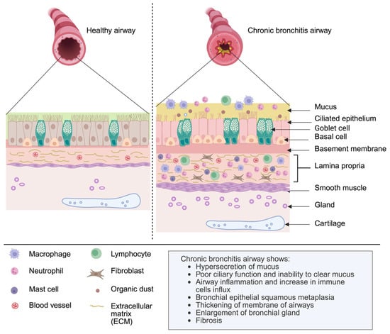
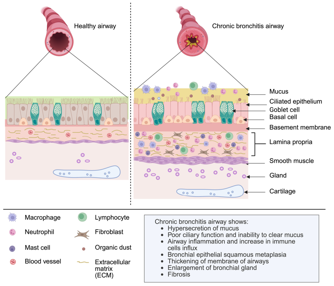
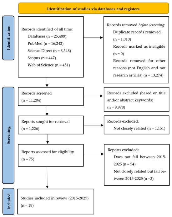
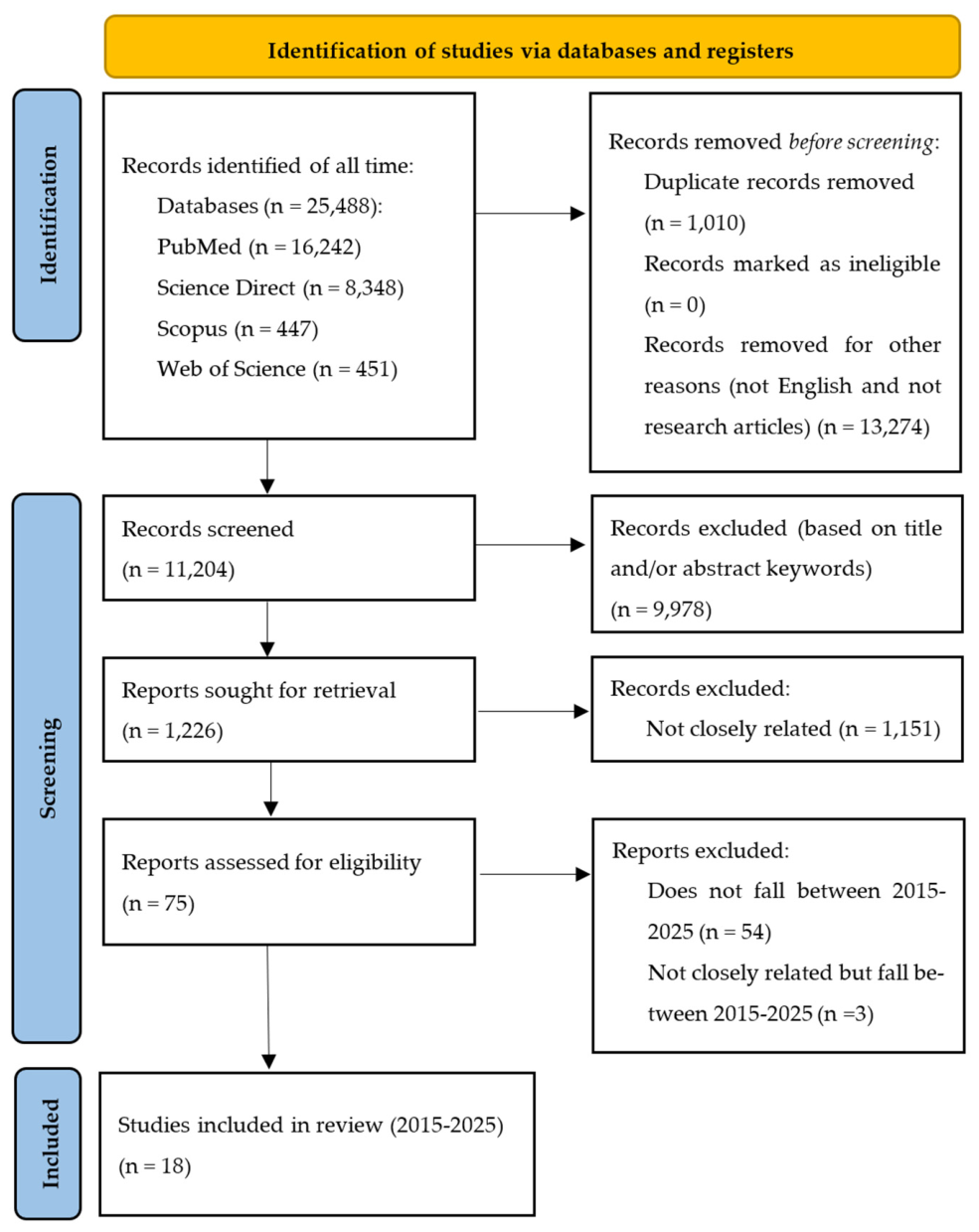
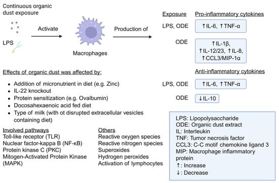
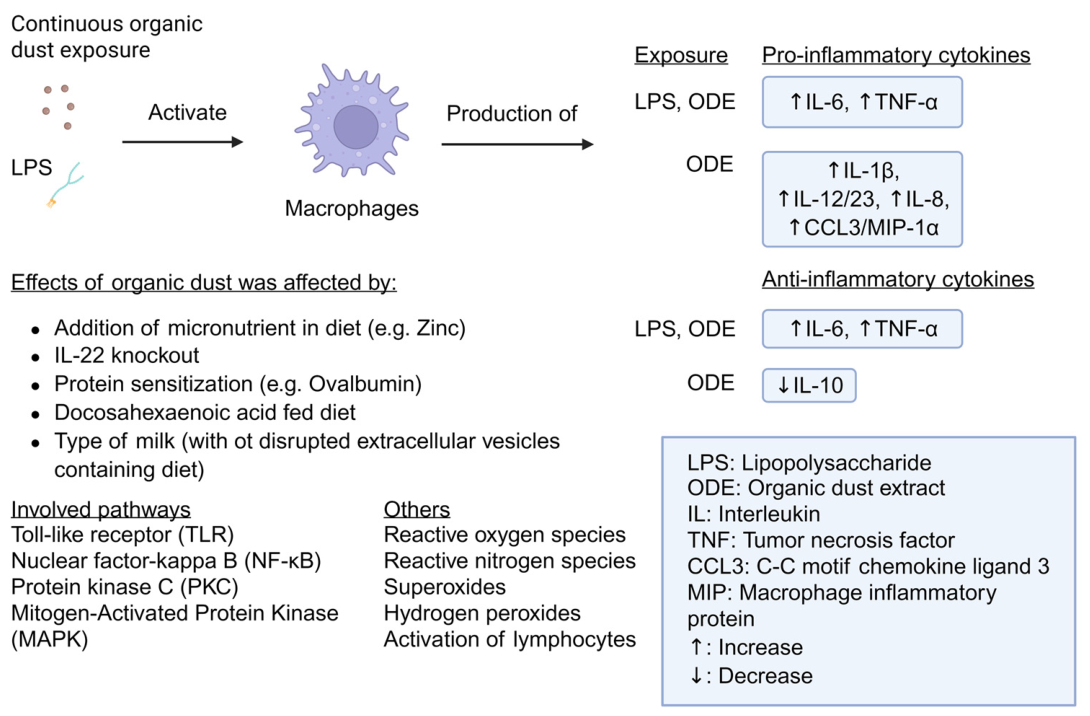

Disclaimer/Publisher’s Note: The statements, opinions and data contained in all publications are solely those of the individual author(s) and contributor(s) and not of MDPI and/or the editor(s). MDPI and/or the editor(s) disclaim responsibility for any injury to people or property resulting from any ideas, methods, instructions or products referred to in the content. |
© 2026 by the authors. Licensee MDPI, Basel, Switzerland. This article is an open access article distributed under the terms and conditions of the Creative Commons Attribution (CC BY) license.