


Highlights
What are the main findings?
- Biological vulnerability (first hit): Diabetes disrupts estrogen-mediated vascular protection, promoting oxidative stress and endothelial dysfunction in women.
- Clinical disadvantage (Second hit): Sex-specific risk factors and clinical care disparities further increase cardiovascular risk in women with diabetes.
What are the implications of the main findings?
- A double-hit framework highlights the need for sex-specific cardiovascular risk assessment and prevention strategies for people with diabetes.
- Further research is needed to clarify sex-specific responses to GLP-1 receptor agonists and SGLT2 inhibitors and to optimize cardiovascular prevention in women with diabetes.
Abstract
Premenopausal women without diabetes have a well-established cardiovascular advantage over men of the same age, mainly due to the benefits of estrogen, which has vasoprotective, anti-inflammatory, and antioxidant effects on the vascular endothelium. However, women with type 1 or type 2 diabetes have a 40–50% higher relative risk of coronary heart disease and suffer a greater burden of heart failure and cardiovascular mortality compared with diabetic men. The mechanisms involved in the disparity are not well understood and are seldom considered in clinical guidelines or trials, where women are underrepresented. The paradoxical relationship between chronic hyperglycemia and estrogen signaling plays a critical role, since estradiol, rather than having its usual cardioprotective effect, instead leads to oxidative stress and endothelial dysfunction. This occurs through eNOS uncoupling, activation of the AGE-RAGE axis, and a shift towards pro-inflammatory signaling via estrogen receptors. Sex-related differences in body composition and the distribution of fat tissue also make a significant contribution to the variations in cardiovascular risk. Modifiable lifestyle factors, especially physical activity and skeletal muscle mass, are important targets for intervention, with evidence showing that their benefits on endothelial function, body composition, and systemic inflammation are particularly noticeable in females. In addition, the sex-specific cardiovascular responses to treatments such as glucagon-like peptide-1 receptor agonists (GLP-1RAs) and sodium-glucose cotransporter-2 (SGLT2) inhibitors should be investigated more closely. This review summarizes the current evidence, identifies critical knowledge gaps, and proposes a framework for sex-informed cardiovascular prevention in diabetes.
1. Introduction
Cardiovascular diseases remain the leading cause of morbidity and mortality worldwide [1]. Despite this global burden, one consistent observation in cardiovascular epidemiology is that premenopausal women exhibit a cardiovascular advantage, where they are protected against coronary heart disease and stroke relative to age-matched men. In the general population, this female advantage delays the onset of clinically significant cardiovascular disease by approximately a decade, which may contribute meaningfully to the longer life expectancy observed in women across high-income countries. The biological basis of this protection is multifactorial, involving favorable differences in lipid profiles, blood pressure regulation, endothelial function, and systemic inflammation, which are modulated at least in part by the actions of female sex hormones, principally estradiol [2].
However, the onset of diabetes fundamentally disrupts this picture. Since the landmark Framingham Heart Study observations nearly five decades ago, it has been recognized that the usual female cardiovascular protection is erased in the presence of diabetes [3]. Subsequent large-scale meta-analyses have confirmed and quantified this reversal: women with type 2 diabetes bear a 44% greater excess relative risk of coronary heart disease and a 27% greater excess risk of stroke compared with diabetic men [4,5]. The excess burden extends to heart failure, peripheral arterial disease, and cardiovascular mortality. In type 1 diabetes, the disparity is equally pronounced: women face roughly 40% greater all-cause excess mortality and twice the excess risk of fatal and nonfatal vascular events compared with men with the same disease [6]. Critically, these figures represent excess relative risk above the non-diabetic population. While men with diabetes carry a higher absolute burden of disease, women experience a far steeper departure from the cardiovascular safety they would otherwise have. Why diabetes so selectively and completely strips women of their cardiovascular advantage remains an important and underexplored question in cardiometabolic medicine. Biological, hormonal, compositional, behavioral, and clinical factors all appear to contribute, yet they are rarely examined together. This knowledge gap is reinforced by the lack of sex-specific analyses in cardiovascular outcome trials, where women have historically been underrepresented [7]. This long-term underrepresentation has perpetuated a cycle in which treatment guidelines remain sex-neutral despite a sex-specific biological problem.
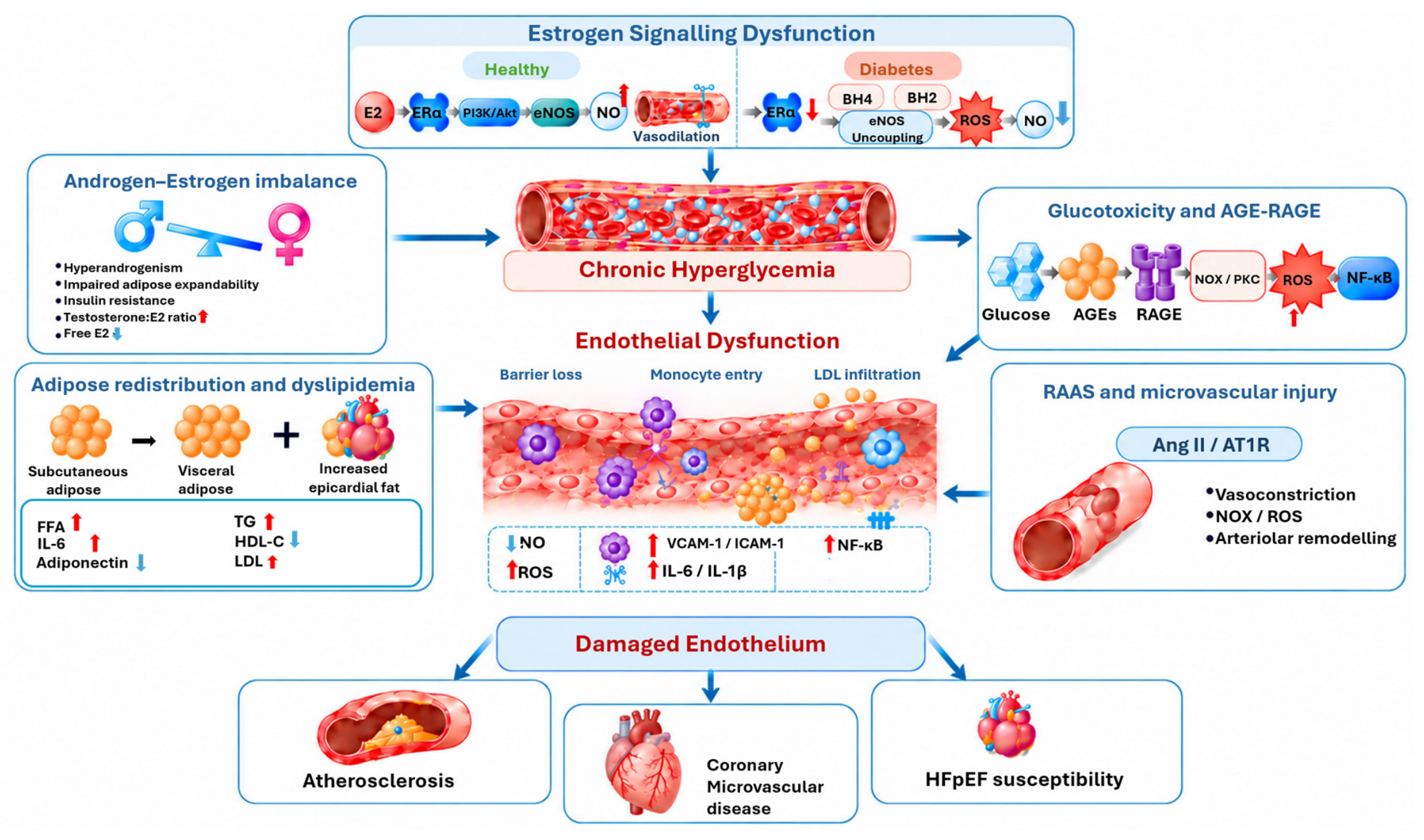
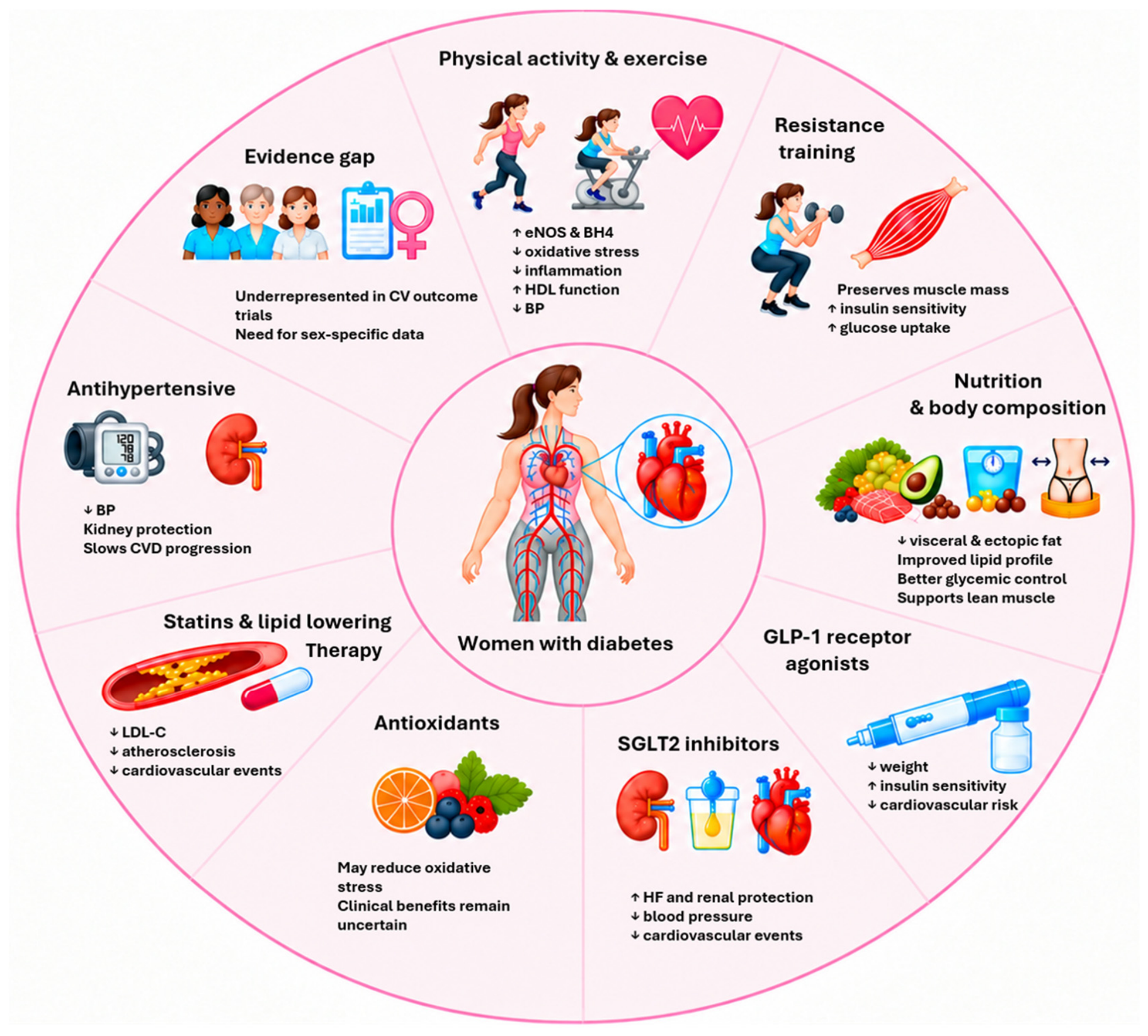
This review aims to dismantle the sex-neutral approach to diabetic cardiovascular care by examining the unique vulnerability of women through a “double-hit” framework. Hit 1 represents the biological inversion of female cardiovascular protection that is driven by hyperglycemia-induced receptor reprogramming, visceral lipotoxicity, and amplified oxidative stress, summarized in Figure 1. Hit 2 represents the clinical compounding of this biological risk through systemic undertreatment, underrepresentation in clinical trials, and clinical inertia. By mapping both the molecular and clinical dimensions of this disparity, we propose targeted, sex-informed interventional strategies to mitigate female cardiovascular risk (Figure 2).
Figure 1.
Proposed pathways linking diabetes, estrogen signaling dysfunction, and metabolic disturbances to cardiovascular complications in women. Connecting arrows indicate proposed relationships among the depicted processes and downstream outcomes, whereas upward arrows (↑) and downward arrows (↓) indicate increased and decreased levels, respectively.
Figure 2.
Lifestyle and pharmacological strategies for cardiovascular risk reduction in women with diabetes. Upward (↑) and downward (↓) arrows indicate increased and decreased levels, activity, or risk, respectively.
2. The Normal Basis of Female Cardiovascular Protection
2.1. Estrogen and Vascular Biology
The cardioprotective effects of estrogen operate through multiple interacting pathways in the vascular endothelium. Estradiol (E2), the principal biologically active estrogen in premenopausal women, exerts both rapid non-genomic effects mediated through membrane-bound estrogen receptors (primarily ERα, ERβ) and G-protein-coupled receptor 30 (GPR30), and slower, sustained genomic effects through nuclear receptor activation [2,8]. Endothelial nitric oxide (NO) bioavailability is the primary molecular mediator in this defense. When bound to its ligand, ERα triggers the PI3K/Akt cascade, phosphorylating and activating endothelial nitric oxide synthase (eNOS), promoting the continuous synthesis of NO [9,10]. NO mediates endothelium-dependent vasodilation, suppresses vascular smooth muscle cell (VSMC) proliferation, and inhibits both platelet aggregation and leukocyte adhesion to the vessel wall. This continuous supply of NO results in a vascular phenotype that is inherently anti-atherogenic.
Beyond stimulating NO generation, estrogen also functions in the protection of NO from degradation. With its antioxidant properties, Estradiol reduces the production of reactive oxygen species (ROS) in endothelial cells, partly by suppressing NADPH oxidase (NOX) activity and limiting the production of superoxide [8,11]. Thus, increased NO production combined with the suppression of ROS production in the endothelium favors vasoprotection. Estrogen also modulates the renin–angiotensin–aldosterone system (RAAS) to favor vasoprotection. The suppression of the RAAS system occurs through the downregulation of the angiotensin II type I receptor (AT1R) and the inhibition of angiotensin-converting enzyme (ACE) activity, therefore favoring vasodilation. Estrogen also protects vasculature from systemic inflammation by suppressing NF-κB activation, reducing circulating levels of C-reactive protein (CRP), interleukin-6 (IL-6), and Tumor Necrosis Factor alpha (TNF-α), and limiting monocyte recruitment to the vascular wall [2]. Estrogen receptor activation in cardiomyocytes and vascular smooth muscle cells further contributes to favorable cardiac remodeling and vascular compliance [8]. Collectively, these mechanisms contribute to a robust, multidimensional cardioprotective environment during the reproductively active years.
2.2. Metabolic Dimensions of Female Cardiovascular Protection
Female cardiovascular protection is not solely related to endothelial effects, but also an estrogen-driven metabolic phenotype that is part of a defense against lipotoxicity and insulin resistance. Due to estrogen, premenopausal women have a more favorable fat distribution characterized by preferential subcutaneous rather than visceral adiposity. This effectively shields the viscera, liver, and cardiovascular system from the accumulation of ectopic fat [12,13]. This contributes highly to women exhibiting higher HDL cholesterol, lower triglycerides, and lower concentrations of small, atherogenic LDL particles. Simultaneously, women demonstrate greater whole-body insulin sensitivity relative to men of equivalent fitness, driven in part by higher skeletal muscle glucose uptake [14]. These systemic metabolic adaptations combined with the vascular defenses maintain a superior profile of endothelium-dependent vasodilation, leading to lower blood pressure in premenopausal women than age-matched men [10].
The reliance of female physiology on these mechanisms may be demonstrated by menopause. As circulating estradiol levels fall, these advantages begin to converge with and eventually fall below male values. This can be viewed as a natural biological experiment that implicates estrogen as the key driver of their preservation throughout the reproductive years [2]. Consequently, the premenopausal metabolic profile is not innate, but actively maintained by an estrogen-dependent state that is pathologically disrupted under diabetic conditions.
3. Hit 1: Biological Mechanisms Erasing Female Cardiovascular Protection
3.1. The Estrogen Paradox in Diabetes
One of the most compelling mechanistic hypotheses rising in this field is that estrogen, normally cardioprotective, may become maladaptive in the presence of diabetes. Studies in premenopausal women with type 1 diabetes suggest that endothelial function, measured by flow-mediated dilation (FMD), actually decreases from the menstrual phase to the late follicular phase (when estradiol concentrations are highest), in direct contrast to the expected FMD increase observed in healthy women across the same hormonal transition [15]. This inversion of the normal estrogen–endothelial relationship represents a fundamental disruption of the female cardiovascular advantage. The underlying mechanism centers on the interaction between chronic hyperglycemia and estrogen receptor signaling. Under physiological conditions, ERα is the primary receptor mediating cardioprotection, coupling to the PI3K/Akt pathway to stimulate NO production [16,17]. In contrast, chronic hyperglycemia and sustained oxidative stress downregulate ERα expression while maladaptively preserving or upregulating ERβ and GPR30 in the diabetic endothelium. As a consequence, E2 signaling is shunted away from its normal PI3K/Akt axis, creating a receptor imbalance. With the ERα-PI3K/Akt pathway compromised, estradiol signaling is actively directed toward these altered receptor populations amidst severe oxidative stress. Concurrently, oxidation of the eNOS cofactor tetrahydrobiopterin (BH4) into the inactive radical BH2 uncouples the eNOS enzyme [18,19]. Consequently, the follicular-phase estradiol surge acts upon this unbalanced receptor profile and uncoupled eNOS system to actively amplify superoxide generation, converting a physiological vasodilatory stimulus into a mechanism of vascular injury [15,20]. This mechanistic inversion may help explain why the female hormonal environment that confers protection in health becomes a driver of harm in diabetes.
It is important to explicitly note the evidentiary weight of this narrative. Clinical evidence for this in vivo inversion is currently sparse, coming primarily from premenopausal women with type 1 diabetes [15]. Whether this paradox operates identically in type 2 diabetes remains to be clinically validated. However, the compounding effects of insulin resistance, visceral adiposity and systemic inflammation inherent to type 2 diabetes establish an oxidative environment that is likely capable of driving parallel receptor disruption. Furthermore, because this paradox relies on cyclic surges of endogenous estradiol acting upon damaged endothelium, the cyclic amplification of oxidative stress is strictly a premenopausal and perimenopausal phenomenon. Following menopause, this cyclic oxidative surging subsides as estradiol levels decline, yet the underlying receptor downregulation and eNOS uncoupling leave the vasculature chronically compromised.
3.2. Androgens, Cardiovascular Health, and Diabetes
The role of androgens and their effect on female cardiovascular health is inconclusive. While some studies argue that increased levels of androgens are associated with CVD in females, others have argued that no such association exists [21,22]. While androgens may not be conclusively linked to CVD, they can be associated with many factors associated with diabetes. Research has found that hyperandrogenism in women has been linked to specific obesity factors, inhibition of adipose hyperplasia, and insulin resistance, indicating a possible link between hyperandrogenism and diabetes [23]. Finally, diabetes and cardiovascular health have been linked to imbalance of androgen/estrogen ratios in women. In a 2023 study, it was found that women with type 1 diabetes and cardiovascular autonomic dysfunction had higher total testosterone/E2 and free testosterone/E2 molar ratios, lower E2, and free E2, compared with those without cardiovascular autonomic dysfunction [24]. These results suggest that increased levels of androgens in females with diabetes could impact cardiovascular health. However, further research is needed to confirm the link between androgens, diabetes, and cardiovascular health in women.
3.3. The AGE-RAGE Axis and Sex-Specific Endothelial Vulnerability
Advanced glycation end-products (AGEs), the irreversible products of non-enzymatic glycation that accumulate in proportion to cumulative glycemic exposure, are central mediators of diabetic vascular injury. Binding AGEs to their receptor for AGEs (RAGE) activates NF-κB, increases NADPH oxidase activity, stimulates the production of proinflammatory cytokines, and directly suppresses eNOS expression and phosphorylation [19,25]. This combination of reduced nitric oxide (NO) bioavailability and increased oxidative stress contributes to endothelial dysfunction, accelerates atherogenesis, and increases vascular stiffness. Emerging preclinical and clinical data suggest that female endothelial cells may be more sensitive to hyperglycemia-induced mitochondrial dysfunction than male cells, exhibiting more pronounced impairments in vasodilation, angiogenesis, and mitochondrial activity under diabetic conditions [26]. In a healthy state, estrogen maintains the endothelial mitochondrial networks through the upregulation of peroxisome proliferator-activated receptor-gamma coactivator-1 alpha (PGC-1α), which is the key regulator of mitochondrial biogenesis and antioxidant defense [27]. In the diabetic environment, ERα downregulation and intracellular glucose toxicity suppress PGC-1α expression, leaving female endothelial cells unable to clear damaged mitochondria or generate healthy replacements as effectively. This leads to electron leak and ROS generation that can overwhelm the antioxidant defenses [28]. Although these findings remain incompletely understood and require more in-depth scientific study, they suggest that the female endothelium may depend more heavily on estrogen-mediated mitochondrial homeostasis that, when disrupted by the diabetic environment, leaves female cells disproportionately exposed to oxidative insult.
3.4. Oxidative Stress and Inflammation: A Female-Amplified Loop
Chronic hyperglycemia generates oxidative stress through multiple converging pathways: excess glucose flux through the polyol pathway, increased protein kinase C (PKC) activation, mitochondrial electron transport chain overload, and AGE-RAGE-mediated NOX activation [19]. In women with diabetes, these pathways intersect with the disrupted estrogen receptor signaling described above to create an amplified oxidative and inflammatory state [29]. A key mechanism underlying this amplification is the loss of estrogen-mediated transrepression of inflammatory signaling. Under physiological conditions, estrogen receptors with their ligands exert anti-inflammatory effects through the upregulation of IκBα (an NF-B inhibitor) and binding to the p65 subunit of the NF-B complex. This interaction blocks p65 from binding to the DNA promoter of inflammatory genes [30]. In the hyperglycemic state of diabetes, AGE accumulation and ROS production, this inhibitory crosstalk is dismantled. IκBα is heavily degraded, and with suppressed estrogen signaling, it is not replaced, resulting in sustained NF-κB activation. Consequently, NF-κB promotes the expression of key inflammatory mediators, including vascular cell adhesion molecule-1 (VCAM-1), intercellular adhesion molecule-1 (ICAM-1), and monocyte chemoattractant protein-1 (MCP-1), thereby exacerbating endothelial inflammation and vascular dysfunction [19].
Systemic markers of inflammation including high-sensitivity CRP, IL-6, and fibrinogen are disproportionately elevated in women with diabetes compared with diabetic men, and the relationship between inflammatory burden and cardiovascular outcomes appears steeper in women [29]. Ultimately, the diabetic environment appears to convert what is normally an anti-inflammatory signaling axis into a driver of vascular NF-κB activation, enhanced expression of inflammatory mediators, and monocyte adhesion.
Although men with diabetes also experience AGE-RAGE-induced NF-B activation, their baseline vascular homeostasis is less dependent on estrogen-mediated transrepression. In contrast, women with diabetes experience both the direct pro-inflammatory effects of chronic hyperglycemia and a collapse of their primary endothelial defense mechanism mediated by ER signaling, which may contribute to the loss of the IκBα “shield” and a steeper escalation of inflammatory markers and a sharper increase in cardiovascular risk.
3.5. Dyslipidemia: A Greater Deterioration in Women
The transition from normoglycemia to overt diabetes is associated with a more profound and destructive deterioration of the lipid profile in women than in men [4,12]. Because premenopausal women preferentially utilize subcutaneous fat storage for excess lipids, women typically develop clinically apparent diabetes at a higher BMI than men, and fat eventually is redirected to visceral depots. This metabolic transition results in greater diabetes-associated reductions in HDL cholesterol, greater increases in triglycerides, and a more pronounced shift toward small dense LDL particles—changes that track more strongly with cardiovascular events in women than in age-matched men [12,31]. The mechanisms are related to the interaction between impaired insulin signaling, adipose tissue dysfunction, and hepatic lipoprotein metabolism; the preferential subcutaneous fat distribution of premenopausal women appears to mask metabolic deterioration during early adiposity expansion, but when visceral and ectopic fat eventually accumulate in diabetes, the resulting dyslipidemia is more and atherogenic per unit of adiposity than in men [12,13].
3.6. Blood Pressure, RAAS, and Microvascular Injury
Hypertension is more strongly associated with cardiovascular outcomes in women with diabetes than in men [29], making it a sex-specific multiplier driving their cardiovascular morbidity and mortality. In healthy vasculature, estrogen normally modulates RAAS to favor angiotensin-(1–7) and bradykinin-mediated vasodilation and suppresses vasoconstrictive AT1R signaling [8]. In the diabetic environment, this hormonal modulation is disrupted. The resulting RAAS imbalance drives endothelial inflammation, sustained oxidative stress, and rapid arteriolar remodeling [8,19]. The female microvasculature is more dependent on estrogen-mediated vasodilatory tone; therefore, the unchecked RAAS activation leaves female arteriolar beds more vulnerable to injury. This susceptibility helps explain why women with diabetes are disproportionately affected by microvascular disease, including ischemic heart disease with non-obstructive coronary arteries (INOCA), a particularly dangerous phenotype that does not match the traditional macrovascular risk that consistently treats male physiology as the baseline, and therefore presents an important clinical blind spot [13,32].
Table 1 summarizes the mechanisms underlying the loss of female cardiovascular protection in women with diabetes.
Table 1.
Potential mechanisms underlying the loss of female cardiovascular protection in women with diabetes.
The mechanisms described above are closely linked to the changes in adipose tissue structure and metabolism. In women with diabetes, increased visceral and ectopic fat is not only a consequence of metabolic dysfunction but also a contributor to endothelial dysfunction, oxidative stress, and inflammation. For example, visceral adipose tissue releases free fatty acids and pro-inflammatory adipokines into the portal circulation, which can enhance AGE-RAGE–mediated NF-κB activation and further impair estrogen receptor signaling that normally helps limit vascular inflammation. Additionally, epicardial fat may also have local lipotoxic and inflammatory effects on the coronary endothelium, adding to the eNOS uncoupling and mitochondrial dysfunction caused by chronic hyperglycemia. Thus, when subcutaneous adipose tissue can no longer adequately store excess energy, the resulting accumulation of visceral and ectopic fat does more than add another cardiovascular risk factor. It can further amplify the molecular pathways that contribute to loss of the female vascular protective effect. For this reason, changes in body composition in women with diabetes should be considered an important part of the mechanisms underlying their increased cardiovascular risk (Figure 1).
4. Body Composition, Fat Distribution, and Sex-Specific Cardiovascular Risk
4.1. Beyond BMI: Why Body Composition Matters Differently in Women
BMI is a notoriously imperfect measure of cardiometabolic risk, and its limitations are particularly pronounced in women. This is highly relevant in the pathogenesis of type 2 diabetes, where body composition heavily dictates metabolic changes. Women generally carry a higher percentage of total body fat than men at the same BMI, concentrated preferentially in subcutaneous depots, a pattern that is metabolically relatively benign compared with visceral adiposity [13,32]. Because of the large storage capacity of subcutaneous fat, women typically develop diabetes at higher BMI values than men, having accumulated more total adipose tissue before reaching the visceral fat thresholds associated with insulin resistance, dyslipidemia, and inflammation [4,12]. This dynamic obscures early cardiometabolic risk: by the time clinical diabetes is diagnosed, women have often already undergone a substantial metabolic change in the redistribution of fat toward visceral and ectopic depots after exhausting subcutaneous storage. As a consequence, the severity of metabolic tissue disruption at the time of diagnosis may be greater in women than in men despite similar or lower BMI [12], as well as the resulting cardiovascular threat.
4.2. Visceral and Ectopic Fat Depots
Once fat begins accumulating viscerally as visceral adipose tissue (VAT) and ectopic depots, a distinct and more destructive secretory profile is generated. Compared with subcutaneous fat, VAT releases higher concentrations of pro-inflammatory adipokines (including leptin, resistin, and IL-6), free fatty acids, and ROS into the portal circulation. This directly exposes the liver and cardiovascular system to lipotoxic and inflammatory signals [13,32]. In women with diabetes, accumulation of VAT is associated with disproportionate reductions in adiponectin, which is a cardioprotective adipokine that normally promotes endothelial NO production, reduces vascular inflammation, and improves insulin sensitivity [13]. The accumulation of VAT in women is also associated with amplified systemic inflammatory responses compared with similarly obese men [13,32].
Ectopic fat deposition is the accumulation of fat in pericardial, hepatic, intramyocardial, and skeletal muscle compartments, which represents an additional and underappreciated dimension of sex-specific cardiovascular risk in diabetes. This can be considered even more dangerous than VAT, particularly epicardial adipose tissue (EAT). EAT is in direct anatomical contact with the coronary arteries and myocardium, lacking separation by fascia. As EAT expands in diabetic women, it exerts toxicity on the heart through a paracrine mechanism, which is associated with coronary microvascular dysfunction, myocardial steatosis, and adverse left ventricular stiffening. In turn, this contributes to the disproportionate burden of heart failure with preserved ejection fraction (HFpEF) observed in women with diabetes and obesity [13,29].
4.3. Skeletal Muscle Mass as a Cardioprotective Factor
Skeletal muscle is the primary site of insulin-stimulated glucose disposal. Consequently, muscle mass is a critical determinant of whole-body insulin sensitivity and metabolic health [14]. In women with diabetes, low skeletal muscle mass (sarcopenia or relative muscle depletion) is common, and when compounded with the effects of aging, insulin resistance and obesity, a phenotype that is increasingly recognized as “sarcopenic obesity” Occurs. This combination of low muscle mass and high visceral adiposity in women with diabetes is associated with higher cardiovascular risk. Without skeletal muscle to uptake glucose, these women suffer from elevated circulating free fatty acids and reduced cardiorespiratory fitness that is independent of total body fat [14,33]. Physical fitness, a functional index of skeletal muscle and cardiorespiratory capacity, is among the strongest predictors of cardiovascular mortality in both T1D and T2D, with cardiorespiratory fitness showing an inverse dose–response relationship with cardiovascular events [34,35]. Maintaining muscle mass and aerobic fitness therefore represents not only a general health goal, but also an absolute cardiovascular imperative for women with diabetes.
5. Hit 2: Clinical Disparities and Behavioral Compounding Factors
5.1. Disparities in Cardiovascular Risk Factor Management
The excess biological risk experienced by women with diabetes is further compounded by systematic disparities in the clinical management of cardiovascular risk factors. Multiple studies consistently suggested that women with diabetes are significantly less likely to be prescribed lipid-lowering therapies and RAAS inhibitors, less likely to achieve recommended LDL cholesterol or blood pressure targets, and less likely to receive antiplatelet agents after cardiovascular events compared with diabetic men [31,36,37,38]. These disparities persist even after adjustment for relevant clinical variables and appear to reflect both provider-level underestimation of cardiovascular risk in women and patient-level undertreatment [36,37]. Notably, one study of over 10,000 individuals with type 2 diabetes found that women were significantly less likely to be prescribed statins and RAAS inhibitors, and these disparities were independently associated with their higher excess risks for cardiovascular and renal complications [38]. These disparities solidify the “second hit” of the female cardiovascular phenotype, as medical providers underestimate diabetic female cardiovascular risk and their previously discussed biological vulnerability. The combination of biological vulnerability and medical undertreatment can lead to adverse outcomes if left unaddressed.
5.2. Underrepresentation in Cardiovascular Outcome Trials
The medical undertreatment of women has roots in their historical exclusion from cardiovascular outcome trials (CVOTs). Women have historically comprised only 28–36% of participants in CVOTs of diabetes medications [7]. This underrepresentation constitutes a fundamental, longstanding limitation in cardiovascular research. It constrains the detection of sex-specific differences in treatment efficacy, safety, and mechanism. Trials that are predominantly male limit the identification of female subgroups that may experience disproportionate benefit or harm, and weaken the sex-stratified evidence required to inform clinical guidelines [7,29]. The consistent sex neutrality of current cardiovascular risk guidelines for diabetes reflects, in part, the sex-neutral composition of the trials on which they rest, a circularity that perpetuates undertreatment of women.
5.3. Female-Specific Life Events as Additional Risk Layers
Beyond traditional cardiovascular risk factors, several female-specific reproductive and endocrine events further influence cardiovascular risk in women with diabetes. Pregnancy involves profound changes to the cardiovascular system and metabolism. These changes include, but are not limited to, increased insulin resistance, hypercoagulability, and elevated lipid levels, which may have long-lasting health impacts [39]. A 2019 systematic review and meta-analysis found that a history of adverse pregnancy outcomes (APO), specifically preeclampsia, gestational hypertension, gestational diabetes mellitus, placental abruption, preterm birth, and stillbirth elevates a woman’s risk of subsequent CVD and cerebrovascular morbidity and mortality [40]. Epidemiological evidence reveals that hypertensive APOs, such as preeclampsia, may cause persistent vascular changes, and statistics have suggested that women with T1D have a higher risk of preeclampsia compared to those without the condition (10–17% vs. 3–5%) [39]. This compounding of cardiovascular risk factors is particularly concerning, as studies link preeclampsia to subclinical atherosclerosis [41].
Gestational diabetes (GDM) is now recognized as a powerful sex-specific cardiovascular risk and prognostic marker: a recent meta-analysis involving over 5.3 million women suggested a twofold higher risk of CVD in women with previous GDM, emerging within the first decade post-pregnancy, and this risk remained elevated even when women who subsequently developed type 2 diabetes were excluded [42]. The combination of GDM history and subsequent type 2 diabetes development accelerates atherosclerosis progression and coronary microvascular dysfunction.
Furthermore, an increasingly recognized risk layer is the condition traditionally known in cardiovascular literature as Polycystic Ovary Syndrome (PCOS). Although readers may be encountering the updated term for the first time here, this review utilizes the recently proposed nomenclature that is Polyendocrine Metabolic Ovarian Syndrome (PMOS), to better represent its widespread endocrine, metabolic and systemic nature. This is a disorder that affects 5–15% of women of reproductive age and confers an independently elevated cardiovascular risk [43]. PMOS intersects additively with diabetic cardiovascular pathology, as it is strongly associated with increased insulin resistance and type 2 diabetes prior to the onset of menopause [44]. Finally, menopause represents a critical acceleration point: the abrupt decline in circulating estradiol triggers rapid deterioration of endothelial function, accelerated visceral fat redistribution, worsening dyslipidemia, and rising blood pressure. These changes in women with pre-existing diabetes compound an already adversely remodeled cardiovascular risk profile [2,45].
5.4. Psychosocial and Behavioral Factors
Navigating a chronic metabolic disease involves a substantial psychosocial burden that is often underestimated in those without firsthand experience. This burden can act as an independent cardiovascular risk multiplier. Depression and diabetes distress are significantly more prevalent in female patients, and both are independently associated with adverse cardiovascular outcomes. These outcomes are not driven solely through behavioral disengagement, but are mediated through direct physiological pathways that include heightened hypothalamic–pituitary–adrenal (HPA) axis activation, sympathetic nervous system dysregulation, poor medication adherence, and reduced engagement with cardiovascular-protective behaviors including physical activity [29]. Disordered eating and insulin omission, a behavior used to induce weight loss through glycosuria, are female-predominant phenomena in patients with type 1 diabetes, particularly in adolescence and early adulthood, and are associated with chronic hyperglycemic exposure, accelerated complications, and elevated cardiovascular mortality [46]. For both type 1 and type 2 diabetic patients, restrictive dieting behaviors, emotional eating, and weight cycling are practices more prevalent in women, which may impede their adherence to cardioprotective dietary and nutritional guidelines, potentially leading to exacerbated glycemic fluctuation and metabolic dysregulation [47].
6. Intervention: Restoring or Preserving Female Cardiovascular Protection
Lifestyle modifications and pharmacological interventions are critical for restoring cardiovascular protection in women. Regular physical activity significantly improves vascular and metabolic health in individuals with diabetes [34]. Pharmacological therapies, including GLP-1 receptor agonists, SGLT2 inhibitors, lipid-lowering agents, and antihypertensive medications, have demonstrated efficacy in reducing cardiovascular risk (Figure 2) [48,49]. Table 2 summarizes clinical evidence supporting cardiovascular risk-reduction interventions in type 1 and type 2 diabetes and highlights gaps in evidence related to women identified in clinical trials.
Table 2.
Summary of Interventions to Restore or Preserve Female Cardiovascular Protection in Patients with Diabetes.
6.1. Physical Activity and Exercise
Regular physical activity is among the most potent and broadly accessible interventions for reducing cardiovascular risk in women with diabetes, operating through mechanisms that directly address the key drivers of lost female protection. Therefore, regular physical exercise should be increasingly regarded as an effective targeted medical intervention, rather than general lifestyle advice. Aerobic exercise actively restores endothelial NO bioavailability by upregulating eNOS expression and restoring BH4 levels, effectively reversing the eNOS uncoupling that underlies the previously discussed estrogen paradox in diabetes [34,53]. It reduces circulating markers of oxidative stress and systemic inflammation, improves HDL functionality, lowers blood pressure, and favorably shifts fat distribution away from visceral and ectopic depots [34,35]. In type 1 diabetes specifically, high-intensity interval training (HIIT) produces superior improvements in endothelial function and cardiorespiratory fitness compared with moderate-intensity continuous training, with FMD improvements demonstrable after 8 weeks [53]. In both T1D and T2D, aerobic training rapidly increases cardiorespiratory fitness, reduces HbA1c, improves lipid profiles, and measurably improves endothelial function [34].
Resistance training can act as a necessary complement to aerobic exercise by preserving and building skeletal muscle mass, a “metabolic sink” in the human body. Skeletal muscle is the body’s primary disposal site for circulating energy. Skeletal muscle absorbs the majority of post-meal glucose, burns fatty acids for resting energy, and protects organs from metabolic overload. Glucose uptake by skeletal muscle can occur independent of insulin, allowing the muscles to clear blood sugar, even in a state of insulin resistance [58,59]. Resistance training, which focuses on weight training to build muscle, can improve insulin-stimulated glucose disposal, reduce free fatty acid flux, and support cardiorespiratory fitness indirectly through improvements in lean body mass [33,35]. A 2025 systematic review and meta-analysis of 25 randomized controlled trials in T1D confirmed that exercise training reduces BMI, fasting glucose, and HbA1c and increases Maximal oxygen uptake (VO2max), with age, exercise mode, and intervention duration as key moderators of effect [35]. Given the particular importance of skeletal muscle mass for cardiovascular function in women, a combined aerobic and resistance training approach offers synergistic benefits.
Despite the clear evidence for these benefits, physical activity levels are substantially lower in women with diabetes than in men, and exercise recommendations have historically been based on predominantly male populations [73]. Societal pressure, widespread and constantly changing beauty standards, and social media often lead women to desire thinness, even at the expense of their health [74]. Sex-stratified evidence on optimal exercise modality, intensity, and duration for cardiovascular protection in women with diabetes remains limited, particularly for type 1 diabetes, representing an urgent research gap [73]. Educating the public on the importance of exercise and the evidence-based differences it can make on cardiovascular health and longevity remains critical, especially as societal pressures shift ideal body types in and out of trends.
6.2. Body Composition-Targeted Interventions
Therapeutic paradigms must shift and change focus from total body weight to body composition remodeling. Targeted reduction in visceral and ectopic fat represents a more precise cardiovascular intervention goal than weight loss per se [13,32]. With VAT contributing to female-specific risk, lifestyle interventions that reduce VAT are important to relieve circulation from lipotoxic stress. Combining caloric modification with physical activity consistently reduce VAT and improve cardiovascular risk markers, and these benefits are disproportionate relative to total weight lost, reflecting the metabolic sensitivity of visceral fat to caloric restriction and physical activity [13]. The preservation of skeletal muscle mass during weight loss is a critical and often overlooked objective in women with diabetes. Conventional caloric restriction without concurrent resistance exercise results in significant lean mass loss alongside fat loss, potentially undermining the insulin-sensitizing and cardiovascular benefits of the intervention [33,35]. Resistance training during intentional weight loss programs effectively preserves lean mass, and the combination of adequate dietary protein intake with resistance exercise represents the most evidence-based approach to favorable body composition remodeling in women with diabetes.
6.3. Pharmacological Interventions with Sex-Specific Potential
6.3.1. GLP-1 Receptor Agonists
GLP-1 receptor agonists (GLP-1 RAs) have emerged as major therapeutic advances in the management of cardiovascular risk in type 2 diabetes, and their clinical utilization has soared in recent years. Landmark trials suggest significant reductions in major adverse cardiovascular events with the use of these medications [49,75]. The SOUL trial (2025) demonstrated that oral semaglutide may reduce major adverse cardiovascular events (MACE) by 14% (HR 0.86; 95% CI, 0.77–0.96; p = 0.006) in patients with T2D and established atherosclerotic cardiovascular disease or chronic kidney disease, over a median follow-up of approximately 49.5 months, primarily driven by reductions in non-fatal myocardial infarction [75,76]. Beyond glycemic control, GLP-1 RAs exert anti-inflammatory and anti-atherogenic effects on the vascular endothelium, reduce visceral fat mass and total body weight, lower blood pressure, and improve endothelial function [49]. The preferential reduction in visceral adiposity associated with GLP-1 RA therapy is particularly relevant for women with diabetes, given the centrality of visceral fat expansion to the loss of female cardiovascular protection [13,32]. Women are disproportionately prescribed these therapies for both glycemic control and weight management; however, they remain systematically underrepresented in CVOTs that define their efficacy. Sex-stratified analyses from large GLP-1 RA trials remain incompletely reported, representing a pressing evidence gap [7]. While the mechanism aligns favorably with female-specific visceral fat patterns, the lack of sex-stratified outcome data prevents definitive conclusions regarding superior efficacy in women.
6.3.2. SGLT2 Inhibitors
SGLT2 inhibitors are known to reduce cardiovascular mortality and heart failure hospitalizations in patients with type 2 diabetes and established cardiovascular disease, with benefits extending to patients without diabetes in the setting of heart failure [49,75]. Their cardiovascular effects operate through multiple non-glycemic mechanisms including natriuresis-mediated blood pressure reduction, hematocrit elevation, reduction in cardiac preload and afterload, and potentially direct cardiomyocyte effects on mitochondrial function and energetics [49]. Sex-stratified data from major SGLT2 inhibitor trials are inconsistent, with some analyses suggesting attenuated benefits in women, an observation that requires mechanistic investigation [7]. As new pharmacological interventions continue to be developed, it is important to also improve knowledge on medications that evidence has already proven to be effective, to maximize their benefit in all populations.
6.3.3. Statins and Lipid-Lowering Therapy
Statins reduce cardiovascular events in women with diabetes at least as effectively as in men on a relative basis, yet women with diabetes remain systematically undertreated with lipid-lowering therapy and are less likely to reach LDL targets [31,37,38]. Women remain under-screened and undertreated for dyslipidemia compared with men, and also comprise a small proportion of clinical trial participants for lipid-lowering agents [31]. Given that dyslipidemia, particularly the HDL reduction and triglyceride elevation associated with diabetic metabolic dysregulation, is a particularly strong cardiovascular risk driver in women, aggressive lipid management is arguably more urgent in diabetic women than current practice reflects [31,37]. Yet, women continue to experience treatment disparities.
6.3.4. Antihypertensive Therapy and RAAS Blockade
Blood pressure control reduces cardiovascular events in women with diabetes, and RAAS blockade with ACE inhibitors or angiotensin receptor blockers appears to confer vascular endothelial benefits beyond blood pressure reduction, including attenuation of oxidative stress, restoration of bradykinin-mediated vasodilation, and reduction in renal and microvascular injury, which may be particularly relevant given the pronounced microvascular susceptibility of women with diabetes [8,38].
6.3.5. Antioxidants
In diabetes, insulin resistance and hyperglycemia increase reactive oxygen species (ROS) through multiple pathways—including NADPH oxidase activation, mitochondrial electron transport chain overload, and advanced glycation end-product formation—overwhelming endogenous antioxidant defenses and elevating cardiovascular disease risk. Antioxidants, including vitamins C and E, selenium, glutathione, coenzyme Q10, carotenoids, and polyphenols, may help mitigate oxidative stress; however, their effects on clinically meaningful cardiovascular outcomes remain uncertain, optimal doses remain uncertain, and long-term clinical benefits have not been consistently demonstrated. Preclinical and limited clinical evidence suggests that N-acetylcysteine (NAC) and α-lipoic acid may improve endothelial function in diabetes, potentially by reducing oxidative stress, inflammation, and enhancing NO bioavailability. Nevertheless, antioxidant vitamin supplementation has not consistently reduced cardiovascular complications in individuals with diabetes [67,77,78]. Declining estrogen after menopause is also associated with increased oxidative stress, oxidized LDL, reduced NO bioavailability, endothelial dysfunction, and increased insulin resistance, potentially contributing to heightened cardiovascular risk in postmenopausal women [79]. Future research should account for sex and menopausal status when evaluating antioxidant therapies and supplementation, but these variables alone may not fully explain the heterogeneity observed in clinical trial outcomes [80].
6.3.6. Hormonal Considerations
The role of hormone therapy (HT) in cardiovascular risk modification in perimenopausal and postmenopausal women with diabetes is complex and incompletely resolved [45]. Observational data from the general population suggest a cardiovascular benefit from timely initiation of HT in early menopause (the “timing hypothesis”), but the presence of diabetes significantly modifies this picture, as estrogen’s cardiovascular effects are fundamentally altered in the hyperglycemic environment [15,45]. A systematic review and meta-analysis of HT in women with T1D and T2D found benefits for glycemic control, but cardiovascular effects remained uncertain [72]. However, this mechanistic divergence presents a testable clinical hypothesis: the vascular response to HT in diabetic women likely depends on the extent of hyperglycemia-induced receptor reprogramming at the time of initiation. If the estrogen paradox is driven by ERα downregulation and eNOS uncoupling, then early initiation in perimenopausal women with strict glycemic control may preserve the physiological, cardioprotective estrogen signaling axis. Conversely, administering HT in the setting of chronic, uncontrolled hyperglycemia and established oxidative stress would likely redirect exogenous estrogen into maladaptive, superoxide-generating pathways, exacerbating endothelial injury. Current evidence does not support routine use of HT for primary cardiovascular prevention in women with diabetes, and its use should be individualized based on menopausal symptom burden, vascular risk profile, and glycemic control status [45,72]. In other words, hormonal interventions should be taken with strict caution.
7. Knowledge Gaps and Future Directions
The profound clinical and biological disparities identified in this review highlight the demand for substantial changes to the current cardiometabolic research agenda. To close the current gaps in female cardiovascular and preventative care, several topics should be addressed in future studies. First, cardiovascular outcome trials in diabetes should require sex-stratified primary and secondary analyses as a prerequisite for publication and regulatory evaluation, as the continued reliance on sex-aggregated outcomes obscures potentially important differences in treatment efficacy and safety [7]. Second, mechanistic studies are needed to clarify the interaction between estrogen receptor signaling and hyperglycemia across the full spectrum of diabetes types, disease duration, and hormonal life stages. Rigorous, reproducible studies are important for future diabetic care and can determine whether the estrogen paradox is reversible with improved glycemic control or whether it reflects longer-term receptor-level reprogramming [15,29]. Third, prospective longitudinal studies should characterize body composition trajectories, particularly visceral fat accumulation, lean mass changes, and ectopic fat deposition, as predictors of cardiovascular events in women with diabetes, essentially tracking the pathology of body composition [12,32]. This way, women’s metabolic risk assessment can move beyond the limitations of standard BMI stratification. Fourth, rather than generalizing exercise physiology from cohorts that are predominantly male, sex-stratified exercise intervention trials in both T1D and T2D are needed to generate the female-specific evidence base currently absent from exercise guidelines [34,73]. Specific exercise modalities can be evaluated and eventually recommended as more than general lifestyle advice, but rather as targeted interventions to preserve skeletal muscle. Fifth, novel sex-specific cardiovascular risk prediction models for diabetes, incorporating body composition, hormonal status, lifetime reproductive and pregnancy history and sex-specific biomarkers beyond traditional risk factors, are needed to replace the current sex-neutral tools that systematically underestimate risk in women [29].
8. Conclusions
Diabetes is a unique cardiovascular risk factor in women. The excess cardiovascular mortality observed in women with diabetes is not an inevitable consequence of the disease, but may be the result of a biological and clinical double-hit. Multiple mechanisms may play important roles: chronic hyperglycemia causes the normally protective effects of estrogen at the vascular endothelium to be reversed; the AGE-RAGE axis together with eNOS uncoupling leads to an oxidative and inflammatory environment that is specific to women; the different patterns of fat distribution according to sex worsen metabolic deterioration in a manner not reflected in BMI; and ongoing disparities in the way diabetes is managed in clinical practice add to the already high biological risk. Overcoming the first “hit” requires targeted interventions, such as resistance training and lipid-lowering therapy that specifically address visceral adiposity and endothelial dysfunction. Overcoming the second “hit” involves a conscious and ongoing commitment to sex-informed cardiovascular research, sex-stratified clinical evidence, and sex-aware clinical practice.
Author Contributions
J.P.G., S.A. and M.V.: Original draft—writing and review and editing. F.D.: Conceptualization, Funding acquisition, Project administration, Supervision, original draft writing—review and editing. All authors have read and agreed to the published version of the manuscript.
Funding
Northeast Ohio Medical University Summer Fellowship (JG/FD).
Institutional Review Board Statement
Not applicable. This study did not involve human participants or human-derived identifiable data.
Informed Consent Statement
Not applicable (there were no human participants).
Data Availability Statement
No new data were created or analyzed in this study. Data sharing is not applicable to this review article.
Acknowledgments
During the preparation of this manuscript/study, the authors used Perplexity Pro for the purposes of modifying the figures. The authors have reviewed and edited the output and take full responsibility for the content of this publication.
Conflicts of Interest
The authors declare no conflicts of interest.
References
- Di Cesare, M.; Perel, P.; Taylor, S.; Kabudula, C.; Bixby, H.; Gaziano, T.A.; McGhie, D.V.; Mwangi, J.; Pervan, B.; Narula, J.; et al. The Heart of the World. Glob. Heart 2024, 19, 11. [Google Scholar] [CrossRef] [Scilit] [PubMed]
- Straub, R.H. The Complex Role of Estrogens in Inflammation. Endocr. Rev. 2007, 28, 521–574. [Google Scholar] [CrossRef] [Scilit] [PubMed]
- Kannel, W.B.; McGee, D.L. Diabetes and Cardiovascular Disease: The Framingham Study. JAMA 1979, 241, 2035. [Google Scholar] [CrossRef] [Scilit] [PubMed]
- Peters, S.A.E.; Huxley, R.R.; Woodward, M. Diabetes as Risk Factor for Incident Coronary Heart Disease in Women Compared with Men: A Systematic Review and Meta-Analysis of 64 Cohorts Including 858,507 Individuals and 28,203 Coronary Events. Diabetologia 2014, 57, 1542–1551. [Google Scholar] [CrossRef] [Scilit] [PubMed]
- Peters, S.A.E.; Huxley, R.R.; Woodward, M. Diabetes as a Risk Factor for Stroke in Women Compared with Men: A Systematic Review and Meta-Analysis of 64 Cohorts, Including 775 385 Individuals and 12 539 Strokes. Lancet 2014, 383, 1973–1980. [Google Scholar] [CrossRef] [Scilit] [PubMed]
- Huxley, R.R.; Peters, S.A.E.; Mishra, G.D.; Woodward, M. Risk of All-Cause Mortality and Vascular Events in Women versus Men with Type 1 Diabetes: A Systematic Review and Meta-Analysis. Lancet Diabetes Endocrinol. 2015, 3, 198–206. [Google Scholar] [CrossRef] [Scilit] [PubMed]
- Clemens, K.K.; Woodward, M.; Neal, B.; Zinman, B. Sex Disparities in Cardiovascular Outcome Trials of Populations with Diabetes: A Systematic Review and Meta-Analysis. Diabetes Care 2020, 43, 1157–1163. [Google Scholar] [CrossRef] [Scilit] [PubMed]
- Iorga, A.; Cunningham, C.M.; Moazeni, S.; Ruffenach, G.; Umar, S.; Eghbali, M. The Protective Role of Estrogen and Estrogen Receptors in Cardiovascular Disease and the Controversial Use of Estrogen Therapy. Biol. Sex. Differ. 2017, 8, 33. [Google Scholar] [CrossRef] [Scilit] [PubMed]
- Orphanides, G.; Reinberg, D. RNA Polymerase II Elongation through Chromatin. Nature 2000, 407, 471–476. [Google Scholar] [CrossRef] [Scilit] [PubMed]
- Chambliss, K.L.; Shaul, P.W. Estrogen Modulation of Endothelial Nitric Oxide Synthase. Endocr. Rev. 2002, 23, 665–686. [Google Scholar] [CrossRef] [Scilit] [PubMed]
- Wagner, A.H.; Schroeter, M.R.; Hecker, M. 17β—Estradiol Inhibition of NADPH Oxidase Expression in Human Endothelial Cells. FASEB J. 2001, 15, 2121–2130. [Google Scholar] [CrossRef] [Scilit] [PubMed]
- Peters, S.A.E.; Woodward, M. Sex Differences in the Burden and Complications of Diabetes. Curr. Diabetes Rep. 2018, 18, 33. [Google Scholar] [CrossRef] [Scilit] [PubMed]
- Chait, A.; Den Hartigh, L.J. Adipose Tissue Distribution, Inflammation and Its Metabolic Consequences, Including Diabetes and Cardiovascular Disease. Front. Cardiovasc. Med. 2020, 7, 22. [Google Scholar] [CrossRef] [Scilit] [PubMed]
- Mauvais-Jarvis, F. Sex Differences in Metabolic Homeostasis, Diabetes, and Obesity. Biol. Sex Differ. 2015, 6, 14. [Google Scholar] [CrossRef] [Scilit] [PubMed]
- Simon, A.B.; Derella, C.C.; Blackburn, M.; Thomas, J.; Layman, L.C.; Nicholson, M.S.; Waller, J.; Elmarakby, A.; Saad, K.M.; Harris, R.A. Endogenous Estradiol Contributes to Vascular Endothelial Dysfunction in Premenopausal Women with Type 1 Diabetes. Cardiovasc. Diabetol. 2023, 22, 243. [Google Scholar] [CrossRef] [Scilit] [PubMed]
- Sacharidou, A.; Chambliss, K.; Peng, J.; Barrera, J.; Tanigaki, K.; Luby-Phelps, K.; Özdemir, İ.; Khan, S.; Sirsi, S.R.; Kim, S.H.; et al. Endothelial ERα Promotes Glucose Tolerance by Enhancing Endothelial Insulin Transport to Skeletal Muscle. Nat. Commun. 2023, 14, 4989. [Google Scholar] [CrossRef] [Scilit] [PubMed]
- Zhao, Y.; Qian, Y.; Sun, Z.; Shen, X.; Cai, Y.; Li, L.; Wang, Z. Role of PI3K in the Progression and Regression of Atherosclerosis. Front. Pharmacol. 2021, 12, 632378. [Google Scholar] [CrossRef] [Scilit] [PubMed]
- Förstermann, U.; Münzel, T. Endothelial Nitric Oxide Synthase in Vascular Disease: From Marvel to Menace. Circulation 2006, 113, 1708–1714. [Google Scholar] [CrossRef] [Scilit] [PubMed]
- Giacco, F.; Brownlee, M. Oxidative Stress and Diabetic Complications. Circ. Res. 2010, 107, 1058–1070. [Google Scholar] [CrossRef] [Scilit] [PubMed]
- Meza, C.A.; La Favor, J.D.; Kim, D.-H.; Hickner, R.C. Endothelial Dysfunction: Is There a Hyperglycemia-Induced Imbalance of NOX and NOS? Int. J. Mol. Sci. 2019, 20, 3775. [Google Scholar] [CrossRef] [Scilit] [PubMed]
- Armeni, E.; Lambrinoudaki, I. Menopause, Androgens, and Cardiovascular Ageing: A Narrative Review. Ther. Adv. Endocrinol. Metab. 2022, 13, 20420188221129946. [Google Scholar] [CrossRef] [Scilit] [PubMed]
- Zhao, D.; Guallar, E.; Ouyang, P.; Subramanya, V.; Vaidya, D.; Ndumele, C.E.; Lima, J.A.; Allison, M.A.; Shah, S.J.; Bertoni, A.G.; et al. Endogenous Sex Hormones and Incident Cardiovascular Disease in Post-Menopausal Women. J. Am. Coll. Cardiol. 2018, 71, 2555–2566. [Google Scholar] [CrossRef] [Scilit] [PubMed]
- Xu, J.; He, B. The Central Role of Hyperandrogenism in Driving Metabolic Dysfunction in Polycystic Ovary Syndrome: From Pathophysiological Mechanisms to Targeted Therapies. J. Ovarian Res. 2026, 19, 252. [Google Scholar] [CrossRef] [Scilit]
- Nattero-Chávez, L.; Insenser, M.; Quintero Tobar, A.; Fernández-Durán, E.; Dorado Avendaño, B.; Fiers, T.; Kaufman, J.-M.; Luque-Ramírez, M.; Escobar-Morreale, H.F. Sex Differences and Sex Steroids Influence on the Presentation and Severity of Cardiovascular Autonomic Neuropathy of Patients with Type 1 Diabetes. Cardiovasc. Diabetol. 2023, 22, 32. [Google Scholar] [CrossRef] [Scilit] [PubMed]
- Cai, W.; Ramdas, M.; Zhu, L.; Chen, X.; Striker, G.E.; Vlassara, H. Oral Advanced Glycation Endproducts (AGEs) Promote Insulin Resistance and Diabetes by Depleting the Antioxidant Defenses AGE Receptor-1 and Sirtuin 1. Proc. Natl. Acad. Sci. USA 2012, 109, 15888–15893. [Google Scholar] [CrossRef] [Scilit] [PubMed]
- Bhargava, P.; Schnellmann, R.G. Mitochondrial Energetics in the Kidney. Nat. Rev. Nephrol. 2017, 13, 629–646. [Google Scholar] [CrossRef] [Scilit] [PubMed]
- Wenz, T. Mitochondria and PGC-1α in Aging and Age-Associated Diseases. J. Aging Res. 2011, 2011, 810619. [Google Scholar] [CrossRef] [Scilit] [PubMed]
- Jia, G.; Aroor, A.R.; Sowers, J.R. Estrogen and Mitochondria Function in Cardiorenal Metabolic Syndrome. In Progress in Molecular Biology and Translational Science; Elsevier: Amsterdam, The Netherlands, 2014; Volume 127, pp. 229–249. [Google Scholar] [CrossRef] [Scilit] [PubMed]
- Mauvais-Jarvis, F.; Bairey Merz, N.; Barnes, P.J.; Brinton, R.D.; Carrero, J.-J.; DeMeo, D.L.; De Vries, G.J.; Epperson, C.N.; Govindan, R.; Klein, S.L.; et al. Sex and Gender: Modifiers of Health, Disease, and Medicine. Lancet 2020, 396, 565–582, Erratum in Lancet 2020, 396, 668. https://doi.org/10.1016/S0140-6736(20)31827-4. [Google Scholar] [CrossRef] [Scilit] [PubMed]
- Xing, D.; Oparil, S.; Yu, H.; Gong, K.; Feng, W.; Black, J.; Chen, Y.-F.; Nozell, S. Estrogen Modulates NFκB Signaling by Enhancing IκBα Levels and Blocking P65 Binding at the Promoters of Inflammatory Genes via Estrogen Receptor-β. PLoS ONE 2012, 7, e36890. [Google Scholar] [CrossRef] [Scilit] [PubMed]
- Aggarwal, N.R.; Patel, H.N.; Mehta, L.S.; Sanghani, R.M.; Lundberg, G.P.; Lewis, S.J.; Mendelson, M.A.; Wood, M.J.; Volgman, A.S.; Mieres, J.H. Sex Differences in Ischemic Heart Disease: Advances, Obstacles, and Next Steps. Circ. Cardiovasc. Qual. Outcomes 2018, 11, e004437. [Google Scholar] [CrossRef] [Scilit] [PubMed]
- Piché, M.-E.; Tchernof, A.; Després, J.-P. Obesity Phenotypes, Diabetes, and Cardiovascular Diseases. Circ. Res. 2020, 126, 1477–1500, Erratum in Circ. Res. 2020, 127, e107. https://doi.org/10.1161/RES.0000000000000421. [Google Scholar] [CrossRef] [Scilit] [PubMed]
- Terada, T.; Reed, J.L.; Vidal-Almela, S.; Mistura, M.; Kamiya, K.; Way, K.L. Sex-Specific Associations of Fat Mass and Muscle Mass with Cardiovascular Disease Risk Factors in Adults with Type 2 Diabetes Living with Overweight and Obesity: Secondary Analysis of the Look AHEAD Trial. Cardiovasc. Diabetol. 2022, 21, 40. [Google Scholar] [CrossRef] [Scilit] [PubMed]
- Colberg, S.R.; Sigal, R.J.; Yardley, J.E.; Riddell, M.C.; Dunstan, D.W.; Dempsey, P.C.; Horton, E.S.; Castorino, K.; Tate, D.F. Physical Activity/Exercise and Diabetes: A Position Statement of the American Diabetes Association. Diabetes Care 2016, 39, 2065–2079. [Google Scholar] [CrossRef] [Scilit] [PubMed]
- Khalafi, M.; Dinizadeh, F.; Rosenkranz, S.K.; Symonds, M.E.; Fatolahi, S. The Effects of Exercise Training on Body Composition and Cardiometabolic Risk Factors in Type 1 Diabetes Mellitus: A Systematic Review and Meta-Analysis. Healthcare 2025, 13, 246. [Google Scholar] [CrossRef] [Scilit] [PubMed]
- Norris, C.M.; Yip, C.Y.Y.; Nerenberg, K.A.; Clavel, M.; Pacheco, C.; Foulds, H.J.A.; Hardy, M.; Gonsalves, C.A.; Jaffer, S.; Parry, M.; et al. State of the Science in Women’s Cardiovascular Disease: A Canadian Perspective on the Influence of Sex and Gender. J. Am. Heart Assoc. 2020, 9, e015634. [Google Scholar] [CrossRef] [Scilit] [PubMed]
- de Jong, M.; Peters, S.A.E.; de Ritter, R.; van der Kallen, C.J.H.; Sep, S.J.S.; Woodward, M.; Stehouwer, C.D.A.; Bots, M.L.; Vos, R.C. Sex Disparities in Cardiovascular Risk Factor Assessment and Screening for Diabetes-Related Complications in Individuals with Diabetes: A Systematic Review. Front. Endocrinol. 2021, 12, 617902. [Google Scholar] [CrossRef] [Scilit] [PubMed]
- Ambrož, M.; Geelink, M.; Smits, K.P.J.; De Vries, S.T.; Denig, P. Sex Disparities in Medication Prescribing amongst Patients with Type 2 Diabetes Mellitus Managed in Primary Care. Diabet. Med. 2023, 40, e14987. [Google Scholar] [CrossRef] [Scilit] [PubMed]
- Mesa, A.; Franch-Nadal, J.; Navas, E.; Mauricio, D. Cardiovascular Disease in Women with Type 1 Diabetes: A Narrative Review and Insights from a Population-Based Cohort Analysis. Cardiovasc. Diabetol. 2025, 24, 217. [Google Scholar] [CrossRef] [Scilit] [PubMed]
- Grandi, S.M.; Filion, K.B.; Yoon, S.; Ayele, H.T.; Doyle, C.M.; Hutcheon, J.A.; Smith, G.N.; Gore, G.C.; Ray, J.G.; Nerenberg, K.; et al. Cardiovascular Disease-Related Morbidity and Mortality in Women with a History of Pregnancy Complications: Systematic Review and Meta-Analysis. Circulation 2019, 139, 1069–1079, Erratum in Circulation 2019, 140, e544. https://doi.org/10.1161/CIR.0000000000000718. [Google Scholar] [CrossRef] [Scilit] [PubMed]
- Amor, A.J.; Vinagre, I.; Valverde, M.; Pané, A.; Urquizu, X.; Meler, E.; López, E.; Quirós, C.; Giménez, M.; Codina, L.; et al. Preeclampsia Is Associated with Increased Preclinical Carotid Atherosclerosis in Women with Type 1 Diabetes. J. Clin. Endocrinol. Metab. 2020, 105, 85–95. [Google Scholar] [CrossRef] [Scilit] [PubMed]
- Kramer, C.K.; Campbell, S.; Retnakaran, R. Gestational Diabetes and the Risk of Cardiovascular Disease in Women: A Systematic Review and Meta-Analysis. Diabetologia 2019, 62, 905–914. [Google Scholar] [CrossRef] [Scilit] [PubMed]
- Teede, H.J.; Khomami, M.B.; Morman, R.; Laven, J.S.E.; Joham, A.E.; Costello, M.F.; Patil, M.; Rees, D.A.; Berry, L.; Cree, M.G.; et al. Polyendocrine Metabolic Ovarian Syndrome, the New Name for Polycystic Ovary Syndrome: A Multistep Global Consensus Process. Lancet 2026, 407, 2329–2339. [Google Scholar] [CrossRef] [Scilit] [PubMed]
- Osibogun, O.; Ogunmoroti, O.; Michos, E.D. Polycystic Ovary Syndrome and Cardiometabolic Risk: Opportunities for Cardiovascular Disease Prevention. Trends Cardiovasc. Med. 2020, 30, 399–404. [Google Scholar] [CrossRef] [Scilit] [PubMed]
- Anagnostis, P.; Bitzer, J.; Cano, A.; Ceausu, I.; Chedraui, P.; Durmusoglu, F.; Erkkola, R.; Goulis, D.G.; Hirschberg, A.L.; Kiesel, L.; et al. Menopause Symptom Management in Women with Dyslipidemias: An EMAS Clinical Guide. Maturitas 2020, 135, 82–88. [Google Scholar] [CrossRef] [Scilit] [PubMed]
- Goebel-Fabbri, A.E. Disturbed Eating Behaviors and Eating Disorders in Type 1 Diabetes: Clinical Significance and Treatment Recommendations. Curr. Diabetes Rep. 2009, 9, 133–139. [Google Scholar] [CrossRef] [Scilit] [PubMed]
- Ábel, T.; Gellért, D.; Csobod Csajbókné, É.; Mák, E. Disproportionate Cardiovascular Risk in Women with Type 2 Diabetes: A Narrative Review of Diet, Metabolic Phenotypes, and Gene–Diet–Epigenetic Interactions Across the Life Course. Nutrients 2026, 18, 1217. [Google Scholar] [CrossRef] [Scilit] [PubMed]
- Joshi, P.; Rick, G.; Mesineni, S.; Bohara, S.; Atif, S.; Poonthottam, N.; Azam, A.; Mehmood, Y.; Ali, R.; Raquib, N.; et al. Antihypertensive Therapy in Diabetes Mellitus: Efficacy, Safety, and Metabolic Impacts in Type 1 and Type 2 Diabetes. Cardiovasc. Diabetol.—Endocrinol. Rep. 2026, 12, 4. [Google Scholar] [CrossRef] [Scilit] [PubMed]
- Cosentino, F.; Grant, P.J.; Aboyans, V.; Bailey, C.J.; Ceriello, A.; Delgado, V.; Federici, M.; Filippatos, G.; Grobbee, D.E.; Hansen, T.B.; et al. 2019 ESC Guidelines on Diabetes, Pre-Diabetes, and Cardiovascular Diseases Developed in Collaboration with the EASD. Eur. Heart J. 2020, 41, 255–323, Erratum in Eur. Heart J. 2020, 41, 4317. https://doi.org/10.1093/eurheartj/ehz828. [Google Scholar] [CrossRef] [Scilit] [PubMed]
- Fuchsjäger-Mayrl, G.; Pleiner, J.; Wiesinger, G.F.; Sieder, A.E.; Quittan, M.; Nuhr, M.J.; Francesconi, C.; Seit, H.-P.; Francesconi, M.; Schmetterer, L.; et al. Exercise Training Improves Vascular Endothelial Function in Patients with Type 1 Diabetes. Diabetes Care 2002, 25, 1795–1801. [Google Scholar] [CrossRef] [Scilit] [PubMed]
- Kwon, H.R.; Min, K.W.; Ahn, H.J.; Seok, H.G.; Lee, J.H.; Park, G.S.; Han, K.A. Effects of Aerobic Exercise vs. Resistance Training on Endothelial Function in Women with Type 2 Diabetes Mellitus. Diabetes Metab. J. 2011, 35, 364. [Google Scholar] [CrossRef] [Scilit] [PubMed]
- Swift, D.L.; Earnest, C.P.; Blair, S.N.; Church, T.S. The Effect of Different Doses of Aerobic Exercise Training on Endothelial Function in Postmenopausal Women with Elevated Blood Pressure: Results from the DREW Study. Br. J. Sports Med. 2012, 46, 753–758. [Google Scholar] [CrossRef] [Scilit] [PubMed]
- Boff, W.; Da Silva, A.M.; Farinha, J.B.; Rodrigues-Krause, J.; Reischak-Oliveira, A.; Tschiedel, B.; Puñales, M.; Bertoluci, M.C. Superior Effects of High-Intensity Interval vs. Moderate-Intensity Continuous Training on Endothelial Function and Cardiorespiratory Fitness in Patients with Type 1 Diabetes: A Randomized Controlled Trial. Front. Physiol. 2019, 10, 450. [Google Scholar] [CrossRef] [Scilit] [PubMed]
- Mitranun, W.; Deerochanawong, C.; Tanaka, H.; Suksom, D. Continuous vs Interval Training on Glycemic Control and Macro- and Microvascular Reactivity in Type 2 Diabetic Patients. Scand. Med. Sci. Sports 2014, 24, e69–e76. [Google Scholar] [CrossRef] [Scilit] [PubMed]
- Cassidy, S.; Thoma, C.; Hallsworth, K.; Parikh, J.; Hollingsworth, K.G.; Taylor, R.; Jakovljevic, D.G.; Trenell, M.I. High Intensity Intermittent Exercise Improves Cardiac Structure and Function and Reduces Liver Fat in Patients with Type 2 Diabetes: A Randomised Controlled Trial. Diabetologia 2016, 59, 56–66. [Google Scholar] [CrossRef] [Scilit] [PubMed]
- Maillard, F.; Rousset, S.; Pereira, B.; Traore, A.; De Pradel Del Amaze, P.; Boirie, Y.; Duclos, M.; Boisseau, N. High-Intensity Interval Training Reduces Abdominal Fat Mass in Postmenopausal Women with Type 2 Diabetes. Diabetes Metab. 2016, 42, 433–441. [Google Scholar] [CrossRef] [Scilit] [PubMed]
- Ahmad, A.M.; Mahmoud, A.M.; Serry, Z.H.; Mohamed, M.M.; Abd Elghaffar, H.A. Effects of Low-versus High-Volume High-Intensity Interval Training on Glycemic Control and Quality of Life in Obese Women with Type 2 Diabetes: A Randomized Controlled Trial. J. Exerc. Sci. Fit. 2023, 21, 395–404. [Google Scholar] [CrossRef] [Scilit] [PubMed]
- Favro, F.; Roma, E.; Gobbo, S.; Bullo, V.; Di Blasio, A.; Cugusi, L.; Bergamin, M. The Influence of Resistance Training on Joint Flexibility in Healthy Adults: A Systematic Review, Meta-Analysis, and Meta-Regression. J. Strength Cond. Res. 2025, 39, 386–397. [Google Scholar] [CrossRef] [Scilit] [PubMed]
- Lin, D.; Zhang, L.; Huang, C.; Shao, W. Skeletal Muscle Metabolism in Health and Disease: Mechanisms, Interventions, and Clinical Perspectives. iScience 2026, 29, 115024. [Google Scholar] [CrossRef] [Scilit] [PubMed]
- Xu, Y.; Malek, N.D.; Chang, A.R.; Echouffo-Tcheugui, J.B.; Selvin, E.; Grams, M.E.; Fang, M.; Shin, J.-I. Glucagon-like Peptide-1 Receptor Agonists for Major Cardiovascular and Kidney Outcomes in Type 1 Diabetes. Nat. Med. 2026, 32, 1682–1685. [Google Scholar] [CrossRef] [Scilit] [PubMed]
- McCrimmon, R.J.; Henry, R.R. SGLT Inhibitor Adjunct Therapy in Type 1 Diabetes. Diabetologia 2018, 61, 2126–2133. [Google Scholar] [CrossRef] [Scilit] [PubMed]
- Eldor, R.; Raz, I. American Diabetes Association Indications for Statins in Diabetes. Diabetes Care 2009, 32, S384–S391. [Google Scholar] [CrossRef] [Scilit] [PubMed]
- Elian, V.; Dorita, A.; Stegaru, D.; Vinereanu, D. Treatment of Dyslipidemia in Patients with Type 1 Diabetes Mellitus: A Review of Current Evidence and Knowledge Gaps. Int. J. Mol. Sci. 2025, 26, 8558. [Google Scholar] [CrossRef] [Scilit] [PubMed]
- Lozano-Maneiro, L.; Puente-García, A. Renin-Angiotensin-Aldosterone System Blockade in Diabetic Nephropathy. Present Evidences. J. Clin. Med. 2015, 4, 1908–1937. [Google Scholar] [CrossRef] [Scilit] [PubMed]
- Przezak, A.; Bielka, W.; Pawlik, A. Hypertension and Type 2 Diabetes—The Novel Treatment Possibilities. Int. J. Mol. Sci. 2022, 23, 6500. [Google Scholar] [CrossRef] [Scilit] [PubMed]
- Paquet, S.; Delpech, R.; Sassenou, J.; Kab, S.; Zins, M.; Ringa, V.; Rigal, L. Are Hypertensive Women with Type 2 Diabetes Treated with RAS Inhibitors as Often as Men? A Cross-Sectional Study in France. BMJ Open 2025, 15, e092205. [Google Scholar] [CrossRef] [Scilit] [PubMed]
- Dilworth, L.; Stennett, D.; Facey, A.; Omoruyi, F.; Mohansingh, S.; Omoruyi, F.O. Diabetes and the Associated Complications: The Role of Antioxidants in Diabetes Therapy and Care. Biomed. Pharmacother. 2024, 181, 117641. [Google Scholar] [CrossRef] [Scilit] [PubMed]
- Scaramuzza, A.; Giani, E.; Redaelli, F.; Ungheri, S.; Macedoni, M.; Giudici, V.; Bosetti, A.; Ferrari, M.; Zuccotti, G.V. Alpha-Lipoic Acid and Antioxidant Diet Help to Improve Endothelial Dysfunction in Adolescents with Type 1 Diabetes: A Pilot Trial. J. Diabetes Res. 2015, 2015, 474561. [Google Scholar] [CrossRef] [Scilit] [PubMed]
- Cook, N.R.; Albert, C.M.; Gaziano, J.M.; Zaharris, E.; MacFadyen, J.; Danielson, E.; Buring, J.E.; Manson, J.E. A Randomized Factorial Trial of Vitamins C and E and Beta Carotene in the Secondary Prevention of Cardiovascular Events in Women: Results from the Women’s Antioxidant Cardiovascular Study. Arch. Intern. Med. 2007, 167, 1610–1618. [Google Scholar] [CrossRef] [Scilit] [PubMed]
- Paschou, S.A.; Athanasiadou, K.I.; Papanas, N. Menopausal Hormone Therapy in Women with Type 2 Diabetes Mellitus: An Updated Review. Diabetes Ther. 2024, 15, 741–748. [Google Scholar] [CrossRef] [Scilit] [PubMed]
- Mackay, L.; Kilbride, L.; Adamson, K.A.; Chisholm, J. Hormone Replacement Therapy for Women with Type 1 Diabetes Mellitus. Cochrane Database Syst. Rev. 2013, 2013, CD008613. [Google Scholar] [CrossRef] [Scilit] [PubMed]
- Speksnijder, E.M.; Ten Noever De Brauw, G.V.; Malekzadeh, A.; Bisschop, P.H.; Stenvers, D.J.; Siegelaar, S.E. Effect of Postmenopausal Hormone Therapy on Glucose Regulation in Women with Type 1 or Type 2 Diabetes: A Systematic Review and Meta-Analysis. Diabetes Care 2023, 46, 1866–1875. [Google Scholar] [CrossRef] [Scilit] [PubMed]
- Riddell, M.C.; Gallen, I.W.; Smart, C.E.; Taplin, C.E.; Adolfsson, P.; Lumb, A.N.; Kowalski, A.; Rabasa-Lhoret, R.; McCrimmon, R.J.; Hume, C.; et al. Exercise Management in Type 1 Diabetes: A Consensus Statement. Lancet Diabetes Endocrinol. 2017, 5, 377–390, Erratum in Lancet Diabetes Endocrinol. 2017, 5, e3. https://doi.org/10.1016/S2213-8587(17)30086-4. [Google Scholar] [CrossRef] [Scilit] [PubMed]
- Aparicio-Martinez, P.; Perea-Moreno, A.-J.; Martinez-Jimenez, M.P.; Redel-Macías, M.D.; Pagliari, C.; Vaquero-Abellan, M. Social Media, Thin-Ideal, Body Dissatisfaction and Disordered Eating Attitudes: An Exploratory Analysis. Int. J. Environ. Res. Public Health 2019, 16, 4177. [Google Scholar] [CrossRef] [Scilit] [PubMed]
- McGuire, D.K.; Marx, N.; Mulvagh, S.L.; Deanfield, J.E.; Inzucchi, S.E.; Pop-Busui, R.; Mann, J.F.E.; Emerson, S.S.; Poulter, N.R.; Engelmann, M.D.M.; et al. Oral Semaglutide and Cardiovascular Outcomes in High-Risk Type 2 Diabetes. N. Engl. J. Med. 2025, 392, 2001–2012. [Google Scholar] [CrossRef] [Scilit] [PubMed]
- Lehenbauer, K.S.; Nelson, A.J.; Nodari, S.; Sverdlov, A.L.; Harrington, J. The Impact of Oral Semaglutide on Cardiovascular Events in Patients with Type 2 Diabetes: Review of Results from the SOUL Trial. Heart Fail. Rev. 2025, 31, 11. [Google Scholar] [CrossRef] [Scilit] [PubMed]
- Li, X.; Zou, J.; Lin, A.; Chi, J.; Hao, H.; Chen, H.; Liu, Z. Oxidative Stress, Endothelial Dysfunction, and N-Acetylcysteine in Type 2 Diabetes Mellitus. Antioxid. Redox Signal. 2024, 40, 968–989. [Google Scholar] [CrossRef] [Scilit] [PubMed]
- Golbidi, S.; Badran, M.; Laher, I. Diabetes and Alpha Lipoic Acid. Front. Pharmacol. 2011, 2, 69. [Google Scholar] [CrossRef] [Scilit] [PubMed]
- Doshi, S.B.; Agarwal, A. The Role of Oxidative Stress in Menopause. J. Midlife Health 2013, 4, 140–146. [Google Scholar] [CrossRef] [Scilit] [PubMed]
- Ventre, A.; Dato, E.; Di Salvo, M.; Casciaro, M.; Gangemi, S. Sex Differences in Oxidative Stress: Role of Dietary and Nutraceutical Antioxidants and Clinical Implications. Appl. Sci. 2026, 16, 5544. [Google Scholar] [CrossRef] [Scilit]

Disclaimer/Publisher’s Note: The statements, opinions and data contained in all publications are solely those of the individual author(s) and contributor(s) and not of MDPI and/or the editor(s). MDPI and/or the editor(s) disclaim responsibility for any injury to people or property resulting from any ideas, methods, instructions or products referred to in the content. |
© 2026 by the authors. Licensee MDPI, Basel, Switzerland. This article is an open access article distributed under the terms and conditions of the Creative Commons Attribution (CC BY) license.