


Highlights
What are the main findings?
- Adhesion G protein-coupled receptors (aGPCRs) are potential therapeutic targets in kidney disease.
- Analysis of known non-renal functions of aGPCRs in context of their renal spatio-temporal expression allows prediction of potential renal functions.
What is the implication of the main finding?
- New tools are required to determine the spatio-temporal and cellular expression of aGPCRs.
- Studies modulating aGPCR function in renal disease models are needed to determine the therapeutic potential of aGPCRs.
Abstract
Recently, adhesion G-protein-coupled receptors (aGPCRs) gained recognition for their essential roles in embryonic development. However, for the majority of them, their exact functions and underlying signaling mechanisms remain poorly understood. Here, we summarize the existing literature on aGPCRs in the context of kidney development, physiology, and disease. We compile available aGPCR expression profiles, discuss their validation statuses, and describe the knockout phenotypes. For instance, Celsr1/Adgrc1 is expressed early in metanephric development, and its deletion results in aberrant oriented cell division and simplified ureteric trees. Furthermore, GPR116/ADGRF5 is important for the integrity of the glomerular filtration barrier and regulation of renal acid secretion. Several aGPCRs are also implicated in kidney diseases, e.g., deletion of Gpr56/Adgrg1 ameliorates diabetic kidney disease. On the other hand, upregulation of GPR97/ADGRG3 exacerbates acute kidney injury and hypertensive nephropathy. Notably, for around two-thirds of the aGPCRs expressed in the kidney, no function has been proposed. For such aGPCRs, we draw hypotheses for potential functions based on their role in other tissues. Our goal is to shed light on how aGPCRs potentially contribute to the complex and highly regulated orchestration of cellular and molecular mechanisms underlying disease and development of a highly specialized organ such as the kidney.
1. Introduction
The kidney is essential for systemic homeostasis, continuously filtering the blood to regulate fluid and electrolyte balance, acid–base status, and the excretion of metabolic waste. In addition, it also acts as an endocrine organ that produces erythropoietin, renin, and the active form of vitamin D. These diverse functions rest on a highly ordered tissue architecture and on tightly regulated communication among specialized cell types, rendering the kidney sensitive to disturbances in cell–cell and cell–matrix interactions, epithelial polarity, and mechanotransduction.
The architecture of the kidney is established during development, when the metanephric kidney arises through reciprocal inductive signaling between the ureteric bud (UB), an epithelial outgrowth of the nephric duct, and the surrounding metanephric mesenchyme [1]. Beginning around embryonic day (E) 10.5 in the mouse (~week 5 in humans), “glial cell line-derived neurotrophic factor” (GDNF) secreted by the mesenchyme signals through the tyrosine kinase receptor “rearranged during transfection” (RET) at the UB tips to drive iterative branching morphogenesis—reinforced by “wingless-related integration site 11” (Wnt11) and “fibroblast growth factor 7” (FGF7) signaling—resulting in the collecting duct (CD) tree [1,2]. In parallel, the cap mesenchyme forms a self-renewing nephron progenitor pool that, in response to UB-derived Wnt9b and Wnt4, undergoes a mesenchymal-to-epithelial transition to build the nephron, with distinct components—Bowman’s capsule, proximal tubule (PT), loop of Henle (LOH), and distal tubule (DT)—patterned by the coordinated action of Wnt/β-catenin, FGF, and Notch signaling [1]. Tubule elongation and the control of tubular diameter depend on planar cell polarity (PCP) signaling, which orients cell division and drives convergent-extension movements along the tubule axis [3,4]. Notably, because the core PCP receptor Frizzled couples to heterotrimeric G proteins and the Gαi–LGN (leucine–glycine–asparagine)–NuMA (nuclear and mitotic apparatus) module positions the mitotic spindle, PCP-directed morphogenesis is mechanistically linked to G protein signaling and oriented cell division [5,6].
The mature human kidney contains on average around one million nephrons [7,8]. These functional units of the mature kidney contain structurally and molecularly distinct segments that together accomplish filtration, reabsorption, and secretion [9]. Filtration occurs at the renal corpuscle, where the glomerular filtration barrier (GFB)—a fenestrated endothelium, the glomerular basement membrane (GBM), and interdigitating podocyte foot processes bridged by slit diaphragms—generates a plasma ultrafiltrate while restricting passage by molecular size and charge; breakdown of this barrier permits protein leakage and manifests as proteinuria, a hallmark of glomerular disease [10,11]. The filtrate then enters the PT, whose dense brush border and high mitochondrial content drive bulk, largely iso-osmotic reabsorption of most filtered sodium and water together with virtually all glucose, amino acids, and bicarbonate—an energetic demand that also renders this segment especially vulnerable to ischemic and toxic injury. In the LOH, the water-permeable thin descending limb and the water-impermeable thick ascending limb, which actively reabsorbs NaCl via the renal Na+-K+-2Cl− cotransporter (NKCC2), function as a countercurrent multiplier that establishes the medullary osmotic gradient required to concentrate urine. The distal convoluted tubule (DCT) subsequently fine-tunes sodium balance through the thiazide-sensitive sodium-chloride cotransporter (NCC) and governs calcium and magnesium handling, while the connecting tubule (CNT) and collecting ducts (CDs) carry out the final, hormonally regulated adjustment of sodium and potassium (via the epithelial sodium channel (ENaC) and principal cells), water reabsorption (via vasopressin-controlled aquaporin-2), and acid–base balance (via intercalated cells, IC), thereby setting the ultimate volume, osmolarity, and pH of the urine [9].
The epithelial and endothelial cells that make up the nephron segments are not passive conduits: they are continuously subjected to mechanical stimuli—fluid shear stress generated by glomerular filtration and tubular flow, hydrostatic pressure, and circumferential wall tension—which they convert into intracellular signals through mechanotransduction [12]. These forces are detected by a range of sensors, including the primary cilium acting as a flow-sensing antenna, stretch- and flow-activated ion channels, and integrin-based focal adhesions, and are relayed through calcium, purinergic, and nitric oxide signaling to modulate epithelial cell polarity, proliferation, survival, differentiation, and transepithelial transport [13,14,15,16,17]. Mechanosensation and adhesion-dependent signaling have accordingly emerged as central principles in kidney biology—operating in normal transport physiology and becoming dysregulated when renal hemodynamics are disturbed in disease.
The clinical stakes of advancing kidney biology are both high and rising. Chronic kidney disease (CKD) already affects more than one in ten individuals worldwide, and its prevalence is rising steeply, driven by increasing rates of type 2 diabetes and hypertension as well as population aging [18]. It now ranks among the fastest-growing causes of death and is projected to become the fifth leading cause of death globally by 2040 [19,20]. Acute kidney injury (AKI) frequently initiates or accelerates this progression, and—although recent agents such as SGLT2 inhibitors have improved outcomes—therapies that directly target the diverse molecular drivers of kidney injury remain limited [21,22]. Closing this gap will require the identification of novel, mechanism-based targets, making the systematic characterization of understudied yet druggable receptor families in the kidney a clear priority.
2. The Role of GPCRs in Kidney Development and Function
G protein-coupled receptors (GPCRs) are the largest superfamily of cell-surface membrane proteins in humans, encoded by ~800–1000 genes and characterized by a conserved seven-transmembrane (7TM) α-helical structure. They detect diverse extracellular cues such as photons, ions, neurotransmitters, hormones, lipids, peptides, as well as proteins, and transduce them into intracellular signals via heterotrimeric G proteins and other effectors [23]. GPCRs also play major roles in kidney development and function. In development, they are required for UB branching, nephron formation, and tubular morphogenesis, as recently reviewed [24,25,26]. In the adult kidney, they regulate water, electrolyte, and/or acid–base homeostasis (e.g., V2R [27,28], GPR39 [29,30], P2Y2R [31], CaSR [32], ADGRF5 [33]). Poll and co-workers have provided a detailed landscape of GPCR expression along the mouse nephron, identifying members of several GPCR classes including adrenergic receptors, lysophosphatidic acid receptors, olfactory receptors, and adhesion GPCRs (aGPCRs) [34].
Notably, a substantial proportion of aGPCRs is expressed in the kidney—during nephrogenesis, in the mature nephron, and in diseased tissue—yet for the great majority, their renal function remains essentially unexplored, and detailed mechanistic information exists for only a handful [35,36]. The aGPCRs are the second largest GPCR class, with 33 members in humans distributed across nine subfamilies. Their features make them compelling candidates for controlling kidney development and function. aGPCRs have an unusual architecture allowing activation by multiple mechanisms, including exogenous ligands, mechanical stimuli, and cell–cell interactions via other membrane proteins [37,38,39,40,41,42,43]. Each receptor carries an extended N-terminal ectodomain composed of varied adhesion modules, followed by a GPCR autoproteolysis-inducing (GAIN) domain and the 7TM core; autoproteolytic cleavage at the GPCR proteolysis site (GPS) within the GAIN domain generates an N-terminal fragment (NTF) and a C-terminal fragment (CTF) that typically remain non-covalently associated at the cell surface (Figure 1) [44].
Figure 1.
Schematic representation of the aGPCR holoreceptor (left) and its disassociated form (right). An aGPCR exhibits a long extracellular region (ECR) containing multiple adhesion domains and the conserved GPCR-autoproteolysis-inducing (GAIN) domain. The GAIN domain undergoes autoproteolysis during protein maturation, thereby generating an N-terminal and a C-terminal fragment (NTF and CTF). NTF and CTF remain non-covalently associated at the cell surface. The N-terminus of the CTF contains a tethered peptide agonist, called Stachel. The dissociation of the NTF from the CTF, via ligand binding or force, exposes the Stachel peptide, representing one of the activation mechanisms of aGPCRs.
NTF and CTF can have distinct independent functions [45,46,47]. Canonical activation depends on a cryptic tethered-agonist sequence—termed the “Stachel”—located at the newly formed N-terminus of the CTF: conformational change, dissociation of the NTF, or mechanical force can unmask this sequence, allowing it to engage the 7TM bundle and initiate signaling [48,49,50]. This force-gated, tethered-agonist mechanism is precisely what enables aGPCRs to convert mechanical and adhesive inputs into intracellular responses [42]. Once activated, aGPCRs couple to multiple heterotrimeric G proteins—Gαs, Gαi/o, Gαq/11, and Gα12/13—to modulate 3′,5′-cyclic adenosine monophosphate (cAMP), calcium, and ras homolog family member A (RhoA)-dependent cytoskeletal remodeling, and several members additionally engage G protein-independent effectors, giving the family a broad and context-dependent signaling repertoire [35,44,51]. To unify the nomenclature of aGPCRs, the Adhesion GPCR Consortium together with the International Union of Basic and Clinical Pharmacology Committee on Receptor Nomenclature and Drug Classification (NC-IUPHAR) renamed aGPCRs with “ADGR” as a common denominator followed by a letter and a number denoting the subfamily and subtype, respectively [44].
Notably, aGPCRs hold considerable therapeutic promise. GPCRs as a whole are the most successfully druggable superfamily of proteins, accounting for roughly one-third of all approved medicines, so a large and disease-associated GPCR class represents an appealing reservoir of potential drug targets [52,53,54]. Furthermore, several preclinical and clinical studies underline the therapeutic potential of aGPCRs. For instance, a clinical study using aerosolized ADGRL3 antibody in pediatric asthma patients reported significant improvements in symptoms and inflammatory markers [55]. In addition, two clinical trials [NCT05463640, NCT05748197] utilize increased expression of the aGPCR EMR2/ADGRE2 for a CAR-T cell therapy to target and eliminate leukemic stem cells in acute myeloid leukemia patients [56,57]. In a preclinical study, structure-enabled in silico screening identified a small-molecule agonist of GPR133/ADGRD1, AP503, that enhances muscle strength but does not exhibit side effects mediated by the androgen 5α-dihydro-testosterone (5α-DHT), which acts in part through ADGRD1 [58]. 5α-DHT is used for androgen replacement therapy [59], but it has considerable adverse side effects, including an increased risk of prostate cancer [58]. Finally, targeting GPR110/ADGRF1 via its agonists synaptamide and dimethyl-synaptamide improved visual deficit in mouse models of optic nerve injury and mild traumatic brain injury [60,61].
aGPCRs are numerous, broadly or segment-specifically expressed across the developing and diseased kidney, and pharmacologically tractable, yet the function of most renal aGPCRs remains undefined, even as the burden of kidney disease continues to climb and disease-modifying therapies remain scarce. Bridging this gap could yield both fundamental insight into renal signal integration and new avenues for therapeutic intervention. Here, we review the current knowledge of aGPCRs in kidney development, physiology, and disease, with emphasis on receptor expression, signaling mechanisms, experimental model systems, and translational potential.
3. aGPCRs in Renal Development, Physiology, and Disease
The expression of aGPCRs during kidney development and disease had previously been summarized [35]. Currently, the expression pattern of 25 members has been described in the developing, adult, and/or diseased kidney, and for 7 members their functional role in kidney development and/or disease is known (Table 1). In addition, the expression of 12 members is altered in the diseased kidney (Table 2). For 7 members, their functional role or at least a knockout (KO) phenotype in another organ is known. Finally, for 5 members, there are only expression and/or cell-based data available. In Figure 2, we have summarized the currently known renal expression patterns of aGPCRs (Table 1), highlighting those with segment- or cell type-specific expression; broadly expressed aGPCRs are omitted for clarity.
Figure 2.
Schematic overview of a metanephric nephron representing known expression patterns of aGPCRs described in Table 1. The aGPCRs are denoted by subfamily and subtype. For expression patterns supported exclusively by snRNA-seq data, only aGPCRs detected in >20% of cells within a given nephron segment or cell type are depicted; patterns reported by other studies are shown regardless of the snRNA-seq detection frequency. The aGPCRs with unknown specific location or broad expression in the kidney, glomerulus, or tubules are listed in the lower-right box. Superscript letters indicate the species in which the expression has been observed: h, human; m, mouse; r, rat; c, canine. PST, proximal straight tubule; PCT, proximal convoluted tubule; LOH, loop of Henle; DCT, distal convoluted tubule; DST, distal straight tubule; CNT, connecting tubule; CD, collecting duct.
Table 1.
Renal expression of aGPCRs.
Table 2.
Loss-of-function phenotypes and human disease associations of aGPCRs in the kidney.
3.1. aGPCRs with a Well-Defined Role in Renal Development and/or Physiology
aGPCRs (the ADGR family) contribute at several levels to kidney development and function, from UB branching and tubular morphogenesis to vascular integrity and acid–base regulation.
3.1.1. CELSR1/ADGRC1 and Planar Cell Polarity in Nephrogenesis
CELSR1/ADGRC1 is a well-characterized component of the PCP machinery in the developing mammalian kidney (Figure 3A). In Adgrc1 KO mouse embryos, analysis at E13.5 revealed rudimentary UB trees with reduced branching complexity—quantified as fewer tips, segments, and branching points—indicating a critical role for ADGRC1 in early branching morphogenesis of the collecting system [85]. Compound heterozygous mutations of Adgrc1 and Vangl2, another core PCP gene, exacerbated these UB branching defects, including a disturbed caudal/rostral branch ratio due to a selective reduction in caudal branches, highlighting genetic interaction within the PCP pathway during renal tract patterning [85].
Figure 3.
aGPCRs in renal development and physiology. (A) Model of CELSR1/ADGRC1 function in planar cell polarity (PCP) during nephrogenesis. In developing kidneys, ADGRC1 localizes to the membranes of ureteric and tubular epithelial cells and contributes to PCP signaling. Proper ADGRC1 function supports correct spindle orientation during ureteric tree branching and tubular morphogenesis. In Adgrc1 knockout (KO) kidneys, loss of PCP signaling caused misoriented spindle poles with respect to the basal lamina. Adgrc1 KO kidneys presented with simplified, hypoplastic ureteric trees with reduced branching. (B) Illustration of ADGRA3/GPR125 function in Madin–Darby canine kidney (MDCK) cells. In the native state (left), DLG1 localizes ADGRA3 to the basolateral membrane, where they associate with other polarity regulators, LGN and NuMA, to ensure proper mitotic spindle orientation. In Adgra3 KO MDCK cells (right), loss of ADGRA3 disrupted the mitotic spindle orientation machinery and resulted in misoriented spindle poles. (C) Schematic representation of GPR116/ADGRF5 function in type A intercalated cells (IC-As) in wild-type (WT, (left)) and renal tubule-specific Adgrf5 KO mice (right), adapted from Zaidman et al. [33]. In WT mice, ADGRF5 localizes to the apical membrane of IC-As, coupling most likely to Gαq/11, limiting apical V-ATPase surface density and proton secretion into the tubular lumen, regulating urinary pH. In Adgrf5 KO (right), V-ATPase translocation is misregulated, leading to increased recruitment of V-ATPase-containing vesicles to the apical membrane, enhanced proton secretion, and a lower urinary pH.
In addition to its role in UB branching, Adgrc1 mutants exhibit defects during late nephrogenesis. By E17.5, Adgrc1 loss led to global kidney growth retardation and dilated cortical tubules. Quantitative assessment of phospho-histone H3–positive mitoses revealed that tubular cell divisions are less frequently aligned with the longitudinal tubular axis and more often oriented at oblique angles than in controls [85]. This association between altered spindle orientation and tubule dilation is consistent with the idea that ADGRC1 helps maintain proper tubular diameter via PCP-dependent control of oriented cell division, although it does not by itself prove a direct mechanistic link.
Clinically, in human fetuses and neonates carrying heterozygous ADGRC1 variants and exhibiting spina bifida, renal tract malformations, such as unilateral renal agenesis, hydronephrosis, and hydroureter, are reported more frequently than expected [85]. The co-occurrence of ADGRC1 mutations and renal disease/malformations suggests that ADGRC1 might also play a role in humans; however, the available data do not allow firm conclusions about penetrance or causality.
3.1.2. GPR125/ADGRA3 in Renal Epithelial Polarity and Cystogenesis
GPR125/ADGRA3 is an aGPCR implicated in PCP and epithelial organization. In zebrafish, Adgra3 acts as a modulator of non-canonical Wnt/PCP signaling, recruiting disheveled (Dvl) to the plasma membrane and clustering it into discrete membrane subdomains during convergence-extension movements; excess Adgra3 impairs these movements and the underlying cell polarities, whereas reduced Adgra3 function exacerbates the defects of PCP mutants [89]. Although this was established in gastrulating embryos rather than in the developing kidney, the PCP machinery involved is the same that governs pronephric tubule elongation, identifying ADGRA3 as a PCP-competent receptor whose renal relevance remains to be tested directly.
Kidney-relevant evidence comes from mammalian epithelial models. In Madin–Darby canine kidney (MDCK) cells, ADGRA3 underwent cis-autoproteolysis at an atypical GPS within the GAIN domain, and CRISPR-mediated deletion of ADGRA3 in three-dimensional MatrigelTM culture led to abnormal multi-lumen cysts, whereas control cells predominantly formed single-lumen cysts [65]. In these cyst models, ADGRA3 localized basolaterally, and its correct localization depended on its C-terminal PDZ-binding motif and its interaction with the polarity scaffolding protein “discs large homolog 1” (DLG1); DLG1 knockdown reduced basolateral ADGRA3 and produced similar cystic defects. Cysts derived from ADGRA3-deficient cells showed a broader distribution of mitotic spindle angles, indicating less constrained spindle orientation, and co-immunoprecipitation experiments demonstrated preferential association of ADGRA3 with Gαi2 and Gαi3. On this basis, a DLG1–ADGRA3–Gαi axis has been proposed to contribute to spindle orientation via the NuMA–LGN–Gαi complex in renal epithelia (Figure 3B). At present, this mechanism is well supported in cyst and cell-culture models, whereas direct in vivo data from kidney tubules are not yet available.
3.1.3. GPR116/ADGRF5 in the Glomerular Endothelium
GPR116/ADGRF5 protein is present on GECs lining the capillary walls [74,76]. The endothelial aGPCRs ADGRF5 and ELTD1/ADGRL4 have been interrogated in double-KO mice to assess their combined cardiovascular and renal roles. Single deletion of either Adgrf5 (targeting exon 8) or Adgrl4 (targeting exon 5) caused neither overt renal pathology nor gross cardiovascular malformation [74]. Mice lacking both Adgrf5 and Adgrl4, however, exhibited malformations of the aortic arch arteries and cardiac outflow tract, and approximately 50% died perinatally [74]. Surviving double-KO mice developed renal thrombotic microangiopathy with schistocytes in glomerular capillaries, narrowed capillary lumina, proteinuria, elevated plasma urea, and reduced lifespan. Ultrastructural analysis revealed loss of endothelial fenestrations and podocyte foot process effacement, consistent with a compromised GFB. Notably, neither endothelial cell-specific nor neural crest-specific double KO recapitulated the renal phenotype, suggesting that ADGRF5/ADGRL4 expression in other cell types, or additional developmental roles, contributed to the kidney pathology. Because the vascular and renal abnormalities co-occurred with major cardiac malformations in this model, it remains difficult to distinguish direct renal effects from systemic hemodynamic or hematological consequences. A subsequent study has demonstrated that ADGRF5 resides on the luminal surface and fenestrae of GECs in the adult mouse kidney. In-depth analysis of another Adgrf5 KO mouse (targeting exon 2) revealed impaired renal function, albuminuria, and glomerular hypertrophy [76]. Complex ultrastructural changes included GBM splitting, thickening and duplication, GEC detachment, diaphragmed fenestrations with reduced open fenestrated area, mesangial interposition, and increased podocyte foot process width. Together, these findings support a model in which ADGRF5 at the blood–urine interface contributes to GFB integrity and to the fenestrated architecture required for selective filtration. The observation of different phenotypes in the two Adgrf5 KO mouse models might be due to the different Adgrf5 gene targeting strategies. According to UniProt and NCBI Gene annotations, exon 2 codes for the start codon and signal peptide but no functional domain, while exon 8 encodes part of the immunoglobulin-like domain after the SEA domain of the NTF. Hence, Adgrf5 KO mice targeting exon 8 might still express a functional truncated NTF up to the SEA domain, potentially compensating for the glomerular phenotype. Furthermore, splicing events might play a role, as 105 different Adgrf5 transcripts encoded by 79 exons have been described [90].
3.1.4. ADGRF5 in Intercalated Cells and Renal Acid–Base Regulation
Beyond the glomerular endothelium, ADGRF5 has also been localized to the acid-secreting IC-A of the cortical CD, first in rat [73] and subsequently in mouse [75] utilizing antibody-based detection [33,75]. Mice with kidney tubule-specific Adgrf5 deletion (Ksp1.3-Cre) produced more acidic urine and exhibited mild alkalemia compared with littermate controls—a pattern the authors termed “renal tubular alkalosis” [33]. Light microscopy did not reveal major structural abnormalities in the kidneys, and blood pressure was not significantly altered, suggesting that the primary defect under baseline conditions is functional dysregulation of acid secretion rather than gross developmental or hemodynamic change. At the cellular level, immunogold labeling and morphometric analysis demonstrated an increased apical surface density of vacuolar-type H+-ATPase (V-ATPase) in IC-As from Adgrf5 KO mice, consistent with enhanced luminal availability of proton pumps in the absence of receptor signaling. In split-open CD preparations, application of a synthetic tethered-agonist peptide (p16) that activates ADGRF5 reduced proton extrusion in ICs and increased intracellular calcium specifically in V-ATPase–positive IC-As, whereas neighboring principal cells showed no comparable response. Together, these data support a model in which ADGRF5 acts as an inhibitory modulator of V-ATPase-dependent proton secretion in IC-As, likely through calcium-dependent signaling (Figure 3C). The endogenous ligand(s), and the extent to which this pathway is engaged under physiological or pathophysiological acid–base challenges, remain to be defined.
In summary, current data implicate ADGRC1 in UB branching and tubular morphogenesis, ADGRA3 in epithelial polarity and spindle orientation in renal cyst models, and ADGRF5 in both glomerular–vascular integrity and IC-A–mediated acid–base regulation. Notably, these receptors converge on a common set of themes—oriented cell division, epithelial polarity, and the structural integrity of specialized renal barriers—reinforcing the view that aGPCRs couple adhesive and mechanical context to renal form and function.
3.2. aGPCRs with a Disease Link and/or an Examined KO Model but Unknown Renal Function
Most of the aGPCRs expressed in the kidney have been characterized in other tissues/organs. Thus, while their renal function remains elusive, this information, e.g., regarding established ligands (Table 3), signaling pathways, and cell behavior, can possibly be utilized, combined with the renal expression data, to hypothesize their role in the kidney. Therefore, we have analyzed whether known ligands of renal aGPCRs are known to be present in the kidney and whether ligand-mediated signaling pathways are known to play a role in kidney development, physiology, and/or disease. Based on this analysis, we discuss potential roles of renal aGPCRs in the following sub-chapters.
Table 3.
Ligands and Interaction Partners of Renal aGPCRs.
3.2.1. GPR126/ADGRG6: N-Cadherin and Basement Membrane Sensing
Adgrg6 is expressed in parietal epithelial cells (PECs), CD, and urothelium, and its expression is modulated in a number of different kidney diseases and disease models [36]. To date, no studies are available investigating a renal phenotype upon Adgrg6 deletion. Straight Adgrg6 KO mice are embryonic lethal, and a renal phenotype in available adult adgrg6 zebrafish mutants has not yet been described. adgrg6 zebrafish mutants exhibit defects in maintaining cell–cell adhesion in cardiomyocytes, with N-cadherin mislocalization [181]. Notably, N-cadherin is mainly expressed in PTs [182], the segment in which Adgrg6 is expressed only during disease [87]. In experimental and human AKI, N-cadherin is depleted from PTs [183]. Potentially, Adgrg6 upregulation is a mechanism to stabilize cell–cell adhesion.
While ligand-induced signaling of aGPCRs is still poorly understood, several ligands of ADGRG6 have been identified: type IV collagen, laminin-211, and collagen VI [152] which, together with mechanical load, trigger Gαs- and Gαi-mediated cAMP signaling [153,154]. Core structural components of basement membranes consist of laminin, type IV collagen, nidogen, and heparan sulfate proteoglycan. However, basement membranes differ significantly in the types of laminin and type IV collagen [184]. For example, the mature GBM consists of collagen α3α4α5 (IV) and laminin-521 [185]. In contrast, the basement membrane of Bowman’s capsule, the DTs, and CDs—where Adgrg6 is expressed in PECs and tubular cells—contains α5α5α6 collagen IV alongside the ubiquitous α1α1α2 collagen IV [186,187]. Thus, ADGRG6 might act as a sensor for the mechanical state of the basement membranes to detect via α5α5α6 collagen IV mechanical load/stretch of the Bowman’s capsule and the CDs. This might also explain the upregulation of Adgrg6 in kidney disease [87]. Future studies utilizing kidney- and PEC-specific Adgrg6 KO mice in renal injury models will reveal whether ADGRG6 might be a potential therapeutic target.
In addition, progesterone and 17-hydroxyprogesterone were identified as activators, both acting through Gαi [157]. Although the reproducibility of steroid binding across adhesion GPCRs is debated [188]. Progesterone is known to act on the CD by competing with aldosterone at the nuclear mineralocorticoid receptor. While nuclear receptors are generally slow in transmitting signals, as their main effect is changing gene transcription and protein synthesis, which usually takes minutes to hours, cell surface receptors are generally faster because they trigger pre-existing intracellular signaling pathways, so responses can occur in seconds to minutes. Thus, Adgrg6, which is also expressed in CDs, provides a potentially faster response mechanism.
3.2.2. CD97/ADGRE5: Primary Cilium
Adgre5 is expressed in the CD system of E14.5 mouse embryos (EURExpress, sections 9–11, 18–21) and in podocytes and mesangial cells in adult mice. Yet, Adgre5 KO mice do not exhibit a kidney phenotype [70]. ADGRE5 is also abundant on granulocytes, monocytes, and activated lymphocytes, mediating leukocyte adhesion and migration, with the complement regulator CD55 as its best-characterized ligand [118,120]. CD55 is expressed on renal endothelium, podocytes, and tubular epithelium as part of the kidney’s intrinsic complement defense. The renal ADGRE5 signal is upregulated in lupus nephritis, vasculitis, and IgA nephropathy [35], which might be due to the infiltrating CD97-expressing leukocytes. In terms of potential renal functions of ADGRE5, it has been described that outside the immune system, ADGRE5 influences cell–cell adhesion, adherens junctions, migration, and invasion in epithelia and tumors [117,189,190,191,192]. Mechanical forces induce protein kinase C- and D-dependent phosphorylation of ADGRE5, disrupting its interaction with the PDZ domain of the scaffold DLG1, resulting in cytoskeleton remodeling [125]. This interaction is noteworthy as DLG1 has been implicated in kidney development. DLG1 regulates primary cilia length and kidney morphology [193]. Conditional Dlg1 KO mice exhibit ciliary elongation, cystogenesis, and impaired ciliary targeting of polycystin-2 [194]. Based on its expression, Adgre5 might play a role in ciliogenesis of the CD, as the primary cilium is especially important in the tubular epithelium of the kidney, mainly the PT and DT as well as the CD [195,196]. Notably, also GPR125/ADGRA3 has been reported to interact with Dlg1. In MDCK cells, ADGRA3 and DLG1 are both recruited to their basolateral domain of the plasma membrane, and knockdown of each protein resulted in cystogenesis [65]. Thus, Adgra3 might be able to compensate for the loss of Adgre5.
3.2.3. BAI1/ADGRB1: Efferocytosis by the Tubular Epithelium
Renal Adgrb1 is well characterized, with expression in PTs and DTs [66]. Adgrb1 KO mice exhibit brain and behavioral phenotypes [197]. Yet, a kidney phenotype has so far not been described. KO studies in hippocampal neuron-glia cultures indicated that BAI1 is required for neuronal development, relying on its interaction with reticulon-4 receptors (RTN4Rs) [100,111]. A correlation to kidney development is not apparent in this case. ADGRB1 is reported to recognize phosphatidylserine on apoptotic cells through its thrombospondin repeats, driving engulfment of apoptotic cells via the ELMO/DOCK180/Rac signaling module [98]. Notably, PT cells are themselves semi-professional phagocytes utilizing the “kidney injury molecule-1/T cell IgG and mucin containing 1” (KIM-1/TIM-1) receptor recognizing phosphatidylserine on apoptotic cells, inducing the binding and uptake of dead cells from the kidney tubule lumen in AKI [198]. Thus, Adgrb1 might enhance the efferocytotic capacity of the tubules or might be involved in KIM-1/TIM-1 signaling.
Overexpression studies indicate that Adgrb1 acts as an anti-angiogenic receptor that suppresses endothelial proliferation and vessel formation via αvβ5 integrin [199].
3.2.4. GPR110/ADGRF1: Metabolism
Adgrf1 is highly expressed in the renal pelvis and ureter urothelium [71]. Yet, Adgrf1 KO mice exhibit no obvious phenotype. Interestingly, there is accumulating evidence that ADGRF1 expression is required to protect from disease. Hepatocyte-specific Adgrf1 KO protects against “metabolic dysfunction-associated steatohepatitis (MASH)” in female, but not male mice, and the ADGRF1 variant rs937057 T > C is associated with a higher prevalence of metabolic dysfunction-associated steatotic liver disease in women. Mechanistically, activation of ADGRF1 signaling results in suppressed expression of hepatic estrogen receptor alpha (Esr1) [200]. In addition, Adgrf1 expression is associated with hepatic de novo lipogenesis by regulating Stearoyl-CoA desaturase 1 (Scd1) [201]. This might explain why, under physiological conditions, an Adgrf1 KO does not exhibit an obvious phenotype. Future studies of renal injury models in Adgrf1 KO mice might reveal whether ADGRF1 acts in a protective manner.
3.2.5. LPHN2/ADGRL2: Endothelial Shear Sensing Under Hyperfiltration
Expression of ADGRL2/Adgrl2 in the kidney is poorly characterized. Northern and western blot analyses revealed expression in the adult rat and human kidney [81,82,202]. Single-nucleus RNA sequencing (snRNA-seq) suggested expression in podocytes, PECs. and endothelial cells. ADGRL2 is a fluid-shear-stress sensor of vascular endothelium acting upstream of PECAM-1 and is required for flow-dependent angiogenesis and arterial remodeling [161]. It is localized at cell–matrix adhesions and signals through cAMP and Rap1, while restraining nuclear translocation of Yes-associated protein (YAP) and transcriptional coactivator with PDZ-binding motif (TAZ) [158]. adgrl2 KO leaves endothelial cells in zebrafish abnormally stretched with defective intercellular junctions [158,161]. Shear stress plays a major role in several compartments of the kidney. Besides the blood vessels in the kidney, shear stress is important in the renal tubules, especially the PT, where tubular fluid flow acts on the apical surface of epithelial cells, regulating transport, protein uptake, barrier function, and metabolism [203,204]. In addition, shear stress affects podocytes and PECs when urinary ultrafiltrate flows between the podocytes into Bowman’s space [205]. Consequently, ADGRL2 could have several functions in the kidney. However, before further speculating about a potential function of ADGRL2 in the kidney, it appears important to validate ADGRL2 protein expression in the kidney, determine the expressing cell type, and assess the subcellular localization.
3.2.6. GPR64/ADGRG2: Apical Fluid and pH Handling in the Collecting Duct
Nephroseq data suggest that ADGRG2 is expressed in a number of kidney diseases but is absent during kidney development or in the healthy kidney. Previously, it has been shown that truncating mutations of ADGRG2 can cause X-Linked Congenital Bilateral Absence of Vas Deferens [206,207]. ADGRG2 occupies the apical membrane of the non-ciliated, microvillous cells of the efferent ductules of the male reproductive system [208], where around 90% of luminal fluid is reabsorbed [209]. Adgrg2 KO in mice resulted in fluid accumulation and obstructive infertility [210]. Notably, “Cystic Fibrosis Transmembrane Conductance Regulator (CFTR)” mutations can cause X-Linked Congenital Bilateral Absence of the Vas Deferens [211]. ADGRG2 is constitutively active and couples through Gαq. It forms a complex with CFTR via β-arrestin-1 [212]. This complex is located apically in the cells and is essential for fluid reabsorption. Loss of any component reduces CFTR current and disturbs luminal pH [212]. While the efferent ductules derive from the mesonephric tubules, the UB and hence the whole collecting system arise from the mesonephric (Wolffian) duct itself, as do the epididymal duct and vas deferens [213]. Notably, epididymal clear cells as well as IC-A are recognized counterparts, both concentrating V-ATPase at the apical membrane to acidify the lumen [33]. This suggests that ADGRG2 might contribute to pH handling in renal IC-A, as described above for ADGRF5. Notably, aldosterone enhances V-ATPase trafficking in the ICs of CD, facilitating proton secretion and acid–base homeostasis. In contrast, ADGRF5 acts to decrease surface accumulation of V-ATPase [33]. In addition, aldosterone also promotes inflammation and fibrosis in the kidney by activating the mineralocorticoid receptor in other cellular compartments, including podocytes, mesangial cells, epithelial cells, and myeloid cells [214]. Considering that dehydroepiandrosterone (DHEA) acutely potentiates the whole-cell chloride current in primary non-ciliated efferent ductule cells, an effect abolished by Adgrg2 deletion or by CFTR inhibition, and that deoxycorticosterone antagonizes it [215], ADGRG2 appears to be downstream of the mineralocorticoid pathway. Whether ADGRG2 contributes during diabetic nephropathy, CKD, and lupus nephritis [35], all conditions with disturbed tubular fluid and acid handling and increased ADGRG2 expression, to enhance apical fluid and pH handling or promote inflammation and fibrosis remains elusive.
3.2.7. CELSR3/ADGRC3: Primary Urinary Tract Morphogenesis
Until recently, ADGRC3 had not been considered to be relevant in kidney development or disease, as no expression was observed in the kidney [35]. However, in 2024, biallelic variants in ADGRC3 were described in humans associated with central nervous system and urinary tract anomalies [86]. The patients with or without parallel central nervous system (CNS) phenotype exhibited duplicated collecting system, ectopic kidney, multicystic dysplastic kidney, vesico-ureteric reflux, hydronephrosis, obstructive uropathies, or irregular bladder wall. While mutations in CNS-affected patients reside in NTF as well as CTF coding regions, all variants identified in individuals with a renal phenotype resided within the NTF. Thus, ADGRC3 might not be expressed in the kidney, but its NTF might affect kidney development in a paracrine manner. The authors of this study performed a detailed ADGRC3 analysis in the human embryonic metanephros and the zebrafish embryonic pronephros. Expression was detected by antibody staining in human medullary CDs, UB branch stalks, Bowman capsule of immature glomeruli, PTs, uncondensed metanephric mesenchyme, and the urogenital sinus tube, the precursor of the bladder urothelium. Notably, it is unclear if the utilized antibodies have been validated with KO tissues. According to the manufacturer, the antibody detects an EGF-like domain in the NTF. To validate the role of adgrc3 in renal development, the authors performed morpholino (MO)-mediated knockdown as well as F0 CRISPR KO studies in zebrafish but no expression analysis. The observed MO-induced renal phenotype at 3 days post-fertilization was significant dilatation of the glomerulus and a reduced size of the neck segments. While these data indicated a role of adgrc3, it is currently difficult to develop a hypothesis, except that ADGRC3 is similar to ADGRC1, known to be involved in PCP. ADGRC3 is required for ependymal ciliogenesis for proper beating and fluid flow and axon guidance via PCP signaling [216,217].
3.3. aGPCRs for Which Only Their Expression Patterns Are Known
For some aGPCRs expressed in the kidney, there is neither a disease association described nor is an analysis available of the kidney in the respective KO animal model.
3.3.1. CELSR2/ADGRC2: Planar Polarity in Partnership with CELSR1
Adgrc2 and Adgrc1 are co-expressed in the developing kidney, whereby ADGRC2 is more broadly expressed [35,218]. ADGRC2 and ADGRC1 are closely related aGPCRs, but they have diverged in their primary biological roles and adhesive properties. Both are core components of the PCP pathway, yet ADGRC1 is the dominant PCP effector in epithelia (e.g., skin, neural tube), whereas ADGRC2 is more specialized for neuronal development, ciliogenesis, and non-epithelial PCP contexts [219,220]. Notably, Adgrc1 KO abolished PCP protein asymmetry and hair follicle polarization, whereby the phenotype was only minimally enhanced by additional Adgrc2 KO. The most apparent difference is the more specialized role of ADGRC2 in cilia. While ependymal cilia are motile 9+2 organelles that generate cerebrospinal fluid flow [221], renal cell primary cilia are non-motile (9+0) sensory antennae that transduce mechanical and chemical signals [195,222]. Primary cilia and motile cilia share core assembly machineries (basal bodies, transition zone, intraflagellar transport) but differ in axonemal architecture, number per cell, motility, and principal physiological role. Notably, ADGRC2 is a putative gene in Joubert syndrome [223], a genetically and clinically heterogeneous group of ciliopathies with the variable feature of fibrocystic disease of the kidneys. However, in the one described 8-year-old female patient carrying an ADGRC2 mutation, kidney-related serum chemistries and abdominal ultrasonography were normal, arguing against a role in renal ciliogenesis. A genome-wide association study of American Indians identified an ADGRC2 mutation, proposing ADGRC2 as a potential regulator of energy metabolism [224], yet no data regarding the kidney were provided. Thus, the current available information is not sufficient to draw any conclusions.
3.3.2. GPR112/ADGRG4: An Apical Sensor at the Proximal Tubule Brush Border
In non-renal tissues, ADGRG4 marks intestinal enterochromaffin cells [225]. ADGRG4 carries a dimerizing N-terminal pentraxin-like domain, followed by an exceptionally long mucin-like serine and threonine stalk, and couples to Gαs and cAMP. The dimerization itself is established (resolved by X-ray crystallography) and shown to be conserved across mammalian ADGRG4 [226] whereas force sensing via the pentraxin-like domain is proposed based on structural grounds for enterochromaffin and Paneth cells of the small intestine [44]. Given the reported PT RNA expression of Adgrg4 [34] and since PT microvilli are established flow sensors whose bending modulates sodium reabsorption [227,228] ADGRG4 appears to be a reasonable candidate as a flow sensor. Determining whether ADGRG4 is expressed in the apical surface of PT cells would further substantiate this hypothesis.
3.3.3. Podocytes vs. Neurons
LPHN1/ADGRL1, LPHN3/ADGRL3, and BAI3/ADGRB3, for which in the kidney only their glomerular and tubular expression is reported [35] are known as synaptic adhesion receptors. ADGRL members are known interactors of teneurins, fibronectin leucine-rich transmembrane (FLRT) family members, and/or neurexins [229]. Also, ADGRB3 (BAI3) has been implicated in synapse formation, as it interacts with high affinity with C1q-like molecules [107]. All these interactors are single-span transmembrane molecules that have roles in axon guidance, neuronal connectivity, and synapse formation [44]. Notably, podocytes and neuronal synapses share striking molecular and functional similarities. Podocytes express many “synaptic” proteins, organize their cytoskeleton in neuron-like ways, and contain neuron-like functional synaptic vesicles [230,231]. The proteins classically associated with synapses are functionally important for the GFB [230,232]. Synaptopodin, a key organizer of dendritic spine structure, is critical for foot process stability. Dynamin, synaptojanin 1, and endophilins, core components of clathrin-mediated endocytosis (CME), are important regulators of the GFB. Selective KO of the respective genes causes foot process effacement, underdeveloped GFB, and/or severe proteinuria leading to kidney failure. Thus, we hypothesize that ADGRL1, ADGRL3, and ADGRB3 play a role in GFB function and/or homeostasis.
3.4. aGPCRs with a Very Weak Link to Renal Function
VLGR1/ADGRV1: Based on databases assessing RNA expression, bulk-array ADGRV1 expression was reported in the human medulla [35]. However, ADGRV1 is expressed almost ubiquitously, with strongest expression in the developing and mature nervous system [233,234,235]. ADGRV1 loss of function causes Usher syndrome type 2C in humans, whereas loss-of-function mutations in the mouse ortholog Adgrv1 caused audiogenic seizures with sensory deficits [236,237,238,239]. Despite the fact that Usher cohorts are thoroughly phenotyped, no renal disease features were detected. Similarly, no kidney phenotype was reported for Adgrv1 KO mice. Recently, it has been shown that ADGRV1 controls glutamate homeostasis in hippocampal astrocytes supporting neurons [240]. Considering that podocytes possess glutamate-containing vesicular structures that undergo spontaneous and regulated exoendocytosis [231,241], ADGRV1 might play a role in this process.
GPR115/ADGRF4: Similar to ADGRV1, ADGRF4 expression was described for many tissues. It is strongly expressed in the skin, and PCR (polymerase chain reaction) gave a weak signal in the kidney as well as in the testis, ovary, epididymis, and thymus. Notably, Adgrf4 KO in mice did not affect development or fertility [71].
GPR123/ADGRA1: In our last review, we stated that the analysis of aGPCRs during zebrafish development suggests that adgra1l (gpr123l) is also expressed in the adult zebrafish kidney [35]. However, revisiting the original publication showed that adgra1 is not expressed, and the relative expression (fold change) of adgra1l is around 0.8 [62]. Notably, fold changes are relative to expression at 21 days post-fertilization and thus difficult to interpret. Main expression for adgra1 and adgra1l was muscle (~2-fold) and brain/eye (2- to 2.5-fold), respectively. The subsequent analysis of Adgra1 KO mice confirmed high brain expression with no detection in the kidney [242].
EMR1/ADGRE1 and EMR3/ADGRE3: The Nephroseq database suggests that ADGRE1 is enriched in the glomerulus. In addition, increased expression of these two aGPCRs is associated with lupus nephritis and diabetic nephropathy [35]. Based on the known functions of these receptors, ADGRE1 is an eosinophil-specific receptor [243] and EMR3 tracks granulocyte maturation [244]. Their expression in kidney disease is most likely due to acute interstitial nephritis, the classic eosinophil-associated renal lesion.
GPR113/ADGRF3, GPR114/ADGRG5, and GPR128/ADGRG7: According to the Nephroseq database, ADGRF3 expression is enriched in the glomerulus [35]. The Human Protein Atlas (HPA) database indicates that ADGRG5 is expressed at low levels in tubules [35] and adgrg7b is expressed based on RT-PCR predominantly in the adult kidney in zebrafish (~3-fold) [62]. In contrast, northern blot analysis of a variety of tissues revealed strong Adgrf3 expression in the testis but none in the adult mouse kidney. This was confirmed by RT-PCR analysis, which detected additional expression only in circumvallate papillae [245]. CRISPR/Cas9-mediated Adgrf3 KO mice exhibited no obvious phenotype, and associated in silico PCR (isPCR) analysis indicated that Adgrf3 is not expressed in the mouse kidney but potentially in the human kidney [246]. A study analyzing Adgrg7 KO mice reported that Adgrg7 mRNA was highly and exclusively detected in intestinal tissues [247]. Therefore, we conclude that these aGPCRs most likely have no role in kidney development and physiology.
4. aGPCRs in Kidney Disease
AKI and CKD together account for a substantial and growing global health burden, with CKD affecting roughly 10–15% of adults in many populations and CKD-related mortality increasing over recent decades [19]. In the absence of therapies that can reliably restore lost nephron mass or reverse established renal dysfunction, there is growing interest in delineating molecular pathways that function both as mechanistic biomarkers and as tractable therapeutic targets to prevent or attenuate kidney injury. Emerging data place several aGPCRs within these pathways, often in cell type-specific and disease-context-dependent manners. Notably, several aGPCRs whose expression is known to be altered during kidney disease are known to be expressed by immune cells. For example, ADGRB1, ADGRE1, ADGRE3, ADGRE5, ADGRF5, ADGRG1, and ADGRG3 have been involved in the immune system [248] as well as the kidney (Table 1 and Table 2). According to snRNA-seq analysis, Adgra3, Adgrc1, Adgrc2, Adgre1, Adgrf5, Adgrl1, and Adgrl4 are expressed in kidneys in immune cells [64]. Thus, it is important to determine which cell types in kidney disease show altered aGPCR expression, as it might reflect immune cell infiltration or activation of resident immune cells.
4.1. GPR56/ADGRG1 and GPR124/ADGRA2 in Diabetic Kidney Disease
Work in human biopsies and diabetic mouse models has revealed that several aGPCRs show reciprocal regulation in glomerular cells during diabetic kidney disease (DKD) [63,78]. GPR124/ADGRA2 has been studied more extensively in brain angiogenesis, but recent work points to a potential protective role in the glomerular compartment in DKD [63]. ADGRA2 protein is significantly reduced in glomeruli from DKD patients, where its expression localizes predominantly to podocytes; glomerular ADGRA2 levels correlate positively with the estimated glomerular filtration rate (eGFR) and negatively with serum creatinine, indicating that loss of ADGRA2 accompanies functional decline. Consistently, ADGRA2 protein is downregulated in podocytes of both type 1 (streptozotocin (STZ)-treated) and type 2 (db/db) diabetic mice, and high glucose or advanced glycation end products similarly suppress ADGRA2 in cultured human podocytes. Podocyte-specific deletion of Adgra2 in these diabetic models aggravates kidney injury, with increased albuminuria, mesangial expansion, podocyte damage, reduced endothelial fenestrations, and enhanced podocyte senescence. In line with this, senescence markers such as SA-β-gal, p53, p21, and p16 were upregulated in podocytes. This suggests that ADGRA2 may restrain podocyte senescence in the diabetic milieu. Mechanistic experiments using genetic and biochemical approaches show that ADGRA2 binds vinculin and restrains focal adhesion kinase (FAK) activation; FAK signaling in turn influences mitochondrial dynamics via dynamin-related protein 1 (DRP1), providing a plausible route by which ADGRA2 protects podocytes from senescence under diabetic stress (Figure 4A).
Figure 4.
aGPCRs in kidney disease. (A) Role of GPR124/ADGRA2 in podocytes in diabetic kidney disease (DKD) and aging. In podocytes, ADGRA2 localizes to the plasma membrane and can directly bind to vinculin. In DKD and aging, ADGRA2 expression in podocytes is reduced. High-glucose treatment of podocytes decreases the levels of GPR124 and vinculin, leading to increased FAK phosphorylation and phosphorylation of the FAK target DRP1. This induces increased mitochondrial fission, exacerbating podocyte senescence. (B) Schematic illustration of GPR56/ADGRG1-mediated glomerular endothelial cell dysfunction in DKD. In diabetes, high glucose and advanced glycation end products (AGEs) promote ADGRG1 expression in glomerular endothelial cells (GECs). In GECs, ADGRG1 signals through Gα12/13 to activate the RhoA-ROCK pathway or Gαi to inhibit the adenylyl cyclase–cAMP–PKA pathway, leading to reduced eNOS phosphorylation and expression, causing endothelial dysfunction and increased oxidative stress. This injury aggravates albuminuria and promotes podocyte damage, contributing to glomerular injury and DKD progression. (C) Overview of the roles of Gpr97/ADGRG3 in acute kidney injury (AKI), hypertensive nephropathy (HTN), and fibrosis. (a) In ischemia-reperfusion injury or toxin-induced AKI, ADGRG3 is upregulated and promotes HuR-dependent stabilization of semaphorin 3A (Sema3A) mRNA, leading to increased Sema3A expression. Elevated Sema3A then activates TLR4 signaling and downregulates STAT3 phosphorylation and survivin, driving inflammatory responses, tubular epithelial cell death, and worsening renal injury. (b) In hypertension-associated renal injury, ADGRG3 is upregulated and enhances RhoA-dependent F-actin reorganization, which facilitates clathrin-mediated endocytosis of TGFβRII and subsequent Smad2/3 phosphorylation and nuclear translocation, promoting epithelial–mesenchymal transition (EMT) and tubulointerstitial fibrosis. (c) ADGRG3 also regulates Wnt1/β-catenin signaling by binding Wnt1 to increase β-catenin accumulation and activity, leading to upregulation of MMP7 and fibronectin and thereby driving inflammatory responses and fibrotic remodeling.
GPR56/ADGRG1, expressed in DTs and PTs [249] as well as in glomeruli, is increased in glomeruli of patients with DKD [78]. In line with the human data, Adgrg1 expression is upregulated in an advanced type 1 diabetic nephropathy model (STZ-treated eNOS-deficient mice) in GECs [78] and in vitro exposure of GECs to high glucose raises ADGRG1 protein abundance [250]. Although Adgrg1 KO mice did not display any gross renal defects, upon induction of diabetes in Adgrg1 KO mice with STZ, kidney injury signatures were reduced. This included reduced albuminuria, mesangial matrix accumulation, glomerular hypertrophy, and podocyte foot process effacement. Using biochemical and cellular techniques, Adgrg1 loss was found to restore the expression of the transcription factor “Krüppel-like factor 4” (KLF4) and “endothelial nitric oxide synthase” (eNOS) in GECs of diabetic mice, thereby reducing oxidative stress known to play a key role in DKD pathogenesis (Figure 4B). Mechanistically, it has been proposed that the activation of ADGRG1 by brief stimulation with the ECM molecule collagen III activates the Gα12/13-RhoA-ROCK pathway, resulting in reduced eNOS phosphorylation. Furthermore, activation of Gαi via ADGRG1 might control the total expression of eNOS over time by inhibiting cAMP/protein kinase A (PKA) signaling to regulate the expression of KLF4. These data indicate that ADGRG1 functions as a maladaptive GEC receptor whose upregulation promotes oxidative damage and proteinuria, such that its genetic ablation improves diabetic glomerular outcomes [78].
4.2. GPR97/ADGRG3 in Acute Kidney Injury
GPR97/ADGRG3 has been implicated as a pro-injury mediator in multiple forms of AKI (Figure 4C). It is expressed in several renal parenchymal cell types in mouse and human kidneys [79]. Importantly, ADGRG3 protein is markedly increased in kidney biopsies from patients with biopsy-proven acute tubular necrosis, the most common intrinsic cause of AKI [79]. Similarly, in mouse renal ischemia-reperfusion injury (IRI), an AKI model, Adgrg3 mRNA and protein are strongly upregulated, especially in PTs and DTs 24 h post-injury [79]. Upon IRI induction in Adgrg3 KO mice, lower serum creatinine and blood urea nitrogen levels were observed, in addition to less tubular damage on histology, reduced tubular cell death, and inflammatory infiltrates compared with wild-type mice.
Similar beneficial effects of Adgrg3 deficiency have been reported in additional AKI settings, including a cisplatin-mediated in vivo AKI model as well as in vitro oxygen-glucose deprivation (OGD) and CoCl-based hypoxia induction, primary hallmarks of AKI. Mechanistically, ADGRG3 has been shown to promote human antigen R (HuR)-dependent stabilization of semaphorin-3A (Sema3a) mRNA under hypoxic conditions, leading to increased SEMA3A expression [79]. SEMA3A in turn drives Toll-like receptor 4 (TLR4)-mediated inflammatory signaling and tubular cell death and negatively regulates “signal transducer and activator of transcription 3” (STAT3)–survivin pathway that is protective in AKI, providing a plausible explanation for the milder injury phenotype in Adgrg3 KO mice. These findings position ADGRG3 as a pro-injury factor in AKI, but the extent to which this pathway contributes to human disease remains to be clarified.
4.3. GPR97/ADGRG3 in Hypertensive Nephropathy and Fibrosis
Beyond AKI, ADGRG3 also contributes to hypertensive kidney injury and fibrogenesis. In patients with hypertensive nephropathy and in deoxycorticosterone acetate (DOCA)–salt hypertensive mice, ADGRG3 expression is increased, alongside activation of β-catenin signaling. In human disease, ADGRG3 levels correlate positively with serum creatinine and proteinuria and negatively with the eGFR [251]. In hypertensive mice, ADGRG3 upregulation is particularly evident in PT epithelial cells, and genetic deletion of Adgrg3 ameliorates mesangial matrix expansion and tubulointerstitial fibrosis [252]. In vitro, silencing Adgrg3 in PT cells reduces transforming growth factor (TGF)-β1–induced inflammation and fibrotic marker expression. Mechanistic studies indicate that ADGRG3 promotes TGF-β1 receptor endocytosis via RhoA, leading to Smad2 activation and fibrosis [251]. In addition, Wnt1 can directly bind ADGRG3 in a TGF-β1-dependent manner to enhance β-catenin and matrix metalloproteinase (MMP) 7 expression, further driving fibrogenesis [252]. Crosstalk between RhoA and β-catenin—through effects on the actin cytoskeleton or direct binding of phosphorylated RhoA to β-catenin—likely integrates these pathways, although the precise downstream network remains to be resolved.
5. Discussion
The kidney performs a wide range of essential physiological functions, including filtration, reabsorption, secretion, and the maintenance of fluid and electrolyte homeostasis. These functions are highly compartmentalized among structurally and molecularly distinct segments of the nephron, each defined by a characteristic repertoire of receptors, ion channels, and transport proteins [253,254,255,256,257,258,259,260,261,262]. In addition, nephron epithelial cells exhibit pronounced apical-basolateral polarity [263,264], and many tubular cells extend a single primary cilium into the lumen, where it serves as a mechano- and chemosensory organelle [13,14,265,266,267]. So far, the expression of 25 of 33 aGPCRs has been detected in the kidney during development, physiology, and disease (Table 1 and Table 2). Given their large and structurally diverse extracellular domains [44], aGPCRs are well suited to detect a broad range of extracellular cues and transduce these into distinct cellular behaviors. Consistent with this concept, ADGRF5 has been reported to localize to the apical membrane of IC-As in the CD, where it contributes to acid–base regulation by modulating the apical trafficking of V-ATPases [33]. In addition, aGPCRs might be interacting with other membrane proteins, modulating their function. Notably, the atypical, 11-transmembrane receptor polycystin-1 with several aGPCR-like features (GAIN-like domain, Stachel, GPS, autoproteolytic cleavage into NTF and CTF, heterotrimeric G protein signaling) and the calcium-permeable, non-selective cation channel polycystin-2 physically associate to form a membrane-localized polycystin complex that integrates extracellular or mechanical cues with intracellular signaling, which is relevant to renal tubular architecture and the pathogenesis of autosomal dominant polycystic kidney disease [268,269].
Over the past decade, the study of aGPCRs in the kidney has progressed from largely descriptive associations based on RNA-expression profiles to functional and mechanistic evidence. These studies suggest that selected aGPCRs participate in kidney development and homeostasis, while genetic or pharmacological perturbation of their expression or signaling can influence renal function and disease progression. The spatially restricted expression pattern of individual aGPCRs within specific renal cell types or nephron segments allows prediction of which distinct, context-dependent physiological and pathological processes are regulated. For example, ADGRC1 is expressed very early in ureteric and tubular epithelial cells and has been implicated in polarity during nephrogenesis and cystogenesis [85] while ADGRF5 is expressed in podocytes and IC-As and is required for the maintenance of glomerular endothelial integrity [76] and control of acid secretion in the CD [33], respectively. However, so far, only 7 of the 25 renally expressed aGPCRs have had their renal function identified.
There might be a simple reason why we have so far elucidated the renal function of only a few aGPCRs using KO mouse models. Besides the classical challenges (e.g., genetic compensation (e.g., Pax2/Pax8 [270]), functional redundancy, developmental adaptation, incomplete gene disruption (adgrg6, myelination vs. cardiac phenotype [46,271]; Adgrf5, no overt renal phenotype vs. impaired renal function [33,74]), background and environmental effects), there are several kidney-specific challenges. Notably, after loss of one kidney or reduction in nephron mass, the remaining kidney can undergo compensatory growth and increase single-nephron filtration. In general, the functional reserve of the kidney is significant, and thus phenotypes can often only be observed during salt or water loading, acid–base stress, diuretic treatment, high-protein feeding, ischemia, hypertension, or nephrotoxic injury. While Adgrg1 and Adgrg3 KO mice did not exhibit an obvious renal phenotype, these mice were protected from renal damage upon AKI and during DKD [78,79]. Importantly, segment-specific impairment can also be compensated by another nephron segment by altered delivery, reabsorption, or secretion in downstream segments. In PTs, individual deletion of NHE3 (Sodium-Hydrogen Exchanger 3) and aquaporin-1, both required for sodium and water reabsorption, resulted in reduced proximal fluid reabsorption, but Na+ loss was minimal, largely because GFR was secondarily reduced via tubuloglomerular feedback, which normalizes distal delivery. In some settings (e.g., Nhe3+/− mice and hypothyroid rats), downstream segments also increase their reabsorptive activity (e.g., upregulation of NKCC2 and ENaC), further preserving Na+ balance [272,273,274,275]. Moreover, the generated phenotype might be so specific that conventional readouts such as gross morphology, GFR, serum creatinine, blood urea nitrogen, urine output, and electrolyte concentrations might not detect it. For example, constitutive KO of Adgrf5 in mice was initially reported to have no phenotype [74]. However, upon closer inspection, enhanced urinary acidification with increased blood pH was observed [33].
In addition to renal-specific considerations, there are also some aGPCR-specific considerations: (i) Maternal compensation. For almost all aGPCRs in zebrafish, it has been reported that maternal mRNAs are loaded into the oocyte before fertilization [62], potentially compensating for zygotic gene loss and preventing early developmental phenotypes [181]. (ii) Non-cell autonomous function. While RNA expression data can be utilized to predict function to perform more specialized investigations, it is important to note that the non-covalent association of the NTF to the CTF on the cell surface makes it possible for the NTF to act in a non-cell autonomous manner [46]. Thus, the NTF of a highly specifically expressed aGPCR might have an unexpected function such as controlling the function of a downstream nephron segment. ADGRG6 is, for example, expressed specifically in endocardial cells in the heart but can bind to cardiomyocytes [46] and controls cell–cell adhesion of cardiomyocytes [181]. (iii) Lack of antibodies. There is a significant lack of validated antibodies against aGPCRs. Current expression information of aGPCRs is largely based on RNA-based approaches—RNA-seq, microarray, in situ hybridization, and quantitative RT-PCR (Table 1). Thus, the localization of shed NTFs cannot be determined. Furthermore, the subcellular localization of aGPCRs cannot be determined. This would be of particular interest as renal epithelial cells are highly polarized and organized into discrete functional domains. The subcellular distribution of a receptor—apical, basolateral, ciliary, intracellular, or matrix-associated—largely dictates which signals it can perceive and how it shapes cellular behavior. For ADGRA3 and ADGRF5, it is known that they are expressed in basolateral and apical membranes, respectively. Notably, these localizations are in alignment with their identified functions, i.e., polarized cell division during cystogenesis [65] and acid–base balance of urine [33] on the luminal side.
The identification of aGPCRs as a novel class of diagnostic and therapeutic targets could have substantial clinical and socioeconomic implications given the increasing global burden of kidney disease. The incidence of kidney disease is rising worldwide [276,277], with AKI occurring in approximately one-third of children and one-fifth of adults during hospitalization [277]. CKD is now among the ten leading causes of death globally and accounts for approximately one death every 20 s [276,278]. Moreover, the kidney’s substantial functional reserve and the compensatory hyperfiltration of surviving nephrons can preserve global renal function until substantial structural damage has occurred, thereby limiting the sensitivity of conventional markers for early disease detection [279,280]. These considerations underscore the need to identify novel biomarkers and therapeutic strategies that enable earlier diagnosis and more effective intervention. An expanding body of evidence indicates that aGPCR expression is dysregulated during the transition from renal homeostasis to disease (Table 2), supporting their potential utility as biomarkers and drug targets. However, there are so far only two aGPCRs, ADGRG1 and ADGRG3, for which functional studies in mouse models have demonstrated a causal contribution to kidney disease.
Four features make aGPCRs particularly attractive therapeutic targets in kidney disease. First, their membership in the GPCR superfamily, which already comprises molecular targets for more than one-third of currently marketed therapeutic drugs [281]. Second, the recent expansion of available aGPCR structural information is beginning to reveal receptor-specific ligand-binding sites, activation mechanisms, and opportunities for rational drug design [282]. Third, several aGPCRs have progressed from being merely associated with renal disease on the basis of differential expression to being mechanistically implicated in defined pathological processes, thereby establishing them as concrete therapeutic candidates. After demonstrating that Adgrg1 KO mice exhibited a significant reduction in diabetes-induced albuminuria and glomerular injury and Adgrg3 KO mice had significantly less renal injury in AKI models, it would be of great interest to validate the results with cell-type-specific and inducible KO models and to utilize selective agonists and antagonists as a therapeutic approach. Importantly, there is significant progress in identifying small molecules capable of modulating ADGRG1 signaling (selective antagonist, dihydromunduletone; partial agonist, 3α-DOG) [282,283,284]. Fourth, there are several ways to activate aGPCRs. Stachel-derived peptidomimetics can be used to identify agonists. The large extracellular domain can be targeted by antibodies, nanobodies, and monobodies. These approaches have been used to modulate ADGRG1, ADGRE2, ADGRE5, and ADGRL3 signaling [70,281,285,286,287,288]. Moreover, PrPC has been identified as an endogenous agonistic ligand of ADGRG6, which can activate ADGRG6-dependent signaling associated with peripheral myelin maintenance [156].
6. Conclusions
We conclude that defining the functional roles of aGPCRs in renal development, homeostasis, and disease represents a promising and rapidly emerging area of investigation. A deeper understanding of their cell type- and segment-specific expression, ligand interactions, activation mechanisms, and downstream signaling pathways may reveal previously unrecognized determinants of kidney function and pathology. These insights could allow the utilization of the immense untapped therapeutic potential of aGPCRs, establishing them as a therapeutically tractable receptor class and supporting the development of selective agonists, antagonists, antibodies, or ligand-mimetic strategies for the treatment of kidney disease. For this purpose, it is necessary to identify the subcellular localization of NTF and CTF of aGPCRs by validated antibodies or by generating knock-in reporter cell or mouse lines to track endogenous aGPCR protein. Note that while some anti-aGPCR antibody-based stainings appear to show highly specific staining patterns, validation is strictly necessary. For multiple proteins, including angiotensin II receptor and cannabinoid receptor, highly specific-appearing staining patterns were shown to be non-specific by utilizing KO animals [289,290]. In the future, it will be important to predict potential renal functions of aGPCRs and design accordingly detailed analyses of renal morphology and function, and to challenge kidney function by overcoming the functional reserve of the kidney and compensatory mechanisms such as tubuloglomerular feedback control to reveal the function of aGPCRs.
Author Contributions
Conceptualization, F.B.E., A.K.S. and A.M.; methodology, A.K.S., A.M. and F.B.E.; investigation, A.M., A.K.S. and F.B.E.; data curation, A.M., A.K.S. and F.B.E.; writing—original draft preparation, A.M., A.K.S. and F.B.E.; writing—review and editing, F.B.E., A.M. and A.K.S.; visualization, A.M., A.K.S. and F.B.E.; supervision, F.B.E.; funding acquisition, F.B.E. and A.K.S. All authors have read and agreed to the published version of the manuscript.
Funding
This research was funded by the Deutsche Forschungsgemeinschaft (DFG), grant number 522261482, EN 453/15-1 (to F.B.E.), the Deutscher Akademischer Austauschdienst (DAAD), personal reference number 91897195 (to A.K.S.), and the Interdisciplinary Centre for Clinical Research Erlangen (IZKF), project F7 (to F.B.E).
Data Availability Statement
No new data were created or analyzed in this study. Data sharing is not applicable to this article.
Acknowledgments
During the preparation of this manuscript, the authors used Perplexity AI (standard and deep research modes, web platform, https://www.perplexity.ai/) and Claude AI (Opus 4.8 Max) to identify potentially relevant literature that may have been overlooked. The authors have reviewed and edited the output and take full responsibility for the content of this publication.
Conflicts of Interest
The authors declare no conflicts of interest.
Abbreviations
The following abbreviations are used in this manuscript:
| 7TM | 7 transmembrane |
| aGPCR | Adhesion G protein-coupled receptor |
| AKI | Acute kidney injury |
| cAMP | 3′,5′-cyclic adenosine monophosphate |
| CD | Collecting duct |
| CFTR | Cystic Fibrosis Transmembrane Conductance Regulator |
| CKD | Chronic kidney disease |
| CME | Clathrin-mediated endocytosis |
| CNS | Central nervous system |
| CNT | Connecting tubule |
| CTF | C-terminal fragment |
| DCT | Distal convoluted tubule |
| DKD | Diabetic kidney disease |
| DLG1 | Discs large homolog 1 |
| DT | Distal tubule |
| E | Embryonic day |
| eGFR | Estimated glomerular filtration rate |
| ENaC | Epithelial sodium channel |
| eNOS | Endothelial nitric oxide synthase |
| FAK | Focal adhesion kinase |
| FGF | Fibroblast growth factor |
| GAIN | GPCR autoproteolysis-inducing |
| GBM | Glomerular basement membrane |
| GDNF | Glial cell line-derived neurotrophic factor |
| GEC | Glomerular endothelial cell |
| GFB | Glomerular filtration barrier |
| GPCR | G protein-coupled receptor |
| GPS | GPCR proteolysis site |
| IC | Intercalated cell |
| IC-A | Intercalated cell, type A |
| KIM-1 | kidney injury molecule-1 |
| KLF4 | Krüppel-like factor 4 |
| KO | Knockout |
| LGN | Leucine–glycine–asparagine |
| LOH | Loop of Henle |
| MDCK | Madin–Darby canine kidney cell |
| MO | Morpholino |
| NTF | N-terminal fragment |
| NuMA | Nuclear and mitotic apparatus |
| PC | Principal cell |
| PCP | Planar cell polarity |
| PCR | Polymerase chain reaction |
| PEC | Parietal epithelial cell |
| PT | Proximal tubule |
| RhoA | Ras homolog family member A |
| RT-PCR | Real-time PCR |
| STZ | Streptozotocin |
| TGF | Transforming growth factor |
| TIM-1 | T cell IgG and mucin containing 1 |
| UB | Ureteric bud |
| V-ATPase | Vacuolar-type H+-ATPase |
| Wnt | Wingless-related integration site |
References
- Costantini, F.; Kopan, R. Patterning a complex organ: Branching morphogenesis and nephron segmentation in kidney development. Dev. Cell 2010, 18, 698–712. [Google Scholar] [CrossRef] [Scilit]
- Majumdar, A.; Vainio, S.; Kispert, A.; McMahon, J.; McMahon, A.P. Wnt11 and Ret/Gdnf pathways cooperate in regulating ureteric branching during metanephric kidney development. Development 2003, 130, 3175–3185. [Google Scholar] [CrossRef] [Scilit]
- Karner, C.M.; Chirumamilla, R.; Aoki, S.; Igarashi, P.; Wallingford, J.B.; Carroll, T.J. Wnt9b signaling regulates planar cell polarity and kidney tubule morphogenesis. Nat. Genet. 2009, 41, 793–799. [Google Scholar] [CrossRef] [Scilit]
- Papakrivopoulou, E.; Dean, C.H.; Copp, A.J.; Long, D.A. Planar cell polarity and the kidney. Nephrol. Dial. Transplant. 2014, 29, 1320–1326. [Google Scholar] [CrossRef] [Scilit][Green Version]
- Schulte, G. International Union of Basic and Clinical Pharmacology. LXXX. The class Frizzled receptors. Pharmacol. Rev. 2010, 62, 632–667. [Google Scholar] [CrossRef] [Scilit]
- Segalen, M.; Bellaiche, Y. Cell division orientation and planar cell polarity pathways. Semin. Cell Dev. Biol. 2009, 20, 972–977. [Google Scholar] [CrossRef] [Scilit]
- Wang, X.; Garrett, M.R. Nephron number, hypertension, and CKD: Physiological and genetic insight from humans and animal models. Physiol. Genom. 2017, 49, 180–192. [Google Scholar] [CrossRef] [Scilit]
- Charlton, J.R.; Baldelomar, E.J.; Hyatt, D.M.; Bennett, K.M. Nephron number and its determinants: A 2020 update. Pediatr. Nephrol. 2021, 36, 797–807. [Google Scholar] [CrossRef] [Scilit]
- Palmer, L.G.; Schnermann, J. Integrated control of Na transport along the nephron. Clin. J. Am. Soc. Nephrol. 2015, 10, 676–687. [Google Scholar] [CrossRef] [Scilit]
- Haraldsson, B.; Nystrom, J.; Deen, W.M. Properties of the glomerular barrier and mechanisms of proteinuria. Physiol. Rev. 2008, 88, 451–487. [Google Scholar] [CrossRef] [Scilit]
- Scott, R.P.; Quaggin, S.E. Review series: The cell biology of renal filtration. J. Cell Biol. 2015, 209, 199–210. [Google Scholar] [CrossRef] [Scilit]
- Verschuren, E.H.J.; Castenmiller, C.; Peters, D.J.M.; Arjona, F.J.; Bindels, R.J.M.; Hoenderop, J.G.J. Sensing of tubular flow and renal electrolyte transport. Nat. Rev. Nephrol. 2020, 16, 337–351. [Google Scholar] [CrossRef] [Scilit]
- Nauli, S.M.; Alenghat, F.J.; Luo, Y.; Williams, E.; Vassilev, P.; Li, X.; Elia, A.E.; Lu, W.; Brown, E.M.; Quinn, S.J.; et al. Polycystins 1 and 2 mediate mechanosensation in the primary cilium of kidney cells. Nat. Genet. 2003, 33, 129–137. [Google Scholar] [CrossRef] [Scilit]
- Praetorius, H.A.; Spring, K.R. The renal cell primary cilium functions as a flow sensor. Curr. Opin. Nephrol. Hypertens. 2003, 12, 517–520. [Google Scholar] [CrossRef] [Scilit]
- Praetorius, H.A.; Frokiaer, J.; Nielsen, S.; Spring, K.R. Bending the primary cilium opens Ca2+-sensitive intermediate-conductance K+ channels in MDCK cells. J. Membr. Biol. 2003, 191, 193–200. [Google Scholar] [CrossRef] [Scilit]
- Liu, W.; Xu, S.; Woda, C.; Kim, P.; Weinbaum, S.; Satlin, L.M. Effect of flow and stretch on the [Ca2+]i response of principal and intercalated cells in cortical collecting duct. Am. J. Physiol. Ren. Physiol. 2003, 285, F998–F1012. [Google Scholar] [CrossRef] [Scilit]
- Bock, F.; Li, S.; Pozzi, A.; Zent, R. Integrins in the kidney—Beyond the matrix. Nat. Rev. Nephrol. 2025, 21, 157–174. [Google Scholar] [CrossRef] [Scilit]
- Kovesdy, C.P. Epidemiology of chronic kidney disease: An update 2022. Kidney Int. Suppl. 2022, 12, 7–11. [Google Scholar] [CrossRef] [Scilit]
- Foreman, K.J.; Marquez, N.; Dolgert, A.; Fukutaki, K.; Fullman, N.; McGaughey, M.; Pletcher, M.A.; Smith, A.E.; Tang, K.; Yuan, C.W.; et al. Forecasting life expectancy, years of life lost, and all-cause and cause-specific mortality for 250 causes of death: Reference and alternative scenarios for 2016-40 for 195 countries and territories. Lancet 2018, 392, 2052–2090. [Google Scholar] [CrossRef] [Scilit]
- Mahfooz, A.; Khan, A.A.; Ahmad, Z.B.; Hussain, S.A.; Meghani, M.A. Five-Decade Trends in Chronic Kidney Disease Mortality Across Demographic and Geographic Subgroups in the US. Am. J. Med. Sci. 2026. [Google Scholar] [CrossRef] [Scilit]
- Strausser, S.A.; Nakano, D.; Souma, T. Acute kidney injury to chronic kidney disease transition: Insufficient cellular stress response. Curr. Opin. Nephrol. Hypertens. 2018, 27, 314–322. [Google Scholar] [CrossRef] [Scilit]
- Vallon, V.; Verma, S. Effects of SGLT2 Inhibitors on Kidney and Cardiovascular Function. Annu. Rev. Physiol. 2021, 83, 503–528. [Google Scholar] [CrossRef] [Scilit]
- Zhang, M.; Chen, T.; Lu, X.; Lan, X.; Chen, Z.; Lu, S. G protein-coupled receptors (GPCRs): Advances in structures, mechanisms, and drug discovery. Signal Transduct. Target. Ther. 2024, 9, 88. [Google Scholar] [CrossRef] [Scilit]
- Rajkumar, P.; Pluznick, J.L. Unsung renal receptors: Orphan G-protein-coupled receptors play essential roles in renal development and homeostasis. Acta Physiol. 2017, 220, 189–200. [Google Scholar] [CrossRef] [Scilit]
- Xu, J.; Shepard, B.D.; Pluznick, J.L. Roles of sensory receptors in non-sensory organs: The kidney and beyond. Nat. Rev. Nephrol. 2025, 21, 253–263. [Google Scholar] [CrossRef] [Scilit]
- Zaidman, N.A.; Pluznick, J.L. Understudied G Protein-Coupled Receptors in the Kidney. Nephron 2022, 146, 278–281. [Google Scholar] [CrossRef] [Scilit]
- Bankir, L.; Fernandes, S.; Bardoux, P.; Bouby, N.; Bichet, D.G. Vasopressin-V2 receptor stimulation reduces sodium excretion in healthy humans. J. Am. Soc. Nephrol. 2005, 16, 1920–1928. [Google Scholar] [CrossRef] [Scilit]
- Schliebe, N.; Strotmann, R.; Busse, K.; Mitschke, D.; Biebermann, H.; Schomburg, L.; Kohrle, J.; Bar, J.; Rompler, H.; Wess, J.; et al. V2 vasopressin receptor deficiency causes changes in expression and function of renal and hypothalamic components involved in electrolyte and water homeostasis. Am. J. Physiol. Ren. Physiol. 2008, 295, F1177–F1190. [Google Scholar] [CrossRef] [Scilit]
- Kui, M.K.; Zhang, J.J.; Ahmed, I.A.; Patel, S.K.; Fallah Rastegar, T.; Rabb, H.; Pluznick, J.L. GPR39 activation inhibits AQP2 trafficking and alters cytoskeletal organization. Am. J. Physiol. Cell Physiol. 2026, 330, C448–C459. [Google Scholar] [CrossRef] [Scilit]
- Liu, L.; Rosenbaek, L.L.; Kui, M.; Svendsen, S.L.; Brethvad, A.O.; Jakobsen, A.; Sparsoe, L.V.; Hamilton, A.; Sorensen, M.V.; Skov, M.; et al. Renal Ghrelin-Family GPR39 Receptor and Urinary Concentrating Ability. J. Am. Soc. Nephrol. 2025, 36, 1548–1560. [Google Scholar] [CrossRef] [Scilit]
- Rieg, T.; Bundey, R.A.; Chen, Y.; Deschenes, G.; Junger, W.; Insel, P.A.; Vallon, V. Mice lacking P2Y2 receptors have salt-resistant hypertension and facilitated renal Na+ and water reabsorption. FASEB J. 2007, 21, 3717–3726. [Google Scholar] [CrossRef] [Scilit]
- Bazua-Valenti, S.; Rojas-Vega, L.; Castaneda-Bueno, M.; Barrera-Chimal, J.; Bautista, R.; Cervantes-Perez, L.G.; Vazquez, N.; Plata, C.; Murillo-de-Ozores, A.R.; Gonzalez-Mariscal, L.; et al. The Calcium-Sensing Receptor Increases Activity of the Renal NCC through the WNK4-SPAK Pathway. J. Am. Soc. Nephrol. 2018, 29, 1838–1848. [Google Scholar] [CrossRef] [Scilit]
- Zaidman, N.A.; Tomilin, V.N.; Hassanzadeh Khayyat, N.; Damarla, M.; Tidmore, J.; Capen, D.E.; Brown, D.; Pochynyuk, O.M.; Pluznick, J.L. Adhesion-GPCR Gpr116 (ADGRF5) expression inhibits renal acid secretion. Proc. Natl. Acad. Sci. USA 2020, 117, 26470–26481. [Google Scholar] [CrossRef] [Scilit]
- Poll, B.G.; Chen, L.; Chou, C.L.; Raghuram, V.; Knepper, M.A. Landscape of GPCR expression along the mouse nephron. Am. J. Physiol. Ren. Physiol. 2021, 321, F50–F68. [Google Scholar] [CrossRef] [Scilit]
- Cazorla-Vazquez, S.; Engel, F.B. Adhesion GPCRs in Kidney Development and Disease. Front. Cell Dev. Biol. 2018, 6, 9. [Google Scholar] [CrossRef] [Scilit]
- Cazorla-Vazquez, S.; Kosters, P.; Bertz, S.; Pfister, F.; Daniel, C.; Dedden, M.; Zundler, S.; Jobst-Schwan, T.; Amann, K.; Engel, F.B. Adhesion GPCR Gpr126 (Adgrg6) Expression Profiling in Zebrafish, Mouse, and Human Kidney. Cells 2023, 12, 1988. [Google Scholar] [CrossRef] [Scilit]
- Chen, H.; Nwe, P.K.; Yang, Y.; Rosen, C.E.; Bielecka, A.A.; Kuchroo, M.; Cline, G.W.; Kruse, A.C.; Ring, A.M.; Crawford, J.M.; et al. A Forward Chemical Genetic Screen Reveals Gut Microbiota Metabolites That Modulate Host Physiology. Cell 2019, 177, 1217–1231.e18. [Google Scholar] [CrossRef] [Scilit]
- Luo, R.; Jeong, S.J.; Jin, Z.; Strokes, N.; Li, S.; Piao, X. G protein-coupled receptor 56 and collagen III, a receptor-ligand pair, regulates cortical development and lamination. Proc. Natl. Acad. Sci. USA 2011, 108, 12925–12930. [Google Scholar] [CrossRef] [Scilit]
- Scholz, N.; Gehring, J.; Guan, C.; Ljaschenko, D.; Fischer, R.; Lakshmanan, V.; Kittel, R.J.; Langenhan, T. The adhesion GPCR latrophilin/CIRL shapes mechanosensation. Cell Rep. 2015, 11, 866–874. [Google Scholar] [CrossRef] [Scilit]
- Ward, Y.; Lake, R.; Yin, J.J.; Heger, C.D.; Raffeld, M.; Goldsmith, P.K.; Merino, M.; Kelly, K. LPA receptor heterodimerizes with CD97 to amplify LPA-initiated RHO-dependent signaling and invasion in prostate cancer cells. Cancer Res. 2011, 71, 7301–7311. [Google Scholar] [CrossRef] [Scilit]
- Scholz, N.; Dahse, A.K.; Kemkemer, M.; Bormann, A.; Auger, G.M.; Vieira Contreras, F.; Ernst, L.F.; Staake, H.; Korner, M.B.; Buhlan, M.; et al. Molecular sensing of mechano- and ligand-dependent adhesion GPCR dissociation. Nature 2023, 615, 945–953. [Google Scholar] [CrossRef] [Scilit]
- Scholz, N.; Monk, K.R.; Kittel, R.J.; Langenhan, T. Adhesion GPCRs as a Putative Class of Metabotropic Mechanosensors. Handb. Exp. Pharmacol. 2016, 234, 221–247. [Google Scholar] [CrossRef] [Scilit]
- Langenhan, T. Adhesion G protein-coupled receptors-Candidate metabotropic mechanosensors and novel drug targets. Basic Clin. Pharmacol. Toxicol. 2020, 126, 5–16. [Google Scholar] [CrossRef] [Scilit]
- Hamann, J.; Aust, G.; Arac, D.; Engel, F.B.; Formstone, C.; Fredriksson, R.; Hall, R.A.; Harty, B.L.; Kirchhoff, C.; Knapp, B.; et al. International Union of Basic and Clinical Pharmacology. XCIV. Adhesion G protein-coupled receptors. Pharmacol. Rev. 2015, 67, 338–367. [Google Scholar] [CrossRef] [Scilit]
- Lehmann, L.; Gross, V.E.; Behlendorf, R.; Promel, S. The N terminus-only function of adhesion GPCRs: Emerging concepts. Trends Pharmacol. Sci. 2025, 46, 231–248. [Google Scholar] [CrossRef] [Scilit]
- Patra, C.; van Amerongen, M.J.; Ghosh, S.; Ricciardi, F.; Sajjad, A.; Novoyatleva, T.; Mogha, A.; Monk, K.R.; Muhlfeld, C.; Engel, F.B. Organ-specific function of adhesion G protein-coupled receptor GPR126 is domain-dependent. Proc. Natl. Acad. Sci. USA 2013, 110, 16898–16903. [Google Scholar] [CrossRef] [Scilit]
- Petersen, S.C.; Luo, R.; Liebscher, I.; Giera, S.; Jeong, S.J.; Mogha, A.; Ghidinelli, M.; Feltri, M.L.; Schoneberg, T.; Piao, X.; et al. The adhesion GPCR GPR126 has distinct, domain-dependent functions in Schwann cell development mediated by interaction with laminin-211. Neuron 2015, 85, 755–769. [Google Scholar] [CrossRef] [Scilit]
- Liebscher, I.; Schon, J.; Petersen, S.C.; Fischer, L.; Auerbach, N.; Demberg, L.M.; Mogha, A.; Coster, M.; Simon, K.U.; Rothemund, S.; et al. A tethered agonist within the ectodomain activates the adhesion G protein-coupled receptors GPR126 and GPR133. Cell Rep. 2014, 9, 2018–2026. [Google Scholar] [CrossRef] [Scilit]
- Liebscher, I.; Schoneberg, T. Tethered Agonism: A Common Activation Mechanism of Adhesion GPCRs. Handb. Exp. Pharmacol. 2016, 234, 111–125. [Google Scholar] [CrossRef] [Scilit]
- Stoveken, H.M.; Hajduczok, A.G.; Xu, L.; Tall, G.G. Adhesion G protein-coupled receptors are activated by exposure of a cryptic tethered agonist. Proc. Natl. Acad. Sci. USA 2015, 112, 6194–6199. [Google Scholar] [CrossRef] [Scilit]
- Kishore, A.; Hall, R.A. Versatile Signaling Activity of Adhesion GPCRs. Handb. Exp. Pharmacol. 2016, 234, 127–146. [Google Scholar] [CrossRef] [Scilit]
- Hauser, A.S.; Chavali, S.; Masuho, I.; Jahn, L.J.; Martemyanov, K.A.; Gloriam, D.E.; Babu, M.M. Pharmacogenomics of GPCR Drug Targets. Cell 2018, 172, 41–54.e19. [Google Scholar] [CrossRef] [Scilit]
- Sriram, K.; Insel, P.A. G Protein-Coupled Receptors as Targets for Approved Drugs: How Many Targets and How Many Drugs? Mol. Pharmacol. 2018, 93, 251–258. [Google Scholar] [CrossRef] [Scilit]
- Lorente, J.S.; Sokolov, A.V.; Ferguson, G.; Schioth, H.B.; Hauser, A.S.; Gloriam, D.E. GPCR drug discovery: New agents, targets and indications. Nat. Rev. Drug Discov. 2025, 24, 458–479. [Google Scholar] [CrossRef] [Scilit]
- Liu, M.; Zhang, J.; Liu, C. Clinical efficacy of recombinant human latrophilin 3 antibody in the treatment of pediatric asthma. Exp. Ther. Med. 2018, 15, 539–547. [Google Scholar] [CrossRef] [Scilit]
- Stillwell, J.D.; Elmi, M.A.; Parag, R.R.; Van Meir, E.G. Therapeutic targeting of adhesion GPCRs: A status update and future potential. Expert. Opin. Drug Discov. 2026, 21, 447–463. [Google Scholar] [CrossRef] [Scilit]
- Haubner, S.; Mansilla-Soto, J.; Nataraj, S.; Kogel, F.; Chang, Q.; de Stanchina, E.; Lopez, M.; Ng, M.R.; Fraser, K.; Subklewe, M.; et al. Cooperative CAR targeting to selectively eliminate AML and minimize escape. Cancer Cell 2023, 41, 1871–1891.e1876. [Google Scholar] [CrossRef] [Scilit]
- Yang, Z.; Ping, Y.Q.; Wang, M.W.; Zhang, C.; Zhou, S.H.; Xi, Y.T.; Zhu, K.K.; Ding, W.; Zhang, Q.Y.; Song, Z.C.; et al. Identification, structure, and agonist design of an androgen membrane receptor. Cell 2025, 188, 1589–1604.e1524. [Google Scholar] [CrossRef] [Scilit]
- Ly, L.P.; Jimenez, M.; Zhuang, T.N.; Celermajer, D.S.; Conway, A.J.; Handelsman, D.J. A double-blind, placebo-controlled, randomized clinical trial of transdermal dihydrotestosterone gel on muscular strength, mobility, and quality of life in older men with partial androgen deficiency. J. Clin. Endocrinol. Metab. 2001, 86, 4078–4088. [Google Scholar] [CrossRef]
- Kwon, H.S.; Kevala, K.; Qian, H.; Abu-Asab, M.; Patnaik, S.; Marugan, J.; Kim, H.Y. Ligand-Induced Activation of GPR110 (ADGRF1) to Improve Visual Function Impaired by Optic Nerve Injury. Int. J. Mol. Sci. 2023, 24, 5340. [Google Scholar] [CrossRef] [Scilit]
- Chen, H.; Kevala, K.; Aflaki, E.; Marugan, J.; Kim, H.Y. GPR110 ligands reduce chronic optic tract gliosis and visual deficit following repetitive mild traumatic brain injury in mice. J. Neuroinflamm. 2021, 18, 157. [Google Scholar] [CrossRef] [Scilit]
- Harty, B.L.; Krishnan, A.; Sanchez, N.E.; Schioth, H.B.; Monk, K.R. Defining the gene repertoire and spatiotemporal expression profiles of adhesion G protein-coupled receptors in zebrafish. BMC Genom. 2015, 16, 62. [Google Scholar] [CrossRef] [Scilit]
- Li, Y.; Duan, Y.; Chu, Q.; Lv, H.; Li, J.; Guo, X.; Gao, Y.; Liu, M.; Tang, W.; Hu, H.; et al. G-protein coupled receptor GPR124 protects against podocyte senescence and injury in diabetic kidney disease. Kidney Int. 2025, 107, 652–665. [Google Scholar] [CrossRef] [Scilit]
- Steichen, H.; Xue, J.; Zaidman, N.A. Identification and localization of adhesion G protein-coupled receptor expression in the murine kidney. Am. J. Physiol. Ren. Physiol. 2025, 329, F11–F19. [Google Scholar] [CrossRef] [Scilit]
- Sakurai, T.; Kamakura, S.; Hayase, J.; Kohda, A.; Nakamura, M.; Sumimoto, H. GPR125 (ADGRA3) is an autocleavable adhesion GPCR that traffics with Dlg1 to the basolateral membrane and regulates epithelial apicobasal polarity. J. Biol. Chem. 2022, 298, 102475. [Google Scholar] [CrossRef] [Scilit]
- Izutsu, T.; Konda, R.; Sugimura, J.; Iwasaki, K.; Fujioka, T. Brain-specific angiogenesis inhibitor 1 is a putative factor for inhibition of neovascular formation in renal cell carcinoma. J. Urol. 2011, 185, 2353–2358. [Google Scholar] [CrossRef] [Scilit]
- Yates, L.L.; Papakrivopoulou, J.; Long, D.A.; Goggolidou, P.; Connolly, J.O.; Woolf, A.S.; Dean, C.H. The planar cell polarity gene Vangl2 is required for mammalian kidney-branching morphogenesis and glomerular maturation. Hum. Mol. Genet. 2010, 19, 4663–4676. [Google Scholar] [CrossRef] [Scilit]
- Qian, Y.M.; Haino, M.; Kelly, K.; Song, W.C. Structural characterization of mouse CD97 and study of its specific interaction with the murine decay-accelerating factor (DAF, CD55). Immunology 1999, 98, 303–311. [Google Scholar] [CrossRef] [Scilit]
- Jaspars, L.H.; Vos, W.; Aust, G.; Van Lier, R.A.; Hamann, J. Tissue distribution of the human CD97 EGF-TM7 receptor. Tissue Antigens 2001, 57, 325–331. [Google Scholar] [CrossRef] [Scilit]
- Veninga, H.; Becker, S.; Hoek, R.M.; Wobus, M.; Wandel, E.; van der Kaa, J.; van der Valk, M.; de Vos, A.F.; Haase, H.; Owens, B.; et al. Analysis of CD97 expression and manipulation: Antibody treatment but not gene targeting curtails granulocyte migration. J. Immunol. 2008, 181, 6574–6583. [Google Scholar] [CrossRef] [Scilit]
- Promel, S.; Waller-Evans, H.; Dixon, J.; Zahn, D.; Colledge, W.H.; Doran, J.; Carlton, M.B.; Grosse, J.; Schoneberg, T.; Russ, A.P.; et al. Characterization and functional study of a cluster of four highly conserved orphan adhesion-GPCR in mouse. Dev. Dyn. 2012, 241, 1591–1602. [Google Scholar] [CrossRef] [Scilit]
- Lum, A.M.; Wang, B.B.; Beck-Engeser, G.B.; Li, L.; Channa, N.; Wabl, M. Orphan receptor GPR110, an oncogene overexpressed in lung and prostate cancer. BMC Cancer 2010, 10, 40. [Google Scholar] [CrossRef] [Scilit]
- Abe, J.; Suzuki, H.; Notoya, M.; Yamamoto, T.; Hirose, S. Ig-hepta, a novel member of the G protein-coupled hepta-helical receptor (GPCR) family that has immunoglobulin-like repeats in a long N-terminal extracellular domain and defines a new subfamily of GPCRs. J. Biol. Chem. 1999, 274, 19957–19964. [Google Scholar] [CrossRef] [Scilit]
- Lu, S.; Liu, S.; Wietelmann, A.; Kojonazarov, B.; Atzberger, A.; Tang, C.; Schermuly, R.T.; Grone, H.J.; Offermanns, S. Developmental vascular remodeling defects and postnatal kidney failure in mice lacking Gpr116 (Adgrf5) and Eltd1 (Adgrl4). PLoS ONE 2017, 12, e0183166. [Google Scholar] [CrossRef] [Scilit]
- Fukuzawa, T.; Ishida, J.; Kato, A.; Ichinose, T.; Ariestanti, D.M.; Takahashi, T.; Ito, K.; Abe, J.; Suzuki, T.; Wakana, S.; et al. Lung surfactant levels are regulated by Ig-Hepta/GPR116 by monitoring surfactant protein D. PLoS ONE 2013, 8, e69451. [Google Scholar] [CrossRef] [Scilit]
- Nagase, M.; Ando, H.; Beppu, Y.; Kurihara, H.; Oki, S.; Kubo, F.; Yamamoto, K.; Nagase, T.; Kaname, S.; Akimoto, Y.; et al. Glomerular Endothelial Cell Receptor Adhesion G-Protein-Coupled Receptor F5 (ADGRF5) and the Integrity of the Glomerular Filtration Barrier. J. Am. Soc. Nephrol. 2024, 35, 1366–1380. [Google Scholar] [CrossRef] [Scilit]
- Chen, L.; Lee, J.W.; Chou, C.L.; Nair, A.V.; Battistone, M.A.; Paunescu, T.G.; Merkulova, M.; Breton, S.; Verlander, J.W.; Wall, S.M.; et al. Transcriptomes of major renal collecting duct cell types in mouse identified by single-cell RNA-seq. Proc. Natl. Acad. Sci. USA 2017, 114, E9989–E9998. [Google Scholar] [CrossRef] [Scilit]
- Wu, J.; Wang, Z.; Cai, M.; Wang, X.; Lo, B.; Li, Q.; He, J.C.; Lee, K.; Fu, J. GPR56 Promotes Diabetic Kidney Disease Through eNOS Regulation in Glomerular Endothelial Cells. Diabetes 2023, 72, 1652–1663. [Google Scholar] [CrossRef] [Scilit]
- Fang, W.; Wang, Z.; Li, Q.; Wang, X.; Zhang, Y.; Sun, Y.; Tang, W.; Ma, C.; Sun, J.; Li, N.; et al. Gpr97 Exacerbates AKI by Mediating Sema3A Signaling. J. Am. Soc. Nephrol. 2018, 29, 1475–1489. [Google Scholar] [CrossRef] [Scilit]
- Thul, P.J.; Lindskog, C. The human protein atlas: A spatial map of the human proteome. Protein Sci. 2018, 27, 233–244. [Google Scholar] [CrossRef] [Scilit]
- Sugita, S.; Ichtchenko, K.; Khvotchev, M.; Sudhof, T.C. alpha-Latrotoxin receptor CIRL/latrophilin 1 (CL1) defines an unusual family of ubiquitous G-protein-linked receptors. G-protein coupling not required for triggering exocytosis. J. Biol. Chem. 1998, 273, 32715–32724. [Google Scholar] [CrossRef] [Scilit]
- Ichtchenko, K.; Bittner, M.A.; Krasnoperov, V.; Little, A.R.; Chepurny, O.; Holz, R.W.; Petrenko, A.G. A novel ubiquitously expressed alpha-latrotoxin receptor is a member of the CIRL family of G-protein-coupled receptors. J. Biol. Chem. 1999, 274, 5491–5498. [Google Scholar] [CrossRef] [Scilit]
- Kaur, H.; Yerra, V.G.; Batchu, S.N.; Tran, D.T.; Kabir, M.D.G.; Liu, Y.; Advani, S.L.; Sedrak, P.; Geldenhuys, L.; Tennankore, K.K.; et al. Single cell G-protein coupled receptor profiling of activated kidney fibroblasts expressing transcription factor 21. Br. J. Pharmacol. 2023, 180, 2898–2915. [Google Scholar] [CrossRef] [Scilit]
- Kudo, S.; Konda, R.; Obara, W.; Kudo, D.; Tani, K.; Nakamura, Y.; Fujioka, T. Inhibition of tumor growth through suppression of angiogenesis by brain-specific angiogenesis inhibitor 1 gene transfer in murine renal cell carcinoma. Oncol. Rep. 2007, 18, 785–791. [Google Scholar] [CrossRef] [Scilit]
- Brzoska, H.L.; d’Esposito, A.M.; Kolatsi-Joannou, M.; Patel, V.; Igarashi, P.; Lei, Y.; Finnell, R.H.; Lythgoe, M.F.; Woolf, A.S.; Papakrivopoulou, E.; et al. Planar cell polarity genes Celsr1 and Vangl2 are necessary for kidney growth, differentiation, and rostrocaudal patterning. Kidney Int. 2016, 90, 1274–1284. [Google Scholar] [CrossRef] [Scilit]
- Stegmann, J.D.; Kalanithy, J.C.; Dworschak, G.C.; Ishorst, N.; Mingardo, E.; Lopes, F.M.; Ho, Y.M.; Grote, P.; Lindenberg, T.T.; Yilmaz, O.; et al. Bi-allelic variants in CELSR3 are implicated in central nervous system and urinary tract anomalies. npj Genom. Med. 2024, 9, 18. [Google Scholar] [CrossRef] [Scilit]
- Kosters, P.; Cazorla-Vazquez, S.; Kruger, R.; Daniel, C.; Vonbrunn, E.; Amann, K.; Engel, F.B. Adhesion G Protein-Coupled Receptor Gpr126 (Adgrg6) Expression Profiling in Diseased Mouse, Rat, and Human Kidneys. Cells 2024, 13, 874. [Google Scholar] [CrossRef] [Scilit]
- Wang, Z.; Zhang, Z. Single-cell analysis reveals ADGRL4+ renal tubule cells as a highly aggressive cell type in clear cell renal cell carcinoma. Sci. Rep. 2024, 14, 2407. [Google Scholar] [CrossRef] [Scilit]
- Li, X.; Roszko, I.; Sepich, D.S.; Ni, M.; Hamm, H.E.; Marlow, F.L.; Solnica-Krezel, L. Gpr125 modulates Dishevelled distribution and planar cell polarity signaling. Development 2013, 140, 3028–3039. [Google Scholar] [CrossRef] [Scilit]
- Knierim, A.B.; Rothe, J.; Cakir, M.V.; Lede, V.; Wilde, C.; Liebscher, I.; Thor, D.; Schoneberg, T. Genetic basis of functional variability in adhesion G protein-coupled receptors. Sci. Rep. 2019, 9, 11036. [Google Scholar] [CrossRef] [Scilit]
- Hernandez-Vasquez, M.N.; Adame-Garcia, S.R.; Hamoud, N.; Chidiac, R.; Reyes-Cruz, G.; Gratton, J.P.; Cote, J.F.; Vazquez-Prado, J. Cell adhesion controlled by adhesion G protein-coupled receptor GPR124/ADGRA2 is mediated by a protein complex comprising intersectins and Elmo-Dock. J. Biol. Chem. 2017, 292, 12178–12191. [Google Scholar] [CrossRef] [Scilit]
- Cho, C.; Smallwood, P.M.; Nathans, J. Reck and Gpr124 Are Essential Receptor Cofactors for Wnt7a/Wnt7b-Specific Signaling in Mammalian CNS Angiogenesis and Blood-Brain Barrier Regulation. Neuron 2017, 95, 1056–1073.e1055. [Google Scholar] [CrossRef] [Scilit]
- Vallon, M.; Yuki, K.; Nguyen, T.D.; Chang, J.; Yuan, J.; Siepe, D.; Miao, Y.; Essler, M.; Noda, M.; Garcia, K.C.; et al. A RECK-WNT7 Receptor-Ligand Interaction Enables Isoform-Specific Regulation of Wnt Bioavailability. Cell Rep. 2018, 25, 339–349.e339. [Google Scholar] [CrossRef] [Scilit]
- Vallon, M.; Essler, M. Proteolytically processed soluble tumor endothelial marker (TEM) 5 mediates endothelial cell survival during angiogenesis by linking integrin alpha(v)beta3 to glycosaminoglycans. J. Biol. Chem. 2006, 281, 34179–34188. [Google Scholar] [CrossRef] [Scilit]
- Xiao, Y.; Shen, H.; Li, R.; Zhou, X.; Xiao, H.; Yan, J. A Novel Octapeptide Derived from G Protein-Coupled Receptor 124 Improves Cognitive Function via Pro-Angiogenesis in a Rat Model of Chronic Cerebral Hypoperfusion-Induced Vascular Dementia. Drug Des. Dev. Ther. 2019, 13, 3669–3682. [Google Scholar] [CrossRef] [Scilit]
- Zhao, Z.; Hu, L.; Song, B.; Jiang, T.; Wu, Q.; Lin, J.; Li, X.; Cai, Y.; Li, J.; Qian, B.; et al. Constitutively active receptor ADGRA3 signaling induces adipose thermogenesis. eLife 2024, 13, RP100205. [Google Scholar] [CrossRef]
- Bagger, S.M.; Schihada, H.; Walser, A.L.S.; Drzazga, A.K.; Gratz, L.; Palmisano, T.; Kuhn, C.K.; Mavri, M.; Molleskov-Jensen, A.S.; Tall, G.G.; et al. Complex G-protein signaling of the adhesion GPCR, ADGRA3. J. Biol. Chem. 2025, 301, 108441. [Google Scholar] [CrossRef] [Scilit]
- Park, D.; Tosello-Trampont, A.C.; Elliott, M.R.; Lu, M.; Haney, L.B.; Ma, Z.; Klibanov, A.L.; Mandell, J.W.; Ravichandran, K.S. BAI1 is an engulfment receptor for apoptotic cells upstream of the ELMO/Dock180/Rac module. Nature 2007, 450, 430–434. [Google Scholar] [CrossRef] [Scilit]
- Das, S.; Owen, K.A.; Ly, K.T.; Park, D.; Black, S.G.; Wilson, J.M.; Sifri, C.D.; Ravichandran, K.S.; Ernst, P.B.; Casanova, J.E. Brain angiogenesis inhibitor 1 (BAI1) is a pattern recognition receptor that mediates macrophage binding and engulfment of Gram-negative bacteria. Proc. Natl. Acad. Sci. USA 2011, 108, 2136–2141. [Google Scholar] [CrossRef] [Scilit]
- Wang, J.; Miao, Y.; Wicklein, R.; Sun, Z.; Wang, J.; Jude, K.M.; Fernandes, R.A.; Merrill, S.A.; Wernig, M.; Garcia, K.C.; et al. RTN4/NoGo-receptor binding to BAI adhesion-GPCRs regulates neuronal development. Cell 2021, 184, 5869–5885.e25. [Google Scholar] [CrossRef] [Scilit]
- Kaur, B.; Cork, S.M.; Sandberg, E.M.; Devi, N.S.; Zhang, Z.; Klenotic, P.A.; Febbraio, M.; Shim, H.; Mao, H.; Tucker-Burden, C.; et al. Vasculostatin inhibits intracranial glioma growth and negatively regulates in vivo angiogenesis through a CD36-dependent mechanism. Cancer Res. 2009, 69, 1212–1220. [Google Scholar] [CrossRef] [Scilit]
- Duman, J.G.; Tzeng, C.P.; Tu, Y.K.; Munjal, T.; Schwechter, B.; Ho, T.S.; Tolias, K.F. The adhesion-GPCR BAI1 regulates synaptogenesis by controlling the recruitment of the Par3/Tiam1 polarity complex to synaptic sites. J. Neurosci. 2013, 33, 6964–6978. [Google Scholar] [CrossRef] [Scilit]
- Koh, J.T.; Kook, H.; Kee, H.J.; Seo, Y.W.; Jeong, B.C.; Lee, J.H.; Kim, M.Y.; Yoon, K.C.; Jung, S.; Kim, K.K. Extracellular fragment of brain-specific angiogenesis inhibitor 1 suppresses endothelial cell proliferation by blocking alphavbeta5 integrin. Exp. Cell Res. 2004, 294, 172–184. [Google Scholar] [CrossRef] [Scilit]
- Stephenson, J.R.; Paavola, K.J.; Schaefer, S.A.; Kaur, B.; Van Meir, E.G.; Hall, R.A. Brain-specific angiogenesis inhibitor-1 signaling, regulation, and enrichment in the postsynaptic density. J. Biol. Chem. 2013, 288, 22248–22256. [Google Scholar] [CrossRef] [Scilit]
- Oda, K.; Shiratsuchi, T.; Nishimori, H.; Inazawa, J.; Yoshikawa, H.; Taketani, Y.; Nakamura, Y.; Tokino, T. Identification of BAIAP2 (BAI-associated protein 2), a novel human homologue of hamster IRSp53, whose SH3 domain interacts with the cytoplasmic domain of BAI1. Cytogenet. Cell Genet. 1999, 84, 75–82. [Google Scholar] [CrossRef] [Scilit]
- Okajima, D.; Kudo, G.; Yokota, H. Brain-specific angiogenesis inhibitor 2 (BAI2) may be activated by proteolytic processing. J. Recept. Signal Transduct. Res. 2010, 30, 143–153. [Google Scholar] [CrossRef] [Scilit]
- Bolliger, M.F.; Martinelli, D.C.; Sudhof, T.C. The cell-adhesion G protein-coupled receptor BAI3 is a high-affinity receptor for C1q-like proteins. Proc. Natl. Acad. Sci. USA 2011, 108, 2534–2539. [Google Scholar] [CrossRef] [Scilit]
- Gupta, R.; Nguyen, D.C.; Schaid, M.D.; Lei, X.; Balamurugan, A.N.; Wong, G.W.; Kim, J.A.; Koltes, J.E.; Kimple, M.E.; Bhatnagar, S. Complement 1q-like-3 protein inhibits insulin secretion from pancreatic beta-cells via the cell adhesion G protein-coupled receptor BAI3. J. Biol. Chem. 2018, 293, 18086–18098. [Google Scholar] [CrossRef] [Scilit]
- Hamoud, N.; Tran, V.; Croteau, L.P.; Kania, A.; Cote, J.F. G-protein coupled receptor BAI3 promotes myoblast fusion in vertebrates. Proc. Natl. Acad. Sci. USA 2014, 111, 3745–3750. [Google Scholar] [CrossRef] [Scilit]
- Hamoud, N.; Tran, V.; Aimi, T.; Kakegawa, W.; Lahaie, S.; Thibault, M.P.; Pelletier, A.; Wong, G.W.; Kim, I.S.; Kania, A.; et al. Spatiotemporal regulation of the GPCR activity of BAI3 by C1qL4 and Stabilin-2 controls myoblast fusion. Nat. Commun. 2018, 9, 4470. [Google Scholar] [CrossRef] [Scilit]
- Wang, J.; Wang, J.; Miao, Y.; Li, Y.; Zhu, S.; Zhang, Y.; Yousif, A.; Huang, M.; Wernig, M.; Sudhof, T.C. BAI adhesion-GPCRs perform distinct functions in neural development differentially controlled by RTN4R and C1ql ligands. Nat. Commun. 2025, 17, 6140. [Google Scholar] [CrossRef] [Scilit]
- Zhan, Y.H.; Luo, Q.C.; Zhang, X.R.; Xiao, N.A.; Lu, C.X.; Yue, C.; Wang, N.; Ma, Q.L. CELSR1 Is a Positive Regulator of Endothelial Cell Migration and Angiogenesis. Biochemistry 2016, 81, 591–599. [Google Scholar] [CrossRef] [Scilit]
- Park, M.C.; Goughnour, P.C.; Jun, S.; Cho, S.; Song, E.; Kim, S.B.; Kim, H.Y.; Hyun, J.K.; Kim, P.; Jung, H.S.; et al. Two distinct receptor-binding domains of human glycyl-tRNA synthetase 1 displayed on extracellular vesicles activate M1 polarization and phagocytic bridging of macrophages to cancer cells. Cancer Lett. 2022, 539, 215698. [Google Scholar] [CrossRef] [Scilit]
- Lindenmaier, L.B.; Parmentier, N.; Guo, C.; Tissir, F.; Wright, K.M. Dystroglycan is a scaffold for extracellular axon guidance decisions. eLife 2019, 8, e42143. [Google Scholar] [CrossRef] [Scilit]
- Feng, B.; Freitas, A.E.; Gorodetski, L.; Wang, J.; Tian, R.; Lee, Y.R.; Grewal, A.S.; Zou, Y. Planar cell polarity signaling components are a direct target of beta-amyloid-associated degeneration of glutamatergic synapses. Sci. Adv. 2021, 7, eabh2307. [Google Scholar] [CrossRef] [Scilit]
- Wang, N.; Qian, Y.; Xia, R.; Zhu, X.; Xiong, Y.; Zhang, A.; Guo, C.; He, Y. Structural basis of CD97 activation and G-protein coupling. Cell Chem. Biol. 2023, 30, 1343–1353.e5. [Google Scholar] [CrossRef] [Scilit]
- Eichler, W.; Lohrenz, A.; Simon, K.U.; Krohn, S.; Lange, J.; Burger, S.; Liebscher, I. The role of ADGRE5/CD97 in human retinal pigment epithelial cell growth and survival. Ann. N. Y. Acad. Sci. 2019, 1456, 64–79. [Google Scholar] [CrossRef] [Scilit]
- Hamann, J.; Vogel, B.; van Schijndel, G.M.; van Lier, R.A. The seven-span transmembrane receptor CD97 has a cellular ligand (CD55, DAF). J. Exp. Med. 1996, 184, 1185–1189. [Google Scholar] [CrossRef] [Scilit]
- Niu, M.; Xu, S.; Yang, J.; Yao, D.; Li, N.; Yan, J.; Zhong, G.; Song, G. Structural basis for CD97 recognition of the decay-accelerating factor CD55 suggests mechanosensitive activation of adhesion GPCRs. J. Biol. Chem. 2021, 296, 100776. [Google Scholar] [CrossRef] [Scilit]
- Tjong, W.Y.; Lin, H.H. The RGD motif is involved in CD97/ADGRE5-promoted cell adhesion and viability of HT1080 cells. Sci. Rep. 2019, 9, 1517. [Google Scholar] [CrossRef] [Scilit]
- Mao, C.; Zhao, R.J.; Dong, Y.J.; Gao, M.; Chen, L.N.; Zhang, C.; Xiao, P.; Guo, J.; Qin, J.; Shen, D.D.; et al. Conformational transitions and activation of the adhesion receptor CD97. Mol. Cell 2024, 84, 570–583.e7. [Google Scholar] [CrossRef] [Scilit]
- Wang, T.; Ward, Y.; Tian, L.; Lake, R.; Guedez, L.; Stetler-Stevenson, W.G.; Kelly, K. CD97, an adhesion receptor on inflammatory cells, stimulates angiogenesis through binding integrin counterreceptors on endothelial cells. Blood 2005, 105, 2836–2844. [Google Scholar] [CrossRef] [Scilit]
- Kwakkenbos, M.J.; Pouwels, W.; Matmati, M.; Stacey, M.; Lin, H.H.; Gordon, S.; van Lier, R.A.; Hamann, J. Expression of the largest CD97 and EMR2 isoforms on leukocytes facilitates a specific interaction with chondroitin sulfate on B cells. J. Leukoc. Biol. 2005, 77, 112–119. [Google Scholar] [CrossRef] [Scilit]
- Wandel, E.; Saalbach, A.; Sittig, D.; Gebhardt, C.; Aust, G. Thy-1 (CD90) is an interacting partner for CD97 on activated endothelial cells. J. Immunol. 2012, 188, 1442–1450. [Google Scholar] [CrossRef] [Scilit]
- Hilbig, D.; Sittig, D.; Hoffmann, F.; Rothemund, S.; Warmt, E.; Quaas, M.; Sturmer, J.; Seiler, L.; Liebscher, I.; Hoang, N.A.; et al. Mechano-Dependent Phosphorylation of the PDZ-Binding Motif of CD97/ADGRE5 Modulates Cellular Detachment. Cell Rep. 2018, 24, 1986–1995. [Google Scholar] [CrossRef] [Scilit]
- Abdulkareem, N.M.; Bhat, R.; Castillo, M.; Jung, S.Y.; Vasaikar, S.; Nanda, S.; Ruiz, A.; Shea, M.; Cao, W.; Veeraraghavan, J.; et al. Interactions between ADGRF1 (GPR110) and extracellular matrix proteins govern its effects on tumorigenesis in HER2-positive breast cancer. Br. J. Pharmacol. 2025, 182, 2524–2541. [Google Scholar] [CrossRef] [Scilit]
- Jones, D.T.D.; Dates, A.N.; Rawson, S.D.; Burruss, M.M.; Lipper, C.H.; Blacklow, S.C. Tethered agonist activated ADGRF1 structure and signalling analysis reveal basis for G protein coupling. Nat. Commun. 2023, 14, 2490. [Google Scholar] [CrossRef] [Scilit]
- Zhu, X.; Qian, Y.; Li, X.; Xu, Z.; Xia, R.; Wang, N.; Liang, J.; Yin, H.; Zhang, A.; Guo, C.; et al. Structural basis of adhesion GPCR GPR110 activation by stalk peptide and G-proteins coupling. Nat. Commun. 2022, 13, 5513. [Google Scholar] [CrossRef] [Scilit]
- Huang, B.X.; Chen, H.; Joo, Y.; Kwon, H.S.; Fu, C.; Spector, A.A.; Kim, H.Y. Interaction between GPR110 (ADGRF1) and tight junction protein occludin implicated in blood-brain barrier permeability. iScience 2023, 26, 106550. [Google Scholar] [CrossRef] [Scilit]
- Lee, J.W.; Huang, B.X.; Kwon, H.; Rashid, M.A.; Kharebava, G.; Desai, A.; Patnaik, S.; Marugan, J.; Kim, H.Y. Orphan GPR110 (ADGRF1) targeted by N-docosahexaenoylethanolamine in development of neurons and cognitive function. Nat. Commun. 2016, 7, 13123. [Google Scholar] [CrossRef] [Scilit]
- Brown, K.; Filuta, A.; Ludwig, M.G.; Seuwen, K.; Jaros, J.; Vidal, S.; Arora, K.; Naren, A.P.; Kandasamy, K.; Parthasarathi, K.; et al. Epithelial Gpr116 regulates pulmonary alveolar homeostasis via Gq/11 signaling. JCI Insight 2017, 2, e93700. [Google Scholar] [CrossRef] [Scilit]
- Georgiadi, A.; Lopez-Salazar, V.; Merahbi, R.E.; Karikari, R.A.; Ma, X.; Mourao, A.; Klepac, K.; Buhler, L.; Alfaro, A.J.; Kaczmarek, I.; et al. Orphan GPR116 mediates the insulin sensitizing effects of the hepatokine FNDC4 in adipose tissue. Nat. Commun. 2021, 12, 2999. [Google Scholar] [CrossRef] [Scilit]
- Rothe, J.; Kraft, R.; Ricken, A.; Kaczmarek, I.; Matz-Soja, M.; Winter, K.; Dietzsch, A.N.; Buchold, J.; Ludwig, M.G.; Liebscher, I.; et al. The adhesion GPCR GPR116/ADGRF5 has a dual function in pancreatic islets regulating somatostatin release and islet development. Commun. Biol. 2024, 7, 104. [Google Scholar] [CrossRef] [Scilit]
- Tang, X.; Jin, R.; Qu, G.; Wang, X.; Li, Z.; Yuan, Z.; Zhao, C.; Siwko, S.; Shi, T.; Wang, P.; et al. GPR116, an adhesion G-protein-coupled receptor, promotes breast cancer metastasis via the Galphaq-p63RhoGEF-Rho GTPase pathway. Cancer Res. 2013, 73, 6206–6218. [Google Scholar] [CrossRef] [Scilit]
- Yeung, J.; Adili, R.; Stringham, E.N.; Luo, R.; Vizurraga, A.; Rosselli-Murai, L.K.; Stoveken, H.M.; Yu, M.; Piao, X.; Holinstat, M.; et al. GPR56/ADGRG1 is a platelet collagen-responsive GPCR and hemostatic sensor of shear force. Proc. Natl. Acad. Sci. USA 2020, 117, 28275–28286. [Google Scholar] [CrossRef] [Scilit]
- Duner, P.; Al-Amily, I.M.; Soni, A.; Asplund, O.; Safi, F.; Storm, P.; Groop, L.; Amisten, S.; Salehi, A. Adhesion G Protein-Coupled Receptor G1 (ADGRG1/GPR56) and Pancreatic beta-Cell Function. J. Clin. Endocrinol. Metab. 2016, 101, 4637–4645. [Google Scholar] [CrossRef] [Scilit]
- Salzman, G.S.; Zhang, S.; Fernandez, C.G.; Arac, D.; Koide, S. Specific and direct modulation of the interaction between adhesion GPCR GPR56/ADGRG1 and tissue transglutaminase 2 using synthetic ligands. Sci. Rep. 2020, 10, 16912. [Google Scholar] [CrossRef] [Scilit]
- Chiang, N.Y.; Chang, G.W.; Huang, Y.S.; Peng, Y.M.; Hsiao, C.C.; Kuo, M.L.; Lin, H.H. Heparin interacts with the adhesion GPCR GPR56, reduces receptor shedding, and promotes cell adhesion and motility. J. Cell Sci. 2016, 129, 2156–2169. [Google Scholar] [CrossRef] [Scilit]
- Chiang, N.Y.; Peng, Y.M.; Juang, H.H.; Chen, T.C.; Pan, H.L.; Chang, G.W.; Lin, H.H. GPR56/ADGRG1 Activation Promotes Melanoma Cell Migration via NTF Dissociation and CTF-Mediated Galpha(12/13)/RhoA Signaling. J. Investig. Dermatol. 2017, 137, 727–736. [Google Scholar] [CrossRef] [Scilit]
- Giera, S.; Luo, R.; Ying, Y.; Ackerman, S.D.; Jeong, S.J.; Stoveken, H.M.; Folts, C.J.; Welsh, C.A.; Tall, G.G.; Stevens, B.; et al. Microglial transglutaminase-2 drives myelination and myelin repair via GPR56/ADGRG1 in oligodendrocyte precursor cells. eLife 2018, 7, e33385. [Google Scholar] [CrossRef] [Scilit]
- Huang, K.Y.; Ng, K.F.; I, K.Y.; Chang, Y.C.; Chen, H.Y.; Chiu, Y.F.; Hung, C.M.; Yu, W.C.; Chen, T.C.; Stacey, M.; et al. GPR56/ADGRG1 induces biased Rho-ROCK-MLC and JAK-STAT3 signaling to promote amoeboid-like morphology and IL-6 upregulation in melanoma cells. Cell Commun. Signal 2025, 23, 251. [Google Scholar] [CrossRef] [Scilit]
- Li, T.; Chiou, B.; Gilman, C.K.; Luo, R.; Koshi, T.; Yu, D.; Oak, H.C.; Giera, S.; Johnson-Venkatesh, E.; Muthukumar, A.K.; et al. A splicing isoform of GPR56 mediates microglial synaptic refinement via phosphatidylserine binding. EMBO J. 2020, 39, e104136. [Google Scholar] [CrossRef] [Scilit]
- Lin, H.; Ma, C.; Zhuang, X.; Liu, S.; Liu, D.; Zhang, M.; Lu, Y.; Zhou, G.; Zhang, C.; Wang, T.; et al. Sensing steroid hormone 17alpha-hydroxypregnenolone by GPR56 enables protection from ferroptosis-induced liver injury. Cell Metab. 2024, 36, 2402–2418.e2410. [Google Scholar] [CrossRef] [Scilit]
- Little, K.D.; Hemler, M.E.; Stipp, C.S. Dynamic regulation of a GPCR-tetraspanin-G protein complex on intact cells: Central role of CD81 in facilitating GPR56-Galpha q/11 association. Mol. Biol. Cell 2004, 15, 2375–2387. [Google Scholar] [CrossRef] [Scilit]
- Luo, R.; Jin, Z.; Deng, Y.; Strokes, N.; Piao, X. Disease-associated mutations prevent GPR56-collagen III interaction. PLoS ONE 2012, 7, e29818. [Google Scholar] [CrossRef] [Scilit]
- Zhu, B.; Li, T.; Rodrigues, B.; Piller, M.; Call, C.L.; Mogha, A.; Belloso Iguerategui, A.; Chiou, B.; Schmidt, R.; Wangzhou, A.; et al. Pre-myelinating oligodendrocyte ADGRG1 is required for axon ensheathment and CNS myelin formation. bioRxiv 2026. [Google Scholar] [CrossRef] [Scilit]
- Wang, J.; Wang, X.; Chen, X.; Lu, S.; Kuang, Y.; Fei, J.; Wang, Z. Gpr97/Adgrg3 ameliorates experimental autoimmune encephalomyelitis by regulating cytokine expression. Acta Biochim. Biophys. Sin. 2018, 50, 666–675. [Google Scholar] [CrossRef] [Scilit]
- Ping, Y.Q.; Mao, C.; Xiao, P.; Zhao, R.J.; Jiang, Y.; Yang, Z.; An, W.T.; Shen, D.D.; Yang, F.; Zhang, H.; et al. Structures of the glucocorticoid-bound adhesion receptor GPR97-G(o) complex. Nature 2021, 589, 620–626. [Google Scholar] [CrossRef] [Scilit]
- Bernadyn, T.; Kwarcinski, F.; Chandan, N.; Gandhi, R.; Feng, Y.; Holinstat, M.; Parent, C.A.; Smrcka, A.V.; Tall, G.G. Activation of GPR97/ADGRG3 by its tethered agonist, but not by beclomethasone, induces neutrophil polarization and migration. Sci. Signal 2026, 19, eabo5234. [Google Scholar] [CrossRef] [Scilit]
- Xu, J.; Zhang, Z.; Ren, D.; Liu, L.; Xing, H.; Wang, D.; Wu, Y.; Zhang, Y.; Chen, Q.; Wang, T. Astragaloside IV negatively regulates Gpr97-TPL2 signaling to protect against hyperhomocysteine-exacerbated sepsis associated acute kidney injury. Phytomedicine 2024, 125, 155346. [Google Scholar] [CrossRef] [Scilit]
- Chu, T.Y.; Zheng-Gerard, C.; Huang, K.Y.; Chang, Y.C.; Chen, Y.W.; I, K.Y.; Lo, Y.L.; Chiang, N.Y.; Chen, H.Y.; Stacey, M.; et al. GPR97 triggers inflammatory processes in human neutrophils via a macromolecular complex upstream of PAR2 activation. Nat. Commun. 2022, 13, 6385. [Google Scholar] [CrossRef] [Scilit]
- Paavola, K.J.; Sidik, H.; Zuchero, J.B.; Eckart, M.; Talbot, W.S. Type IV collagen is an activating ligand for the adhesion G protein-coupled receptor GPR126. Sci. Signal 2014, 7, ra76. [Google Scholar] [CrossRef] [Scilit]
- Mitgau, J.; Franke, J.; Schinner, C.; Stephan, G.; Berndt, S.; Placantonakis, D.G.; Kalwa, H.; Spindler, V.; Wilde, C.; Liebscher, I. The N Terminus of Adhesion G Protein-Coupled Receptor GPR126/ADGRG6 as Allosteric Force Integrator. Front. Cell Dev. Biol. 2022, 10, 873278. [Google Scholar] [CrossRef] [Scilit]
- Wilde, C.; Chaudhry, P.M.; Luo, R.; Simon, K.U.; Piao, X.; Liebscher, I. Collagen VI Is a Gi-Biased Ligand of the Adhesion GPCR GPR126/ADGRG6. Cells 2023, 12, 1551. [Google Scholar] [CrossRef] [Scilit]
- Lizano, E.; Hayes, J.L.; Willard, F.S. A synthetic method to assay adhesion-family G-protein coupled receptors. Determination of the G-protein coupling profile of ADGRG6(GPR126). Biochem. Biophys. Res. Commun. 2021, 534, 317–322. [Google Scholar] [CrossRef] [Scilit]
- Kuffer, A.; Lakkaraju, A.K.; Mogha, A.; Petersen, S.C.; Airich, K.; Doucerain, C.; Marpakwar, R.; Bakirci, P.; Senatore, A.; Monnard, A.; et al. The prion protein is an agonistic ligand of the G protein-coupled receptor Adgrg6. Nature 2016, 536, 464–468. [Google Scholar] [CrossRef] [Scilit]
- An, W.; Lin, H.; Ma, L.; Zhang, C.; Zheng, Y.; Cheng, Q.; Ma, C.; Wu, X.; Zhang, Z.; Zhong, Y.; et al. Progesterone activates GPR126 to promote breast cancer development via the Gi pathway. Proc. Natl. Acad. Sci. USA 2022, 119, e2117004119. [Google Scholar] [CrossRef] [Scilit]
- Camillo, C.; Facchinello, N.; Villari, G.; Mana, G.; Gioelli, N.; Sandri, C.; Astone, M.; Tortarolo, D.; Clapero, F.; Gays, D.; et al. LPHN2 inhibits vascular permeability by differential control of endothelial cell adhesion. J. Cell Biol. 2021, 220, e202006033. [Google Scholar] [CrossRef] [Scilit]
- Sarmoko; Toriyama, M.; Kaji, N.; Itoh, H. Functional Analysis of Adhesion GPCR Latrophilin 2 (ADGRL2) in MDA-MB-231 Human Breast Cancer Cells. Genes Cells 2025, 30, e70030. [Google Scholar] [CrossRef] [Scilit]
- Jakobs, P.; Rafflenbeul, A.; Post, W.B.; Ale-Agha, N.; Gross, V.E.; Pick, S.; Dolata, S.; Cox, F.F.; von Ameln, F.; Eckermann, O.; et al. The Adhesion GPCR ADGRL2/LPHN2 Can Protect Against Cellular and Organismal Dysfunction. Cells 2024, 13, 1826. [Google Scholar] [CrossRef] [Scilit]
- Tanaka, K.; Chen, M.; Prendergast, A.; Zhuang, Z.; Nasiri, A.; Joshi, D.; Hintzen, J.; Chung, M.; Kumar, A.; Mani, A.; et al. Latrophilin-2 mediates fluid shear stress mechanotransduction at endothelial junctions. EMBO J. 2024, 43, 3175–3191. [Google Scholar] [CrossRef] [Scilit]
- Huang, W.R.; Wu, Y.Y.; Liao, T.L.; Nielsen, B.L.; Liu, H.J. Cell Entry of Avian Reovirus Modulated by Cell-Surface Annexin A2 and Adhesion G Protein-Coupled Receptor Latrophilin-2 Triggers Src and p38 MAPK Signaling Enhancing Caveolin-1- and Dynamin 2-Dependent Endocytosis. Microbiol. Spectr. 2023, 11, e0000923. [Google Scholar] [CrossRef] [Scilit]
- Yin, G.N.; Kim, D.K.; Kang, J.I.; Im, Y.; Lee, D.S.; Han, A.R.; Ock, J.; Choi, M.J.; Kwon, M.H.; Limanjaya, A.; et al. Latrophilin-2 is a novel receptor of LRG1 that rescues vascular and neurological abnormalities and restores diabetic erectile function. Exp. Mol. Med. 2022, 54, 626–638. [Google Scholar] [CrossRef] [Scilit]
- Del Toro, D.; Carrasquero-Ordaz, M.A.; Chu, A.; Ruff, T.; Shahin, M.; Jackson, V.A.; Chavent, M.; Berbeira-Santana, M.; Seyit-Bremer, G.; Brignani, S.; et al. Structural Basis of Teneurin-Latrophilin Interaction in Repulsive Guidance of Migrating Neurons. Cell 2020, 180, 323–339.e19. [Google Scholar] [CrossRef] [Scilit]
- Zhou, S.H.; Wang, M.W.; Song, Z.C.; Yang, Z.; Zhu, K.K.; Wen, X.; Zhang, Q.Y.; Guan, Y.; Gao, J.R.; Wang, X.H.; et al. The force-sensing GPCR LPHN2 is indispensable for normal auditory function. Cell Rep. 2025, 44, 116519. [Google Scholar] [CrossRef] [Scilit]
- Goto, T.; Teramoto, Y.; Nagata, Y.; Miyamoto, H. Latrophilin-3 as a downstream effector of the androgen receptor induces bladder cancer progression. Discov. Oncol. 2024, 15, 440. [Google Scholar] [CrossRef] [Scilit]
- Lu, Y.C.; Nazarko, O.V.; Sando, R., 3rd; Salzman, G.S.; Li, N.S.; Sudhof, T.C.; Arac, D. Structural Basis of Latrophilin-FLRT-UNC5 Interaction in Cell Adhesion. Structure 2015, 23, 1678–1691. [Google Scholar] [CrossRef] [Scilit]
- Barros-Alvarez, X.; Nwokonko, R.M.; Vizurraga, A.; Matzov, D.; He, F.; Papasergi-Scott, M.M.; Robertson, M.J.; Panova, O.; Yardeni, E.H.; Seven, A.B.; et al. The tethered peptide activation mechanism of adhesion GPCRs. Nature 2022, 604, 757–762. [Google Scholar] [CrossRef] [Scilit]
- Wang, S.; DeLeon, C.; Sun, W.; Quake, S.R.; Roth, B.L.; Sudhof, T.C. Alternative splicing of latrophilin-3 controls synapse formation. Nature 2024, 626, 128–135. [Google Scholar] [CrossRef] [Scilit]
- Moreno-Salinas, A.L.; Holleran, B.J.; Ojeda-Muniz, E.Y.; Correoso-Brana, K.G.; Ribalta-Mena, S.; Ovando-Zambrano, J.C.; Leduc, R.; Boucard, A.A. Convergent selective signaling impairment exposes the pathogenicity of latrophilin-3 missense variants linked to inheritable ADHD susceptibility. Mol. Psychiatry 2022, 27, 2425–2438. [Google Scholar] [CrossRef] [Scilit]
- Rothe, J.; Thor, D.; Winkler, J.; Knierim, A.B.; Binder, C.; Huth, S.; Kraft, R.; Rothemund, S.; Schoneberg, T.; Promel, S. Involvement of the Adhesion GPCRs Latrophilins in the Regulation of Insulin Release. Cell Rep. 2019, 26, 1573–1584.e1575. [Google Scholar] [CrossRef] [Scilit]
- Amisten, S.; Rezeli, M.; Grossi, M.; Bryl-Gorecka, P.; Hakansson, A.; Torngren, K.; Marko-Varga, G.; Erlinge, D.; Olde, B. The DNA repair factor ku80 binds and activates the adhesion receptor ELTD1/ADGRL4. Biochem. Biophys. Res. Commun. 2025, 764, 151785. [Google Scholar] [CrossRef] [Scilit]
- Chen, Q.; Gusach, A.; Diamante, A.; Patel, J.C.; Edwards, P.C.; Tate, C.G.; Favara, D.M. Structure of the G(q)-coupled adhesion receptor ADGRL4. Nat. Commun. 2025, 17, 907. [Google Scholar] [CrossRef] [Scilit]
- Knapp, B.; Roedig, J.; Roedig, H.; Krzysko, J.; Horn, N.; Guler, B.E.; Kusuluri, D.K.; Yildirim, A.; Boldt, K.; Ueffing, M.; et al. Affinity Proteomics Identifies Interaction Partners and Defines Novel Insights into the Function of the Adhesion GPCR VLGR1/ADGRV1. Molecules 2022, 27, 3108. [Google Scholar] [CrossRef] [Scilit]
- Kusuluri, D.K.; Guler, B.E.; Knapp, B.; Horn, N.; Boldt, K.; Ueffing, M.; Aust, G.; Wolfrum, U. Adhesion G protein-coupled receptor VLGR1/ADGRV1 regulates cell spreading and migration by mechanosensing at focal adhesions. iScience 2021, 24, 102283. [Google Scholar] [CrossRef] [Scilit]
- van Wijk, E.; van der Zwaag, B.; Peters, T.; Zimmermann, U.; Te Brinke, H.; Kersten, F.F.; Marker, T.; Aller, E.; Hoefsloot, L.H.; Cremers, C.W.; et al. The DFNB31 gene product whirlin connects to the Usher protein network in the cochlea and retina by direct association with USH2A and VLGR1. Hum. Mol. Genet. 2006, 15, 751–765. [Google Scholar] [CrossRef] [Scilit]
- Linnert, J.; Knapp, B.; Guler, B.E.; Boldt, K.; Ueffing, M.; Wolfrum, U. Usher syndrome proteins ADGRV1 (USH2C) and CIB2 (USH1J) interact and share a common interactome containing TRiC/CCT-BBS chaperonins. Front. Cell Dev. Biol. 2023, 11, 1199069. [Google Scholar] [CrossRef] [Scilit]
- Zou, J.; Yu, D.; Mathur, P.D.; Nordhagen, C.P.; Zou, X.; Bonaldo, P.; Yang, J. Characterization of Usher Syndrome Type 2-Associated Proteins in the Retina via Affinity Purification-Mass Spectrometry. Mol. Cell Proteom. 2026, 25, 101526. [Google Scholar] [CrossRef] [Scilit]
- Colcombet-Cazenave, B.; Cordier, F.; Zhu, Y.; Bouvier, G.; Litsardaki, E.; Laserre, L.; Prevost, M.S.; Raynal, B.; Caillet-Saguy, C.; Wolff, N. Deciphering the Molecular Interaction Between the Adhesion G Protein-Coupled Receptor ADGRV1 and its PDZ-Containing Regulator PDZD7. Front. Mol. Biosci. 2022, 9, 923740. [Google Scholar] [CrossRef] [Scilit]
- Shin, D.; Lin, S.T.; Fu, Y.H.; Ptacek, L.J. Very large G protein-coupled receptor 1 regulates myelin-associated glycoprotein via Galphas/Galphaq-mediated protein kinases A/C. Proc. Natl. Acad. Sci. USA 2013, 110, 19101–19106. [Google Scholar] [CrossRef] [Scilit]
- Srivastava, S.; Gunawan, F.; Vergarajauregui, S.; Gentile, A.; Angeloni, M.; Petersen, S.C.; Gunther, S.; Ferrazzi, F.; Stainier, D.Y.R.; Engel, F.B. Adgrg6/Gpr126 is required for compact wall integrity and establishing trabecular identity during cardiac trabeculation. Nat. Commun. 2026, 17, 1484. [Google Scholar] [CrossRef] [Scilit]
- Terada, N.; Karim, M.R.; Izawa, T.; Kuwamura, M.; Yamate, J. Immunolocalization of beta-catenin, E-cadherin and N-cadherin in neonate and adult rat kidney. J. Vet. Med. Sci. 2017, 79, 1785–1790. [Google Scholar] [CrossRef] [Scilit]
- Nurnberger, J.; Feldkamp, T.; Kavapurackal, R.; Opazo Saez, A.; Becker, J.; Horbelt, M.; Kribben, A. N-cadherin is depleted from proximal tubules in experimental and human acute kidney injury. Histochem. Cell Biol. 2010, 133, 641–649. [Google Scholar] [CrossRef] [Scilit]
- Pozzi, A.; Yurchenco, P.D.; Iozzo, R.V. The nature and biology of basement membranes. Matrix Biol. 2017, 57–58, 1–11. [Google Scholar] [CrossRef] [Scilit]
- Miner, J.H. The glomerular basement membrane. Exp. Cell Res. 2012, 318, 973–978. [Google Scholar] [CrossRef] [Scilit]
- Massoudi, D.; Miner, J.H.; Gould, D.B. Collagen IV in Gould syndrome and Alport syndrome. Nat. Rev. Nephrol. 2025, 21, 778–793. [Google Scholar] [CrossRef] [Scilit]
- Ninomiya, Y.; Kagawa, M.; Iyama, K.; Naito, I.; Kishiro, Y.; Seyer, J.M.; Sugimoto, M.; Oohashi, T.; Sado, Y. Differential expression of two basement membrane collagen genes, COL4A6 and COL4A5, demonstrated by immunofluorescence staining using peptide-specific monoclonal antibodies. J. Cell Biol. 1995, 130, 1219–1229. [Google Scholar] [CrossRef] [Scilit]
- Fleming, B.; Guseinov, A.A.; Tikhonova, I.G.; Milligan, G.; Perry-Hauser, N.A. Steroid Recognition in Adhesion GPCRs: A Structural and Pharmacological Perspective. Pharmacol. Res. Perspect. 2026, 14, e70221. [Google Scholar] [CrossRef] [Scilit]
- Safaee, M.; Clark, A.J.; Ivan, M.E.; Oh, M.C.; Bloch, O.; Sun, M.Z.; Oh, T.; Parsa, A.T. CD97 is a multifunctional leukocyte receptor with distinct roles in human cancers (Review). Int. J. Oncol. 2013, 43, 1343–1350. [Google Scholar] [CrossRef] [Scilit]
- Becker, S.; Wandel, E.; Wobus, M.; Schneider, R.; Amasheh, S.; Sittig, D.; Kerner, C.; Naumann, R.; Hamann, J.; Aust, G. Overexpression of CD97 in intestinal epithelial cells of transgenic mice attenuates colitis by strengthening adherens junctions. PLoS ONE 2010, 5, e8507. [Google Scholar] [CrossRef] [Scilit]
- Hsiao, C.C.; Chen, H.Y.; Chang, G.W.; Lin, H.H. GPS autoproteolysis is required for CD97 to up-regulate the expression of N-cadherin that promotes homotypic cell-cell aggregation. FEBS Lett. 2011, 585, 313–318. [Google Scholar] [CrossRef] [Scilit]
- Aust, G.; Zheng, L.; Quaas, M. To Detach, Migrate, Adhere, and Metastasize: CD97/ADGRE5 in Cancer. Cells 2022, 11, 1538. [Google Scholar] [CrossRef] [Scilit]
- Rezi, C.K.; Doganli, C.; Aslanyan, M.G.; Chamlali, M.; Mary, B.; Callesen, N.L.; Abu-Khesha, R.R.K.; Diwan, G.D.; Russell, R.B.; Lorentzen, E.; et al. ZDHHC5 interacts physically and functionally with DLG1 at primary cilia and regulates ciliary length and kidney morphology. Front. Cell Dev. Biol. 2026, 14, 1805468. [Google Scholar] [CrossRef] [Scilit]
- Rezi, C.K.; Aslanyan, M.G.; Diwan, G.D.; Cheng, T.; Chamlali, M.; Junger, K.; Anvarian, Z.; Lorentzen, E.; Pauly, K.B.; Afshar-Bahadori, Y.; et al. DLG1 functions upstream of SDCCAG3 and IFT20 to control ciliary targeting of polycystin-2. EMBO Rep. 2024, 25, 3040–3063. [Google Scholar] [CrossRef] [Scilit]
- Bai, Y.; Wei, C.; Li, P.; Sun, X.; Cai, G.; Chen, X.; Hong, Q. Primary cilium in kidney development, function and disease. Front. Endocrinol. 2022, 13, 952055. [Google Scholar] [CrossRef] [Scilit]
- Wang, S.; Hu, J.; Dong, Z. From Primary Cilia and Planar Cell Polarity to Kidney Injury and Repair. Nephron 2023, 147, 721–724. [Google Scholar] [CrossRef] [Scilit]
- Shiu, F.H.; Wong, J.C.; Yamamoto, T.; Lala, T.; Purcell, R.H.; Owino, S.; Zhu, D.; Van Meir, E.G.; Hall, R.A.; Escayg, A. Mice lacking full length Adgrb1 (Bai1) exhibit social deficits, increased seizure susceptibility, and altered brain development. Exp. Neurol. 2022, 351, 113994. [Google Scholar] [CrossRef] [Scilit]
- Ichimura, T.; Asseldonk, E.J.; Humphreys, B.D.; Gunaratnam, L.; Duffield, J.S.; Bonventre, J.V. Kidney injury molecule-1 is a phosphatidylserine receptor that confers a phagocytic phenotype on epithelial cells. J. Clin. Investig. 2008, 118, 1657–1668. [Google Scholar] [CrossRef] [Scilit]
- Yoon, K.C.; Ahn, K.Y.; Lee, J.H.; Chun, B.J.; Park, S.W.; Seo, M.S.; Park, Y.G.; Kim, K.K. Lipid-mediated delivery of brain-specific angiogenesis inhibitor 1 gene reduces corneal neovascularization in an in vivo rabbit model. Gene Ther. 2005, 12, 617–624. [Google Scholar] [CrossRef] [Scilit]
- Yang, F.; Wang, W.; Qiu, F.; Qing, R.; Gao, Q.; Yan, X.; Wu, D.; Hui, H.X.; Dang, R.; Jiang, G.; et al. Hepatic GPR110 contributes to sex disparity in the development of MASH through oestrogen receptor alpha-dependent signalling. Nat. Metab. 2026, 8, 116–138. [Google Scholar] [CrossRef] [Scilit]
- Wu, M.; Lo, T.H.; Li, L.; Sun, J.; Deng, C.; Chan, K.Y.; Li, X.; Yeh, S.T.; Lee, J.T.H.; Lui, P.P.Y.; et al. Amelioration of non-alcoholic fatty liver disease by targeting adhesion G protein-coupled receptor F1 (Adgrf1). eLife 2023, 12, e85131. [Google Scholar] [CrossRef] [Scilit]
- Matsushita, H.; Lelianova, V.G.; Ushkaryov, Y.A. The latrophilin family: Multiply spliced G protein-coupled receptors with differential tissue distribution. FEBS Lett. 1999, 443, 348–352. [Google Scholar] [CrossRef] [Scilit]
- Raghavan, V.; Weisz, O.A. Discerning the role of mechanosensors in regulating proximal tubule function. Am. J. Physiol. Ren. Physiol. 2016, 310, F1–F5. [Google Scholar] [CrossRef] [Scilit][Green Version]
- Khan, M.; Bond, K.; Grilli, E.; Cameron, D.; Zhao, L.; Sims-Lucas, S.; McMahon, A.P.; Carroll, T.J.; Oxburgh, L. Mitochondrial organization in the developing proximal tubule is controlled by LRRK2. Nat. Commun. 2025, 16, 9611. [Google Scholar] [CrossRef] [Scilit]
- Chagnac, A.; Zingerman, B.; Rozen-Zvi, B.; Herman-Edelstein, M. Consequences of Glomerular Hyperfiltration: The Role of Physical Forces in the Pathogenesis of Chronic Kidney Disease in Diabetes and Obesity. Nephron 2019, 143, 38–42. [Google Scholar] [CrossRef] [Scilit]
- Patat, O.; Pagin, A.; Siegfried, A.; Mitchell, V.; Chassaing, N.; Faguer, S.; Monteil, L.; Gaston, V.; Bujan, L.; Courtade-Saidi, M.; et al. Truncating Mutations in the Adhesion G Protein-Coupled Receptor G2 Gene ADGRG2 Cause an X-Linked Congenital Bilateral Absence of Vas Deferens. Am. J. Hum. Genet. 2016, 99, 437–442. [Google Scholar] [CrossRef] [Scilit]
- Pagin, A.; Bergougnoux, A.; Girodon, E.; Reboul, M.P.; Willoquaux, C.; Kesteloot, M.; Raynal, C.; Bienvenu, T.; Humbert, M.; Lalau, G.; et al. Novel ADGRG2 truncating variants in patients with X-linked congenital absence of vas deferens. Andrology 2020, 8, 618–624. [Google Scholar] [CrossRef] [Scilit]
- Obermann, H.; Samalecos, A.; Osterhoff, C.; Schroder, B.; Heller, R.; Kirchhoff, C. HE6, a two-subunit heptahelical receptor associated with apical membranes of efferent and epididymal duct epithelia. Mol. Reprod. Dev. 2003, 64, 13–26. [Google Scholar] [CrossRef] [Scilit]
- Clulow, J.; Jones, R.C.; Hansen, L.A. Micropuncture and cannulation studies of fluid composition and transport in the ductuli efferentes testis of the rat: Comparisons with the homologous metanephric proximal tubule. Exp. Physiol. 1994, 79, 915–928. [Google Scholar] [CrossRef] [Scilit]
- Davies, B.; Baumann, C.; Kirchhoff, C.; Ivell, R.; Nubbemeyer, R.; Habenicht, U.F.; Theuring, F.; Gottwald, U. Targeted deletion of the epididymal receptor HE6 results in fluid dysregulation and male infertility. Mol. Cell Biol. 2004, 24, 8642–8648. [Google Scholar] [CrossRef] [Scilit]
- Yuan, P.; Liang, Z.; Zhou, L.; Ji, X.; Wang, S.; Zhang, J.; Li, J.; Xie, S.; Li, Y.; Yalikun, T.; et al. Spectrum and Classification of CFTR and ADGRG2 Variants in Chinese Patients with Isolated CAVD: A Large Cohort Study and Risk Assessment of CFTR Variant Carriage in Couples. Hum. Mutat. 2026, 2026, 5588277. [Google Scholar] [CrossRef] [Scilit]
- Zhang, D.L.; Sun, Y.J.; Ma, M.L.; Wang, Y.J.; Lin, H.; Li, R.R.; Liang, Z.L.; Gao, Y.; Yang, Z.; He, D.F.; et al. Gq activity- and beta-arrestin-1 scaffolding-mediated ADGRG2/CFTR coupling are required for male fertility. eLife 2018, 7, e33432. [Google Scholar] [CrossRef] [Scilit]
- Joseph, A.; Yao, H.; Hinton, B.T. Development and morphogenesis of the Wolffian/epididymal duct, more twists and turns. Dev. Biol. 2009, 325, 6–14. [Google Scholar] [CrossRef] [Scilit]
- Luther, J.M.; Fogo, A.B. The role of mineralocorticoid receptor activation in kidney inflammation and fibrosis. Kidney Int. Suppl. 2022, 12, 63–68. [Google Scholar] [CrossRef] [Scilit]
- Jiang, D.; Han, J.F.; Guo, X.Z.; Ping, Y.Q.; Yang, F.; Xiao, P.; Sun, J.P.; Yu, X.; Yang, Z.; Lin, H. Dehydroepiandrosterone activates ADGRG2 to regulate chloride homeostasis and sperm motility via Gs-cAMP pathways. Sci. China Life Sci. 2026, 69, 1650–1661. [Google Scholar] [CrossRef] [Scilit]
- Goffinet, A.M.; Tissir, F. Seven pass Cadherins CELSR1-3. Semin. Cell Dev. Biol. 2017, 69, 102–110. [Google Scholar] [CrossRef] [Scilit]
- Hakanen, J.; Parmentier, N.; Sommacal, L.; Garcia-Sanchez, D.; Aittaleb, M.; Vertommen, D.; Zhou, L.; Ruiz-Reig, N.; Tissir, F. The Celsr3-Kif2a axis directs neuronal migration in the postnatal brain. Prog. Neurobiol. 2022, 208, 102177. [Google Scholar] [CrossRef] [Scilit]
- Shima, Y.; Copeland, N.G.; Gilbert, D.J.; Jenkins, N.A.; Chisaka, O.; Takeichi, M.; Uemura, T. Differential expression of the seven-pass transmembrane cadherin genes Celsr1-3 and distribution of the Celsr2 protein during mouse development. Dev. Dyn. 2002, 223, 321–332. [Google Scholar] [CrossRef] [Scilit]
- Basta, L.P.; Sil, P.; Jones, R.A.; Little, K.A.; Hayward-Lara, G.; Devenport, D. Celsr1 and Celsr2 exhibit distinct adhesive interactions and contributions to planar cell polarity. Front. Cell Dev. Biol. 2022, 10, 1064907. [Google Scholar] [CrossRef] [Scilit]
- Tissir, F.; Qu, Y.; Montcouquiol, M.; Zhou, L.; Komatsu, K.; Shi, D.; Fujimori, T.; Labeau, J.; Tyteca, D.; Courtoy, P.; et al. Lack of cadherins Celsr2 and Celsr3 impairs ependymal ciliogenesis, leading to fatal hydrocephalus. Nat. Neurosci. 2010, 13, 700–707. [Google Scholar] [CrossRef] [Scilit]
- Groh, A.M.R.; Song, Y.L.; Tea, F.; Lu, B.; Huynh, S.; Afanasiev, E.; Bigotte, M.; Del Bigio, M.R.; Stratton, J.A. Multiciliated ependymal cells: An update on biology and pathology in the adult brain. Acta Neuropathol. 2024, 148, 39. [Google Scholar] [CrossRef] [Scilit]
- Silva, L.M.; Wang, W.; Allard, B.A.; Pottorf, T.S.; Jacobs, D.T.; Tran, P.V. Analysis of primary cilia in renal tissue and cells. Methods Cell Biol. 2019, 153, 205–229. [Google Scholar] [CrossRef] [Scilit]
- Vilboux, T.; Malicdan, M.C.; Roney, J.C.; Cullinane, A.R.; Stephen, J.; Yildirimli, D.; Bryant, J.; Fischer, R.; Vemulapalli, M.; Mullikin, J.C.; et al. CELSR2, encoding a planar cell polarity protein, is a putative gene in Joubert syndrome with cortical heterotopia, microophthalmia, and growth hormone deficiency. Am. J. Med. Genet. A 2017, 173, 661–666. [Google Scholar] [CrossRef] [Scilit]
- Bandesh, K.; Freeland, K.; Traurig, M.; Hanson, R.L.; Bogardus, C.; Piaggi, P.; Baier, L.J. Pleiotropic Effects of an eQTL in the CELSR2/PSRC1/SORT1 Cluster That Associates with LDL-C and Resting Metabolic Rate. J. Clin. Endocrinol. Metab. 2025, 110, 480–488. [Google Scholar] [CrossRef] [Scilit]
- Leja, J.; Essaghir, A.; Essand, M.; Wester, K.; Oberg, K.; Totterman, T.H.; Lloyd, R.; Vasmatzis, G.; Demoulin, J.B.; Giandomenico, V. Novel markers for enterochromaffin cells and gastrointestinal neuroendocrine carcinomas. Mod. Pathol. 2009, 22, 261–272. [Google Scholar] [CrossRef] [Scilit]
- Kieslich, B.; Weisse, R.H.; Brendler, J.; Ricken, A.; Schoneberg, T.; Strater, N. The dimerized pentraxin-like domain of the adhesion G protein-coupled receptor 112 (ADGRG4) suggests function in sensing mechanical forces. J. Biol. Chem. 2023, 299, 105356. [Google Scholar] [CrossRef] [Scilit]
- Du, Z.; Duan, Y.; Yan, Q.; Weinstein, A.M.; Weinbaum, S.; Wang, T. Mechanosensory function of microvilli of the kidney proximal tubule. Proc. Natl. Acad. Sci. USA 2004, 101, 13068–13073. [Google Scholar] [CrossRef] [Scilit]
- Du, Z.; Yan, Q.; Duan, Y.; Weinbaum, S.; Weinstein, A.M.; Wang, T. Axial flow modulates proximal tubule NHE3 and H-ATPase activities by changing microvillus bending moments. Am. J. Physiol. Ren. Physiol. 2006, 290, F289–F296. [Google Scholar] [CrossRef] [Scilit]
- Silva, J.P.; Lelianova, V.G.; Ermolyuk, Y.S.; Vysokov, N.; Hitchen, P.G.; Berninghausen, O.; Rahman, M.A.; Zangrandi, A.; Fidalgo, S.; Tonevitsky, A.G.; et al. Latrophilin 1 and its endogenous ligand Lasso/teneurin-2 form a high-affinity transsynaptic receptor pair with signaling capabilities. Proc. Natl. Acad. Sci. USA 2011, 108, 12113–12118. [Google Scholar] [CrossRef] [Scilit]
- Kobayashi, N. Mechanism of the process formation; podocytes vs. neurons. Microsc. Res. Tech. 2002, 57, 217–223. [Google Scholar] [CrossRef] [Scilit]
- Rastaldi, M.P.; Armelloni, S.; Berra, S.; Calvaresi, N.; Corbelli, A.; Giardino, L.A.; Li, M.; Wang, G.Q.; Fornasieri, A.; Villa, A.; et al. Glomerular podocytes contain neuron-like functional synaptic vesicles. FASEB J. 2006, 20, 976–978. [Google Scholar] [CrossRef] [Scilit]
- Soda, K.; Balkin, D.M.; Ferguson, S.M.; Paradise, S.; Milosevic, I.; Giovedi, S.; Volpicelli-Daley, L.; Tian, X.; Wu, Y.; Ma, H.; et al. Role of dynamin, synaptojanin, and endophilin in podocyte foot processes. J. Clin. Investig. 2012, 122, 4401–4411. [Google Scholar] [CrossRef] [Scilit]
- McMillan, D.R.; White, P.C. Studies on the very large G protein-coupled receptor: From initial discovery to determining its role in sensorineural deafness in higher animals. Adv. Exp. Med. Biol. 2010, 706, 76–86. [Google Scholar] [CrossRef] [Scilit]
- O’Sullivan, M.L.; de Wit, J.; Savas, J.N.; Comoletti, D.; Otto-Hitt, S.; Yates, J.R., 3rd; Ghosh, A. FLRT proteins are endogenous latrophilin ligands and regulate excitatory synapse development. Neuron 2012, 73, 903–910. [Google Scholar] [CrossRef] [Scilit]
- Sando, R.; Jiang, X.; Sudhof, T.C. Latrophilin GPCRs direct synapse specificity by coincident binding of FLRTs and teneurins. Science 2019, 363, eaav7969. [Google Scholar] [CrossRef] [Scilit]
- McMillan, D.R.; White, P.C. Loss of the transmembrane and cytoplasmic domains of the very large G-protein-coupled receptor-1 (VLGR1 or Mass1) causes audiogenic seizures in mice. Mol. Cell Neurosci. 2004, 26, 322–329. [Google Scholar] [CrossRef] [Scilit]
- Weston, M.D.; Luijendijk, M.W.; Humphrey, K.D.; Moller, C.; Kimberling, W.J. Mutations in the VLGR1 gene implicate G-protein signaling in the pathogenesis of Usher syndrome type II. Am. J. Hum. Genet. 2004, 74, 357–366. [Google Scholar] [CrossRef] [Scilit]
- Yagi, H.; Noguchi, Y.; Kitamura, K.; Sato, M. Deficiency of Vlgr1 resulted in deafness and susceptibility to audiogenic seizures while the degree of hearing impairment was not correlated with seizure severity in C57BL/6- and 129-backcrossed lines of Vlgr1 knockout mice. Neurosci. Lett. 2009, 461, 190–195. [Google Scholar] [CrossRef] [Scilit]
- Yagi, H.; Takamura, Y.; Yoneda, T.; Konno, D.; Akagi, Y.; Yoshida, K.; Sato, M. Vlgr1 knockout mice show audiogenic seizure susceptibility. J. Neurochem. 2005, 92, 191–202. [Google Scholar] [CrossRef] [Scilit]
- Guler, B.E.; Zorin, M.; Linnert, J.; Nagel-Wolfrum, K.; Wolfrum, U. The adhesion GPCR ADGRV1 controls glutamate homeostasis in hippocampal astrocytes supporting neurons. Acta Neuropathol. Commun. 2026, 14, 98. [Google Scholar] [CrossRef] [Scilit]
- Giardino, L.; Armelloni, S.; Corbelli, A.; Mattinzoli, D.; Zennaro, C.; Guerrot, D.; Tourrel, F.; Ikehata, M.; Li, M.; Berra, S.; et al. Podocyte glutamatergic signaling contributes to the function of the glomerular filtration barrier. J. Am. Soc. Nephrol. 2009, 20, 1929–1940. [Google Scholar] [CrossRef] [Scilit]
- Zhang, X.H.; Tang, L.Y.; Wang, X.Y.; Shen, C.L.; Xiong, W.F.; Shen, Y.; Wan, Y.H.; Wu, Y.B.; Wang, Y.C.; Zhang, H.X.; et al. ADGRA1 negatively regulates energy expenditure and thermogenesis through both sympathetic nervous system and hypothalamus-pituitary-thyroid axis in male mice. Cell Death Dis. 2021, 12, 362. [Google Scholar] [CrossRef] [Scilit]
- Hamann, J.; Koning, N.; Pouwels, W.; Ulfman, L.H.; van Eijk, M.; Stacey, M.; Lin, H.H.; Gordon, S.; Kwakkenbos, M.J. EMR1, the human homolog of F4/80, is an eosinophil-specific receptor. Eur. J. Immunol. 2007, 37, 2797–2802. [Google Scholar] [CrossRef] [Scilit]
- Stacey, M.; Lin, H.H.; Hilyard, K.L.; Gordon, S.; McKnight, A.J. Human epidermal growth factor (EGF) module-containing mucin-like hormone receptor 3 is a new member of the EGF-TM7 family that recognizes a ligand on human macrophages and activated neutrophils. J. Biol. Chem. 2001, 276, 18863–18870. [Google Scholar] [CrossRef] [Scilit]
- LopezJimenez, N.D.; Sainz, E.; Cavenagh, M.M.; Cruz-Ithier, M.A.; Blackwood, C.A.; Battey, J.F.; Sullivan, S.L. Two novel genes, Gpr113, which encodes a family 2 G-protein-coupled receptor, and Trcg1, are selectively expressed in taste receptor cells. Genomics 2005, 85, 472–482. [Google Scholar] [CrossRef] [Scilit][Green Version]
- Suzuki, A.; Yabuta, N.; Shimada, K.; Mashiko, D.; Tokuhiro, K.; Oyama, Y.; Miyata, H.; Garcia, T.X.; Matzuk, M.M.; Ikawa, M. Individual disruption of 12 testis-enriched genes via the CRISPR/Cas9 system does not affect the fertility of male mice. J. Reprod. Immunol. 2024, 163, 104252. [Google Scholar] [CrossRef] [Scilit]
- Ni, Y.Y.; Chen, Y.; Lu, S.Y.; Sun, B.Y.; Wang, F.; Wu, X.L.; Dang, S.Y.; Zhang, G.H.; Zhang, H.X.; Kuang, Y.; et al. Deletion of Gpr128 results in weight loss and increased intestinal contraction frequency. World J. Gastroenterol. 2014, 20, 498–508. [Google Scholar] [CrossRef] [Scilit]
- Lin, H.H.; Hsiao, C.C.; Pabst, C.; Hebert, J.; Schoneberg, T.; Hamann, J. Adhesion GPCRs in Regulating Immune Responses and Inflammation. Adv. Immunol. 2017, 136, 163–201. [Google Scholar] [CrossRef] [Scilit]
- Kaiser, F.; Morawski, M.; Krohn, K.; Rayes, N.; Hsiao, C.C.; Quaas, M.; Aust, G. Adhesion GPCR GPR56 Expression Profiling in Human Tissues. Cells 2021, 10, 3557. [Google Scholar] [CrossRef] [Scilit]
- Fu, J.; Wei, C.; Zhang, W.; Schlondorff, D.; Wu, J.; Cai, M.; He, W.; Baron, M.H.; Chuang, P.Y.; Liu, Z.; et al. Gene expression profiles of glomerular endothelial cells support their role in the glomerulopathy of diabetic mice. Kidney Int. 2018, 94, 326–345. [Google Scholar] [CrossRef] [Scilit]
- Wu, J.C.; Wang, X.J.; Zhu, J.H.; Huang, X.Y.; Liu, M.; Qiao, Z.; Zhang, Y.; Sun, Y.; Wang, Z.Y.; Zhan, P.; et al. GPR97 deficiency ameliorates renal interstitial fibrosis in mouse hypertensive nephropathy. Acta Pharmacol. Sin. 2023, 44, 1206–1216. [Google Scholar] [CrossRef] [Scilit]
- Gao, P.; Zhu, J.; Xiangyun, G.; Li, J.; Wu, J. GPR97 deficiency suppresses Wnt/beta-catenin signaling in hypertensive nephropathy. FASEB J. 2024, 38, e23479. [Google Scholar] [CrossRef] [Scilit]
- Burg, M.; Grantham, J.; Abramow, M.; Orloff, J. Preparation and study of fragments of single rabbit nephrons. Am. J. Physiol. 1966, 210, 1293–1298. [Google Scholar] [CrossRef] [Scilit]
- Morel, F.; Chabardes, D.; Imbert, M. Functional segmentation of the rabbit distal tubule by microdetermination of hormone-dependent adenylate cyclase activity. Kidney Int. 1976, 9, 264–277. [Google Scholar] [CrossRef] [Scilit]
- Gamba, G.; Miyanoshita, A.; Lombardi, M.; Lytton, J.; Lee, W.S.; Hediger, M.A.; Hebert, S.C. Molecular cloning, primary structure, and characterization of two members of the mammalian electroneutral sodium-(potassium)-chloride cotransporter family expressed in kidney. J. Biol. Chem. 1994, 269, 17713–17722. [Google Scholar] [CrossRef] [Scilit]
- Ho, K.; Nichols, C.G.; Lederer, W.J.; Lytton, J.; Vassilev, P.M.; Kanazirska, M.V.; Hebert, S.C. Cloning and expression of an inwardly rectifying ATP-regulated potassium channel. Nature 1993, 362, 31–38. [Google Scholar] [CrossRef] [Scilit]
- Canessa, C.M.; Schild, L.; Buell, G.; Thorens, B.; Gautschi, I.; Horisberger, J.D.; Rossier, B.C. Amiloride-sensitive epithelial Na+ channel is made of three homologous subunits. Nature 1994, 367, 463–467. [Google Scholar] [CrossRef] [Scilit]
- Biemesderfer, D.; Pizzonia, J.; Abu-Alfa, A.; Exner, M.; Reilly, R.; Igarashi, P.; Aronson, P.S. NHE3: A Na+/H+ exchanger isoform of renal brush border. Am. J. Physiol. 1993, 265, F736–F742. [Google Scholar] [CrossRef] [Scilit]
- Hoenderop, J.G.; van der Kemp, A.W.; Hartog, A.; van de Graaf, S.F.; van Os, C.H.; Willems, P.H.; Bindels, R.J. Molecular identification of the apical Ca2+ channel in 1,25-dihydroxyvitamin D3-responsive epithelia. J. Biol. Chem. 1999, 274, 8375–8378. [Google Scholar] [CrossRef] [Scilit]
- Estevez, R.; Boettger, T.; Stein, V.; Birkenhager, R.; Otto, E.; Hildebrandt, F.; Jentsch, T.J. Barttin is a Cl− channel beta-subunit crucial for renal Cl− reabsorption and inner ear K+ secretion. Nature 2001, 414, 558–561. [Google Scholar] [CrossRef] [Scilit]
- Limbutara, K.; Chou, C.L.; Knepper, M.A. Quantitative Proteomics of All 14 Renal Tubule Segments in Rat. J. Am. Soc. Nephrol. 2020, 31, 1255–1266. [Google Scholar] [CrossRef] [Scilit]
- Lee, J.W.; Chou, C.L.; Knepper, M.A. Deep Sequencing in Microdissected Renal Tubules Identifies Nephron Segment-Specific Transcriptomes. J. Am. Soc. Nephrol. 2015, 26, 2669–2677. [Google Scholar] [CrossRef] [Scilit]
- Nielsen, S.; DiGiovanni, S.R.; Christensen, E.I.; Knepper, M.A.; Harris, H.W. Cellular and subcellular immunolocalization of vasopressin-regulated water channel in rat kidney. Proc. Natl. Acad. Sci. USA 1993, 90, 11663–11667. [Google Scholar] [CrossRef] [Scilit]
- Rodriguez-Boulan, E.; Macara, I.G. Organization and execution of the epithelial polarity programme. Nat. Rev. Mol. Cell Biol. 2014, 15, 225–242. [Google Scholar] [CrossRef] [Scilit]
- Upadhyay, V.S.; Muntean, B.S.; Kathem, S.H.; Hwang, J.J.; Aboualaiwi, W.A.; Nauli, S.M. Roles of dopamine receptor on chemosensory and mechanosensory primary cilia in renal epithelial cells. Front. Physiol. 2014, 5, 72. [Google Scholar] [CrossRef] [Scilit]
- Siroky, B.J.; Kleene, N.K.; Kleene, S.J.; Varnell, C.D., Jr.; Comer, R.G.; Liu, J.; Lu, L.; Pachciarz, N.W.; Bissler, J.J.; Dixon, B.P. Primary cilia regulate the osmotic stress response of renal epithelial cells through TRPM3. Am. J. Physiol. Ren. Physiol. 2017, 312, F791–F805. [Google Scholar] [CrossRef] [Scilit]
- Sherpa, R.T.; Mohieldin, A.M.; Pala, R.; Wachten, D.; Ostrom, R.S.; Nauli, S.M. Sensory primary cilium is a responsive cAMP microdomain in renal epithelia. Sci. Rep. 2019, 9, 6523. [Google Scholar] [CrossRef] [Scilit]
- Qian, F.; Germino, F.J.; Cai, Y.; Zhang, X.; Somlo, S.; Germino, G.G. PKD1 interacts with PKD2 through a probable coiled-coil domain. Nat. Genet. 1997, 16, 179–183. [Google Scholar] [CrossRef] [Scilit]
- Maser, R.L.; Calvet, J.P. Adhesion GPCRs as a paradigm for understanding polycystin-1 G protein regulation. Cell Signal 2020, 72, 109637. [Google Scholar] [CrossRef] [Scilit]
- Bouchard, M.; Souabni, A.; Mandler, M.; Neubuser, A.; Busslinger, M. Nephric lineage specification by Pax2 and Pax8. Genes. Dev. 2002, 16, 2958–2970. [Google Scholar] [CrossRef] [Scilit]
- Monk, K.R.; Oshima, K.; Jors, S.; Heller, S.; Talbot, W.S. Gpr126 is essential for peripheral nerve development and myelination in mammals. Development 2011, 138, 2673–2680. [Google Scholar] [CrossRef] [Scilit]
- Lorenz, J.N.; Schultheis, P.J.; Traynor, T.; Shull, G.E.; Schnermann, J. Micropuncture analysis of single-nephron function in NHE3-deficient mice. Am. J. Physiol. 1999, 277, F447–F453. [Google Scholar] [CrossRef] [Scilit]
- Schnermann, J.; Chou, C.L.; Ma, T.; Traynor, T.; Knepper, M.A.; Verkman, A.S. Defective proximal tubular fluid reabsorption in transgenic aquaporin-1 null mice. Proc. Natl. Acad. Sci. USA 1998, 95, 9660–9664. [Google Scholar] [CrossRef] [Scilit]
- Amlal, H.; Ledoussal, C.; Sheriff, S.; Shull, G.E.; Soleimani, M. Downregulation of renal AQP2 water channel and NKCC2 in mice lacking the apical Na+-H+ exchanger NHE3. J. Physiol. 2003, 553, 511–522. [Google Scholar] [CrossRef] [Scilit]
- Moreno, J.M.; Perez-Abud, R.; Wangensteen, R.; Rodriguez Gomez, I.; Lopez Merino, I.; Osuna, A.; Vargas, F. Function and expression of renal epithelial sodium transporters in rats with thyroid dysfunction. J. Endocrinol. Investig. 2012, 35, 735–741. [Google Scholar] [CrossRef] [Scilit]
- GBD 2023 Chronic Kidney Disease Collaborators. Global, regional, and national burden of chronic kidney disease in adults, 1990-2023, and its attributable risk factors: A systematic analysis for the Global Burden of Disease Study 2023. Lancet 2025, 406, 2461–2482. [Google Scholar] [CrossRef] [Scilit]
- Cerda, J.; Kashani, K.; Ostermann, M.; Basu, R.K.; Bell, S.; Cantaluppi, V.; Chakaravarthi, R.; Costa, J.M.; Claure-Del Granado, R.; Macedo, E.; et al. The global epidemiology of acute kidney injury: Challenges and opportunities. Nat. Rev. Nephrol. 2026, 22, 179–198. [Google Scholar] [CrossRef] [Scilit]
- Ortiz, A.; Lees, J.S.; Torra, R.; Stel, V.S.; Kramer, A.; Mark, P.B. The updated global burden of chronic kidney disease: One death every 20 seconds. Nephrol. Dial. Transplant. 2026, gfag040. [Google Scholar] [CrossRef] [Scilit]
- Kanbay, M.; Copur, S.; Bakir, C.N.; Covic, A.; Ortiz, A.; Tuttle, K.R. Glomerular hyperfiltration as a therapeutic target for CKD. Nephrol. Dial. Transplant. 2024, 39, 1228–1238. [Google Scholar] [CrossRef] [Scilit]
- Hostetter, T.H.; Olson, J.L.; Rennke, H.G.; Venkatachalam, M.A.; Brenner, B.M. Hyperfiltration in remnant nephrons: A potentially adverse response to renal ablation. Am. J. Physiol. 1981, 241, F85–F93. [Google Scholar] [CrossRef] [Scilit]
- Demberg, L.M.; Winkler, J.; Wilde, C.; Simon, K.U.; Schon, J.; Rothemund, S.; Schoneberg, T.; Promel, S.; Liebscher, I. Activation of Adhesion G Protein-coupled Receptors: Agonist Specificity of Stachel Sequence-Derived Peptides. J. Biol. Chem. 2017, 292, 4383–4394. [Google Scholar] [CrossRef] [Scilit]
- Liessmann, F.; von Bredow, L.; Meiler, J.; Liebscher, I. Targeting adhesion G protein-coupled receptors. Current status and future perspectives. Structure 2024, 32, 2188–2205. [Google Scholar] [CrossRef] [Scilit]
- Bondarev, A.D.; Attwood, M.M.; Jonsson, J.; Chubarev, V.N.; Tarasov, V.V.; Schioth, H.B. Opportunities and challenges for drug discovery in modulating Adhesion G protein-coupled receptor (GPCR) functions. Expert. Opin. Drug Discov. 2020, 15, 1291–1307. [Google Scholar] [CrossRef] [Scilit]
- Stoveken, H.M.; Larsen, S.D.; Smrcka, A.V.; Tall, G.G. Gedunin- and Khivorin-Derivatives Are Small-Molecule Partial Agonists for Adhesion G Protein-Coupled Receptors GPR56/ADGRG1 and GPR114/ADGRG5. Mol. Pharmacol. 2018, 93, 477–488. [Google Scholar] [CrossRef] [Scilit]
- Salzman, G.S.; Ackerman, S.D.; Ding, C.; Koide, A.; Leon, K.; Luo, R.; Stoveken, H.M.; Fernandez, C.G.; Tall, G.G.; Piao, X.; et al. Structural Basis for Regulation of GPR56/ADGRG1 by Its Alternatively Spliced Extracellular Domains. Neuron 2016, 91, 1292–1304. [Google Scholar] [CrossRef] [Scilit]
- Chatterjee, T.; Zhang, S.; Posey, T.A.; Jacob, J.; Wu, L.; Yu, W.; Francisco, L.E.; Liu, Q.J.; Carmon, K.S. Anti-GPR56 monoclonal antibody potentiates GPR56-mediated Src-Fak signaling to modulate cell adhesion. J. Biol. Chem. 2021, 296, 100261. [Google Scholar] [CrossRef] [Scilit]
- Yona, S.; Lin, H.H.; Dri, P.; Davies, J.Q.; Hayhoe, R.P.; Lewis, S.M.; Heinsbroek, S.E.; Brown, K.A.; Perretti, M.; Hamann, J.; et al. Ligation of the adhesion-GPCR EMR2 regulates human neutrophil function. FASEB J. 2008, 22, 741–751. [Google Scholar] [CrossRef] [Scilit]
- Kordon, S.P.; Dutka, P.; Adamska, J.M.; Bandekar, S.J.; Leon, K.; Erramilli, S.K.; Adams, B.; Li, J.; Kossiakoff, A.A.; Arac, D. Isoform- and ligand-specific modulation of the adhesion GPCR ADGRL3/Latrophilin3 by a synthetic binder. Nat. Commun. 2023, 14, 635. [Google Scholar] [CrossRef] [Scilit]
- Marchalant, Y.; Brownjohn, P.W.; Bonnet, A.; Kleffmann, T.; Ashton, J.C. Validating Antibodies to the Cannabinoid CB2 Receptor: Antibody Sensitivity Is Not Evidence of Antibody Specificity. J. Histochem. Cytochem. 2014, 62, 395–404. [Google Scholar] [CrossRef] [Scilit]
- Hafko, R.; Villapol, S.; Nostramo, R.; Symes, A.; Sabban, E.L.; Inagami, T.; Saavedra, J.M. Commercially available angiotensin II At(2) receptor antibodies are nonspecific. PLoS ONE 2013, 8, e69234. [Google Scholar] [CrossRef] [Scilit]
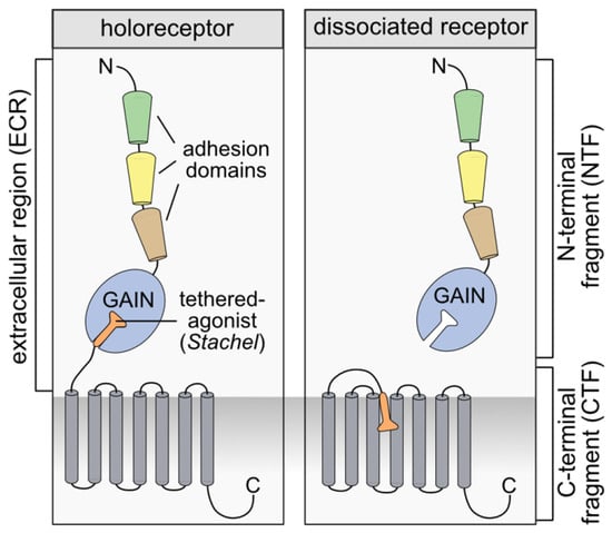
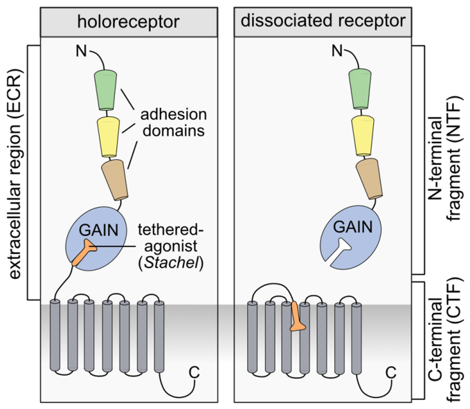
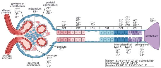
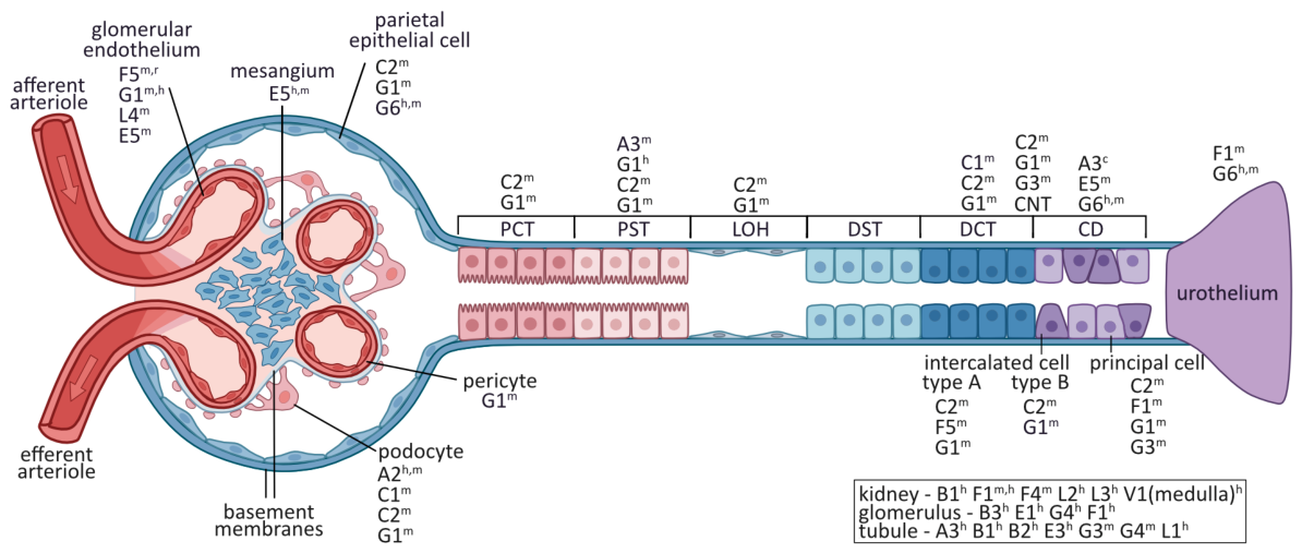
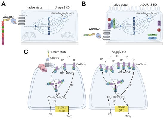
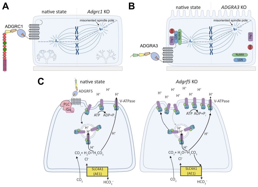
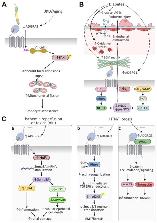
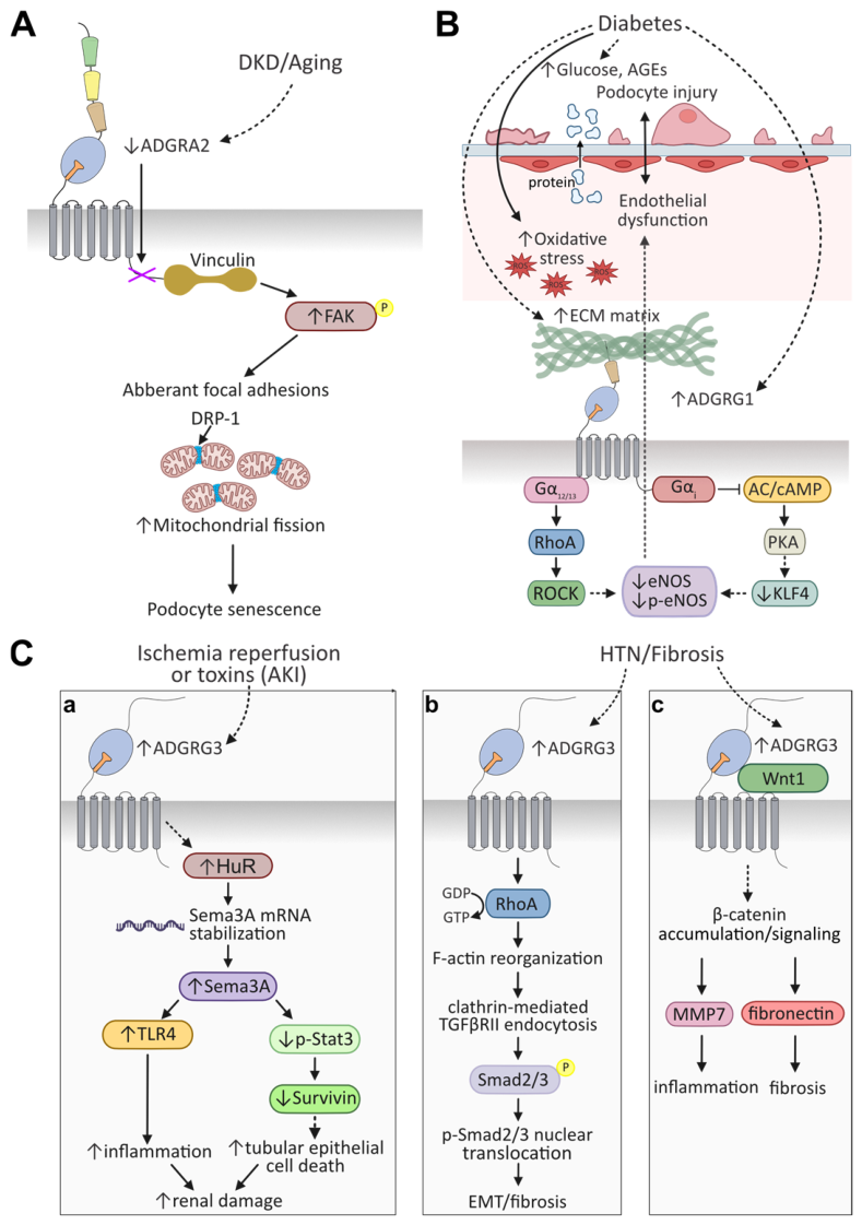

Disclaimer/Publisher’s Note: The statements, opinions and data contained in all publications are solely those of the individual author(s) and contributor(s) and not of MDPI and/or the editor(s). MDPI and/or the editor(s) disclaim responsibility for any injury to people or property resulting from any ideas, methods, instructions or products referred to in the content. |
© 2026 by the authors. Licensee MDPI, Basel, Switzerland. This article is an open access article distributed under the terms and conditions of the Creative Commons Attribution (CC BY) license.