


Abstract
Dynamic insulin secretion and glucose homeostasis are dependent on the appropriate maturation and function of pancreatic β-cells. The islet-enriched musculoaponeurotic fibrosarcoma oncogene family A (MAFA) transcription factor acts as a master regulator of β-cell identity and function, coordinating gene expression networks required for glucose-stimulated insulin secretion. Dysregulation of MAFA contributes to β-cell dysfunction, as reduced expression is detected early in the pathogenesis of Type 1 Diabetes and Type 2 Diabetes, while long-lived variants can drive monogenic forms of diabetes. In this review, we summarize the current understanding of MAFA on β-cell maturation and function in both the mouse and human. This includes structural features of the MAFA protein, regulation of MAFA transcription, post-translational modifications, and emerging areas of research for therapeutic potential.
1. Introduction
As of 2024, 1 in 9 adults are living with diabetes worldwide, resulting in over 3 million deaths in 2024 and an estimated 1 trillion dollars in economic burden [1]. Diabetes mellitus is a heterogeneous collection of diseases characterized by eventual beta (β)-cell failure to regulate blood glucose levels through insulin secretion. β-cells reside in the pancreatic islets of Langerhans—small clusters of endocrine cells within the pancreas—and are the only cells in the body to secrete insulin in response to glucose. β-cells coordinate with other islet cell types, including glucagon-secreting α-cells and somatostatin-secreting δ-cells, to ultimately regulate systemic glucose levels. β-cell failure can result from a range of insults, including autoimmune destruction (Type 1 Diabetes; T1D), relative insulin deficiency driven by chronic metabolic stress and insulin resistance (Type 2 Diabetes; T2D), stressors during pregnancy (Gestational Diabetes), hereditary mutations that impair β-cell function (Monogenic Diabetes), and diabetogenic viral infections [2,3] and exposure to β-cell toxins, including immunosuppressive drugs [4].
Several islet-enriched transcription factors (TFs) have been identified as critical regulators of gene expression to promote β-cell identity and function. Among these, musculoaponeurotic fibrosarcoma oncogene family A (MAFA) is uniquely enriched in pancreatic islet β-cells and is a critical regulator of β-cell identity and maturation [5,6,7,8,9,10,11]. MAF TFs were first identified as cellular proto-oncogene counterparts of the avian musculoaponeurotic fibrosarcoma virus AS42 in the late 1980s [12,13]. Since then, 7 MAF family TFs have been identified and characterized based on size: the large MAFs (c-MAF, MAFA, MAFB, NRL) and the small MAFs (MAFF, MAFG, MAFK) [14,15]. These TFs contribute to the development, differentiation, and maintenance of a diverse range of cellular functions in many organs and tissues, such as in the lens [16,17] and pancreatic islet [18]. 3 MAF TFs (c-MAF, MAFA, and MAFB) are detected within the islet and can bind to and activate Insulin (Ins1/2, mouse) and INSULIN (INS, human) expression [18].
Previous reviews have established MAFA as a critical regulator of β-cell maturation and Insulin gene transcription [7,8,9,10,11]. Here, we review the structure, expression, and function of MAFA in β-cells under physiologic and metabolic stress conditions. We further highlight recent advances that have substantially expanded our understanding of MAFA biology. These include the identification of MAFA as a causative monogenic Maturity-Onset Diabetes of the Young (MODY) gene, emerging roles in circadian timing of MAFA activity, insights into the functional relevance of MAFA structural elements predicted by AlphaFold, and growing evidence that MAFA is a key determinant of stem-cell-derived (SC)-β-cell maturation. By integrating findings from mouse and human islet studies, stem-cell models, and natural history of monogenic disease, we highlight how MAFA functions as a dynamic regulator of β-cell identity and glucose homeostasis across developmental, physiological, and disease contexts. We also discuss major unresolved questions regarding MAFA regulation and function that may inform future therapeutic strategies for diabetes and β-cell replacement therapies.
2. Protein Structure and Functional Domains of MAFA
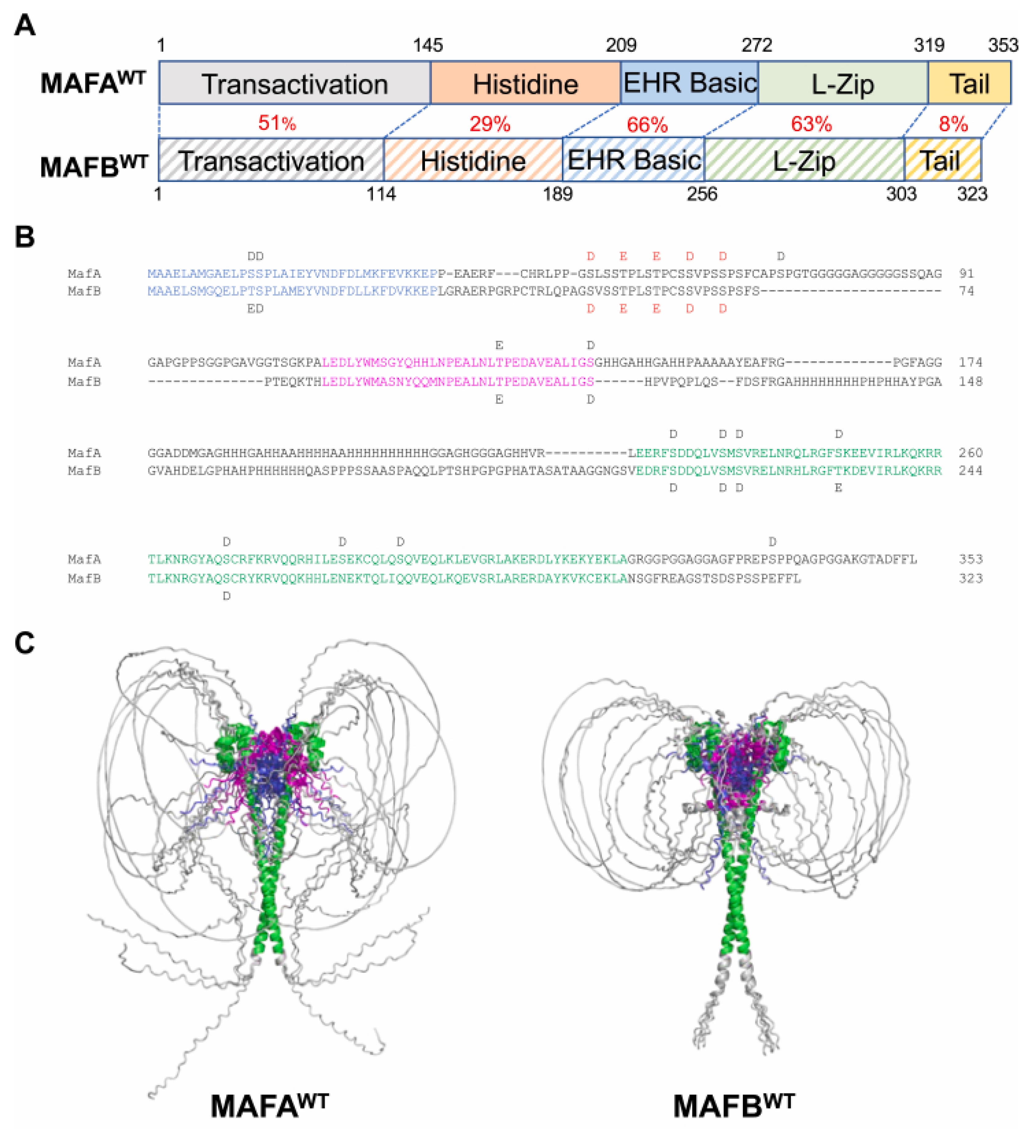
MAFA is 353 amino acids (aa) in length and includes several structural domains (Figure 1), including a coregulatory binding domain (N-terminus transcriptional activation domain [TAD; aa1–145]), DNA-binding domains (a histidine-rich region [aa146–208], an extended homology region [EHR; aa209–258], a basic region [aa259–272]), a dimerization region (leucine-zipper [aa273–318]) and a C-terminal tail [aa319–353] [19,20]. The highest sequence variability between MAFA and other large MAF proteins lies within the C-terminal tail and histidine-rich regions, whereas the EHR and basic-leucine zipper (bZIP) domains are highly conserved (Figure 2A) [19].
Figure 1.
Musculoaponeurotic fibrosarcoma oncogene family A (MAFA) protein structural elements work together to coordinate MAFA biology. The MAFA protein is 353 amino acids in length and comprises six domains with distinct functions: a transcriptional activation domain (Transactivation), a histidine-rich domain (Histidine), an extended homology region (EHR), a basic region (Basic), a leucine-zipper (L-Zip), and a C-terminal tail (C-Tail). The transactivation domain is involved in binding coregulators that influence gene transcription, while the histidine-rich domain, EHR, and basic region work together to bind MAFA to DNA via MAF recognition elements (MAREs). The EHR does not directly interact with MARE sequences but instead stabilizes DNA binding by interacting with the basic domain, as indicated with an arrow. The leucine zipper allows dimerization with another MAFA protein or other transcription factor (TF), which impacts the transcriptional activity. Figure made with BioRender.
2.1. Transactivation Domain (TAD)
A defining feature of large MAF proteins is the presence of the TAD, which enables MAF dimers to bind to coregulatory proteins. Coregulators influence MAFA-regulated gene expression by participating in the recruitment of transcriptional machinery and/or chromatin modifiers such as islet-enriched TFs, methyl transferases/de-transferases, coactivators, and scaffolding proteins [21] (See Section 5).
2.2. DNA-Binding Domains
The enhanced (extended) homology region (EHR), basic domain, and histidine-rich region coordinate MAFA DNA-binding specificity and transcriptional activity. Large MAF proteins utilize the EHR domains to enhance binding to 13–14 base pair DNA motifs known as MAF recognition elements (MAREs) in target genes [22,23,24]. There are two types of MARE sequences: a 13-base pair (bp) T-MARE sequence and a 14-bp C-MARE sequence. Within each of these sequences, a 7-bp TPA-response element (TRE; 5′-TGA(G/C)TCA-3′) or an 8-bp cyclic AMP-response element (CRE; 5′-TGACGTCA-3′) is at the core of the T-MARE and C-MARE sequences, respectively, both flanked by extended GC-rich sequences (5′-TGC-(TRE/CRE)-GCA-3′) [23,24,25].
MAFA interaction with MARE sequences is phosphorylation-dependent [20,26] (See Section 4.1). The basic domain directly binds the core regions of target MARE sequences [25]. The EHR does not directly interact with MARE sequences but instead stabilizes DNA binding by interacting with the basic domain [22]. Inference from other basic leucine-zipper (bZIP) TFs suggests that this interaction induces a conformational change in the basic domain α-helix from a flexible to a structured state to stabilize DNA binding [27].
Histidine-rich regions are known to affect protein stability and DNA binding [28,29,30], and can readily interact with Cu2+ and Zn2+ metal ions [31]. How these metal-ion interactions coordinate specific MAFA coregulator recruitment or DNA-binding specificity is not well described and will require structural and proteomic approaches to identify metal-dependent cofactors and target-site selection mechanisms.
2.3. Leucine-Zipper Domain
The leucine-zipper domain is a leucine-rich coiled region that facilitates dimerization through hydrophobic interactions. MAFA imparts transcriptional activity as a dimer and can form either homodimers or heterodimers to work synergistically with other TFs. Such interactions have been characterized in a variety of cell types for MAF family proteins, including dimerization with other MAFs, BACH2 [32], NRF2 [33], FOS and JUN [24,34,35] to increase transactivation [36]. This domain also coordinates with other MAFA domains by modulating interactions with phosphorylation sites in the N-terminus, affecting DNA-binding and MAFA stability [20,35,37,38].
2.4. C-Terminal Tail
There have been limited studies investigating the direct role of the C-terminal tail of MAFA. A recent study using AlphaFold 2 structural predictions of MAFA homodimers has facilitated understanding of this disordered region and shows the non-conserved C-terminal tail likely holds a secondary structure that lies near the DNA-binding domain (Figure 2C). Functional assessment of chimeric proteins with C-terminal tails interchanged between MAFA and MAFB reveals that the C-terminal tail is important for cooperativity with other islet-enriched TFs in target gene activation, independent of DNA binding [19].
Figure 2.
Protein sequence and AlphaFold 2 modeling of MAFAwild-type (WT) and MAF family B (MAFBWT) proteins. (A) protein sequence identities (%) between various domains of MAFAWT and MAFBWT are denoted in red. Extended homology domain (EHR); Leucine-zipper (L-Zip). (B) The aligned amino acid sequences of MAFAWT and MAFBWT, with aspartic acid (D) and glutamic acid (E) used as phosphomimics at bona fide sites of phosphorylation [20]. The red-labeled E and D represent the phosphosites absent in the underphosphorylated variants MAFAS64F and MAFBS70A. The regions marked in blue (i.e., 83%) and magenta (i.e., 79%) have higher sequence conservation than the overall transactivation domain and have structural elements in the AlphaFold 2 models. The region in green is the basic-leucine-zipper domain structured by AlphaFold 2 and determined by crystallography (i.e., MAFA residues 226–318 and MAFB residues 211–302) [22,39]. (C) Comparing the five independently derived representative models for MAFAWT (left) and MAFBWT (right) illustrates a potential structural difference. For example, the longer C-terminal tail of MAFAWT leads to a more divergent conformation than the MAFBWT, which may interact with N-terminal region sequences. These structural differences may contribute to the distinct transcriptional activities of MAFA and MAFB in β-cells despite their shared DNA-binding domain. Adapted from Cha et al., 2024 [19].
3. Islet MAFA Expression During Development and Adulthood
While the expression and regulation of other islet-enriched TFs are similar between humans and rodents [40,41,42,43], there are distinct species-specific expression patterns for human MAFA and mouse MafA. Rodent models continue to provide essential fundamental mechanistic insight into MAFA biology, but considering species-specific differences is essential for improving the translational relevance of preclinical studies. These key differences are summarized in Table 1.
Table 1.
Species-Specific Differences in β-cell MAFA Biology.
3.1. Rodent Models
In mice, pancreatic development is characterized by two sequential stages: the primary wave, lasting until embryonic day (E) 13.5, and the secondary wave, lasting until birth (E18.5). Organ determination is established during the primary wave, and initial pancreas formation is observed by E9.5 [50,56]. By E9.5, primary β-cell specification begins with multipotent progenitor cell differentiation within the pancreatic epithelium. Glucagon-producing cells appear first, followed by insulin-producing cells at approximately E10.5 [57,58]. During this primary wave of development, these hormone-producing cells exist in a “protodifferentiated state”, in which they produce minimal amounts of hormone and are unresponsive to glucose stimulation due to a lack of key glucose uptake proteins [59]. MafA expression is induced during the transition to the secondary wave (E12.5–E13.5) exclusively in insulin-producing cells [6,44].
The secondary wave is characterized by a significant increase in protein synthesis [60]. MafA expression drives acquisition of the glucose-responsive, mature β-cell phenotype, in part through induction of Slc2a2 (encoding Glut2) and Ins1/2 expression [9,61,62]. Most pancreatic β-cells are generated during this differentiation wave through the contribution of many transcriptional regulators in conjunction with MafA (Figure 3). MafA remains highly expressed in this period and is the dominant Maf factor in β-cells postnatally throughout adulthood [44,45].
Figure 3.
MafA expression drives β-cell maturity during development, characterized by glucose-regulated insulin secretion in mice. This schematic summarizes the timing of MafA expression and other TFs governing the development and differentiation of mouse β-cells. The pink section depicts the first wave of development from embryonic day (E)9.5, the beginning of pancreatic formation and multipotent progenitor cells, to the first time point MafA is detectable in insulin-producing cells. The blue section displays the second wave of development from E13.5 to birth (E18.5), characterized by increased protein synthesis driven by MafA, resulting in glucose-responsive β-cells. The yellow section illustrates the postnatal result of the mature β-cell with high MafA expression. Figure made with BioRender.
3.2. Human Models
Human MAFA expression is undetectable within the pancreas until the second trimester and remains low during early postnatal life in β-cells until approximately 9 years of age [46,47,48]. Similarly, postnatal MAFA protein levels are rarely detected in juvenile human β-cells until approximately 10 years of age [48]. In contrast, rodent MafA expression is induced during embryonic development and becomes the dominant Maf TF in mature β-cells shortly after birth (Figure 3) [44].
To better understand this discrepancy, investigators have leveraged collaborative large-scale initiatives to acquire human pancreata, such as the Network for Pancreatic Organ Donors with Diabetes (nPOD), Human Pancreas Analysis Program (HPAP), and the Human Islet Research Network (HIRN) [63,64,65]. These efforts have enhanced our understanding of human MAFA expression through integrated transcriptomic, epigenomic, and single-cell analyses of healthy and diabetic islets. These datasets support a model in which MAFA expression is associated with mature β-cell states [51] and is diminished in dysfunctional β-cells from individuals with diabetes [52].
Human single-cell transcriptomic studies identify MAFA expression as one of the most reliable markers of β-cell identity, with expression correlating with improved β-cell function [51]. Adult human β-cells frequently co-express MAFA and another MAF TF, MAFB, unlike rodents, in which MafB expression is largely restricted to α-cells [45]. Human β-cells co-expressing MAFA and MAFB display enriched expression of genes involved in insulin secretion, mitochondrial metabolism, and β-cell function [51]. This suggests species-specific MAFA transcriptional networks within the β-cell. Transcriptionally, studies in both the human β-cell line EndoC-βH1 and human islets demonstrate MAFA as a regulator of exocytotic machinery [66]. Functionally, loss of MAFA impairs both glucose- and K+- stimulated insulin secretion in EndoC-βH1 cells [66]. Moreover, rodent MafA is required for the maintenance of β-cell identity and repression of non-β-cell hormones such as glucagon, somatostatin, and gastrin [67]. However, this role is fulfilled by MAFB in human β-cells [67]. These studies indicate that while MAFA serves a conserved role in promoting β-cell maturity across species, the timing of expression, interactions with other TFs, and pathways dysregulated in the absence of these factors differ substantially between rodents and humans.
Human embryonic stem (hES) cells are capable of directed differentiation to generate pancreatic and endocrine precursor cells, and ultimately insulin-secreting cells [68,69,70,71,72,73,74,75,76,77]. In these SC-β-cells, glucose-responsiveness is correlated with sustained MAFA expression [71,78]. Although hES cell models can provide insight into MAFA function in a human context, defining MAFA expression patterns during differentiation does not fully recapitulate the human condition. Continued integration of data from human islet-derived datasets and stem-cell models will be important for defining pathways and strategies regulating MAFA expression and activity in human β-cells [63,64,65] (See Section 7).
4. Regulation of MAFA Activity via Post-Translational Modifications
4.1. Phosphorylation
Blood glucose concentrations are regulated by dynamic insulin secretion from β-cells and precise regulation of MAFA via post-translational modifications (PTMs). Among these PTMs, the phosphorylation status of MAFA dictates many aspects of MAFA function, including dimerization [20], DNA binding [20,26,38], coregulator interactions [79], and ubiquitin-targeting for protein turnover [80]. Specifically, phosphorylation within the MAFA N-terminus is required for C-terminus dimerization with other proteins and coactivators [81]. Chemical dephosphorylation of MAFA by treatment with calf intestinal alkaline phosphatase reduced DNA-binding activity. These effects were largely prevented with the phosphatase inhibitor Na3VO4, suggesting that phosphorylation of MAFA positively impacts DNA binding [20,26].
Phosphorylation in the TAD has a dual role in regulating human MAFA activity, as it promotes both transcriptional activation and protein turnover. A mass spectrometry study revealed 10 phosphorylation sites within the MAFA TAD, 7 in the bZIP domain, and 1 in the C-terminus [20]. Of these sites, MAFA biological functions depend on the priming phosphorylation event of the TAD residue serine (Ser [S]) 65 [80]. Although the kinase responsible for this priming phosphorylation event is still unknown, phosphorylation of Ser65 triggers the sequential phosphorylation of TAD residues: S61, threonine (Thr [T]) 57, T53, and S49 by GSK-3 [81,82]. Phosphorylation of S14 is distinct from the GSK-3 cascade and not required for protein turnover, but instead is required for MAFA transcriptional activity [38,80,81,82].
4.2. Ubiquitination
Under low-glucose conditions, MAFA is rapidly marked for degradation by polyubiquitination [82]. Ubiquitination is a PTM in which the small 76 amino acid protein ubiquitin is conjugated onto a protein [83]. Following the key phosphorylation event at S65, MAFA is polyubiquitinated in the C-terminus and targeted for proteasomal degradation [80]. By a cycloheximide chase experiment, it was determined in vitro that MAFA has rapid protein turnover with a t1/2 of ~30 min [54] under low-glucose conditions. In contrast, under high-glucose conditions, the TAD residues are phosphorylated, and subsequent acetylation transiently prevents MAFA degradation, thereby promoting transcriptional activity [81,82]. Following transcription initiation, these same phosphorylated sites serve as signals for polyubiquitin via the E3 ligase HRD1 and eventual degradation [81,84].
4.3. Acetylation
PCAF (Lysine acetyltransferase 2b, or KAT2B) acetylates MAFA and protects it from ubiquitin-tagged degradation [81], although specific lysine residues or deacetylases involved have not yet been identified. Consistently, KAT2b (encoding PCAF) expression is decreased in both db/db mice and T2D human islets [85]. Disruption of Kat2b in mice also results in increased MAFA proteasomal degradation [86], although it has minimal impact on MafA mRNA levels [85]. Functionally, loss of PCAF protein impairs insulin secretion and results in overall glucose intolerance in mice from a loss of β-cell adaptation to stress [85]. Although the importance of acetylation in MAFA regulation of β-cell function is evident, future studies are needed to define signaling pathways that control PCAF activity and determine how altered MAFA acetylation contributes to β-cell dysfunction during diabetes pathogenesis.
4.4. SUMOylation
SUMOylation is a series of enzymatic reactions that covalently conjugate small ubiquitin-related modifiers (SUMOs) to lysine residues of target proteins. These PTMs are dynamic and readily removed by SUMO-specific proteases. MAFA can be post-translationally modified by two SUMO isoforms, SUMO-1 and SUMO-2, on Lys32, resulting in decreased transcriptional activity [87]. SUMOylation is known to regulate a wide range of processes in different cell types and tissues, including DNA repair, cell cycle, and gene transcription [83,88]. However, its exact influence on MAFA activity and protein levels within the β-cell requires examination of relevant SUMO ligases, proteases, and downstream transcriptional consequences.
5. Transcriptional Regulation of β-Cell Function by MAFA
β-cell maturation is defined by the ability to secrete insulin in response to glucose and the acquisition of β-cell-enriched identity genes, including MAFA [89,90,91]. Loss of MafA in the mouse pancreas [45,49], islet [5], or specifically in the β-cell [61] similarly results in altered β-cell identity and function. This includes impaired glucose-stimulated insulin secretion (GSIS) and transdifferentiation into other islet cell types. For example, β-cell-specific deletion of MafA under stress conditions induces inappropriate gastrin hormone production in mouse islets [67]. Notably, complete loss of rodent MafA is not required for dysfunction; hyperglycemia alone impairs MafA nuclear translocation and promotes progressive loss of β-cell identity [52,53,92].
The first identified MafA regulatory target in the pancreatic β-cell was the rat insulin promoter element 3b (RIPE3b) via the C1 element within the RIPE3b promoter [26,93,94]. This led to the original identification of MAFA as the RIPE3b activator [94]. MAFA has since been shown to regulate the expression of several genes involved in β-cell transcriptional maturation and functional competence in humans and mouse models, as summarized in Table 2.
Table 2.
Known β-cell MAFA target genes.
The establishment and maintenance of β-cell GSIS requires complex coordination of transcriptional networks driven by islet-enriched factors such as MAFA, PDX1, NGN3, and NEUROD1 [108,109,110]. MAFA functions cooperatively with other TFs through heterodimerization and coregulator interactions mediated by its TAD (See Section 2.3), including other large MAF bZIP TFs such as MAFB and c-MAF [22] within the islet. β-cells co-expressing MAFA and another family member, MAFB, are functionally and transcriptionally more active than β-cells expressing MAFA homodimers alone [51], suggesting unique interactions with coregulatory proteins. MAFA also participates in multi-protein transcriptional complexes to regulate gene expression. Notably, MAFA acts synergistically with PDX1 and NEUROD1, although MAFA can independently activate INS expression [110].
In addition, MAFA recruits chromatin-modifying coactivators to facilitate the recruitment and assembly of RNA polymerase II and transcriptional machinery at target promoters. The mixed-lineage leukemia 3 (MLL3) and mixed-lineage leukemia 4 (MLL4) complexes, which catalyze enhancer-associated mono-methylation of histone 3 lysine residue 4 (H3K4me1), contribute to MAFA-dependent gene regulation [21]. Loss of β-cell-specific Ncoa6 in mice, a core subunit of these complexes, reduces expression of target genes such as Ins2 and Slc2a2, and impairs GSIS [21]. Similarly, histone acetyltransferases, including p300, CREB binding protein (CBP), and PCAF, can bind to Maf proteins [111], and are required for glucose-responsive, MAFA-mediated gene transcription [112]. Together, these findings highlight the importance of chromatin remodeling in enabling robust MAFA-mediated transcriptional programs to regulate the expression of downstream targets.
6. MAFA in Diabetes Pathogenesis
6.1. Type 1 Diabetes (T1D)
T1D is characterized by a tissue-specific autoimmune attack against insulin-producing β-cells, leading to an inability to appropriately regulate blood glucose levels [113]. Insulin is the dominant autoantigen in T1D [113], and reduced thymic Insulin expression is associated with increased insulin autoantibody production [114,115]. MafA is the only insulin-promoting TF identified thus far in the mouse thymus [116,117]. Global MafA knockout (KO) in mice decreased Ins2 expression in the thymus and increased β-cell autoantibodies in serum [116,117]. NOD mice, a model of T1D, also show decreased MafA and Ins2 thymic expression compared to control mice [117]. Genetic variants of MAFA affecting its expression or activity can influence T1D susceptibility in both mice and humans. MafA variants decreasing its promoter activity were identified exclusively in NOD mice compared to control and T2D mouse models [116]. Conversely, a single nucleotide polymorphism (Gly346Cys, rs6252187) increasing thymic MAFA transcriptional activity is correlated with a decreased risk of T1D in humans [118]. These results suggest that thymic MAFA expression or activity can potentially influence T1D susceptibility in mice and humans.
The immune microenvironment of the islet also plays a role in the pathogenesis of T1D. The expression of pro-inflammatory molecules in healthy islets is tightly regulated to avoid inappropriate immune activation. Decreased MAFA expression in human islets strongly correlated with cytokine-induced signaling and T1D susceptibility genes. Loss of MafA in mouse islets similarly resulted in an increase in pro-inflammatory genes in isolated islets, improved viral clearance compared to control islets [119], and increased accumulation of immune cells [120]. This suggests that islet MafA expression may also be critical for preventing autoimmune attack in T1D. However, an underlying mechanism for these changes remains unclear, and more research is needed to identify a causative link to T1D pathogenesis.
6.2. Type 2 Diabetes (T2D)
T2D is a chronic condition where β-cells fail to produce sufficient insulin alongside peripheral insulin resistance, resulting in high blood glucose levels. Both MAFA mRNA and MAFA protein levels are reduced in islets from T2D patients compared to non-diabetic controls [52,53,119]. Similarly, MafA expression is reduced in db/db mice, a model for T2D, obesity, and insulin resistance [121]. Restoring pancreas-specific MafA expression in mice improves β-cell mass, enhances function, and lowers blood glucose levels [122]. Notably, MAFA is susceptible to post-translational oxidative stress, demonstrating a 97% decrease in protein levels in response to glucotoxicity [123]. Chronic hyperglycemia creates glucolipotoxic conditions, which overwhelm β-cell mitochondria to accumulate reactive oxygen species (ROS). This oxidative stress increases MAFA turnover, decreases MAFA expression, and promotes MAFA mislocation to the cytoplasm, collectively reducing binding to the Insulin promoter [52,124]. Consequently, this impairs Insulin expression and GSIS [123,125], which can be prevented by treatment with antioxidants in vitro in a dose-dependent manner [123].
Given that MAFA is downregulated in islets from T2D donors and plays a pivotal role in regulating GSIS, MAFA overexpression has been explored as a potential therapeutic target in restoring β-cell function. MAFA overexpression alone or in combination with other islet-enriched TF’s can drive non-β-cells into functional insulin-secreting cells in vitro [126,127,128]. However, these approaches have remained unable to fully rescue diabetic phenotypes in vivo [129]. This discrepancy may reflect the exceptionally tight regulation of MAFA, where sustained overexpression is insufficiently maintained by autoregulation, likely via region 3 of the MAFA promoter [130]. These findings highlight the need for improved characterization of MAFA production and strategies that recapitulate precise endogenous MAFA regulation to achieve therapeutic benefit for T2D.
6.3. Maturity-Onset Diabetes of the Young (MODY)
Two missense mutations within the TAD of MAFA—Ser64Phe [54] (S64F) and Thr57Arg [131] (T57R)—have been identified to drive a form of monogenic diabetes called MODY diabetes [132]. Typical clinical characteristics of MODYs include noninsulin-dependent diabetes and early-onset, typically before the age of 25 years [132,133]. MAFA-MODY differs phenotypically with clinical presentation at age ~40 and presents in a sex-dependent manner. Female carriers are more likely to develop insulinomas while male carriers develop MODY, though this bias appears stronger with the Ser64Phe mutation.
These clinical phenotypes are partially recapitulated in a mouse model: male mutant mice harboring the Ser64Phe mutation develop diabetes, whereas female mutants are persistently hypoglycemic. Furthermore, sex-biased signatures of MafAS64F point to downstream disruption of calcium dynamics in females versus early onset senescence and dysregulated circadian rhythm in males [55,107]. Although ovariectomy studies in mice have demonstrated that these differences are not principally due to postnatal estrogen [55], underlying etiology for these diametrically opposed phenotypes remains to be explored.
At the protein level, Ser64Phe mimics the effects of the kinase-priming defective mutation, Ser65Ala, suggesting that Ser64Phe disrupts the phosphorylation at Ser65 (See Section 4.1). As a result, MAFAS64F has both increased protein stability and increased transactivation capacity. This disrupts the requisite and precise regulation of MAFA and downstream target regulation by MAFA, ultimately leading to impaired GSIS [54]. Similarly, Thr57 lies within the TAD and is recognized to be one of the subsequent phosphorylation events following the Ser65 phosphorylation. Therefore, while the mechanisms behind Thr57Arg have not been thoroughly studied, it is proposed to have similar functional consequences as Ser64Phe, including enhanced stability [131]. These familial cases of MAFA-MODY highlight the importance of MAFA phosphorylation and appropriate turnover on cellular function, supporting a model in which MAFA activity must be maintained within a functional range.
7. Emerging Roles and Novel Studies
7.1. Roles of MAFA in Glucose Homeostasis Beyond the Pancreatic β-Cell
While MAFA expression is enriched in the pancreatic islet, it is also highly expressed in other tissues, including skeletal muscle, adipose, testes, and regions of the brain [134,135]. Among these, its role in the skeletal muscle is well characterized. Skeletal muscle is a major contributor to whole-body glucose homeostasis, accounting for the majority of insulin-stimulated glucose uptake and serving as a key site of insulin resistance in diabetes [136]. Prolonged skeletal muscle inactivity, such as that which occurs in neuromuscular disease, induces a shift from oxidative type I myofibers to more glycolytic type II myofibers [137]. MafA is expressed in a subset of fetal myofibers in lambs, where it promotes maturation through activation of myofiber-specific maturity programs [138]. MafA also functions as a determinant of myofiber subtype specification in mice, specifically promoting type IIb myofiber identity during development and adaptation [139]. Since myofiber composition influences glucose utilization and metabolic flexibility [140], these findings position MAFA as a regulator of metabolic function in skeletal muscle. Modulation of MAFA activity may represent novel therapeutic avenues for insulin resistance related to muscular dystrophy.
In contrast, the roles of MAFA in other metabolic tissues remain less defined. In adipose tissue, loss of rodent MafA is associated with reduced expression of genes required for mature adipocyte differentiation, as well as alterations in cell growth and lipid droplet accumulation [141,142]. MAFA is also highly enriched in the testes, where it may influence male sex hormone production, although its functional role in this context remains unclear. Notably, nuclear androgen signaling can regulate islet function [143], raising the possibility of indirect crosstalk between MAFA activity in reproductive tissues and β-cell physiology. Consistent with this idea, the pronounced sex-specific phenotypes observed clinically in patients with mutations in MAFA (MAFAS64F and MAFAT57R) suggest that MAFA-dependent pathways may contribute to differential metabolic vulnerability between males and females [144] (See Section 6.3). Investigation of sex-biased differences in expression and activity of MAFA regulators or MAFA variants is warranted.
MAFA expression has also been reported in discrete regions of the hypothalamus, including the ventromedial nucleus (VMH), arcuate nucleus (ARC), paraventricular nucleus (PVN), and lateral hypothalamus (LH), which are key regulators of energy balance and glycemic control [145]. Loss of MafA in the mouse brain is associated with downregulation of genes involved in food consumption and metabolism, including growth hormone, pro-melanin-concentrating hormone (MCH), and orexin [142]. These findings suggest that MAFA may also contribute to central regulation of energy homeostasis, although direct functional studies in specific neuronal populations are limited.
Taken together, emerging evidence suggests that MAFA may function beyond the β-cell to integrate tissue-specific aspects of metabolism across multiple organ systems. Addressing this question will require tissue-specific genetic models and metabolic phenotyping to determine how MAFA functions across organs are coordinated.
7.2. Circadian Regulation of MAFA
Circadian regulation of whole-body metabolism was first described over 50 years ago, when studies demonstrated diurnal variation in β-cell insulin secretion in humans [146,147,148]. Epidemiological studies have since shown that circadian misalignment, such as in night shift workers, is associated with a 60% increase in the development of diabetes [149,150]. Conversely, T2D patients exhibit blunted rhythmic insulin in response to mixed meals [151], suggesting that disruption of circadian control contributes to impaired insulin secretion. Together, these findings indicate that β-cell insulin secretory machinery is intrinsically tuned with circadian timing.
The circadian system is driven by transcriptional-translational feedback loops present in nearly all cell types, including pancreatic β-cells. These molecular clocks coordinate environmental cues, such as the light-dark cycles and feeding cycles, with cellular function, including cyclic insulin secretion. In β-cells, intrinsic autonomous oscillatory transcriptional cycles enable islets to anticipate nutrient availability and be primed for insulin demand [152]. The detailed molecular aspects of the circadian clock of the pancreatic β-cell have been extensively reviewed elsewhere [153,154,155]. Briefly, circadian activators, Circadian Locomotor Output Cycles Kaput (CLOCK) and Brain and Muscle ARNT-Like 1 (BMAL1), drive expression of clock-controlled genes, including circadian repressors Period (encoding PER1, PER2, and PER3) and Cryptochrome (encoding CRY1 and CRY2), as well as additional modifiers such as Basic Helix-Loop-Helix Family Member E40 (BHLHE40; encodes DEC1). These proteins form autoregulatory feedback loops that generate oscillations in gene expression and cellular function.
Recent studies have linked circadian entrainment to postnatal β-cell maturation. Establishment and maintenance of circadian rhythmicity are critical for β-cell maturity [156,157,158], which includes the induction of key maturation factors such as MAFA. Circadian entrainment promotes human islet maturation through chromatin remodeling, increasing accessibility at loci associated with insulin secretion, including MAFA, and enhancing GSIS responsiveness, although the specific chromatin modifiers remain unclear [157]. In differentiating hES cells, acquisition of glucose responsiveness coincides with chromatin remodeling events that enable access to both insulin-secretory genes and circadian regulators such as CLOCK and BMAL1 [157]. Notably, MafA is one of the only essential β-cell TFs that exhibits rhythmic expression in mice [159]. Disruption of the circadian regulator DEC1 in human SC-islets abolishes MAFA oscillatory expression and impairs GSIS [75]. Conversely, mouse models expressing stabilized MafAS64F show impaired glucose tolerance with downregulated islet expression of circadian genes, including Bhlhe40 [107], suggesting the existence of a conserved feedback relationship between MAFA and circadian regulators in mice and humans. Dec1 is also suggested to mediate hypoxia-induced β-cell function through suppression of MafA in mice [160]. The downregulation of MafA compromises ATP generation and exocytotic machinery, resulting in impaired β-cell GSIS [160].
Together, these findings suggest a bidirectional relationship between circadian regulation and β-cell transcriptional networks, in which MAFA both responds to and influences circadian timing mechanisms. Understanding how essential β-cell-specific TFs such as MAFA integrate circadian timing with specialized insulin-secretory programs will require integrating circadian perturbation, chromatin accessibility, transcriptomic profiling, and MAFA occupancy analysis in mature human β-cells. However, these results reveal that optimal β-cell function may require not only maintenance of appropriate MAFA levels but also cyclical periods of MAFA activity.
7.3. Stem Cell (SC)-Derived Therapies
Current therapies for diabetes effectively manage hyperglycemia but do not cure the disease. Standard treatment includes glucose monitoring and exogenous insulin administration, which requires frequent monitoring and dose adjustments, which increases risk for adverse events such as hypoglycemia. As a result, there is a need for alternative therapies to improve the overall treatment of diabetes while minimizing adverse outcomes. Transplantation of β-cells provides an attractive therapeutic approach, as these cells can sense glucose and secrete insulin in real-time. Cadaveric β-cells are often limited and difficult to acquire, while SC-β-cells offer a renewable source of β-cells, which may improve access to treatment [161].
Recent advances in SC-β-cell therapies have demonstrated SC-β-cell potential for clinical translation. As of 2025, Vertex Pharmaceuticals has completed Phase 1/2 clinical trials of an SC-derived islet transplantation therapy (Zimislecel) in individuals with T1D. One year after treatment, all 12 participants who received a full dose exhibited elimination of hypoglycemic events, improved glycemic control (HbA1c < 7%), and increased time in the target glucose range (70–180 mg/dL), with the majority achieving insulin independence thus far [162]. However promising, these pilot studies require longitudinal outcomes monitoring for safety and efficacy. In addition, there remain outstanding issues in this field preventing the broad clinical use of SC-β-cell therapy. These include improving scalability and dosing, eliminating the reliance on systemic immunosuppression in non-encapsulation strategies, and optimizing transplant site outcomes [163].
Another major challenge in the field remains in the generation of fully mature, glucose-responsive β-cells. While there are many protocols developed to differentiate SCs into immature, insulin-producing cells [68,69,70,71,72,73,74,75,76,77], there is difficulty in developing mature β-cells that dynamically secrete insulin in response to glucose to the degree of native human islets. Notably, transplantation of immature SC-β-cells promotes their functional maturation in vivo, suggesting key maturation signals are missing from current in vitro systems. MAFA expression is significantly upregulated following transplantation, consistent with its role in establishing functional β-cell maturity. Indeed, induction of MAFA expression has been shown to directly induce β-cell maturation from immature SCs or induce insulin production from non-β-cells [6,164].
Thus, strategies to increase MAFA expression in SC-β-cells, either through direct or indirect methods, are an important area of research. Current potential approaches include reducing oxidative stress, which contributes to post-translational MAFA loss during glucolipotoxicity [123], promoting chromatin states permissive for MAFA expression during β-cell maturation [165,166,167], enhancing circadian entrainment pathways which regulate MAFA rhythmic expression [75,160], or developing inducible systems to regulate the MAFA protein. While these strategies remain largely experimental, they may provide opportunities to restore endogenous MAFA activity under conditions of metabolic stress. New findings of rhythmic MAFA expression in mature SC-β-cells collectively point towards temporal and rhythmic regulation of MAFA expression for optimal SC-β-cell function [11,75]. These findings underscore the importance of better characterizing and precisely coordinating MAFA expression with developmental cues to optimize the generation of therapeutically relevant SC-β-cells.
8. Conclusions
Recent advances have transformed the view of MAFA from a β-cell maturation factor to a dynamically regulated integrator of developmental, metabolic, circadian, and stress-responsive signaling pathways. Collectively, studies from genetic mouse models, human donor islets, and SC-β-cells have revealed that MAFA activity must be maintained within a narrow functional range. Loss of MAFA impairs β-cell identity and GSIS, whereas stabilization of MAFA, as observed in MAFA-MODY, similarly disrupts β-cell function and glucose homeostasis. These findings support a model in which the timing, amplitude, and duration of MAFA activity are all critical determinants of β-cell health.
Despite significant advances, key questions remain. The upstream pathways governing MAFA induction during human β-cell maturation remain poorly defined, particularly given the substantial differences in developmental timing between rodents and humans. Likewise, recent evidence linking MAFA to circadian entrainment raises important questions regarding how rhythmic MAFA expression is established, maintained, and integrated with nutrient sensing and metabolic stress. Defining the transcriptional, epigenic, and environmental signals that coordinate these processes will be essential for understanding how mature β-cell identity is established and maintained.
Finally, emerging studies in SC-β-cells and circadian regulation of MAFA suggest that future therapeutic strategies will likely require restoration of physiological MAFA dynamics rather than simple enhancement of MAFA expression. Determining whether temporal MAFA expression and MAFA activity can be leveraged to improve β-cell replacement therapies, SC-β-cell maturation, or preservation of endogenous β-cell function represents an important challenge for the field. As efforts to develop therapies for diabetes advance, defining mechanisms that regulate MAFA abundance, activity, and rhythmicity will be critical to translating MAFA biology for therapeutic benefit.
Author Contributions
Conceptualization, L.J., M.A.M., and J.C.; investigation, L.J., M.A.M., and J.C.; writing—original draft preparation, L.J., M.A.M., and J.C.; writing—review and editing, L.J., M.A.M., and J.C.; visualization, L.J. and M.A.M.; supervision, J.C.; project administration, L.J., M.A.M., and J.C.; funding acquisition, L.J. and J.C. All authors have read and agreed to the published version of the manuscript.
Funding
This work was supported by NIH funding provided to LJ (T32 DK007563) and JC (K08 DK132507). JC was also supported by the Doris Duke Foundation Physician Scientist Fellowship (2020063) and Burroughs Welcome Fund Career Award for Medical Scientists.
Data Availability Statement
No new data were created or analyzed in this study.
Acknowledgments
We regret that space limitations precluded us from citing many relevant references. We thank members of the Cha lab for helpful discussions and feedback on writing.
Conflicts of Interest
The authors declare no conflicts of interest.
References
- International Diabetes Federation. IDF Diabetes Atlas 2025, 11th ed.; International Diabetes Federation: Brussels, Belgium, 2025; Available online: https://diabetesatlas.org (accessed on 23 April 2026).
- Kotsiri, I.; Xanthi, M.; Domazinaki, C.-M.; Magiorkinis, E. The Role of Viral Infections in the Immunopathogenesis of Type 1 Diabetes Mellitus: A Narrative Review. Biology 2025, 14, 981. [Google Scholar] [CrossRef] [Scilit] [PubMed]
- Nekoua, M.P.; Bertin, A.; Sane, F.; Alidjinou, E.K.; Lobert, D.; Trauet, J.; Hober, C.; Engelmann, I.; Moutairou, K.; Yessoufou, A.; et al. Pancreatic beta cells persistently infected with coxsackievirus B4 are targets of NK cell-mediated cytolytic activity. Cell. Mol. Life Sci. 2019, 77, 179–194. [Google Scholar] [CrossRef] [Scilit] [PubMed]
- Kaestner, K.H. β Cell transplantation and immunosuppression: Can’t live with it, can’t live without it. J. Clin. Investig. 2007, 117, 2380–2382. [Google Scholar] [CrossRef] [Scilit] [PubMed]
- Zhang, C.; Moriguchi, T.; Kajihara, M.; Esaki, R.; Harada, A.; Shimohata, H.; Oishi, H.; Hamada, M.; Morito, N.; Hasegawa, K.; et al. MafA Is a Key Regulator of Glucose-Stimulated Insulin Secretion. Mol. Cell. Biol. 2005, 25, 4969–4976. [Google Scholar] [CrossRef] [Scilit] [PubMed]
- Matsuoka, T.; Artner, I.; Henderson, E.; Means, A.; Sander, M.; Stein, R. The MafA transcription factor appears to be responsible for tissue-specific expression of insulin. Proc. Natl. Acad. Sci. USA 2004, 101, 2930–2933. [Google Scholar] [CrossRef] [Scilit] [PubMed]
- Nishimura, W.; Iwasa, H.; Tumurkhuu, M. Role of the Transcription Factor MAFA in the Maintenance of Pancreatic β-Cells. Int. J. Mol. Sci. 2022, 23, 4478. [Google Scholar] [CrossRef] [Scilit] [PubMed]
- Liang, J.; Chirikjian, M.; Pajvani, U.B.; Bartolomé, A. MafA Regulation in β-Cells: From Transcriptional to Post-Translational Mechanisms. Biomolecules 2022, 12, 535. [Google Scholar] [CrossRef] [Scilit] [PubMed]
- Hang, Y.; Stein, R. MafA and MafB activity in pancreatic β cells. Trends Endocrinol. Metab. 2011, 22, 364–373. [Google Scholar] [CrossRef] [Scilit] [PubMed]
- Aramata, S.; Han, S.-I.; Kataoka, K. Roles and regulation of transcription factor MafA in islet beta-cells. Endocr. J. 2007, 54, 659–666. [Google Scholar] [CrossRef] [Scilit] [PubMed]
- El Khattabi, I.; Sharma, A. Proper activation of MafA is required for optimal differentiation and maturation of pancreatic β-cells. Best Pract. Res. Clin. Endocrinol. Metab. 2015, 29, 821–831. [Google Scholar] [CrossRef] [Scilit] [PubMed]
- Nishizawa, M.; Kataoka, K.; Goto, N.; Fujiwara, K.T.; Kawai, S. v-maf, a viral oncogene that encodes a “leucine zipper” motif. Proc. Natl. Acad. Sci. USA 1989, 86, 7711–7715. [Google Scholar] [CrossRef] [Scilit] [PubMed]
- Kataoka, K.; Nishizawa, M.; Kawai, S. Structure-function analysis of the maf oncogene product, a member of the b-Zip protein family. J. Virol. 1993, 67, 2133–2141. [Google Scholar] [CrossRef] [Scilit] [PubMed]
- Kannan, M.B.; Solovieva, V.; Blank, V. The small MAF transcription factors MAFF, MAFG and MAFK: Current knowledge and perspectives. Biochim. Biophys. Acta Mol. Cell Res. 2012, 1823, 1841–1846. [Google Scholar] [CrossRef] [Scilit] [PubMed]
- Fujino, M.; Ojima, M.; Takahashi, S. Exploring Large MAF Transcription Factors: Functions, Pathology, and Mouse Models with Point Mutations. Genes 2023, 14, 1883. [Google Scholar] [CrossRef] [Scilit] [PubMed]
- Ogino, H.; Yasuda, K. Induction of Lens Differentiation by Activation of a bZIP Transcription Factor, L-Maf. Science 1998, 280, 115–118. [Google Scholar] [CrossRef] [Scilit] [PubMed]
- Reza, H.M.; Ogino, H.; Yasuda, K. L-Maf, a downstream target of Pax6, is essential for chick lens development. Mech. Dev. 2002, 116, 61–73. [Google Scholar] [CrossRef] [Scilit] [PubMed]
- Matsuoka, T.; Zhao, L.; Artner, I.; Jarrett, H.W.; Friedman, D.; Means, A.; Stein, R. Members of the large Maf transcription family regulate insulin gene transcription in islet beta cells. Mol. Cell. Biol. 2003, 23, 6049–6062. [Google Scholar] [CrossRef] [Scilit] [PubMed]
- Cha, J.; Tong, X.; Coate, K.C.; Guo, M.; Liu, J.; Reynolds, G.; Walker, E.M.; Stein, R.A.; Mchaourab, H.; Stein, R. Defining unique structural features in the MAFA and MAFB transcription factors that control Insulin gene activity. J. Biol. Chem. 2024, 300, 107938. [Google Scholar] [CrossRef] [Scilit] [PubMed]
- Guo, S.; Vanderford, N.L.; Stein, R. Phosphorylation within the MafA N Terminus Regulates C-terminal Dimerization and DNA Binding. J. Biol. Chem. 2010, 285, 12655–12661. [Google Scholar] [CrossRef] [Scilit] [PubMed]
- Scoville, D.W.; Cyphert, H.A.; Liao, L.; Xu, J.; Reynolds, A.; Guo, S.; Stein, R. MLL3 and MLL4 Methyltransferases Bind to the MAFA and MAFB Transcription Factors to Regulate Islet β-Cell Function. Diabetes 2015, 64, 3772–3783. [Google Scholar] [CrossRef] [Scilit] [PubMed]
- Lu, X.; Guanga, G.P.; Wan, C.; Rose, R.B. A Novel DNA Binding Mechanism for maf Basic Region-Leucine Zipper Factors Inferred from a MafA–DNA Complex Structure and Binding Specificities. Biochemistry 2012, 51, 9706–9717. [Google Scholar] [CrossRef] [Scilit] [PubMed]
- Kerppola, T.K.; Curran, T. A conserved region adjacent to the basic domain is required for recognition of an extended DNA binding site by Maf/Nrl family proteins. Oncogene 1994, 9, 3149–3158. [Google Scholar] [PubMed]
- Kataoka, K.; Noda, M.; Nishizawa, M. Maf nuclear oncoprotein recognizes sequences related to an AP-1 site and forms heterodimers with both Fos and Jun. Mol. Cell. Biol. 1994, 14, 700–712. [Google Scholar] [CrossRef] [Scilit] [PubMed]
- Kurokawa, H.; Motohashi, H.; Sueno, S.; Kimura, M.; Takagawa, H.; Kanno, Y.; Yamamoto, M.; Tanaka, T. Structural Basis of Alternative DNA Recognition by Maf Transcription Factors. Mol. Cell. Biol. 2009, 29, 6232–6244. [Google Scholar] [CrossRef] [Scilit] [PubMed]
- Zhao, L.; Cissell, M.A.; Henderson, E.; Colbran, R.; Stein, R. The RIPE3b1 Activator of the Insulin Gene Is Composed of a Protein(s) of Approximately 43 kDa, Whose DNA Binding Activity Is Inhibited by Protein Phosphatase Treatment. J. Biol. Chem. 2000, 275, 10532–10537. [Google Scholar] [CrossRef] [Scilit] [PubMed]
- Ellenberger, T.E.; Brandl, C.J.; Struhl, K.; Harrison, S.C. The GCN4 basic region leucine zipper binds DNA as a dimer of uninterrupted alpha helices: Crystal structure of the protein-DNA complex. Cell 1992, 71, 1223–1237. [Google Scholar] [CrossRef] [Scilit] [PubMed]
- Umadevi, P.; Senthilkumar, L. Influence of metal ions (Zn2+, Cu2+, Ca2+, Mg2+ and Na+) on the water coordinated neutral and zwitterionic L-histidine dimer. RSC Adv. 2014, 4, 49040–49052. [Google Scholar] [CrossRef] [Scilit]
- Chen, S.-H.; Chen, L.; Russell, D.H. Metal-Induced Conformational Changes of Human Metallothionein-2A: A Combined Theoretical and Experimental Study of Metal-Free and Partially Metalated Intermediates. J. Am. Chem. Soc. 2014, 136, 9499–9508. [Google Scholar] [CrossRef] [Scilit] [PubMed]
- Paul, N.K.; Baksh, K.A.; Arias, J.F.; Zamble, D.B. The impact of a His-tag on DNA binding by RNA polymerase alpha-C-terminal domain from Helicobacter pylori. Protein Expr. Purif. 2020, 167, 105541. [Google Scholar] [CrossRef] [Scilit] [PubMed]
- Hecel, A.; Wątły, J.; Rowińska-Żyrek, M.; Świątek-Kozłowska, J.; Kozłowski, H. Histidine tracts in human transcription factors: Insight into metal ion coordination ability. J. Biol. Inorg. Chem. 2018, 23, 81–90. [Google Scholar] [CrossRef] [Scilit] [PubMed]
- Muto, A.; Hoshino, H.; Madisen, L.; Yanai, N.; Obinata, M.; Karasuyama, H.; Hayashi, N.; Nakauchi, H.; Yamamoto, M.; Groudine, M.; et al. Identification of Bach2 as a B-cell-specific partner for small Maf proteins that negatively regulate the immunoglobulin heavy chain gene 3′ enhancer. EMBO J. 1998, 17, 5734–5743. [Google Scholar] [CrossRef] [Scilit] [PubMed]
- Motohashi, H.; Katsuoka, F.; Engel, J.D.; Yamamoto, M. Small Maf proteins serve as transcriptional cofactors for keratinocyte differentiation in the Keap1–Nrf2 regulatory pathway. Proc. Natl. Acad. Sci. USA 2004, 101, 6379–6384. [Google Scholar] [CrossRef] [Scilit] [PubMed]
- Kataoka, K.; Fujiwara, K.T.; Noda, M.; Nishizawa, M. MafB, a New Maf Family Transcription Activator That Can Associate with Maf and Fos but Not with Jun. Mol. Cell. Biol. 1994, 14, 7581–7591. [Google Scholar] [CrossRef] [Scilit] [PubMed]
- Patel, L.; Abate, C.; Curran, T. Altered protein conformation on DNA binding by Fos and Jun. Nature 1990, 347, 572–575. [Google Scholar] [CrossRef] [Scilit] [PubMed]
- Zhao, L.; Guo, M.; Matsuoka, T.-A.; Hagman, D.K.; Parazzoli, S.D.; Poitout, V.; Stein, R. The islet beta cell-enriched MafA activator is a key regulator of insulin gene transcription. J. Biol. Chem. 2005, 280, 11887–11894. [Google Scholar] [CrossRef] [Scilit] [PubMed]
- Dlakić, M.; Grinberg, A.V.; Leonard, D.A.; Kerppola, T.K. DNA sequence-dependent folding determines the divergence in binding specificities between Maf and other bZIP proteins. EMBO J. 2001, 20, 828–840. [Google Scholar] [CrossRef] [Scilit] [PubMed]
- Benkhelifa, S.; Provot, S.; Nabais, E.; Eychène, A.; Calothy, G.; Felder-Schmittbuhl, M.-P. Phosphorylation of MafA Is Essential for Its Transcriptional and Biological Properties. Mol. Cell. Biol. 2001, 21, 4441–4452. [Google Scholar] [CrossRef] [Scilit] [PubMed]
- Textor, L.C.; Wilmanns, M.; Holton, S.J. Expression, purification, crystallization and preliminary crystallographic analysis of the mouse transcription factor MafB in complex with its DNA-recognition motif Cmare. Acta Crystallogr. Sect. F Struct. Biol. Cryst. Commun. 2007, 63, 657–661. [Google Scholar] [CrossRef] [Scilit] [PubMed]
- Gao, T.; McKenna, B.; Li, C.; Reichert, M.; Nguyen, J.; Singh, T.; Yang, C.; Pannikar, A.; Doliba, N.; Zhang, T.; et al. Pdx1 Maintains β Cell Identity and Function by Repressing an α Cell Program. Cell Metab. 2014, 19, 259–271. [Google Scholar] [CrossRef] [Scilit] [PubMed]
- Gannon, M.; Tweedie Ables, E.; Crawford, L.; Lowe, D.; Offield, M.F.; Magnuson, M.A.; Wright, C.V.E. pdx-1 function is specifically required in embryonic β cells to generate appropriate numbers of endocrine cell types and maintain glucose homeostasis. Dev. Biol. 2008, 314, 406–417. [Google Scholar] [CrossRef] [Scilit] [PubMed]
- Romer, A.I.; Singer, R.A.; Sui, L.; Egli, D.; Sussel, L. Murine Perinatal β-Cell Proliferation and the Differentiation of Human Stem Cell–Derived Insulin-Expressing Cells Require NEUROD1. Diabetes 2019, 68, 2259–2271. [Google Scholar] [CrossRef] [Scilit] [PubMed]
- Bohuslavova, R.; Smolik, O.; Malfatti, J.; Berkova, Z.; Novakova, Z.; Saudek, F.; Pavlinkova, G. NEUROD1 Is Required for the Early α and β Endocrine Differentiation in the Pancreas. Int. J. Mol. Sci. 2021, 22, 6713. [Google Scholar] [CrossRef] [Scilit] [PubMed]
- Nishimura, W.; Kondo, T.; Salameh, T.; El Khattabi, I.; Dodge, R.; Bonner-Weir, S.; Sharma, A. A switch from MafB to MafA expression accompanies differentiation to pancreatic β-cells. Dev. Biol. 2006, 293, 526–539. [Google Scholar] [CrossRef] [Scilit] [PubMed]
- Artner, I.; Hang, Y.; Mazur, M.; Yamamoto, T.; Guo, M.; Lindner, J.; Magnuson, M.A.; Stein, R. MafA and MafB Regulate Genes Critical to β-Cells in a Unique Temporal Manner. Diabetes 2010, 59, 2530–2539. [Google Scholar] [CrossRef] [Scilit] [PubMed]
- Jeon, J.; Correa-Medina, M.; Ricordi, C.; Edlund, H.; Diez, J.A. Endocrine cell clustering during human pancreas development. J. Histochem. Cytochem. Off. J. Histochem. Soc. 2009, 57, 811–824. [Google Scholar] [CrossRef] [Scilit] [PubMed]
- Arda, H.E.; Li, L.; Tsai, J.; Torre, E.A.; Rosli, Y.; Peiris, H.; Spitale, R.C.; Dai, C.; Gu, X.; Qu, K.; et al. Age-Dependent Pancreatic Gene Regulation Reveals Mechanisms Governing Human β Cell Function. Cell Metab. 2016, 23, 909–920. [Google Scholar] [CrossRef] [Scilit] [PubMed]
- Cyphert, H.A.; Walker, E.M.; Hang, Y.; Dhawan, S.; Haliyur, R.; Bonatakis, L.; Avrahami, D.; Brissova, M.; Kaestner, K.H.; Bhushan, A.; et al. Examining How the MAFB Transcription Factor Affects Islet β-Cell Function Postnatally. Diabetes 2019, 68, 337–348. [Google Scholar] [CrossRef] [Scilit] [PubMed]
- Hang, Y.; Yamamoto, T.; Benninger, R.K.P.; Brissova, M.; Guo, M.; Bush, W.; Piston, D.W.; Powers, A.C.; Magnuson, M.; Thurmond, D.C.; et al. The MafA Transcription Factor Becomes Essential to Islet β-Cells Soon After Birth. Diabetes 2014, 63, 1994–2005. [Google Scholar] [CrossRef] [Scilit] [PubMed]
- Kim, S.K.; Hebrok, M. Intercellular signals regulating pancreas development and function. Genes Dev. 2001, 15, 111–127. [Google Scholar] [CrossRef] [Scilit] [PubMed]
- Shrestha, S.; Saunders, D.C.; Walker, J.T.; Camunas-Soler, J.; Dai, X.-Q.; Haliyur, R.; Aramandla, R.; Poffenberger, G.; Prasad, N.; Bottino, R.; et al. Combinatorial transcription factor profiles predict mature and functional human islet α and β cells. JCI Insight 2021, 6, e151621. [Google Scholar] [CrossRef] [Scilit] [PubMed]
- Guo, S.; Dai, C.; Guo, M.; Taylor, B.; Harmon, J.S.; Sander, M.; Robertson, R.P.; Powers, A.C.; Stein, R. Inactivation of specific β cell transcription factors in type 2 diabetes. J. Clin. Investig. 2013, 123, 3305–3316. [Google Scholar] [CrossRef] [Scilit] [PubMed]
- Butler, A.E.; Robertson, R.P.; Hernandez, R.; Matveyenko, A.V.; Gurlo, T.; Butler, P.C. Beta cell nuclear musculoaponeurotic fibrosarcoma oncogene family A (MafA) is deficient in type 2 diabetes. Diabetologia 2012, 55, 2985–2988. [Google Scholar] [CrossRef] [Scilit] [PubMed]
- Iacovazzo, D.; Flanagan, S.E.; Walker, E.; Quezado, R.; de Sousa Barros, F.A.; Caswell, R.; Johnson, M.B.; Wakeling, M.; Brändle, M.; Guo, M.; et al. MAFA missense mutation causes familial insulinomatosis and diabetes mellitus. Proc. Natl. Acad. Sci. USA 2018, 115, 1027–1032. [Google Scholar] [CrossRef] [Scilit] [PubMed]
- Walker, E.M.; Cha, J.; Tong, X.; Guo, M.; Liu, J.-H.; Yu, S.; Iacovazzo, D.; Mauvais-Jarvis, F.; Flanagan, S.E.; Korbonits, M.; et al. Sex-biased islet β cell dysfunction is caused by the MODY MAFA S64F variant by inducing premature aging and senescence in males. Cell Rep. 2021, 37, 109813. [Google Scholar] [CrossRef] [Scilit] [PubMed]
- Kim, S.K.; Melton, D.A. Pancreas development is promoted by cyclopamine, a Hedgehog signaling inhibitor. Proc. Natl. Acad. Sci. USA 1998, 95, 13036–13041. [Google Scholar] [CrossRef] [Scilit] [PubMed]
- Herrera, P.-L.; Huarte, J.; Sanvito, F.; Meda, P.; Orci, L.; Vassalli, J.-D. Embryogenesis of the murine endocrine pancreas; early expression of pancreatic polypeptide gene. Development 1991, 113, 1257–1265. [Google Scholar] [CrossRef] [Scilit] [PubMed]
- Teitelman, G.; Alpert, S.; Polak, J.M.; Martinez, A.; Hanahan, D. Precursor cells of mouse endocrine pancreas coexpress insulin, glucagon and the neuronal proteins tyrosine hydroxylase and neuropeptide Y, but not pancreatic polypeptide. Dev. Camb. Engl. 1993, 118, 1031–1039. [Google Scholar] [CrossRef] [Scilit] [PubMed]
- Pang, K.; Mukonoweshuro, C.; Wong, G.G. Beta cells arise from glucose transporter type 2 (Glut2)-expressing epithelial cells of the developing rat pancreas. Proc. Natl. Acad. Sci. USA 1994, 91, 9559–9563. [Google Scholar] [CrossRef] [Scilit] [PubMed]
- Kemp, J.D.; Walther, B.T.; Rutter, W.J. Protein Synthesis during the Secondary Developmental Transition of the Embryonic Rat Pancreas. J. Biol. Chem. 1972, 247, 3941–3952. [Google Scholar] [CrossRef] [Scilit]
- Nishimura, W.; Takahashi, S.; Yasuda, K. MafA is critical for maintenance of the mature beta cell phenotype in mice. Diabetologia 2015, 58, 566–574. [Google Scholar] [CrossRef] [Scilit] [PubMed]
- Aguayo-Mazzucato, C.; Koh, A.; El Khattabi, I.; Li, W.-C.; Toschi, E.; Jermendy, A.; Juhl, K.; Mao, K.; Weir, G.C.; Sharma, A.; et al. Mafa expression enhances glucose-responsive insulin secretion in neonatal rat beta cells. Diabetologia 2011, 54, 583–593. [Google Scholar] [CrossRef] [Scilit] [PubMed]
- Campbell-Thompson, M.; Wasserfall, C.; Kaddis, J.; Albanese-O’Neill, A.; Staeva, T.; Nierras, C.; Moraski, J.; Rowe, P.; Gianani, R.; Eisenbarth, G.; et al. Network for Pancreatic Organ Donors with Diabetes (nPOD): Developing a Tissue Biobank for Type 1 Diabetes. Diabetes Metab. Res. Rev. 2012, 28, 608–617. [Google Scholar] [CrossRef] [Scilit] [PubMed]
- Kaestner, K.H.; Powers, A.C.; Naji, A.; Atkinson, M.A. NIH Initiative to Improve Understanding of the Pancreas, Islet, and Autoimmunity in Type 1 Diabetes: The Human Pancreas Analysis Program (HPAP). Diabetes 2019, 68, 1394–1402. [Google Scholar] [CrossRef] [Scilit] [PubMed]
- Shapira, S.N.; Naji, A.; Atkinson, M.A.; Powers, A.C.; Kaestner, K.H. Understanding islet dysfunction in type 2 diabetes through multidimensional pancreatic phenotyping: The Human Pancreas Analysis Program—Type 2 Diabetes (HPAP-T2D). Cell Metab. 2022, 34, 1906–1913. [Google Scholar] [CrossRef] [Scilit] [PubMed]
- Cataldo, L.R.; Singh, T.; Achanta, K.; Bsharat, S.; Prasad, R.B.; Luan, C.; Renström, E.; Eliasson, L.; Artner, I. MAFA and MAFB regulate exocytosis-related genes in human β-cells. Acta Physiol. 2022, 234, e13761. [Google Scholar] [CrossRef] [Scilit] [PubMed]
- Cha, J.; Tong, X.; Walker, E.M.; Dahan, T.; Cochrane, V.A.; Ashe, S.; Russell, R.; Osipovich, A.B.; Mawla, A.M.; Guo, M.; et al. Species-specific roles for the MAFA and MAFB transcription factors in regulating islet β cell identity. JCI Insight 2023, 8, e166386. [Google Scholar] [CrossRef] [Scilit] [PubMed]
- Knofczynski, K.R.; Law, E.W.; Lewis-Brinkman, S.; Khashim, Z.; Schornack, A.M.R.; Shrestha, S.; Jennings, L.T.; Peterson, Q.P. Disruption of Cell-Adhesion Signaling Resolves Unwanted Progenitor Specification in Stem Cell-Derived α and β Cell Grafts. Cells 2026, 15, 314. [Google Scholar] [CrossRef] [Scilit] [PubMed]
- D’Amour, K.A.; Bang, A.G.; Eliazer, S.; Kelly, O.G.; Agulnick, A.D.; Smart, N.G.; Moorman, M.A.; Kroon, E.; Carpenter, M.K.; Baetge, E.E. Production of pancreatic hormone–expressing endocrine cells from human embryonic stem cells. Nat. Biotechnol. 2006, 24, 1392–1401. [Google Scholar] [CrossRef] [Scilit] [PubMed]
- Hogrebe, N.J.; Maxwell, K.G.; Augsornworawat, P.; Millman, J.R. Generation of insulin-producing pancreatic β cells from multiple human stem cell lines. Nat. Protoc. 2021, 16, 4109–4143. [Google Scholar] [CrossRef] [Scilit] [PubMed]
- Cheng, X.; Ying, L.; Lu, L.; Galvão, A.M.; Mills, J.A.; Lin, H.C.; Kotton, D.N.; Shen, S.S.; Nostro, M.C.; Choi, J.K.; et al. Self-Renewing Endodermal Progenitor Lines Generated from Human Pluripotent Stem Cells. Cell Stem Cell 2012, 10, 371–384. [Google Scholar] [CrossRef] [Scilit] [PubMed]
- Kao, D.-I.; Chen, S. Pluripotent Stem Cell-Derived Pancreatic β-Cells: Potential for Regenerative Medicine in Diabetes. Regen. Med. 2012, 7, 583–593. [Google Scholar] [CrossRef] [Scilit] [PubMed]
- Melton, D. The promise of stem cell-derived islet replacement therapy. Diabetologia 2021, 64, 1030–1036. [Google Scholar] [CrossRef] [Scilit] [PubMed]
- Russell, R.; Carnese, P.P.; Hennings, T.G.; Walker, E.M.; Russ, H.A.; Liu, J.S.; Giacometti, S.; Stein, R.; Hebrok, M. Loss of the transcription factor MAFB limits β-cell derivation from human PSCs. Nat. Commun. 2020, 11, 2742. [Google Scholar] [CrossRef] [Scilit] [PubMed]
- Preza, S.; Zheng, B.; Gao, Z.; Biju, A.; Liu, M.; Cheng, Z.; Choi, M.; Alvarez-Dominguez, J.R. DEC1 regulates human β cell functional maturation and circadian rhythm. Cell Rep. 2025, 44, 116666. [Google Scholar] [CrossRef] [Scilit] [PubMed]
- Shilleh, A.H.; Russ, H.A. Cell Replacement Therapy for Type 1 Diabetes Patients: Potential Mechanisms Leading to Stem-Cell-Derived Pancreatic β-Cell Loss upon Transplant. Cells 2023, 12, 698. [Google Scholar] [CrossRef] [Scilit] [PubMed]
- Hrvatin, S.; O’Donnell, C.W.; Deng, F.; Millman, J.R.; Pagliuca, F.W.; DiIorio, P.; Rezania, A.; Gifford, D.K.; Melton, D.A. Differentiated human stem cells resemble fetal, not adult, β cells. Proc. Natl. Acad. Sci. USA 2014, 111, 3038–3043. [Google Scholar] [CrossRef] [Scilit] [PubMed]
- Kroon, E.; Martinson, L.A.; Kadoya, K.; Bang, A.G.; Kelly, O.G.; Eliazer, S.; Young, H.; Richardson, M.; Smart, N.G.; Cunningham, J.; et al. Pancreatic endoderm derived from human embryonic stem cells generates glucose-responsive insulin-secreting cells in vivo. Nat. Biotechnol. 2008, 26, 443–452. [Google Scholar] [CrossRef] [Scilit] [PubMed]
- Han, S.; Tsunekage, Y.; Kataoka, K. Phosphorylation of MafA enhances interaction with Beta2/NeuroD1. Acta Diabetol. 2016, 53, 651–660. [Google Scholar] [CrossRef] [Scilit] [PubMed]
- Guo, S.; Burnette, R.; Zhao, L.; Vanderford, N.L.; Poitout, V.; Hagman, D.K.; Henderson, E.; Özcan, S.; Wadzinski, B.E.; Stein, R. The Stability and Transactivation Potential of the Mammalian MafA Transcription Factor Are Regulated by Serine 65 Phosphorylation. J. Biol. Chem. 2009, 284, 759–765. [Google Scholar] [CrossRef] [Scilit] [PubMed]
- Rocques, N.; Zeid, N.A.; Sii-Felice, K.; Lecoin, L.; Felder-Schmittbuhl, M.-P.; Eychène, A.; Pouponnot, C. GSK-3-Mediated Phosphorylation Enhances Maf-Transforming Activity. Mol. Cell 2007, 28, 584–597. [Google Scholar] [CrossRef] [Scilit] [PubMed]
- Han, S.; Aramata, S.; Yasuda, K.; Kataoka, K. MafA Stability in Pancreatic β Cells Is Regulated by Glucose and Is Dependent on Its Constitutive Phosphorylation at Multiple Sites by Glycogen Synthase Kinase 3. Mol. Cell. Biol. 2007, 27, 6593–6605. [Google Scholar] [CrossRef] [Scilit] [PubMed]
- Kerscher, O.; Felberbaum, R.; Hochstrasser, M. Modification of Proteins by Ubiquitin and Ubiquitin-Like Proteins. Annu. Rev. Cell Dev. Biol. 2006, 22, 159–180. [Google Scholar] [CrossRef] [Scilit] [PubMed]
- Wu, T.; Zhang, S.; Xu, J.; Zhang, Y.; Sun, T.; Shao, Y.; Wang, J.; Tang, W.; Chen, F.; Han, X. HRD1, an Important Player in Pancreatic β-Cell Failure and Therapeutic Target for Type 2 Diabetic Mice. Diabetes 2020, 69, 940–953. [Google Scholar] [CrossRef] [Scilit] [PubMed]
- Rabhi, N.; Denechaud, P.-D.; Gromada, X.; Hannou, S.A.; Zhang, H.; Rashid, T.; Salas, E.; Durand, E.; Sand, O.; Bonnefond, A.; et al. KAT2B Is Required for Pancreatic Beta Cell Adaptation to Metabolic Stress by Controlling the Unfolded Protein Response. Cell Rep. 2016, 15, 1051–1061. [Google Scholar] [CrossRef] [Scilit] [PubMed]
- Bartolome, A.; Zhu, C.; Sussel, L.; Pajvani, U.B. Notch signaling dynamically regulates adult β cell proliferation and maturity. J. Clin. Investig. 2019, 129, 268–280. [Google Scholar] [CrossRef] [Scilit] [PubMed]
- Shao, C.; Cobb, M.H. Sumoylation Regulates the Transcriptional Activity of MafA in Pancreatic β Cells. J. Biol. Chem. 2009, 284, 3117–3124. [Google Scholar] [CrossRef] [Scilit] [PubMed]
- Gill, G. Something about SUMO inhibits transcription. Curr. Opin. Genet. Dev. 2005, 15, 536–541. [Google Scholar] [CrossRef] [Scilit] [PubMed]
- Kaneto, H.; Miyatsuka, T.; Kawamori, D.; Yamamoto, K.; Kato, K.; Shiraiwa, T.; Katakami, N.; Yamasaki, Y.; Matsuhisa, M.; Matsuoka, T. PDX-1 and MafA Play a Crucial Role in Pancreatic β-Cell Differentiation and Maintenance of Mature β-Cell Function. Endocr. J. 2008, 55, 235–252. [Google Scholar] [CrossRef] [Scilit] [PubMed]
- Kaestner, K.H.; Campbell–Thompson, M.; Dor, Y.; Gill, R.G.; Glaser, B.; Kim, S.K.; Sander, M.; Stabler, C.; Stewart, A.F.; Powers, A.C. What is a β cell?—Chapter I in the Human Islet Research Network (HIRN) review series. Mol. Metab. 2021, 53, 101323. [Google Scholar] [CrossRef] [Scilit] [PubMed]
- Barsby, T.; Otonkoski, T. Maturation of beta cells: Lessons from in vivo and in vitro models. Diabetologia 2022, 65, 917–930. [Google Scholar] [CrossRef] [Scilit] [PubMed]
- Cinti, F.; Bouchi, R.; Kim-Muller, J.Y.; Ohmura, Y.; Sandoval, P.R.; Masini, M.; Marselli, L.; Suleiman, M.; Ratner, L.E.; Marchetti, P.; et al. Evidence of β-Cell Dedifferentiation in Human Type 2 Diabetes. J. Clin. Endocrinol. Metab. 2016, 101, 1044–1054. [Google Scholar] [CrossRef] [Scilit] [PubMed]
- Harrington, R.H.; Sharma, A. Transcription Factors Recognizing Overlapping C1-A2 Binding Sites Positively Regulate Insulin Gene Expression. J. Biol. Chem. 2001, 276, 104–113. [Google Scholar] [CrossRef] [Scilit] [PubMed]
- Olbrot, M.; Rud, J.; Moss, L.G.; Sharma, A. Identification of β-cell-specific insulin gene transcription factor RIPE3b1 as mammalian MafA. Proc. Natl. Acad. Sci. USA 2002, 99, 6737–6742. [Google Scholar] [CrossRef] [Scilit] [PubMed]
- Wang, H.; Brun, T.; Kataoka, K.; Sharma, A.J.; Wollheim, C.B. MAFA controls genes implicated in insulin biosynthesis and secretion. Diabetologia 2007, 50, 348–358. [Google Scholar] [CrossRef] [Scilit] [PubMed]
- Martin, C.C.; Flemming, B.P.; Wang, Y.; Oeser, J.K.; O’Brien, R.M. Foxa2 and MafA Regulate Islet-specific Glucose-6-Phosphatase Catalytic Subunit-Related Protein (IGRP/G6PC2) Gene Expression. J. Mol. Endocrinol. 2008, 41, 315–328. [Google Scholar] [CrossRef] [Scilit] [PubMed]
- Kajihara, M.; Sone, H.; Amemiya, M.; Katoh, Y.; Isogai, M.; Shimano, H.; Yamada, N.; Takahashi, S. Mouse MafA, homologue of zebrafish somite Maf 1, contributes to the specific transcriptional activity through the insulin promoter. Biochem. Biophys. Res. Commun. 2003, 312, 831–842. [Google Scholar] [CrossRef] [Scilit] [PubMed]
- Kataoka, K.; Han, S.; Shioda, S.; Hirai, M.; Nishizawa, M.; Handa, H. MafA Is a Glucose-regulated and Pancreatic β-Cell-specific Transcriptional Activator for the Insulin Gene. J. Biol. Chem. 2002, 277, 49903–49910. [Google Scholar] [CrossRef] [Scilit] [PubMed]
- Wang, H.; Ishizaki, R.; Kobayashi, E.; Fujiwara, T.; Akagawa, K.; Izumi, T. Loss of granuphilin and loss of syntaxin-1A cause differential effects on insulin granule docking and fusion. J. Biol. Chem. 2011, 286, 32244–32250. [Google Scholar] [CrossRef] [Scilit] [PubMed]
- Luan, C.; Ye, Y.; Singh, T.; Barghouth, M.; Eliasson, L.; Artner, I.; Zhang, E.; Renström, E. The calcium channel subunit gamma-4 is regulated by MafA and necessary for pancreatic beta-cell specification. Commun. Biol. 2019, 2, 106. [Google Scholar] [CrossRef] [Scilit] [PubMed]
- Lai, C.; Chen, Y.; Han, X.; Fu, Y.; Chen, J.; Tan, D.; Shan, X.; Jiang, H. Transcription factor MAFA regulates muscle growth via calcium ion channels and receptor tyrosine kinase activation. Int. J. Biol. Macromol. 2025, 321, 146518. [Google Scholar] [CrossRef] [Scilit] [PubMed]
- Eto, K.; Nishimura, W.; Oishi, H.; Udagawa, H.; Kawaguchi, M.; Hiramoto, M.; Fujiwara, T.; Takahashi, S.; Yasuda, K. MafA Is Required for Postnatal Proliferation of Pancreatic β-Cells. PLoS ONE 2014, 9, e104184, Correction in PLoS ONE 2014, 9, e113892. https://doi.org/10.1371/journal.pone.0113892. [Google Scholar] [CrossRef] [Scilit] [PubMed]
- Vanhoose, A.M.; Samaras, S.; Artner, I.; Henderson, E.; Hang, Y.; Stein, R. MafA and MafB Regulate Pdx1 Transcription through the Area II Control Region in Pancreatic β Cells. J. Biol. Chem. 2008, 283, 22612–22619. [Google Scholar] [CrossRef] [Scilit] [PubMed]
- Cataldo, L.R.; Vishnu, N.; Singh, T.; Bertonnier-Brouty, L.; Bsharat, S.; Luan, C.; Renström, E.; Prasad, R.B.; Fex, M.; Mulder, H.; et al. The MafA-target gene PPP1R1A regulates GLP1R-mediated amplification of glucose-stimulated insulin secretion in β-cells. Metab.—Clin. Exp. 2021, 118, 154734. [Google Scholar] [CrossRef] [Scilit] [PubMed]
- Ganic, E.; Singh, T.; Luan, C.; Fadista, J.; Johansson, J.K.; Cyphert, H.A.; Bennet, H.; Storm, P.; Prost, G.; Ahlenius, H.; et al. MafA-Controlled Nicotinic Receptor Expression Is Essential for Insulin Secretion and Is Impaired in Patients with Type 2 Diabetes. Cell Rep. 2016, 14, 1991–2002. [Google Scholar] [CrossRef] [Scilit] [PubMed]
- Ganic, E.; Johansson, J.K.; Bennet, H.; Fex, M.; Artner, I. Islet-specific monoamine oxidase A and B expression depends on MafA transcriptional activity and is compromised in type 2 diabetes. Biochem. Biophys. Res. Commun. 2015, 468, 629–635. [Google Scholar] [CrossRef] [Scilit] [PubMed]
- Loyd, Z.; Lee, D.; Maurer, M.; Buzzelli, L.; Liu, J.-H.; Reynolds, G.; Keller, M.P.; Cartailler, J.-P.; Magnuson, M.A.; Stein, R.; et al. Genetic background influences MAFAS64F-mediated diabetes penetrance in male mice. bioRxiv 2025. [Google Scholar] [CrossRef] [Scilit] [PubMed]
- Ebrahim, N.; Shakirova, K.; Dashinimaev, E. PDX1 is the cornerstone of pancreatic β-cell functions and identity. Front. Mol. Biosci. 2022, 9, 1091757. [Google Scholar] [CrossRef] [Scilit] [PubMed]
- Azzarelli, R.; Hurley, C.; Sznurkowska, M.K.; Rulands, S.; Hardwick, L.; Gamper, I.; Ali, F.; McCracken, L.; Hindley, C.; McDuff, F.; et al. Multi-site Neurogenin3 Phosphorylation Controls Pancreatic Endocrine Differentiation. Dev. Cell 2017, 41, 274–286.e5. [Google Scholar] [CrossRef] [Scilit] [PubMed]
- Aramata, S.; Han, S.i.; Yasuda, K.; Kataoka, K. Synergistic activation of the insulin gene promoter by the β-cell enriched transcription factors MafA, Beta2, and Pdx1. Biochim. Biophys. Acta Gene Struct. Expr. 2005, 1730, 41–46. [Google Scholar] [CrossRef] [Scilit] [PubMed]
- Chen, Q.; Dowhan, D.H.; Liang, D.; Moore, D.D.; Overbeek, P.A. CREB-binding Protein/p300 Co-activation of Crystallin Gene Expression. J. Biol. Chem. 2002, 277, 24081–24089. [Google Scholar] [CrossRef] [Scilit] [PubMed]
- Sampley, M.L.; Ozcan, S. Regulation of insulin gene transcription by multiple histone acetyltransferases. DNA Cell Biol. 2012, 31, 8–14. [Google Scholar] [CrossRef] [Scilit] [PubMed]
- Kent, S.C.; Chen, Y.; Bregoli, L.; Clemmings, S.M.; Kenyon, N.S.; Ricordi, C.; Hering, B.J.; Hafler, D.A. Expanded T cells from pancreatic lymph nodes of type 1 diabetic subjects recognize an insulin epitope. Nature 2005, 435, 224–228. [Google Scholar] [CrossRef] [Scilit] [PubMed]
- Fan, Y.; Rudert, W.A.; Grupillo, M.; He, J.; Sisino, G.; Trucco, M. Thymus-specific deletion of insulin induces autoimmune diabetes. EMBO J. 2009, 28, 2812–2824. [Google Scholar] [CrossRef] [Scilit] [PubMed]
- Smith, J.A.; Yuen, B.T.K.; Purtha, W.; Balolong, J.M.; Phipps, J.D.; Crawford, F.; Bluestone, J.A.; Kappler, J.W.; Anderson, M.S. Aire mediates tolerance to insulin through thymic trimming of high-affinity T cell clones. Proc. Natl. Acad. Sci. USA 2024, 121, e2320268121. [Google Scholar] [CrossRef] [Scilit] [PubMed]
- Brimnes, M.K.; Jensen, T.; Jørgensen, T.N.; Michelsen, B.K.; Troelsen, J.; Werdelin, O. Low Expression of Insulin in the Thymus of Non-obese Diabetic Mice. J. Autoimmun. 2002, 19, 203–213. [Google Scholar] [CrossRef] [Scilit] [PubMed]
- Noso, S.; Kataoka, K.; Kawabata, Y.; Babaya, N.; Hiromine, Y.; Yamaji, K.; Fujisawa, T.; Aramata, S.; Kudo, T.; Takahashi, S.; et al. Insulin transactivator MafA regulates intrathymic expression of insulin and affects susceptibility to type 1 diabetes. Diabetes 2010, 59, 2579–2587. [Google Scholar] [CrossRef] [Scilit] [PubMed]
- Noso, S. Association Study of MAFA and MAFB Genes Related to Organ-Specific Autoimmunity, with Susceptibility to Type-1 Diabetes in Japanese and Caucasian Populations. J. Genet. Syndr. Gene Ther. 2013, 4, 204. [Google Scholar] [CrossRef]
- Singh, T.; Sarmiento, L.; Luan, C.; Prasad, R.B.; Johansson, J.; Cataldo, L.R.; Renström, E.; Soneji, S.; Cilio, C.; Artner, I. MafA Expression Preserves Immune Homeostasis in Human and Mouse Islets. Genes 2018, 9, 644. [Google Scholar] [CrossRef] [Scilit] [PubMed]
- Singh, T.; Colberg, J.K.; Sarmiento, L.; Chaves, P.; Hansen, L.; Bsharat, S.; Cataldo, L.R.; Dudenhöffer-Pfeifer, M.; Fex, M.; Bryder, D.; et al. Loss of MafA and MafB expression promotes islet inflammation. Sci. Rep. 2019, 9, 9074. [Google Scholar] [CrossRef] [Scilit] [PubMed]
- Matsuoka, T.; Kaneto, H.; Miyatsuka, T.; Yamamoto, T.; Yamamoto, K.; Kato, K.; Shimomura, I.; Stein, R.; Matsuhisa, M. Regulation of MafA Expression in Pancreatic β-Cells in db/db Mice with Diabetes. Diabetes 2010, 59, 1709–1720. [Google Scholar] [CrossRef] [Scilit] [PubMed]
- Matsuoka, T.; Kaneto, H.; Kawashima, S.; Miyatsuka, T.; Tochino, Y.; Yoshikawa, A.; Imagawa, A.; Miyazaki, J.; Gannon, M.; Stein, R.; et al. Preserving Mafa expression in diabetic islet β-cells improves glycemic control in vivo. J. Biol. Chem. 2015, 290, 7647–7657. [Google Scholar] [CrossRef] [Scilit] [PubMed]
- Harmon, J.S.; Stein, R.; Robertson, R.P. Oxidative Stress-mediated, Post-translational Loss of MafA Protein as a Contributing Mechanism to Loss of Insulin Gene Expression in Glucotoxic Beta Cells. J. Biol. Chem. 2005, 280, 11107–11113. [Google Scholar] [CrossRef] [Scilit] [PubMed]
- Harmon, J.S.; Bogdani, M.; Parazzoli, S.D.; Mak, S.S.M.; Oseid, E.A.; Berghmans, M.; Leboeuf, R.C.; Robertson, R.P. beta-Cell-specific overexpression of glutathione peroxidase preserves intranuclear MafA and reverses diabetes in db/db mice. Endocrinology 2009, 150, 4855–4862. [Google Scholar] [CrossRef] [Scilit]
- Poitout, V.; Olson, L.K.; Robertson, R.P. Chronic exposure of betaTC-6 cells to supraphysiologic concentrations of glucose decreases binding of the RIPE3b1 insulin gene transcription activator. J. Clin. Investig. 1996, 97, 1041–1046. [Google Scholar] [CrossRef] [Scilit] [PubMed]
- Akinci, E.; Banga, A.; Greder, L.V.; Dutton, J.R.; Slack, J.M.W. Reprogramming of pancreatic exocrine cells towards a beta (β) cell character using Pdx1, Ngn3 and MafA. Biochem. J. 2012, 442, 539–550. [Google Scholar] [CrossRef] [Scilit] [PubMed]
- Chen, Y.-J.; Finkbeiner, S.R.; Weinblatt, D.; Emmett, M.J.; Tameire, F.; Yousefi, M.; Yang, C.; Maehr, R.; Zhou, Q.; Shemer, R.; et al. De novo formation of insulin-producing “neo-β cell islets” from intestinal crypts. Cell Rep. 2014, 6, 1046–1058. [Google Scholar] [CrossRef] [Scilit] [PubMed]
- Zhou, Q.; Brown, J.; Kanarek, A.; Rajagopal, J.; Melton, D.A. In vivo reprogramming of adult pancreatic exocrine cells to beta-cells. Nature 2008, 455, 627–632. [Google Scholar] [CrossRef] [Scilit] [PubMed]
- Dayer, D.; Tabandeh, M.R.; Moghimipour, E.; Hashemi Tabar, M.; Ghadiri, A.; Allah Bakhshi, E.; Orazizadeh, M.; Ghafari, M.A. MafA Overexpression: A New Efficient Protocol for In Vitro Differentiation of Adipose-Derived Mesenchymal Stem Cells into Functional Insulin-Producing Cells. Cell J. 2019, 21, 169–178. [Google Scholar] [CrossRef] [Scilit] [PubMed]
- Raum, J.C.; Hunter, C.S.; Artner, I.; Henderson, E.; Guo, M.; Elghazi, L.; Sosa-Pineda, B.; Ogihara, T.; Mirmira, R.G.; Sussel, L.; et al. Islet β-Cell-Specific MafA Transcription Requires the 5′-Flanking Conserved Region 3 Control Domain. Mol. Cell. Biol. 2010, 30, 4234–4244. [Google Scholar] [CrossRef] [Scilit] [PubMed]
- Fottner, C.; Sollfrank, S.; Ghiasi, M.; Adenaeuer, A.; Musholt, T.; Schad, A.; Miederer, M.; Schadmand-Fischer, S.; Weber, M.M.; Lackner, K.J.; et al. Second MAFA Variant Causing a Phosphorylation Defect in the Transactivation Domain and Familial Insulinomatosis. Cancers 2022, 14, 1798. [Google Scholar] [CrossRef] [Scilit] [PubMed]
- Bonnefond, A.; Unnikrishnan, R.; Doria, A.; Vaxillaire, M.; Kulkarni, R.N.; Mohan, V.; Trischitta, V.; Froguel, P. Monogenic diabetes. Nat. Rev. Dis. Primer 2023, 9, 12. [Google Scholar] [CrossRef] [Scilit] [PubMed]
- Gardner, D.S.; Tai, E.S. Clinical features and treatment of maturity onset diabetes of the young (MODY). Diabetes Metab. Syndr. Obes. Targets Ther. 2012, 5, 101–108. [Google Scholar] [CrossRef] [Scilit] [PubMed]
- The Human Protein Atlas. Available online: https://www.proteinatlas.org/ (accessed on 26 April 2026).
- Uhlén, M.; Fagerberg, L.; Hallström, B.M.; Lindskog, C.; Oksvold, P.; Mardinoglu, A.; Sivertsson, Å.; Kampf, C.; Sjöstedt, E.; Asplund, A.; et al. Tissue-based map of the human proteome. Science 2015, 347, 1260419. [Google Scholar] [CrossRef] [Scilit] [PubMed]
- Kanzaki, M. Insulin Receptor Signals Regulating GLUT4 Translocation and Actin Dynamics. Endocr. J. 2006, 53, 267–293. [Google Scholar] [CrossRef] [Scilit] [PubMed]
- Lloyd, E.M.; Pinniger, G.J.; Murphy, R.M.; Grounds, M.D. Slow or fast: Implications of myofibre type and associated differences for manifestation of neuromuscular disorders. Acta Physiol. 2023, 238, e14012. [Google Scholar] [CrossRef] [Scilit] [PubMed]
- Dos Santos, M.; Shah, A.M.; Zhang, Y.; Bezprozvannaya, S.; Chen, K.; Xu, L.; Lin, W.; McAnally, J.R.; Bassel-Duby, R.; Liu, N.; et al. Opposing gene regulatory programs governing myofiber development and maturation revealed at single nucleus resolution. Nat. Commun. 2023, 14, 4333. [Google Scholar] [CrossRef] [Scilit] [PubMed]
- Sadaki, S.; Fujita, R.; Hayashi, T.; Nakamura, A.; Okamura, Y.; Fuseya, S.; Hamada, M.; Warabi, E.; Kuno, A.; Ishii, A.; et al. Large Maf transcription factor family is a major regulator of fast type IIb myofiber determination. Cell Rep. 2023, 42, 112289. [Google Scholar] [CrossRef] [Scilit] [PubMed]
- Patti, M.E.; Butte, A.J.; Crunkhorn, S.; Cusi, K.; Berria, R.; Kashyap, S.; Miyazaki, Y.; Kohane, I.; Costello, M.; Saccone, R.; et al. Coordinated reduction of genes of oxidative metabolism in humans with insulin resistance and diabetes: Potential role of PGC1 and NRF1. Proc. Natl. Acad. Sci. USA 2003, 100, 8466–8471. [Google Scholar] [CrossRef] [Scilit] [PubMed]
- Tsuchiya, M.; Maeda, A.; Suzuki, A.; Yasuda, K.; Yoshida, T.; Nitta, K.; Tsuchiya, K. Suppression of MafA mRNA with siRNA prevents adipose cell differentiation in 3T3-L1 cells. Int. J. Mol. Med. 2009, 23, 725–732. [Google Scholar] [CrossRef] [Scilit] [PubMed]
- Tsuchiya, M.; Tsuchiya, K.; Yasuda, K.; Fujita, M.; Takinishi, A.; Furukawa, M.; Nitta, K.; Maeda, A. MafA is a Key Molecule in Glucose and Energy Balance in the Central Nervous System and Peripheral Organs. Int. J. Biomed. Sci. 2011, 7, 19–26. [Google Scholar] [CrossRef] [Scilit] [PubMed]
- Xu, W.; Morford, J.; Mauvais-Jarvis, F. Emerging role of testosterone in pancreatic β–cell function and insulin secretion. J. Endocrinol. 2019, 240, R97–R105. [Google Scholar] [CrossRef] [Scilit] [PubMed]
- Gannon, M.; Kulkarni, R.N.; Tse, H.M.; Mauvais-Jarvis, F. Sex differences underlying pancreatic islet biology and its dysfunction. Mol. Metab. 2018, 15, 82–91. [Google Scholar] [CrossRef] [Scilit] [PubMed]
- Allen Institute for Brain Science. Allen Human Brain Atlas. Allen Institute. Available online: https://alleninstitute.org/ (accessed on 26 April 2026).
- Rigas, A.N.; Bittles, A.H.; Hadden, D.R.; Montgomery, D.A.D. Circadian Variation of Glucose, Insulin, and Free Fatty Acids During Long-term Use of Oral Hypoglycaemic Agents in Diabetes Mellitus. Br. Med. J. 1968, 4, 25–28. [Google Scholar] [CrossRef] [Scilit] [PubMed]
- Malherbe, C.; de Gasparo, M.; de Hertogh, R.; Hoem, J.J. Circadian variations of blood sugar and plasma insulin levels in man. Diabetologia 1969, 5, 397–404. [Google Scholar] [CrossRef] [Scilit] [PubMed]
- Barter, P.J.; Carroll, K.F.; Nestel, P.J. Diurnal fluctuations in triglyceride, free fatty acids, and insulin during sucrose consumption and insulin infusion in man. J. Clin. Investig. 1971, 50, 583–591. [Google Scholar] [CrossRef] [Scilit] [PubMed]
- Pan, A.; Schernhammer, E.S.; Sun, Q.; Hu, F.B. Rotating Night Shift Work and Risk of Type 2 Diabetes: Two Prospective Cohort Studies in Women. PLoS Med. 2011, 8, e1001141. [Google Scholar] [CrossRef] [Scilit] [PubMed]
- Kivimäki, M.; Batty, G.D.; Hublin, C. Shift Work as a Risk Factor for Future Type 2 Diabetes: Evidence, Mechanisms, Implications, and Future Research Directions. PLoS Med. 2011, 8, e1001138. [Google Scholar] [CrossRef] [Scilit] [PubMed]
- Polonsky, K.S.; Given, B.D.; Hirsch, L.J.; Tillil, H.; Shapiro, E.T.; Beebe, C.; Frank, B.H.; Galloway, J.A.; Cauter, E.V. Abnormal Patterns of Insulin Secretion in Non-Insulin-Dependent Diabetes Mellitus. N. Engl. J. Med. 1988, 318, 1231–1239. [Google Scholar] [CrossRef] [Scilit] [PubMed]
- Perelis, M.; Marcheva, B.; Ramsey, K.M.; Schipma, M.J.; Hutchison, A.L.; Taguchi, A.; Peek, C.B.; Hong, H.; Huang, W.; Omura, C.; et al. Pancreatic β-cell Enhancers Regulate Rhythmic Transcription of Genes Controlling Insulin Secretion. Science 2015, 350, aac4250. [Google Scholar] [CrossRef] [Scilit] [PubMed]
- Ko, C.H.; Takahashi, J.S. Molecular components of the mammalian circadian clock. Hum. Mol. Genet. 2006, 15, R271–R277. [Google Scholar] [CrossRef] [Scilit] [PubMed]
- Seshadri, N.; Doucette, C.A. Circadian Regulation of the Pancreatic Beta Cell. Endocrinology 2021, 162, bqab089. [Google Scholar] [CrossRef] [Scilit] [PubMed]
- Ding, M.; Zhou, H.; Li, Y.-M.; Zheng, Y.-W.; Ding, M.; Zhou, H.; Li, Y.-M.; Zheng, Y.-W. Molecular Pathways Regulating Circadian Rhythm and Associated Diseases. Front. Biosci.-Landmark 2024, 29, 206. [Google Scholar] [CrossRef] [Scilit] [PubMed]
- Rakshit, K.; Qian, J.; Gaonkar, K.S.; Dhawan, S.; Colwell, C.S.; Matveyenko, A.V. Postnatal Ontogenesis of the Islet Circadian Clock Plays a Contributory Role in β-Cell Maturation Process. Diabetes 2018, 67, 911–922. [Google Scholar] [CrossRef] [Scilit] [PubMed]
- Alvarez-Dominguez, J.R.; Donaghey, J.; Rasouli, N.; Kenty, J.H.R.; Helman, A.; Charlton, J.; Straubhaar, J.R.; Meissner, A.; Melton, D.A. Circadian Entrainment Triggers Maturation of Human In Vitro Islets. Cell Stem Cell 2020, 26, 108–122.e10. [Google Scholar] [CrossRef] [Scilit] [PubMed]
- Weidemann, B.J.; Marcheva, B.; Kobayashi, M.; Omura, C.; Newman, M.V.; Kobayashi, Y.; Waldeck, N.J.; Perelis, M.; Lantier, L.; McGuinness, O.P.; et al. Repression of Latent NF-κB Enhancers by PDX1 Regulates β-cell Functional Heterogeneity. Cell Metab. 2024, 36, 90–102.e7. [Google Scholar] [CrossRef] [Scilit] [PubMed]
- Petrenko, V.; Saini, C.; Giovannoni, L.; Gobet, C.; Sage, D.; Unser, M.; Heddad Masson, M.; Gu, G.; Bosco, D.; Gachon, F.; et al. Pancreatic α- and β-cellular clocks have distinct molecular properties and impact on islet hormone secretion and gene expression. Genes Dev. 2017, 31, 383–398. [Google Scholar] [CrossRef] [Scilit] [PubMed]
- Tsuyama, T.; Sato, Y.; Yoshizawa, T.; Matsuoka, T.; Yamagata, K. Hypoxia causes pancreatic β-cell dysfunction and impairs insulin secretion by activating the transcriptional repressor BHLHE40. EMBO Rep. 2023, 24, e56227. [Google Scholar] [CrossRef] [Scilit] [PubMed]
- Langlois, A.; Pinget, M.; Kessler, L.; Bouzakri, K. Islet Transplantation: Current Limitations and Challenges for Successful Outcomes. Cells 2024, 13, 1783. [Google Scholar] [CrossRef] [Scilit] [PubMed]
- Reichman, T.W.; Markmann, J.F.; Odorico, J.; Witkowski, P.; Fung, J.J.; Wijkstrom, M.; Kandeel, F.; de Koning, E.J.P.; Peters, A.L.; Mathieu, C.; et al. Stem Cell–Derived, Fully Differentiated Islets for Type 1 Diabetes. N. Engl. J. Med. 2025, 393, 858–868. [Google Scholar] [CrossRef] [Scilit] [PubMed]
- Usama, M.; Deng, Y.; Chen, Y.; Milland, T.; Malleshaiah, M.; Aghazadeh, Y. Navigating challenges in human pluripotent stem cell-derived islet therapy for type 1 diabetes. Front. Immunol. 2025, 16, 1625439. [Google Scholar] [CrossRef] [Scilit] [PubMed]
- Aguayo-Mazzucato, C.; DiIenno, A.; Hollister-Lock, J.; Cahill, C.; Sharma, A.; Weir, G.; Colton, C.; Bonner-Weir, S. MAFA and T3 Drive Maturation of Both Fetal Human Islets and Insulin-Producing Cells Differentiated From hESC. J. Clin. Endocrinol. Metab. 2015, 100, 3651–3659. [Google Scholar] [CrossRef] [Scilit] [PubMed]
- Hota, S.K.; Bruneau, B.G. ATP-dependent chromatin remodeling during mammalian development. Dev. Camb. Engl. 2016, 143, 2882–2897. [Google Scholar] [CrossRef] [Scilit] [PubMed]
- Xie, R.; Everett, L.J.; Lim, H.-W.; Patel, N.A.; Schug, J.; Kroon, E.; Kelly, O.G.; Wang, A.; D’Amour, K.A.; Robins, A.J.; et al. Dynamic Chromatin Remodeling Mediated by Polycomb Proteins Orchestrates Pancreatic Differentiation of Human Embryonic Stem Cells. Cell Stem Cell 2013, 12, 224–237. [Google Scholar] [CrossRef] [Scilit] [PubMed]
- Davidson, R.K.; Wu, W.; Kanojia, S.; George, R.M.; Huter, K.; Sandoval, K.; Osmulski, M.; Casey, N.; Spaeth, J.M. The SWI/SNF chromatin remodelling complex regulates pancreatic endocrine cell expansion and differentiation in mice in vivo. Diabetologia 2024, 67, 2275–2288. [Google Scholar] [CrossRef] [Scilit] [PubMed]
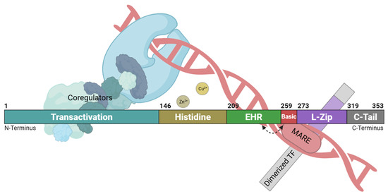
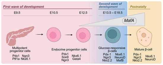
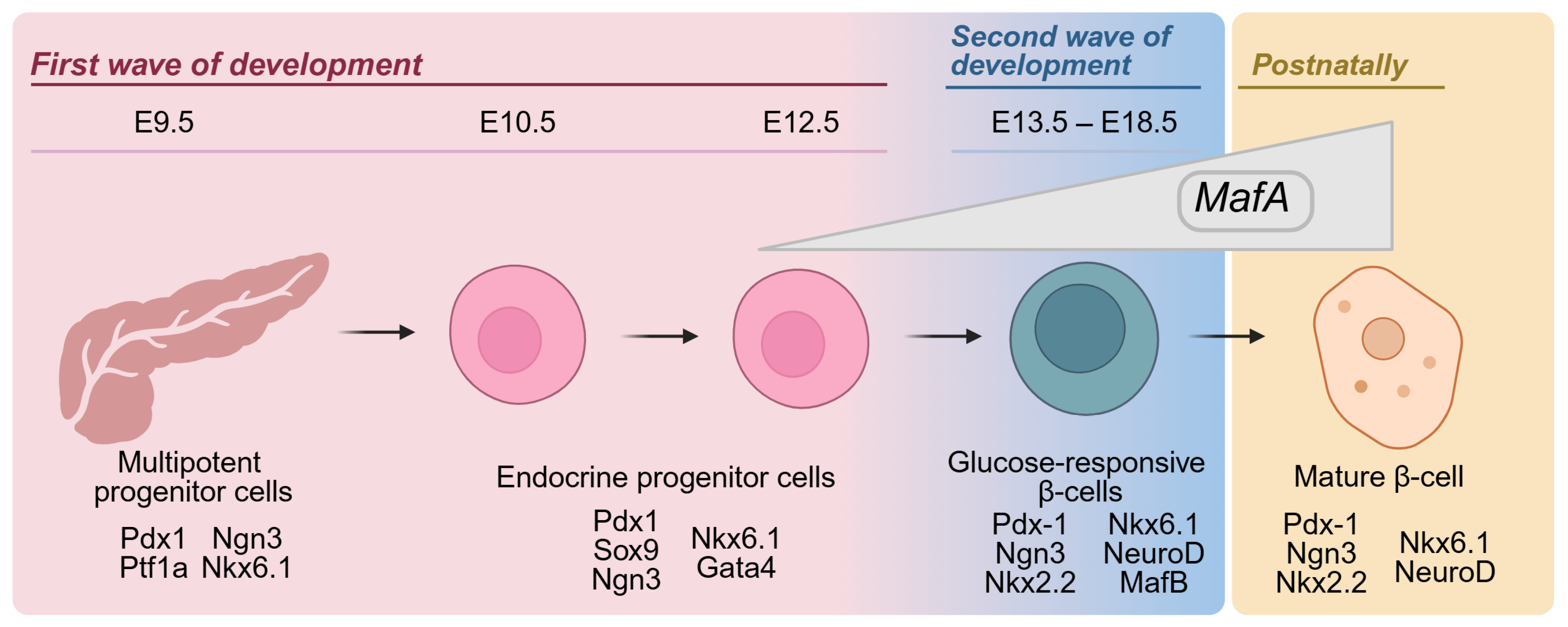

Disclaimer/Publisher’s Note: The statements, opinions and data contained in all publications are solely those of the individual author(s) and contributor(s) and not of MDPI and/or the editor(s). MDPI and/or the editor(s) disclaim responsibility for any injury to people or property resulting from any ideas, methods, instructions or products referred to in the content. |
© 2026 by the authors. Licensee MDPI, Basel, Switzerland. This article is an open access article distributed under the terms and conditions of the Creative Commons Attribution (CC BY) license.