


Abstract
Cell death is a vital physiological or pathological phenomenon in the development process of the organism. Ferroptosis is a kind of newly-discovered regulated cell death (RCD), which is different from other RCD patterns, such as apoptosis, necrosis and autophagy at the morphological, biochemical and genetic levels. It is a kind of iron-dependent mode of death mediated by lipid peroxides and lipid reactive oxygen species aggregation. Noteworthily, the number of studies focused on ferroptosis has been increasing exponentially since ferroptosis was first found in 2012. The liver is the organ that stores the most iron in the human body. Recently, it was frequently found that there are different degrees of iron metabolism disorder and lipid peroxidation and other ferroptosis characteristics in various liver diseases. Numerous investigators have discovered that the progression of various liver diseases can be affected via the regulation of ferroptosis, which may provide a potential therapeutic strategy for clinical hepatic diseases. This review aims to summarize the mechanism and update research progress of ferroptosis, so as to provide novel promising directions for the treatment of liver diseases.
1. Introduction
Liver disease with high morbidity and mortality accounts for the heavy burden of disease and costs worldwide. Statistically, about 2 million people around the world die of liver diseases [1]. Cell death plays a pleiotropic role in the development and process of various forms of liver diseases. Unlike accidental cell death triggered by serious mechanical, physical and chemical injury, regulated cell death (RCD) can be controlled pharmacologically and genetically. Thus, it is under the regulation of specific intracellular mechanisms [2]. A better understanding of the molecular mechanisms governing cell death is vital for confirming its role in various diseases. The first found form of RCD at the molecular level was caspase-dependent apoptosis. In recent years, necroptosis, autophagy, pyroptosis, and other forms of RCD have been found and explored. Notably, ferroptosis, a newly discovered form of RCD, found by Dixon and his team in 2012 [3], is considered as one of the most common and oldest forms of cell death. Ferroptosis-like cell death was not only observed in mammals, but also in remote species, such as plants, fungi and protozoa [4,5,6], etc. Over recent years, the field of ferroptosis research in liver diseases has been increased exponentially, since the concept “ferroptosis” was created in 2012.
Ferroptosis is an iron-dependent form of RCD, which is mediated by high levels of lipid peroxides and lipid reactive oxygen species (ROS) [7]. It is different from other types of RCD in cell morphology and biochemical characteristics. In morphological aspect, ferroptotic cells mainly show mitochondria shrinkage with accessorial membrane density, reduction or disappearance of mitochondrial crista, and the rupture of outer mitochondrial membrane, which are absolutely different features from other modes of RCD [7,8]. In contrast to apoptosis, ferroptosis does not show cell shrinkage, condensation of chromatin, apoptotic body formation and cytoskeleton disintegration. Compared to autophagy, ferroptosis lacks classical autophagic vacuoles, which is a kind of closed bilayer membrane structure. Unlike necrosis, ferroptosis does not exhibit cytoplasm and organelles swelling and cell membrane rupture. In biochemical aspects, ferroptotic cells show a variety of biological processes and chemical changes, including but not limited to iron, amino acid and polyunsaturated fatty acid, glutathione biosynthesis, GPX4, FSP1, GCH1, NADPH, and coenzyme Q10, etc. [9].
The liver is a pivotal organ for regulating various physiological metabolic processes, such as the metabolism of glucose, lipids and amino acids. Abnormal expression of these elements can result in oxidative stress or severe liver disease [10,11]. An increasing number of studies have shown that hepatic metabolic pathways are closely related to ferroptosis [12,13,14,15]. After dietary intake, glucose will enter the liver rapidly and sequester as glycogen. Glucose can generate energy through glycolysis and generate NADPH. The metabolic disorder of essential fatty acids is vitally important, since the excessive accumulation of lipid peroxidation is regarded as one of the critical characteristics of ferroptosis [16]. With respect to amino acids, some amino acids are directly associated with ferroptosis via regulating oxidative stress. Amino acids cannot transport nutrients such as sugar into cells directly. They must be diffused across cell membrane with the aid of specific transporters, such as system Xc−, which is comprised of SLC3A2 and SLC7A11. System Xc− accelerates the exchange of cystine and glutamate across the cell plasma membrane. Once transported into the cell, cystine will be reduced to cysteine, which is the limiting amino acid in GSH synthesis. Reduced GSH is the essential intracellular antioxidant in mammals and is generated from glycine, glutamate and cysteine. Thus, the biosynthesis of GSH is influenced by cystine and cysteine availability [17]. Inhibiting the import of cystine through system Xc− is sufficient to trigger ferroptosis via depleting GSH levels [18]. Of note, an increasing number of studies have shown that cystine deprivation can induce ferroptosis in a variety of diseases [19,20,21,22,23].
There are about 2 million people dying of different liver diseases per year worldwide. Some of them are due to the complications of cirrhosis, and others are due to acute or chronic hepatitis and hepatocellular carcinoma [1,24]. The liver is the most important organ for storing iron in the human body. A growing number of research studies indicated that ferroptosis plays an essential role in the progression of various forms of liver diseases, such as alcohol liver disease, virus hepatitis, non-alcohol steatohepatitis, liver fibrosis, hepatocellular carcinoma, etc. [25,26]. In this review, we summarize the recent advance of ferroptosis in liver diseases, proposing the possibility of explaining how ferroptosis cures or leads to diseases. We also discussed the possibility of treating clinical liver diseases through targeting ferroptosis-related genes.
2. Mechanisms Regulating Ferroptosis
As mentioned above, ferroptosis is a type of RCD, characterized by excessive lipid hydroperoxides caused by iron overload. Since ferroptosis, this newly discovered form of cell death was first reported in 2012 by Dixon et al. [3], and the mechanisms governing ferroptosis were gradually clearly explored, mainly focusing on the metabolism of cysteine and glutathione, GPX4 (the phospholipid peroxidase) [27], FSP1 (ferroptosis suppressor protein 1) [14] and GCH1/BH4 [28], etc. (Figure 1). Unrestrained peroxidation of phospholipids is the central part of ferroptosis. Phospholipids with polyunsaturated acyl tails (PUFA-PLs), because of their susceptibility to peroxidation chemistry, are the main substrate for peroxidation during the process of ferroptosis. Consistent with this, based on CRISPR/Cas9 and genome-wide haploid screening, two portal drivers of ferroptosis, acyl-CoA synthetase long-chain family member 4 (ACSL4) and lysophosphatidylcholine acyltransferase 3 (LPCAT3), were found, which could generate PUFA-PLs by activating and incorporating free PUFAs into phospholipids [29,30,31]. Thus, controlling the content of PUFA-PLs, the inducers of peroxidation and the factors that inhibit lipid peroxides are all strategies to regulate ferroptosis.
Figure 1.
Summary of core regulators and main mechanisms of ferroptosis. The major systems regulating ferroptosis include GPX4-related ferroptosis defense, FSP1-associated system, GCH1/BH4-related system and DHODH-mediated ferroptosis resistance. The core components and regulators involved in the regulation of ferroptosis are exhibited and the detailed description is described in the main text. Abbreviations: ACSL4: acyl-CoA synthetase long-chain family member 4; BH4: Tetrahydrobiopterin; CoQ: oxidized coenzyme Q; CoQH2: reduced coenzyme Q; GCH1: GTP Cyclohydrolase1; GPX4: glutathione peroxidase 4; GSH: reduced glutathione; GSSG: oxidized glutathione; IFN-γ: Interferon γ; LPCAT3: lysophosphatidylcholine acyltransferase 3; MDM2: Murine double minute 2; MDMX: Murine double minute X; MUFA: monounsaturated fatty acid; NAD(P)+: oxidized nicotinamide adenine dinucleotide (phosphate); NAD(P)H: reduced nicotinamide adenine dinucleotide (phosphate); Nrf2: nuclear factor erythroid 2-related factor2; PLOO·: phospholipid hydroperoxyl radical; PLOOH: phospholipid hydroperoxyl; PL-PUFA-OOH: polyunsaturated fatty acid-phospholipid ethanolamine hydroperoxide. The black arrow means to accelerate the process. The red T bar means to inhibit the process.
2.1. GPX4-Regulated Ferroptosis
In the early 1980s, Ursini and colleagues originally discovered glutathione peroxidase 4 (GPX4, a selenoprotein) through biochemical purification, and identified its role in catalyzing PLOOH to PLOH in mammalian cells [32,33]. As one of 25 specific selenoproteins in humans, the expression of GPX4 is precisely regulated [34] and GPX4 is the major PLOOH-neutralizing enzyme. It is worth noting that GSH, the richest reductant in mammalian cells, is the essential cofactor for glutathione peroxidase (GPX). GSH can be generated from cysteine that is generated from methionine by trans-sulfuration reaction [35], or from system Xc−, which pumps glutamate out and cystine into cells. Furthermore, cystine can be mutated into cysteine through redox reactions [2]. Previous research has shown that cyst(e)ine-deficient cells would die in a short time but increasing the synthesis of endogenous cysteine with methionine and glucose would reduce this kind of cell death [36,37]. System Xc− cystine/glutamate antiporter, composed by subunits SLC71A11 and SLC3A2, is the central part for the regulation of ferroptosis [38,39]. Further mechanistic investigation revealed that Erastin, RSL3 and ML210 promote ferroptosis by inhibiting system Xc− and GPX4, respectively [27,40,41,42]. Additionally, Wang and colleagues discovered IFN-γ released from CD8+ T cells could enhance ferroptosis in tumor cells by downregulating the expression of SLC3A2 and SLC7A11 [43]. In contrast, p62-Keap1-NRF2 was reported to diminish ferroptosis in cancer cells by upregulating SLC7A11 and increasing the secretion of glutamate [44].
2.2. FSP1-Regulated Ferroptosis
FSP1 (ferroptosis-suppressor-protein 1, formerly known as AIFM2) has the ability to inhibit ferroptosis, which was initially found through expression cloning technology in GPX4-deficient cells. Additionally, the ferroptosis-resistant function of FSP1 is independent of GSH content, GPX4 activity and ACSL4 levels [12,14]. Thus, FSP1 became the second major anti-ferroptosis mechanism after the cyst(e)ine–GSH–GPX4 axis. Mechanically, FSP1 acts as an oxidoreductase to convert ubiquinone (CoQ) to ubiquinol (CoQH2). CoQH2 functions as a lipophilic radical-trapping antioxidant to clear lipid peroxyl radicals [45]. Moreover, Verkatesh et al. discovered FSP1 can be activated by PPARα, which is directed by the MDM2.MDMX complex [46].
2.3. GCH1-Regulated Ferroptosis
GTP cyclohydrolase-1 (GCH1) and its antioxidant metabolites tetrahydrobiopterin/dihydrobiopterin (BH4/BH2) were found to have antagonistic properties against ferroptosis by genome-wide activation screen technology [28]. Mechanistically, a high expression of GCH1 and BH4/BH2 could lead to lipid remodeling, prevent the depletion of phospholipids with two polyunsaturated fatty acyl tails, and ultimately inhibit ferroptosis. In addition, the GCH1–BH4–phospholipid axis could also suppress the formation of PL–PUFA–OOH through regulating the abundance of CoQ [28].
2.4. Other Mechanisms Regulating Ferroptosis Sensitivity
After GPX4 and FSP1, the two major mechanisms against ferroptosis were discovered; Mao et al. [45] found supplementation with the product orotate and substrate dihydroorotate of dihydroorotate dehydrogenase (DHODH) could enhance or attenuate ferroptosis in GPX4low cancer cells. Mechanistically, the DHODH gene could encode dihydroorotate dehydrogenase, which is located on the outer surface of mitochondrial intima and involved in pyrimidine synthesis. Furthermore, DHODH works synergistically with mitochondrial GPX4 to suppress ferroptosis via reducing ubiquinone to ubiquinol.
What is more, while monounsaturated fatty acids (MUFA) are activated by ACSL3, the sensitivity of phospholipids to lethal oxidation can be reduced significantly in a short time [47]. Consequently, the process of ferroptosis is suppressed. Additionally, AMPK could inhibit ferroptosis by inhibiting ACC [48]. Furthermore, antagonizing the Merlin–Hippo signalling axis triggers the activity of YAP to induce ferroptosis by upregulation of ACSL4 and transferrin receptor [49]. Transcription factor p53 can decrease the uptake of cystine through inhibiting SLC7A11 expression, and ultimately increase cellular sensitivity to ferroptosis [50].
3. Links between Ferroptosis and Liver Diseases
Structurally, the liver is composed of parenchymal cells (hepatocytes) and nonparenchymal cells. Hepatocytes play a critical role in maintaining stable glucose and lipoprotein content in the plasma. Hepatocyte death occurs in almost every type of liver disease and is viewed as a major index for the examination of various acute and chronic hepatic diseases of toxic, viral, alcohol, metabolic or autoimmune origin [51]. Liver sinusoidal endothelial cells, hepatic macrophages and hepatic stellate cells (HSCs) constitute the liver microcirculatory milieu and control liver homeostasis. Macrophages are extensively involved in the initiation and development of a variety of liver diseases [52,53]. HSCs account for 5–8% of cells and store lots of vitamin A lipid droplets in a healthy liver [54,55]. When the liver is damaged by toxins, viral infection or other injury factors, HSCs can be activated and become proliferative, contractile and fibrogenic, serving as the major ECM producer in the liver, which is the main pathological feature of liver fibrosis [56]. Noteworthily, the occurrence and development of liver diseases are the result of crosstalk among different types of cells. Fortunately, increasing studies have shown that the progression of liver disease can be moderated by targeting the cells [57,58,59,60]. Iron overload and oxidative stress are the two most important causes of liver damage and disease development in most hepatopathies [61,62,63]. Ferroptosis, characterized by iron overloading and lipid peroxidation, has been widely reported to regulate multiple liver diseases by targeting the cells mentioned above. The following review will concentrate on summarizing the proven function of ferroptosis in a wide spectrum of hepatic diseases.
3.1. Ferroptosis and Acute Liver Injury
Acute liver injury (ALI) is a rare but life-threatening critical disease characterized by a rapid reduction in hepatocyte function that occurs most frequently in individuals without pre-existing liver disease [64,65]. Drugs, hepatic ischemia reperfusion injury (IRI), alcohol and viral hepatitis are common pathogenic factors of ALI [65]. Among the drugs with hepatotoxicity, the typical nonsteroidal anti-inflammatory drug paracetamol (N-acetyl-para-aminophenol, or APAP) is the leading cause of ALI [66].
3.1.1. Ferroptosis and APAP-Induced Acute Liver Injury
APAP-induced ALI often occurs after intentional overdose or unintentional ingestion (therapeutic error) and APAP-induced hepatoxicity is dose-dependent [66]. After APAP at the therapeutic dose enters the liver, approximately 85–90% of APAP undergoes a phase II metabolic reaction with the participation of UDP-glucuronyltransferase (UGTs) and sulfotransferase (SULTs) and transforms to non-toxic compounds, which are then excreted in urine [67,68]. About 2% of APAP is excreted in prototype form with urine [69]. The remaining 10% of APAP undergoes phase I oxidation with cytochrome P450 2E1 (CYP2E1) and is converted into a highly reactive and toxic intermediate, N-acetyl-para-benzoquinoneimine (NAPQI), which will combine with GSH to form non-toxic compounds and be excreted in urine [68,69,70]. Excessive APAP can cause the production of NAPQI, which will bind to GSH, further leading to the depletion of GSH in hepatocytes. Thus, GSH decline, iron overload and lipid peroxidation have been considered as the basic mechanisms of APAP-caused liver injury [71]. Increasing numbers of researchers have verified that ferroptosis is involved in APAP-induced cell death [72,73]. Lőrincz et al. [72] found that in addition to necroptosis and apoptosis, ferroptosis is also involved in APAP-induced hepatocyte death. Subsequently, Yamada et al. [26] reported that ferroptosis mediates the hepatotoxicity and mortality that APAP induces in a murine model. The ferroptosis inhibitor Ferrostatin (Fer-1), α-tocopherol and the iron chelator deferoxamine could effectively suppress APAP-induced hepatic injury, lipid peroxidation and GSH exhaustion. Mass spectrometry and gene inhibition of ACSL4 further revealed that radical oxidation of n-6 polyunsaturated fatty acids (mainly arachidonic acid) contributed to APAP-induced ferroptosis in hepatocytes. What is more, Wang et al. [74]. have verified that ulinastatin could reduce APAP-induced liver injury via suppressing ferroptosis through the SIRT1/Nrf2/HO-1 pathway.
In addition, some compounds, such as 3,4-dihydroxyphenylethyl alcohol glycoside isolated from sargentodoxa cuneata, have been proven to inhibit ferroptosis in APAP-induced liver injury [75]. In conclusion, ferroptosis may play an important role in APAP-induced ALI and a better understanding of the regulatory role of ferroptosis may provide a potential clinical therapeutic approach.
3.1.2. Ferroptosis and Ischemia-Reperfusion Injury (IRI)
IRI is the result of revascularization following a temporary reduction in blood supply [76], which will aggravate pre-existing disease in the liver. Liver IRI is still a tough clinical problem, which can be induced by liver transplantation, hepatic resection surgery and shock (such as sepsis and hemorrhage). Moreover, liver IRI can be divided into two phases: ischemia and reperfusion. These two stages are the consequence of oxidative stress accompanied by loss of blood flow, nutrition deficiency, inflammation and other mechanisms [77]. During reperfusion, iron-mediated death is thought to be related to oxidative stress from ROS [78,79]. What is more, ferroptosis is a form of iron-dependent cell death characterized by excessive accumulation of ROS. Friedmann Angeli et al. [80] reported that knockout of GPX4 in cells results in severe ferroptosis pathologically, and Liprostatin-1, a kind of ferroptosis inhibitor, can suppress ferroptosis significantly in GPX4−/− mice. Further pre-clinical trials show that kidney IRI and liver IRI are obviously decreased by Liprostatin-1, indicating that ferroptosis plays a significant role in IRI [80]. Wu et al. [53] found that targeting macrophages and inhibiting the release of macrophage extracellular traps (METs) could prevent iron overload and further inhibit hepatocyte ferroptosis, and eventually protect liver from damage caused by IRI. What is more, liver IRI can be significantly prevented by ferrostatin-1 and α-tocopherol or by an iron chelator such as deferoxamine [81]. Collectively, these studies demonstrated that ferroptosis is an innovative hazardous factor for liver IRI, and inhibition of ferroptosis may effectively relieve the symptoms of IRI.
3.2. Ferroptosis and Chronic Liver Injury
At present, chronic liver injury (CLI) is a kind of refractory public disease that impairs human health, with annual increasing incidence [82,83]. When the body is exposed to alcohol, harmful chemical materials such as CCl4, chronic infection viruses such as HBV/HCV or intermediary metabolism, the liver is the most susceptible and vulnerable organ with the occurrence of CLI [84,85]. The persistence of damaged elements will result in the excessive accumulation of lipids and peroxidation, which will further lead to chronic inflammation and steatohepatitis [86,87]. Then, the over-deposition of extracellular matrix (ECM) can disrupt the normal physiological architecture of the liver [55]. In the early stages, once the intervention is not performed in a timely manner, CLI will develop into hepatitis, fibrosis, cirrhosis and even hepatocellular carcinoma [88]. Recently, an increasing number of studies have demonstrated the function of ferroptosis in CLI. This will be described in detail in the following text.
3.2.1. Ferroptosis and Virus Hepatitis
Virus hepatitis is the leading cause of CLI, and chronic HCV infection promotes iron accumulation, upregulating duodenal ferroportin-1 and downregulating hepatic hepcidin, which is the master regulator of systemic iron homeostasis [89,90]. In hepatocytes, iron is stored in ferritin and exported by ferroportin. Hepcidin suppresses intracellular iron efflux via binding to and facilitating the degradation of ferroportin. Excessive iron accumulation directly correlates with the worsening of hepatic damage. Currently, whether ferroptosis plays an effective role in the development of HBV/HCV remains to be further explored. The study conducted by Yamane et al. [91]. showed that HCV replication was suppressed by ferroptosis and mediated by the non-classical desaturation of oleate to mead acid and other unsaturated fatty acids by fatty acid desaturase 2 (FADS2), which is the critical determinant of cell sensitivity to ferroptosis. Thus, inhibition of FADS2 significantly suppresses ferroptosis and further markedly enhances the replication of HCV [91]. Conversely, Erastin, the ferroptosis-inducing compound, changes the construction of HCV replicases and further sensitizes it to anti-virus agents targeting the viral protease [91]. What is more, Zhang et al. [92]. reported that the exosomes secreted by HBV-infected hepatocytes aggravate hepatic fibrosis through the miR-222/TFRC axis. Altogether, the above studies show that ferroptosis may serve as a new therapeutic strategy for the virus hepatitis.
3.2.2. Ferroptosis and Non-Alcoholic Fatty Liver Disease
Non-alcoholic fatty liver disease (NAFLD) is a kind of CLI that spans from simple steatosis to non-alcoholic steatohepatitis (NASH) and is assessed to influence up to one-third of the total population globally [93]. The development of NAFLD is closely related to obesity and metabolic syndrome [94]. Recently, Gautheron and his team proposed an iron-centered hypothesis during the progression of NAFLD [94]. It is widely known that excessive iron accumulation is a common index in patients with NAFLD, and lipid peroxide induced by iron is one of the prime contributors to NAFLD [95,96]. What is more, iron overload is closely related to insulin resistance and obesity, two representative traits of NAFLD patients [97]. Tsurusaki et al. [98]. verified that ferroptosis, but not necroptosis, is the major cell death to initiate inflammation and cell death in NASH, and the ferroptosis inhibitors (such as trolox or deferoxamine) can strongly reverse hepatocyte death, inflammation and lipid peroxidation during the initial phase of NASH. Iron overload triggered by metabolic dysfunction (such as hereditary hemochromatosis and liver ironosis) can accelerate the progression of NASH [99,100]. In addition, ferroptosis inhibitor (such as ferrostatin-1), and iron removal (with deferoxamine mesylate salt) can effectively improve liver injury in NAFLD [101,102,103,104]. In addition to iron accumulation, MDA and 4-HNE (two lipid peroxidation markers) are also increased in NASH patients [105]. The lipid peroxidation and serum transaminases are suppressed by Vitamin E in NASH patients [106]. All these studies indicate that ferroptosis induced by iron accumulation and lipid peroxidation might be associated with the pathological progression of NAFLD and that inhibition of ferroptosis might provide a novel therapeutic strategy for NAFLD.
Further mechanistic studies have found that GPX4 can slow down NASH progression via inhibiting ferroptosis, and RSL-3, an inhibitor of GPX4, aggravated the liver image of NASH by facilitating ferroptosis in MCD diet-fed mice. Conversely, sodium selenite, an activator of GPX4, improved the pathologic characteristics of NASH [102]. Consistently, liver cell death is also influenced by GPX4-induced ferroptosis in a palmitic acid-induced NASH model in vitro [107]. What is more, Thymosin β4 [103] and Enolase 3 [108] were reported to regulate the progression of NASH via inhibiting or increasing GPX4 expression. Wei et al. found that the expression of ACSL4 was elevated in arsenic-induced NASH models in vivo and in vitro. In addition, ACSL4 inhibition with rosiglitazone or specific siRNA can significantly suppress ferroptosis and further alleviate the progression of arsenic-induced NASH via restraining 5-HETE content [109]. Moreover, Nrf2 was also reported to regulate the synthesis of fatty acids by inhibiting enzyme expression and suppress NASH progression via the p62–Keap1–Nrf2 axis [110]. Additionally, Nrf2 plays an essential role in antioxidation response and alleviating ferroptosis. Notably, activation of the Nrf2 pathway can decrease liver lipid deposition and effectively improve the pathology of NAFLD in vivo [111]. Dehydroabietic acid [95] and Ginkgolide B [111] improve NAFLD via inhibiting ferroptosis through upregulating Nrf2 and its downstream genes (such as HO-1, GPX4, etc.). In addition, other mechanisms, such as ECH1 and miR-33, have also been reported to participate in the progression of NAFLD via regulating ferroptosis [112,113]. Altogether, studies of ferroptosis in NAFLD need further exploration in relevant animal models and in patients, especially since no accurate choices are available for NAFLD treatment at present.
3.2.3. Ferroptosis and Hepatic Fibrosis
Physiologically, hepatic stellate cells (HSCs) are quiescent and rich in vitamin A lipid droplets [54]. When the liver is injured by multi-stimuli such as toxins or chemical material, quiescent HSCs will differentiate into activated contractile, proliferative and fibrogenic HSCs, which are the leading target for the treatment of liver fibrosis [56]. Overexpression of TRIM26 inhibits HSCs proliferation, accelerates HSCs ferroptosis and further alleviates CCl4-induced liver fibrosis via interacting with SLC7A11 and promoting its ubiquitination [114]. In addition, many other compounds or plant extracts (such as magnesium isoglycyrrhizinate [115], artemether [116], artesunate [117], and Sorafenib [118], etc.) are able to target activated HSCs, trigger their ferroptosis and eventually slow down the progression of liver fibrosis. Moreover, ZFP36 [119], IRP2 [120], ELAVL1/HuR [121], and the BRD7–P53–SLC25A28 axis [122], etc. are all regulatory factors involved in the ferroptosis of HSCs. What is more, epigenetic m6A modification [123] was also reported to be associated with the process of ferroptosis in HSCs. Altogether, these results manifest that regulating ferroptosis by targeting HSCs may be a new therapeutic approach for treating liver fibrosis in the clinic. A growing number of studies indicate that superfluous iron is relevant to liver injury via ferroptosis [3,124]. Transferrin (Trf) is responsible for transferring Fe3+ to other organs or bone marrow. When the level of Trf decreases, excessive non-Trf-bound-iron (NTBI) will increase in hepatocytes. Further study showed that when Trf-KO mice were treated with ferrostatin, a ferroptosis inhibitor, the symptoms of liver fibrosis induced by either CCl4 injection or high dietary iron were rescued potently [125]. In addition, combined with the clinical data, we can conclude that Trf plays a protective role in liver fibrosis via suppressing ferroptosis [125].
3.2.4. Ferroptosis and Hepatocellular Carcinoma
In economically developed and developing countries, carcinoma is respectively the leading and second leading cause of cancer-associated death [126,127]. Among the primary hepatic cancers, hepatocellular carcinoma (HCC) takes up 70–85% of the total burden of liver cancer globally, representing the main histological subtype [128]. Surgical resection and conservative non-surgical treatment are both therapeutic strategies for advanced HCC, but both are of restricted treatment effect. Sorafenib, as the first and only approved first-line drug for advanced HCC, improves the survival rate of patients to some extent [129,130]. However, emerging drug resistance and severe adverse reactions to Sorafenib are the leading causes of poor clinical prognosis. Interestingly, a large number of researchers have manifested that Sorafenib can induce ferroptosis via inhibiting SLC7A11 [40,131,132], blocking oxidized cysteine from entering cells. Excessive intake of dietary iron can raise the risk of HCC, indicating that excessive iron content can accelerate the progression of HCC [133]. In addition, a growing number of pathways and mechanisms have been found to regulate Sorafenib resistance and the progression of HCC via aiming at ferroptosis [25,134]. Collectively, Sorafenib-induced ferroptosis may be an effective and promising strategy for the induction of cancer cell death in HCC. In this review, we will summarize recent studies on the relationship between HCC and ferroptosis, and further evaluate the possibility of ferroptosis as a therapeutic target for HCC.
In recent years, more and more studies have clarified the essential role of ferroptosis in HCC. Many genes, such as retinoblastoma (Rb) protein [135], Nrf2 [25], metallothionein-1G (MT-1G) [136], CDGSH iron sulfur domain 1 (CISD1, also named mitoNEET) [137], TP53 gene (S47 variant) [138], etc. are known as the negative regulators of ferroptosis. Louandre et al. [135] reported that the expression and function of Rb protein is reduced during liver carcinogenesis. Further examination showed that Rb-negative HCC cells are more sensitive to death induced by Sorafenib compared with Rb-expressing HCC cells. The transcription factor Nrf2 was originally considered as a pivotal regulator of antioxidant intercellularly and was negatively controlled by Keap1 [139]. Overexpression of Nrf2 induces apoptosis and caused chemoresistance in lung carcinoma, breast adenocarcinoma and nephroblastoma [140]. Moreover, Sun et al. [25] reported that the p62–Keap1–Nrf2 pathway plays a pivotal role in protecting HCC cells from ferroptosis induced by ferroptosis-inducing compounds (such as Sorafenib, Erastin, and buthionine sulfoximine) via increasing the expression of several Nrf2-targeting genes (NQO1, HO1, FTH1) associated with ROS and iron metabolism. The anticancer effect of Sorafenib and Erastin in HCC cells increased markedly while the expression of Nrf2 was inhibited in vivo and in vitro. When these Nrf2-targeted genes were knocked down, the growth inhibition of ferroptotic inducer-treated HCC cells was significantly increased [25]. Another gene that negatively regulates Sorafenib-induced ferroptosis is MT-1G, which has been a prospective therapeutic target for improving Sorafenib resistance in HCC cells [136]. In addition, CISD1, an iron-containing outer mitochondrial membrane protein, negatively regulates ferroptosis in HCC cells. The genetic knockdown of CISD1 enhances iron-mediated lipid peroxidation, resulting in Erastin-induced ferroptosis in human HCC cells, such as HepG2 and Hep3B [137]. What is more, Jennies et al. [138]. found that when TP53 gene mutated into mutant p53 with Pro47 (S47 variant), it exhibited a slight decrease in apoptosis induced by genotoxic stress but showed significant resistance to ferroptosis induced by Erastin. What is more, compared to WT p53 HCC cells, after cisplatin stimulus, the S47 variant cells showed a higher expression level of SLC7A11 and lower PTGS content (a biomarker of ferroptosis in vivo), which manifested the resistance to ferroptosis [138]. Haloperidol was reported to promote both Sorafenib and Erastin-induced ferroptosis at a comparatively low dose, which would benefit HCC patients treated with Sorafenib by decreasing the dosage or enhancing its curative effect [141]. The traits of ferroptosis such as excessive iron accumulation, GSH depletion and lipid peroxidation were detected following haloperidol administration. Meanwhile, Nrf2, HO-1, GPX4 and other ferroptosis-associated targets were influenced by haloperidol [141]. Altogether, the above studies provide an innovative strategy for the treatment of HCC, that is, inducing ferroptosis of HCC cells may act as a novel and promising approach for HCC.
The pivotal regulator of lipid peroxidation in ferroptosis needs further exploration, though that lipid peroxidation has as an essential role in promoting ferroptosis is known. Lipoxygenase, an iron-containing enzyme, is the central inducer of ferroptosis via generating lipid hydroperoxides, and its effect depends on the excitation of ACSL4-dependent lipid biosynthesis [142]. Feng and colleagues discovered that the expression of ACSL-4 can forecast sensitivity to Sorafenib in a series of HCC cell lines. ACSL4 inhibition by pharmacological methods and specific siRNA/sgRNA could significantly suppress Sorafenib-induced lipid peroxidation and ferroptotic cell death in Huh7 cells and rescue xenograft growth suppression in vivo [143]. In addition, low-density lipoprotein-docosahexaenoic acid (LDL-DHA) can also induce ferroptosis and further kill HCC cells, accompanied by GSH depletion and GPX4 inactivation [144]. Moreover, LDL-DHA nanoparticles were reported to lessen intracellular GSH via downregulating GSH/GSSG and NADPH/NADP+ (redox couples) and reducing GSH-aldehyde adducts in HCC cells [145]. Then, due to the elimination of substrate, GPX4 failed to function within its enzymatic activity. Recently, IFN-γ [146], YAP/TAZ and ATF4 [134], LIFR–NF–κB–LCN2 axis [147], lncRNA GABPB1-AS1 and GABPB1 [148], and circular RNA cIARS [149], etc. were all reported to play a functional role in the progression of HCC by targeting ferroptosis.
To sum up, a number of genes have been demonstrated to be capable of contributing to cell death via the ferroptosis pathway in HCC cells, which provides new molecular mechanisms of anti-hepatoma therapy. In addition to potential therapeutic effects, the prognosis prediction of patients with HCC is another important utility of ferroptosis. Intriguingly, a recent study detected a new ferroptosis-associated gene signature via leveraging a machine learning-based model to divide patients with HCC into two groups [150]. Fortunately, this signature can be utilized for prognosis prediction in HCC patients, proved by the truth that the overall survival rate decreased significantly in patients in the high-risk group compared to the low-risk group.
4. Pharmacologic Regulation of Ferroptosis
Many compounds directly or indirectly regulating ferroptosis (inducers or inhibitors) and ferroptosis-associated genes have been discovered and play essential roles in illuminating the mechanisms and progression of ferroptosis-associated diseases [45,151,152]. Apart from genetic regulators, iron overload, superabundant lipid peroxidation and some small molecules are also able to induce ferroptosis [2,3]. Up to now, the well-studied and widespread-used ferroptosis inducers (FINs) are categorized into four classes. Firstly, class I FINs activate ferroptosis via decreasing intracellular GSH and targeting system Xc− (such as Erastin, Erastin analogs (e.g., imidazole ketone Erastin and piperazine Erastin), sulfasalazine, Sorafenib and glutamate). Then, class II ferroptosis stimulants (e.g., RSL3 and ML162) induce ferroptosis via inactivating GPX4 directly, which suppress the function of GPX4 through covalent binding to GPX4, resulting in an excessive accumulation of lipid peroxides and ferroptosis [3,153]. Similarly, JKE-1674 and JKE-1716 can also trigger ferroptosis via covalently targeting selenocysteine residue of GPX4 [42,154]. On the contrary, class III ferroptosis inducers, such as FIN56, function via indirect suppressing and inactivating GPX4 through the squalene–mevalonate pathway [155]. Eventually, class IV FINs accelerate ferroptosis through iron overloading or through activating HO-1 [156]. Beyond these, a number of ferroptosis inducers do not belong to the above four categories and are also listed in Table 1. For instance, zalcitabine has the ability to damage mitochondrial DNA and induce autophagy-relevant ferroptosis in human pancreatic cancer cells [157]. In addition, Llabani et al. [158] identified that the compound ferroptocide served as a ferroptosis agonist via covalently binding to thioredoxin (TXN), an oxidoreductase.
Concerning ferroptosis inhibitors, iron chelation, lipophilic antioxidants and cleaning lipid peroxides are widely recognized as three main effective methods to inhibit ferroptosis. Deferoxamine (DFO), deferiprone, ciclopirox and other iron chelators can chelate iron and prevent lipid peroxidation via inhibiting the Fenton reaction [3]. Additionally, lipophilic antioxidants, such as α-tocopherol, Lip-1 and Fer-1 act as the radical scavengers to decrease lipid peroxides and ultimately [3,80]. However, too short a biological half-life may be the main reason that restricts Fer-1 to clinical application. In addition, necrostatin-1 (Nec-1) [159] (a necroptosis inhibitor), selenium [160], cycloheximide [3], beta-mercaptoethanol [3], etc., are identified as possessing the ability to inhibit ferroptosis. The identified ferroptosis inhibitors are listed in Table 2.
Table 2.
Major Inhibitors of Ferroptosis.
Table 1.
Major Inducers of Ferroptosis.
5. Summary and Prospect
Ferroptosis is a novel-identified cell death process inhibited by both iron elimination and lipophilic RTAs, such as ferrostatin-1, vitamin E, or CoQ10 [2]. Both conditions are essential and critical, because ferroptosis is not the only mode of iron-dependent cell death [173] that may be associated with lysosomal toxicity and oxidative stress iron-independent mechanisms [3]. Over the past few years, there has been a great improvement in our comprehension of the role of ferroptosis during the progression and pathogenesis of hepatic diseases. Based on the increasing studies of ferroptosis in the progression of a variety of liver diseases, drugs targeting ferroptosis act as a prospective therapeutic strategy (Table 3). This should be taken into account while designing therapeutic drugs targeting ferroptosis: ferroptosis is a two-edged sword. That is, ferroptosis can not only function as the liver fibrosis and HCC therapeutic strategy, but can also induce many other liver diseases, such as NAFLD, ALD, NASH, IRI, etc. As shown in Figure 1, the common mechanisms of ferroptosis include GPX4 inhibition, FSP1 suppression, GCH-1 restraint, system Xc− suppression, lipid peroxide accumulation and iron overload. It is worth noting that the key regulators of ferroptosis such as GPX4 [80,107], ACSL4 [109], SLC7A11 [114,118], p62–Keap1–Nrf2 signaling pathway [25,110], and p53 signaling pathway [122], etc., are of great significance for regulating ferroptosis-related liver diseases. In addition, as transcriptional coactivators, YAP and TAZ are vital regulators of ferroptosis in NAFLD [174], IRI [175], liver regeneration [176], liver fibrosis [177] and HCC [178]. It was found that Fuzheng Yanggan mixture has the ability to inhibit ferroptosis via upregulating the expression levels of GPX4 and SLC7A11, thereby preventing the liver from APAP-induced damage [179]. Additionally, the p62–Keap1–Nrf2 signaling pathway is closely associated with ferroptosis during the development of HCC. Sorafenib and Erastin exhibit their anticancer activity via promotion of ferroptosis and suppression of Nrf2 expression in HCC. Retinoblastoma and metallothionein are negative regulators of Nrf2 and are closely related to the progression of HCC [135,136]. Thus, drugs targeting the above components or signaling pathways may provide promising therapeutic methods for the clinical treatment of various liver diseases. As noted in Table 1, Table 2 and Table 3, this review offers a comprehensive summary of genes and drugs that can induce, inhibit or target ferroptosis, laying the foundation for the clinical treatment of liver diseases. What is more, it should be noted that though the treatment strategy based on ferroptosis is prospective for HCC, other normal cells and tissues can also be damaged by ferroptosis because of the lack of drug specificity during the treatment of HCC. Nanomedicine offers the possibility to achieve cancerous tissue targeted drug delivery due to the acidic tumor microenvironment or conjugating arginine–glycine–aspartic acid peptide, which will not appear in normal tissues [180,181,182]. Thus, ferroptosis-inducing nanomedicines via targeting the HCC microenvironment may be a potential effective approach for achieving targeted therapy for HCC. However, there still remain many challenges to overcome before ferroptosis can be applied to clinical practice.
Although much effort has been made in exploring the pathological role and mechanisms of ferroptosis in hepatic diseases, several critical and essential questions are waiting to be addressed and answered for future clinical application of ferroptosis-targeted therapies. Firstly, it is urgent and necessary to fill in the gap in exploring the downstream pathways or essential molecular junctions following the start of ferroptosis, which could lay a solid foundation for future diagnosis or treatment of clinical diseases via intervention of ferroptosis. Secondly, there is a serious lack of available clinical data involving ferroptosis [136,141], since the concept of ferroptosis was first proposed just in 2012 [3]. Thirdly, ferroptosis occurring in hepatocytes, HSCs [119] or liver tumor cells may result in disparate consequences, as shown in Figure 2. In general, hepatocyte ferroptosis can aggravate the progression of multiple liver diseases, while the ferroptosis of HSCs and HCC cells could repair the damage of liver fibrosis and HCC to a certain extent, respectively. Generally, many chronic liver injuries would eventually develop into liver fibrosis and even hepatocellular carcinoma. Thus, it may be fruitful to explore an optimal time point when intervention with ferroptosis can protect chronic liver injury from progression to hepatocellular carcinoma. Fourthly, numerous studies have shown that different types of regulated cell death (e.g., ferroptosis, apoptosis, necroptosis, pyroptosis, and autophagy) can simultaneously coexist in a pathological environment, and several RCD patterns share overlapping mechanisms which may serve as the “backup” dying tactics to maintain body homeostasis when the cellular death-inducing threshold is reached [94,102,183,184]. Arakawa et al. [185]. reported that Atg5/Bax/Bak triple-knockout (TKO) mice showed more severe development abnormalities than Bax/Bak double-knockout (DKO) mice, indicating that autophagic cell death can compensate for the dysregulation or deficiency of apoptosis in mammal development. Likewise, autophagy-dependent death deficiency can also be compensated by apoptosis. Another case in point is that researchers found that ferroptosis can be promoted by autophagy via iron overloaded inhibition of system Xc−, and removing Atg5/Atg7 inhibited Erastin-induced ferroptosis [63,186,187]. The interplay during multiple regulated cell deaths makes it difficult to get a precise perception of ferroptosis in liver diseases.
Figure 2.
Various effects of ferroptosis on different liver diseases. In most cases, hepatocyte ferroptosis can aggravate the progression of liver damage. However, ferroptosis occurring in HSCs and liver cancer cells can improve the symptoms of liver fibrosis and hepatocellular carcinoma, respectively. Abbreviations: ALI: acute liver injury; HSC: hepatocyte stellate cell; IRI: ischemia reperfusion injury; NAFLD: non-alcoholic fatty liver disease.
At present, there are still many other questions about ferroptosis that need to be addressed. For instance, how do we identify sensitive and reliable biomarkers of ferroptosis in various liver diseases? How can we find out the specific time point when we can target ferroptosis in different pathological liver disease stages? How can we activate ferroptosis particularly in liver cancer cells without damaging other healthy cells? Solving the key scientific issues mentioned above in this review will improve our comprehension of the important role that ferroptosis plays in multiple pathological liver conditions, hence supplying a scientific theoretical basis for the treatment and prevention of liver diseases via the targeting of ferroptosis.
Table 3.
Drugs Targeting Ferroptosis in Liver Diseases.
Author Contributions
H.Y., L.W. and F.B. wrote the manuscript, analyzed articles, and finalized the figures. H.M. and C.H. collected the relevant studies. J.L. and G.F. participated in the design of this paper and revised the manuscript. All authors have read and agreed to the published version of the manuscript.
Funding
This study was supported by grants from the National Natural Science Foundation of China (No. U19A2001).
Institutional Review Board Statement
Not applicable.
Informed Consent Statement
Not applicable.
Data Availability Statement
Not applicable.
Conflicts of Interest
The authors declare no conflict of interest for this article.
References
- Asrani, S.K.; Devarbhavi, H.; Eaton, J.; Kamath, P.S. Burden of liver diseases in the world. J. Hepatol. 2019, 70, 151–171. [Google Scholar] [CrossRef] [Scilit] [PubMed]
- Stockwell, B.R.; Friedmann Angeli, J.P.; Bayir, H.; Bush, A.I.; Conrad, M.; Dixon, S.J.; Fulda, S.; Gascon, S.; Hatzios, S.K.; Kagan, V.E.; et al. Ferroptosis: A Regulated Cell Death Nexus Linking Metabolism, Redox. Biol.ogy, and Disease. Cell 2017, 171, 273–285. [Google Scholar] [CrossRef] [Scilit] [PubMed]
- Dixon, S.J.; Lemberg, K.M.; Lamprecht, M.R.; Skouta, R.; Zaitsev, E.M.; Gleason, C.E.; Patel, D.N.; Bauer, A.J.; Cantley, A.M.; Yang, W.S.; et al. Ferroptosis: An iron-dependent form of nonapoptotic cell death. Cell 2012, 149, 1060–1072. [Google Scholar] [CrossRef] [Scilit] [PubMed]
- Bogacz, M.; Krauth-Siegel, R.L. Tryparedoxin peroxidase-deficiency commits trypanosomes to ferroptosis-type cell death. Elife 2018, 7, e37053. [Google Scholar] [CrossRef] [Scilit] [PubMed]
- Distefano, A.M.; Martin, M.V.; Cordoba, J.P.; Bellido, A.M.; D’Ippolito, S.; Colman, S.L.; Soto, D.; Roldan, J.A.; Bartoli, C.G.; Zabaleta, E.J.; et al. Heat stress induces ferroptosis-like cell death in plants. J. Cell Biol. 2017, 216, 463–476. [Google Scholar] [CrossRef] [Scilit]
- Shen, Q.; Liang, M.; Yang, F.; Deng, Y.Z.; Naqvi, N.I. Ferroptosis contributes to developmental cell death in rice blast. New Phytol. 2020, 227, 1831–1846. [Google Scholar] [CrossRef] [Scilit]
- Li, J.; Cao, F.; Yin, H.L.; Huang, Z.J.; Lin, Z.T.; Mao, N.; Sun, B.; Wang, G. Ferroptosis: Past, present and future. Cell Death Dis. 2020, 11, 88. [Google Scholar] [CrossRef] [Scilit]
- Yang, W.S.; Stockwell, B.R. Synthetic lethal screening identifies compounds activating iron-dependent, nonapoptotic cell death in oncogenic-RAS-harboring cancer cells. Chem. Biol. 2008, 15, 234–245. [Google Scholar] [CrossRef] [Scilit]
- Hadian, K.; Stockwell, B.R. SnapShot: Ferroptosis. Cell 2020, 181, 1188–1188.e1. [Google Scholar] [CrossRef] [Scilit]
- Cusi, K. Role of obesity and lipotoxicity in the development of nonalcoholic steatohepatitis: Pathophysiology and clinical implications. Gastroenterology 2012, 142, 711–725.e716. [Google Scholar] [CrossRef] [Scilit]
- Masoodi, M.; Gastaldelli, A.; Hyotylainen, T.; Arretxe, E.; Alonso, C.; Gaggini, M.; Brosnan, J.; Anstee, Q.M.; Millet, O.; Ortiz, P.; et al. Metabolomics and lipidomics in NAFLD: Biomarkers and non-invasive diagnostic tests. Nat. Rev. Gastroenterol. Hepatol. 2021, 18, 835–856. [Google Scholar] [CrossRef] [Scilit] [PubMed]
- Bersuker, K.; Hendricks, J.M.; Li, Z.; Magtanong, L.; Ford, B.; Tang, P.H.; Roberts, M.A.; Tong, B.; Maimone, T.J.; Zoncu, R.; et al. The CoQ oxidoreductase FSP1 acts parallel to GPX4 to inhibit ferroptosis. Nature 2019, 575, 688–692. [Google Scholar] [CrossRef] [Scilit] [PubMed]
- Shimada, K.; Hayano, M.; Pagano, N.C.; Stockwell, B.R. Cell-Line Selectivity Improves the Predictive Power of Pharmacogenomic Analyses and Helps Identify NADPH as Biomarker for Ferroptosis Sensitivity. Cell Chem. Biol. 2016, 23, 225–235. [Google Scholar] [CrossRef] [Scilit] [PubMed]
- Doll, S.; Freitas, F.P.; Shah, R.; Aldrovandi, M.; da Silva, M.C.; Ingold, I.; Goya Grocin, A.; Xavier da Silva, T.N.; Panzilius, E.; Scheel, C.H.; et al. FSP1 is a glutathione-independent ferroptosis suppressor. Nature 2019, 575, 693–698. [Google Scholar] [CrossRef] [Scilit] [PubMed]
- Chen, S.; Zhu, J.Y.; Zang, X.; Zhai, Y.Z. The Emerging Role of Ferroptosis in Liver Diseases. Front. Cell Dev. Biol. 2021, 9, 801365. [Google Scholar] [CrossRef] [Scilit]
- Yang, W.S.; Stockwell, B.R. Ferroptosis: Death by Lipid Peroxidation. Trends Cell Biol. 2016, 26, 165–176. [Google Scholar] [CrossRef] [Scilit]
- Liang, C.; Zhang, X.; Yang, M.; Dong, X. Recent Progress in Ferroptosis Inducers for Cancer Therapy. Adv. Mater. 2019, 31, e1904197. [Google Scholar] [CrossRef] [Scilit]
- Sun, Y.; Zheng, Y.; Wang, C.; Liu, Y. Glutathione depletion induces ferroptosis, autophagy, and premature cell senescence in retinal pigment epithelial cells. Cell Death Dis. 2018, 9, 753. [Google Scholar] [CrossRef] [Scilit]
- Kang, Y.P.; Mockabee-Macias, A.; Jiang, C.; Falzone, A.; Prieto-Farigua, N.; Stone, E.; Harris, I.S.; DeNicola, G.M. Non-canonical Glutamate-Cysteine Ligase Activity Protects against Ferroptosis. Cell Metab. 2021, 33, 174–189.e7. [Google Scholar] [CrossRef] [Scilit]
- Tarangelo, A.; Magtanong, L.; Bieging-Rolett, K.T.; Li, Y.; Ye, J.; Attardi, L.D.; Dixon, S.J. p53 Suppresses Metabolic Stress-Induced Ferroptosis in Cancer Cells. Cell Rep. 2018, 22, 569–575. [Google Scholar] [CrossRef] [Scilit]
- Wang, X.; Li, Y.; Li, Z.; Lin, S.; Wang, H.; Sun, J.; Lan, C.; Wu, L.; Sun, D.; Huang, C.; et al. Mitochondrial Calcium Uniporter Drives Metastasis and Confers a Targetable Cystine Dependency in Pancreatic Cancer. Cancer Res. 2022, 82, 2254–2268. [Google Scholar] [CrossRef] [Scilit] [PubMed]
- Badgley, M.A.; Kremer, D.M.; Maurer, H.C.; DelGiorno, K.E.; Lee, H.J.; Purohit, V.; Sagalovskiy, I.R.; Ma, A.; Kapilian, J.; Firl, C.E.M.; et al. Cysteine depletion induces pancreatic tumor ferroptosis in mice. Science 2020, 368, 85–89. [Google Scholar] [CrossRef] [Scilit] [PubMed]
- Kim, D.H.; Kim, W.D.; Kim, S.K.; Moon, D.H.; Lee, S.J. TGF-beta1-mediated repression of SLC7A11 drives vulnerability to GPX4 inhibition in hepatocellular carcinoma cells. Cell Death Dis. 2020, 11, 406. [Google Scholar] [CrossRef] [Scilit]
- Mokdad, A.A.; Lopez, A.D.; Shahraz, S.; Lozano, R.; Mokdad, A.H.; Stanaway, J.; Murray, C.J.; Naghavi, M. Liver cirrhosis mortality in 187 countries between 1980 and 2010: A systematic analysis. BMC Med. 2014, 12, 145. [Google Scholar] [CrossRef] [Scilit] [PubMed]
- Sun, X.; Ou, Z.; Chen, R.; Niu, X.; Chen, D.; Kang, R.; Tang, D. Activation of the p62-Keap1-NRF2 pathway protects against ferroptosis in hepatocellular carcinoma cells. Hepatology 2016, 63, 173–184. [Google Scholar] [CrossRef] [Scilit]
- Yamada, N.; Karasawa, T.; Kimura, H.; Watanabe, S.; Komada, T.; Kamata, R.; Sampilvanjil, A.; Ito, J.; Nakagawa, K.; Kuwata, H.; et al. Ferroptosis driven by radical oxidation of n-6 polyunsaturated fatty acids mediates acetaminophen-induced acute liver failure. Cell Death Dis. 2020, 11, 144. [Google Scholar] [CrossRef] [Scilit]
- Yang, W.S.; SriRamaratnam, R.; Welsch, M.E.; Shimada, K.; Skouta, R.; Viswanathan, V.S.; Cheah, J.H.; Clemons, P.A.; Shamji, A.F.; Clish, C.B.; et al. Regulation of ferroptotic cancer cell death by GPX4. Cell 2014, 156, 317–331. [Google Scholar] [CrossRef] [Scilit]
- Kraft, V.A.N.; Bezjian, C.T.; Pfeiffer, S.; Ringelstetter, L.; Muller, C.; Zandkarimi, F.; Merl-Pham, J.; Bao, X.; Anastasov, N.; Kossl, J.; et al. GTP Cyclohydrolase 1/Tetrahydrobiopterin Counteract Ferroptosis through Lipid Remodeling. ACS Cent. Sci. 2020, 6, 41–53. [Google Scholar] [CrossRef] [Scilit]
- Dixon, S.J.; Winter, G.E.; Musavi, L.S.; Lee, E.D.; Snijder, B.; Rebsamen, M.; Superti-Furga, G.; Stockwell, B.R. Human Haploid Cell Genetics Reveals Roles for Lipid Metabolism Genes in Nonapoptotic Cell Death. ACS Chem. Biol. 2015, 10, 1604–1609. [Google Scholar] [CrossRef] [Scilit]
- Doll, S.; Proneth, B.; Tyurina, Y.Y.; Panzilius, E.; Kobayashi, S.; Ingold, I.; Irmler, M.; Beckers, J.; Aichler, M.; Walch, A.; et al. ACSL4 dictates ferroptosis sensitivity by shaping cellular lipid composition. Nat. Chem. Biol. 2017, 13, 91–98. [Google Scholar] [CrossRef] [Scilit]
- Zou, Y.; Palte, M.J.; Deik, A.A.; Li, H.; Eaton, J.K.; Wang, W.; Tseng, Y.Y.; Deasy, R.; Kost-Alimova, M.; Dancik, V.; et al. A GPX4-dependent cancer cell state underlies the clear-cell morphology and confers sensitivity to ferroptosis. Nat. Commun 2019, 10, 1617. [Google Scholar] [CrossRef] [Scilit]
- Ursini, F.; Maiorino, M.; Valente, M.; Ferri, L.; Gregolin, C. Purification from pig liver of a protein which protects liposomes and biomembranes from peroxidative degradation and exhibits glutathione peroxidase activity on phosphatidylcholine hydroperoxides. Biochim. Biophys. Acta Lipids Lipid Metab. 1982, 710, 197–211. [Google Scholar] [CrossRef] [Scilit]
- Ursini, F.; Maiorino, M.; Gregolin, C. The selenoenzyme phospholipid hydroperoxide glutathione peroxidase. Biochim. Biophys. Acta Gen. Subj. 1985, 839, 62–70. [Google Scholar] [CrossRef] [Scilit]
- Conrad, M.; Proneth, B. Selenium: Tracing Another Essential Element of Ferroptotic Cell Death. Cell Chem. Biol. 2020, 27, 409–419. [Google Scholar] [CrossRef] [Scilit] [PubMed]
- McBean, G.J. The transsulfuration pathway: A source of cysteine for glutathione in astrocytes. Amino Acids 2012, 42, 199–205. [Google Scholar] [CrossRef] [Scilit]
- Coltorti, M.; De Ritis, F.; Giusti, G. Enzymatic mechanisms of transsulfuration in biology and clinical practice. G. Di Clin. Med. 1956, 37, 285–323. [Google Scholar] [PubMed]
- Eagle, H.; Piez, K.A.; Oyama, V.I. The biosynthesis of cystine in human cell cultures. J. Biol. Chem. 1961, 236, 1425–1428. [Google Scholar] [CrossRef] [Scilit]
- Bannai, S.; Kitamura, E. Transport interaction of L-cystine and L-glutamate in human diploid fibroblasts in culture. J. Biol. Chem. 1980, 255, 2372–2376. [Google Scholar] [CrossRef] [Scilit]
- Sato, H.; Tamba, M.; Ishii, T.; Bannai, S. Cloning and expression of a plasma membrane cystine/glutamate exchange transporter composed of two distinct proteins. J. Biol. Chem. 1999, 274, 11455–11458. [Google Scholar] [CrossRef] [Scilit]
- Dixon, S.J.; Patel, D.N.; Welsch, M.; Skouta, R.; Lee, E.D.; Hayano, M.; Thomas, A.G.; Gleason, C.E.; Tatonetti, N.P.; Slusher, B.S.; et al. Pharmacological inhibition of cystine-glutamate exchange induces endoplasmic reticulum stress and ferroptosis. Elife 2014, 3, e02523. [Google Scholar] [CrossRef] [Scilit]
- Conrad, M.; Angeli, J.P.; Vandenabeele, P.; Stockwell, B.R. Regulated necrosis: Disease relevance and therapeutic opportunities. Nat. Rev. Drug Discov. 2016, 15, 348–366. [Google Scholar] [CrossRef] [Scilit] [PubMed]
- Eaton, J.K.; Furst, L.; Ruberto, R.A.; Moosmayer, D.; Hilpmann, A.; Ryan, M.J.; Zimmermann, K.; Cai, L.L.; Niehues, M.; Badock, V.; et al. Selective covalent targeting of GPX4 using masked nitrile-oxide electrophiles. Nat. Chem. Biol. 2020, 16, 497–506. [Google Scholar] [CrossRef] [Scilit] [PubMed]
- Wang, W.; Green, M.; Choi, J.E.; Gijon, M.; Kennedy, P.D.; Johnson, J.K.; Liao, P.; Lang, X.; Kryczek, I.; Sell, A.; et al. CD8+ T cells regulate tumour ferroptosis during cancer immunotherapy. Nature 2019, 569, 270–274. [Google Scholar] [CrossRef] [Scilit] [PubMed]
- Fan, Z.; Wirth, A.K.; Chen, D.; Wruck, C.J.; Rauh, M.; Buchfelder, M.; Savaskan, N. Nrf2-Keap1 pathway promotes cell proliferation and diminishes ferroptosis. Oncogenesis 2017, 6, e371. [Google Scholar] [CrossRef] [Scilit]
- Mao, C.; Liu, X.; Zhang, Y.; Lei, G.; Yan, Y.; Lee, H.; Koppula, P.; Wu, S.; Zhuang, L.; Fang, B.; et al. DHODH-mediated ferroptosis defence is a targetable vulnerability in cancer. Nature 2021, 593, 586–590. [Google Scholar] [CrossRef] [Scilit]
- Venkatesh, D.; O’Brien, N.A.; Zandkarimi, F.; Tong, D.R.; Stokes, M.E.; Dunn, D.E.; Kengmana, E.S.; Aron, A.T.; Klein, A.M.; Csuka, J.M.; et al. MDM2 and MDMX promote ferroptosis by PPARalpha-mediated lipid remodeling. Genes Dev. 2020, 34, 526–543. [Google Scholar] [CrossRef] [Scilit]
- Magtanong, L.; Ko, P.J.; To, M.; Cao, J.Y.; Forcina, G.C.; Tarangelo, A.; Ward, C.C.; Cho, K.; Patti, G.J.; Nomura, D.K.; et al. Exogenous Monounsaturated Fatty Acids Promote a Ferroptosis-Resistant Cell State. Cell Chem. Biol. 2019, 26, 420–432.e9. [Google Scholar] [CrossRef] [Scilit]
- Lee, H.; Zandkarimi, F.; Zhang, Y.; Meena, J.K.; Kim, J.; Zhuang, L.; Tyagi, S.; Ma, L.; Westbrook, T.F.; Steinberg, G.R.; et al. Energy-stress-mediated AMPK activation inhibits ferroptosis. Nat. Cell Biol. 2020, 22, 225–234. [Google Scholar] [CrossRef] [Scilit]
- Wu, J.; Minikes, A.M.; Gao, M.; Bian, H.; Li, Y.; Stockwell, B.R.; Chen, Z.N.; Jiang, X. Intercellular interaction dictates cancer cell ferroptosis via NF2-YAP signalling. Nature 2019, 572, 402–406. [Google Scholar] [CrossRef] [Scilit]
- Jiang, L.; Kon, N.; Li, T.; Wang, S.J.; Su, T.; Hibshoosh, H.; Baer, R.; Gu, W. Ferroptosis as a p53-mediated activity during tumour suppression. Nature 2015, 520, 57–62. [Google Scholar] [CrossRef] [Scilit]
- Luedde, T.; Kaplowitz, N.; Schwabe, R.F. Cell death and cell death responses in liver disease: Mechanisms and clinical relevance. Gastroenterology 2014, 147, 765–783.e4. [Google Scholar] [CrossRef] [Scilit] [PubMed]
- Wang, C.; Ma, C.; Gong, L.; Guo, Y.; Fu, K.; Zhang, Y.; Zhou, H.; Li, Y. Macrophage Polarization and Its Role in Liver Disease. Front. Immunol. 2021, 12, 803037. [Google Scholar] [CrossRef] [Scilit] [PubMed]
- Wu, S.; Yang, J.; Sun, G.; Hu, J.; Zhang, Q.; Cai, J.; Yuan, D.; Li, H.; Hei, Z.; Yao, W. Macrophage extracellular traps aggravate iron overload-related liver ischaemia/reperfusion injury. Br. J. Pharmacol. 2021, 178, 3783–3796. [Google Scholar] [CrossRef] [Scilit]
- Blaner, W.S.; O’Byrne, S.M.; Wongsiriroj, N.; Kluwe, J.; D’Ambrosio, D.M.; Jiang, H.; Schwabe, R.F.; Hillman, E.M.; Piantedosi, R.; Libien, J. Hepatic stellate cell lipid droplets: A specialized lipid droplet for retinoid storage. Biochim. Biophys. Acta 2009, 1791, 467–473. [Google Scholar] [CrossRef] [Scilit] [PubMed]
- You, H.; Wang, L.; Bu, F.; Meng, H.; Pan, X.; Li, J.; Zhang, Y.; Wang, A.; Yin, N.; Huang, C.; et al. The miR-455-3p/HDAC2 axis plays a pivotal role in the progression and reversal of liver fibrosis and is regulated by epigenetics. FASEB J. 2021, 35, e21700. [Google Scholar] [CrossRef] [Scilit] [PubMed]
- Yin, C.; Evason, K.J.; Asahina, K.; Stainier, D.Y. Hepatic stellate cells in liver development, regeneration, and cancer. J. Clin. Investig. 2013, 123, 1902–1910. [Google Scholar] [CrossRef] [Scilit]
- Higashi, T.; Friedman, S.L.; Hoshida, Y. Hepatic stellate cells as key target in liver fibrosis. Adv. Drug Deliv. Rev. 2017, 121, 27–42. [Google Scholar] [CrossRef] [Scilit] [PubMed]
- Krenkel, O.; Tacke, F. Liver macrophages in tissue homeostasis and disease. Nat. Rev. Immunol. 2017, 17, 306–321. [Google Scholar] [CrossRef] [Scilit]
- Michalopoulos, G.K.; Bhushan, B. Liver regeneration: Biological and pathological mechanisms and implications. Nat. Rev. Gastroenterol. Hepatol. 2021, 18, 40–55. [Google Scholar] [CrossRef] [Scilit]
- Poisson, J.; Lemoinne, S.; Boulanger, C.; Durand, F.; Moreau, R.; Valla, D.; Rautou, P.E. Liver sinusoidal endothelial cells: Physiology and role in liver diseases. J. Hepatol. 2017, 66, 212–227. [Google Scholar] [CrossRef] [Scilit]
- Brown, K.E.; Dennery, P.A.; Ridnour, L.A.; Fimmel, C.J.; Kladney, R.D.; Brunt, E.M.; Spitz, D.R. Effect of iron overload and dietary fat on indices of oxidative stress and hepatic fibrogenesis in rats. Liver Int. 2003, 23, 232–242. [Google Scholar] [CrossRef] [Scilit]
- Mancardi, D.; Mezzanotte, M.; Arrigo, E.; Barinotti, A.; Roetto, A. Iron Overload, Oxidative Stress, and Ferroptosis in the Failing Heart and Liver. Antioxidants 2021, 10, 1864. [Google Scholar] [CrossRef] [Scilit] [PubMed]
- Wu, J.; Wang, Y.; Jiang, R.; Xue, R.; Yin, X.; Wu, M.; Meng, Q. Ferroptosis in liver disease: New insights into disease mechanisms. Cell Death Dis. Cov. 2021, 7, 276. [Google Scholar] [CrossRef] [Scilit] [PubMed]
- Bernal, W.; Wendon, J. Acute liver failure. N. Engl. J. Med. 2013, 369, 2525–2534. [Google Scholar] [CrossRef] [Scilit] [PubMed]
- Stravitz, R.T.; Lee, W.M. Acute liver failure. Lancet 2019, 394, 869–881. [Google Scholar] [CrossRef] [Scilit]
- Bunchorntavakul, C.; Reddy, K.R. Acetaminophen (APAP or N-Acetyl-p-Aminophenol) and Acute Liver Failure. Clin. Liver Dis. 2018, 22, 325–346. [Google Scholar] [CrossRef] [Scilit]
- Bunchorntavakul, C.; Reddy, K.R. Acetaminophen-related hepatotoxicity. Clin. Liver Dis. 2013, 17, 587–607. [Google Scholar] [CrossRef] [Scilit]
- Larson, A.M. Acetaminophen hepatotoxicity. Clin. Liver Dis. 2007, 11, 525–548. [Google Scholar] [CrossRef] [Scilit]
- Hodgman, M.J.; Garrard, A.R. A review of acetaminophen poisoning. Crit. Care Clin. 2012, 28, 499–516. [Google Scholar] [CrossRef] [Scilit]
- Graham, G.G.; Scott, K.F.; Day, R.O. Tolerability of paracetamol. Drug Saf. 2005, 28, 227–240. [Google Scholar] [CrossRef] [Scilit]
- Krenkel, O.; Mossanen, J.C.; Tacke, F. Immune mechanisms in acetaminophen-induced acute liver failure. Hepatobiliary Surg. Nutr. 2014, 3, 331–343. [Google Scholar] [CrossRef] [Scilit] [PubMed]
- Lorincz, T.; Jemnitz, K.; Kardon, T.; Mandl, J.; Szarka, A. Ferroptosis is Involved in Acetaminophen Induced Cell Death. Pathol. Oncol. Res. 2015, 21, 1115–1121. [Google Scholar] [CrossRef] [Scilit] [PubMed]
- Shojaie, L.; Iorga, A.; Dara, L. Cell Death in Liver Diseases: A Review. Int. J. Mol. Sci. 2020, 21, 9628. [Google Scholar] [CrossRef] [Scilit] [PubMed]
- Wang, C.; Liu, T.; Tong, Y.; Cui, R.; Qu, K.; Liu, C.; Zhang, J. Ulinastatin protects against acetaminophen-induced liver injury by alleviating ferroptosis via the SIRT1/NRF2/HO-1 pathway. Am. J. Transl. Res. 2021, 13, 6031–6042. [Google Scholar]
- Liu, T.; Yang, L.; Gao, H.; Zhuo, Y.; Tu, Z.; Wang, Y.; Xun, J.; Zhang, Q.; Zhang, L.; Wang, X. 3,4-dihydroxyphenylethyl alcohol glycoside reduces acetaminophen-induced acute liver failure in mice by inhibiting hepatocyte ferroptosis and pyroptosis. PeerJ 2022, 10, e13082. [Google Scholar] [CrossRef] [Scilit]
- Saidi, R.F.; Kenari, S.K. Liver ischemia/reperfusion injury: An overview. J. Investig. Surg. 2014, 27, 366–379. [Google Scholar] [CrossRef] [Scilit]
- Decuypere, J.P.; Ceulemans, L.J.; Agostinis, P.; Monbaliu, D.; Naesens, M.; Pirenne, J.; Jochmans, I. Autophagy and the Kidney: Implications for Ischemia-Reperfusion Injury and Therapy. Am. J. Kidney Dis. 2015, 66, 699–709. [Google Scholar] [CrossRef] [Scilit]
- Oliveira, T.H.C.; Marques, P.E.; Proost, P.; Teixeira, M.M.M. Neutrophils: A cornerstone of liver ischemia and reperfusion injury. Lab. Invest. 2018, 98, 51–62. [Google Scholar] [CrossRef] [Scilit]
- Luo, L.; Mo, G.; Huang, D. Ferroptosis in hepatic ischemiareperfusion injury: Regulatory mechanisms and new methods for therapy (Review). Mol. Med. Rep. 2021, 23, 225. [Google Scholar] [CrossRef] [Scilit]
- Friedmann Angeli, J.P.; Schneider, M.; Proneth, B.; Tyurina, Y.Y.; Tyurin, V.A.; Hammond, V.J.; Herbach, N.; Aichler, M.; Walch, A.; Eggenhofer, E.; et al. Inactivation of the ferroptosis regulator Gpx4 triggers acute renal failure in mice. Nat. Cell Biol. 2014, 16, 1180–1191. [Google Scholar] [CrossRef] [Scilit]
- Yamada, N.; Karasawa, T.; Wakiya, T.; Sadatomo, A.; Ito, H.; Kamata, R.; Watanabe, S.; Komada, T.; Kimura, H.; Sanada, Y.; et al. Iron overload as a risk factor for hepatic ischemia-reperfusion injury in liver transplantation: Potential role of ferroptosis. Am. J. Transplant 2020, 20, 1606–1618. [Google Scholar] [CrossRef] [Scilit] [PubMed]
- Andrade, R.J.; Robles-Diaz, M. Diagnostic and prognostic assessment of suspected drug-induced liver injury in clinical practice. Liver Int. 2020, 40, 6–17. [Google Scholar] [CrossRef] [Scilit] [PubMed]
- Pimpin, L.; Cortez-Pinto, H.; Negro, F.; Corbould, E.; Lazarus, J.V.; Webber, L.; Sheron, N.; Committee, E.H.S. Burden of liver disease in Europe: Epidemiology and analysis of risk factors to identify prevention policies. J. Hepatol. 2018, 69, 718–735. [Google Scholar] [CrossRef] [Scilit] [PubMed]
- Ortega-Alonso, A.; Andrade, R.J. Chronic liver injury induced by drugs and toxins. J. Dig. Dis. 2018, 19, 514–521. [Google Scholar] [CrossRef] [Scilit] [PubMed]
- Capelletti, M.M.; Manceau, H.; Puy, H.; Peoc’h, K. Ferroptosis in Liver Diseases: An Overview. Int. J. Mol. Sci. 2020, 21, 4908. [Google Scholar] [CrossRef] [Scilit] [PubMed]
- Neuschwander-Tetri, B.A. Hepatic lipotoxicity and the pathogenesis of nonalcoholic steatohepatitis: The central role of nontriglyceride fatty acid metabolites. Hepatology 2010, 52, 774–788. [Google Scholar] [CrossRef] [Scilit]
- Vergani, L. Fatty Acids and Effects on In Vitro and In Vivo Models of Liver Steatosis. Curr. Med. Chem. 2019, 26, 3439–3456. [Google Scholar] [CrossRef] [Scilit]
- Allard, J.; Le Guillou, D.; Begriche, K.; Fromenty, B. Drug-induced liver injury in obesity and nonalcoholic fatty liver disease. Adv. Pharmacol. 2019, 85, 75–107. [Google Scholar] [CrossRef] [Scilit]
- Chen, J.; Li, X.; Ge, C.; Min, J.; Wang, F. The multifaceted role of ferroptosis in liver disease. Cell Death Differ. 2022, 29, 467–480. [Google Scholar] [CrossRef] [Scilit]
- Kanda, T.; Goto, T.; Hirotsu, Y.; Moriyama, M.; Omata, M. Molecular Mechanisms Driving Progression of Liver Cirrhosis towards Hepatocellular Carcinoma in Chronic Hepatitis B and C Infections: A Review. Int. J. Mol. Sci. 2019, 20, 1358. [Google Scholar] [CrossRef] [Scilit]
- Yamane, D.; Hayashi, Y.; Matsumoto, M.; Nakanishi, H.; Imagawa, H.; Kohara, M.; Lemon, S.M.; Ichi, I. FADS2-dependent fatty acid desaturation dictates cellular sensitivity to ferroptosis and permissiveness for hepatitis C virus replication. Cell Chem. Biol. 2021. [Google Scholar] [CrossRef] [Scilit] [PubMed]
- Zhang, Q.; Qu, Y.; Zhang, Q.; Li, F.; Li, B.; Li, Z.; Dong, Y.; Lu, L.; Cai, X. Exosomes derived from hepatitis B virus-infected hepatocytes promote liver fibrosis via miR-222/TFRC axis. Cell Biol. Toxicol. 2022. [Google Scholar] [CrossRef] [Scilit]
- Tsochatzis, E.A.; Newsome, P.N. Non-alcoholic fatty liver disease and the interface between primary and secondary care. Lancet Gastroenterol. Hepatol. 2018, 3, 509–517. [Google Scholar] [CrossRef] [Scilit]
- Gautheron, J.; Gores, G.J.; Rodrigues, C.M.P. Lytic cell death in metabolic liver disease. J. Hepatol. 2020, 73, 394–408. [Google Scholar] [CrossRef] [Scilit] [PubMed]
- Gao, G.; Xie, Z.; Li, E.W.; Yuan, Y.; Fu, Y.; Wang, P.; Zhang, X.; Qiao, Y.; Xu, J.; Holscher, C.; et al. Dehydroabietic acid improves nonalcoholic fatty liver disease through activating the Keap1/Nrf2-ARE signaling pathway to reduce ferroptosis. J. Nat. Med. 2021, 75, 540–552. [Google Scholar] [CrossRef] [Scilit]
- Kowdley, K.V.; Belt, P.; Wilson, L.A.; Yeh, M.M.; Neuschwander-Tetri, B.A.; Chalasani, N.; Sanyal, A.J.; Nelson, J.E.; Network, N.C.R. Serum ferritin is an independent predictor of histologic severity and advanced fibrosis in patients with nonalcoholic fatty liver disease. Hepatology 2012, 55, 77–85. [Google Scholar] [CrossRef] [Scilit]
- Folgueras, A.R.; Freitas-Rodriguez, S.; Ramsay, A.J.; Garabaya, C.; Rodriguez, F.; Velasco, G.; Lopez-Otin, C. Matriptase-2 deficiency protects from obesity by modulating iron homeostasis. Nat. Commun. 2018, 9, 1350. [Google Scholar] [CrossRef] [Scilit]
- Tsurusaki, S.; Tsuchiya, Y.; Koumura, T.; Nakasone, M.; Sakamoto, T.; Matsuoka, M.; Imai, H.; Yuet-Yin Kok, C.; Okochi, H.; Nakano, H.; et al. Hepatic ferroptosis plays an important role as the trigger for initiating inflammation in nonalcoholic steatohepatitis. Cell Death Dis. 2019, 10, 449. [Google Scholar] [CrossRef] [Scilit]
- Bonkovsky, H.L.; Jawaid, Q.; Tortorelli, K.; LeClair, P.; Cobb, J.; Lambrecht, R.W.; Banner, B.F. Non-alcoholic steatohepatitis and iron: Increased prevalence of mutations of the HFE gene in non-alcoholic steatohepatitis. J. Hepatol. 1999, 31, 421–429. [Google Scholar] [CrossRef] [Scilit]
- Nelson, J.E.; Wilson, L.; Brunt, E.M.; Yeh, M.M.; Kleiner, D.E.; Unalp-Arida, A.; Kowdley, K.V.; Nonalcoholic Steatohepatitis Clinical Research, N. Relationship between the pattern of hepatic iron deposition and histological severity in nonalcoholic fatty liver disease. Hepatology 2011, 53, 448–457. [Google Scholar] [CrossRef] [Scilit]
- Li, X.; Wang, T.X.; Huang, X.; Li, Y.; Sun, T.; Zang, S.; Guan, K.L.; Xiong, Y.; Liu, J.; Yuan, H.X. Targeting ferroptosis alleviates methionine-choline deficient (MCD)-diet induced NASH by suppressing liver lipotoxicity. Liver Int. 2020, 40, 1378–1394. [Google Scholar] [CrossRef] [Scilit]
- Qi, J.; Kim, J.W.; Zhou, Z.; Lim, C.W.; Kim, B. Ferroptosis Affects the Progression of Nonalcoholic Steatohepatitis via the Modulation of Lipid Peroxidation-Mediated Cell Death in Mice. Am. J. Pathol. 2020, 190, 68–81. [Google Scholar] [CrossRef] [Scilit] [PubMed]
- Zhu, Z.; Zhang, Y.; Huang, X.; Can, L.; Zhao, X.; Wang, Y.; Xue, J.; Cheng, M.; Zhu, L. Thymosin beta 4 alleviates non-alcoholic fatty liver by inhibiting ferroptosis via up-regulation of GPX4. Eur. J. Pharmacol. 2021, 908, 174351. [Google Scholar] [CrossRef] [Scilit]
- Valenti, L.; Moscatiello, S.; Vanni, E.; Fracanzani, A.L.; Bugianesi, E.; Fargion, S.; Marchesini, G. Venesection for non-alcoholic fatty liver disease unresponsive to lifestyle counselling—A propensity score-adjusted observational study. QJM 2011, 104, 141–149. [Google Scholar] [CrossRef] [Scilit]
- Loguercio, C.; De Girolamo, V.; de Sio, I.; Tuccillo, C.; Ascione, A.; Baldi, F.; Budillon, G.; Cimino, L.; Di Carlo, A.; Di Marino, M.P.; et al. Non-alcoholic fatty liver disease in an area of southern Italy: Main clinical, histological, and pathophysiological aspects. J. Hepatol. 2001, 35, 568–574. [Google Scholar] [CrossRef] [Scilit]
- Sanyal, A.J.; Chalasani, N.; Kowdley, K.V.; McCullough, A.; Diehl, A.M.; Bass, N.M.; Neuschwander-Tetri, B.A.; Lavine, J.E.; Tonascia, J.; Unalp, A.; et al. Pioglitazone, vitamin E, or placebo for nonalcoholic steatohepatitis. N. Engl. J. Med. 2010, 362, 1675–1685. [Google Scholar] [CrossRef] [Scilit] [PubMed]
- Carlson, B.A.; Tobe, R.; Yefremova, E.; Tsuji, P.A.; Hoffmann, V.J.; Schweizer, U.; Gladyshev, V.N.; Hatfield, D.L.; Conrad, M. Glutathione peroxidase 4 and vitamin E cooperatively prevent hepatocellular degeneration. Redox. Biol. 2016, 9, 22–31. [Google Scholar] [CrossRef] [Scilit] [PubMed]
- Lu, D.; Xia, Q.; Yang, Z.; Gao, S.; Sun, S.; Luo, X.; Li, Z.; Zhang, X.; Han, S.; Li, X.; et al. ENO3 promoted the progression of NASH by negatively regulating ferroptosis via elevation of GPX4 expression and lipid accumulation. Ann. Transl. Med. 2021, 9, 661. [Google Scholar] [CrossRef] [Scilit] [PubMed]
- Wei, S.; Qiu, T.; Wang, N.; Yao, X.; Jiang, L.; Jia, X.; Tao, Y.; Zhang, J.; Zhu, Y.; Yang, G.; et al. Ferroptosis mediated by the interaction between Mfn2 and IREalpha promotes arsenic-induced nonalcoholic steatohepatitis. Environ. Res. 2020, 188, 109824. [Google Scholar] [CrossRef] [Scilit]
- Slocum, S.L.; Skoko, J.J.; Wakabayashi, N.; Aja, S.; Yamamoto, M.; Kensler, T.W.; Chartoumpekis, D.V. Keap1/Nrf2 pathway activation leads to a repressed hepatic gluconeogenic and lipogenic program in mice on a high-fat diet. Arch. Biochem. Biophys. 2016, 591, 57–65. [Google Scholar] [CrossRef] [Scilit]
- Yang, Y.; Chen, J.; Gao, Q.; Shan, X.; Wang, J.; Lv, Z. Study on the attenuated effect of Ginkgolide B on ferroptosis in high fat diet induced nonalcoholic fatty liver disease. Toxicology 2020, 445, 152599. [Google Scholar] [CrossRef] [Scilit] [PubMed]
- Liu, B.; Yi, W.; Mao, X.; Yang, L.; Rao, C. Enoyl coenzyme A hydratase 1 alleviates nonalcoholic steatohepatitis in mice by suppressing hepatic ferroptosis. Am. J. Physiol. Endocrinol. Metab. 2021, 320, E925–E937. [Google Scholar] [CrossRef] [Scilit] [PubMed]
- Pan, J.H.; Cha, H.; Tang, J.; Lee, S.; Lee, S.H.; Le, B.; Redding, M.C.; Kim, S.; Batish, M.; Kong, B.C.; et al. The role of microRNA-33 as a key regulator in hepatic lipogenesis signaling and a potential serological biomarker for NAFLD with excessive dietary fructose consumption in C57BL/6N mice. Food Funct. 2021, 12, 656–667. [Google Scholar] [CrossRef] [Scilit]
- Zhu, Y.; Zhang, C.; Huang, M.; Lin, J.; Fan, X.; Ni, T. TRIM26 Induces Ferroptosis to Inhibit Hepatic Stellate Cell Activation and Mitigate Liver Fibrosis Through Mediating SLC7A11 Ubiquitination. Front. Cell Dev. Biol. 2021, 9, 644901. [Google Scholar] [CrossRef] [Scilit] [PubMed]
- Sui, M.; Jiang, X.; Chen, J.; Yang, H.; Zhu, Y. Magnesium isoglycyrrhizinate ameliorates liver fibrosis and hepatic stellate cell activation by regulating ferroptosis signaling pathway. Biomed. Pharmacother. 2018, 106, 125–133. [Google Scholar] [CrossRef] [Scilit]
- Wang, L.; Zhang, Z.; Li, M.; Wang, F.; Jia, Y.; Zhang, F.; Shao, J.; Chen, A.; Zheng, S. P53-dependent induction of ferroptosis is required for artemether to alleviate carbon tetrachloride-induced liver fibrosis and hepatic stellate cell activation. IUBMB Life 2019, 71, 45–56. [Google Scholar] [CrossRef] [Scilit]
- Kong, Z.; Liu, R.; Cheng, Y. Artesunate alleviates liver fibrosis by regulating ferroptosis signaling pathway. Biomed. Pharmacother. 2019, 109, 2043–2053. [Google Scholar] [CrossRef] [Scilit]
- Yuan, S.; Wei, C.; Liu, G.; Zhang, L.; Li, J.; Li, L.; Cai, S.; Fang, L. Sorafenib attenuates liver fibrosis by triggering hepatic stellate cell ferroptosis via HIF-1alpha/SLC7A11 pathway. Cell Prolif. 2022, 55, e13158. [Google Scholar] [CrossRef] [Scilit]
- Zhang, Z.; Guo, M.; Li, Y.; Shen, M.; Kong, D.; Shao, J.; Ding, H.; Tan, S.; Chen, A.; Zhang, F.; et al. RNA-binding protein ZFP36/TTP protects against ferroptosis by regulating autophagy signaling pathway in hepatic stellate cells. Autophagy 2020, 16, 1482–1505. [Google Scholar] [CrossRef] [Scilit]
- Li, Y.; Jin, C.; Shen, M.; Wang, Z.; Tan, S.; Chen, A.; Wang, S.; Shao, J.; Zhang, F.; Zhang, Z.; et al. Iron regulatory protein 2 is required for artemether -mediated anti-hepatic fibrosis through ferroptosis pathway. Free Radic. Biol. Med. 2020, 160, 845–859. [Google Scholar] [CrossRef] [Scilit]
- Zhang, Z.; Yao, Z.; Wang, L.; Ding, H.; Shao, J.; Chen, A.; Zhang, F.; Zheng, S. Activation of ferritinophagy is required for the RNA-binding protein ELAVL1/HuR to regulate ferroptosis in hepatic stellate cells. Autophagy 2018, 14, 2083–2103. [Google Scholar] [CrossRef] [Scilit] [PubMed]
- Zhang, Z.; Guo, M.; Shen, M.; Kong, D.; Zhang, F.; Shao, J.; Tan, S.; Wang, S.; Chen, A.; Cao, P.; et al. The BRD7-P53-SLC25A28 axis regulates ferroptosis in hepatic stellate cells. Redox. Biol. 2020, 36, 101619. [Google Scholar] [CrossRef] [Scilit] [PubMed]
- Shen, M.; Li, Y.; Wang, Y.; Shao, J.; Zhang, F.; Yin, G.; Chen, A.; Zhang, Z.; Zheng, S. N(6)-methyladenosine modification regulates ferroptosis through autophagy signaling pathway in hepatic stellate cells. Redox. Biol. 2021, 47, 102151. [Google Scholar] [CrossRef] [Scilit] [PubMed]
- Wang, H.; An, P.; Xie, E.; Wu, Q.; Fang, X.; Gao, H.; Zhang, Z.; Li, Y.; Wang, X.; Zhang, J.; et al. Characterization of ferroptosis in murine models of hemochromatosis. Hepatology 2017, 66, 449–465. [Google Scholar] [CrossRef] [Scilit]
- Yu, Y.; Jiang, L.; Wang, H.; Shen, Z.; Cheng, Q.; Zhang, P.; Wang, J.; Wu, Q.; Fang, X.; Duan, L.; et al. Hepatic transferrin plays a role in systemic iron homeostasis and liver ferroptosis. Blood 2020, 136, 726–739. [Google Scholar] [CrossRef] [Scilit]
- Jemal, A.; Bray, F.; Center, M.M.; Ferlay, J.; Ward, E.; Forman, D. Global cancer statistics. CA Cancer J. Clin. 2011, 61, 69–90. [Google Scholar] [CrossRef] [Scilit]
- Torre, L.A.; Bray, F.; Siegel, R.L.; Ferlay, J.; Lortet-Tieulent, J.; Jemal, A. Global cancer statistics, 2012. CA Cancer J. Clin. 2015, 65, 87–108. [Google Scholar] [CrossRef] [Scilit]
- Perz, J.F.; Armstrong, G.L.; Farrington, L.A.; Hutin, Y.J.; Bell, B.P. The contributions of hepatitis B virus and hepatitis C virus infections to cirrhosis and primary liver cancer worldwide. J. Hepatol. 2006, 45, 529–538. [Google Scholar] [CrossRef] [Scilit]
- Llovet, J.M.; Ricci, S.; Mazzaferro, V.; Hilgard, P.; Gane, E.; Blanc, J.F.; de Oliveira, A.C.; Santoro, A.; Raoul, J.L.; Forner, A.; et al. Sorafenib in advanced hepatocellular carcinoma. N. Engl. J. Med. 2008, 359, 378–390. [Google Scholar] [CrossRef] [Scilit]
- Hsu, C.; Shen, Y.C.; Cheng, A.L. Sorafenib for the treatment of hepatocellular carcinoma across geographic regions. Expert Rev. Clin. Pharmacol. 2009, 2, 129–136. [Google Scholar] [CrossRef] [Scilit]
- Louandre, C.; Ezzoukhry, Z.; Godin, C.; Barbare, J.C.; Maziere, J.C.; Chauffert, B.; Galmiche, A. Iron-dependent cell death of hepatocellular carcinoma cells exposed to sorafenib. Int. J. Cancer 2013, 133, 1732–1742. [Google Scholar] [CrossRef] [Scilit] [PubMed]
- Angeli, J.P.F.; Shah, R.; Pratt, D.A.; Conrad, M. Ferroptosis Inhibition: Mechanisms and Opportunities. Trends. Pharmacol. Sci. 2017, 38, 489–498. [Google Scholar] [CrossRef] [Scilit] [PubMed]
- Fonseca-Nunes, A.; Jakszyn, P.; Agudo, A. Iron and cancer risk—A systematic review and meta-analysis of the epidemiological evidence. Cancer Epidemiol. Biomarkers Prev. 2014, 23, 12–31. [Google Scholar] [CrossRef] [Scilit] [PubMed]
- Gao, R.; Kalathur, R.K.R.; Coto-Llerena, M.; Ercan, C.; Buechel, D.; Shuang, S.; Piscuoglio, S.; Dill, M.T.; Camargo, F.D.; Christofori, G.; et al. YAP/TAZ and ATF4 drive resistance to Sorafenib in hepatocellular carcinoma by preventing ferroptosis. EMBO Mol. Med. 2021, 13, e14351. [Google Scholar] [CrossRef] [Scilit]
- Louandre, C.; Marcq, I.; Bouhlal, H.; Lachaier, E.; Godin, C.; Saidak, Z.; Francois, C.; Chatelain, D.; Debuysscher, V.; Barbare, J.C.; et al. The retinoblastoma (Rb) protein regulates ferroptosis induced by sorafenib in human hepatocellular carcinoma cells. Cancer Lett. 2015, 356, 971–977. [Google Scholar] [CrossRef] [Scilit]
- Sun, X.; Niu, X.; Chen, R.; He, W.; Chen, D.; Kang, R.; Tang, D. Metallothionein-1G facilitates sorafenib resistance through inhibition of ferroptosis. Hepatology 2016, 64, 488–500. [Google Scholar] [CrossRef] [Scilit]
- Yuan, H.; Li, X.; Zhang, X.; Kang, R.; Tang, D. CISD1 inhibits ferroptosis by protection against mitochondrial lipid peroxidation. Biochem. Biophys. Res. Commun. 2016, 478, 838–844. [Google Scholar] [CrossRef] [Scilit]
- Jennis, M.; Kung, C.P.; Basu, S.; Budina-Kolomets, A.; Leu, J.I.; Khaku, S.; Scott, J.P.; Cai, K.Q.; Campbell, M.R.; Porter, D.K.; et al. An African-specific polymorphism in the TP53 gene impairs p53 tumor suppressor function in a mouse model. Genes Dev. 2016, 30, 918–930. [Google Scholar] [CrossRef] [Scilit]
- Ma, Q. Role of nrf2 in oxidative stress and toxicity. Annu. Rev. Pharmacol. Toxicol. 2013, 53, 401–426. [Google Scholar] [CrossRef] [Scilit]
- Wang, X.J.; Sun, Z.; Villeneuve, N.F.; Zhang, S.; Zhao, F.; Li, Y.; Chen, W.; Yi, X.; Zheng, W.; Wondrak, G.T.; et al. Nrf2 enhances resistance of cancer cells to chemotherapeutic drugs, the dark side of Nrf2. Carcinogenesis 2008, 29, 1235–1243. [Google Scholar] [CrossRef] [Scilit]
- Bai, T.; Wang, S.; Zhao, Y.; Zhu, R.; Wang, W.; Sun, Y. Haloperidol, a sigma receptor 1 antagonist, promotes ferroptosis in hepatocellular carcinoma cells. Biochem. Biophys. Res. Commun. 2017, 491, 919–925. [Google Scholar] [CrossRef] [Scilit] [PubMed]
- Chen, X.; Li, J.; Kang, R.; Klionsky, D.J.; Tang, D. Ferroptosis: Machinery and regulation. Autophagy 2021, 17, 2054–2081. [Google Scholar] [CrossRef] [Scilit] [PubMed]
- Feng, J.; Lu, P.Z.; Zhu, G.Z.; Hooi, S.C.; Wu, Y.; Huang, X.W.; Dai, H.Q.; Chen, P.H.; Li, Z.J.; Su, W.J.; et al. ACSL4 is a predictive biomarker of sorafenib sensitivity in hepatocellular carcinoma. Acta Pharmacol. Sin. 2021, 42, 160–170. [Google Scholar] [CrossRef] [Scilit] [PubMed]
- Ou, W.; Mulik, R.S.; Anwar, A.; McDonald, J.G.; He, X.; Corbin, I.R. Low-density lipoprotein docosahexaenoic acid nanoparticles induce ferroptotic cell death in hepatocellular carcinoma. Free Radic. Biol. Med. 2017, 112, 597–607. [Google Scholar] [CrossRef] [Scilit]
- Hayes, J.D.; McLellan, L.I. Glutathione and glutathione-dependent enzymes represent a co-ordinately regulated defence against oxidative stress. Free Radic. Res. 1999, 31, 273–300. [Google Scholar] [CrossRef] [Scilit] [PubMed]
- Kong, R.; Wang, N.; Han, W.; Bao, W.; Lu, J. IFNgamma-mediated repression of system xc(-) drives vulnerability to induced ferroptosis in hepatocellular carcinoma cells. J. Leukoc Biol. 2021, 110, 301–314. [Google Scholar] [CrossRef] [Scilit] [PubMed]
- Yao, F.; Deng, Y.; Zhao, Y.; Mei, Y.; Zhang, Y.; Liu, X.; Martinez, C.; Su, X.; Rosato, R.R.; Teng, H.; et al. A targetable LIFR-NF-kappaB-LCN2 axis controls liver tumorigenesis and vulnerability to ferroptosis. Nat. Commun. 2021, 12, 7333. [Google Scholar] [CrossRef] [Scilit]
- Qi, W.; Li, Z.; Xia, L.; Dai, J.; Zhang, Q.; Wu, C.; Xu, S. LncRNA GABPB1-AS1 and GABPB1 regulate oxidative stress during erastin-induced ferroptosis in HepG2 hepatocellular carcinoma cells. Sci. Rep. 2019, 9, 16185. [Google Scholar] [CrossRef] [Scilit]
- Liu, Z.; Wang, Q.; Wang, X.; Xu, Z.; Wei, X.; Li, J. Circular RNA cIARS regulates ferroptosis in HCC cells through interacting with RNA binding protein ALKBH5. Cell Death Dis.cov 2020, 6, 72. [Google Scholar] [CrossRef] [Scilit]
- Liang, J.Y.; Wang, D.S.; Lin, H.C.; Chen, X.X.; Yang, H.; Zheng, Y.; Li, Y.H. A Novel Ferroptosis-related Gene Signature for Overall Survival Prediction in Patients with Hepatocellular Carcinoma. Int. J. Biol. Sci. 2020, 16, 2430–2441. [Google Scholar] [CrossRef] [Scilit]
- Xie, Y.; Hou, W.; Song, X.; Yu, Y.; Huang, J.; Sun, X.; Kang, R.; Tang, D. Ferroptosis: Process and function. Cell Death Differ. 2016, 23, 369–379. [Google Scholar] [CrossRef] [Scilit] [PubMed]
- Ding, Y.; Chen, X.; Liu, C.; Ge, W.; Wang, Q.; Hao, X.; Wang, M.; Chen, Y.; Zhang, Q. Identification of a small molecule as inducer of ferroptosis and apoptosis through ubiquitination of GPX4 in triple negative breast cancer cells. J. Hematol. Oncol. 2021, 14, 19. [Google Scholar] [CrossRef] [Scilit] [PubMed]
- Weiwer, M.; Bittker, J.A.; Lewis, T.A.; Shimada, K.; Yang, W.S.; MacPherson, L.; Dandapani, S.; Palmer, M.; Stockwell, B.R.; Schreiber, S.L.; et al. Development of small-molecule probes that selectively kill cells induced to express mutant RAS. Bioorg. Med. Chem. Lett. 2012, 22, 1822–1826. [Google Scholar] [CrossRef] [Scilit] [PubMed]
- Eaton, J.K.; Ruberto, R.A.; Kramm, A.; Viswanathan, V.S.; Schreiber, S.L. Diacylfuroxans Are Masked Nitrile Oxides That Inhibit GPX4 Covalently. J. Am. Chem. Soc. 2019, 141, 20407–20415. [Google Scholar] [CrossRef] [Scilit]
- Shimada, K.; Skouta, R.; Kaplan, A.; Yang, W.S.; Hayano, M.; Dixon, S.J.; Brown, L.M.; Valenzuela, C.A.; Wolpaw, A.J.; Stockwell, B.R. Global survey of cell death mechanisms reveals metabolic regulation of ferroptosis. Nat. Chem. Biol. 2016, 12, 497–503. [Google Scholar] [CrossRef] [Scilit]
- Abrams, R.P.; Carroll, W.L.; Woerpel, K.A. Five-Membered Ring Peroxide Selectively Initiates Ferroptosis in Cancer Cells. ACS Chem. Biol. 2016, 11, 1305–1312. [Google Scholar] [CrossRef] [Scilit]
- Li, C.; Zhang, Y.; Liu, J.; Kang, R.; Klionsky, D.J.; Tang, D. Mitochondrial DNA stress triggers autophagy-dependent ferroptotic death. Autophagy 2021, 17, 948–960. [Google Scholar] [CrossRef] [Scilit]
- Llabani, E.; Hicklin, R.W.; Lee, H.Y.; Motika, S.E.; Crawford, L.A.; Weerapana, E.; Hergenrother, P.J. Diverse compounds from pleuromutilin lead to a thioredoxin inhibitor and inducer of ferroptosis. Nat. Chem. 2019, 11, 521–532. [Google Scholar] [CrossRef] [Scilit]
- Tonnus, W.; Meyer, C.; Steinebach, C.; Belavgeni, A.; von Massenhausen, A.; Gonzalez, N.Z.; Maremonti, F.; Gembardt, F.; Himmerkus, N.; Latk, M.; et al. Dysfunction of the key ferroptosis-surveilling systems hypersensitizes mice to tubular necrosis during acute kidney injury. Nat. Commun 2021, 12, 4402. [Google Scholar] [CrossRef] [Scilit]
- Ingold, I.; Berndt, C.; Schmitt, S.; Doll, S.; Poschmann, G.; Buday, K.; Roveri, A.; Peng, X.; Porto Freitas, F.; Seibt, T.; et al. Selenium Utilization by GPX4 Is Required to Prevent Hydroperoxide-Induced Ferroptosis. Cell 2018, 172, 409–422.e21. [Google Scholar] [CrossRef] [Scilit]
- Gout, P.W.; Buckley, A.R.; Simms, C.R.; Bruchovsky, N. Sulfasalazine, a potent suppressor of lymphoma growth by inhibition of the x(c)- cystine transporter: A new action for an old drug. Leukemia 2001, 15, 1633–1640. [Google Scholar] [CrossRef] [Scilit] [PubMed]
- Gao, M.; Monian, P.; Quadri, N.; Ramasamy, R.; Jiang, X. Glutaminolysis and Transferrin Regulate Ferroptosis. Mol. Cell 2015, 59, 298–308. [Google Scholar] [CrossRef] [Scilit] [PubMed]
- Yang, W.S.; Kim, K.J.; Gaschler, M.M.; Patel, M.; Shchepinov, M.S.; Stockwell, B.R. Peroxidation of polyunsaturated fatty acids by lipoxygenases drives ferroptosis. Proc. Natl. Acad. Sci. USA 2016, 113, E4966–E4975. [Google Scholar] [CrossRef] [Scilit] [PubMed]
- Shin, D.; Kim, E.H.; Lee, J.; Roh, J.L. Nrf2 inhibition reverses resistance to GPX4 inhibitor-induced ferroptosis in head and neck cancer. Free Radic. Biol. Med. 2018, 129, 454–462. [Google Scholar] [CrossRef] [Scilit]
- Gaschler, M.M.; Andia, A.A.; Liu, H.; Csuka, J.M.; Hurlocker, B.; Vaiana, C.A.; Heindel, D.W.; Zuckerman, D.S.; Bos, P.H.; Reznik, E.; et al. FINO2 initiates ferroptosis through GPX4 inactivation and iron oxidation. Nat. Chem. Biol. 2018, 14, 507–515. [Google Scholar] [CrossRef] [Scilit]
- Gaschler, M.M.; Hu, F.; Feng, H.; Linkermann, A.; Min, W.; Stockwell, B.R. Determination of the Subcellular Localization and Mechanism of Action of Ferrostatins in Suppressing Ferroptosis. ACS Chem. Biol. 2018, 13, 1013–1020. [Google Scholar] [CrossRef] [Scilit]
- Skouta, R.; Dixon, S.J.; Wang, J.; Dunn, D.E.; Orman, M.; Shimada, K.; Rosenberg, P.A.; Lo, D.C.; Weinberg, J.M.; Linkermann, A.; et al. Ferrostatins inhibit oxidative lipid damage and cell death in diverse disease models. J. Am. Chem. Soc. 2014, 136, 4551–4556. [Google Scholar] [CrossRef] [Scilit]
- Zilka, O.; Shah, R.; Li, B.; Friedmann Angeli, J.P.; Griesser, M.; Conrad, M.; Pratt, D.A. On the Mechanism of Cytoprotection by Ferrostatin-1 and Liproxstatin-1 and the Role of Lipid Peroxidation in Ferroptotic Cell Death. ACS Cent. Sci. 2017, 3, 232–243. [Google Scholar] [CrossRef] [Scilit]
- Liu, C.Y.; Wang, M.; Yu, H.M.; Han, F.X.; Wu, Q.S.; Cai, X.J.; Kurihara, H.; Chen, Y.X.; Li, Y.F.; He, R.R. Ferroptosis is involved in alcohol-induced cell death in vivo and in vitro. Biosci. Biotechnol. Biochem. 2020, 84, 1621–1628. [Google Scholar] [CrossRef] [Scilit]
- Soula, M.; Weber, R.A.; Zilka, O.; Alwaseem, H.; La, K.; Yen, F.; Molina, H.; Garcia-Bermudez, J.; Pratt, D.A.; Birsoy, K. Metabolic determinants of cancer cell sensitivity to canonical ferroptosis inducers. Nat Chem. Biol. 2020, 16, 1351–1360. [Google Scholar] [CrossRef] [Scilit]
- Do Van, B.; Gouel, F.; Jonneaux, A.; Timmerman, K.; Gele, P.; Petrault, M.; Bastide, M.; Laloux, C.; Moreau, C.; Bordet, R.; et al. Ferroptosis, a newly characterized form of cell death in Parkinson’s disease that is regulated by PKC. Neurobiol. Dis. 2016, 94, 169–178. [Google Scholar] [CrossRef] [Scilit] [PubMed]
- Hasegawa, M.; Takahashi, H.; Rajabi, H.; Alam, M.; Suzuki, Y.; Yin, L.; Tagde, A.; Maeda, T.; Hiraki, M.; Sukhatme, V.P.; et al. Functional interactions of the cystine/glutamate antiporter, CD44v and MUC1-C oncoprotein in triple-negative breast cancer cells. Oncotarget 2016, 7, 11756–11769. [Google Scholar] [CrossRef] [Scilit] [PubMed]
- Mai, T.T.; Hamai, A.; Hienzsch, A.; Caneque, T.; Muller, S.; Wicinski, J.; Cabaud, O.; Leroy, C.; David, A.; Acevedo, V.; et al. Salinomycin kills cancer stem cells by sequestering iron in lysosomes. Nat. Chem. 2017, 9, 1025–1033. [Google Scholar] [CrossRef] [Scilit] [PubMed]
- Song, K.; Kwon, H.; Han, C.; Chen, W.; Zhang, J.; Ma, W.; Dash, S.; Gandhi, C.R.; Wu, T. Yes-Associated Protein in Kupffer Cells Enhances the Production of Proinflammatory Cytokines and Promotes the Development of Nonalcoholic Steatohepatitis. Hepatology 2020, 72, 72–87. [Google Scholar] [CrossRef] [Scilit] [PubMed]
- Liu, Y.; Lu, T.; Zhang, C.; Xu, J.; Xue, Z.; Busuttil, R.W.; Xu, N.; Xia, Q.; Kupiec-Weglinski, J.W.; Ji, H. Activation of YAP attenuates hepatic damage and fibrosis in liver ischemia-reperfusion injury. J. Hepatol. 2019, 71, 719–730. [Google Scholar] [CrossRef] [Scilit] [PubMed]
- Jiang, Y.; Feng, D.; Ma, X.; Fan, S.; Gao, Y.; Fu, K.; Wang, Y.; Sun, J.; Yao, X.; Liu, C.; et al. Pregnane X Receptor Regulates Liver Size and Liver Cell Fate by Yes-Associated Protein Activation in Mice. Hepatology 2019, 69, 343–358. [Google Scholar] [CrossRef] [Scilit]
- Mooring, M.; Fowl, B.H.; Lum, S.Z.C.; Liu, Y.; Yao, K.; Softic, S.; Kirchner, R.; Bernstein, A.; Singhi, A.D.; Jay, D.G.; et al. Hepatocyte Stress Increases Expression of Yes-Associated Protein and Transcriptional Coactivator With PDZ-Binding Motif in Hepatocytes to Promote Parenchymal Inflammation and Fibrosis. Hepatology 2020, 71, 1813–1830. [Google Scholar] [CrossRef] [Scilit]
- Yuan, W.C.; Pepe-Mooney, B.; Galli, G.G.; Dill, M.T.; Huang, H.T.; Hao, M.; Wang, Y.; Liang, H.; Calogero, R.A.; Camargo, F.D. NUAK2 is a critical YAP target in liver cancer. Nat. Commun. 2018, 9, 4834. [Google Scholar] [CrossRef] [Scilit]
- He, Y.; Liu, C.Y.; He, C.C.; Zhao, J.; Sun, Y.H.; Xu, H.S.; Cai, X.Q.; Li, Y.F.; Kurihara, H.; He, R.R. [Protective effect of Fuzheng Yanggan Mixture on drug-induced liver injury]. Zhongguo Zhong Yao Za Zhi 2018, 43, 4685–4691. [Google Scholar] [CrossRef]
- Elnaggar, M.H.; Abushouk, A.I.; Hassan, A.H.E.; Lamloum, H.M.; Benmelouka, A.; Moatamed, S.A.; Abd-Elmegeed, H.; Attia, S.; Samir, A.; Amr, N.; et al. Nanomedicine as a putative approach for active targeting of hepatocellular carcinoma. Semin. Cancer Biol. 2021, 69, 91–99. [Google Scholar] [CrossRef] [Scilit]
- Wu, H.; Wang, M.D.; Liang, L.; Xing, H.; Zhang, C.W.; Shen, F.; Huang, D.S.; Yang, T. Nanotechnology for Hepatocellular Carcinoma: From Surveillance, Diagnosis to Management. Small 2021, 17, e2005236. [Google Scholar] [CrossRef] [Scilit]
- Lu, J.; Wang, J.; Ling, D. Surface Engineering of Nanoparticles for Targeted Delivery to Hepatocellular Carcinoma. Small 2018, 14, 1702037. [Google Scholar] [CrossRef] [Scilit] [PubMed]
- Majdi, A.; Aoudjehane, L.; Ratziu, V.; Islam, T.; Afonso, M.B.; Conti, F.; Mestiri, T.; Lagouge, M.; Foufelle, F.; Ballenghien, F.; et al. Inhibition of receptor-interacting protein kinase 1 improves experimental non-alcoholic fatty liver disease. J. Hepatol. 2020, 72, 627–635. [Google Scholar] [CrossRef] [Scilit]
- Xu, B.; Jiang, M.; Chu, Y.; Wang, W.; Chen, D.; Li, X.; Zhang, Z.; Zhang, D.; Fan, D.; Nie, Y.; et al. Gasdermin D plays a key role as a pyroptosis executor of non-alcoholic steatohepatitis in humans and mice. J. Hepatol. 2018, 68, 773–782. [Google Scholar] [CrossRef] [Scilit] [PubMed]
- Arakawa, S.; Tsujioka, M.; Yoshida, T.; Tajima-Sakurai, H.; Nishida, Y.; Matsuoka, Y.; Yoshino, I.; Tsujimoto, Y.; Shimizu, S. Role of Atg5-dependent cell death in the embryonic development of Bax/Bak double-knockout mice. Cell Death Differ. 2017, 24, 1598–1608. [Google Scholar] [CrossRef] [Scilit] [PubMed]
- Hou, W.; Xie, Y.; Song, X.; Sun, X.; Lotze, M.T.; Zeh, H.J., 3rd; Kang, R.; Tang, D. Autophagy promotes ferroptosis by degradation of ferritin. Autophagy 2016, 12, 1425–1428. [Google Scholar] [CrossRef] [Scilit] [PubMed]
- Song, X.; Zhu, S.; Chen, P.; Hou, W.; Wen, Q.; Liu, J.; Xie, Y.; Liu, J.; Klionsky, D.J.; Kroemer, G.; et al. AMPK-Mediated BECN1 Phosphorylation Promotes Ferroptosis by Directly Blocking System Xc(-) Activity. Curr. Biol. 2018, 28, 2388–2399.e5. [Google Scholar] [CrossRef] [Scilit]
- Darwish, S.F.; El-Bakly, W.M.; El-Naga, R.N.; Awad, A.S.; El-Demerdash, E. Antifibrotic mechanism of deferoxamine in concanavalin A induced-liver fibrosis: Impact on interferon therapy. Biochem. Pharmacol. 2015, 98, 231–242. [Google Scholar] [CrossRef] [Scilit]
- Zhang, Y.; Zhao, S.; Fu, Y.; Yan, L.; Feng, Y.; Chen, Y.; Wu, Y.; Deng, Y.; Zhang, G.; Chen, Z.; et al. Computational repositioning of dimethyl fumarate for treating alcoholic liver disease. Cell Death Dis. 2020, 11, 641. [Google Scholar] [CrossRef] [Scilit]
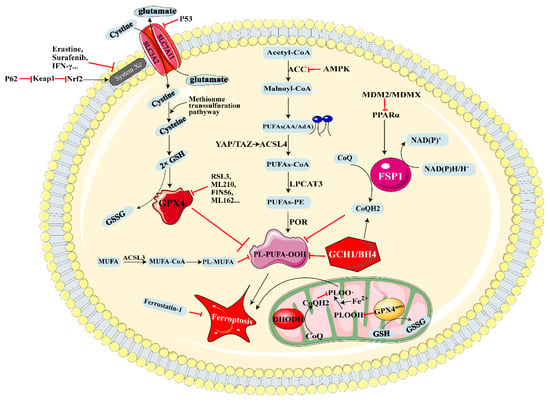
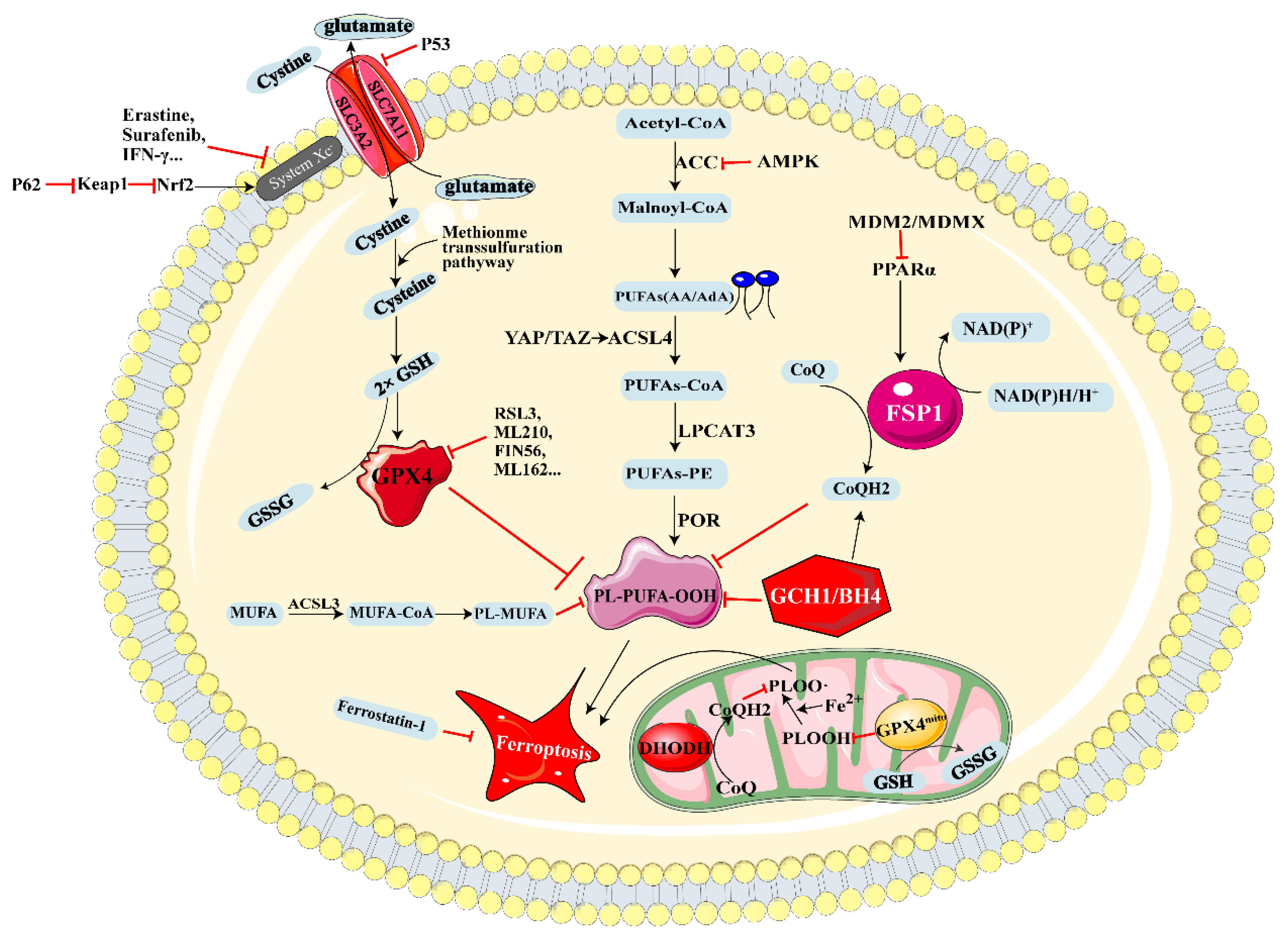
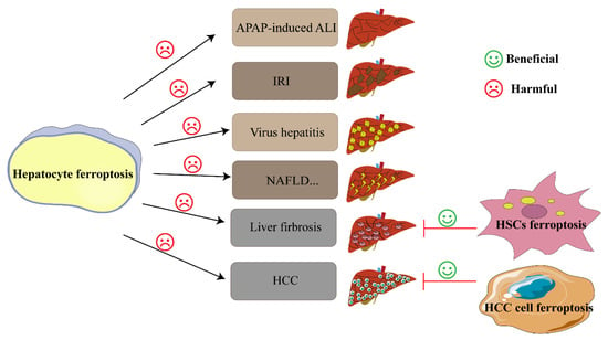
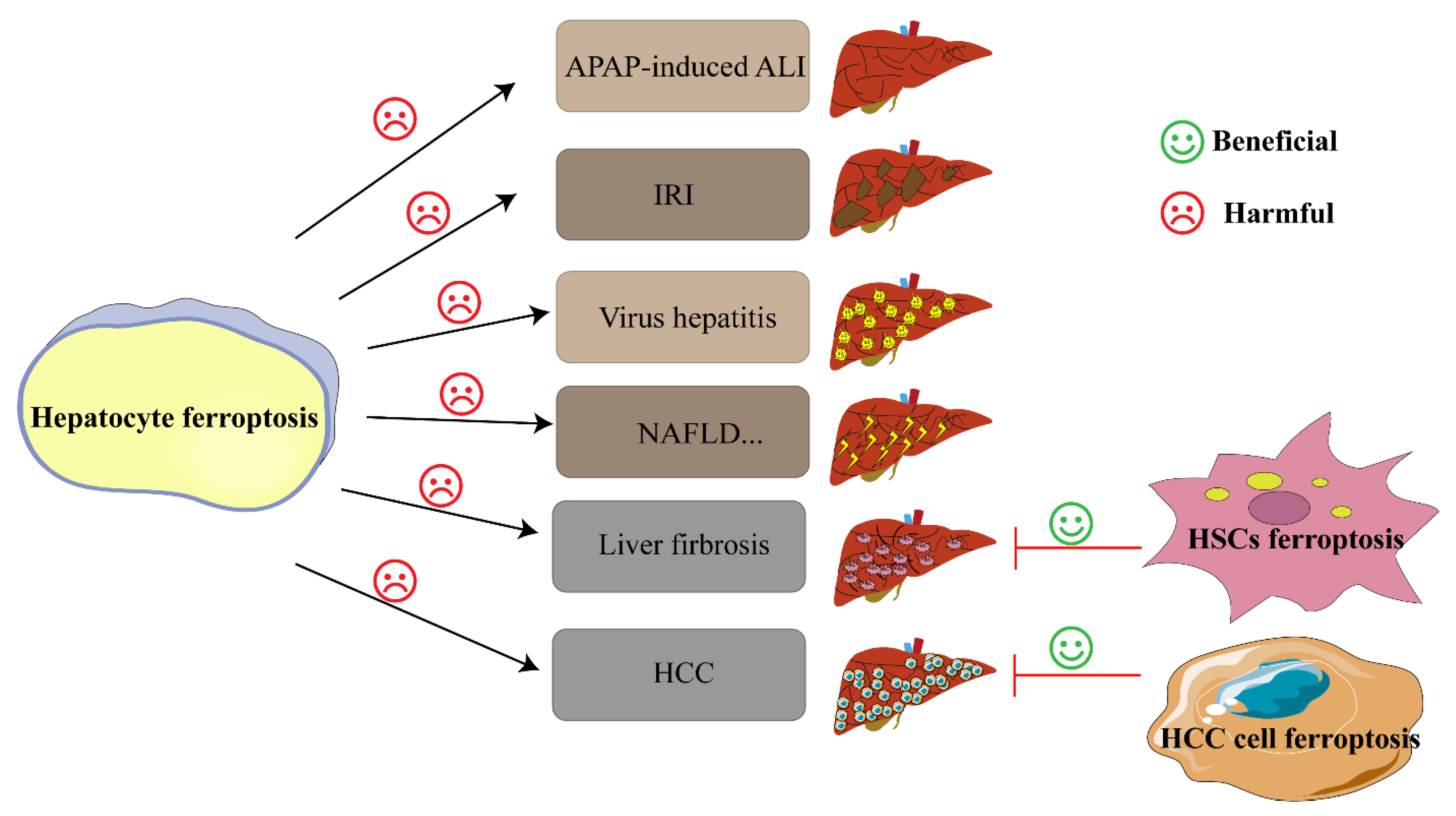

Publisher’s Note: MDPI stays neutral with regard to jurisdictional claims in published maps and institutional affiliations. |
© 2022 by the authors. Licensee MDPI, Basel, Switzerland. This article is an open access article distributed under the terms and conditions of the Creative Commons Attribution (CC BY) license (https://creativecommons.org/licenses/by/4.0/).