


Abstract
The viscoelastic properties of a cell cytoskeleton contain abundant information about the state of a cell. Cells show a response to a specific environment or an administered drug through changes in their viscoelastic properties. Studies of single cells have shown that chemical agents that interact with the cytoskeleton can alter mechanical cell properties and suppress mitosis. This envisions using rheological measurements as a non-specific tool for drug development, the pharmacological screening of new drug agents, and to optimize dosage. Although there exists a number of sophisticated methods for studying mechanical properties of single cells, studying concentration dependencies is difficult and cumbersome with these methods: large cell-to-cell variations demand high repetition rates to obtain statistically significant data. Furthermore, method-induced changes in the cell mechanics cannot be excluded when working in a nonlinear viscoelastic range. To address these issues, we not only compared narrow-gap rheometry with commonly used single cell techniques, such as atomic force microscopy and microfluidic-based approaches, but we also compared existing cell monolayer studies used to estimate cell mechanical properties. This review provides insight for whether and how narrow-gap rheometer could be used as an efficient drug screening tool, which could further improve our current understanding of the mechanical issues present in the treatment of human diseases.
1. Introduction
The mechanical properties of cells have vital functional implications, such as mechanical stability, adjustment to environmental load, motility, proliferation, phagocytosis, contraction, and morphogenesis [1]. Cell mechanics defines response of the cells to mechanical forces exerted by the cell microenvironment, such as the extracellular matrix [2]. Cells, by detecting changes in the extracellular matrix via a process known as mechanosensing [3], modify their responses, such as cytoskeletal organization, adhesion, stiffness, and motility [4]. In eukaryotic cells, the cytoskeleton is responsible for controlling these mechanical properties, cell shape, locomotion, and division. Their mechanical properties are governed by the actin–myosin, the microtubule, and the intermediate filament networks. These cytoskeleton components are connected to each other. The contractile forces built up by myosin motors are counterbalanced by forces transmitted from the adhesive contacts of a cell to its environment via the actin–myosin network. Intermediate filaments contribute significantly to the mechanical response of the cells at large cell deformation when they are stretched [5]. Compared to those, microtubules are stiff [6] and play a vital role in mitosis [7].
The mechanical cell properties are useful and promise label-free markers of cell state, indicating cytoskeletal or nuclear changes due to various diseases that change the mechanical cell properties [8]. Malignant transformation and neoplasm are related to significant changes in the cellular cytoskeleton and surface alterations, hence in the mechanical cell properties [9,10]. Many diseases show deviations in the structural and mechanical cell properties. Among them are arthritis, asthma, elliptocytosis, malaria, sickle cell anemia, spherocytosis, osteoarthritis, inflammation, and, particularly, cancer [11,12], which is the second leading cause of death worldwide [10]. The changes in the cytoskeleton are crucial in the pathology of diseases like cancer where growth, spreading, and metastasis are facilitated by altered cell mechanics [13,14,15].
The decrease in cell stiffness, due to changes in internal and cytoskeletal structures, could predict an aggressive and metastatic state of cancer cells [16,17,18]. Cancer cells are found to be softer at small deformations [8,10,13,14,19,20]. It has been shown that cell softening is a good indicator for increased cell proliferation and may help detect early dysplasia, for instance, in oral cancer [14]. Hence, cell-rheological measurements are envisioned to serve as a tool for personalized medicine [8]. The cancer cell characteristics are highly affected during the epithelial–mesenchymal transition, resulting in altered cell–cell and cell–matrix interactions, motility, and invasiveness [21,22]. Invasiveness, on the other hand, is believed to be related to the ability of cells to generate traction forces [23]. Differentiating healthy cells from cancerous ones and between various states of cancer by their mechanical properties is expected to be of great medical benefit [10]. A better understanding of mechanical changes during cancer progression is expected to result in novel drugs and treatments, hindering neoplasm propagation by altering mechanical properties of cancerous cells [14].
2. Single Cell Studies vs. Average Rheological Properties
For the general understanding of cells, significant efforts have been put into studying the mechanical cell behavior. During the last decades, different techniques, like optical and magnetic tweezers, atomic force microscopy, magnetic twisting cytometry, micropipette aspiration, cell poking, particle tracking micro-rheology, optical stretching, and high-throughput microfluidic techniques have been employed to measure the viscoelastic behavior of single cells [2,6,24,25,26,27,28]. Frequency-dependent changes in the viscoelastic properties of single cells have been studied extensively. Over a wide frequency range, cells show a power-law rheology, like soft glassy materials. The exponents typically range between 0.1 and 0.5, being smaller for stiffer cells [6]. Yet, quantitative data on the absolute value of the cell stiffness or the storage and loss moduli can differ depending on the method employed [12]. One reason is that most of the single-cell studies have been conducted in the nonlinear viscoelastic regime, where the response depends in a nonlinear fashion on the mechanical load. For general mechanical material characterization, the linear regime is mandatory to avoid the damage of the material. Yet, it is often difficult, if not impossible, with methods like optical tweezers, micropipettes, or cell poking to reach or resolve the linear viscoelastic regime. Moreover, optical methods, such as optical stretching with high power input may suffer from considerable heating [2,24]. Different rheological techniques measure various aspects of cell mechanics, and the choice of an appropriate method depends on the question being addressed. Common methods for measuring cell mechanics have been briefly compared in Table 1.
Table 1.
Comparison of common methods for measuring cell mechanical properties.
In single-cell studies, it is cumbersome to quantify the average viscoelastic properties of adherent cells. Depending for instance on cell identity, life cycle, shape, structure, and level of proteins, cells show large cell-to-cell variations [62]. Stiffness and dynamic moduli may vary by orders of magnitude [2,62]. On the other hand, drugs, aging, or diseases may strongly affect the cytoskeleton and thus the viscoelastic cellular properties [13]. Hence, to quantify their impact, it is important to determine the average viscoelastic cell properties in the linear viscoelastic range. In single-cell studies, experiments with several tens up to several thousand cells per hour have been conducted [29,62,63,64,65]. Although these methods provide quantitative data for single cells, complex chip design and operational control of microfluidic devices, non-standard culture protocols, reduced proliferation rates, and small volumes challenging subsequent analytical chemistry hold back their application in drug screening or in clinical trials [27,59,60]. Moreover, microfluidic devices are usually made from polydimethylsiloxane. Compared to polystyrene or glass, of which cell culture plates are usually made, it is rather compliant [59,66]. Since adherent cells may be affected by the substrate mechanics, it may result in differences to widely established studies with cell culture plates. Averaging a large number of cells (of the order of a million) can be achieved by studying them in a monolayer between rheometer disks [2,56,67]. Dakhil et al. have extended the method to enable the quantitative determination of average linear viscoelastic cell properties in single experimental runs [20]. This permits the quantifying of the impact of diseases and of drugs on the cells in single experimental runs that would otherwise be difficult if not impossible to determine with low-content methodologies.
Atomic force microscopy is a widely used technique in cellular biophysics, allowing high special resolution including the topography of living cells and the fast and highly sensitive measurement of mechanical cell properties [68]. It allows discriminating pathological and physiological conditions of cells by the mechanical fingerprinting of diseased and healthy cells and distinguishing cancer cells at different stages of malignancy [69,70,71,72,73]. In addition, it allows the investigation of changes in adhesiveness, stiffness, and deformability of cells underlying different processes that affect cell phenotype, including cancer cell transformation and metastasis [28,74,75,76,77]. It is commonly used to quantify the mechanical properties of adherent cells at a subcellular resolution [2]. The quantitative information received by atomic force microscopy is based on the indentation of the cell membrane with a probe or tip situated on the border of a cantilever. Indentations with atomic force microscopy probes of a few nanometer apexes may affect inner cell components due to the pressing of the membrane in between the cortex filaments for depths < 100 nm [26]. Furthermore, cell mechanical measurements using atomic force microscopy could show more than tenfold differences depending on the measurement parameters and the probed region of the cells [2].
The cell mechanical properties could function as a biophysical marker of its state and functions, showing its potential for clinical diagnostics. Recently, single-cell mechanophenotyping using microfluidic-based methods has shown comparable results to flow cytometry [27,78,79,80]. Urbanska et al. compared cell mechanical properties using three different microfluidic-based techniques: constriction-based deformability, shear flow deformability cytometry, and extensional flow deformability cytometry [27]. Traditional single cell techniques, such as atomic force microscopy, micropipette aspiration, optical stretching, and parallel-plate rheology, used to quantify cell deformation under exposure of external stresses, suffer from technically demanding and time-consuming procedures, which limit their use beyond specialized laboratories [27]. Contrary to the traditional single cell studies, the recently developed microfluidic-based techniques allow for the robust, high-throughput assessment of the cell deformability under homogeneous and heterogeneous cell populations. Furthermore, due to the ease of handling, these techniques could be used in biological laboratories and clinical settings. However, the current lack of standardization among various microfluidic techniques poses analytical challenges and limits cross-study comparison.
Due to the difficulty of obtaining representative quantitative data of linear viscoelastic cell properties in single cell studies, the impact of drugs on mechanical cell properties are usually studied first by measuring the viscoelastic properties of a single cell and repeating this measurement after treating the cells with drugs. In this way, it has been shown that substances that affect actin or intermediate filaments like cytochalasin D, latrunculin A, 2,3-butanedione monoxime, blebbistatin, or ML-7 [38,62,81] affect the mechanical cell properties. As a tendency, it has been observed that fixation and contractile agents like histamine or serotonin increase cell stiffness, while relaxing agents like isoproterenol, DBcAMP, and ML-7 decrease it [82]. Yet, depending on their mechanism, some relaxing agents like blebbistatin [38] and latrunculin A [81] have a considerable impact on frequency dependence, while others, like cytochalasin D [38] and sodium azide/deoxyglucose [81], do not.
Wu et al. compared the cell mechanical properties of the breast cancer cell line MCF-7, using most widely adopted methods for cell mechanics, including atomic force microscopy, magnetic twisting cytometry, particle tracking micro-rheology, parallel-plate rheometry, cell monolayer rheology, and optical stretching [2]. Among the compared techniques, they observed a variation by at least two orders of magnitude in the average values of the tested moduli. Apart from the general method employed, they showed that the data depends on specificities of the method. For instance, when using atomic force microscopy, there was more than a tenfold difference in the mechanical properties depending on the measurement parameters and the probed region of the cells. The mechanical cell response also depends sensitively on the forcing and the force profile [2] and on the pre-stress [83]. The positive correlation between pre-stress and elastic modulus observed in cell monolayer rheology was also consistent with atomic force microscopy experiments using a sharp conical probe [2]. These results highlight the importance of selecting an adequate technique for the biological question being addressed.
3. Narrow-Gap Rheology
Rheometers are versatile machines to characterize the rheology of materials. To this end, they detect the relation between shear stress, , deformation, γ, and shear rate, , respectively. In parallel-disk rotational rheometers, these quantities are derived for the maximum values within the gap, i.e., at the outer radius, R, of the gap, from the torque, M, and the angular displacements between the disks, θ, respectively, to the angular velocity, Ω, by Equation (1) [84].
where H is the gap width between the rheometer disks.
Viscoelastic solids are usually characterized by exposing them to oscillatory load. While for a perfectly elastic solid shear deformation and shear stress are in phase, the phase difference, δ, between them is π/2 for a perfect liquid. For a viscoelastic material, the phase difference is in-between. The elastic response is quantified by the storage modulus, G′, and the viscous one by the loss modulus, G″. They are related to the former quantities by Equation (2) [84].
where the index 0 indicates the amplitude. The moduli usually depend on the oscillation frequency.
Rheometers are usually employed to characterize the bulk behavior of the samples working at characteristic sample thickness in the millimeter range. To study cell rheology, the sample size needs to be reduced to that of the cells, i.e., in the range of a few micrometers. Fernández et al. introduced a narrow-gap rheometer that allowed the studying of the rheology of cells in shear oscillation [56]. To this end, cells were adhered in a monolayer between two flat plates via a thin fibronectin coating. They measured mechanical properties of the fibroblast monolayer and observed a significant drop in cell stiffness when using the actin-depolymerizing drug, latrunculin-A. However, as the cell coverage remained unknown, they were unable to quantify the average cell mechanical properties. Dakhil et al. introduced a narrow-gap device that also allows working at narrow gaps in unidirectional shear [85]. With this device, a parallelism of the plates of better than ±1 µm for unidirectional flows and about ±0.4 µm for oscillation can be reached. This is far below the reported effective zero-gap error of commercial rheometers of 25 µm to 70 µm [86], which, apart from being due to disk roughness and residual disk inclination, can be caused by squeeze flow during zeroing the rheometer [85]. The enhanced precision allows the extending of the measurement window to much higher shear rates [87,88] as well as studying the adhesion [89] and load limits [90] of cells in low viscous environments. With their device, Dakhil et al. reached the linear viscoelastic range at small deformations, where the stress amplitude is proportional to the deformation amplitude [20,91]. By determining the cell coverage in the monolayer, i.e., the number of cells in the measurement gap, they were able to quantify the average mechanical cell properties.
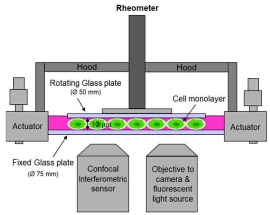
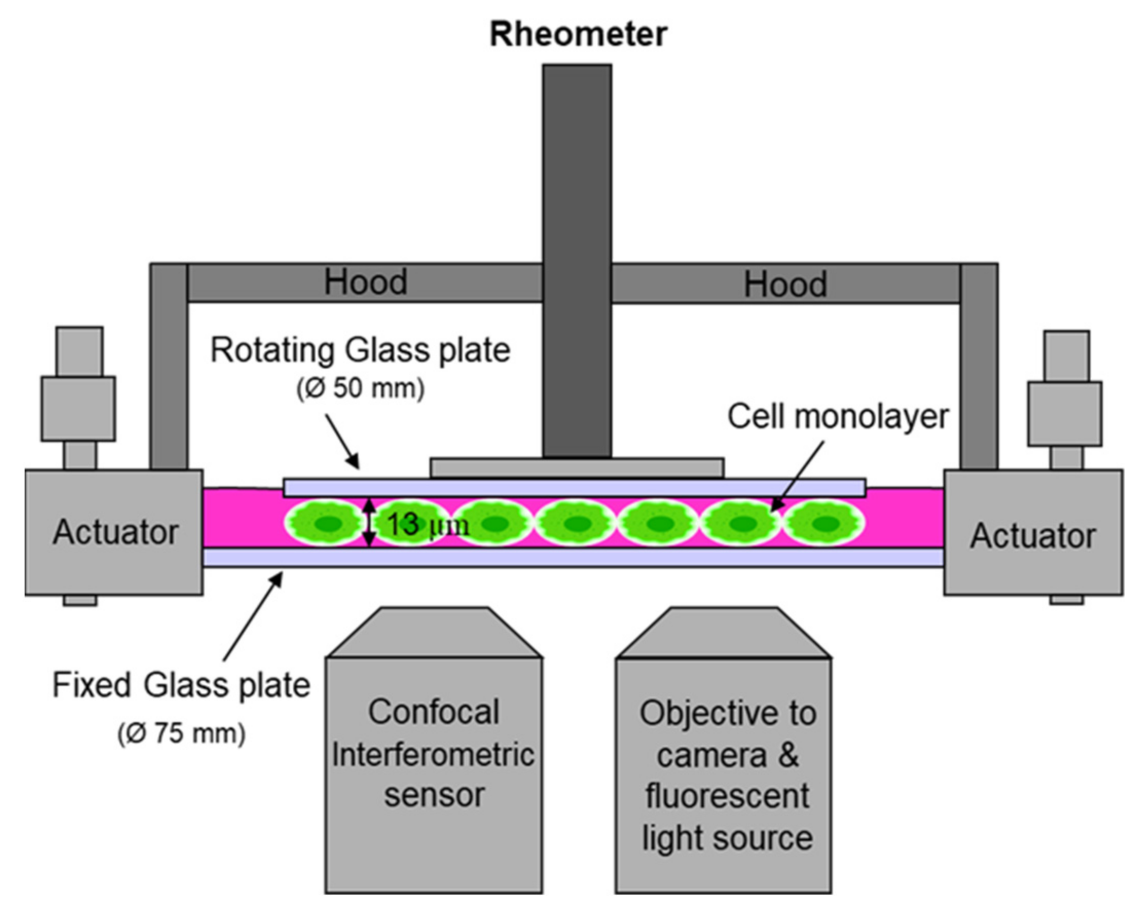
The setup of the narrow-gap device used by Dakhil et al. [85,91] is sketched in Figure 1. The fixed bottom plate has a diameter of 75 mm and an evenness of λ/4. The rotating plate has a diameter of 50 mm with an evenness of λ/4. The top plate is attached to a measurement head of the rheometer. The bottom plate is aligned perpendicularly to the axis of rotation using three actuators. For disk alignment, the gap width is measured with a customized confocal interferometric sensor. It has a working distance of 42 mm, a measuring range of 90 µm and allows the independent detection of the gap width with an axial accuracy of 10 nm. The sensor is placed underneath the fixed glass plate. It is placed next to a fluorescence microscope equipped with a 5x objective, which is used for detecting cell coverage and cell viability. They determined the average area covered by the cells from multiple images at distinct locations after staining the cells with a fluorescent dye. Since deformation is strongest at maximum distance from the turning axis of the rheometer, and, hence, the rheometer signal is caused by the cells at the outer rim, the images are taken close to the edge of the upper plate. To enable measurements at various locations sensor and microscope are both fixed to a traverse. Sander et al. used a similar setup [57]. Differently to Dakhil et al. [20,91], they used small beads for setting up the gap width. Instead of the upper plate, they used a ring to disregard the cells near the axis of oscillation.
Figure 1.
Sketch of the narrow-gap rheometer.
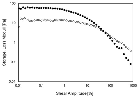
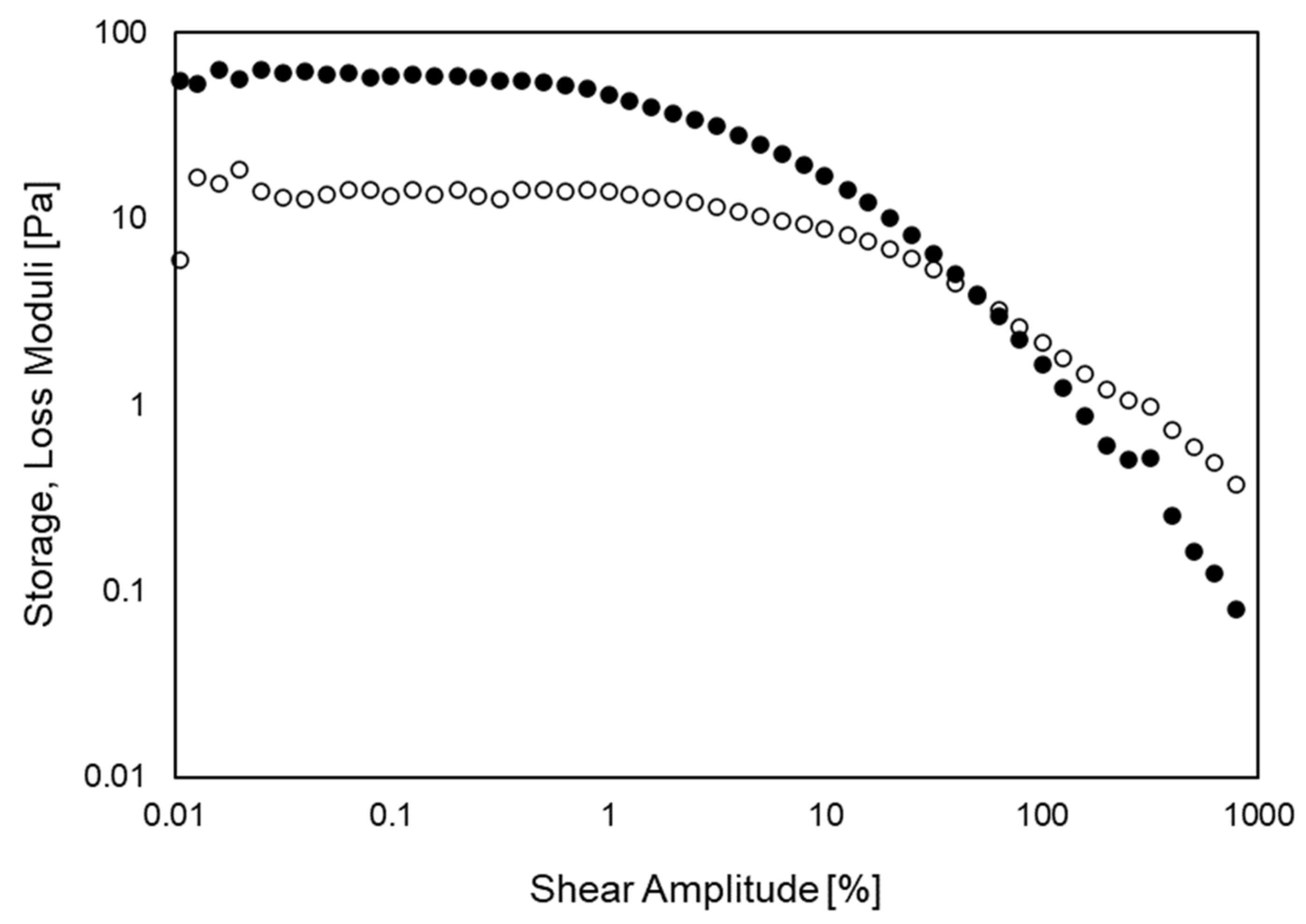
The setup of Dakhil et al. [20] enabled the detection of the linear viscoelastic regime of the cells. Figure 2 shows a representative amplitude sweep of MCF-7 cells detected with a setup similar to theirs. In this example, the cells were treated with paclitaxel, a well-known anticancer drug. Storage and loss moduli were determined from the maximum shear-stress and shear-deformation amplitudes, i.e., at the radius of the upper plate. The linear viscoelastic regime appears as a plateau in storage and loss moduli. As is apparent in Figure 2, it is constrained to small amplitudes. With increasing amplitudes, the moduli deviate from the plateau and tend to decline. At larger amplitudes, the cells start to detach from the plates and a cross-over of the moduli appears.
Figure 2.
Amplitude sweep of MCF-7 cells treated with paclitaxel. Storage and loss moduli are indicated by closed and open symbols, respectively. Angular frequency: 1 rad/s; Gap width: 13 µm.
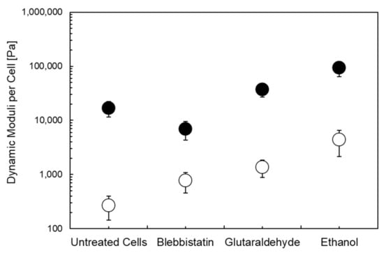
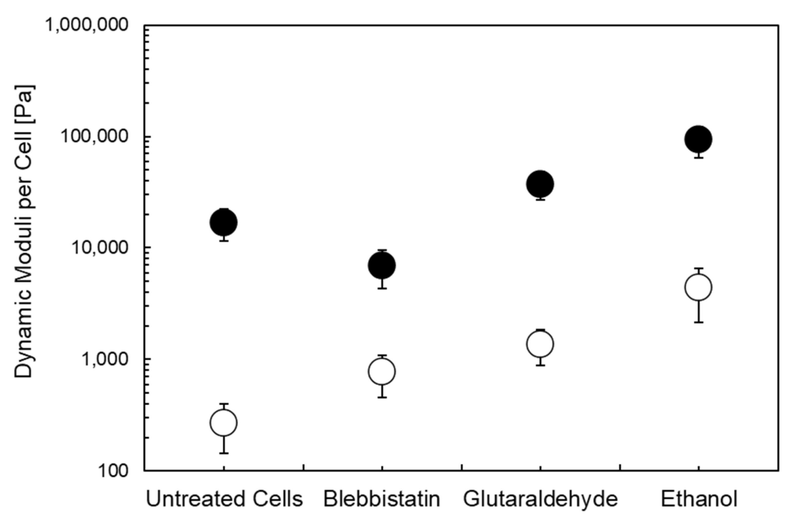
Biochemical treatments may affect the dynamic moduli and their frequency dependence. To study the impact of drugs that are known to affect the cytoskeleton structure, in a recent study we applied ethanol (0.5 vol. %), glutaraldehyde (0.1 vol. %), and blebbistatin (150 µM) to murine fibroblasts (Figure 3) [91]. Figure 3 shows the average moduli per cell to the linear viscoelastic regime for murine fibroblasts [91]. Compared to untreated cells, blebbistatin lowers the storage modulus while glutaraldehyde and ethanol tend to enhance the storage modulus. Except blebbistatin, all other drugs yielded an increase in the loss modulus. These results correlate with the effects of drug treatment and for setting the experimental limits accordingly.
Figure 3.
Dynamic moduli per cell for untreated fibroblasts and fibroblasts treated with different drugs [91]. Storage and loss moduli are indicated by close and open symbols, respectively. Frequency: 1 Hz; Gap width: 5 µm.
Recently, Kiran et al. [58] used cell monolayer rheology to study the rheological properties of a serum-starved, fully confluent Madin–Darby canine kidney cell monolayer in shear oscillation with a setup similar to that used by Dakhil et al. [20] and Fernández et al. [20,56]. The circular cell monolayer was grown on rectangular glass coverslips using polydimethylsiloxane strips. Both plates were coated with fibronectin to adhere cells. For plate alignment and optical observation, they used a microscope with a 20x objective.
Elkins et al. introduced a linear cell monolayer rheometer to study the cell mechanics of confluent stromal vascular cells [67]. As an upper plate, they used a slightly curved plano-convex lens with the curves side to the bottom. The curvature let a variation in gap width of less than 5%. They adhered the plate to a mount fixed to a force transducer. To shear the monolayer, the transducer was mounted on a piezoelectric stage and displaced laterally. The device was centered on a metal dish with a glass coverslip mounted onto an inverted microscope with a 100x objective to optically monitor cell density and cell deformation. Both plates were coated with collagen to adhere cells. With this device, Elkins et al. [67] conducted step-strain measurements and detected changes in the average relaxation modulus of the cell monolayer.
The linear monolayer device of Elkins and coworkers differs in a number of ways from that introduced by Fernández et al. [56]: (1) eliminating the need for perfectly parallel top and bottom plates by employing a top plate with gentle curvature; (2) provides high cell spreading area; (3) allows the live quantification of cell strain values during deformation due to the incorporation of live cell imaging system; and (4) uses a linear shear geometry, ensuring in principle a uniform strain exposure of all cells in the system. Similar to other monolayer studies [2,20,56], the linear monolayer device does not require culturing cells on the rheometer plates and show high cell viability. However, their device has the following limitations as compared to the system introduced by Dakhil et al. [20]: (1) does not allow gap width adjustment; (2) requires 24 h for cell attachment; (3) gap width was determined using spacer beads of known diameter, however, this gap width was not well-defined and precise due to the small curvature of the top plate; (4) does not allow changes in normal stresses as the normal force on the cell monolayer was solely due to the weight of the top plate and the pyramid mount.
The rotational devices, as presented by Dakhil et al. [20,91], on the other hand, offer following possibilities: (1) cells can be plated on the rheometer plates a few hours before the experiment thus reducing the time scale of the actual experiment; (2) allows optical determination of cell concentration and live quantification of cell strain values during deformation; (3) allows gap width adjustment with a precision of about ±0.4 μm; (4) allows changes, setting and measuring the normal stresses; (5) allows elongation and compression of cells, hence quantifying the impact of pre-stress on storage and loss modulus; and (6) shows high reproducibility. Together, all these features make the narrow-gap rheometer an attractive choice as a diagnostic tool to quantify variation in cell mechanics, such as in transgenic cell lines. The above-mentioned rheological studies employing narrow-gap rheometer are summed up in Table 2.
Table 2.
Applications of narrow-gap rheology for cell mechanical studies.
4. Summary and Future Perspectives
The mechanical characterization of cellular behavior allows a comprehensive understanding of physiological and pathological processes [94,95] with potential applications in clinical diagnostics [8,27,96,97]. Cell mechanical properties define its function and behavior against the mechanical forces exerted by the cell microenvironment or by the presence of external components [98]. This cellular response to the mechanical forces can be elastic, viscous, or viscoelastic as well as active or passive [2]. Changes in cellular or nuclear mechanics can indicate human diseases, such as inflammation, cardiovascular diseases, infectious diseases, aging, and, most importantly, cancer, as one of the leading causes of death worldwide [13,44,99,100,101].
It has been reported that the detected values of cell mechanical properties, such as viscosity and elasticity, may vary considerably depending on the method used [2,12,26,27,80]. In single-cell studies, such as atomic force microscopy, magnetic twisting cytometry, particle tracking micro-rheology, optical and magnetic tweezers, micropipette aspiration, and cell poking, it is cumbersome to quantify average viscoelastic properties of cells due to the repetition of the experiment to obtain reliable data. Although several single cell studies, including cytometric methods and microfluidic systems, provide high through-put quantitative measurement of mechanical properties, their use is limited due to complex chip design and operational control. Culturing matrix-based differences in mechanical cell properties show limitations in case of microfluidic devices, for instance, for drug screening or in clinical studies [27,59,60]. Atomic force microscopy provides rich viscoelastic properties. Depending on the probe size, cell structures with significantly different responses can be detected. However, discrepancies in the atomic force microscopy could arise due to cantilever calibration errors of 10–15%, speed of indentation, type of probe used, the probed area, and probed timescale [2]. Similarly, each technique is developed under the trade-off relationship between information content and the throughput [96,102].
Quantifying the impact of drugs on the mechanical cell properties can help in the pharmacological screening, for instance, of new components to screen their efficiency, minimize side effects, and to overcome drug resistance [7,103]. To this end, the detection of their influence on the linear viscoelastic cell properties needs to be established. To unravel this regime, a reliable quantitative determination of the average rheological quantities is necessary. The recently modified narrow-gap rotational rheometer provides a promising tool to quantitatively determine those average linear viscoelastic cell properties in single experimental runs [20]. In this technique, cells are adhered to both plates by an adhesive protein, such as fibronectin. The elastic modulus measured with this technique is comparable to atomic force microscopy using a sharp conical probe [2]. In cell monolayer rheology, with the increasing stress, the tensile pre-stress changes the apparent elastic modulus of cells [2,83]. These findings are comparable to the typical tensile stresses measured in single cell studies [2].
The choice of a cell-based mechanical method depends critically on the biological material and the specific question addressed. Similar to other single cell-based mechanical techniques, the recently developed narrow-gap device can also evaluate samples in in vitro and ex vivo settings and can provide reliable data by measuring the average mechanical properties of cell monolayers. By measuring the characteristic response of cells to the drugs, efficient and accurate results can be obtained in a wide variety of areas, such as the quantification of chemical agents and measuring the impact of new drug candidates.
Author Contributions
Conceptualization, K.M.I.B. and A.W.; methodology, K.M.I.B. and A.W.; formal analysis, K.M.I.B., S.L. and D.H.J.; resources, M.-G.C. and A.W.; data curation, K.M.I.B., S.L. and D.H.J.; writing—original draft preparation, K.M.I.B., D.H.J. and A.W.; writing—review and editing, S.K.B., M.-G.C. and A.W.; supervision, A.W.; funding acquisition, K.M.I.B., M.-G.C. and A.W. All authors have read and agreed to the published version of the manuscript.
Funding
This work was supported by the National Research Foundation of Korea (NRF) grant funded by the Korean government (MSIT) (No. 2020R1A2C201479211); the authors are thankful for their support.
Conflicts of Interest
The authors declare no conflict of interest.
References
- Kollmannsberger, P.; Fabry, B. Active Soft Glassy Rheology of Adherent Cells. Soft Matter 2009, 5, 1771–1774. [Google Scholar] [CrossRef] [Scilit]
- Wu, P.H.; Aroush, D.R.B.; Asnacios, A.; Chen, W.C.; Dokukin, M.E.; Doss, B.L.; Durand-Smet, P.; Ekpenyong, A.; Guck, J.; Guz, N.V.; et al. A Comparison of Methods to Assess Cell Mechanical Properties. Nat. Methods 2018, 15, 491–498. [Google Scholar] [CrossRef] [Scilit] [PubMed]
- Janmey, P.A.; Fletcher, D.A.; Reinhart-King, C.A. Stiffness Sensing by Cells. Physiol. Rev. 2020, 100, 695–724. [Google Scholar] [CrossRef] [Scilit] [PubMed]
- Discher, D.E.; Janmey, P.; Wang, Y.L. Tissue Cells Feel and Respond to the Stiffness of Their Substrate. Science 2005, 310, 1139–1143. [Google Scholar] [CrossRef] [Scilit]
- Bertaud, J.; Qin, Z.; Buehler, M.J. Intermediate Filament-Deficient Cells Are Mechanically Softer at Large Deformation: A Multi-Scale Simulation Study. Acta Biomater. 2010, 6, 2457–2466. [Google Scholar] [CrossRef] [Scilit]
- Kollmannsberger, P.; Fabry, B. Linear and Nonlinear Rheology of Living Cells. Annu. Rev. Mater. Res. 2011, 41, 75–97. [Google Scholar] [CrossRef] [Scilit]
- Jordan, M.A.; Wilson, L. Microtubules as a Target for Anticancer Drugs. Nat. Rev. Cancer 2004, 4, 253–265. [Google Scholar] [CrossRef] [Scilit]
- Di Carlo, D. A Mechanical Biomarker of Cell State in Medicine. J. Lab. Autom. 2012, 17, 32–42. [Google Scholar] [CrossRef] [Scilit]
- Weinberg, R.A. The Biology of Cancer, 2nd ed.; W. W. Norton & Company: New York, NY, USA, 2013; pp. 1–876. [Google Scholar] [CrossRef] [Scilit]
- Yallapu, M.M.; Katti, K.S.; Katti, D.R.; Mishra, S.R.; Khan, S.; Jaggi, M.; Chauhan, S.C. The Roles of Cellular Nanomechanics in Cancer. Med. Res. Rev. 2015, 35, 198–223. [Google Scholar] [CrossRef] [Scilit]
- Lee, G.Y.H.; Lim, C.T. Biomechanics Approaches to Studying Human Diseases. Trends Biotechnol. 2007, 25, 111–118. [Google Scholar] [CrossRef] [Scilit]
- Chen, J.; Zhang, Z.; Li, L.; Chen, B.C.; Revyakin, A.; Hajj, B.; Legant, W.; Dahan, M.; Lionnet, T.; Betzig, E.; et al. Single-Molecule Dynamics of Enhanceosome Assembly in Embryonic Stem Cells. Cell 2014, 156, 1274–1285. [Google Scholar] [CrossRef] [Scilit]
- Guck, J.; Schinkinger, S.; Lincoln, B.; Wottawah, F.; Ebert, S.; Romeyke, M.; Lenz, D.; Erickson, H.M.; Ananthakrishnan, R.; Mitchell, D.; et al. Optical Deformability as an Inherent Cell Marker for Testing Malignant Transformation and Metastatic Competence. Biophys. J. 2005, 88, 3689–3698. [Google Scholar] [CrossRef] [Scilit]
- Fritsch, A.; Höckel, M.; Kiessling, T.; Nnetu, K.D.; Wetzel, F.; Zink, M.; Käs, J.A. Are Biomechanical Changes Necessary for Tumour Progression? Nat. Phys. 2010, 6, 730–732. [Google Scholar] [CrossRef] [Scilit]
- Swaminathan, V.; Mythreye, K.; Tim O’Brien, E.; Berchuck, A.; Blobe, G.C.; Superfine, R. Mechanical Stiffness Grades Metastatic Potential in Patient Tumor Cells and in Cancer Cell Lines. Cancer Res. 2011, 71, 5075–5080. [Google Scholar] [CrossRef] [Scilit]
- Mierke, C.T.; Kollmannsberger, P.; Zitterbart, D.P.; Diez, G.; Koch, T.M.; Marg, S.; Ziegler, W.H.; Goldmann, W.H.; Fabry, B. Vinculin Facilitates Cell Invasion into Three-Dimensional Collagen Matrices. J. Biol. Chem. 2010, 285, 13121–13130. [Google Scholar] [CrossRef] [Scilit]
- Li, B.; Cai, Y.; Qi, C.; Hansen, R.; Ding, C.; Thomas, C.; Yan, J. Orally Administered Particular β-Glucan Modulates Tumor-Capturing Dendritic Cells and Improves Anti-Tumor T Cell Responses in Cancer. Clin. Cancer Res. 2010, 16, 5153–5164. [Google Scholar] [CrossRef] [Scilit]
- Gal, N.; Weihs, D. Intracellular Mechanics and Activity of Breast Cancer Cells Correlate with Metastatic Potential. Cell Biochem. Biophys. 2012, 63, 199–209. [Google Scholar] [CrossRef] [Scilit]
- Suresh, S. Biomechanics and Biophysics of Cancer Cells. Acta Biomater. 2007, 3, 413–438. [Google Scholar] [CrossRef] [Scilit]
- Dakhil, H.; Gilbert, D.F.; Malhotra, D.; Limmer, A.; Engelhardt, H.; Amtmann, A.; Hansmann, J.; Hübner, H.; Buchholz, R.; Friedrich, O.; et al. Measuring Average Rheological Quantities of Cell Monolayers in the Linear Viscoelastic Regime. Rheol. Acta 2016, 55, 527–536. [Google Scholar] [CrossRef] [Scilit]
- Voulgari, A.; Pintzas, A. Epithelial–Mesenchymal Transition in Cancer Metastasis: Mechanisms, Markers and Strategies to Overcome Drug Resistance in the Clinic. Biochim. Biophys. Acta—Rev. Cancer 2009, 1796, 75–90. [Google Scholar] [CrossRef] [Scilit]
- Ceppi, P.; Peter, M.E. MicroRNAs Regulate Both Epithelial-to-Mesenchymal Transition and Cancer Stem Cells. Oncogene 2014, 33, 269–278. [Google Scholar] [CrossRef] [Scilit]
- Koch, T.M.; Münster, S.; Bonakdar, N.; Butler, J.P.; Fabry, B. 3D Traction Forces in Cancer Cell Invasion. PLoS ONE 2012, 7, e33476. [Google Scholar] [CrossRef] [Scilit]
- Bhat, S.; Jun, D.; Paul, B.C.; Dahms, T.E.S. Viscoelasticity in Biological Systems: A Special Focus on Microbes; Chapter 6; IntechOpen: London, UK, 2012. [Google Scholar] [CrossRef] [Scilit]
- Sarkar, R.; Rybenkov, V.V. A Guide to Magnetic Tweezers and Their Applications. Front. Phys. 2016, 4, 48. [Google Scholar] [CrossRef] [Scilit]
- Brill-Karniely, Y. Mechanical Measurements of Cells Using AFM: 3D or 2D Physics? Front. Bioeng. Biotechnol. 2020, 8, 605153. [Google Scholar] [CrossRef] [Scilit]
- Urbanska, M.; Muñoz, H.E.; Shaw Bagnall, J.; Otto, O.; Manalis, S.R.; Di Carlo, D.; Guck, J. A Comparison of Microfluidic Methods for High-Throughput Cell Deformability Measurements. Nat. Methods 2020, 17, 587–593. [Google Scholar] [CrossRef] [Scilit]
- Gil-Redondo, J.C.; Weber, A.; Zbiral, B.; Vivanco, M.d.M.; Toca-Herrera, J.L. Substrate Stiffness Modulates the Viscoelastic Properties of MCF-7 Cells. J. Mech. Behav. Biomed. Mater. 2022, 125, 104979. [Google Scholar] [CrossRef] [Scilit]
- Hiratsuka, S.; Mizutani, Y.; Tsuchiya, M.; Kawahara, K.; Tokumoto, H.; Okajima, T. The Number Distribution of Complex Shear Modulus of Single Cells Measured by Atomic Force Microscopy. Ultramicroscopy 2009, 109, 937–941. [Google Scholar] [CrossRef] [Scilit]
- Davidovits, P. Physics in Biology and Medicine, 4th ed.; Academic Press: Cambridge, MA, USA, 2013; pp. 1–331. [Google Scholar] [CrossRef] [Scilit]
- Guz, N.; Dokukin, M.; Kalaparthi, V.; Sokolov, I. If Cell Mechanics Can Be Described by Elastic Modulus: Study of Different Models and Probes Used in Indentation Experiments. Biophys. J. 2014, 107, 564–575. [Google Scholar] [CrossRef] [Scilit] [PubMed]
- Staunton, J.R.; Doss, B.L.; Lindsay, S.; Ros, R. Correlating Confocal Microscopy and Atomic Force Indentation Reveals Metastatic Cancer Cells Stiffen during Invasion into Collagen i Matrices. Sci. Rep. 2016, 6, 19686. [Google Scholar] [CrossRef] [Scilit]
- Deng, L.; Trepat, X.; Butler, J.P.; Millet, E.; Morgan, K.G.; Weitz, D.A.; Fredberg, J.J. Fast and Slow Dynamics of the Cytoskeleton. Nat. Mater. 2006, 5, 636–640. [Google Scholar] [CrossRef] [Scilit]
- Zhang, Y.; Wei, F.; Poh, Y.C.; Jia, Q.; Chen, J.; Chen, J.; Luo, J.; Yao, W.; Zhou, W.; Huang, W.; et al. Interfacing 3D Magnetic Twisting Cytometry with Confocal Fluorescence Microscopy to Image Force Responses in Living Cells. Nat. Protoc. 2017, 12, 1437–1450. [Google Scholar] [CrossRef] [Scilit] [PubMed]
- Kollmannsberger, P.; Fabry, B. High-Force Magnetic Tweezers with Force Feedback for Biological Applications. Rev. Sci. Instrum. 2007, 78, 114301. [Google Scholar] [CrossRef] [Scilit] [PubMed]
- Vilfan, I.D.; Lipfert, J.; Koster, D.A.; Lemay, S.G.; Dekker, N.H. Magnetic Tweezers for single-molecule experiments. In Handbook of Single-Molecule Biophysics; Springer: Berlin/Heidelberg, Germany, 2009; pp. 371–396. [Google Scholar] [CrossRef] [Scilit]
- Chen, H.; Fu, H.; Zhu, X.; Cong, P.; Nakamura, F.; Yan, J. Improved High-Force Magnetic Tweezers for Stretching and Refolding of Proteins and Short DNA. Biophys. J. 2011, 100, 517–523. [Google Scholar] [CrossRef] [Scilit] [PubMed]
- Balland, M.; Desprat, N.; Icard, D.; Féréol, S.; Asnacios, A.; Browaeys, J.; Hénon, S.; Gallet, F. Power Laws in Microrheology Experiments on Living Cells: Comparative Analysis and Modeling. Phys. Rev. E.—Stat. Nonlinear Soft Matter Phys. 2006, 74, 021911. [Google Scholar] [CrossRef] [Scilit]
- Zhang, H.; Liu, K.K. Optical Tweezers for Single Cells. J. R. Soc. Interface 2008, 5, 671–690. [Google Scholar] [CrossRef] [Scilit]
- Ayala, Y.A.; Pontes, B.; Ether, D.S.; Pires, L.B.; Araujo, G.R.; Frases, S.; Romão, L.F.; Farina, M.; Moura-Neto, V.; Viana, N.B.; et al. Rheological Properties of Cells Measured by Optical Tweezers. BMC Biophys. 2016, 9, 5. [Google Scholar] [CrossRef] [Scilit]
- Desprat, N.; Guiroy, A.; Asnacios, A. Microplates-Based Rheometer for a Single Living Cell. Rev. Sci. Instrum. 2006, 77, 055111. [Google Scholar] [CrossRef] [Scilit]
- Desprat, N.; Richert, A.; Simeon, J.; Asnacios, A. Creep Function of a Single Living Cell. Biophys. J. 2005, 88, 2224–2233. [Google Scholar] [CrossRef] [Scilit]
- Fernández, P.; Pullarkat, P.A.; Ott, A. A Master Relation Defines the Nonlinear Viscoelasticity of Single Fibroblasts. Biophys. J. 2006, 90, 3796. [Google Scholar] [CrossRef] [Scilit]
- Bufi, N.; Durand-Smet, P.; Asnacios, A. Single-Cell Mechanics: The Parallel Plates Technique. Methods Cell Biol. 2015, 125, 187–209. [Google Scholar] [CrossRef] [Scilit]
- Yamada, S.; Wirtz, D.; Kuo, S.C. Mechanics of Living Cells Measured by Laser Tracking Microrheology. Biophys. J. 2000, 78, 1736–1747. [Google Scholar] [CrossRef] [Scilit]
- Wirtz, D. Particle-Tracking Microrheology of Living Cells: Principles and Applications. Annu. Rev. Biophys. 2009, 38, 301–326. [Google Scholar] [CrossRef] [Scilit]
- McGlynn, J.A.; Wu, N.; Schultz, K.M. Multiple Particle Tracking Microrheological Characterization: Fundamentals, Emerging Techniques and Applications. J. Appl. Phys. 2020, 127, 201101. [Google Scholar] [CrossRef] [Scilit]
- Hochmuth, R.M. Micropipette Aspiration of Living Cells. J. Biomech. 2000, 33, 15–22. [Google Scholar] [CrossRef] [Scilit]
- Guilak, F.; Alexopoulos, L.G.; Haider, M.A.; Ting-Beall, H.P.; Setton, L.A. Zonal Uniformity in Mechanical Properties of the Chondrocyte Pericellular Matrix: Micropipette Aspiration of Canine Chondrons Isolated by Cartilage Homogenization. Ann. Biomed. Eng. 2005, 33, 1312–1318. [Google Scholar] [CrossRef] [Scilit]
- Rodriguez, M.L.; McGarry, P.J.; Sniadecki, N.J. Review on Cell Mechanics: Experimental and Modeling Approaches. Appl. Mech. Rev. 2013, 65, 060801. [Google Scholar] [CrossRef] [Scilit]
- Gangotra, A.; Willmott, G.R. Cellular and Sub-Cellular Mechanics: Measurement of Material Properties. In Reference Module in Materials Science and Materials Engineering, 2nd ed.; Elsavier: Amsterdam, The Netherlands, 2019; Volume 3, pp. 227–244. [Google Scholar] [CrossRef] [Scilit]
- Guck, J.; Ananthakrishnan, R.; Mahmood, H.; Moon, T.J.; Cunningham, C.C.; Käs, J. The Optical Stretcher: A Novel Laser Tool to Micromanipulate Cells. Biophys. J. 2001, 81, 767. [Google Scholar] [CrossRef] [Scilit]
- Lincoln, B.; Wottawah, F.; Schinkinger, S.; Ebert, S.; Guck, J. High-Throughput Rheological Measurements with an Optical Stretcher. Methods Cell Biol. 2007, 83, 397–423. [Google Scholar] [CrossRef] [Scilit]
- Yang, T.; Bragheri, F.; Minzioni, P. A Comprehensive Review of Optical Stretcher for Cell Mechanical Characterization at Single-Cell Level. Micromachines 2016, 7, 90. [Google Scholar] [CrossRef] [Scilit]
- Mierke, C.T. The Role of the Optical Stretcher Is Crucial in the Investigation of Cell Mechanics Regulating Cell Adhesion and Motility. Front. Cell Dev. Biol. 2019, 7, 184. [Google Scholar] [CrossRef] [Scilit]
- Fernández, P.; Heymann, L.; Ott, A.; Aksel, N.; Pullarkat, P.A. Shear Rheology of a Cell Monolayer. New J. Phys. 2007, 9, 149. [Google Scholar] [CrossRef] [Scilit]
- Sander, M.; Flesch, J.; Ott, A. Using Cell Monolayer Rheology to Probe Average Single Cell Mechanical Properties. Biorheology 2015, 52, 269–278. [Google Scholar] [CrossRef] [Scilit]
- Kiran, A.; Shekhar, C.; Sabapathy, M.; Mishra, M.; Kumar, L.; Kumar, N.; Mehandia, V. Effect of Serum Starvation on Rheology of Cell Monolayers. Phys. Fluids 2021, 33, 071908. [Google Scholar] [CrossRef] [Scilit]
- Dahl, J.B.; Lin, J.M.G.; Muller, S.J.; Kumar, S. Microfluidic Strategies for Understanding the Mechanics of Cells and Cell-Mimetic Systems. Annu. Rev. Chem. Biomol. Eng. 2015, 6, 293–317. [Google Scholar] [CrossRef] [Scilit] [PubMed]
- Halldorsson, S.; Lucumi, E.; Gómez-Sjöberg, R.; Fleming, R.M.T. Advantages and Challenges of Microfluidic Cell Culture in Polydimethylsiloxane Devices. Biosens. Bioelectron. 2015, 63, 218–231. [Google Scholar] [CrossRef] [Scilit] [PubMed]
- Luo, T.; Fan, L.; Zhu, R.; Sun, D. Microfluidic Single-Cell Manipulation and Analysis: Methods and Applications. Micromachines 2019, 10, 104. [Google Scholar] [CrossRef] [Scilit] [PubMed]
- Cai, P.; Mizutani, Y.; Tsuchiya, M.; Maloney, J.M.; Fabry, B.; Van Vliet, K.J.; Okajima, T. Quantifying Cell-to-Cell Variation in Power-Law Rheology. Biophys. J. 2013, 105, 1093–1102. [Google Scholar] [CrossRef] [Scilit]
- Mierke, C.T.; Kollmannsberger, P.; Zitterbart, D.P.; Smith, J.; Fabry, B.; Goldmann, W.H. Mechano-Coupling and Regulation of Contractility by the Vinculin Tail Domain. Biophys. J. 2008, 94, 661–670. [Google Scholar] [CrossRef] [Scilit]
- Lange, J.R.; Steinwachs, J.; Kolb, T.; Lautscham, L.A.; Harder, I.; Whyte, G.; Fabry, B. Microconstriction Arrays for High-Throughput Quantitative Measurements of Cell Mechanical Properties. Biophys. J. 2015, 109, 26–34. [Google Scholar] [CrossRef] [Scilit]
- Herbig, M.; Tessmer, K.; Nötzel, M.; Nawaz, A.A.; Santos-Ferreira, T.; Borsch, O.; Gasparini, S.J.; Guck, J.; Ader, M. Label-Free Imaging Flow Cytometry for Analysis and Sorting of Enzymatically Dissociated Tissues. Sci. Rep. 2022, 12, 963. [Google Scholar] [CrossRef] [Scilit]
- Paguirigan, A.L.; Beebe, D.J. From the Cellular Perspective: Exploring Differences in the Cellular Baseline in Macroscale and Microfluidic Cultures. Integr. Biol. 2009, 1, 182. [Google Scholar] [CrossRef] [Scilit]
- Elkins, C.M.; Shen, W.-J.; Khor, V.K.; Kraemer, F.B.; Fuller, G.G. Quantification of Stromal Vascular Cell Mechanics with a Linear Cell Monolayer Rheometer. J. Rheol. 2015, 59, 33–50. [Google Scholar] [CrossRef] [Scilit]
- Pérez-Domínguez, S.; Kulkarni, S.G.; Rianna, C.; Radmacher, M. Atomic Force Microscopy for Cell Mechanics and Diseases. Neuroforum 2020, 26, 101–109. [Google Scholar] [CrossRef] [Scilit]
- Lekka, M. Atomic Force Microscopy: A Tip for Diagnosing Cancer. Nat. Nanotechnol. 2012, 7, 691–692. [Google Scholar] [CrossRef] [Scilit]
- Lekka, M. Discrimination Between Normal and Cancerous Cells Using AFM. Bionanoscience 2016, 6, 65–80. [Google Scholar] [CrossRef] [Scilit]
- Rianna, C.; Radmacher, M. Cell Mechanics as a Marker for Diseases: Biomedical Applications of AFM. AIP Conf. Proc. 2016, 1760, 020057. [Google Scholar] [CrossRef] [Scilit]
- Rother, J.; Nöding, H.; Mey, I.; Janshoff, A. Atomic Force Microscopy-Based Microrheology Reveals Significant Differences in the Viscoelastic Response between Malign and Benign Cell Lines. Open Biol. 2014, 4, 140046. [Google Scholar] [CrossRef] [Scilit]
- Nematbakhsh, Y.; Pang, K.T.; Lim, C.T. Correlating the Viscoelasticity of Breast Cancer Cells with Their Malignancy. Converg. Sci. Phys. Oncol. 2017, 3, 034003. [Google Scholar] [CrossRef] [Scilit]
- Smolyakov, G.; Thiebot, B.; Campillo, C.; Labdi, S.; Severac, C.; Pelta, J.; Dague, É. Elasticity, Adhesion, and Tether Extrusion on Breast Cancer Cells Provide a Signature of Their Invasive Potential. ACS Appl. Mater. Interfaces 2016, 8, 27426–27431. [Google Scholar] [CrossRef] [Scilit]
- Gavara, N. A Beginner’s Guide to Atomic Force Microscopy Probing for Cell Mechanics. Microsc. Res. Tech. 2017, 80, 75–84. [Google Scholar] [CrossRef] [Scilit]
- Zemła, J.; Danilkiewicz, J.; Orzechowska, B.; Pabijan, J.; Seweryn, S.; Lekka, M. Atomic Force Microscopy as a Tool for Assessing the Cellular Elasticity and Adhesiveness to Identify Cancer Cells and Tissues. Semin. Cell Dev. Biol. 2018, 73, 115–124. [Google Scholar] [CrossRef] [Scilit]
- Efremov, Y.M.; Okajima, T.; Raman, A. Measuring Viscoelasticity of Soft Biological Samples Using Atomic Force Microscopy. Soft Matter 2019, 16, 64–81. [Google Scholar] [CrossRef] [Scilit]
- Darling, E.M.; Carlo, D. Di High-Throughput Assessment of Cellular Mechanical Properties. Annu. Rev. Biomed. Eng. 2015, 17, 35–62. [Google Scholar] [CrossRef] [Scilit]
- Liu, N.; Du, P.; Xiao, X.; Liu, Y.; Peng, Y.; Yang, C.; Yue, T. Microfluidic-Based Mechanical Phenotyping of Androgen-Sensitive and Non-Sensitive Prostate Cancer Cells Lines. Micromachines 2019, 10, 602. [Google Scholar] [CrossRef] [Scilit]
- Choi, G.; Tang, Z.; Guan, W. Microfluidic High-Throughput Single-Cell Mechanotyping: Devices and Applications. Nami Jishu Yu Jingmi Gongcheng/Nanotechnol. Precis. Eng. 2021, 4, 045002. [Google Scholar] [CrossRef] [Scilit]
- Van Citters, K.M.; Hoffman, B.D.; Massiera, G.; Crocker, J.C. The Role of F-Actin and Myosin in Epithelial Cell Rheology. Biophys. J. 2006, 91, 3946–3956. [Google Scholar] [CrossRef] [Scilit]
- Stamenović, D. Rheological Behavior of Mammalian Cells. Cell. Mol. Life Sci. 2008, 65, 3592–3605. [Google Scholar] [CrossRef] [Scilit]
- Chowdhury, F.; Huang, B.; Wang, N. Cytoskeletal Prestress: The Cellular Hallmark in Mechanobiology and Mechanomedicine. Cytoskeleton 2021, 78, 249–276. [Google Scholar] [CrossRef] [Scilit]
- Macosko, C.W. Rheology: Principles, Measurements, and Applications (Advances in Interfacial Engineering); NY Weinheim Cambridge VCH: New York, NY, USA, 1994; pp. 1–568. [Google Scholar]
- Dakhil, H.; Wierschem, A. Measuring Low Viscosities and High Shear Rates with a Rotational Rheometer in a Thin-Gap Parallel-Disk Configuration. Appl. Rheol. 2014, 24, 1–6. [Google Scholar] [CrossRef] [Scilit]
- Pipe, C.J.; Majmudar, T.S.; McKinley, G.H. High Shear Rate Viscometry. Rheol. Acta 2008, 47, 621–642. [Google Scholar] [CrossRef] [Scilit]
- Dakhil, H.; Auhl, D.; Wierschem, A. Infinite-Shear Viscosity Plateau of Salt-Free Aqueous Xanthan Solutions. J. Rheol. 2019, 63, 63–69. [Google Scholar] [CrossRef] [Scilit]
- Dakhil, H.; Basu, S.K.; Steiner, S.; Gerlach, Y.; Soller, A.; Pan, S.; Germann, N.; Leidenberger, M.; Kappes, B.; Wierschem, A. Buffered λ-DNA Solutions at High Shear Rates. J. Rheol. 2021, 65, 159–169. [Google Scholar] [CrossRef] [Scilit]
- Dakhil, H.; Do, H.; Hübner, H.; Wierschem, A. Measuring the Adhesion Limit of Fibronectin for Fibroblasts with a Narrow-Gap Rotational Rheometer. Bioprocess Biosyst. Eng. 2018, 41, 353–358. [Google Scholar] [CrossRef] [Scilit] [PubMed]
- Kokkinos, D.; Dakhil, H.; Wierschem, A.; Briesen, H.; Braun, A. Deformation and Rupture of Dunaliella Salina at High Shear Rates without the Use of Thickeners. Biorheology 2016, 53, 1–11. [Google Scholar] [CrossRef] [Scilit]
- Dakhil, H.; Basu, S.K.; Lee, S.; Bashir, K.M.I.; Wierschem, A. Quantitative Measurement of Rheological Cell Properties and the Impact of Drugs. In Proceedings of the ANBRE19 Conference, Jeju, Korea, 17–21 September 2019; pp. 17–21. [Google Scholar]
- Dakhil, H.; Do, H.; Wierschem, A. Adhesion Limit of Fibroblasts Measured in a Narrow-Gap Rotational Rheometer. In Proceedings of the Experimentelle Strömungsmechanik Conference, Cottbus, Germany, 6–8 September 2016; pp. 2–7. [Google Scholar]
- Lee, S.; Bashir, K.M.I.; Jung, D.H.; Basu, S.K.; Seo, G.; Cho, M.-G.; Wierschem, A. Measuring the Linear Viscoelastic Regime of MCF-7 Cells with a Monolayer Rheometer in the Presence of Microtubule-Active Anticancer Drugs at High Concentrations. Interface Focus 2022. Manuscript in review. [Google Scholar]
- Mammoto, T.; Ingber, D.E. Mechanical Control of Tissue and Organ Development. Development 2010, 137, 1407–1420. [Google Scholar] [CrossRef] [Scilit]
- Chan, C.J.; Heisenberg, C.P.; Hiiragi, T. Coordination of Morphogenesis and Cell-Fate Specification in Development. Curr. Biol. 2017, 27, R1024–R1035. [Google Scholar] [CrossRef] [Scilit]
- Guck, J.; Chilvers, E.R. Mechanics Meets Medicine. Sci. Transl. Med. 2013, 5, 212fs41. [Google Scholar] [CrossRef] [Scilit]
- Nematbakhsh, Y.; Lim, C.T. Cell Biomechanics and Its Applications in Human Disease Diagnosis. Acta Mech. Sinica 2015, 31, 268–273. [Google Scholar] [CrossRef] [Scilit]
- Lautenschläger, F.; Paschke, S.; Schinkinger, S.; Bruel, A.; Beil, M.; Guck, J. The Regulatory Role of Cell Mechanics for Migration of Differentiating Myeloid Cells. Proc. Natl. Acad. Sci. USA 2009, 106, 15696–15701. [Google Scholar] [CrossRef] [Scilit]
- Lammerding, J.; Schulze, P.C.; Takahashi, T.; Kozlov, S.; Sullivan, T.; Kamm, R.D.; Stewart, C.L.; Lee, R.T. Lamin A/C Deficiency Causes Defective Nuclear Mechanics and Mechanotransduction. J. Clin. Investig. 2004, 113, 370–378. [Google Scholar] [CrossRef]
- Wirtz, D.; Konstantopoulos, K.; Searson, P.C. The Physics of Cancer: The Role of Physical Interactions and Mechanical Forces in Metastasis. Nat. Rev. Cancer 2011, 11, 512. [Google Scholar] [CrossRef] [Scilit]
- Phillip, J.M.; Aifuwa, I.; Walston, J.; Wirtz, D. The Mechanobiology of Aging. Annu. Rev. Biomed. Eng. 2015, 17, 113–141. [Google Scholar] [CrossRef] [Scilit]
- Ito, H.; Kaneko, M. On-Chip Cell Manipulation and Applications to Deformability Measurements. ROBOMECH J. 2020, 7, 1–11. [Google Scholar] [CrossRef] [Scilit]
- Ojima, I.; Wang, X.; Jing, Y.; Wang, C. Quest for Efficacious Next-Generation Taxoid Anticancer Agents and Their Tumor-Targeted Delivery. J. Nat. Prod. 2018, 81, 703–721. [Google Scholar] [CrossRef] [Scilit]



Publisher’s Note: MDPI stays neutral with regard to jurisdictional claims in published maps and institutional affiliations. |
© 2022 by the authors. Licensee MDPI, Basel, Switzerland. This article is an open access article distributed under the terms and conditions of the Creative Commons Attribution (CC BY) license (https://creativecommons.org/licenses/by/4.0/).