Skip Content
You are currently on the new version of our website. Access the old version .

Cells

Cells is an international, peer-reviewed, open access journal on cell biology, molecular biology, and biophysics, published semimonthly online by MDPI.
The Nordic Autophagy Society (NAS), the Spanish Society of Hematology and Hemotherapy (SEHH) and the International Cell Death Society (ICDS) are affiliated with Cells and their members receive discounts on the article processing charges.
Indexed in PubMed | Quartile Ranking JCR - Q2 (Cell Biology)

All Articles (20,046)

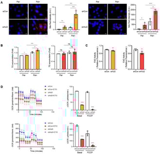
Metabolic stress caused by lipid overload is a key driver of cellular dysfunction in aging and disease. Excess saturated fatty acids such as palmitate impair fatty acid oxidation (FAO), promote lipid accumulation, and increase reactive oxygen species (ROS), ultimately triggering premature senescence-like states. Senescence further amplifies vulnerability by worsening mitochondrial dysfunction, enhancing lipid imbalance, and sustaining pro-inflammatory signaling. Here, we investigated the role of the neuron-enriched RNA-binding protein HuD (ELAVL4) in protecting cells against lipotoxic stress. Using Neuro2a neuroblastoma cells, we found that HuD knockdown suppressed FAO, leading to increased lipid accumulation and elevated ROS following palmitate exposure. HuD-deficient cells also exhibited cytosolic mitochondrial DNA release, IRF phosphorylation, and upregulation of senescence markers. Mechanistically, RNA immunoprecipitation revealed that HuD binds directly to PPARα mRNA, sustaining its expression by competing with the PPARα-targeting microRNAs miR-9-5p and miR-22-3p. Loss of HuD reduced PPARα levels, thereby weakening the FAO capacity and sensitizing cells to palmitate-induced lipotoxic stress. These findings identify a previously unrecognized HuD–PPARα–FAO axis that restrains metabolic stress and senescence. By linking post-transcriptional regulation to lipid metabolism and inflammatory signaling, this work highlights stress-induced premature senescence as both an outcome and a propagator of metabolic dysfunction, providing insight into mechanisms of aging-related vulnerability.

7 February 2026

HuD preserves FAO and restrains lipid droplets under palmitate. N2a cells transfected with siRNAs (siCon or siHuD) or stable N2a cells expressing shRNAs (shCtrl or shHuD) were incubated with palmitate. (A) Cells were stained with 0.1 μM Nile Red to visualize lipid droplets (red) and counterstained with DAPI to label nuclei (blue). Fluorescence intensity of lipid droplets was quantified using ImageJ software. Scale bar, 10 µm. (B) Intracellular triglyceride (TG) levels were measured to assess lipid accumulation. (C) Cells were stained with FAOBlue and fluorescence was measured for each group. (D) Cells were pretreated with etomoxir and oxygen consumption rate (OCR) was measured using Seahorse XFe24 Extracellular Flux Analyzer; data were acquired and analyzed via the Wave software. ΔOCR was used to evaluate FAO-dependent respiration. Data represent mean ± SD from three independent experiments. Statistical significance was determined using Student’s t-test for (A–C) and one-way ANOVA with Tukey’s multiple comparisons test for (D); * p < 0.05; ** p < 0.01; *** p < 0.001; ****, p < 0.0001; ns, not significant.

Pancreatic cancer is a highly intractable malignancy that necessitates personalized treatment strategies. Conventional patient-derived models, such as three-dimensional organoids, are often limited by intellectual property constraints and high costs. In this study, we developed an affordable adherent culture system for patient-derived pancreatic cancer cells using a proprietary medium and laminin-coated dishes. Primary cultures were successfully established from 28 patients with pancreatic ductal adenocarcinoma, exceeding a 90% success rate. Validation of eight samples confirmed maintenance of epithelial cell adhesion molecule expression and preservation of oncogenic KRAS mutations. Transcriptomic profiling revealed consistent upregulation of a six-gene signature (FAP, IGFBP5, PRRX1, SPARC, WNT5A, and ADAMTS12), which is associated with malignancy. In vitro drug sensitivity assays revealed interpatient heterogeneity with preliminary clinical associations. In conclusion, this simplified platform provides high-purity cancer cells and serves as a functional precision medicine tool. Beyond conventional chemotherapy, this platform has the potential to support applications ranging from biomarker validation and exploratory preclinical testing of novel therapeutics, including immune checkpoint inhibitors and antibody–drug conjugates. This optimization can lead to personalized therapeutic strategies for pancreatic cancer.

7 February 2026

  • Perspective
  • Open Access

New and Emerging Research Models for Sepsis

  • Saichaitanya Nallajennugari,
  • Xiang Li and
  • Mingui Fu

Human sepsis is a complex disease that manifests with a diverse range of phenotypes and inherent variability among individuals, making it hard to develop a comprehensive animal model. Despite this difficulty, numerous animal models have been developed that capture many key aspects of human sepsis. Though the animal models have contributed to the fundamental advances in understanding the pathogenesis of septic patients, the translational value of these models has been constantly questioned because many clinical trials of targeted therapies based on the advances in animal models have failed, highlighting the urgent need for developing new research models or refining previous animal models for sepsis research. In this review, we will summarize recent advances in new and emerging research models for sepsis, including human-based in vitro systems, highly tailored animal models, AI and digital models analyzing vast datasets to define patient subgroups and predict outcomes, and the FAMOUS framework ensuring that therapies are tested against the specific mechanism they are designed to target. We will discuss the strengths and limitations of these models, reflecting the clinical course of sepsis, and discuss the future directions in this subject area.

7 February 2026

Decoding Glioblastoma Through Liquid Biopsy: Molecular Insights and Clinical Prospects

  • Tomasz Wasiak,
  • Maria Jaskólska and
  • Adrianna Rutkowska
  • + 2 authors

Liquid biopsy (LB) offers a minimally invasive approach to characterizing and monitoring glioblastoma (GB), a tumor marked by extensive heterogeneity, limited surgical accessibility and rapid molecular evolution. By analyzing circulating tumor-derived components such as circulating tumor DNA (ctDNA), extracellular vesicles (EVs), circulating RNA species and circulating tumor cells (CTC), LB provides dynamic molecular information that cannot be captured by neuroimaging or single-site tissue sampling. Cerebrospinal fluid (CSF) currently yields the highest sensitivity for detecting tumor-specific alterations, while plasma enables repeat monitoring despite lower biomarker abundance. EVs have gained particular prominence due to their ability to preserve DNA, RNA, and protein cargo that reflects key genomic changes, treatment resistance mechanisms, and immune evasion. Although advances are substantial, clinical implementation remains constrained by low analyte concentrations, methodological variability, limited standardization and the high cost of testing, which is rarely reimbursed by insurers. This review summarizes current evidence on circulating biomarkers in GB and highlights research priorities essential for integrating LB into future diagnostic and therapeutic workflows.

6 February 2026

News & Conferences

Issues

Open for Submission

Editor's Choice

Reprints of Collections

Inflammation
Reprint

Inflammation

The Cause of all Diseases 2.0
Editors: Vasso Apostolopoulos, Jack Feehan, Vivek P. Chavda
Cellular and Molecular Mechanisms in Immune Regulation
Reprint

Cellular and Molecular Mechanisms in Immune Regulation

Editors: Fábio Rinaldo Santori, Natalia B. Ivanova

Get Alerted

Add your email address to receive forthcoming issues of this journal.

XFacebookLinkedIn
Cells - ISSN 2073-4409