Decoding Glioblastoma Through Liquid Biopsy: Molecular Insights and Clinical Prospects

 , ,
, ,
Highlights
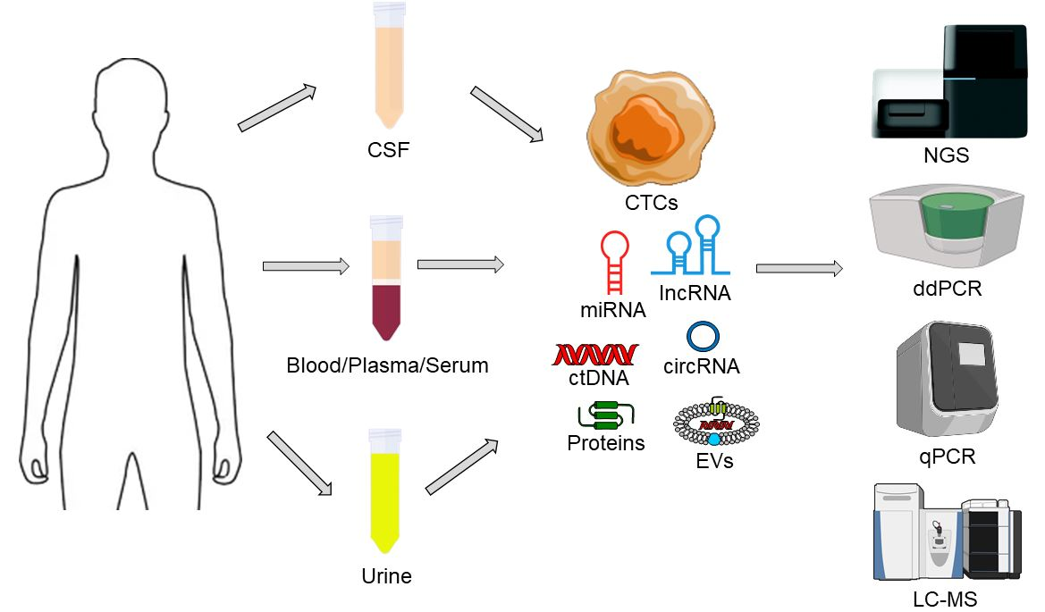
- Extracellular vesicles, ctDNA, circulating RNA species, and CTCs provide complementary molecular information that reflects glioblastoma heterogeneity and treatment dynamics.
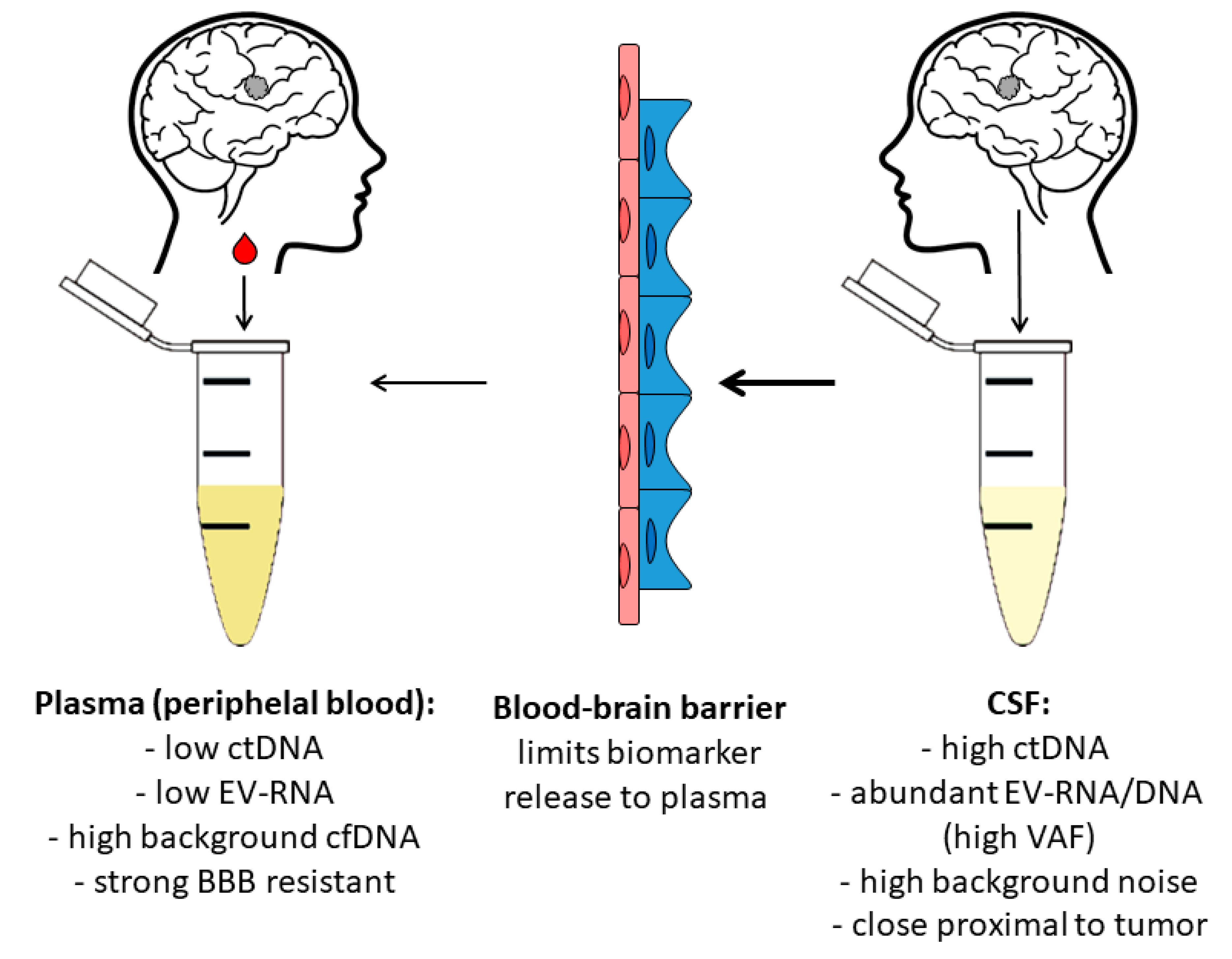
- CSF-derived biomarkers show consistently higher sensitivity than plasma biomarkers, while EV-associated signatures strongly correlate with tumor biology, therapy resistance, and immune evasion.
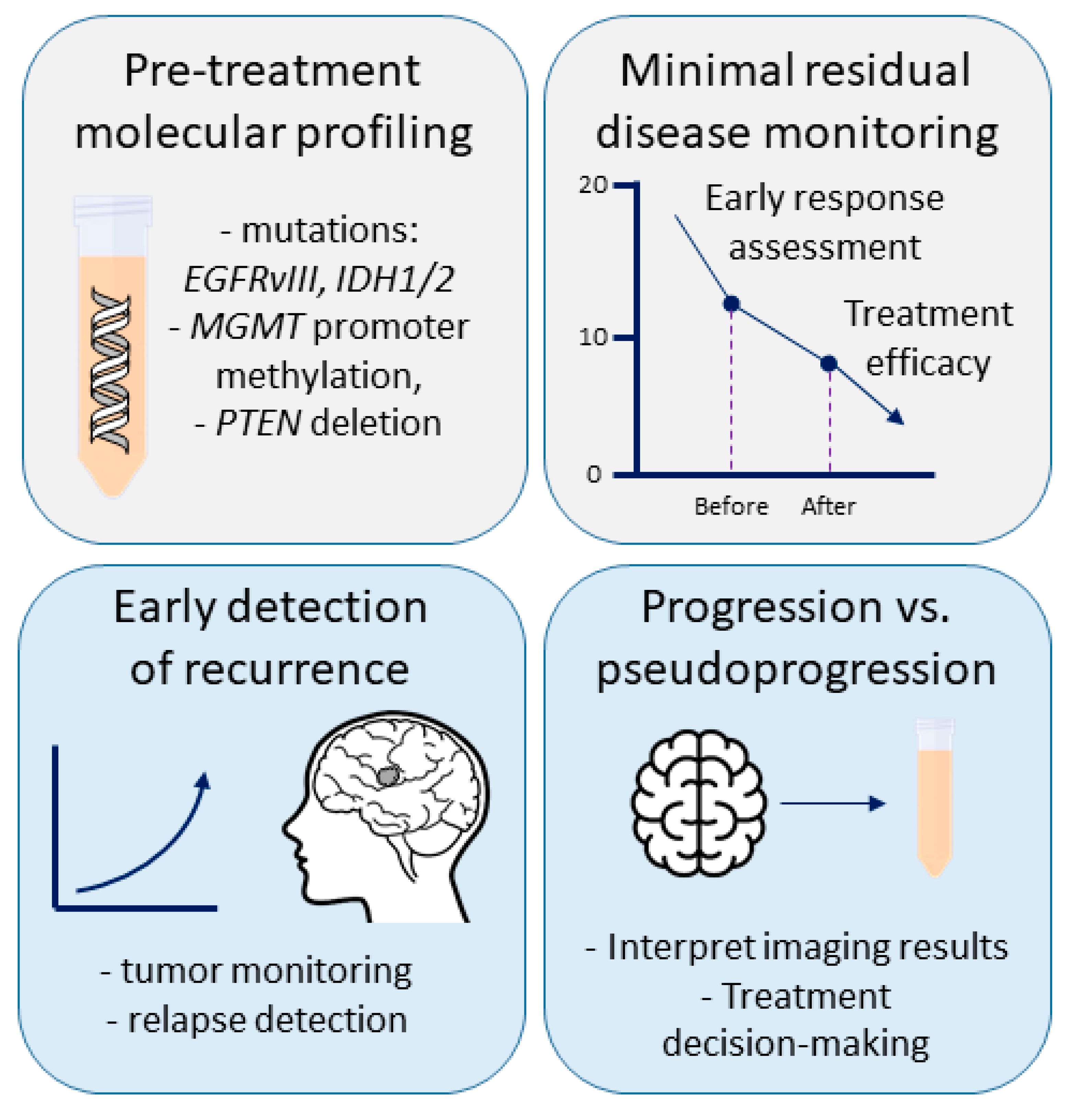
- Liquid biopsy can support diagnosis, early detection of recurrence and differentiation between progression and pseudoprogression when integrated with neuroimaging.
- Standardized multi-marker assays combining ctDNA, EV-RNA (Extracellular Vesicle RNA) and circulating RNA profiling may accelerate the translation of liquid biopsy into routine glioblastoma management.
Abstract
1. Introduction and Clinical Background
2. Unmet Clinical Needs in Glioblastoma Diagnosis
2.1. Early, Non-Invasive Diagnosis and Preoperative Stratification
2.2. Differentiating True Progression from Pseudoprogression and Radionecrosis
2.3. Monitoring Minimal Residual Disease and Early Detection of Recurrence
2.4. Capturing the Spatial and Temporal Heterogeneity of the Tumor
2.5. Standardization, Validation, and Integration with Decision Algorithms
3. The Biology of Liquid Biopsy
4. Biofluid Sources: CSF, Blood, Tumor-Proximal Blood, Urine
5. Markers for Liquid Biopsy
5.1. Circulating Tumor DNA and Methylation Markers
5.2. Circulating RNAs
5.3. Extracellular Vesicles (EVs)
5.4. Circulating Tumor Cells
5.5. Prognostic Relevance of Circulating Biomarkers
6. Technological Platforms and Pre-Analytical Variables
6.1. Digital (Droplet) PCR
6.2. Next-Generation Sequencing
6.3. Not Only Molecular Biology
6.4. Pre-Analytical Factors
7. Scenarios Where Liquid Biopsy Can Change Clinical Practice
7.1. Preoperative Diagnosis and Triage
7.2. Molecular Profiling Is Used When Tissue Is Limited or Risky to Obtain
7.3. Monitoring Treatment Response, Resistance Mechanisms
7.4. Pseudoprogression and True Progression
7.5. Differential Diagnosis of Intracranial Lesions
8. Evidence Level, Clinical Trials, and Regulatory Landscape
9. Integration with Imaging, Radiomics, and AI/ML
10. Challenges, Standardization, and Implementation
11. Ethical, Logistical, and Patient-Centered Aspects
12. Future Directions and Conclusions
Author Contributions
Funding
Institutional Review Board Statement
Data Availability Statement
Acknowledgments
Conflicts of Interest
Abbreviations
| ADC | Apparent Diffusion Coefficient |
| AI | Artificial Intelligence |
| APC | Adenomatous Polyposis Coli |
| BBB | Blood–Brain Barrier |
| CDKN2A/B | Cyclin-Dependent Kinase Inhibitor 2A and 2B |
| cfDNA | Cell-Free DNA |
| cfRNA | Cell-Free RNA |
| Cho/NAA | Choline (Cho) to N-acetylaspartate (NAA) ratio |
| circRNA | Circular RNA |
| CSF | Cerebrospinal Fluid |
| CTCs | Circulating Tumor Cells |
| ctDNA | Circulating Tumor DNA |
| ddPCR | Digital Droplet PCR |
| DIPG | Diffuse Intrinsic Pontine Glioma |
| EANO | European Association of Neuro-Oncology |
| EBM | Evidence Based Medicine |
| EGFR | Epidermal Growth Factor Receptor |
| EGFRvIII | Epidermal Growth Factor Receptor variant III |
| ERBB2 | ERB-B2 Receptor Tyrosine Kinase 2 |
| EVs | Extracellular Vesicles |
| FA | Fractional anisotropy |
| FUS-BBBO | Focused Ultrasound-Mediated Blood–Brain Barrier Opening |
| FT-IR | Fourier Transform Infrared Spectroscopy |
| GB | Glioblastoma |
| GFAP | Glial Fibrillary Acidic Protein |
| HOTAIR | HOX Transcript Antisense RNA |
| IDH1 | Isocitrate Dehydrogenase 1 |
| IL-13Rα2 | Interleukin-13 Receptor subunit alpha-2 |
| KDR | Kinase Insert Domain Receptor |
| LB | Liquid Biopsy |
| lncRNA | Long Noncoding RNA |
| LOD | Limit Of Detection |
| MET | Mesenchymal–Epithelial Transition Factor |
| ML | Machine Learning |
| miR | Micro RNA |
| MHC | Major Histocompatibility Complex |
| MGMT | Methylated-DNA–protein-cysteine methyltransferase |
| MRD | Minimal Residual Disease |
| MRI | Magnetic Resonance Imaging |
| NCCN | National Comprehensive Cancer Network |
| NF1 | Neurofibromin 1 |
| NGS | Next-Generation Sequencing |
| OR | Overall Response |
| PDGFRA | Platelet-Derived Growth Factor Receptor alpha |
| PD-L1 | Programmed Death-Ligand 1 |
| PET | Positron Emission Tomography |
| PFS | Progression Free Survival |
| PIK3CA | Phosphatidylinositol 3-Kinase Catalytic Subunit alpha |
| PsP | Pseudoprogression |
| PTEN | Phosphatase and Tensin Homolog |
| rCBV | Relative Cerebral Blood Volume |
| SNO | Society of Neuro-Oncology |
| TEPs | Tumor-Educated Platelets |
| TERT | Telomerase Reverse Transcriptase |
| TMZ | Temozolomide |
| TP | True Progression |
| TP53 | Human Tumor Protein p53 gene |
| VAF | Variant Allele Frequency |
References
- van Linde, M.E.; Brahm, C.G.; de Witt Hamer, P.C.; Reijneveld, J.C.; Bruynzeel, A.M.E.; Vandertop, W.P.; van de Ven, P.M.; Wagemakers, M.; van der Weide, H.L.; Enting, R.H.; et al. Treatment outcome of patients with recurrent glioblastoma multiforme: A retrospective multicenter analysis. J. Neuro-Oncol. 2017, 135, 183–192. [Google Scholar] [CrossRef]
- Davis, M.E. Epidemiology and Overview of Gliomas. Semin. Oncol. Nurs. 2018, 34, 420–429. [Google Scholar] [CrossRef] [PubMed]
- Louis, D.N.; Perry, A.; Wesseling, P.; Brat, D.J.; Cree, I.A.; Figarella-Branger, D.; Hawkins, C.; Ng, H.K.; Pfister, S.M.; Reifenberger, G.; et al. The 2021 WHO Classification of Tumors of the Central Nervous System: A summary. Neuro-Oncology 2021, 23, 1231–1251. [Google Scholar] [CrossRef] [PubMed]
- Piper, R.J.; Senthil, K.K.; Yan, J.L.; Price, S.J. Neuroimaging classification of progression patterns in glioblastoma: A systematic review. J. Neuro-Oncol. 2018, 139, 77–88. [Google Scholar] [CrossRef]
- Wagle, N.; Grabiner, B.C.; Van Allen, E.M.; Amin-Mansour, A.; Taylor-Weiner, A.; Rosenberg, M.; Gray, N.; Barletta, J.A.; Guo, Y.; Swanson, S.J.; et al. Response and acquired resistance to everolimus in anaplastic thyroid cancer. N. Engl. J. Med. 2014, 371, 1426–1433. [Google Scholar] [CrossRef] [PubMed]
- Li, K.; Zhu, Q.; Yang, J.; Zheng, Y.; Du, S.; Song, M.; Peng, Q.; Yang, R.; Liu, Y.; Qi, L. Imaging and Liquid Biopsy for Distinguishing True Progression From Pseudoprogression in Gliomas, Current Advances and Challenges. Acad. Radiol. 2024, 31, 3366–3383. [Google Scholar] [CrossRef]
- Seyhan, A.A. Circulating Liquid Biopsy Biomarkers in Glioblastoma: Advances and Challenges. Int. J. Mol. Sci. 2024, 25, 7974. [Google Scholar] [CrossRef]
- Elias, M.G.; Hadjiyiannis, H.; Vafaee, F.; Scott, K.F.; de Souza, P.; Becker, T.M.; Fatima, S. The Quest for Non-Invasive Diagnosis: A Review of Liquid Biopsy in Glioblastoma. Cancers 2025, 17, 2700. [Google Scholar] [CrossRef]
- Ronvaux, L.; Riva, M.; Coosemans, A.; Herzog, M.; Rommelaere, G.; Donis, N.; D’Hondt, L.; Douxfils, J. Liquid Biopsy in Glioblastoma. Cancers 2022, 14, 3394. [Google Scholar] [CrossRef]
- Kurokawa, R.; Kurokawa, M.; Baba, A.; Ota, Y.; Pinarbasi, E.; Camelo-Piragua, S.; Capizzano, A.A.; Liao, E.; Srinivasan, A.; Moritani, T. Major Changes in 2021 World Health Organization Classification of Central Nervous System Tumors. RadioGraphics 2022, 42, 1474–1493. [Google Scholar] [CrossRef]
- Luo, Y.; Gadd, M.E.; Qie, Y.; Otamendi-Lopez, A.; Sanchez-Garavito, J.E.; Brooks, M.M.; Ulloa Navas, M.J.; Hundal, T.; Li, S.; Jones, V.K.; et al. Solid cancer-directed CAR T cell therapy that attacks both tumor and immunosuppressive cells via targeting PD-L1. Mol. Therap. Oncol. 2024, 32, 200891. [Google Scholar] [CrossRef] [PubMed]
- Bagley, S.J.; Desai, A.S.; Fraietta, J.A.; Silverbush, D.; Chafamo, D.; Freeburg, N.F.; Gopikrishna, G.K.; Rech, A.J.; Nabavizadeh, A.; Bagley, L.J.; et al. Intracerebroventricular bivalent CAR T cells targeting EGFR and IL-13Ralpha2 in recurrent glioblastoma: A phase 1 trial. Nat. Med. 2025, 31, 2778–2787. [Google Scholar] [CrossRef] [PubMed]
- Poon, M.T.C.; Keni, S.; Vimalan, V.; Ip, C.; Smith, C.; Erridge, S.; Weir, C.J.; Brennan, P.M. Extent of MGMT promoter methylation modifies the effect of temozolomide on overall survival in patients with glioblastoma: A regional cohort study. Neuro-Oncol. Adv. 2021, 3, vdab171. [Google Scholar] [CrossRef] [PubMed]
- Zarychta, J.; Kowalczyk, A.; Marszolek, A.; Zawitkowska, J.; Lejman, M. Strategies to overcome tumor microenvironment immunosuppressive effect on the functioning of CAR-T cells in high-grade glioma. Ther. Adv. Med. Oncol. 2024, 16, 17588359241266140. [Google Scholar] [CrossRef]
- Yekula, A.; Muralidharan, K.; Rosh, Z.S.; Youngkin, A.E.; Kang, K.M.; Balaj, L.; Carter, B.S. Liquid Biopsy Strategies to Distinguish Progression from Pseudoprogression and Radiation Necrosis in Glioblastomas. Adv. Biosyst. 2020, 4, e2000029. [Google Scholar] [CrossRef]
- Neagu, M.R.; Huang, R.Y.; Reardon, D.A.; Wen, P.Y. How treatment monitoring is influencing treatment decisions in glioblastomas. Curr. Treat. Options Neurol. 2015, 17, 343. [Google Scholar] [CrossRef]
- Young, J.S.; Al-Adli, N.; Scotford, K.; Cha, S.; Berger, M.S. Pseudoprogression versus true progression in glioblastoma: What neurosurgeons need to know. J. Neurosurg. 2023, 139, 748–759. [Google Scholar] [CrossRef]
- Tsakiris, C.; Siempis, T.; Alexiou, G.A.; Zikou, A.; Sioka, C.; Voulgaris, S.; Argyropoulou, M.I. Differentiation Between True Tumor Progression of Glioblastoma and Pseudoprogression Using Diffusion-Weighted Imaging and Perfusion-Weighted Imaging: Systematic Review and Meta-analysis. World Neurosurg. 2020, 144, e100–e109. [Google Scholar] [CrossRef]
- Le Fevre, C.; Constans, J.M.; Chambrelant, I.; Antoni, D.; Bund, C.; Leroy-Freschini, B.; Schott, R.; Cebula, H.; Noel, G. Pseudoprogression versus true progression in glioblastoma patients: A multiapproach literature review. Part 2—Radiological features and metric markers. Crit. Rev. Oncol. Hematol. 2021, 159, 103230. [Google Scholar] [CrossRef]
- Wen, P.Y.; van den Bent, M.; Youssef, G.; Cloughesy, T.F.; Ellingson, B.M.; Weller, M.; Galanis, E.; Barboriak, D.P.; de Groot, J.; Gilbert, M.R.; et al. RANO 2.0: Update to the Response Assessment in Neuro-Oncology Criteria for High- and Low-Grade Gliomas in Adults. J. Clin. Oncol. Off. J. Am. Soc. Clin. Oncol. 2023, 41, 5187–5199. [Google Scholar] [CrossRef]
- Dwarshuis, G.; Kroon, L.L.; Brandsma, D.; Noske, D.P.; Best, M.G.; Sol, N. Liquid biopsies for the monitoring of gliomas and brain metastases in adults. Acta Neuropathol. 2025, 149, 37. [Google Scholar] [CrossRef] [PubMed]
- Song, M.; Pan, W.; Yu, X.; Ren, J.; Tang, C.; Chen, Z.; Wang, Z.; Deng, Y.; He, N.; Liu, H.; et al. Minimal Residual Disease Detection: Implications for Clinical Diagnosis and Cancer Patient Treatment. MedComm 2025, 6, e70193. [Google Scholar] [CrossRef] [PubMed]
- Eibl, R.H.; Schneemann, M. Liquid biopsy and glioblastoma. Explor. Target. Anti-Tumor Ther. 2023, 4, 28–41. [Google Scholar] [CrossRef] [PubMed]
- Ma, L.; Guo, H.; Zhao, Y.; Liu, Z.; Wang, C.; Bu, J.; Sun, T.; Wei, J. Liquid biopsy in cancer current: Status, challenges and future prospects. Signal Transduct. Target. Ther. 2024, 9, 336. [Google Scholar] [CrossRef]
- Janik, K.; Och, W.; Popeda, M.; Rosiak, K.; Peciak, J.; Rieske, P.; Kulbacki, K.; Szostak, B.; Parda, A.; Stoczynska-Fidelus, E. Glioblastoma with BRAFV600E mutation and numerous metastatic foci: A case report. Folia Neuropathol. 2019, 57, 72–79. [Google Scholar] [CrossRef]
- Garcia-Montano, L.A.; Licon-Munoz, Y.; Martinez, F.J.; Keddari, Y.R.; Ziemke, M.K.; Chohan, M.O.; Piccirillo, S.G.M. Dissecting Intra-tumor Heterogeneity in the Glioblastoma Microenvironment Using Fluorescence-Guided Multiple Sampling. Mol. Cancer Res. MCR 2023, 21, 755–767. [Google Scholar] [CrossRef]
- Berzero, G.; Pieri, V.; Mortini, P.; Filippi, M.; Finocchiaro, G. The coming of age of liquid biopsy in neuro-oncology. Brain J. Neurol. 2023, 146, 4015–4024. [Google Scholar] [CrossRef]
- Trivedi, S.; Kawadkar, M.; Pawar, D.; Agade, R.; Husain, U. Advances and challenges in personalized diagnosis and therapies for the management of recurrent glioblastoma. Precis. Medicat. 2025, 2, 100052. [Google Scholar] [CrossRef]
- Soffietti, R.; Bettegowda, C.; Mellinghoff, I.K.; Warren, K.E.; Ahluwalia, M.S.; De Groot, J.F.; Galanis, E.; Gilbert, M.R.; Jaeckle, K.A.; Le Rhun, E.; et al. Liquid biopsy in gliomas: A RANO review and proposals for clinical applications. Neuro-Oncology 2022, 24, 855–871. [Google Scholar] [CrossRef]
- Chahardehi, A.M.; Faraji, N.; Emtiazi, N.; Nasiri, R.; Daghagheleh, M.; Mohammadaein, H.; Masoudi, F.; Vakili, K.G.; Azar, A.S.; Fatemian, H.; et al. The emerging role of circulating tumor DNA in brain tumor research. IBRO Neurosci. Rep. 2025, 18, 714–725. [Google Scholar] [CrossRef]
- Riviere-Cazaux, C.; Keough, M.B.; Zuccato, J.A.; Kumar, R.; Schulz, S.; Warrington, A.E.; Ruff, M.W.; Ellingson, B.M.; Sanai, N.; Campian, J.L.; et al. A hitchhiker’s guide to cerebrospinal fluid biomarkers for neuro-oncology. Neuro-Oncology 2025, 27, 1165–1179. [Google Scholar] [CrossRef] [PubMed]
- Rafanan, J.; Ghani, N.; Kazemeini, S.; Nadeem-Tariq, A.; Shih, R.; Vida, T.A. Modernizing Neuro-Oncology: The Impact of Imaging, Liquid Biopsies, and AI on Diagnosis and Treatment. Int. J. Mol. Sci. 2025, 26, 917. [Google Scholar] [CrossRef] [PubMed]
- Medina, J.E.; Dracopoli, N.C.; Bach, P.B.; Lau, A.; Scharpf, R.B.; Meijer, G.A.; Andersen, C.L.; Velculescu, V.E. Cell-free DNA approaches for cancer early detection and interception. J. Immunother. Cancer 2023, 11, e006013. [Google Scholar] [CrossRef] [PubMed]
- Mandel, P.; Metais, P. Nuclear Acids In Human Blood Plasma. Comptes Rendus Séances Société Biol. Ses Fil. 1948, 142, 241–243. [Google Scholar]
- Otsuji, R.; Fujioka, Y.; Hata, N.; Kuga, D.; Hatae, R.; Sangatsuda, Y.; Nakamizo, A.; Mizoguchi, M.; Yoshimoto, K. Liquid Biopsy for Glioma Using Cell-Free DNA in Cerebrospinal Fluid. Cancers 2024, 16, 1009. [Google Scholar] [CrossRef]
- Panditharatna, E.; Kilburn, L.B.; Aboian, M.S.; Kambhampati, M.; Gordish-Dressman, H.; Magge, S.N.; Gupta, N.; Myseros, J.S.; Hwang, E.I.; Kline, C.; et al. Clinically Relevant and Minimally Invasive Tumor Surveillance of Pediatric Diffuse Midline Gliomas Using Patient-Derived Liquid Biopsy. Clin. Cancer Res. Off. J. Am. Assoc. Cancer Res. 2018, 24, 5850–5859. [Google Scholar] [CrossRef]
- Moss, J.; Magenheim, J.; Neiman, D.; Zemmour, H.; Loyfer, N.; Korach, A.; Samet, Y.; Maoz, M.; Druid, H.; Arner, P.; et al. Comprehensive human cell-type methylation atlas reveals origins of circulating cell-free DNA in health and disease. Nat. Commun. 2018, 9, 5068. [Google Scholar] [CrossRef]
- Wong, F.C.; Sun, K.; Jiang, P.; Cheng, Y.K.; Chan, K.C.; Leung, T.Y.; Chiu, R.W.; Lo, Y.M. Cell-free DNA in maternal plasma and serum: A comparison of quantity, quality and tissue origin using genomic and epigenomic approaches. Clin. Biochem. 2016, 49, 1379–1386. [Google Scholar] [CrossRef]
- Merker, J.D.; Oxnard, G.R.; Compton, C.; Diehn, M.; Hurley, P.; Lazar, A.J.; Lindeman, N.; Lockwood, C.M.; Rai, A.J.; Schilsky, R.L.; et al. Circulating Tumor DNA Analysis in Patients With Cancer: American Society of Clinical Oncology and College of American Pathologists Joint Review. J. Clin. Oncol. Off. J. Am. Soc. Clin. Oncol. 2018, 36, 1631–1641. [Google Scholar] [CrossRef]
- Snyder, M.W.; Kircher, M.; Hill, A.J.; Daza, R.M.; Shendure, J. Cell-free DNA Comprises an In Vivo Nucleosome Footprint that Informs Its Tissues-Of-Origin. Cell 2016, 164, 57–68. [Google Scholar] [CrossRef]
- Muhanna, N.; Di Grappa, M.A.; Chan, H.H.L.; Khan, T.; Jin, C.S.; Zheng, Y.; Irish, J.C.; Bratman, S.V. Cell-Free DNA Kinetics in a Pre-Clinical Model of Head and Neck Cancer. Sci. Rep. 2017, 7, 16723. [Google Scholar] [CrossRef] [PubMed]
- Vagner, T.; Spinelli, C.; Minciacchi, V.R.; Balaj, L.; Zandian, M.; Conley, A.; Zijlstra, A.; Freeman, M.R.; Demichelis, F.; De, S.; et al. Large extracellular vesicles carry most of the tumour DNA circulating in prostate cancer patient plasma. J. Extracell. Vesicles 2018, 7, 1505403. [Google Scholar] [CrossRef] [PubMed]
- Thakur, B.K.; Zhang, H.; Becker, A.; Matei, I.; Huang, Y.; Costa-Silva, B.; Zheng, Y.; Hoshino, A.; Brazier, H.; Xiang, J.; et al. Double-stranded DNA in exosomes: A novel biomarker in cancer detection. Cell Res. 2014, 24, 766–769. [Google Scholar] [CrossRef] [PubMed]
- Tie, J.; Wang, Y.; Tomasetti, C.; Li, L.; Springer, S.; Kinde, I.; Silliman, N.; Tacey, M.; Wong, H.L.; Christie, M.; et al. Circulating tumor DNA analysis detects minimal residual disease and predicts recurrence in patients with stage II colon cancer. Sci. Transl. Med. 2016, 8, 346ra392. [Google Scholar] [CrossRef]
- Zhang, Y.; Liu, Y.; Liu, H.; Tang, W.H. Exosomes: Biogenesis, biologic function and clinical potential. Cell Biosci. 2019, 9, 19. [Google Scholar] [CrossRef]
- Jin, Y.; Ma, L.; Zhang, W.; Yang, W.; Feng, Q.; Wang, H. Extracellular signals regulate the biogenesis of extracellular vesicles. Biol. Res. 2022, 55, 35. [Google Scholar] [CrossRef]
- Sedzik, M.; Rakoczy, K.; Sleziak, J.; Kisiel, M.; Kraska, K.; Rubin, J.; Luniewska, W.; Choromanska, A. Comparative Analysis of Exosomes and Extracellular Microvesicles in Healing Pathways: Insights for Advancing Regenerative Therapies. Molecules 2024, 29, 3681. [Google Scholar] [CrossRef]
- Chen, Y.F.; Luh, F.; Ho, Y.S.; Yen, Y. Exosomes: A review of biologic function, diagnostic and targeted therapy applications, and clinical trials. J. Biomed. Sci. 2024, 31, 67. [Google Scholar] [CrossRef]
- Trovato, E.; Di Felice, V.; Barone, R. Extracellular Vesicles: Delivery Vehicles of Myokines. Front. Physiol. 2019, 10, 522. [Google Scholar] [CrossRef]
- Lv, Y.; Tan, J.; Miao, Y.; Zhang, Q. The role of microvesicles and its active molecules in regulating cellular biology. J. Cell. Mol. Med. 2019, 23, 7894–7904. [Google Scholar] [CrossRef]
- Beer, K.B.; Wehman, A.M. Mechanisms and functions of extracellular vesicle release in vivo-What we can learn from flies and worms. Cell Adhes. Migr. 2017, 11, 135–150. [Google Scholar] [CrossRef] [PubMed]
- Curtin, J.F.; Cotter, T.G. Apoptosis: Historical perspectives. Essays Biochem. 2003, 39, 1–10. [Google Scholar] [CrossRef] [PubMed]
- Yu, L.; Zhu, G.; Zhang, Z.; Yu, Y.; Zeng, L.; Xu, Z.; Weng, J.; Xia, J.; Li, J.; Pathak, J.L. Apoptotic bodies: Bioactive treasure left behind by the dying cells with robust diagnostic and therapeutic application potentials. J. Nanobiotechnology 2023, 21, 218. [Google Scholar] [CrossRef]
- Mustafa, M.; Ahmad, R.; Tantry, I.Q.; Ahmad, W.; Siddiqui, S.; Alam, M.; Abbas, K.; Moinuddin; Hassan, M.I.; Habib, S.; et al. Apoptosis: A Comprehensive Overview of Signaling Pathways, Morphological Changes, and Physiological Significance and Therapeutic Implications. Cells 2024, 13, 1838. [Google Scholar] [CrossRef] [PubMed]
- Cabel, L.; Proudhon, C.; Gortais, H.; Loirat, D.; Coussy, F.; Pierga, J.Y.; Bidard, F.C. Circulating tumor cells: Clinical validity and utility. Int. J. Clin. Oncol. 2017, 22, 421–430. [Google Scholar] [CrossRef]
- Muller Bark, J.; Kulasinghe, A.; Hartel, G.; Leo, P.; Warkiani, M.E.; Jeffree, R.L.; Chua, B.; Day, B.W.; Punyadeera, C. Isolation of Circulating Tumour Cells in Patients With Glioblastoma Using Spiral Microfluidic Technology—A Pilot Study. Front. Oncol. 2021, 11, 681130. [Google Scholar] [CrossRef]
- Joosse, S.A.; Gorges, T.M.; Pantel, K. Biology, detection, and clinical implications of circulating tumor cells. EMBO Mol. Med. 2015, 7, 1–11. [Google Scholar] [CrossRef]
- Abbott, N.J.; Ronnback, L.; Hansson, E. Astrocyte-endothelial interactions at the blood-brain barrier. Nat. Rev. Neurosci. 2006, 7, 41–53. [Google Scholar] [CrossRef]
- Chistiakov, D.A.; Chekhonin, V.P. Circulating tumor cells and their advances to promote cancer metastasis and relapse, with focus on glioblastoma multiforme. Exp. Mol. Pathol. 2018, 105, 166–174. [Google Scholar] [CrossRef]
- Miller, A.M.; Shah, R.H.; Pentsova, E.I.; Pourmaleki, M.; Briggs, S.; Distefano, N.; Zheng, Y.; Skakodub, A.; Mehta, S.A.; Campos, C.; et al. Tracking tumour evolution in glioma through liquid biopsies of cerebrospinal fluid. Nature 2019, 565, 654–658. [Google Scholar] [CrossRef]
- Piccioni, D.E.; Achrol, A.S.; Kiedrowski, L.A.; Banks, K.C.; Boucher, N.; Barkhoudarian, G.; Kelly, D.F.; Juarez, T.; Lanman, R.B.; Raymond, V.M.; et al. Analysis of cell-free circulating tumor DNA in 419 patients with glioblastoma and other primary brain tumors. CNS Oncol. 2019, 8, CNS34. [Google Scholar] [CrossRef]
- Muller Bark, J.; Kulasinghe, A.; Chua, B.; Day, B.W.; Punyadeera, C. Circulating biomarkers in patients with glioblastoma. Br. J. Cancer 2020, 122, 295–305. [Google Scholar] [CrossRef] [PubMed]
- Pan, W.; Gu, W.; Nagpal, S.; Gephart, M.H.; Quake, S.R. Brain tumor mutations detected in cerebral spinal fluid. Clin. Chem. 2015, 61, 514–522. [Google Scholar] [CrossRef] [PubMed]
- De Mattos-Arruda, L.; Mayor, R.; Ng, C.K.Y.; Weigelt, B.; Martinez-Ricarte, F.; Torrejon, D.; Oliveira, M.; Arias, A.; Raventos, C.; Tang, J.; et al. Cerebrospinal fluid-derived circulating tumour DNA better represents the genomic alterations of brain tumours than plasma. Nat. Commun. 2015, 6, 8839. [Google Scholar] [CrossRef] [PubMed]
- Khier, S.; Lohan, L. Kinetics of circulating cell-free DNA for biomedical applications: Critical appraisal of the literature. Future Sci. OA 2018, 4, FSO295. [Google Scholar] [CrossRef]
- Legato, M.J. Dyslipidemia, gender, and the role of high-density lipoprotein cholesterol: Implications for therapy. Am. J. Cardiol. 2000, 86, 15–18. [Google Scholar] [CrossRef]
- Aimaletdinov, A.M.; Gomzikova, M.O. Tracking of Extracellular Vesicles’ Biodistribution: New Methods and Approaches. Int. J. Mol. Sci. 2022, 23, 11312. [Google Scholar] [CrossRef]
- Diehl, F.; Schmidt, K.; Choti, M.A.; Romans, K.; Goodman, S.; Li, M.; Thornton, K.; Agrawal, N.; Sokoll, L.; Szabo, S.A.; et al. Circulating mutant DNA to assess tumor dynamics. Nat. Med. 2008, 14, 985–990. [Google Scholar] [CrossRef]
- Wang, C.; Liu, H. Factors influencing degradation kinetics of mRNAs and half-lives of microRNAs, circRNAs, lncRNAs in blood in vitro using quantitative PCR. Sci. Rep. 2022, 12, 7259. [Google Scholar] [CrossRef]
- Izhar, M.; Ahmad, Z.; Moazzam, M.; Jader, A. Targeted liquid biopsy for brain tumors. J. Liq. Biopsy 2024, 6, 100170. [Google Scholar] [CrossRef]
- Hallal, S.M.; Tűzesi, Á.; Sida, L.A.; Xian, E.; Madani, D.; Muralidharan, K.; Shivalingam, B.; Buckland, M.E.; Satgunaseelan, L.; Alexander, K.L. Glioblastoma biomarkers in urinary extracellular vesicles reveal the potential for a ‘liquid gold’ biopsy. Br. J. Cancer 2024, 130, 836–851. [Google Scholar] [CrossRef]
- De Lima, B.P.; Ferraz, L.S.; Devalle, S.; Borges, H.L. Liquid Biopsy-Derived Tumor Biomarkers for Clinical Applications in Glioblastoma. Biomolecules 2025, 15, 658. [Google Scholar] [CrossRef] [PubMed]
- Bogucki, A.; Białecka, M.; Gajos, A.; Budrewicz, S.; Siemiński, M.; Rudzińska-Bar, M. Punkcja lędźwiowa—Zalecenia grupy ekspertów Polskiego Towarzystwa Neurologicznego. Pol. Przegląd Neurol. 2020, 16, 199–208. [Google Scholar]
- Hitchen, N.; Shahnam, A.; Tie, J. Circulating Tumor DNA: A Pan-Cancer Biomarker in Solid Tumors with Prognostic and Predictive Value. Annu. Rev. Med. 2025, 76, 207–223. [Google Scholar] [CrossRef] [PubMed]
- Nassiri, F.; Chakravarthy, A.; Feng, S.; Shen, S.Y.; Nejad, R.; Zuccato, J.A.; Voisin, M.R.; Patil, V.; Horbinski, C.; Aldape, K.; et al. Detection and discrimination of intracranial tumors using plasma cell-free DNA methylomes. Nat. Med. 2020, 26, 1044–1047. [Google Scholar] [CrossRef]
- Lavon, I.; Refael, M.; Zelikovitch, B.; Shalom, E.; Siegal, T. Serum DNA can define tumor-specific genetic and epigenetic markers in gliomas of various grades. Neuro-Oncology 2010, 12, 173–180. [Google Scholar] [CrossRef]
- Wang, Y.; Springer, S.; Zhang, M.; McMahon, K.W.; Kinde, I.; Dobbyn, L.; Ptak, J.; Brem, H.; Chaichana, K.; Gallia, G.L.; et al. Detection of tumor-derived DNA in cerebrospinal fluid of patients with primary tumors of the brain and spinal cord. Proc. Natl. Acad. Sci. USA 2015, 112, 9704–9709. [Google Scholar] [CrossRef]
- Fernandez-Mercado, M.; Manterola, L.; Larrea, E.; Goicoechea, I.; Arestin, M.; Armesto, M.; Otaegui, D.; Lawrie, C.H. The circulating transcriptome as a source of non-invasive cancer biomarkers: Concepts and controversies of non-coding and coding RNA in body fluids. J. Cell. Mol. Med. 2015, 19, 2307–2323. [Google Scholar] [CrossRef]
- Kapplingattu, S.V.; Bhattacharya, S.; Adlakha, Y.K. MiRNAs as major players in brain health and disease: Current knowledge and future perspectives. Cell Death Discov. 2025, 11, 7. [Google Scholar] [CrossRef]
- Qu, S.; Guan, J.; Liu, Y. Identification of microRNAs as novel biomarkers for glioma detection: A meta-analysis based on 11 articles. J. Neurol. Sci. 2015, 348, 181–187. [Google Scholar] [CrossRef]
- Garcia, C.M.; Toms, S.A. The Role of Circulating MicroRNA in Glioblastoma Liquid Biopsy. World Neurosurg. 2020, 138, 425–435. [Google Scholar] [CrossRef]
- Wu, L.; Li, G.; Feng, D.; Qin, H.; Gong, L.; Zhang, J.; Zhang, Z. MicroRNA-21 expression is associated with overall survival in patients with glioma. Diagn. Pathol. 2013, 8, 200. [Google Scholar] [CrossRef] [PubMed]
- Saenz-Antonanzas, A.; Auzmendi-Iriarte, J.; Carrasco-Garcia, E.; Moreno-Cugnon, L.; Ruiz, I.; Villanua, J.; Egana, L.; Otaegui, D.; Sampron, N.; Matheu, A. Liquid Biopsy in Glioblastoma: Opportunities, Applications and Challenges. Cancers 2019, 11, 950. [Google Scholar] [CrossRef] [PubMed]
- Wang, Q.; Li, P.; Li, A.; Jiang, W.; Wang, H.; Wang, J.; Xie, K. Plasma specific miRNAs as predictive biomarkers for diagnosis and prognosis of glioma. J. Exp. Clin. Cancer Res. 2012, 31, 97. [Google Scholar] [CrossRef] [PubMed]
- Yang, C.; Wang, C.; Chen, X.; Chen, S.; Zhang, Y.; Zhi, F.; Wang, J.; Li, L.; Zhou, X.; Li, N.; et al. Identification of seven serum microRNAs from a genome-wide serum microRNA expression profile as potential noninvasive biomarkers for malignant astrocytomas. Int. J. Cancer 2013, 132, 116–127. [Google Scholar] [CrossRef]
- Swellam, M.; Bakr, N.M.; El Magdoub, H.M.; Hamza, M.S.; Ezz El Arab, L.R. Emerging role of miRNAs as liquid biopsy markers for prediction of glioblastoma multiforme prognosis. J. Mol. Neurosci. 2021, 71, 836–844. [Google Scholar] [CrossRef]
- Lagarde, J.; Uszczynska-Ratajczak, B.; Santoyo-Lopez, J.; Gonzalez, J.M.; Tapanari, E.; Mudge, J.M.; Steward, C.A.; Wilming, L.; Tanzer, A.; Howald, C.; et al. Extension of human lncRNA transcripts by RACE coupled with long-read high-throughput sequencing (RACE-Seq). Nat. Commun. 2016, 7, 12339. [Google Scholar] [CrossRef]
- Shi, J.; Dong, B.; Cao, J.; Mao, Y.; Guan, W.; Peng, Y.; Wang, S. Long non-coding RNA in glioma: Signaling pathways. Oncotarget 2017, 8, 27582–27592. [Google Scholar] [CrossRef]
- Zhou, X.; Ren, Y.; Zhang, J.; Zhang, C.; Zhang, K.; Han, L.; Kong, L.; Wei, J.; Chen, L.; Yang, J.; et al. HOTAIR is a therapeutic target in glioblastoma. Oncotarget 2015, 6, 8353–8365. [Google Scholar] [CrossRef]
- Shen, J.; Hodges, T.R.; Song, R.; Gong, Y.; Calin, G.A.; Heimberger, A.B.; Zhao, H. Serum HOTAIR and GAS5 levels as predictors of survival in patients with glioblastoma. Mol. Carcinog. 2018, 57, 137–141. [Google Scholar] [CrossRef]
- Wang, Y.; Sui, X.; Zhao, H.; Cong, L.; Li, Y.; Xin, T.; Guo, M.; Hao, W. Decreased circular RNA hsa_circ_0001649 predicts unfavorable prognosis in glioma and exerts oncogenic properties in vitro and in vivo. Gene 2018, 676, 117–122. [Google Scholar] [CrossRef] [PubMed]
- Zhu, J.; Ye, J.; Zhang, L.; Xia, L.; Hu, H.; Jiang, H.; Wan, Z.; Sheng, F.; Ma, Y.; Li, W.; et al. Differential Expression of Circular RNAs in Glioblastoma Multiforme and Its Correlation with Prognosis. Transl. Oncol. 2017, 10, 271–279. [Google Scholar] [CrossRef] [PubMed]
- Meng, X.; Chen, Q.; Zhang, P.; Chen, M. CircPro: An integrated tool for the identification of circRNAs with protein-coding potential. Bioinformatics 2017, 33, 3314–3316. [Google Scholar] [CrossRef] [PubMed]
- Chen, A.; Zhong, L.; Ju, K.; Lu, T.; Lv, J.; Cao, H. Plasmatic circRNA Predicting the Occurrence of Human Glioblastoma. Cancer Manag. Res. 2020, 12, 2917–2923. [Google Scholar] [CrossRef]
- Sun, J.; Li, B.; Shu, C.; Ma, Q.; Wang, J. Functions and clinical significance of circular RNAs in glioma. Mol. Cancer 2020, 19, 34. [Google Scholar] [CrossRef]
- Schmidl, B.; Wei, K.; Ghasemimehr, N.; Dezfouli, A.B.; Wollenberg, B. Tumor-educated platelets as liquid biopsy: Uncovering the mechanisms of cancer progression. Crit. Rev. Oncol. Hematol. 2025, 218, 105106. [Google Scholar] [CrossRef]
- Sol, N.; In‘t Veld, S.; Vancura, A.; Tjerkstra, M.; Leurs, C.; Rustenburg, F.; Schellen, P.; Verschueren, H.; Post, E.; Zwaan, K.; et al. Tumor-Educated Platelet RNA for the Detection and (Pseudo)progression Monitoring of Glioblastoma. Cell Rep. Med. 2020, 1, 100101. [Google Scholar] [CrossRef]
- Zuo, M.; Li, T.; Wang, Z.; Xiang, Y.; Chen, S.; Liu, Y. Research progress on platelets in glioma. Chin. Med. J. 2025, 138, 28–37. [Google Scholar] [CrossRef]
- Hu, H.; Song, H.; Han, B.; Zhao, H.; He, J. Tumor-Educated Platelet RNA and Circulating Free RNA: Emerging Liquid Biopsy Markers for Different Tumor Types. Front. Biosci. 2024, 29, 80. [Google Scholar] [CrossRef]
- Yang, L.; Jiang, Q.; Li, D.Z.; Zhou, X.; Yu, D.S.; Zhong, J. TIMP1 mRNA in tumor-educated platelets is diagnostic biomarker for colorectal cancer. Aging 2019, 11, 8998–9012. [Google Scholar] [CrossRef]
- Ricklefs, F.L.; Wollmann, K.; Salviano-Silva, A.; Drexler, R.; Maire, C.L.; Kaul, M.G.; Reimer, R.; Schuller, U.; Heinemann, S.; Kolbe, K.; et al. Circulating extracellular vesicles as biomarker for diagnosis, prognosis, and monitoring in glioblastoma patients. Neuro-Oncology 2024, 26, 1280–1291. [Google Scholar] [CrossRef] [PubMed]
- Indira Chandran, V.; Gopala, S.; Venkat, E.H.; Kjolby, M.; Nejsum, P. Extracellular vesicles in glioblastoma: A challenge and an opportunity. NPJ Precis. Oncol. 2024, 8, 103. [Google Scholar] [CrossRef] [PubMed]
- Salviano-Silva, A.; Wollmann, K.; Brenna, S.; Reimer, R.; Neumann, J.E.; Dottermusch, M.; Woythe, L.; Maire, C.L.; Puig, B.; Schuller, U.; et al. Extracellular Vesicles Carrying Tenascin-C are Clinical Biomarkers and Improve Tumor-Derived DNA Analysis in Glioblastoma Patients. ACS Nano 2025, 19, 9844–9859. [Google Scholar] [CrossRef] [PubMed]
- Tzaridis, T.; Weller, J.; Bachurski, D.; Shakeri, F.; Schaub, C.; Hau, P.; Buness, A.; Schlegel, U.; Steinbach, J.P.; Seidel, C.; et al. A novel serum extracellular vesicle protein signature to monitor glioblastoma tumor progression. Int. J. Cancer 2023, 152, 308–319. [Google Scholar] [CrossRef]
- Musatova, O.E.; Rubtsov, Y.P. Effects of glioblastoma-derived extracellular vesicles on the functions of immune cells. Front. Cell Dev. Biol. 2023, 11, 1060000. [Google Scholar] [CrossRef]
- Kielbowski, K.; Plewa, P.; Szulc, J.; Cmil, M.; Bakinowska, E.; Pawlik, A. Extracellular Vesicles and PD-L1-A Review of Complex Immunoregulatory Properties and Clinical Importance. Biomedicines 2025, 13, 1356. [Google Scholar] [CrossRef]
- Lu, Y.; Wang, Z.; Zhang, D.; Luo, N.; Yang, H.; Chen, D.; Huang, H. Application of Circulating Tumor DNA in the Auxiliary Diagnosis and Prognosis Prediction of Glioma. Cell. Mol. Neurobiol. 2024, 45, 6. [Google Scholar] [CrossRef]
- Zhang, H.; Yuan, F.; Qi, Y.; Liu, B.; Chen, Q. Circulating Tumor Cells for Glioma. Front. Oncol. 2021, 11, 607150. [Google Scholar] [CrossRef]
- Cabezas-Camarero, S.; Perez-Alfayate, R.; Garcia-Barberan, V.; Gandia-Gonzalez, M.L.; Garcia-Feijoo, P.; Lopez-Cade, I.; Lorca, V.; Casado-Farinas, I.; Ceron, M.A.; Paz-Cabezas, M.; et al. ctDNA detection in cerebrospinal fluid and plasma and mutational concordance with the primary tumor in a multicenter prospective study of patients with glioma. Ann. Oncol. Off. J. Eur. Soc. Med. Oncol. 2025, 36, 660–672. [Google Scholar] [CrossRef]
- Jones, J.J.; Nguyen, H.; Wong, S.Q.; Whittle, J.; Iaria, J.; Stylli, S.; Towner, J.; Pieters, T.; Gaillard, F.; Kaye, A.H.; et al. Plasma ctDNA liquid biopsy of IDH1, TERTp, and EGFRvIII mutations in glioma. Neuro-Oncol. Adv. 2024, 6, vdae027. [Google Scholar] [CrossRef]
- Riviere-Cazaux, C.; Dong, X.; Mo, W.; Kumar, R.; Dai, C.; Carlstrom, L.P.; Munoz-Casabella, A.; Ghadimi, K.; Nesvick, C.L.; Andersen, K.M.; et al. Longitudinal Glioma Monitoring via Cerebrospinal Fluid Cell-Free DNA. Clin. Cancer Res. Off. J. Am. Assoc. Cancer Res. 2025, 31, 881–889. [Google Scholar] [CrossRef] [PubMed]
- Wang, Q.; Liang, Q.; Wei, W.; Niu, W.; Liang, C.; Wang, X.; Pan, H. Concordance analysis of cerebrospinal fluid with the tumor tissue for integrated diagnosis in gliomas based on next-generation sequencing. Pathol. Oncol. Res. 2023, 29, 1611391. [Google Scholar] [CrossRef] [PubMed]
- Perez-Alfayate, R.; Paz-Cabezas, M.; Perez-Segura, P.; Sanchez Del Hoyo, R.; Cabezas-Camarero, S. Cerebrospinal fluid ctDNA as a diagnostic and prognostic tool in gliomas: A systematic review and meta-analysis. Front. Oncol. 2025, 15, 1714287. [Google Scholar] [CrossRef] [PubMed]
- Fontanilles, M.; Marguet, F.; Beaussire, L.; Magne, N.; Pepin, L.F.; Alexandru, C.; Tennevet, I.; Hanzen, C.; Langlois, O.; Jardin, F.; et al. Cell-free DNA and circulating TERT promoter mutation for disease monitoring in newly-diagnosed glioblastoma. Acta Neuropathol. Commun. 2020, 8, 179. [Google Scholar] [CrossRef]
- Olmedillas-Lopez, S.; Garcia-Arranz, M.; Garcia-Olmo, D. Current and Emerging Applications of Droplet Digital PCR in Oncology. Mol. Diagn. Ther. 2017, 21, 493–510. [Google Scholar] [CrossRef]
- Kojabad, A.A.; Farzanehpour, M.; Galeh, H.E.G.; Dorostkar, R.; Jafarpour, A.; Bolandian, M.; Nodooshan, M.M. Droplet digital PCR of viral DNA/RNA, current progress, challenges, and future perspectives. J. Med. Virol. 2021, 93, 4182–4197. [Google Scholar] [CrossRef]
- Zhang, R.; Chen, B.; Tong, X.; Wang, Y.; Wang, C.; Jin, J.; Tian, P.; Li, W. Diagnostic accuracy of droplet digital PCR for detection of EGFR T790M mutation in circulating tumor DNA. Cancer Manag. Res. 2018, 10, 1209–1218. [Google Scholar] [CrossRef]
- Palacin-Aliana, I.; Garcia-Romero, N.; Asensi-Puig, A.; Carrion-Navarro, J.; Gonzalez-Rumayor, V.; Ayuso-Sacido, A. Clinical Utility of Liquid Biopsy-Based Actionable Mutations Detected via ddPCR. Biomedicines 2021, 9, 906. [Google Scholar] [CrossRef]
- Pott, C.; Bruggemann, M.; Ritgen, M.; van der Velden, V.H.J.; van Dongen, J.J.M. MRD Detection in B-Cell Non-Hodgkin Lymphomas Using Ig Gene Rearrangements and Chromosomal Translocations as Targets for Real-Time Quantitative PCR and ddPCR. Methods Mol. Biol. 2025, 2865, 189–219. [Google Scholar] [CrossRef]
- Gomes, I.P.; Miguita, L.; Gomes-Fernandes, B.; Coura, B.P.; Castro, W.H.; Carneiro, J.G.; De Marco, L.A.; Gomez, R.S.; Bastos-Rodrigues, L.; Gomes, C.C. No BRAF p.V600E mutation detection in ameloblastoma liquid biopsy-based analysis by ddPCR. Oral Oncol. 2024, 149, 106666. [Google Scholar] [CrossRef]
- Henriksen, T.V.; Drue, S.O.; Frydendahl, A.; Demuth, C.; Rasmussen, M.H.; Reinert, T.; Pedersen, J.S.; Andersen, C.L. Error Characterization and Statistical Modeling Improves Circulating Tumor DNA Detection by Droplet Digital PCR. Clin. Chem. 2022, 68, 657–667. [Google Scholar] [CrossRef]
- He, Y.; Lu, Z.; Liu, K.; Wang, L.; Xu, Q.; Fan, H.; Liu, C.; Zhang, T. Photofabricated channel-digital microfluidics (pCDMF): A promising lab-on-a-chip platform for fully integrated digital PCR. Sens. Actuators B Chem. 2024, 399, 134851. [Google Scholar] [CrossRef]
- Wood-Bouwens, C.M.; Haslem, D.; Moulton, B.; Almeda, A.F.; Lee, H.; Heestand, G.M.; Nadauld, L.D.; Ji, H.P. Therapeutic Monitoring of Circulating DNA Mutations in Metastatic Cancer with Personalized Digital PCR. J. Mol. Diagn. 2020, 22, 247–261. [Google Scholar] [CrossRef] [PubMed]
- Sanchez-Herrero, E.; Serna-Blasco, R.; Ivanchuk, V.; Garcia-Campelo, R.; Domine Gomez, M.; Sanchez, J.M.; Massuti, B.; Reguart, N.; Camps, C.; Sanz-Moreno, S.; et al. NGS-based liquid biopsy profiling identifies mechanisms of resistance to ALK inhibitors: A step toward personalized NSCLC treatment. Mol. Oncol. 2021, 15, 2363–2376. [Google Scholar] [CrossRef] [PubMed]
- Sposito, M.; Belluomini, L.; Nocini, R.; Insolda, J.; Scaglione, I.M.; Menis, J.; Simbolo, M.; Lugini, A.; Buzzacchino, F.; Verderame, F.; et al. Tissue- and liquid-biopsy based NGS profiling in advanced non-small-cell lung cancer in a real-world setting: The IMMINENT study. Front. Oncol. 2024, 14, 1436588. [Google Scholar] [CrossRef]
- Brynjulvsen, M.; Solli, E.; Walewska, M.; Zucknick, M.; Djirackor, L.; Langmoen, I.A.; Mughal, A.A.; Skaga, E.; Vik-Mo, E.O.; Sandberg, C.J. Functional and Molecular Heterogeneity in Glioma Stem Cells Derived from Multiregional Sampling. Cancers 2023, 15, 5826. [Google Scholar] [CrossRef]
- Keup, C.; Benyaa, K.; Hauch, S.; Sprenger-Haussels, M.; Tewes, M.; Mach, P.; Bittner, A.K.; Kimmig, R.; Hahn, P.; Kasimir-Bauer, S. Targeted deep sequencing revealed variants in cell-free DNA of hormone receptor-positive metastatic breast cancer patients. Cell. Mol. Life Sci. 2020, 77, 497–509. [Google Scholar] [CrossRef]
- Li, J.; Takamatsu, S.; Little, L.; Celestino, J.; Lawson, B.; Hillman, R.T. Liquid biopsy detection of structural variant breakpoints to monitor ovarian cancer clonal evolution. J. Clin. Oncol. 2024, 42, e17538. [Google Scholar] [CrossRef]
- Comba, A.; Faisal, S.M.; Varela, M.L.; Hollon, T.; Al-Holou, W.N.; Umemura, Y.; Nunez, F.J.; Motsch, S.; Castro, M.G.; Lowenstein, P.R. Uncovering Spatiotemporal Heterogeneity of High-Grade Gliomas: From Disease Biology to Therapeutic Implications. Front. Oncol. 2021, 11, 703764. [Google Scholar] [CrossRef]
- Liu, J.H.; Horiachok, M.; Guru, S.; Maire, C.L. Unraveling Glioblastoma Heterogeneity: Advancing Immunological Insights and Therapeutic Innovations. Brain Sci. 2025, 15, 833. [Google Scholar] [CrossRef]
- Silverman, I.M.; Schonhoft, J.D.; Herzberg, B.; Yablonovitch, A.; Lagow, E.; Fiaux, P.C.; Safabakhsh, P.; Sethuraman, S.; Ulanet, D.; Yang, J.; et al. Genomic and Epigenomic ctDNA Profiling in Liquid Biopsies from Heavily Pretreated Patients with DNA Damage Response-Deficient Tumors. Clin. Cancer Res. Off. J. Am. Assoc. Cancer Res. 2025, 31, 4136–4149. [Google Scholar] [CrossRef] [PubMed]
- Manzano, A.; Svedman, C.; Hofmarcher, T.; Wilking, N. Comparator Report on Cancer in Europe 2025—Disease Burden, Costs and Access to Medicines and Molecular Diagnostics. In IHE REPORT 2025:2; The Swedish Institute for Health Economics: Lund, Sweden, 2025. [Google Scholar]
- Afflerbach, A.K.; Rohrandt, C.; Brandl, B.; Sonksen, M.; Hench, J.; Frank, S.; Bornigen, D.; Alawi, M.; Mynarek, M.; Winkler, B.; et al. Classification of Brain Tumors by Nanopore Sequencing of Cell-Free DNA from Cerebrospinal Fluid. Clin. Chem. 2024, 70, 250–260. [Google Scholar] [CrossRef] [PubMed]
- Addissouky, T.A. Nanopore Sequencing of Cell-free DNA: An Emerging Liquid Biopsy Approach for Brain Tumor Molecular Profilin. J. Exp. Pathol. 2024, 5, 49–60. [Google Scholar] [CrossRef]
- Vermeulen, C.; Pages-Gallego, M.; Kester, L.; Kranendonk, M.E.G.; Wesseling, P.; Verburg, N.; de Witt Hamer, P.; Kooi, E.J.; Dankmeijer, L.; van der Lugt, J.; et al. Ultra-fast deep-learned CNS tumour classification during surgery. Nature 2023, 622, 842–849. [Google Scholar] [CrossRef]
- Wang, Z.; Fang, Y.; Liu, Z.; Hao, N.; Zhang, H.H.; Sun, X.; Que, J.; Ding, H. Adapting nanopore sequencing basecalling models for modification detection via incremental learning and anomaly detection. Nat. Commun. 2024, 15, 7148. [Google Scholar] [CrossRef]
- Dohm, J.C.; Peters, P.; Stralis-Pavese, N.; Himmelbauer, H. Benchmarking of long-read correction methods. NAR Genom. Bioinform. 2020, 2, lqaa037. [Google Scholar] [CrossRef]
- Bettegowda, C.; Sausen, M.; Leary, R.J.; Kinde, I.; Wang, Y.; Agrawal, N.; Bartlett, B.R.; Wang, H.; Luber, B.; Alani, R.M.; et al. Detection of circulating tumor DNA in early- and late-stage human malignancies. Sci. Transl. Med. 2014, 6, 224ra224. [Google Scholar] [CrossRef]
- Zhong, Q.; Bhattacharya, S.; Kotsopoulos, S.; Olson, J.; Taly, V.; Griffiths, A.D.; Link, D.R.; Larson, J.W. Multiplex digital PCR: Breaking the one target per color barrier of quantitative PCR. Lab A Chip 2011, 11, 2167–2174. [Google Scholar] [CrossRef]
- Shin, H.; Choi, B.H.; Shim, O.; Kim, J.; Park, Y.; Cho, S.K.; Kim, H.K.; Choi, Y. Single test-based diagnosis of multiple cancer types using Exosome-SERS-AI for early stage cancers. Nat. Commun. 2023, 14, 1644. [Google Scholar] [CrossRef]
- Huber, M.; Kepesidis, K.V.; Voronina, L.; Fleischmann, F.; Fill, E.; Hermann, J.; Koch, I.; Milger-Kneidinger, K.; Kolben, T.; Schulz, G.B.; et al. Infrared molecular fingerprinting of blood-based liquid biopsies for the detection of cancer. eLife 2021, 10, e68758. [Google Scholar] [CrossRef]
- Smok-Kalwat, J.; Gozdz, S.; Macek, P.; Kalwat, Z.; Khalavka, M.; Rzad, W.; Stepulak, A.; Depciuch, J. Serum and plasma as a good candidates of body fluids for detection lung cancer by FTIR liquid biopsy. Sci. Rep. 2024, 14, 31678. [Google Scholar] [CrossRef]
- Galant, N.; Nicos, M.; Khalavka, M.; Krzyzanowska, N.; Chmielewska, I.; Kuznar-Kaminska, B.; Krawczyk, P.; Sroka-Bartnicka, A.; Depciuch, J. Application of Fourier transform infrared (FTIR) spectroscopy in liquid biopsy to predict the response to the first-line immunotherapy in non-small-cell lung cancer (NSCLC) patients. Biochem. Biophys. Res. Commun. 2025, 771, 152007. [Google Scholar] [CrossRef]
- Ilieva, V.; Tenchov, B.; Virovska, D.; Nencheva, D.; Kalayanov, M.; Farfarov, A.; Yamakova, Y.; Abarova, S. Differential Scanning Calorimetry as a Monitoring Tool for the Effectiveness of Therapeutic Plasma Exchange in Anti-AChR Myasthenia Gravis, Anti-MuSK Myasthenia Gravis, and Myasthenic Syndrome: A Case Series. J. Clin. Med. 2025, 14, 4968. [Google Scholar] [CrossRef] [PubMed]
- Tsvetkov, P.O.; Tabouret, E.; Roman, A.Y.; Romain, S.; Bequet, C.; Ishimbaeva, O.; Honore, S.; Figarella-Branger, D.; Chinot, O.; Devred, F. Differential scanning calorimetry of plasma in glioblastoma: Toward a new prognostic/monitoring tool. Oncotarget 2018, 9, 9391–9399. [Google Scholar] [CrossRef] [PubMed]
- Gerber, T.; Taschner-Mandl, S.; Saloberger-Sindhoringer, L.; Popitsch, N.; Heitzer, E.; Witt, V.; Geyeregger, R.; Hutter, C.; Schwentner, R.; Ambros, I.M.; et al. Assessment of Pre-Analytical Sample Handling Conditions for Comprehensive Liquid Biopsy Analysis. J. Mol. Diagn. 2020, 22, 1070–1086. [Google Scholar] [CrossRef] [PubMed]
- Warton, K.; Yuwono, N.L.; Cowley, M.J.; McCabe, M.J.; So, A.; Ford, C.E. Evaluation of Streck BCT and PAXgene Stabilised Blood Collection Tubes for Cell-Free Circulating DNA Studies in Plasma. Mol. Diagn. Ther. 2017, 21, 563–570. [Google Scholar] [CrossRef]
- Andersson, D.; Kristiansson, H.; Luna Santamaria, M.; Zafar, H.; Mijakovic, I.; Torinsson Naluai, A.; Stahlberg, A. Evaluation of automatic cell free DNA extraction metrics using different blood collection tubes. Sci. Rep. 2025, 15, 19364. [Google Scholar] [CrossRef]
- Simon, N.K.; Volz, S.; Rios de los Rios Reséndiz, J.; Wedig, T.; Montigel, S.H.; Schwarz, N.; Richter, K.; Helm, D.; Neßling, M.; Zielske, L.; et al. Preanalytical framework for routine clinical use of liquid biopsies: Combining EVs and cfDNA. Extracell. Vesicles Circ. Nucleic Acids 2025, 6, 626–650. [Google Scholar] [CrossRef]
- Shin, K.H.; Lee, S.M.; Park, K.; Choi, H.; Kim, I.S.; Yoon, S.H.; Oh, S.H. Effects of Different Centrifugation Protocols on the Detection of EGFR Mutations in Plasma Cell-Free DNA. Am. J. Clin. Pathol. 2022, 158, 206–211. [Google Scholar] [CrossRef]
- Markus, H.; Contente-Cuomo, T.; Farooq, M.; Liang, W.S.; Borad, M.J.; Sivakumar, S.; Gollins, S.; Tran, N.L.; Dhruv, H.D.; Berens, M.E.; et al. Evaluation of pre-analytical factors affecting plasma DNA analysis. Sci. Rep. 2018, 8, 7375. [Google Scholar] [CrossRef]
- Diaz, I.M.; Nocon, A.; Held, S.A.E.; Kobilay, M.; Skowasch, D.; Bronkhorst, A.J.; Ungerer, V.; Fredebohm, J.; Diehl, F.; Holdenrieder, S.; et al. Pre-Analytical Evaluation of Streck Cell-Free DNA Blood Collection Tubes for Liquid Profiling in Oncology. Diagnostics 2023, 13, 1288. [Google Scholar] [CrossRef]
- Risberg, B.; Tsui, D.W.Y.; Biggs, H.; Ruiz-Valdepenas Martin de Almagro, A.; Dawson, S.J.; Hodgkin, C.; Jones, L.; Parkinson, C.; Piskorz, A.; Marass, F.; et al. Effects of Collection and Processing Procedures on Plasma Circulating Cell-Free DNA from Cancer Patients. J. Mol. Diagn. 2018, 20, 883–892. [Google Scholar] [CrossRef]
- Shin, S.H.; Park, W.Y.; Park, D. Characterization of DNA lesions associated with cell-free DNA by targeted deep sequencing. BMC Med. Genom. 2021, 14, 192. [Google Scholar] [CrossRef]
- Kim, S.H.; Weigelt, B. Circulating cell-free (cf)DNA analysis: Current technologies and applications in gynecologic cancer. Gynecol. Oncol. Rep. 2024, 54, 101431. [Google Scholar] [CrossRef] [PubMed]
- Stokowski, R.; White, K.; Hacker, C.; Doshi, J.; Schmid, M. Hemolysis and Fetal Fraction in Cell-Free DNA Blood Collection Tubes for Noninvasive Prenatal Testing. Mol. Diagn. Ther. 2020, 24, 185–190. [Google Scholar] [CrossRef] [PubMed]
- Nishimura, F.; Uno, N.; Chiang, P.C.; Kaku, N.; Morinaga, Y.; Hasegawa, H.; Yanagihara, K. The Effect of In Vitro Hemolysis on Measurement of Cell-Free DNA. J. Appl. Lab. Med. 2019, 4, 235–240. [Google Scholar] [CrossRef] [PubMed]
- Anichini, G.; Fulmali, P.; O’Neill, K.; Datta, V.; Crook, T.; Syed, N. Path-24. Accurate identification of glial malignancies from peripheral blood. Neuro-Oncology 2022, 24, vii155. [Google Scholar] [CrossRef]
- Sava, J. FDA Grants Breakthrough Designation for TriNetra™-Glio to Detect Brain Tumors. Targeted Oncology—Immunotherapy, Biomarkers, and Cancer Pathways. 2023. Available online: https://www.targetedonc.com/view/fda-grants-breakthrough-designation-for-trinetra--glio-to-detect-brain-tumors (accessed on 25 November 2025).
- Seoane, J.; De Mattos-Arruda, L.; Le Rhun, E.; Bardelli, A.; Weller, M. Cerebrospinal fluid cell-free tumour DNA as a liquid biopsy for primary brain tumours and central nervous system metastases. Ann. Oncol. Off. J. Eur. Soc. Med. Oncol. 2019, 30, 211–218. [Google Scholar] [CrossRef]
- Shu, Y.; Wu, X.; Tong, X.; Wang, X.; Chang, Z.; Mao, Y.; Chen, X.; Sun, J.; Wang, Z.; Hong, Z.; et al. Circulating Tumor DNA Mutation Profiling by Targeted Next Generation Sequencing Provides Guidance for Personalized Treatments in Multiple Cancer Types. Sci. Rep. 2017, 7, 583. [Google Scholar] [CrossRef]
- Benfatto, S.; Sill, M.; Jones, D.T.W.; Pfister, S.M.; Sahm, F.; von Deimling, A.; Capper, D.; Hovestadt, V. Explainable artificial intelligence of DNA methylation-based brain tumor diagnostics. Nat. Commun. 2025, 16, 1787. [Google Scholar] [CrossRef]
- Leao, D.J.; Craig, P.G.; Godoy, L.F.; Leite, C.C.; Policeni, B. Response Assessment in Neuro-Oncology Criteria for Gliomas: Practical Approach Using Conventional and Advanced Techniques. AJNR Am. J. Neuroradiol. 2020, 41, 10–20. [Google Scholar] [CrossRef]
- Guo, G.; Zhang, Z.; Zhang, J.; Wang, D.; Xu, S.; Wu, S.; Deng, K.; Bu, Y.; Sheng, Z.; Yu, J.; et al. Dynamic Monitoring of Circulating Tumor DNA to Predict the Risk of Non In Situ Recurrence of Postoperative Glioma: A Prospective Cohort Study. Cancer Med. 2025, 14, e70733. [Google Scholar] [CrossRef] [PubMed]
- Husain, A.; Mishra, S.; Hadi, R.; Sahu, A.; Kumari, S.; Rastogi, M.; Khurana, R.; Shukla, S.; Siddiqui, M.H.; Husain, N. Dynamics of cell-free DNA in predicting response in adult diffuse glioma on chemoradiotherapy. Cancer Genet. 2022, 268–269, 55–63. [Google Scholar] [CrossRef] [PubMed]
- Ordonez-Rubiano, E.G.; Rincon-Arias, N.; Espinosa, S.; Shelton, W.J.; Salazar, A.F.; Combita, A.; Baldoncini, M.; Luzzi, S.; Payan-Gomez, C.; Gomez-Amarillo, D.F.; et al. The potential of miRNA-based approaches in glioblastoma: An update in current advances and future perspectives. Curr. Res. Pharmacol. Drug Discov. 2024, 7, 100193. [Google Scholar] [CrossRef] [PubMed]
- Palizkaran Yazdi, M.; Barjasteh, A.; Moghbeli, M. MicroRNAs as the pivotal regulators of Temozolomide resistance in glioblastoma. Mol. Brain 2024, 17, 42. [Google Scholar] [CrossRef]
- Wang, X.; Yu, X.; Xu, H.; Wei, K.; Wang, S.; Wang, Y.; Han, J. Serum-derived extracellular vesicles facilitate temozolomide resistance in glioblastoma through a HOTAIR-dependent mechanism. Cell Death Dis. 2022, 13, 344. [Google Scholar] [CrossRef]
- Marangon, D.; Lecca, D. Exosomal non-coding RNAs in glioma progression: Insights into tumor microenvironment dynamics and therapeutic implications. Front. Cell Dev. Biol. 2023, 11, 1275755. [Google Scholar] [CrossRef]
- Chen, Y.; Jin, Y.; Wu, N. Role of Tumor-Derived Extracellular Vesicles in Glioblastoma. Cells 2021, 10, 512. [Google Scholar] [CrossRef]
- Dai, J.; Jiang, Y.; Hu, H.; Zhang, S.; Chen, Y. Extracellular vesicles as modulators of glioblastoma progression and tumor microenvironment. Pathol. Oncol. Res. 2024, 30, 1611549. [Google Scholar] [CrossRef]
- Rogachevsky, D.; Yalon, M.; Toren, A.; Mehrian-Shai, R. Circulating miRNAs as potential liquid biomarkers for pediatric gliomas. Pediatr. Res. 2025, 99, 1–10. [Google Scholar] [CrossRef]
- Fitzpatrick, A.; Iravani, M.; Mills, A.; Childs, L.; Alaguthurai, T.; Clifford, A.; Garcia-Murillas, I.; Van Laere, S.; Dirix, L.; Harries, M.; et al. Assessing CSF ctDNA to Improve Diagnostic Accuracy and Therapeutic Monitoring in Breast Cancer Leptomeningeal Metastasis. Clin. Cancer Res. Off. J. Am. Assoc. Cancer Res. 2022, 28, 1180–1191. [Google Scholar] [CrossRef]
- Mathios, D.; Niknafs, N.; Annapragada, A.V.; Bobeff, E.J.; Chiao, E.J.; Boyapati, K.; Short, S.; Bartolomucci, A.L.; Cristiano, S.; Koul, S.; et al. Detection of Brain Cancer Using Genome-wide Cell-free DNA Fragmentomes. Cancer Discov. 2025, 15, 1593–1608. [Google Scholar] [CrossRef] [PubMed]
- Kim, S.H.; Lim, K.H.; Yang, S.; Joo, J.Y. Long non-coding RNAs in brain tumors: Roles and potential as therapeutic targets. J. Hematol. Oncol. 2021, 14, 77. [Google Scholar] [CrossRef] [PubMed]
- Yadav, B.; Pal, S.; Rubstov, Y.; Goel, A.; Garg, M.; Pavlyukov, M.; Pandey, A.K. LncRNAs associated with glioblastoma: From transcriptional noise to novel regulators with a promising role in therapeutics. Mol. Ther. Nucleic Acids 2021, 24, 728–742. [Google Scholar] [CrossRef] [PubMed]
- Zhang, P.; Chen, K.; Liu, W.; Niu, X.; Wang, X.; Wang, J.; Yao, W.; Tang, X.; Tian, W. Engineered exosomes: A promising design platform for overcoming cancer therapy resistance. Front. Cell Dev. Biol. 2025, 13, 1608480. [Google Scholar] [CrossRef]
- Holland, M.; Krishnan, C.; Sotoudeh, H.; Nabors, L.; Fiveash, J.; Riley, K.; Huang, W.; Barboriak, D.; Kim, H. Detecting pseudo versus true progression of glioblastoma via accurate quantitative DCE-MRI using point-of-care portable perfusion phantoms: A pilot study. Quant. Imaging Med. Surg. 2025, 15, 4321–4332. [Google Scholar] [CrossRef]
- Van Dorth, D.; Croese, R.J.I.; Jiang, F.Y.; Schmitz-Abecassis, B.; Taphoorn, M.J.B.; Smits, M.; Dirven, L.; van Osch, M.J.P.; de Bresser, J.; Koekkoek, J.A.F. Perfusion MRI-based differentiation between early tumor progression and pseudoprogression in glioblastoma and its use in clinical practice. Neuro-Oncol. Pract. 2025, 12, 281–290. [Google Scholar] [CrossRef]
- Kertmen, N.; Kavgaci, G.; Akin, S.; Coban, G.; Isikay, A.I.; Yazici, G. Evaluating Immunotherapy Responses in Neuro-Oncology for Glioblastoma and Brain Metastases: A Brief Review Featuring Three Cases. Cancer Control. 2025, 32, 1–8. [Google Scholar] [CrossRef]
- Wurtemberger, U.; Rau, A.; Reisert, M.; Becker, L.; Elsheikh, S.; Hauser, T.K.; Grauvogel, J.; Hohenhaus, M.; Erny, D.; Urbach, H.; et al. Microstructural differentiation of cerebral metastases, glioblastoma, meningioma, and primary CNS lymphoma using advanced diffusion imaging techniques. Neuro-Oncol. Adv. 2025, 7, vdaf076. [Google Scholar] [CrossRef]
- Yekula, A.; Muralidharan, K.; Kang, K.; Carter, B.S.; Balaj, L. Extracellular Vesicles in Glioblastoma Tumor Microenvironment. Front. Immunol. 2019, 10, 3137. [Google Scholar] [CrossRef]
- Hasani, F.; Masrour, M.; Jazi, K.; Ahmadi, P.; Hosseini, S.s.; Lu, V.M.; Alborzi, A. MicroRNA as a potential diagnostic and prognostic biomarker in brain gliomas: A systematic review and meta-analysis. Front. Neurol. 2024, 15, 1357321. [Google Scholar] [CrossRef] [PubMed]
- An, Y.; Fan, F.; Jiang, X.; Sun, K. Recent Advances in Liquid Biopsy of Brain Cancers. Front. Genet. 2021, 12, 720270. [Google Scholar] [CrossRef] [PubMed]
- Zachariah, M.A.; Oliveira-Costa, J.P.; Carter, B.S.; Stott, S.L.; Nahed, B.V. Blood-based biomarkers for the diagnosis and monitoring of gliomas. Neuro-Oncology 2018, 20, 1155–1161. [Google Scholar] [CrossRef] [PubMed]
- Roberto Katrina, L.-F.M.J. Circulating Biomarkers in Diffuse Gliomas: Current Landscape and Future Directions. Pract. Neurol. 2024, 23, 44–48. [Google Scholar]
- Ali, H.; Harting, R.; de Vries, R.; Ali, M.; Wurdinger, T.; Best, M.G. Blood-Based Biomarkers for Glioma in the Context of Gliomagenesis: A Systematic Review. Front. Oncol. 2021, 11, 665235. [Google Scholar] [CrossRef]
- Eid, T.; Ghandour, L.; Ghanem, J.A.; Assi, H.; Mahfouz, R. Liquid biopsy in brain tumors: Potential for impactful clinical applications. Hum. Gene 2024, 42, 201333. [Google Scholar] [CrossRef]
- Mellinghoff, I.; Bale, T.; Graham, M. CHAPTER FOUR: Liquid biopsy in the diagnosis of primary brain tumors. Adv. Cancer Res. 2025, 166, 81–102. [Google Scholar] [CrossRef]
- Torp, S.H.; Solheim, O.; Skjulsvik, A.J. The WHO 2021 Classification of Central Nervous System tumours: A practical update on what neurosurgeons need to know-a minireview. Acta Neurochir. 2022, 164, 2453–2464. [Google Scholar] [CrossRef]
- Tataranu, L.G. Liquid Biopsy as a Diagnostic and Monitoring Tool in Glioblastoma. Medicina 2025, 61, 716. [Google Scholar] [CrossRef]
- Escudero, L.; Martinez-Ricarte, F.; Seoane, J. ctDNA-Based Liquid Biopsy of Cerebrospinal Fluid in Brain Cancer. Cancers 2021, 13, 1989. [Google Scholar] [CrossRef]
- Del Bene, M.; Osti, D.; Faletti, S.; Beznoussenko, G.V.; DiMeco, F.; Pelicci, G. Extracellular vesicles: The key for precision medicine in glioblastoma. Neuro-Oncology 2022, 24, 184–196. [Google Scholar] [CrossRef] [PubMed]
- Raza, I.J.; Tingate, C.A.; Gkolia, P.; Romero, L.; Tee, J.W.; Hunn, M.K. Blood Biomarkers of Glioma in Response Assessment Including Pseudoprogression and Other Treatment Effects: A Systematic Review. Front. Oncol. 2020, 10, 1191. [Google Scholar] [CrossRef] [PubMed]
- Ahanger, A.B.; Aalam, S.W.; Masoodi, T.A.; Shah, A.; Khan, M.A.; Bhat, A.A.; Assad, A.; Macha, M.A.; Bhat, M.R. Radiogenomics and machine learning predict oncogenic signaling pathways in glioblastoma. J. Transl. Med. 2025, 23, 121. [Google Scholar] [CrossRef] [PubMed]
- Zhu, M.; Li, S.; Kuang, Y.; Hill, V.B.; Heimberger, A.B.; Zhai, L.; Zhai, S. Artificial intelligence in the radiomic analysis of glioblastomas: A review, taxonomy, and perspective. Front. Oncol. 2022, 12, 924245. [Google Scholar] [CrossRef]
- Peng, H.; Pan, M.; Zhou, Z.; Chen, C.; Xing, X.; Cheng, S.; Zhang, S.; Zheng, H.; Qian, K. The impact of preanalytical variables on the analysis of cell-free DNA from blood and urine samples. Front. Cell Dev. Biol. 2024, 12, 1385041. [Google Scholar] [CrossRef]
- Zhu, L.; Cheng, G.; Ye, D.; Nazeri, A.; Yue, Y.; Liu, W.; Wang, X.; Dunn, G.P.; Petti, A.A.; Leuthardt, E.C.; et al. Focused Ultrasound-enabled Brain Tumor Liquid Biopsy. Sci. Rep. 2018, 8, 6553. [Google Scholar] [CrossRef]
- Meng, Y.; Pople, C.B.; Suppiah, S.; Llinas, M.; Huang, Y.; Sahgal, A.; Perry, J.; Keith, J.; Davidson, B.; Hamani, C.; et al. MR-guided focused ultrasound liquid biopsy enriches circulating biomarkers in patients with brain tumors. Neuro-Oncology 2021, 23, 1789–1797. [Google Scholar] [CrossRef]
- Bakker, A.; Ixkes, A.E.; Venugopal, H.; Ries, M.G.; Lak, N.S.M.; de Vos, F.; van Vuurden, D.G.; Snijders, T.J. Focused Ultrasound-Enhanced Liquid Biopsy: A Promising Diagnostic Tool for Brain Tumor Patients. Cancers 2024, 16, 1576. [Google Scholar] [CrossRef]
- Zhu, H.; Allwin, C.; Bassous, M.G.; Pouliopoulos, A.N. Focused ultrasound-mediated enhancement of blood-brain barrier permeability for brain tumor treatment: A systematic review of clinical trials. J. Neuro-Oncol. 2024, 170, 235–252. [Google Scholar] [CrossRef]
- Anastasiadis, P.; Gandhi, D.; Guo, Y.; Ahmed, A.-K.; Bentzen, S.M.; Arvanitis, C.; Woodworth, G.F. Localized blood–brain barrier opening in infiltrating gliomas with MRI-guided acoustic emissions–controlled focused ultrasound. Proc. Natl. Acad. Sci. USA 2021, 118, e2103280118. [Google Scholar] [CrossRef]
- Chien, C.Y.; Xu, L.; Yuan, J.; Fadera, S.; Stark, A.H.; Athiraman, U.; Leuthardt, E.C.; Chen, H. Quality assurance for focused ultrasound-induced blood-brain barrier opening procedure using passive acoustic detection. eBioMedicine 2024, 102, 105066. [Google Scholar] [CrossRef] [PubMed]
- Bell, M.S.; Walton, C.M.; Williams, M.J.; Eckert, T.; Brown, J.C.; Rowland, N.C.; Sahin, O.; Strickland, B.A. Engineering focused ultrasound for glioblastoma. Brain Stimul. 2025, 19, 102986. [Google Scholar] [CrossRef] [PubMed]
- Gupta, S.; Smith, T.R.; Broekman, M.L. Ethical considerations of neuro-oncology trial design in the era of precision medicine. J. Neuro-Oncol. 2017, 134, 1–7. [Google Scholar] [CrossRef] [PubMed]
- Hasner, M.C.; van Opijnen, M.P.; de Vos, F.Y.F.; Cuppen, E.; Broekman, M.L.D. Whole genome sequencing in (recurrent) glioblastoma: Challenges related to informed consent procedures and data sharing. Acta Neurochir. 2024, 166, 266. [Google Scholar] [CrossRef]
- Bhandari, A. Revolutionizing Radiology With Artificial Intelligence. Cureus 2024, 16, e72646. [Google Scholar] [CrossRef]
- Ziller, A.; Mueller, T.T.; Stieger, S.; Feiner, L.F.; Brandt, J.; Braren, R.; Rueckert, D.; Kaissis, G. Reconciling privacy and accuracy in AI for medical imaging. Nat. Mach. Intell. 2024, 6, 764–774. [Google Scholar] [CrossRef]
- Mikolajewicz, N.; Yee, P.P.; Bhanja, D.; Trifoi, M.; Miller, A.M.; Metellus, P.; Bagley, S.J.; Balaj, L.; de Macedo Filho, L.J.M.; Zacharia, B.E.; et al. Systematic Review of Cerebrospinal Fluid Biomarker Discovery in Neuro-Oncology: A Roadmap to Standardization and Clinical Application. J. Clin. Oncol. Off. J. Am. Soc. Clin. Oncol. 2024, 42, 1961–1974. [Google Scholar] [CrossRef]
- Horgan, D.; Cufer, T.; Gatto, F.; Lugowska, I.; Verbanac, D.; Carvalho, A.; Lal, J.A.; Kozaric, M.; Toomey, S.; Ivanov, H.Y.; et al. Accelerating the Development and Validation of Liquid Biopsy for Early Cancer Screening and Treatment Tailoring. Healthcare 2022, 10, 1714. [Google Scholar] [CrossRef]
- Pantel, K.; Alix-Panabieres, C.; Hofman, P.; Stoecklein, N.H.; Lu, Y.J.; Lianidou, E.; Giacomini, P.; Koch, C.; de Jager, V.; Deans, Z.C.; et al. Fostering the implementation of liquid biopsy in clinical practice: Meeting report 2024 of the European Liquid Biopsy Society (ELBS). J. Exp. Clin. Cancer Res. CR 2025, 44, 156. [Google Scholar] [CrossRef]
- Shen, F.; Zailaie, S.A.; Chiu, B.; Magliocco, A.; Sergi, C.M. Liquid biopsy—A narrative review with an update on current US governmental clinical trials targeting immunotherapy. Future Sci. OA 2025, 11, 2527598. [Google Scholar] [CrossRef]
- Friedman, J.S.; Hertz, C.A.J.; Karajannis, M.A.; Miller, A.M. Tapping into the genome: The role of CSF ctDNA liquid biopsy in glioma. Neuro-Oncol. Adv. 2022, 4, ii33–ii40. [Google Scholar] [CrossRef] [PubMed]
- Mair, R.; Mouliere, F. Cell-free DNA technologies for the analysis of brain cancer. Br. J. Cancer 2022, 126, 371–378. [Google Scholar] [CrossRef] [PubMed]
- Izhar, M.; Kattaa, A.H.; Akhavan-Sigari, A.; Shaghaghian, E.; Hori, Y.S.; Lam, F.C.; AbuReesh, D.; Emrich, S.C.; Ustrzynski, L.; Tayag, A.; et al. Role of extracellular vesicles in immune modulation, angiogenesis, progression and therapeutic resistance of glioblastoma. Extracell. Vesicle 2025, 5, 100075. [Google Scholar] [CrossRef]
- Ghazi, B.; Harmak, Z.; Rghioui, M.; Kone, A.S.; El Ghanmi, A.; Badou, A. Decoding the secret of extracellular vesicles in the immune tumor microenvironment of the glioblastoma: On the border of kingdoms. Front. Immunol. 2024, 15, 1423232. [Google Scholar] [CrossRef]
- Nelson, H.; Qu, S.; Franklin, J.L.; Liu, Q.; Pua, H.H.; Vickers, K.C.; Weaver, A.M.; Coffey, R.J.; Patton, J.G. Extracellular RNA in oncogenesis, metastasis and drug resistance. RNA Biol. 2024, 21, 17–31. [Google Scholar] [CrossRef]
- Balana, C.; Castaner, S.; Carrato, C.; Moran, T.; Lopez-Paradis, A.; Domenech, M.; Hernandez, A.; Puig, J. Preoperative Diagnosis and Molecular Characterization of Gliomas With Liquid Biopsy and Radiogenomics. Front. Neurol. 2022, 13, 865171. [Google Scholar] [CrossRef]
- Aredo, J.V.; Jamali, A.; Zhu, J.; Heater, N.; Wakelee, H.A.; Vaklavas, C.; Anagnostou, V.; Lu, J. Liquid Biopsy Approaches for Cancer Characterization, Residual Disease Detection, and Therapy Monitoring. Am. Soc. Clin. Oncol. Educ. Book 2025, 45, e481114. [Google Scholar] [CrossRef]
- Ray, A.; Vohra, T.K. Liquid biopsy-from bench to bedside. Neuro-Oncol. Adv. 2022, 4, ii66–ii72. [Google Scholar] [CrossRef]



| Biofluid | Molecular Yield | Detection Sensitivity | Advantages | Limitations | Bibliography |
|---|---|---|---|---|---|
| Cerebrospinal fluid | Highest (high variant allele frequency (VAF), ctDNA, abundant EV-RNA, low background cfDNA) | High (most sensitive biofluid; detects GB alterations in >60–70%) | Tumor-proximal; best representation of heterogeneity; high signal-to-noise; suitable for genomic profiling and clonal evolution | Invasive sampling; not feasible in all patients; requires neuraxial access | [21] |
| Plasma | Low–moderate (low VAF, diluted by systemic cfDNA) | Low–moderate (~20–40% depending on assay) | Minimally invasive; ideal for longitudinal monitoring; widely accessible; repeatable | BBB limits release of tumor DNA/EV; high background cfDNA; poor sensitivity for GB vs systemic tumors | [27] |
| Serum | Moderate (similar to plasma but higher background noise) | Low–moderate | Simple collection; can be paired with plasma comparative studies | Clotting-related release of genomic DNA from leukocytes; reduced ctDNA specificity | [23,70] |
| Urine | Very low (detectable EV-RNA; cfDNA extremely diluted) | Low | Non-invasive; patient-friendly; potential for home sampling | Very low tumor signal; still exploratory; needs ultrasensitive assays | [71] |
| Tumor-proximal venous blood | Moderate–high | Low–moderate | Higher concentration of tumor DNA than peripheral plasma; promising emerging method | Procedural complexity; limited clinical validation | [70] |
| ClinicalTrials.gov ID | Short Study Title | Population | Biofluid/Analyte | Study Focus | LB Objectives |
|---|---|---|---|---|---|
| NCT05925218 | Circulating Tumor DNA Collection From Patients With High Grade Glioma or Other Brain Tumors | High-grade glioma/brain tumors | Blood/ctDNA | Observational collection of blood samples | Characterize ctDNA from high-grade glioma and other brain tumors as a basis for LB–based monitoring and diagnosis |
| NCT05934630 | Testing Cerebrospinal Fluid for Cell-free Tumor DNA in Brain Tumor Patients | Brain tumor patients (including glioma/ GB) | CSF and blood/ cfDNA and ctDNA | Diagnostic/feasibility | Assess feasibility and sensitivity of detecting tumor cfDNA in CSF (and blood) as a non-invasive tool for brain tumor profiling |
| NCT05383872 | Blood-Brain Barrier Disruption for Liquid Biopsy in Subjects With Glioblastoma Brain Tumors | Suspected or confirmed GB | Blood/ctDNA after BBB disruption | Interventional (BBB opening, diagnostic) | Evaluate whether ultrasound-mediated BBB disruption improves yield of blood-based LBin GB |
| NCT05133154 | LIQUID BIOPSY IN Low-grade Glioma Patients | Low-grade glioma (with potential extension to higher grade) | Blood and CSF/ multiple markers | Prospective observational | Determine the value of blood/CSF LB as non-invasive, disease-associated biomarkers in glioma |
| NCT05964153 | Analysis of Circulating DNA in Blood Samples of Glioma Patients | Glioma (various grades) | Blood/ ctDNA and CFDNA | Pilot diagnostic study | Investigate a new strategy for glioma diagnosis using circulating DNA-based LB protocol |
| NCT04940507 (BRAINFUL) | BRAINFUL: Brain Tumor Liquid Biopsy Stud | GB and other brain tumors | Blood and CSF/ multiple markers | Prospective, multi-modal biomarker study | Explore LB signatures in brain tumors; assess impact on GB management and prognostication |
| NCT04539431 | Glioma Brain Tumours: Tissue and Liquid Biopsy Collection | Glioma/GB | Blood and CSF/ liquid biopsy | Prospective cohort with intraoperative sampling | Collect and analyze tumor tissue and matched LB (blood, CSF) obtained during surgery to correlate LB with tissue genomics |
| NCT04692324 | Cerebrospinal Fluid Biomarkers for Brain Tumors | Primary metastatic brain tumors (including GB) | CSF/ multiple markers | Observational biomarker study | Identify and validate CSF biomarkers, including tumor-derived DNA and proteins, for diagnosis and monitoring of brain tumors |
| NCT05864534 | Phase 2a Immune Modulation With Ultrasound for Newly Diagnosed Glioblastoma | Newly diagnosed GB | Blood/ctDNA (pre/post sonication) | Interventional (FUS + ICI) with LB correlative arm | Collect serial ctDNA to correlate with focused ultrasound–induced BBB modulation, treatment response and disease evolution |
Disclaimer/Publisher’s Note: The statements, opinions and data contained in all publications are solely those of the individual author(s) and contributor(s) and not of MDPI and/or the editor(s). MDPI and/or the editor(s) disclaim responsibility for any injury to people or property resulting from any ideas, methods, instructions or products referred to in the content. |
© 2026 by the authors. Licensee MDPI, Basel, Switzerland. This article is an open access article distributed under the terms and conditions of the Creative Commons Attribution (CC BY) license.
Share and Cite
Wasiak, T.; Jaskólska, M.; Filiks, K.; Bartkowiak, J.; Rutkowska, A. Decoding Glioblastoma Through Liquid Biopsy: Molecular Insights and Clinical Prospects. Cells 2026, 15, 309. https://doi.org/10.3390/cells15030309
Wasiak T, Jaskólska M, Filiks K, Bartkowiak J, Rutkowska A. Decoding Glioblastoma Through Liquid Biopsy: Molecular Insights and Clinical Prospects. Cells. 2026; 15(3):309. https://doi.org/10.3390/cells15030309
Chicago/Turabian StyleWasiak, Tomasz, Maria Jaskólska, Kamil Filiks, Jakub Bartkowiak, and Adrianna Rutkowska. 2026. "Decoding Glioblastoma Through Liquid Biopsy: Molecular Insights and Clinical Prospects" Cells 15, no. 3: 309. https://doi.org/10.3390/cells15030309
APA StyleWasiak, T., Jaskólska, M., Filiks, K., Bartkowiak, J., & Rutkowska, A. (2026). Decoding Glioblastoma Through Liquid Biopsy: Molecular Insights and Clinical Prospects. Cells, 15(3), 309. https://doi.org/10.3390/cells15030309