


Abstract
Bacterial soft rot of potato is predominantly associated with members of the genera Pectobacterium and Dickeya, whereas the contribution of other bacterial taxa to the etiology of the disease remains insufficiently characterized. This study aimed to characterize a bacterial isolate recovered from potato tubers exhibiting naturally occurring soft rot symptoms in Kazakhstan and to evaluate its pathogenic potential. Twelve bacterial isolates were obtained from symptomatic tissue, of which only isolate ZA4 reproducibly caused pronounced tissue softening and maceration following wound inoculation of healthy tubers, whereas disease development remained minimal in the negative control. Reproduction of characteristic symptoms and subsequent reisolation of the bacterium from experimentally affected tissue fulfilled the principal criteria of Koch’s postulates. Multilocus phylogenetic analysis based on 16S rRNA, gyrB, rpoB, and pyrE sequences assigned isolate ZA4 to Bacillus altitudinis. When tested across five potato cultivars and one commercial sample of unknown cultivar identity, the proportion of macerated tissue ranged from approximately 60–85% at 24 h and varied among the tested materials, increasing to 95–98% at 48 h, indicating rapid progression of tissue maceration under the bioassay conditions. ZA4 also exhibited pectinolytic, cellulolytic, proteolytic, and amylolytic activities. These findings demonstrate that B. altitudinis ZA4 is capable of inducing pronounced tissue maceration and soft-rot symptoms in wounded potato tubers under artificial wound inoculation conditions. However, broader studies encompassing a larger number of samples and a wider geographic area are required to determine the occurrence and epidemiological significance of B. altitudinis in association with potato soft rot in Kazakhstan.
1. Introduction
Potato (Solanum tuberosum L.) is one of the world’s most important food crops, playing a key role in global food security and ranking fourth in production after wheat, rice, and maize [1,2]. In the Republic of Kazakhstan, potato is considered a strategically important agricultural crop, with annual production exceeding 2.6 million tonnes. Despite relatively stable production levels, bacterial diseases remain an important constraint on crop yield, tuber quality, and storability, causing substantial losses both during the growing season and throughout postharvest storage [3,4,5].
Among the most economically important bacterial diseases of potato are blackleg and tuber soft rot, which are primarily caused by members of the Soft Rot Pectobacteriaceae (SRP). The principal causal agents of these diseases belong to the genera Pectobacterium and Dickeya, which are characterized by high virulence, broad geographical distribution, and substantial economic impact on potato production worldwide [6,7,8,9]. Members of the SRP are necrotrophic phytopathogens whose pathogenicity is largely associated with the secretion of an array of extracellular enzymes that degrade plant cell-wall components, resulting in tissue maceration and the characteristic symptoms of soft rot [10,11,12].
However, recent studies indicate that the taxonomic diversity of bacteria associated with maceration of plant tissues is broader than previously recognized. Individual members of the genus Bacillus, particularly species within the Bacillus pumilus group, including B. pumilus and B. altitudinis, have been reported to cause diseases of various agricultural and woody plants, including soft rots, bacterial leaf spots, rhizome rots, and necrotic lesions of woody tissues [13,14,15,16,17,18,19,20,21,22,23]. These observations are particularly noteworthy because members of the B. pumilus group occur in diverse environmental and plant-associated niches, and numerous strains have been characterized as plant growth-promoting or biocontrol microorganisms [24,25,26,27]. Consequently, biological properties within this group may differ substantially at the strain level, and the mere recovery of a Bacillus isolate from diseased plant tissue is insufficient to establish its etiological role, which requires experimental confirmation of pathogenicity and re-isolation from the inoculated host.
Information on the phytopathogenic potential of Bacillus altitudinis toward potato is particularly limited. Despite its close phylogenetic relationship to other members of the B. pumilus group and reports describing interactions of individual B. altitudinis strains with plants, its ability to cause soft rot of potato tubers remains insufficiently characterized. Data on atypical bacterial agents isolated from potato tubers exhibiting soft rot symptoms are also scarce in Central Asia, including Kazakhstan. To the best of our knowledge, B. altitudinis has not previously been reported in Kazakhstan as a bacterium isolated from naturally diseased potato tubers and experimentally demonstrated to induce maceration of tuber tissue. Establishing the etiological significance of such isolates is important not only for refining our understanding of the bacterial diversity associated with potato soft rot, but also for the accurate diagnosis of cases in which microorganisms other than the classical SRP pathogens are recovered from diseased tissues.
The aim of this study was to isolate a bacterial strain associated with naturally occurring soft-rot symptoms in potato tubers in Kazakhstan and to determine its taxonomic position based on multilocus phylogenetic analysis. Subsequently, the hydrolytic activity of the isolate and its ability to induce soft-rot symptoms in artificially wounded tubers of five potato cultivars and one commercial sample of unknown cultivar identity were evaluated, followed by a comparative assessment of symptom development among the tested potato materials.
2. Materials and Methods
2.1. Sample Collection and Bacterial Isolation
Potato tubers of the cultivar Colombo exhibiting characteristic soft-rot symptoms were collected in January 2026 from a single stored potato lot at the Maria farm, Zhauqashty village, Ili District, Almaty Region, Kazakhstan. A total of 26 symptomatic tubers were collected from different locations within the stored lot. Sampling was targeted toward tubers showing pronounced soft-rot symptoms and was not intended to estimate disease incidence or bacterial prevalence within the lot.
Following transportation to the laboratory, the tubers were washed under running tap water to remove adhering soil and surface debris. The tubers were surface-disinfected with 0.5% sodium hypochlorite for 3 min and subsequently rinsed three times with sterile distilled water. The efficacy of surface disinfection was not independently verified by plating aliquots of the final rinse water.
Under aseptic conditions, approximately 0.5 g of tissue was excised from the transition zone between visually healthy and macerated parenchyma. Each tissue sample was aseptically minced and homogenized in 1 mL of sterile distilled water. Tenfold serial dilutions of the resulting homogenates were prepared to 10−4, and 100 μL aliquots of appropriate dilutions were spread onto potato agar (PA). PA was used for primary isolation of bacteria from symptomatic potato tissue. The plates were incubated at 28 °C for 24–48 h.
Following incubation, colonies displaying distinct macroscopic characteristics were individually selected and purified by successive streaking on fresh PA until pure cultures were obtained. In total, 12 bacterial isolates were recovered and designated ZA1–ZA12. The tuber of origin was not recorded for each isolate; therefore, the number of individual tubers contributing to the final set of 12 isolates could not be determined retrospectively.
After purification, the isolates were transferred to nutrient agar (NA) and maintained at 4 °C until further analysis. PA was used for primary isolation from symptomatic potato tissue, whereas NA was used for routine maintenance and short-term storage of the purified cultures.
2.2. Primary Pathogenicity Screening of Bacterial Isolates on Potato Tubers
The pathogenicity of bacterial isolates ZA1–ZA12 was initially assessed on healthy potato tubers of the cultivar Colombo by artificial wound inoculation. Prior to inoculation, the tubers were thoroughly washed under running tap water, surface-disinfected with 0.5% sodium hypochlorite for 3 min, and rinsed three times with sterile distilled water to remove residual disinfectant. All isolates were tested under identical inoculation and incubation conditions, and pathogenicity was evaluated based on the development of tissue softening and maceration at the inoculation site.
Among the 12 isolates, ZA4 was the only isolate that reproducibly induced pronounced soft-rot symptoms resembling those observed in naturally affected tubers. Based on the results of this initial pathogenicity screening, ZA4 was selected for subsequent re-isolation, molecular confirmation, and further pathogenicity assays.
Isolate ZA4 was cultured on nutrient agar (NA) at 28 °C for 24 h. Bacterial cells were harvested from the agar surface using a sterile inoculating loop and suspended in sterile distilled water. The suspension was mixed thoroughly to obtain a homogeneous cell suspension.
The optical density of the suspension was measured at 600 nm (OD600), and the bacterial cell density was adjusted to approximately 1 × 10⁸ CFU mL−1 using a calibration curve previously established for isolate ZA4. A calibration curve relating OD600 values to viable cell concentration was established by serial dilution and plate counting of ZA4 suspensions on nutrient agar. This curve was used to adjust the bacterial suspension to the required concentration for pathogenicity assays.
Before inoculation, each tuber was aseptically cut longitudinally into two approximately equal halves, which were used as paired experimental treatments. An inoculation well 5 mm in diameter and 5 mm in depth was made in each half. One half was inoculated with 50 μL of the bacterial suspension of the corresponding isolate, whereas the second half of the same tuber received 50 μL of sterile distilled water and served as the paired negative control. The inoculated tuber halves were incubated at 28 °C and 70–85% relative humidity for 24 h.
Pathogenicity was assessed qualitatively based on the presence or absence of tissue softening and maceration at the inoculation site relative to the corresponding paired negative control treated with sterile distilled water. An isolate was classified as pathogenic when tissue softening and maceration were consistently observed in the inoculated tubers across the independent biological replicates, whereas no comparable tissue alterations were observed in the corresponding negative controls. The assay was conducted in three independent experimental repetitions, with three tubers used per bacterial isolate in each repetition, resulting in a total of nine tubers per isolate across the three repetitions. As the experiment was designed as a preliminary, strain-specific pathogenicity screening rather than a quantitative assessment of virulence, no numerical disease-severity scoring system was applied.
Reisolation and Confirmation of Koch’s Postulates
Following the development of characteristic soft-rot symptoms, bacteria were re-isolated from experimentally inoculated potato tubers to assess Koch’s postulates. Under aseptic conditions, tissue was collected from the transition zone between macerated and visually healthy parenchyma, homogenized in sterile distilled water, and plated onto PA. Plates were incubated at 28 °C for 24 h. Colonies exhibiting cultural and macroscopic characteristics consistent with those of the original ZA4 isolate were selected and purified by successive streaking on fresh PA until pure cultures were obtained. The same re-isolation procedure was applied to the corresponding negative-control tubers.
The identity of the re-isolated strain was confirmed by sequencing of the 16S rRNA, gyrB, rpoB, and pyrE loci and comparison with the corresponding sequences of the original ZA4 isolate. The confirmed re-isolated ZA4 strain was subsequently re-inoculated into healthy potato tubers under the same wound inoculation conditions used in the initial pathogenicity assay. Re-inoculation reproducibly resulted in the development of soft-rot symptoms comparable to those induced by the original ZA4 isolate.
2.3. Microscopic and Cultural Characterization
The cellular characteristics and Gram reaction of isolate ZA4 were examined using a 24 h bacterial culture and the standard Gram-staining procedure described by Coico [28]. Stained preparations were examined by light microscopy using a MICROMED microscope (Observarory Instruments LLC, St. Petersburg, Russia).
The cultural characteristics and colony characteristics of isolate ZA4 were evaluated on six solid media selected to provide complementary information on bacterial growth, colony development, and surface properties. (Titan Biotech Ltd., Delhi, India; potato, 200 g/L; agar, 20 g/L) was used to assess bacterial growth and colony characteristics on a potato-based substrate and to facilitate comparison with the cultural characteristics of the bacterium recovered from potato tuber tissue. Meat-peptone agar (Titan Biotech Ltd., Delhi, India; peptone, 10 g/L; meat extract, 3 g/L; NaCl, 5 g/L; agar, 20 g/L) and nutrient agar (Titan Biotech Ltd., Delhi, India; peptone, 5 g/L; meat extract, 3 g/L; agar, 15 g/L) were used as general-purpose media to assess colony size, form, elevation, margin, surface texture, and consistency under nutrient-rich conditions. Lysogeny broth (LB) agar (tryptone, 10 g/L (Titan Biotech Ltd., Delhi, India); yeast extract, 5 g/L (Titan Biotech Ltd., Delhi, India); NaCl, 10 g/L (Merck, Darmstadt, Germany); agar, 15 g/L) was included to assess colony development and cultural characteristics under a standard rich-medium formulation. Levan medium (yeast extract, 2 g/L; peptone, 5 g/L; NaCl, 5 g/L; sucrose, 50 g/L (Titan Biotech Ltd., Delhi, India); agar, 20 g/L) was selected to assess bacterial growth under a high sucrose concentration and levan-associated colony characteristics. King’s B medium (peptone, 20 g/L; glycerol, 10 mL/L (Sigma-Aldrich, St. Louis, MO, USA); K2HPO4, 1.5 g/L (Merck, Darmstadt, Germany); MgSO4·7H2O, 1.5 g/L (Merck, Darmstadt, Germany); agar, 15 g/L) was used to assess bacterial growth and colony characteristics under a medium commonly used for the characterization of bacterial cultural properties. On all media, colony form, size, elevation, margin, surface texture, consistency, and opacity were recorded after incubation.
The isolate was streak-inoculated onto each of the six culture media and incubated at 28 °C for 48 h. Following incubation, colony characteristics, including form, size, elevation, surface texture, margin, consistency, and opacity, were recorded. The assessment was performed in three independent experimental runs. In each run, three replicate plates were prepared for each medium, resulting in nine plates per medium across the three runs.
2.4. Assessment of Extracellular Hydrolytic Enzyme Activities
Pectinolytic, cellulolytic, proteolytic, and amylolytic activities of isolate ZA4 were assessed using semi-quantitative plate assays based on the formation of hydrolysis zones on media containing the corresponding substrates. A 24 h pure culture of ZA4 was used for inoculum preparation. Bacterial cells were resuspended in sterile distilled water, and the suspension was standardized to 1 × 10⁸ CFU/mL. Aliquots of 2 μL of the bacterial suspension were spot-inoculated onto the surface of agar media containing the respective substrates. The assays were performed in three independent experiments using a freshly prepared bacterial culture for each experiment, with three technical replicates per treatment.
Pectinolytic activity was assessed on M63 agar medium supplemented with 0.25% (w/v) polygalacturonate (PGA; Sigma-Aldrich, St. Louis, MO, USA) as the substrate [29]. The M63 medium contained, per liter: KH2PO4 (Merck, Darmstadt, Germany), 13.6 g; (NH4)2SO4 (Merck, Darmstadt, Germany), 2.0 g; FeSO4·7H2O (Merck, Darmstadt, Germany), 0.5 mg; MgSO4·7H2O (Merck, Darmstadt, Germany), 0.2 g; and microbiological agar, 15–18 g, with PGA added to a final concentration of 0.25% (w/v). Following incubation at 28 °C for 48 h, the surface of the medium was treated with 10% (w/v) copper acetate solution to visualize residual non-hydrolyzed PGA. Pectinolytic activity was identified by the formation of a pale or clear hydrolysis zone surrounding the bacterial colony against the stained background. The diameter of the hydrolysis zone was measured in millimeters across the center of the colony.
Cellulolytic activity was assessed on agar medium containing 1% (w/v) carboxymethyl cellulose (CMC; Sigma-Aldrich, St. Louis, MO, USA), as described in [30]. The medium contained, per liter: tryptone, 1.0 g; K2HPO4, 5.8 g; KH2PO4, 3.0 g; (NH4)2SO4, 1.0 g; sodium carboxymethyl cellulose, 10 g; and microbiological agar, 18 g. A MgSO4·7H2O stock solution (150 g L−1) was prepared separately and added aseptically at 1/100 of the final medium volume. Following incubation at 28 °C for 48 h, the plates were stained with 0.1% (w/v) Congo red solution for 15 min and subsequently washed with 1 M NaCl. The formation of a clear zone around the colony was considered indicative of CMC hydrolysis and cellulolytic activity. The diameter of the hydrolysis zone was measured in millimeters across the center of the colony.
Proteolytic activity was determined on agar medium containing 3% (w/v) skim milk powder (Sigma-Aldrich, St. Louis, MO, USA) as described in [31]. The basal medium contained, per liter: K2HPO4, 5.8 g; KH2PO4, 3.0 g; (NH4)2SO4, 1.0 g; and microbiological agar, 18 g. Skim milk powder was added to a final concentration of 3% (w/v). A MgSO4·7H2O stock solution (150 g L−1) was prepared separately and added aseptically at 1/100 of the final medium volume. Following spot inoculation, the plates were incubated at 28 °C for 48 h. The formation of a clear zone around the bacterial colony was considered evidence of casein hydrolysis and proteolytic activity. The diameter of the hydrolysis zone was measured in millimeters across the center of the colony.
Amylolytic activity was assessed on nutrient agar supplemented with 1% (w/v) soluble starch (Sigma-Aldrich, St. Louis, MO, USA), as described in [32]. The medium contained, per liter: peptone, 0.5 g; KCl, 0.1 g; MgSO4·7H2O, 0.5 g; (NH4)2SO4, 0.1 g; soluble starch, 10 g; and microbiological agar, 16 g. Following incubation at 28 °C for 48 h, the plates were treated with an I2/KI (Sigma-Aldrich, St. Louis, MO, USA) solution. The formation of a clear zone around the bacterial colony against the stained starch-containing medium indicated starch hydrolysis and amylolytic activity. The diameter of the hydrolysis zone was measured in millimeters across the center of the colony.
For each assay, uninoculated plates containing the corresponding medium were subjected to the same incubation and visualization procedures and served as negative controls for background effects associated with the medium and visualization reagents. For each independent experiment, technical replicates were averaged, and the results from three independent experiments were expressed as the mean ± standard deviation (SD).
2.5. Pathogenicity Assessment of Isolate ZA4 on Different Potato Cultivars
The pathogenicity of isolate ZA4 was evaluated on tubers of five potato cultivars: Astana, Bolashak, Edem, Zhanaisan, and Ulan—as well as on a commercial potato sample of unknown cultivar identity. Tuber preparation and bacterial inoculum preparation were performed according to the procedure described in Section 2.2.
Tissue maceration was assessed at 24 and 48 h post-inoculation. Three independent tubers were used for each cultivar (n = 3). The same tuber halves were evaluated sequentially at 24 and 48 h post-inoculation without additional cutting of the tissue. At each assessment time point, the exposed tuber surface was photographed under standardized conditions using a fixed camera-to-sample distance and uniform lighting.
Digital images were analyzed using ImageJ v1.54 (National Institutes of Health, USA). Macerated areas were distinguished from visually intact tissue using the Color Threshold function. Identical thresholding parameters were applied to all images. After generation of a binary mask, the area of macerated tissue and the total area of the analyzed tuber-half surface were measured. The extent of tissue maceration was calculated as follows: Tissue maceration (%) = (macerated tissue area/total analyzed tuber surface area) × 100.
2.6. Molecular Identification of Isolate ZA4
Genomic DNA was extracted from a pure culture of isolate ZA4 using the Genomic DNA Purification Kit (Thermo Fisher Scientific, Waltham, MA, USA) according to the manufacturer’s instructions. The extracted DNA was used as a template for amplification of fragments of the 16S rRNA, gyrB, rpoB, and pyrE genes using primer pairs described previously [33]. The primer sequences and expected amplicon sizes are listed in Supplementary Table S1.
PCR amplification was performed in a final reaction volume of 20 μL using Phusion™ High-Fidelity DNA Polymerase (Thermo Fisher Scientific, Waltham, MA, USA). Each reaction mixture contained 4 μL of 5× Phusion HF Buffer, 1 μL of dNTP mix (2 mM), 10 pmol each of the forward and reverse primers, 0.2 μL of Phusion High-Fidelity DNA Polymerase, and 2 μL of template DNA. The final volume was adjusted to 20 μL with nuclease-free water.
PCR amplification was carried out under the following cycling conditions: initial denaturation at 98 °C for 30 s; 30 cycles of denaturation at 98 °C for 10 s, primer annealing at the temperature specified for each primer pair for 30 s, and extension at 72 °C for 30 s; followed by a final extension at 72 °C for 10 min.
PCR products were purified using ExoSAP-IT™ PCR Product Cleanup Reagent (Applied Biosystems, Foster City, CA, USA) according to the manufacturer’s instructions.
Cycle sequencing reactions were performed using the BigDye™ Terminator v3.1 Cycle Sequencing Kit (Applied Biosystems, Foster City, CA, USA) with 0.8 pmol of the corresponding forward or reverse primer. The cycling conditions consisted of an initial denaturation at 96 °C for 1 min, followed by cycles of 96 °C for 10 s, 50 °C for 5 s, and 60 °C for 4 min.
Sequencing reaction products were purified using the BigDye XTerminator™ Purification Kit (Applied Biosystems, Foster City, CA, USA) and analyzed on a 3500xL Genetic Analyzer (Applied Biosystems, Foster City, CA, USA). The resulting nucleotide sequences were compared with sequences available in the NCBI GenBank database using the BLASTn algorithm.
2.7. Multilocus Phylogenetic Analysis
Nucleotide sequences of the 16S rRNA, gyrB, rpoB, and pyrE loci from isolate ZA4 and reference strains of closely related species within the Bacillus pumilus group available in the NCBI GenBank database were used for phylogenetic analysis. The dataset primarily included type and reference strains of B. altitudinis, B. pumilus, and B. safensis for which sequences of the analyzed loci were available. Preference was given to strains with well-established taxonomic assignments and the most complete representation of the four target loci. Bacillus cereus ATCC 14579 was used as the outgroup. The strains included in the phylogenetic analysis and their corresponding GenBank accession numbers are listed in Supplementary Table S1.
Phylogenetic relationships were reconstructed using a concatenated dataset comprising four loci: 16S rRNA, gyrB, rpoB, and pyrE. Sequences for each locus were initially aligned separately using the MUSCLE algorithm implemented in MEGA 12 [34]. The alignments were inspected manually, and ambiguously aligned and low-quality terminal regions were excluded prior to concatenation. Individual locus alignments were subsequently combined into a single concatenated matrix using SequenceMatrix v1.10 [35], maintaining the same locus order for all analyzed strains. After trimming and alignment, the final sequence lengths used for phylogenetic analysis were 1027 bp for 16S rRNA, 718 bp for gyrB, 670 bp for rpoB, and 383 bp for pyrE. The concatenated multilocus dataset therefore comprised 2798 nucleotide positions.
The phylogenetic tree was reconstructed using the maximum likelihood (ML) method implemented in MEGA 12. The best-fitting nucleotide substitution model was selected based on the Bayesian Information Criterion (BIC), with the Kimura two-parameter (K2P) model identified as the optimal model. The robustness of the inferred tree topology was assessed by bootstrap analysis with 1000 pseudoreplicates. The resulting phylogenetic tree was visualized and graphically edited using FigTree v1.4.4. The phylogenetic placement of isolate ZA4 was determined based on its clustering with the corresponding type and reference strains and the bootstrap support of the resulting clades.
2.8. Statistical Analysis
Tissue maceration data were analyzed using a three-way mixed ANOVA. Potato cultivar/sample was treated as the between-subject factor, whereas treatment (ZA4-inoculated vs. sterile-water control) and time post-inoculation (24 and 48 h) were treated as within-subject factors. In each tuber, one half was inoculated with ZA4 and the other half received sterile distilled water, thereby forming paired treatment observations within the same experimental unit. The same tuber halves were evaluated at both time points.
Normality of residuals was assessed using the Shapiro–Wilk test and by visual inspection of residual distributions, and homogeneity of variances was evaluated using Levene’s test. Because both within-subject factors contained only two levels, the assumption of sphericity was inherently satisfied and no correction for sphericity was required.
Significant interaction effects were further examined using pairwise comparisons of estimated marginal means. Comparisons between ZA4 and the corresponding sterile-water control within each cultivar/sample and time point were adjusted using Šídák’s method. Differences among ZA4-inoculated cultivars/samples within each time point were assessed using Tukey’s multiple-comparisons test. Statistical significance was accepted at p < 0.05.
All statistical analyses were performed using GraphPad Prism version 11.0.2 (GraphPad Software, San Diego, CA, USA). Statistical significance was set at α = 0.05.
3. Results
3.1. Isolation and Primary Pathogenicity Screening of Bacterial Isolates from Potato Tubers with Soft Rot Symptoms
Twelve bacterial isolates displaying distinct colony characteristics were recovered from Colombo potato tubers exhibiting pronounced soft rot symptoms and purified by successive subculturing. The isolates were designated ZA1–ZA12 and subjected to an initial pathogenicity screening on healthy potato tubers by artificial wound inoculation.
Among the 12 isolates tested, only ZA4 reproducibly induced pronounced disease symptoms resembling those observed in naturally affected tubers. Inoculation with ZA4 resulted in marked tissue softening and maceration, accompanied by the development of a water-soaked lesion at the inoculation site. None of the remaining 11 isolates produced visible soft rot symptoms during the observation period. Likewise, no tissue softening, maceration, or other visible alterations were observed in the negative controls.
Following inoculation with ZA4, bacteria exhibiting cultural characteristics consistent with those of the original isolate were reisolated from symptomatic tuber tissue. Molecular analysis confirmed the identity of the reisolated bacteria as ZA4, whereas no corresponding isolate was recovered from the negative-control tissue. These findings provide experimental support for the association of ZA4 with the development of soft-rot symptoms under the conditions of artificial wound inoculation.
Based on its reproducible ability to induce maceration of potato tuber tissue, ZA4 was selected for further characterization of its cultural characteristics and growth on different culture media, assessment of extracellular hydrolytic activities, molecular identification, and pathogenicity evaluation.
3.2. Microscopic and Cultural–Morphological Characterization of Isolate ZA4
Gram staining of a pure culture of isolate ZA4 revealed Gram-positive, rod-shaped cells. These observations provided an initial phenotypic characterization of the isolate and were complemented by an assessment of its cultural characteristics and colony features on different growth media.
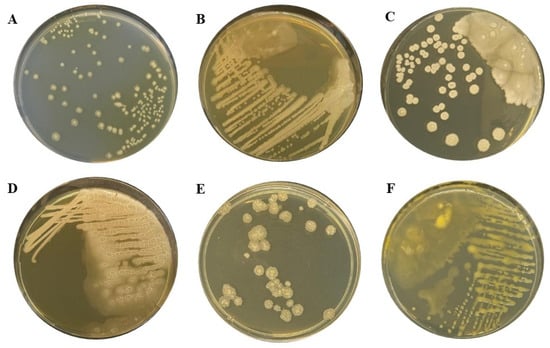
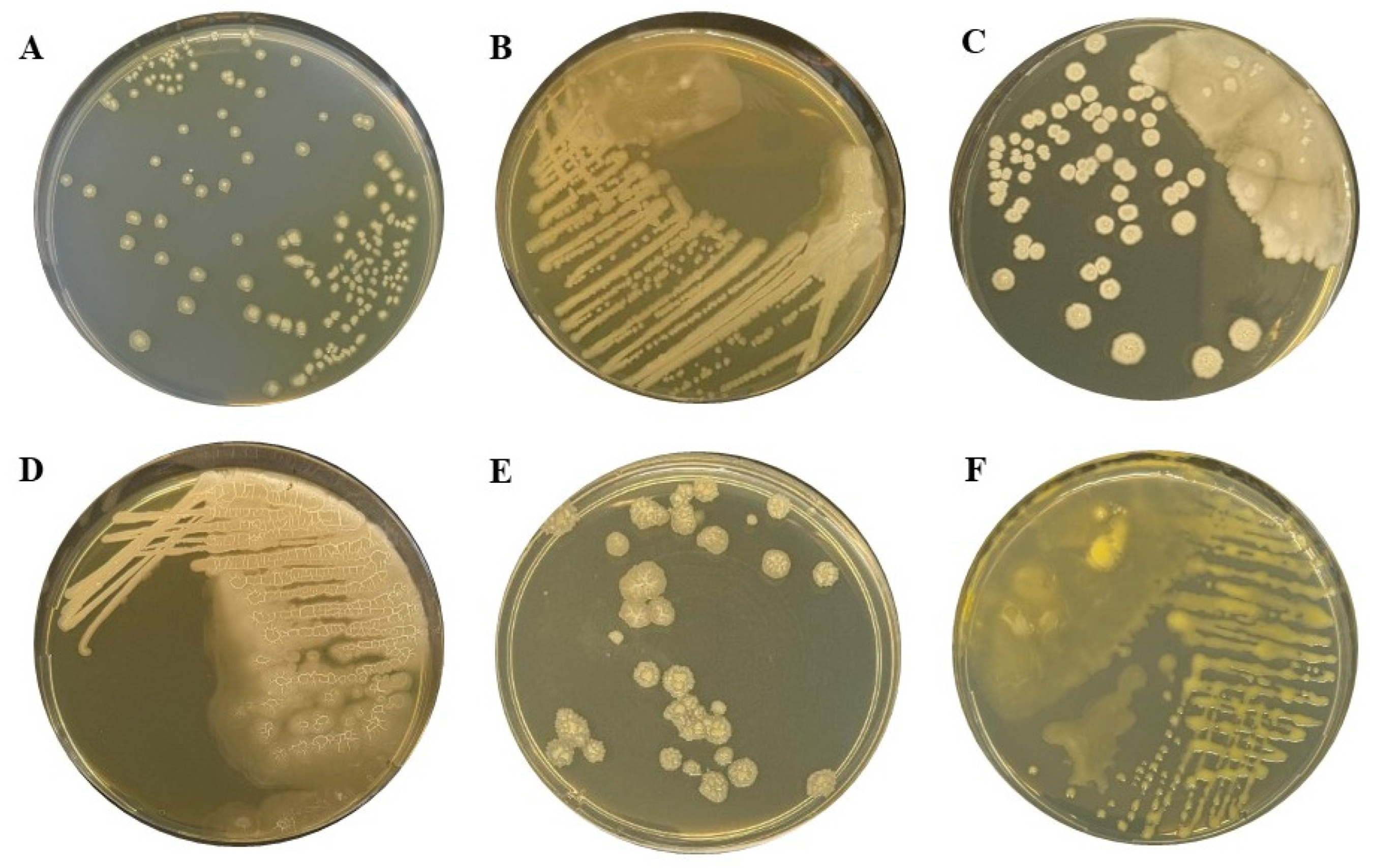
Isolate ZA4 grew on all media tested, although colony characteristics and growth characteristics varied depending on the composition of the medium (Figure 1). Across the media, isolated colonies were predominantly cream-white and circular, whereas colony size, elevation, surface texture, and margin characteristics differed among media.
Figure 1.
Colony characteristics of bacterial isolate ZA4 on different culture media. (A) potato agar (PA); (B) meat-peptone agar (MPA); (C) nutrient agar (NA); (D) lysogeny broth medium (LB); (E) King’s B medium (KB); (F) levan medium (LM).
On PA, numerous well-isolated, cream-white colonies were formed, predominantly circular in shape. The colonies were convex, with relatively smooth to slightly granular surfaces and predominantly entire margins. Some larger colonies showed a denser central region and slight surface heterogeneity.
On meat-peptone agar (MPA), abundant bacterial growth was observed. In areas of dense inoculation, the colonies coalesced to form confluent cream-beige growth, whereas individual circular colonies were observed in less densely inoculated areas. Colony surfaces were predominantly smooth to slightly uneven, and the margins ranged from entire to slightly undulate.
On nutrient agar (NA), numerous opaque, cream-white, circular colonies of variable size were formed. Most isolated colonies were convex and had entire or slightly undulate margins. Some larger colonies exhibited more pronounced surface granularity and slight wrinkling, while partial coalescence of adjacent colonies occurred in areas of dense growth.
On lysogeny broth (LB) medium, abundant growth was observed, with predominantly confluent cream-white bacterial growth in the area of the primary streak. Individual colonies became distinguishable in areas of lower inoculum density. The surface of the bacterial growth was heterogeneous, and the margins of individual colonies were undulate or irregular.
On levan medium (LM), isolate ZA4 exhibited abundant growth along the streak lines. The culture was cream-yellowish in color, and colonies coalesced in densely inoculated areas to form confluent growth. In less densely inoculated areas, individual colonies were predominantly circular, with smooth, glossy surfaces and entire or slightly undulate margins. Growth on this medium appeared visually more moist and mucoid than the colonies formed on most of the other media tested.
On King B medium (KB), predominantly discrete cream-beige colonies were formed, with pronounced surface heterogeneity. The colonies were raised, with relatively dry, rough, and distinctly wrinkled surfaces, and the margins of most larger colonies were undulate or irregular. In some colonies, wrinkling was particularly pronounced in the central region, giving them a characteristic wrinkled appearance.
Overall, isolate ZA4 was Gram-positive and rod-shaped, with marked variation in colony characteristics across the tested media.
3.3. Extracellular Hydrolytic Activities of Isolate ZA4
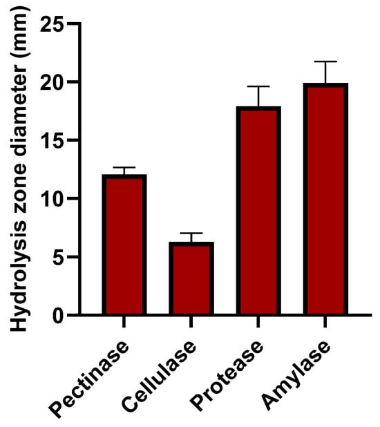
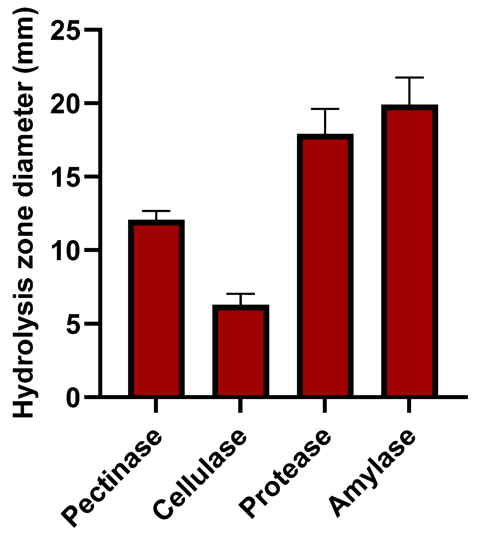
Isolate ZA4 exhibited pectinolytic, cellulolytic, proteolytic, and amylolytic activities on agar media containing the corresponding substrates. Hydrolytic activity was evidenced by the formation of clear hydrolysis zones around the bacterial growth, and the diameters of these zones were used for semi-quantitative assessment of the respective activities (Figure 2).
Figure 2.
Extracellular hydrolytic activities of bacterial isolate ZA4. Pectinolytic, cellulolytic, proteolytic, and amylolytic activities were assessed semi-quantitatively by measuring the diameters of hydrolysis zones (mm) on agar media containing the corresponding substrates. Technical replicates were averaged within each independent experiment. Data are presented as the mean ± SD of three independent experiments (n = 3).
The largest mean hydrolysis-zone diameter was observed in the amylolytic assay, reaching 19.90 ± 1.85 mm. The mean hydrolysis-zone diameter in the proteolytic assay was 17.93 ± 1.68 mm. Pectinolytic activity produced a mean hydrolysis-zone diameter of 12.07 ± 0.60 mm, whereas the smallest mean diameter was recorded in the cellulolytic assay, at 6.30 ± 0.75 mm.
3.4. Pathogenicity of Isolate ZA4 on Tubers of Different Potato Cultivars
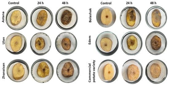
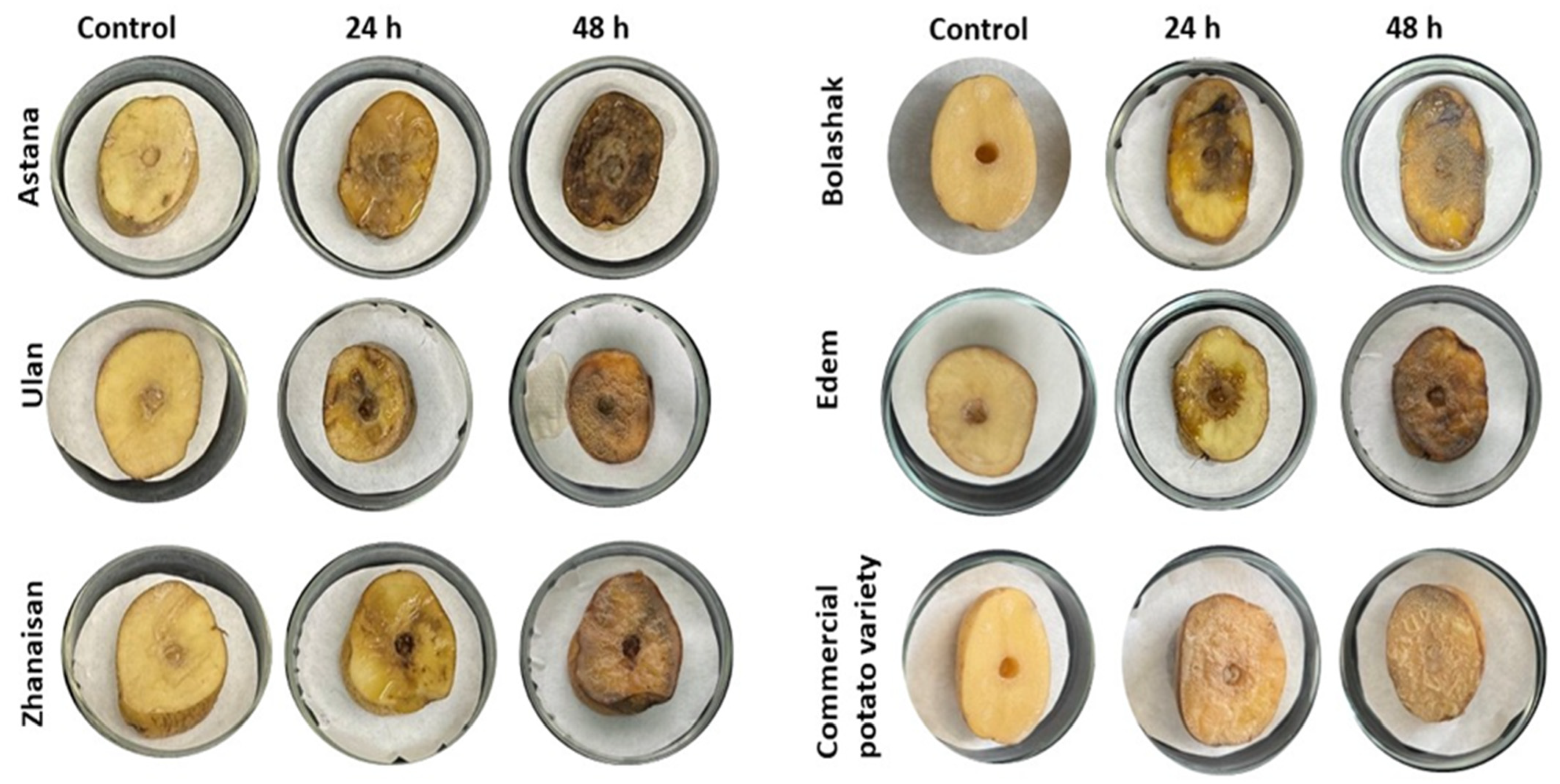
Inoculation with isolate ZA4 induced pronounced soft-rot symptoms in all potato cultivars/samples tested. At 24 h post-inoculation, tissue softening, darkening, and maceration were evident, with the extent of tissue damage varying among cultivars (Figure 3).
Figure 3.
Development of soft rot symptoms on tubers of different potato cultivars following inoculation with bacterial isolate ZA4. Control tubers and representative symptoms observed at 24 and 48 h post-inoculation are shown for the cultivars Astana, Bolashak, Edem, Zhanaisan, and Ulan, as well as for a commercial potato sample of unknown cultivar identity.
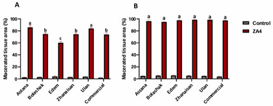
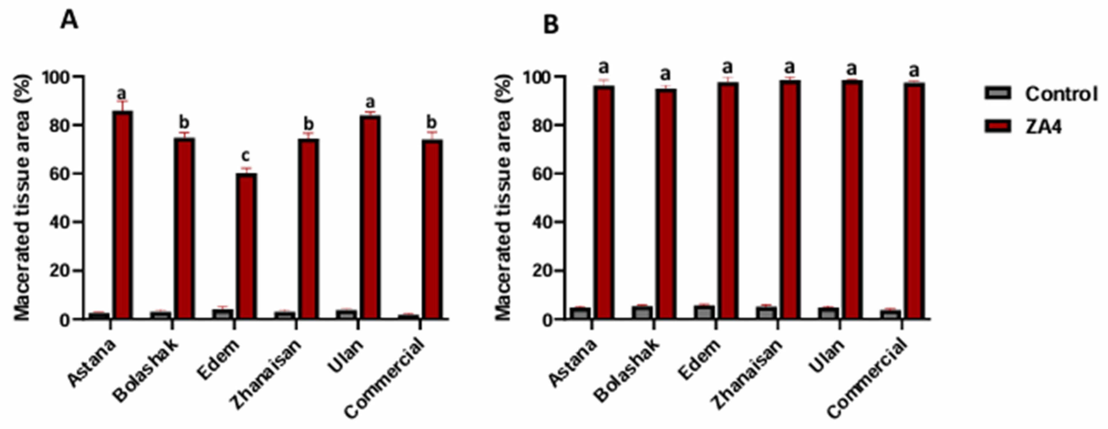
The macerated area ranged from approximately 59% to 85% of the analyzed cut surface. The highest values were observed for Astana (~85%) and Ulan (~83%), whereas Edem showed the lowest maceration (~59%). Bolashak, Zhanaisan, and the commercial potato sample showed intermediate values of approximately 73–75% (Figure 4A).
Figure 4.
Quantitative assessment of potato tuber tissue maceration following inoculation with bacterial isolate ZA4. (A) 24 h post-inoculation; (B) 48 h post-inoculation. Tissue maceration is expressed as the percentage of macerated tissue area relative to the total analyzed cut-surface area. For each tuber, one half was inoculated with ZA4 and the paired half received sterile distilled water as the negative control. Data are presented as the mean ± SD of three independent tubers (n = 3) for each cultivar/sample. Comparisons between ZA4-inoculated and paired control halves within each cultivar/sample were performed using Šídák-adjusted pairwise comparisons following three-way mixed ANOVA. Lowercase letters above ZA4 bars indicate the results of Tukey-adjusted pairwise comparisons among cultivars/samples within the corresponding time point; bars sharing the same letter do not differ significantly (p > 0.05).
By 48 h post-inoculation, tissue maceration had progressed markedly in all tested potato materials, reaching approximately 95–98% of the analyzed cut surface (Figure 4B). Thus, differences among cultivars were most apparent at 24 h, whereas by 48 h maceration had extended over nearly the entire analyzed surface in all ZA4-inoculated samples.
ZA4-inoculated tuber halves exhibited significantly greater tissue maceration than their paired sterile-water controls in all cultivars/samples at both assessment times (Šídák-adjusted p < 0.0001). Control halves showed no pronounced soft-rot symptoms, although quantitative image analysis detected minor localized tissue changes around the wound site. These changes remained substantially lower than those observed in the corresponding ZA4-inoculated halves.
3.5. Molecular Identification and Multilocus Phylogenetic Analysis of Isolate ZA4
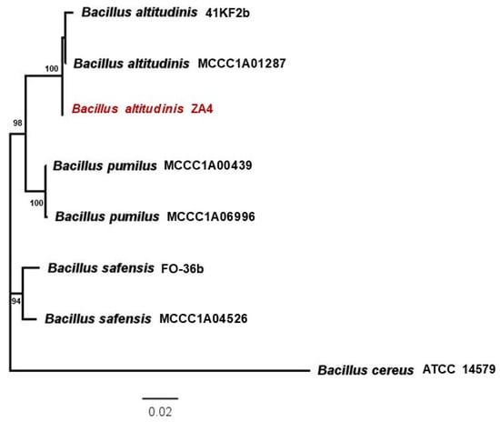
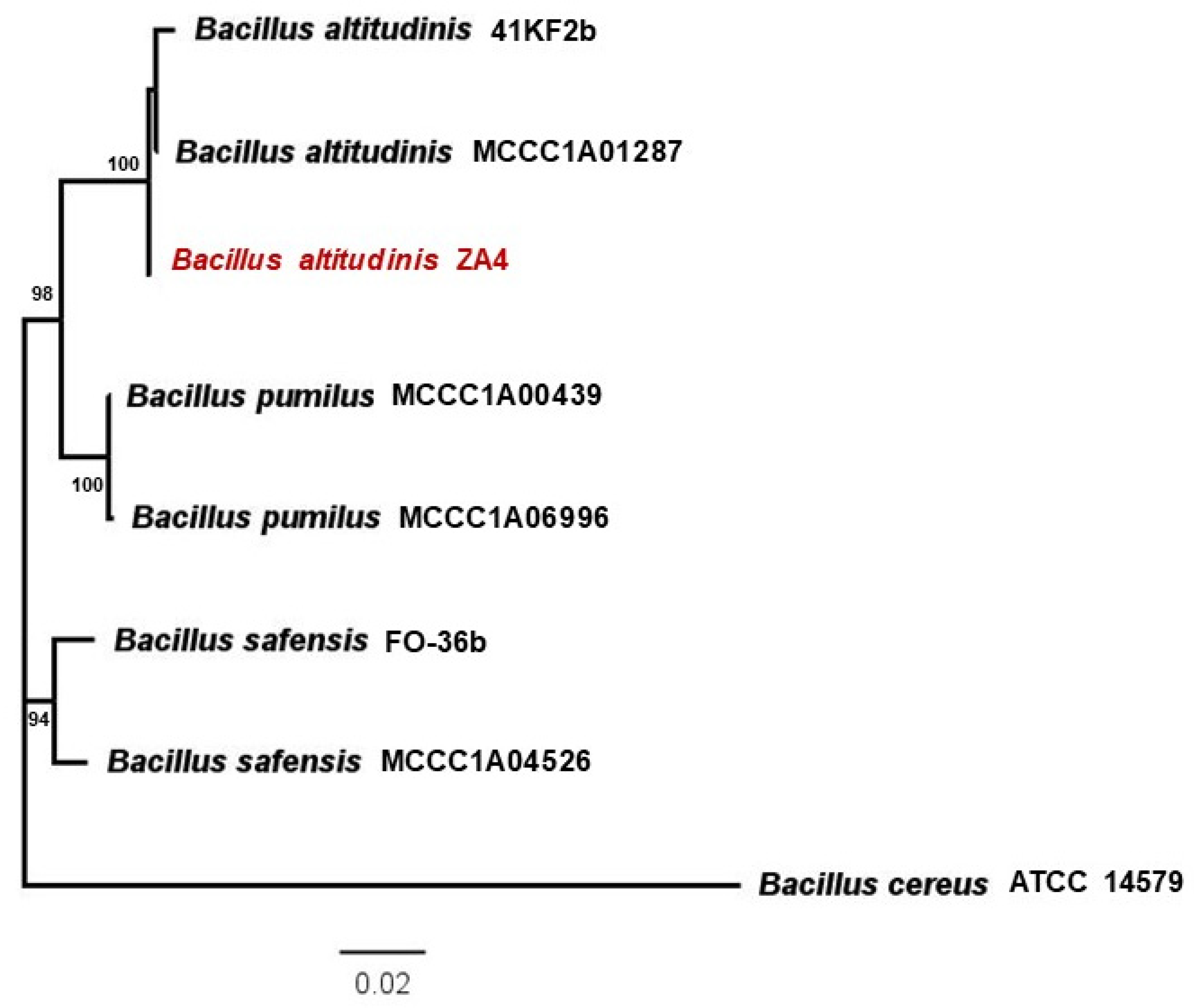
In the multilocus phylogenetic analysis, isolate ZA4 clustered with the B. altitudinis reference strains 41KF2b and MCCC1A01287, forming a strongly supported clade with a bootstrap value of 100% (Figure 5).
Figure 5.
Multilocus phylogenetic tree showing the position of Bacillus altitudinis isolate ZA4 based on concatenated nucleotide sequences of the 16S rRNA, gyrB, rpoB, and pyrE genes. Numbers at the nodes indicate bootstrap support values (%). The scale bar represents 0.02 nucleotide substitutions per site.
Reference strains B. pumilus MCCC1A00439 and MCCC1A06996 formed a distinct clade with 100% bootstrap support, whereas B. safensis strains FO-36b and MCCC1A04526 formed a separate phylogenetic lineage supported by a bootstrap value of 94%. The B. altitudinis and B. pumilus clades were grouped within a broader, strongly supported cluster with 98% bootstrap support. Bacillus cereus ATCC 14579 occupied a distinct phylogenetic position and was used as the outgroup.
4. Discussion
Although members of the Soft Rot Pectobacteriaceae, particularly Pectobacterium spp. and Dickeya spp., are regarded as the principal causal agents of bacterial soft rot of potato, phytopathogenic and macerative activity has also been reported in certain members of the Bacillus pumilus group. For example, B. pumilus strain Od23, isolated from potato tubers affected by soft rot during storage, reproduced characteristic symptoms following artificial inoculation, with disease severity varying among potato cultivars [14]. Similarly, Yahyaoui et al. [28] identified B. pumilus isolates from diseased potato tubers that caused tissue maceration and exhibited pectinolytic, proteolytic, amylolytic, and, to varying degrees, cellulolytic activities. These findings indicate that the capacity to macerate potato tissue is not restricted to the classical bacterial soft-rot pathogens.
Phytopathogenic properties have also been reported for Bacillus altitudinis. Kumvinit and Akarapisan [36] isolated two strains of this species from potato plants showing blackleg and soft-rot symptoms, following artificial inoculation, the isolates caused leaf yellowing, wilting, and subsequent tissue damage. In addition, individual B. altitudinis strains have been shown to induce maceration or necrotic lesions in apple, pear, and other plant hosts [37,38]. Whole-genome analysis of strain B10 revealed a broad repertoire of putative carbohydrate-active enzyme (CAZyme) proteins potentially involved in the modification and degradation of plant polysaccharides [38]. The phytopathogenic potential of B. altitudinis has also been reported in association with pomegranate seed rot and strawberry leaf spot [39,40]. Collectively, these studies suggest that phytopathogenicity in B. altitudinis is strongly strain dependent and is not confined to a single host or symptom type.
The present findings should be interpreted within this strain-specific context. Of the 12 bacterial isolates recovered from potato tubers exhibiting naturally occurring soft-rot symptoms, only ZA4 reproducibly induced pronounced softening and maceration of healthy potato tissue following artificial inoculation. This observation underscores that the recovery of a bacterium from diseased tissue alone is insufficient to establish that it was responsible for the naturally occurring symptoms and that the pathogenic properties observed for ZA4 should not be generalized to B. altitudinis as a species. A similar strain-specific pattern has previously been reported for B. altitudinis B10 [38].
The pathogenic potential of ZA4 was further supported by the re-isolation and re-inoculation experiments. The isolate was initially recovered from naturally diseased potato tubers, reproducibly induced soft-rot symptoms following wound inoculation, was re-isolated from the experimentally induced lesions and molecularly confirmed, and subsequently reproduced comparable symptoms after re-inoculation into healthy tuber tissue. These findings are consistent with key experimental criteria of Koch’s postulates within the wound inoculation model used in this study. However, they do not establish the natural route of infection or demonstrate that ZA4 was responsible for the naturally occurring soft-rot symptoms observed in the sampled tubers.
ZA4 was characterized by rapid progression of tissue maceration. Differences among potato cultivars were most evident at 24 h post-inoculation, whereas by 48 h the extent of tissue damage approached the upper limit of the measured response. This pattern likely reflects a ceiling effect resulting from near-complete degradation of the available tissue rather than true convergence in cultivar susceptibility. Accordingly, the earlier assessment was more informative for detecting differences in the rate of symptom development. Nevertheless, these differences are insufficient to classify the tested cultivars as resistant or susceptible in an agronomic sense without additional experiments conducted under different inoculum pressures and under production or storage conditions. The observed cultivar-dependent variation is consistent with previous reports indicating that disease severity is influenced by interactions between strain-specific bacterial traits and host responses [14,28,37,38]. More broadly, the severity of soft-rot diseases is determined by the combined effects of pathogen virulence, the physiological status of host tissue, and environmental conditions [41].
Multilocus analysis based on 16S rRNA, gyrB, rpoB, and pyrE sequences placed ZA4 together with reference strains of B. altitudinis within a well-supported clade. These data support the taxonomic assignment of ZA4 to B. altitudinis, which is particularly relevant given the close phylogenetic relationships among members of the B. pumilus group. Nevertheless, multilocus phylogeny should be regarded as strong supporting evidence for taxonomic placement rather than as absolute proof of species identity, particularly among closely related taxa with limited interspecific divergence.
This distinction is especially relevant because B. altitudinis is far more commonly described as an endophyte, plant growth-promoting bacterium, or biocontrol agent than as a phytopathogen. For example, strain AMCC 101304 suppressed potato common scab [42], P32-3 reduced postharvest black rot of sweet potato [43], KRS010 showed antagonistic activity against Verticillium dahliae and enhanced plant defense responses [26], and strain GG-22 combined plant growth-promoting and antagonistic properties [27]. These contrasting phenotypes highlight the substantial functional heterogeneity within B. altitudinis and indicate that neither beneficial nor pathogenic properties of individual strains should be extrapolated to the species as a whole.
Interpretation of ZA4 pathogenicity must also take the mode of inoculation into account. In the present study, the bacterial suspension was introduced directly into wounded tissue. The experiment therefore demonstrates the ability of ZA4 to colonize and extensively macerate internal potato tuber tissue once the surface barrier has been breached, but does not establish its ability to penetrate an intact periderm independently. A similar dependence on tissue injury has been reported for other phytopathogenic B. altitudinis strains [37]. Nevertheless, wound-mediated infection is agriculturally relevant because mechanical injury during harvesting, grading, transportation, and storage may provide natural entry sites for bacterial colonization.
The detected pectinolytic, cellulolytic, proteolytic, and amylolytic activities are consistent with the capacity of ZA4 to degrade components of plant tissue and therefore provide supporting evidence for its pathogenic potential. However, these assays do not demonstrate that the corresponding hydrolytic systems are directly responsible for soft-rot development in planta. Establishing the mechanistic contribution of these enzymes will require quantitative biochemical assays, gene-expression studies, and functional genetic analyses.
The present study has several limitations that should be considered when interpreting the findings. Isolate ZA4 was recovered from a limited number of tubers originating from a single surveyed farm; therefore, the available data do not allow conclusions to be drawn regarding the prevalence of B. altitudinis, the frequency of its association with potato soft rot, or its epidemiological significance in Kazakhstan. In addition, the wound inoculation model used in this study does not reproduce all stages of the natural infection process. Accordingly, the results demonstrate the pathogenic potential of ZA4 in wounded tuber tissue but do not establish whether the isolate is capable of infecting intact potato plants, reaching tubers through a natural infection pathway, or acting predominantly as a field or post-harvest pathogen. For the same reason, the cultivar-dependent differences observed under controlled experimental conditions should not be interpreted as evidence of field resistance or susceptibility.
The absence of a well-characterized soft-rot pathogen, such as Pectobacterium or Dickeya, as a positive control also limited direct comparison of the maceration capacity of ZA4 with that of established potato soft-rot pathogens. A further limitation was the near-complete tissue maceration observed at 48 h, which reduced the discriminatory power of the later assessment and limited the ability to distinguish among cultivar responses. Moreover, in the absence of whole-genome analysis, the observed macerative phenotype cannot currently be linked directly to specific genetic determinants of virulence. Finally, the host range of ZA4 remains unknown because pathogenicity assays were restricted to potato. Further studies involving other economically important plant species will therefore be required to determine whether the pathogenic potential of ZA4 is confined to potato or extends to a broader range of hosts.
Despite these limitations, several independent lines of evidence consistently support the pathogenic potential of ZA4. The isolate was recovered from naturally diseased tubers, reproducibly induced soft-rot symptoms following artificial wound inoculation, was successfully re-isolated and re-inoculated, exhibited hydrolytic activities consistent with plant tissue degradation, and was assigned to B. altitudinis by multilocus phylogenetic analysis. Taken together, these findings support the pathogenic potential of ZA4 under the artificial wound inoculation conditions examined and its association with soft-rotted potato tubers. They do not, however, establish that ZA4 was responsible for the naturally occurring disease or demonstrate its ability to infect intact tubers under natural conditions.
The novelty of this study lies in the integrated characterization of a B. altitudinis isolate associated with soft-rotted potato tubers in Kazakhstan and in the experimental demonstration of its ability to reproducibly induce pronounced tissue maceration following wound inoculation, followed by re-isolation and reproduction of comparable symptoms after re-inoculation. To our knowledge, such a characterization has not previously been reported from Kazakhstan. This conclusion, however, applies specifically to ZA4 and does not imply that B. altitudinis is widely associated with potato soft rot or has established epidemiological significance in Kazakhstan.
Further work should include surveys across independent farms and production regions, evaluation of the ability of ZA4 to infect intact tubers, and assessment of its behavior under storage conditions. Comparative whole-genome analyses of B. altitudinis strains exhibiting contrasting pathogenic phenotypes would also be valuable for identifying genetic determinants associated with the macerative phenotype and for clarifying the molecular basis of functional divergence within this ecologically diverse species.
5. Conclusions
In the present study, isolate ZA4, recovered from potato tubers exhibiting soft rot symptoms, was identified as Bacillus altitudinis and reproducibly induced pronounced maceration of healthy tuber tissue. The etiological association of ZA4 with the disease was supported by reproduction of characteristic symptoms and subsequent reisolation of the bacterium in accordance with the principal criteria of Koch’s postulates. The isolate exhibited pectinolytic, cellulolytic, proteolytic, and amylolytic activities, while the severity of early disease development varied among the potato cultivars tested. To our knowledge, this is the first documented report of a phytopathogenic B. altitudinis strain associated with potato soft rot in Kazakhstan. These findings broaden current understanding of the etiological diversity of bacterial soft rot of potato and highlight the need for further investigation of the prevalence of ZA4, its natural routes of infection, and the molecular mechanisms underlying its pathogenicity.
Supplementary Materials
The following supporting information can be downloaded at https://www.mdpi.com/article/10.3390/agronomy16212118/s1. Table S1: Primers used for amplification and sequencing of the 16S rRNA, gyrB, rpoB, and pyrE loci; Table S2: Reference strains and GenBank accession numbers of sequences included in the multilocus phylogenetic analysis.
Author Contributions
Conceptualization, A.T. and A.U.; methodology, A.T., A.Z., S.T. and A.S.; validation, A.T., A.Z. and A.S.; formal analysis, A.T., A.R. and X.T.; investigation, A.T., A.Z., S.T., A.S., A.R., R.I. and N.S.; resources, A.U.; data curation, A.T., S.T. and X.T.; writing—original draft preparation, A.T.; writing—review and editing, A.T., N.S. and B.D.; visualization, A.T., A.Z. and R.I.; supervision, A.U.; project administration A.U. and B.D. All authors have read and agreed to the published version of the manuscript.
Funding
This research was funded by the Ministry of Agriculture of the Republic of Kazakhstan within the framework of the Scientific and Technical Program under program-targeted financing for 2024–2026, grant number BR22885887, «Improvement and implementation of pest management system».
Data Availability Statement
The nucleotide sequence data generated in this study have been deposited in the NCBI GenBank database under accession numbers (The nucleotide sequences generated in this study were submitted to the NCBI GenBank database. The corresponding accession numbers are currently pending and will be provided upon assignment by GenBank.). The remaining data supporting the findings of this study are included in the article and its Supplementary Materials. Additional data are available from the corresponding author upon reasonable request.
Acknowledgments
The authors gratefully acknowledge the Kazakh Research Institute of Fruit and Vegetable Growing LLP (Almaty, Kazakhstan) for providing the potato cultivars used in this study. The authors have reviewed and edited the output and take full responsibility for the content of this publication.
Conflicts of Interest
The authors declare no conflicts of interest. The funder had no role in the design of the study; in the collection, analyses, or interpretation of data; in the writing of the manuscript; or in the decision to publish the results.
Abbreviations
The following abbreviations are used in this manuscript:
| ANOVA | Analysis of variance |
| BIC | Bayesian information criterion |
| BLASTn | Basic Local Alignment Search Tool for nucleotide sequences |
| bp | Base pairs |
| CAZymes | Carbohydrate-active enzymes |
| CFU | Colony-forming units |
| CMC | Carboxymethyl cellulose |
| DNA | Deoxyribonucleic acid |
| K2P | Kimura two-parameter model |
| KB | King’s B medium |
| LB | Lysogeny broth medium |
| LV | Levan medium |
| ML | Maximum likelihood |
| MPA | Meat-peptone agar |
| NA | Nutrient agar |
| NCBI | National Center for Biotechnology Information |
| PA | Potato agar |
| PCR | Polymerase chain reaction |
| PGA | Polygalacturonate |
| RH | Relative humidity |
| rRNA | Ribosomal RNA |
| SD | Standard deviation |
| SRP | Soft Rot Pectobacteriaceae |
References
- Gouerou, M.; Visse-Mansiaux, M.; Brostaux, Y.; Deleu, C.; Val, F.; Leport, L.; Dupuis, B. Effect of climate change on potato yield and starch content. Field Crops Res. 2025, 330, 109951. [Google Scholar] [CrossRef] [Scilit]
- Li, L.; Zhu, T.; Wen, L.; Zhang, T.; Ren, M. Biofortification of potato nutrition. J. Adv. Res. 2025, 75, 23–34. [Google Scholar] [CrossRef] [Scilit]
- Vasilyeva, A.A.; Evseev, P.V.; Ignatov, A.N.; Dzhalilov, F.S.-U. Pectobacterium punjabense causing blackleg and soft rot of potato: The first report in the Russian Federation. Plants 2024, 13, 2144. [Google Scholar] [CrossRef] [Scilit]
- Mainello-Land, A.M.; Bibi, S.; Gugino, B.; Bull, C.T. Multilocus sequence and phenotypic analysis of Pectobacterium and Dickeya type strains for identification of soft rot Pectobacteriaceae from symptomatic potato stems and tubers in Pennsylvania. Syst. Appl. Microbiol. 2024, 47, 126476. [Google Scholar] [CrossRef] [Scilit]
- Keithellakpam, L.B.; Karunakaran, C.; Singh, C.B.; Jayas, D.S.; Danielski, R. A comprehensive review on pre- and post-harvest perspectives of potato quality and non-destructive assessment approaches. Appl. Sci. 2026, 16, 190. [Google Scholar] [CrossRef] [Scilit]
- Charkowski, A.O. The changing face of bacterial soft-rot diseases. Annu. Rev. Phytopathol. 2018, 56, 269–288. [Google Scholar] [CrossRef] [Scilit]
- Youdkes, D.; Helman, Y.; Burdman, S.; Matan, O.; Jurkevitch, E. Potential control of potato soft rot disease by the obligate predators Bdellovibrio and like organisms. Appl. Environ. Microbiol. 2020, 86, e02543-19. [Google Scholar] [CrossRef] [Scilit]
- Charkowski, A.O.; Sharma, K.; Parker, M.L.; Secor, G.A.; Elphinstone, J. Bacterial diseases of potato. In The Potato Crop: Its Agricultural, Nutritional and Social Contribution to Humankind; Campos, H., Ortiz, O., Eds.; Springer: Cham, Switzerland, 2020; pp. 351–388. [Google Scholar] [CrossRef] [Scilit]
- Van Gijsegem, F.; van der Wolf, J.M.; Toth, I.K. (Eds.) Plant Diseases Caused by Dickeya and Pectobacterium Species; Springer: Cham, Switzerland, 2021. [Google Scholar] [CrossRef] [Scilit]
- Kvitko, B.H.; Charkowski, A.O.; Joshi, J.R. Mechanisms of Enterobacterales plant necrotrophy and host specificity: Two types of bacterial necrotroph. Annu. Rev. Phytopathol. 2025, 63, 309–332. [Google Scholar] [CrossRef] [Scilit]
- Gorshkov, V.; Parfirova, O. Host plant physiological transformation and microbial population heterogeneity as important determinants of the Soft Rot Pectobacteriaceae–plant interactions. Semin. Cell Dev. Biol. 2023, 148–149, 33–41. [Google Scholar] [CrossRef] [Scilit]
- Hugouvieux-Cotte-Pattat, N.; Pédron, J.; Van Gijsegem, F. Insight into biodiversity of the recently rearranged genus Dickeya. Front. Plant Sci. 2023, 14, 1168480. [Google Scholar] [CrossRef] [Scilit]
- Galal, A.A.; El-Bana, A.A.; Janse, J. Bacillus pumilus, a new pathogen on mango plants. Egypt. J. Phytopathol. 2006, 34, 17–29. [Google Scholar]
- Bathily, H.; Babana, A.H.; Samaké, F. Bacillus pumilus, a new pathogen on potato tubers in storage in Mali. Afr. J. Microbiol. Res. 2010, 4, 2067–2071. [Google Scholar]
- Peng, Q.; Yuan, Y.; Gao, M. Bacillus pumilus, a novel ginger rhizome rot pathogen in China. Plant Dis. 2013, 97, 1308–1315. [Google Scholar] [CrossRef] [Scilit]
- Kovaleva, V.A.; Shalovylo, Y.I.; Gorovik, Y.N.; Lagonenko, A.L.; Evtushenkov, A.N.; Gout, R.T. Bacillus pumilus—A new phytopathogen of Scots pine. J. For. Sci. 2015, 61, 131–137. [Google Scholar] [CrossRef] [Scilit]
- Saleh, O.I.; Huang, P.-Y.; Huang, J.-S. Bacillus pumilus, the cause of bacterial blotch of immature Balady peach in Egypt. J. Phytopathol. 1997, 145, 447–453. [Google Scholar] [CrossRef] [Scilit]
- Song, J.H.; Wu, Z.R.; Zhang, L.X.; Tan, G.J.; Wang, S.; Wang, J.J. First report of Bacillus pumilus causing fruit rot on muskmelon (Cucumis melo) in China. Plant Dis. 2018, 102, 439. [Google Scholar] [CrossRef] [Scilit]
- Hakim, S.; Liaquat, F.; Gul, S.; Chaudhary, H.J.; Munis, M.F.H. Presence of Bacillus pumilus causing fruit rot of Ficus lacor in Pakistan. J. Plant Pathol. 2015, 97, 543. [Google Scholar] [CrossRef] [Scilit]
- Ahmadi, E.; Kowsari, M.; Azadfar, D.; Salehi Jouzani, G. Bacillus pumilus and Stenotrophomonas maltophilia as two potentially causative agents involved in Persian oak decline in Zagros forests (Iran). For. Pathol. 2019, 49, e12541. [Google Scholar] [CrossRef] [Scilit]
- Mazlan, S.; Zulperi, D.; Wahab, A.; Jaafar, N.M.; Sulaiman, Z.; Rajandas, H. First report of Bacillus pumilus causing trunk bulges of rubber tree (Hevea brasiliensis) in Malaysia. Plant Dis. 2019, 103, 1016. [Google Scholar] [CrossRef] [Scilit]
- Font, M.I.; Bassimba, D.D.M.; Cebrián, M.C.; Molina, L.M.; Jordá, C. First report of Bacillus pumilus on Phaseolus vulgaris in Spain. Plant Pathol. 2010, 59, 400. [Google Scholar] [CrossRef] [Scilit]
- Evdokimova, O.V.; Miamin, V.E.; Valentovich, L.N. Biochemical and molecular genetic characteristics of Bacillus pumilus bacteria isolated in Belarus. J. Belarusian State Univ. Biol. 2018, 1, 38–49. [Google Scholar]
- Dobrzyński, J.; Jakubowska, Z.; Dybek, B. Potential of Bacillus pumilus to directly promote plant growth. Front. Microbiol. 2022, 13, 1069053. [Google Scholar] [CrossRef] [Scilit]
- Dobrzyński, J.; Jakubowska, Z.; Kulkova, I.; Kowalczyk, P.; Kramkowski, K. Biocontrol of fungal phytopathogens by Bacillus pumilus. Front. Microbiol. 2023, 14, 1194606. [Google Scholar] [CrossRef] [Scilit]
- Shan, Y.; Wang, D.; Zhao, F.-H.; Song, J.; Zhu, H.; Li, Y.; Zhang, X.-J.; Dai, X.-F.; Han, D.; Chen, J.-Y. Insights into the biocontrol and plant growth promotion functions of Bacillus altitudinis strain KRS010 against Verticillium dahliae. BMC Biol. 2024, 22, 116. [Google Scholar] [CrossRef] [Scilit]
- Falcón-Piñeiro, A.; Baños, A.; Molin, E.M.; González-Gragera, E.; Giampetruzzi, A.; Abou Kubaa, R.; Antonielli, L.; Wallner, A.; Brader, G.; Verma, S.K.; et al. Bacillus altitudinis GG-22: A novel plant growth-promoting bacterium with beneficial agronomic properties. Biotechnol. Rep. 2026, 49, e00945. [Google Scholar] [CrossRef] [Scilit]
- Coico, R. Gram staining. Curr. Protoc. Immunol. 2001, 23, A.3O.1–A.3O.2. [Google Scholar] [CrossRef] [Scilit]
- Yahyaoui, A.; Oueslati, M.; Motyka-Pomagruk, A.; Kaczynska, N.; Sledz, W.; Tarhouni, B.; Hajlaoui, M.R.; Lojkowska, E.; Sadfi-Zouaoui, N. Characterisation of pectinolytic Bacillus pumilus and Paenibacillus amyloliticus strains, new pathogens of potato in Tunisia. Agriculture 2023, 13, 1275. [Google Scholar] [CrossRef] [Scilit]
- Maghboli Balasjin, N.; Maki, J.S.; Schläppi, M.R.; Marshall, C.W. Plant growth-promoting activity of bacteria isolated from Asian rice (Oryza sativa L.) depends on rice genotype. Microbiol. Spectr. 2022, 10, e02787-21. [Google Scholar] [CrossRef] [Scilit]
- Shin, J.-H.; Lee, H.-K.; Lee, S.-C.; Han, Y.-K. Biological control of Fusarium oxysporum, the causal agent of Fusarium basal rot in onion by Bacillus spp. Plant Pathol. J. 2023, 39, 600–613. [Google Scholar] [CrossRef] [Scilit]
- Hasanin, M.S.; Abdella, M.A.A. Green starch nanoparticles production in situ using α-amylase from a newly isolated Bacillus subtilis strain-MA6: Statistical designs and characterizations. Microb. Cell Fact. 2025, 24, 187. [Google Scholar] [CrossRef] [Scilit]
- Liu, Y.; Lai, Q.; Dong, C.; Sun, F.; Wang, L.; Li, G.; Shao, Z. Phylogenetic diversity of the Bacillus pumilus group and the marine ecotype revealed by multilocus sequence analysis. PLoS ONE 2013, 8, e80097. [Google Scholar] [CrossRef] [Scilit]
- Kumar, S.; Stecher, G.; Suleski, M.; Sanderford, M.; Sharma, S.; Tamura, K. MEGA12: Molecular Evolutionary Genetic Analysis Version 12 for Adaptive and Green Computing. Mol. Biol. Evol. 2024, 41, msae263. [Google Scholar] [CrossRef] [Scilit]
- Vaidya, G.; Lohman, D.J.; Meier, R. SequenceMatrix: Concatenation software for the fast assembly of multi-gene datasets with character set and codon information. Cladistics 2011, 27, 171–180. [Google Scholar] [CrossRef] [Scilit]
- Kumvinit, A.; Akarapisan, A. Characterization of blackleg and soft rot from potato in northern Thailand. J. Phytopathol. 2019, 167, 655–666. [Google Scholar] [CrossRef] [Scilit]
- Elbanna, K.; Elnaggar, S.; Bakeer, A. Characterization of Bacillus altitudinis as a new causative agent of bacterial soft rot. J. Phytopathol. 2014, 162, 712–722. [Google Scholar] [CrossRef] [Scilit]
- Lemjiber, N.; Naamani, K.; Merieau, A.; Dihazi, A.; Zhar, N.; Jediyi, H.; Boukerb, A.M. Identification and genomic characterization of pathogenic Bacillus altitudinis from common pear trees in Morocco. Agronomy 2021, 11, 1344. [Google Scholar] [CrossRef] [Scilit]
- Liu, Q.; Li, D.; Hu, H.; Huang, Q.; Zhang, Y.; Zhou, F.; Song, P.; Guan, Y.; Bu, R.; Hu, P.; et al. First report of Bacillus altitudinis causing seed rot of pomegranate in China. Australas. Plant Pathol. 2021, 50, 427–429. [Google Scholar] [CrossRef] [Scilit]
- Zhang, L.; Liu, Z.; Pu, Y.; Zhang, B.; Wang, B.; Xing, L.; Li, Y.; Zhang, Y.; Gu, R.; Jia, F.; et al. Antagonistic strain Bacillus velezensis JZ mediates the biocontrol of Bacillus altitudinis m-1, a cause of leaf spot disease in strawberry. Int. J. Mol. Sci. 2024, 25, 8872. [Google Scholar] [CrossRef] [Scilit]
- Maciag, T.; Kozieł, E.; Otulak-Kozieł, K.; Jafra, S.; Czajkowski, R. Looking for resistance to soft rot disease of potatoes facing environmental hypoxia. Int. J. Mol. Sci. 2024, 25, 3757. [Google Scholar] [CrossRef] [Scilit]
- Li, B.; Wang, B.; Pan, P.; Li, P.; Qi, Z.; Zhang, Q.; Shi, C.; Hao, W.; Zhou, B.; Lin, R. Bacillus altitudinis strain AMCC 101304: A novel potential biocontrol agent for potato common scab. Biocontrol Sci. Technol. 2019, 29, 1009–1022. [Google Scholar] [CrossRef] [Scilit]
- Zhang, Y.-J.; Cao, X.-Y.; Chen, Y.-J.; Cong, H.; Wang, Y.-M.; Jiang, J.-H.; Li, L.-D. Potential utility of endophytic Bacillus altitudinis strain P32-3 as a biocontrol agent for the postharvest prevention of sweet potato black rot. Biol. Control 2023, 186, 105350. [Google Scholar] [CrossRef] [Scilit]
Disclaimer/Publisher’s Note: The statements, opinions and data contained in all publications are solely those of the individual author(s) and contributor(s) and not of MDPI and/or the editor(s). MDPI and/or the editor(s) disclaim responsibility for any injury to people or property resulting from any ideas, methods, instructions or products referred to in the content. |
© 2026 by the authors. Licensee MDPI, Basel, Switzerland. This article is an open access article distributed under the terms and conditions of the Creative Commons Attribution (CC BY) license.