


Abstract
Cold stress severely limits the productivity of maize, especially in Northeast China. Hydroxymethylbutenyl diphosphate synthase (HDS) is a key enzyme in the methylerythritol phosphate (MEP) pathway, which is responsible for the generation of precursors for carotenoid biosynthesis, thereby playing a central role in facilitating the cellular response to stress. In the present study, virus-induced gene silencing (VIGS)-mediated ZmHDS1 silencing on the B73 background led to enhanced cold stress sensitivity, as evidenced by growth retardation, leaf shriveling, disrupted Kranz anatomy, elevated reactive oxygen species (ROS) accumulation, impaired antioxidant enzyme activity, increased membrane lipid peroxidation, and reduced photosynthetic performance. Hormonal profiling revealed that ZmHDS1 silencing decreased the content of indole-3-acetic acid (IAA), gibberellin (GA4), and abscisic acid (ABA) and disrupted ABA signaling. Furthermore, ZmHDS1 silencing reduced the chlorophyll content, Rubisco activity, and ZmRCA (Rubisco activase) expression and caused a more severe reduction in carotenoid levels and the transcriptional suppression of the entire carotenoid biosynthetic gene network. Collectively, ZmHDS1 acts as a key component of the MEP pathway that contributes to maize cold tolerance by coordinating antioxidant defense, phytohormone homeostasis, photosynthetic maintenance, and carotenoid metabolism. These findings highlight the importance of the MEP pathway as a whole in cold adaptation. Disruption of ZmHDS1 impairs these interconnected protective mechanisms, rendering maize highly susceptible to cold-induced damage.
1. Introduction
As one of the most important staple crops, maize (Zea mays L.) is widely planted worldwide [1,2]. However, as a crop originating from tropical and subtropical regions, maize is more susceptible to low temperatures, especially during the seedling stage [3,4]. Cold stress is one of the major environmental factors that adversely impacts seedling establishment and vigor, culminating in dwarfism and the substantial suppression of stem and root growth in maize [5,6]. Cold stress induces a range of alterations in maize, including in membrane fluidity, the antioxidative defense system, and photosynthetic performance, among others [7,8]. Thus, fully clarifying how maize tolerates cold is critical to improving its ability to withstand cold injury. This helps to maize sustain growth under cold stress and reduce possible yield losses.
Plants have evolved a range of sophisticated and coordinated strategies to withstand low temperatures [9,10]. These strategies include modulating the composition of membrane lipids to maintain cellular membrane stability, the accumulation of protective solutes for osmotic adjustment, and augmenting the antioxidant defense system to efficiently scavenge cold-induced reactive oxygen species [11]. Plants rapidly launch a series of physiological, biochemical, and molecular responses that help them to adjust to environmental changes and reduce cold-induced injury [12,13]. Cold-tolerant maize is achieved through maintaining a stable photosynthetic system and increasing the levels of peroxidase (POD), catalase (CAT), reduced glutathione (GSH), and proline, which effectively mitigate oxidative damage [14]. At the molecular level, low temperature-induced genes are broadly divided into functional and regulatory protein genes, which jointly form the molecular network underlying plant cold tolerance [15].
Plants commonly activate the synthesis of secondary metabolites in response to environmental stresses [16,17]. The methylerythritol phosphate (MEP) pathway is a core route for the synthesis of terpenoids in plant plastids [18], such as gibberellins, ABA, and carotenoids. Phytohormones play an essential role in physiological changes and gene expression regulation during cold stress. The survival capacity of maize upon cold stress exhibits a strong correlation with the upregulation of ABA synthesis [19]. Methylerythritol cyclodiphosphate (MEcPP) is an intermediate of MEP that can mediate retrograde signaling, which alters nuclear gene expression during stress conditions [20]. HDS is a key enzyme in the MEP pathway that catalyzes the conversion of MEcPP to (2E)-4-hydroxy-3-methylbut-2-enyl diphosphate (HMBPP) [21,22]. Although the HDS enzyme is almost universal in the plant kingdom, previous work has shown that its primary structure varies from species to species, and, occasionally, multiple gene family members encode this enzyme [23,24]. HDS catalyzes a key step in the MEP pathway and interacts with heat shock transcription factor A1 to regulate thermotolerance [25]. The heterologous overexpression of PmHDS in Arabidopsis thaliana resulted in a significant increase in carotenoid and chlorophyll accumulation, enhanced activity of associated enzymes, and elevated titers of terpenoid-derived phytohormones such as GA and ABA, which collectively contribute to augmented abiotic stress resistance [26]. Our previous proteomics analysis revealed that the HDS1 protein abundance increased 3.65-fold in cold-tolerant maize, but there were no significant changes in cold-sensitive maize [27]. Hence, it was hypothesized that HDS1 was involved in the cold stress response associated with cold tolerance in maize.
Here, the aim of this study is to elucidate the specific role of ZmHDS1 in maize. The silencing of ZmHDS1 via virus-induced gene silencing (VIGS) technology was used to examine the function of ZmHDS1 under cold stress. The mechanisms by which ZmHDS1 contributes to cold stress resistance were explored by investigating the antioxidant system, alterations of phytohormones, and photosynthesis.
2. Materials and Methods
2.1. In Silico Analysis of ZmHDS1 Gene Structure
The genomic and coding sequences of ZmHDS1 were obtained from the maize B73 reference genome (Zm-B73-REFERENCE-NAM-5.0) via MaizeGDB. The CDS was aligned against the corresponding genomic sequence using BLASTN (2.17.0) and Splign (29.7.0) to determine the exon–intron organization. Conserved domains were predicted using NCBI CD-Search.
2.2. Investigation of the Expression of ZmHDS1 by Real-Time Quantitative PCR
Seeds of the B73 maize genotype were sown in pots filled with soil and grown in a greenhouse under a 16 h light (24 °C)/8 h dark (22 °C) photoperiod, with 60% relative humidity and a photosynthetic photon flux density of 300 μmol m−2 s−1. At the three-true-leaf stage, seedlings were transferred to a growth chamber set at 4 °C with the same photoperiod (16 h light/8 h dark), light intensity (300 μmol m−2 s−1), and relative humidity (60%) for 3 d. Seedlings that were kept in the greenhouse under the same photoperiod and humidity conditions without cold treatment served as the controls. Each treatment consisted of three independent biological replicates, with ten seedlings per replicate. The positions of pots were randomized. Leaf samples were collected at 0, 3, 6, 9, 12, 24, and 72 h after cold stress for real-time quantitative PCR (RT-qPCR) analysis. RNA was extracted using an extraction kit (Ultrapure RNA Kit (CW0597S), Cwbio, Beijing, China) according to the manufacturer’s instructions. The RNA concentration and purity were assessed using a NanoDrop spectrophotometer (Thermo Fisher Scientific, Waltham, MA, USA), and RNA integrity was checked by 1% agarose gel electrophoresis. First-strand cDNA was synthesized from 1 μg total RNA using a reverse transcription kit (HiFiScript (CW2582M), Cwbio, China) in a 20 μL reaction. The reverse transcription program was 42 °C for 2 min for gDNA removal, 45 °C for 15 min, and 85 °C for 5 min. The resulting cDNA was used as the template for RT-qPCR. Then, each 20 μL reaction contained 10 μL 2× SYBR Green Master Mix ((CW3008M), Cwbio, China), 0.4 μL forward primer (10 μM), 0.4 μL reverse primer (10 μM), 2 μL diluted cDNA, and 7.2 μL ddH2O. The final primer concentration was 0.2 μM. The qPCR program was as follows: 95 °C for 30 s and 40 cycles of 95 °C for 10 s and 60 °C for 30 s, followed by melting curve analysis from 65 °C to 95 °C. All RT-qPCR analyses were performed with three replicates. All primers used in this study were designed based on the maize B73 reference genome (Zm-B73-REFERENCE-NAM-5.0) using Primer Premier 5.0 and verified by NCBI Primer-BLAST. The primers used are listed in Supplementary Table S1. The specificity of each primer pair was confirmed by melting curve analysis, which showed a single peak for all primer pairs, and by agarose gel electrophoresis, which showed a single band of the expected size. The relative expression levels of genes were calculated using the 2−ΔΔCt method, with the ZmGAPDH gene as the internal reference.
2.3. Construction of ZmHDS1-Silenced Seedlings by VIGS and Growth Environment
The SGN VIGS Tool (https://vigs.solgenomics.net/ (accessed on 10 March 2025)) systematically compares all 21 nt siRNA sequences derived from the query fragment against all annotated gene models in the maize genome and calculates an off-target score for each candidate region. Using the SGN VIGS tool and the Zea mays Zm-B73-REFERENCE-NAM-5.0 reference database, the recommended 305 bp (CDS positions 1–305) region with the lowest off-target score was selected. Based on this 305 bp sequence, VIGS-specific primers containing restriction enzyme sites and protective bases were designed using Primer Premier 5.0. The primer sequences are listed in Supplementary Table S1. The target VIGS fragment amplified by these primers was 236 bp (CDS position from 66 bp to 301 bp) in length (Supplementary Figure S1C). The off-target prediction results confirmed that the 236 bp fragment had no significant sequence similarity to any other gene in the maize genome. When mismatches were set to 0 (stringent condition), the fragment targeted only ZmHDS1, with no other genes identified as potential off-targets. The TRV-based VIGS system was used as described previously [28]. Electrophoresis was used to confirm product specificity, and sequencing was applied to verify the fragment sequence (Supplementary Figure S2). Then, the fragment was ligated into the pTRV2 vector via double enzyme digestion (EcoRI and BamHI) to create the pTRV2:ZmHDS1 recombinant vector. Subsequently, the pTRV1, pTRV2, and pTRV2:ZmHDS1 vectors were transformed into the GV3101 strain separately. Vacuum-assisted infiltration was performed on germinated maize seeds (about 1 cm radicles). Agrobacterium suspensions carrying pTRV2-ZmHDS1 and pTRV1 were mixed in equal volumes. The germinated seeds (approximately 1 cm radicles) were submerged in the mixture under a vacuum for 3 min. Inoculated seeds were washed three times with sterile water and planted in moist soil, designated as TRV:ZmHDS1. As the negative control, Agrobacterium suspensions carrying empty pTRV2 and pTRV1 were mixed in equal volumes., and seeds were infiltrated using the same method, designated as TRV:00.
A total of 200 B73 maize seedlings were subjected to VIGS infiltration. At the three-true-leaf stage, all seedlings were analyzed by RT-qPCR to evaluate the silencing efficiency. In TRV:ZmHDS1, about 70 out of 200 seedlings (35%) showed more than a 60% reduction in the ZmHDS1 transcript level and were used for the subsequent cold stress experiments. Then, both the control (TRV:00) and ZmHDS1-silenced seedlings were subjected to cold stress (4 °C) for 3 d. The seedlings still grown in the greenhouse served as controls. In addition, the expression of ZmHDS1 was examined after 3 d of cold stress to confirm that silencing was maintained under cold conditions. For phenotypic, physiological, and biochemical analyses, three independent biological replicates were performed, each containing ten seedlings.
2.4. Phenotypic Investigation and Biomass Measurement
Roots and shoots were harvested separately after cold stress for 3 d. After being washed with deionized water, the surface moisture of the roots and shoots was absorbed using absorbent paper. The fresh wight (FW) was measured, and the samples were wrapped in a kraft envelope and baked at 105 °C for 15 min. Then, they were dried at 70 °C to a constant weight, and the dry weight (DW) was measured.
Paraffin sectioning was performed as described [29]. Sections were stained with 0.1% toluidine blue. Microscopic images were captured using a Zeiss Axio Imager A2 microscope (Carl Zeiss, Oberkochen, Germany) equipped with a Zeiss Axiocam 506 (Carl Zeiss, Oberkochen, Germany) color camera. Images were acquired using the ZEN 3.0 software under brightfield illumination.
2.5. Antioxidant System Assay
3,3-Diaminobenzidine (DAB) staining for qualitative detection of H2O2 accumulation: The DAB staining solution was freshly prepared by dissolving DAB in 10 mM phosphate buffer (pH 7.0). Leaf discs (1 cm in diameter) were excised from maize leaves using a leaf punch. The leaf discs were completely immersed in 1.0 mg·mL−1 DAB staining solution and kept in the dark for 12 h at 25 °C. Then, the leaf discs were transferred into 95% (v/v) ethyl alcohol and decolorized in a boiling water bath for 10 min. The leaf discs were decolorized until colorless. They were then observed and photographed using a Canon PowerShot G15 digital camera (Canon, Inc., Tokyo, Japan).
Nitroblue tetrazolium (NBT) staining for qualitative detection of superoxide anion (O2−): The NBT staining solution was freshly prepared by dissolving NBT in 10 mM phosphate buffer (pH 7.8). Leaf discs (1 cm in diameter) were excised from the leaves using a leaf punch. The leaf discs were completely immersed in 0.2% NBT staining solution and kept in the dark for 12 h at 25 °C. Then, the leaf discs were transferred into 95% (v/v) ethyl alcohol and decolorized in a boiling water bath for 10 min. The leaf discs were decolorized until colorless. Photographs were taken using a Canon PowerShot G15 digital camera (Canon, Inc., Tokyo, Japan).
Assay of antioxidant enzyme activity: A 0.2 g (FW) sample of the roots or shoots was homogenized thoroughly with 2 mL of pre-cooling 50 mM phosphate buffer (pH 7.0) and then centrifuged at 8000× g for 10 min (4 °C). The supernatant was used for further enzyme assays. The activity of POD, SOD, CAT, APX, and GPX was measured with assay kits according to the corresponding instructions (Suzhou Comin Biotechnology Co., Ltd., Suzhou, China).
2.6. Lipid Peroxidation and Relative Electrical Conductivity Assay
The content of malonaldehyde (MDA) was measured to evaluate the lipid peroxide level, as described previously [30].
The relative electrical conductivity assay was performed by measuring the relative conductivity, as described in our previous study [27].
2.7. Photosynthesis Assay
The net photosynthetic rate (Pn), intercellular CO2 concentration (Ci), stomatal conductance (Gs), and transpiration rate (Tr) were measured using the LI-6800 photosynthesis system (Li-Cor, Inc., Lincoln, NE, USA) under controlled conditions consisting of a photosynthetic photon flux density of 1500 μmol·m−2·s−1, reference CO2 concentration of 400 µmol·mol−1, vapor pressure deficit of 1.1 kPa, and relative humidity of 50%.
A 0.2 g (FW) leaf was ground thoroughly with 3 mL of pre-cooled 95% (v/v) ethanol and then centrifuged at 500× g for 10 min (4 °C). Then, 1 mL of the supernatant was pipetted after centrifugation and mixed well with 9 mL of 95% ethanol. The absorbance of the mixture was measured using a spectrophotometer at 665 nm, 649 nm, and 470 nm. The content of photosynthetic pigments was calculated as follows: chlorophyll a (mg·L−1) = 13.95 × A665 − 6.88 × A649; chlorophyll b (mg·L−1) = 24.96 × A649 − 7.32 × A665; carotenoid (mg·L−1) = (1000 × A470 − 2.05 × chlorophyll a − 114.8 × chlorophyll b) ÷ 245.
The determination of Rubisco enzyme activity was performed via spectrophotometry according to the instructions of the assay kit (Suzhou Comin Biotechnology Co., Ltd., Suzhou, China).
2.8. Measurement of Hormone Content
The determination of indole-3-acetic acid (IAA), gibberellin 4 (GA4), and abscisic acid (ABA) was performed via high-performance liquid chromatography–mass spectrometry (HPLC-MS). Briefly, 0.5 g (FW) tissue was ground with liquid nitrogen and extracted with 8 mL 75% methanol and 5% formic acid for 45 min under ultrasonication. After centrifugation (8000 rpm, 10 min), the supernatants were purified with an Oasis HLB cartridge and Oasis MCX cartridge. The eluate was dried and reconstituted in 500 μL methanol and filtrated through a 0.22 μm filter before HPLC-MS analysis. Chromatography was performed on an Agilent 1260 HPLC system (Agilent Technologies, Santa Clara, CA, USA) equipped with a ZORBAX Eclipse XDB-C18 column (2.1 × 150 mm, 3.5 μm) and an XDB-C8 guard column. The column temperature was maintained at 40 °C. The mobile phase consisted of 0.1% formic acid in water and 0.1% formic acid in acetonitrile. Separation was achieved using gradient elution at a flow rate of 0.2 mL·min−1. Mass spectrometric detection was performed on an Agilent 6410 triple-quadrupole mass spectrometer equipped with an electrospray ionization (ESI) source. MS detection was operated under multiple reaction monitoring (MRM) mode for IAA, ABA, and GA4, quantified by an internal standard calibration curve. The ESI source parameters were set as follows: drying gas temperature, 350 °C; drying gas flow, 10 L/min; nebulizer pressure, 35 psi; capillary voltage, −4000 V; collision gas, argon. The optimized MRM transitions were IAA, m/z 174.1 → 130.1 (fragmentor 110 V, collision energy 14 V); GA4, m/z 331.2 → 243.1 (fragmentor 120 V, collision energy 18 V); ABA, m/z 263.1 → 153.1 (fragmentor 115 V, collision energy 12 V). Three biological replicates were analyzed.
2.9. Gene Expression Assay
After cold stress for 3 d, the leaves of both cold stress (TRV:00, TRV:ZmHDS1) and control (TRV:00, TRV:ZmHDS1) seedlings were collected. RNA was extracted using the Ultrapure RNA Kit (CWBIO, China) and reverse-transcribed into cDNA as the template for real-time quantitative PCR (RT-qPCR) analysis. The primers for these genes are listed in Supplementary Table S1. ZmRCA (Rubisco activase), ZmNCED1 (9-cis-epoxycarotenoid dioxygenase 1), ZmCYP707A2 (Cytochrome P450, Family 707, Subfamily A, Member 2), ZmPYL8 (Pyrabactin Resistance 1-Like 8), ZmPP2C1 (Protein Phosphatase 2C, Clade A, Member 1), ZmPP2C4 (Protein Phosphatase 2C, Clade A, Member 4), ZmSnRK2.2 (Sucrose Non-Fermenting-1-Related Protein Kinase 2.2), ZmHDR (hydroxy-methyl-butenyl diphosphate reductase), ZmGGPPS1 (geranylgeranyl diphosphate synthase 1), ZmPSY2 (phytoene synthase 2), ZmPDS (phytoene desaturase), ZmRTISO (carotene isomerase), ZmZDS (ζ-carotene desaturase), ZmLCYB (lycopene β-cyclase), ZmCRT-B1 (β-carotene hydroxylase 1), and ZmZEP (zeaxanthin epoxidase) were analyzed. The relative expression levels of these genes were calculated using the 2−ΔΔCt method, with the ZmGAPDH gene as the internal reference.
2.10. Statistical Analysis
Experimental data are presented as the mean ± standard deviation (SD) of three biological replicates. SAS9.3 (SAS Institute, Cary, NC, USA) with the SAS PROC ANOVA LSD model was used to perform the analysis of variance for the physiological data (*, p < 0.05; **, p < 0.01; ***, p < 0.001).
3. Results
3.1. Expression and Gene Structure of ZmHDS1
The in silico analysis shows that ZmHDS1 corresponds to the gene model GRMZM2G137409 (also annotated as Zm00001eb246530/lw2/scd) and is located on chromosome 5 (Zm-B73-REFERENCE-NAM-5.0); it consists of 20 exons and 19 introns (Supplementary Figure S1A). ZmHDS1 is a single-copy gene in the maize B73 genome, and the CDS is 2241 bp, encoding a polypeptide of 746 amino acids. The encoded protein contains a conserved HDS domain (Supplementary Figure S1B) that catalyzes the conversion of MEcPP to HMBPP in the MEP pathway. The ZmHDS1 pan-gene record confirms that GRMZM2G137409 is the only B73 gene model associated with this pan-gene set. No paralogous HDS-encoding gene has been identified in the maize genome. These structural features provide the basis for the subsequent functional analysis of ZmHDS1.
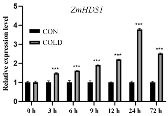
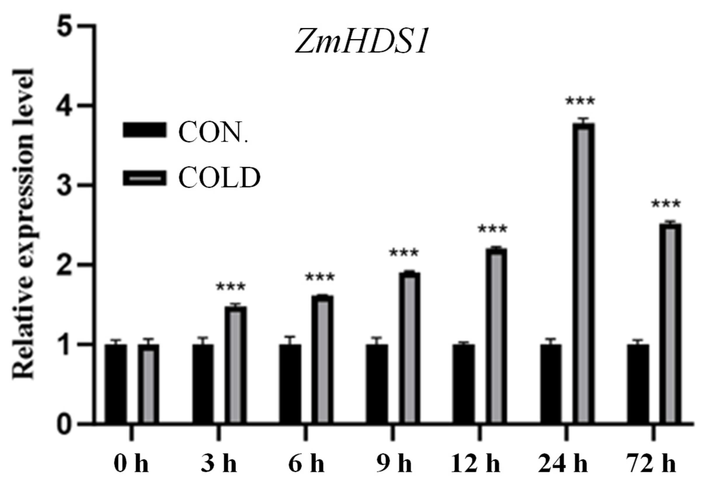
Our previous proteomics analysis revealed that cold stress upregulated the HDS1 protein abundance 3.65-fold in cold-tolerant maize, with no alteration in cold-sensitive maize. Further analyses were performed to investigate the expression profile of ZmHDS1 under cold stress. The expression of ZmHDS1 progressively increased following cold stress and reached its maximum at 24 h (Figure 1). These results indicate that ZmHDS1 is responsive to cold stress and might be involved in the cold stress response in maize.
Figure 1.
An expression analysis of ZmHDS1 in B73 in response to cold stress. Maize seedlings with three leaves were shifted to cold stress (4 °C) for 3 d. Values represents the mean ± SD of three independent replicates. A Student’s t-test was used to assess differences between different treatments (*** p < 0.001).
3.2. Suppression of ZmHDS1 Inhibits Growth of Maize Under Cold Stress
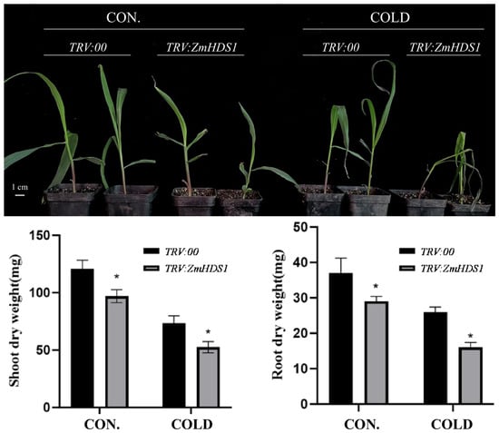
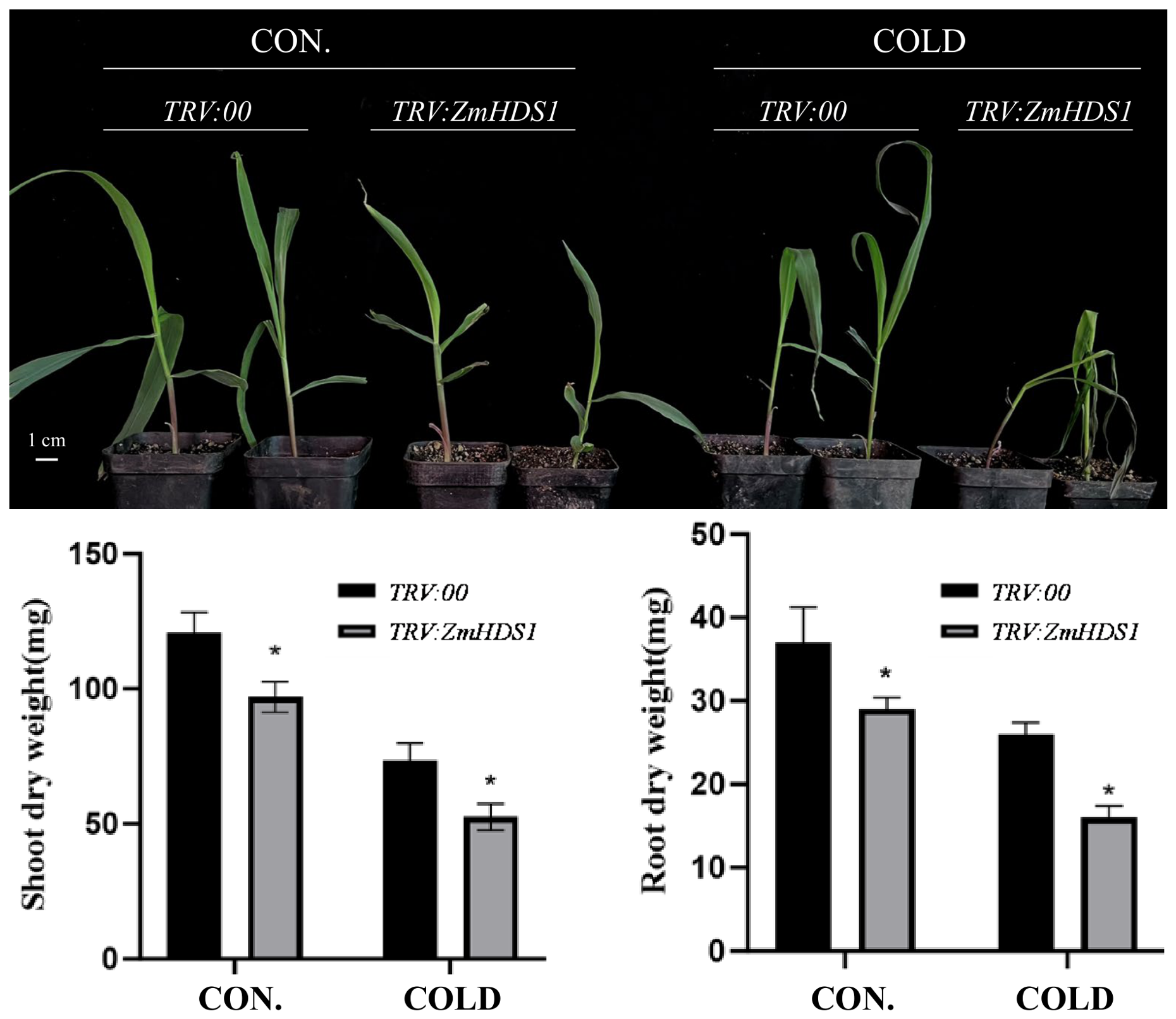
To further validate the function of ZmHDS1 in maize cold tolerance, ZmHDS1-knockdown seedlings were generated using VIGS. The silencing efficiency was evaluated at the three-true-leaf stage by RT-qPCR, using ZmGAPDH as the internal reference. Only TRV:ZmHDS1 plants showing more than a 60% reduction in ZmHDS1 transcript abundance relative to TRV:00 were used for subsequent cold stress experiments (Supplementary Figure S3). Without cold stress, silenced plants grew slightly smaller than the control plants but exhibited similar growth performance. Under cold stress for 3 d, ZmHDS1-silenced plants displayed severe leaf shriveling and wilting accompanied by a darker green leaf phenotype (Figure 2).
Figure 2.
Phenotypic characteristics of ZmHDS1-silenced maize plants via VIGS (TRV:00 and TRV:ZmHDS1) under cold stress for 3 d. TRV:00 represents seedlings infected with the empty TRV vector. Maize seedlings with three true leaves were shifted to cold stress (4 °C) for 3 d. Values represent the mean ± SD of three independent replicates. Significance was detected using SAS PROC ANOVA LSD (*, p < 0.05).
Biomass accumulation serves as a core physiological marker for stress-caused growth retardation. Under cold stress for 3 d, both the shoot and root dry weights of ZmHDS1-silenced maize were significantly lower than those of control plants (Figure 2). The silencing of ZmHDS1 exacerbated cold injury, suggesting that ZmHDS1 contributes to maize cold tolerance.
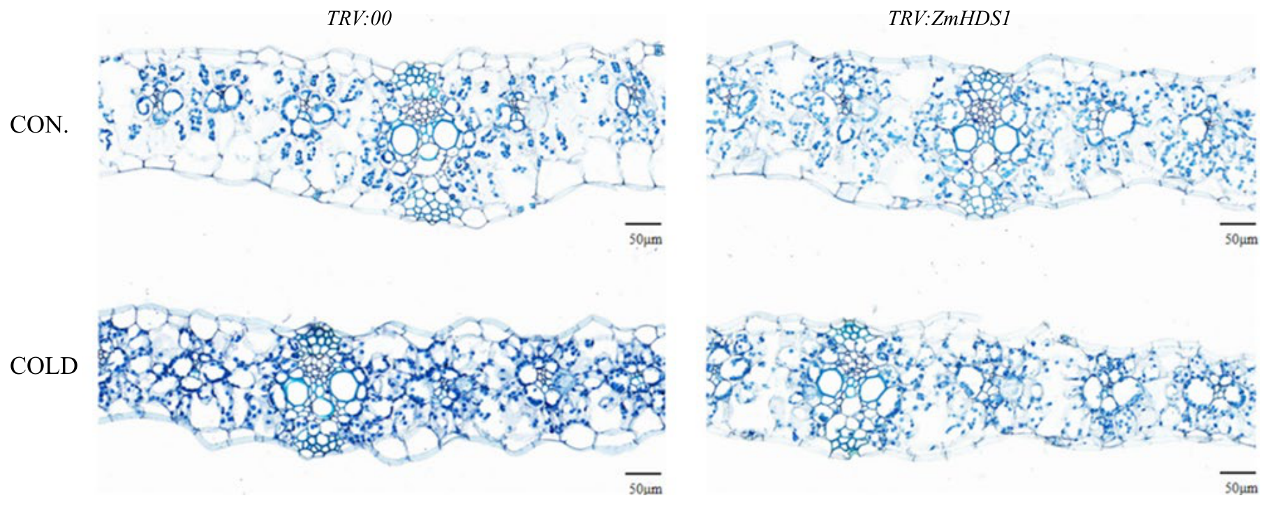
Paraffin section analysis revealed severe anatomical destruction in the leaves of ZmHDS1-silenced plants under cold stress, including a disordered epidermis, degenerated mesophyll, and a disrupted Kranz structure, whereas the leaves of control plants maintained an intact basic cellular architecture with only slight cytological changes (Figure 3). These results indicate that the loss of ZmHDS1 aggravates cold damage to leaf cells in maize.
Figure 3.
Microstructure changes in ZmHDS1-silenced maize plants via VIGS (TRV:00 and TRV:ZmHDS1) under cold stress for 3 d. TRV:00 represents seedlings infected with the empty TRV vector. Maize seedlings with three true leaves were shifted to cold stress (4 °C) for 3 d.
3.3. Suppression of ZmHDS1 Affects Antioxidant System of Maize Under Cold Stress
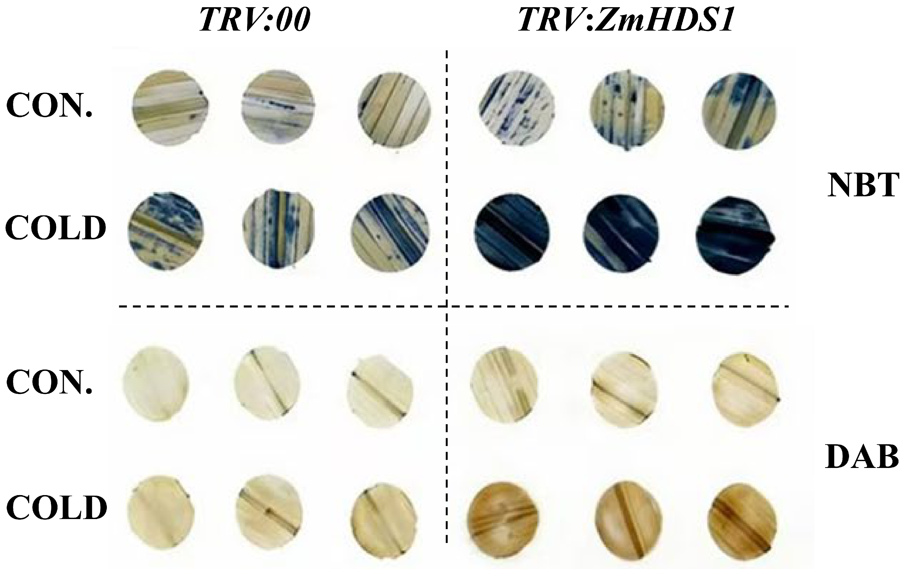
Cold stress induces massive reactive oxygen species (ROS) overproduction and oxidative injury in plants. To explore the regulatory effect of ZmHDS1 on ROS accumulation under cold stress, in situ histochemical staining was performed. NBT staining was used to detect superoxide anion (O2−), while DAB staining visualized hydrogen peroxide (H2O2), with the staining intensity positively correlated with the endogenous ROS concentrations. ZmHDS1-silenced plants exhibited significantly deeper DAB and NBT staining (Figure 4), demonstrating that ZmHDS1-silenced maize accumulated significantly higher ROS levels upon cold stress.
Figure 4.
In situ detection of superoxide anion and H2O2 in leaves under cold stress for 3 d by NBT and DAB staining, respectively. TRV:00 represents seedlings infected with the empty TRV vector. Maize seedlings with three true leaves were shifted to cold stress (4 °C) for 3 d.
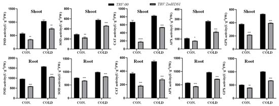
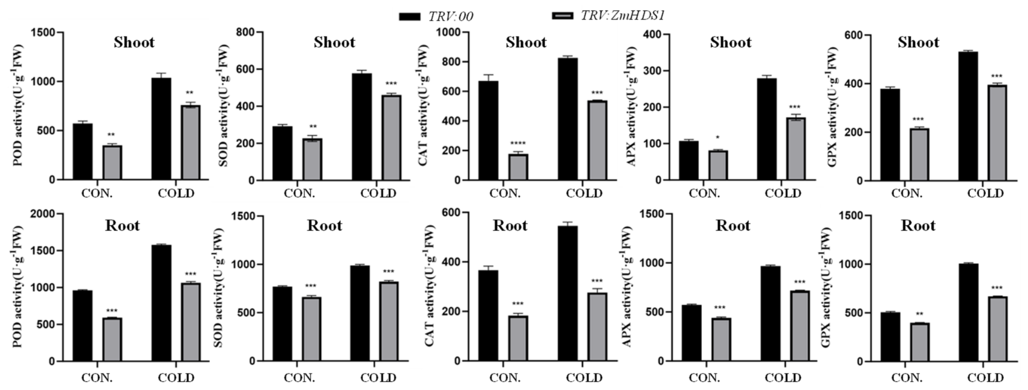
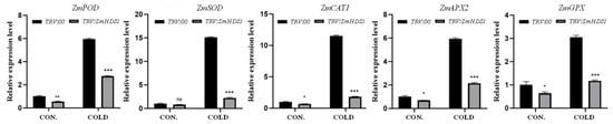
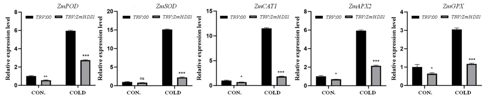
In the present study, the activity of peroxidase (POD), superoxide dismutase (SOD), catalase (CAT), ascorbate peroxidase (APX), and glutathione peroxidase (GPX) in both the shoots and roots was investigated. Although cold stress induced increases in the activity of these antioxidant enzymes, the enzyme activity in ZmHDS1-silenced plants was significantly lower than that in control seedlings in response to cold stress (Figure 5). The overall expression levels of ZmPOD, ZmSOD, ZmCAT1, ZmAPX2, and ZmGPX in ZmHDS1-silenced plants were significantly lower than those in the controls in response to cold stress (Figure 6). These results suggest that ZmHDS1 might enhance low-temperature tolerance by regulating the expression of antioxidant enzyme-related genes.
Figure 5.
Antioxidant enzyme activity of ZmHDS1-silenced maize plants via VIGS (TRV:00 and TRV:ZmHDS1) under cold stress for 3 d. TRV:00 represents seedlings infected with the empty TRV vector. Maize seedlings with three true leaves were shifted to cold stress (4 °C) for 3 d. Values represent the mean ± SD of three independent replicates. Significance was detected using SAS PROC ANOVA LSD (*, p < 0.05; **, p < 0.01; ***, p < 0.001; ****, p < 0.0001).
Figure 6.
An expression analysis of the antioxidant enzymes of ZmHDS1-silenced maize plants via VIGS (TRV:00 and TRV:ZmHDS1) under cold stress. TRV:00 represents seedlings infected with the empty TRV vector. Maize seedlings with three true leaves were shifted to cold stress (4 °C) for 3 d. Values represent the mean ± SD of three independent replicates. Significance was detected using SAS PROC ANOVA LSD (ns, not significant; *, p < 0.05; **, p < 0.01; ***, p < 0.001).
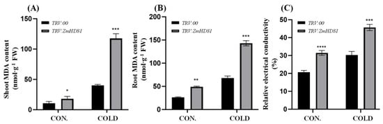
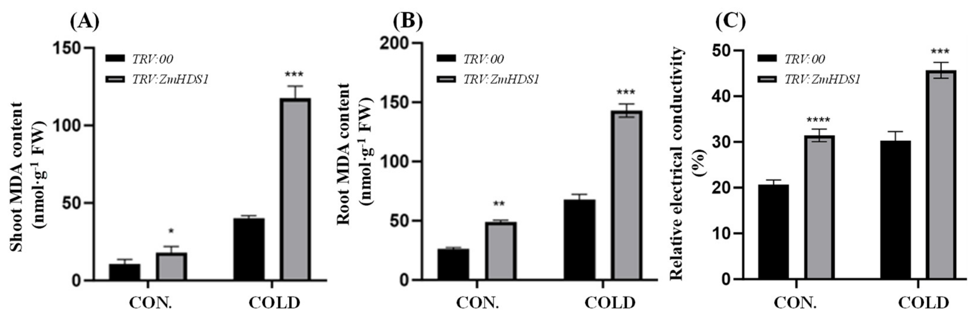
Malondialdehyde (MDA) serves as a pivotal marker for evaluating the extent of membrane lipid peroxidation, and its accumulation directly reflects oxidative damage to plant cell membrane systems. The MDA content was significantly increased in both the roots and leaves of maize seedlings. Notably, ZmHDS1-silenced plants accumulated distinctly higher MDA levels than control plants in both the shoot and root (Figure 7A,B). In addition, the relative electrolyte leakage was significantly higher in ZmHDS1-silenced maize than in control plants (Figure 7C). These results indicate that ZmHDS1-silenced plants suffered from more severe membrane injury under cold stress.
Figure 7.
Malondialdehyde (MDA) content of shoots (A) and roots (B) and relative electrical conductivity (C) of ZmHDS1-silenced maize plants via VIGS (TRV:00 and TRV:ZmHDS1) under cold stress for 3 d. TRV:00 represents seedlings infected with the empty TRV vector. Maize seedlings with three true leaves were shifted to cold stress (4 °C) for 3 d. Values represent the mean ± SD of three independent replicates. Significance was detected using SAS PROC ANOVA LSD (*, p < 0.05; **, p < 0.01; ***, p < 0.001; ****, p < 0.0001).
3.4. Suppression of ZmHDS1 Affects Hormone Balance of Maize Under Cold Stress
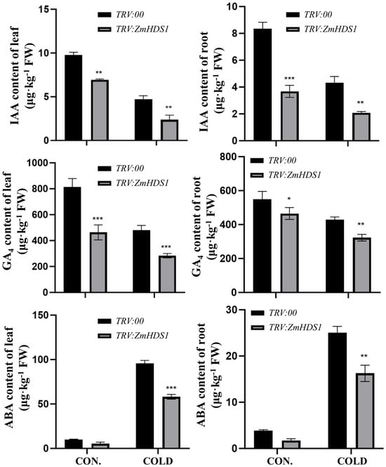
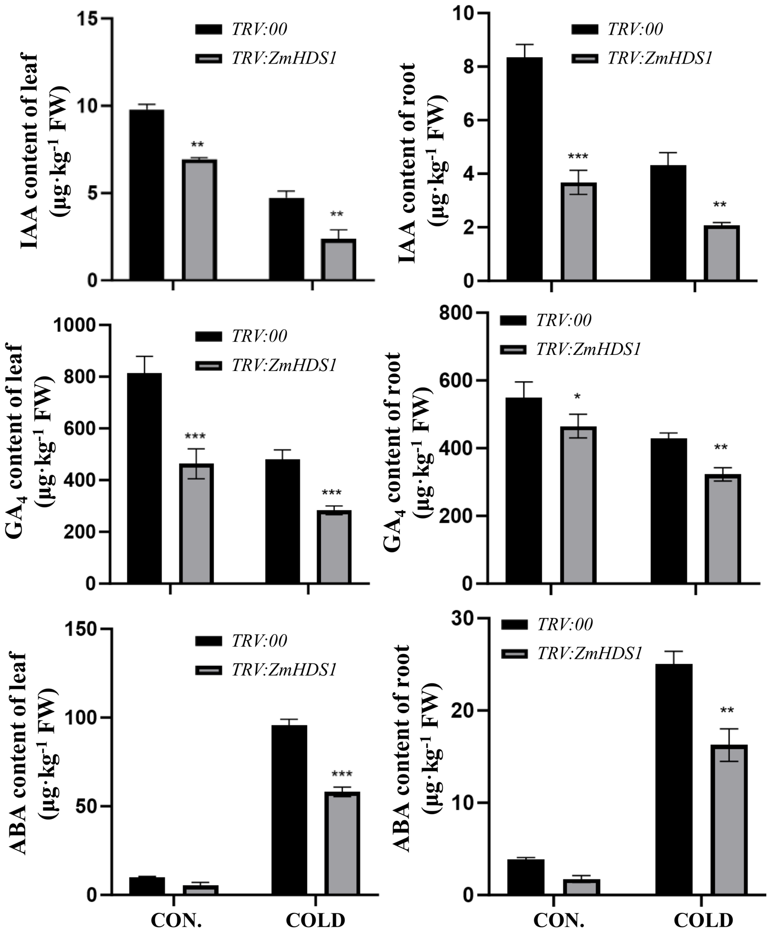
As an essential phytohormone regulating plant growth, development, and stress adaptation, indole-3-acetic acid (IAA) was decreased by 50–65% in both the shoots and roots of maize seedlings. The IAA levels in both the shoots and roots in ZmHDS1-silenced plants were significantly lower than in control plants, regardless of whether there was low temperature stress (Figure 8). GA4 displayed similar changes to IAA in response to cold stress (Figure 8). As one of the predominant gibberellins in maize, GA4 directly modulates cell elongation and cell division and serves as a critical determinant of biomass accumulation. The lower IAA and GA4 content in ZmHDS1-silenced plants may partly explain their retarded biomass formation.
Figure 8.
Hormone alterations in maize seedlings of ZmHDS1-silenced maize plants via VIGS (TRV:00 and TRV:ZmHDS1) under cold stress for 3 d. TRV:00 represents seedlings infected with the empty TRV vector. Maize seedlings with three true leaves were shifted to cold stress (4 °C) for 3 d. Values represent the mean ± SD of three independent replicates. Significance was detected using SAS PROC ANOVA LSD (*, p < 0.05; **, p < 0.01; ***, p < 0.001).
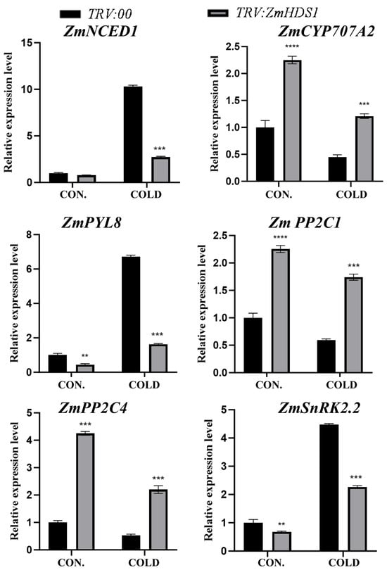
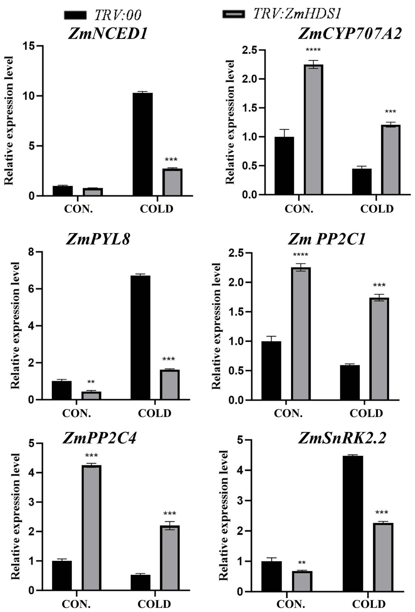
ABA accumulation triggers enhanced cold tolerance in plants. Being a core rate-limiting enzyme of the MEP pathway, HDS controls ABA production via regulating isoprenoid metabolism. Cold stress triggered massive ABA accumulation in both the leaves and roots. The leaf ABA levels increased 10.1-fold in control plants and 10.5-fold in ZmHDS1-silenced plants, whereas the root ABA content rose 6.5-fold and 10.3-fold in control and silenced seedlings, respectively. However, the ABA content of ZmHDS1-silenced plants was significantly lower than in the control plants under cold stress in both the leaf and root (Figure 8). To examine how ZmHDS1 confers cold resistance through ABA-dependent pathways, expression profiling was performed for genes involved in ABA synthesis, degradation, and signaling. ZmNCED1 and ZmCYP707A2 dominate ABA synthesis and catabolism, respectively. Cold stress downregulated the expression of ZmNCED1 but upregulated the expression of ZmCYP707A2 in ZmHDS1-silenced plants compared with control plants (Figure 9). As a crucial ABA receptor-encoding gene, ZmPYL8 was cold-inducible, yet its expression in ZmHDS1-silenced plants was only 0.23-fold relative to the controls under cold stress (Figure 9). In addition, irrespective of cold treatment, the expression levels of ABA signaling-related genes ZmPP2C1 and ZmPP2C4 were significantly higher in ZmHDS1-silenced plants than in control plants (Figure 9). However, the expression of ZmSnRK2.2 in response to cold stress was similar to that of ZmPYL8 in maize (Figure 9). These results indicate that ZmHDS1 positively enhanced maize cold tolerance by fine-tuning ABA metabolism and signaling. The silencing of ZmHDS1 decreased the endogenous ABA content and disturbed downstream ABA signaling networks, thereby drastically increasing cold susceptibility in maize.
Figure 9.
Relative expression of genes associated with ABA metabolism and signal transduction in ZmHDS1-silenced maize plants via VIGS (TRV:00 and TRV:ZmHDS1) under cold stress for 3 d. TRV:00 represents seedlings infected with the empty TRV vector. Maize seedlings with three true leaves were shifted to cold stress (4 °C) for 3 d. Values represent the mean ± SD of three independent replicates. Significance was detected using SAS PROC ANOVA LSD (**, p < 0.01; ***, p < 0.001; ****, p < 0.0001).
3.5. Suppression of ZmHDS1 Affects Photosynthesis in Maize Under Cold Stress
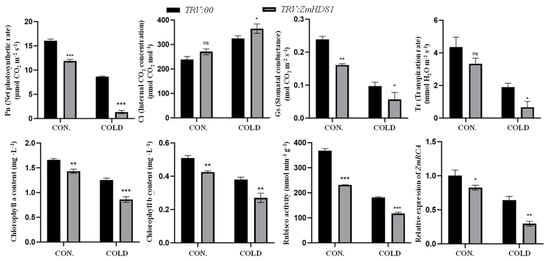
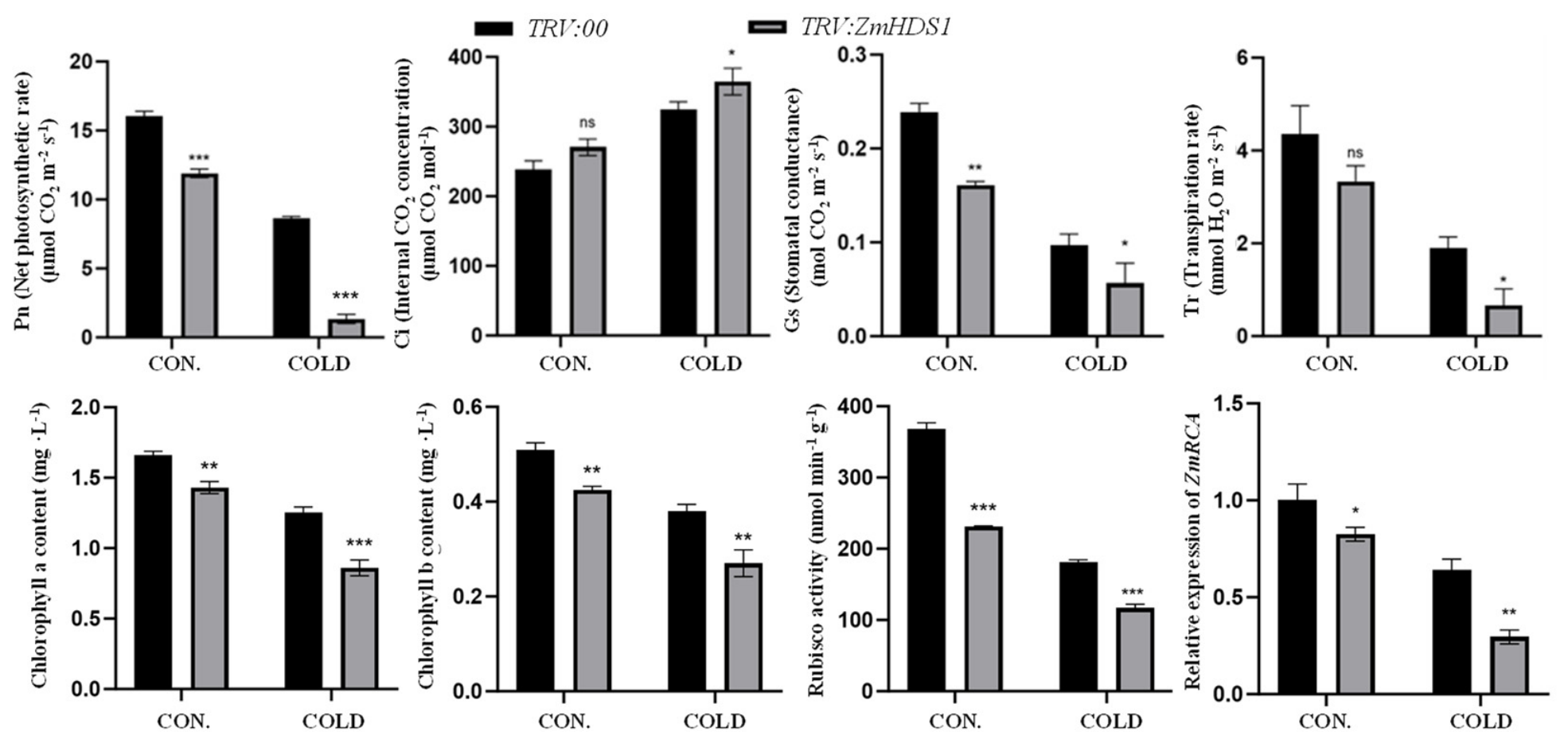
ZmHDS1 localizes to chloroplasts. Given the central role of chloroplasts in plant photosynthesis, photosynthetic parameters were determined to explore ZmHDS1’s potential function in photosynthetic regulation. Under cold stress, silencing ZmHDS1 reduced net photosynthesis (Pn), stomatal conductance (Gs), and the transpiration rate (Tr) but increased the intercellular CO2 concentration (Ci) in maize compared with the controls (Figure 10). The silencing of ZmHDS1 decreased the chlorophyll a and b content, especially under cold conditions (Figure 10), which weakened light capture and energy transformation and ultimately inhibited photosynthesis.
Figure 10.
Photosynthetic parameters, chlorophyll content, Rubisco activity, and the expression of ZmRCA in ZmHDS1-silenced maize plants via VIGS (TRV:00 and TRV:ZmHDS1) under cold stress for 3 d. TRV:00 represents seedlings infected with the empty TRV vector. Maize seedlings with three true leaves were shifted to cold stress (4 °C) for 3 d. Values represent the mean ± SD of three independent replicates. Significance was detected using SAS PROC ANOVA LSD (ns, not significant; *, p < 0.05; **, p < 0.01; ***, p < 0.001).
As the key rate-limiting enzyme of the Calvin cycle, Rubisco relies on Rubisco activase (RCA) for activation, and together they determine the efficiency of photosynthetic carbon assimilation. Regardless of cold treatment, ZmHDS1 silencing repressed Rubisco activity and reduced the expression of the RCA-encoding gene ZmRCA (Figure 10). These results suggest that ZmHDS1 contributed to the maintenance of photosynthetic performance under cold stress, at least in part by sustaining ZmRCA expression and Rubisco activity.
3.6. Suppression of ZmHDS1 Affects Carotenoids in Maize Under Cold Stress
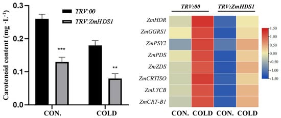
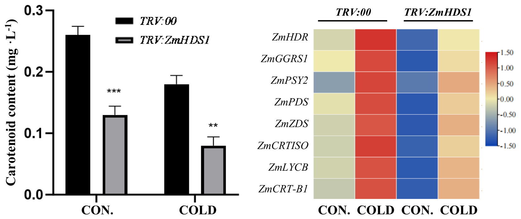
As secondary metabolites, carotenoids function as signaling molecules to mediate plant environmental responses. In the present study, the endogenous carotenoid content and the expression of genes involved in carotenoid biosynthesis were determined to clarify the regulatory role of ZmHDS1 in the tetraterpene carotenoid biosynthetic pathway in maize. Cold stress reduced the carotenoid levels universally, and the reduction was more severe in ZmHDS1-silenced plants than in control plants (Figure 11).
Figure 11.
Carotenoid content and the expression of genes related to carotenoid synthesis in ZmHDS1-silenced maize plants via VIGS (TRV:00 and TRV:ZmHDS1) under cold stress for 3 d. TRV:00 represents seedlings infected with the empty TRV vector. Maize seedlings with three true leaves were shifted to cold stress (4 °C) for 3 d. Values represent the mean ± SD of three independent replicates. Significance was detected using SAS PROC ANOVA LSD (**, p < 0.01; ***, p < 0.001).
Genes governing MEP upstream steps (ZmHDR, ZmGGPS1), carotenoid synthesis (ZmPSY2, ZmPDS, ZmZDS, ZmCRTISO), and carotenoid cyclization (ZmLCYB, ZmCRT-B1) were upregulated by cold in control plants. In ZmHDS1-silenced maize, however, these genes were downregulated under normal conditions, and their expression levels remained significantly lower than those in control plants under cold stress (Figure 11). The results suggest that ZmHDS1 is important for carotenoid homeostasis and contributes to the full cold-induced transcriptional activation of carotenoid biosynthetic genes.
4. Discussion
A candidate protein, ZmHDS1, was significantly increased in protein abundance by cold stress in cold-tolerant maize [27]. The expression of ZmHDS1 progressively increased following cold stress and reached its maximum at 24 h (Figure 1), suggesting that the differential expression of ZmHDS1 might underlie, at least in part, the genotypic variation in maize cold tolerance. In the present study, VIGS-mediated gene silencing and comprehensive physiological, biochemical, and molecular characterization were performed. Our findings suggest that ZmHDS1, a key enzyme in the MEP pathway, may act as an important regulator of cold tolerance in maize, potentially by modulating several downstream processes, including antioxidant defense, phytohormone homeostasis, photosynthetic performance, and carotenoid metabolism. The silencing of ZmHDS1 not only compromised these protective mechanisms but also rendered maize seedlings significantly more susceptible to cold-induced damage, underscoring the essential role of this enzyme in cold acclimation.
The phenotypic consequences of ZmHDS1 silencing were striking. Cold stress induced severe leaf shriveling, wilting, and darker green leaves despite reduced total chlorophyll content in ZmHDS1-silenced plants, accompanied by significantly reduced shoot and root dry weights. The growth retardation phenotype was consistent with the general role of the MEP pathway in providing isoprenoid precursors that are essential for plant growth and development [20,23]. More importantly, paraffin section analysis revealed extensive anatomical disruption in the leaves of ZmHDS1-silenced plants under cold stress, including a disordered epidermis, degenerated mesophyll, and disrupted Kranz anatomy (a hallmark of C4 photosynthesis) in maize (Figure 3). Similar anatomical defects have been reported in the leaves of maize in response to cold stress, where chloroplast development and cellular organization are severely compromised [31]. These results indicate that ZmHDS1 is required for maintaining cellular integrity and growth under cold stress.
Cold stress is well known to induce massive ROS overproduction, leading to oxidative damage [32]. Histochemical staining with NBT and DAB demonstrated that ZmHDS1-silenced maize seedlings accumulated significantly higher levels of superoxide anion and H2O2 under cold stress compared to controls (Figure 4). Excessive ROS accumulation was accompanied by markedly lower activity of key antioxidant enzymes (POD, SOD, CAT, APX, and GPX) in both the shoots and roots of ZmHDS1-silenced maize seedlings (Figure 5). The MEP pathway serves not only as a biosynthetic route for isoprenoid precursors but also as an oxidative stress sensing and response system [33]. Under stress, MEcPP accumulates and acts as a retrograde signal, and elevated MEcPP levels have been shown to reduce APX abundance via a plastidial metalloprotease-mediated mechanism, directly linking MEP pathway flux to enzymatic antioxidant capacity [20,33]. In addition, the MEP pathway supplies precursor pools for non-enzymatic antioxidants, including carotenoids, tocopherols, and plastoquinone. These non-enzymatic antioxidants are not independent of the enzymatic system: ascorbate serves as the electron donor for APX, and GSH regenerates ascorbate via the AsA-GSH cycle, so their depletion directly compromises enzymatic H2O2 detoxification [34]. Therefore, the disruption of ZmHDS1 impairs MEP pathway flux, which simultaneously reduces the supply of non-enzymatic antioxidants and, through MEcPP-mediated signaling and protein-level effects, compromises the enzymatic antioxidant system, rather than one system compensating for the other. The significantly elevated MDA content and relative electrolyte leakage in ZmHDS1-silenced maize seedlings (Figure 7) further confirmed that membrane lipid peroxidation and loss of membrane integrity were exacerbated upon ZmHDS1 silencing. These results align with previous findings indicating that flavonoids and terpenoids serve as vital ROS scavengers in plant stress responses and that MEP pathway-derived metabolites play crucial roles in oxidative stress [35,36]. The results therefore suggest that ZmHDS1 plays an important role in regulating the antioxidant system and membrane integrity in maize in response to cold stress.
The MEP pathway provides the isoprenoid precursors for a wide array of terpenoid-derived phytohormones, including ABA and gibberellins [37]. The IAA and GA4 content was consistently lower in ZmHDS1-silenced maize seedlings regardless of cold stress (Figure 8). Because IAA and GA4 promote cell elongation and cell division, their reduction is consistent with, and may partly explain, the retarded biomass accumulation in these plants. Cold stress triggered substantial ABA accumulation in both the leaves and roots, but the ABA levels in ZmHDS1-silenced maize seedlings were significantly lower as compared with control seedlings (Figure 8). Considering that ABA accumulation is a critical determinant of enhanced cold tolerance in rice [38], these results indicate that intact MEP pathway metabolism mediated by ZmHDS1 is indispensable for cold-induced ABA biosynthesis in maize. Furthermore, ZmHDS1 silencing profoundly disrupted ABA signaling. The expression of ZmNCED1 (for ABA synthesis) was downregulated while that of ZmCYP707A2 (for ABA catabolism) was upregulated in ZmHDS1-silenced maize seedlings response to cold stress. More importantly, the ABA receptor gene ZmPYL8 and the downstream kinase gene ZmSnRK2.2 showed reduced cold inducibility in ZmHDS1-silenced maize seedlings, whereas the negative regulators ZmPP2C1 and ZmPP2C4 were constitutively upregulated (Figure 9). These results suggest that ZmHDS1 may act as a key node that potentially integrates metabolic precursor supply with the transcriptional regulation of the ABA pathway.
Silencing ZmHDS1 reduced net photosynthesis (Pn), stomatal conductance (Gs), and the transpiration rate (Tr) while increasing the intercellular CO2 concentration (Ci) under cold stress. The chlorophyll a and b content was also significantly decreased in ZmHDS1-silenced maize seedlings in response to cold stress (Figure 10). These results suggest that the photosynthetic inhibition was primarily due to non-stomatal limitations, likely reflecting damage to the photosynthetic apparatus within chloroplasts. The MEP pathway provides the phytol chain for chlorophyll biosynthesis [39]; thus, an impaired MEP by ZmHDS1 silencing would directly compromise chlorophyll synthesis and the light-harvesting capacity. Furthermore, ZmHDS1 silencing repressed Rubisco activity and reduced the expression of ZmRCA (which encodes Rubisco activase) (Figure 10). Rubisco activase is particularly sensitive to environmental stresses and plays a crucial role in photosynthesis [40]. Our findings establish a novel link between the MEP pathway and the Calvin cycle machinery, highlighting the interconnectedness of plastidial metabolic pathways in stress adaptation.
Carotenoids are tetraterpenoid metabolites synthesized entirely via the MEP pathway and function not only as photosynthetic pigments and photoprotectants but also as signaling molecules in plant environmental responses [41,42]. Cold stress universally reduced carotenoid levels, with a more severe reduction in ZmHDS1-silenced plants than in controls (Figure 10). This observation is consistent with a previous report that low temperatures significantly reduced carotenoid accumulation in maize seedlings [43]. Strikingly, the expression of genes governing MEP upstream steps (ZmHDR, ZmGGPS1), carotenoid synthesis (ZmPSY2, ZmPDS, ZmZDS, ZmCRTISO), and carotenoid cyclization (ZmLCYB, ZmCRT-B1) was upregulated by cold stress in control plants but remained significantly lower in ZmHDS1-silenced maize seedlings under both normal and cold conditions (Figure 11). These results suggest that ZmHDS1 is required for the full cold-induced transcriptional activation of carotenoid biosynthetic genes. Because ZmHDS1 encodes a key enzyme in the MEP pathway, and because the expression of these genes was reduced rather than abolished in ZmHDS1-silenced plants, we propose that this effect is likely mediated, at least in part, by altered MEP pathway flux or MEcPP-mediated retrograde signaling, rather than by direct transcriptional regulation by ZmHDS1. Under stress, the accumulation of the MEP pathway intermediate MEcPP can act as a plastidial retrograde signal that alters nuclear gene expression [20].
5. Conclusions
The MEP pathway’s key enzyme gene ZmHDS1 is a cold-responsive gene that contributes to maize cold tolerance. Functional analysis via VIGS showed that ZmHDS1 silencing severely compromised seedling growth and cellular structural stability under cold stress. ZmHDS1 is required for maintaining ROS homeostasis by enhancing antioxidant enzyme activity and reducing membrane damage. Meanwhile, ZmHDS1 modulates the homeostasis of IAA, GA4, and ABA and sustains ABA biosynthesis and signaling transduction, which are critical for cold adaptation. Furthermore, ZmHDS1 preserves photosynthetic efficiency by stabilizing chlorophyll accumulation, Rubisco activity, and carbon assimilation capacity. Importantly, ZmHDS1 acts as a key permissive regulator for cold-induced carotenoid accumulation by transcriptionally activating the entire carotenoid biosynthetic pathway. In summary, ZmHDS1 acts as a key component of the MEP pathway that affects multiple downstream physiological processes to enhance maize cold resistance. This study provides new insights into the molecular mechanism of plant cold tolerance and offers a valuable target for the genetic improvement of cold stress resilience in maize.
Supplementary Materials
The following supporting information can be downloaded at: https://www.mdpi.com/article/10.3390/agronomy16202036/s1, Table S1. Primer sequences used for VIGS and RT-qPCR. Figure S1. Gene structure, conserved protein domains, and VIGS target region of ZmHDS1. (A) Exon–intron organization of ZmHDS1 based on the B73 reference genome (Zm-B73-REFERENCE-NAM-5.0). Filled boxes represent exons, and lines represent introns. (B) Conserved protein domain architecture of ZmHDS1 predicted by NCBI CD-Search. The HDS domain is indicated. (C) Schematic mapping of the 236 bp VIGS target fragment (CDS positions 66–301) used for pTRV2:ZmHDS1 construction; the targeted region is highlighted in orange. Figure S2. Electrophoretic detection of PCR-amplified ZmHDS1 silencing fragments (A) and sequencing of PCR-amplified ZmHDS1 silencing fragments (B). M: DNA marker 2000; Lane 1 and 2: PCR-amplified ZmHDS1 silencing fragments. Figure S3. An expression analysis following ZmHDS1-silenced maize plants via VIGS (TRV:ZmHDS1) and its control (TRV:00). Values represent the mean ± SD of three independent replicates. A Student’s t-test was used to assess differences between different treatments (*** p < 0.001).
Author Contributions
All authors contributed to the study conception and design. Z.G.: conceptualization, methodology, investigation writing—original draft; R.Y.: methodology, investigation, validation; X.W.: methodology, investigation, validation; D.J.: methodology, investigation, validation; S.Z.: methodology, investigation; F.Z.: methodology, investigation; H.M.: data curation, writing—review and editing; M.Z.: data curation, writing—review and editing; S.C.: conceptualization, resources, writing—review and editing, funding acquisition. All authors have read and agreed to the published version of the manuscript.
Funding
This research was funded by the National Natural Science Foundation of China (31601808).
Data Availability Statement
The original contributions presented in this study are included in the article. Further inquiries can be directed to the corresponding author, Shuisen Chen.
Conflicts of Interest
The authors declare no conflicts of interest.
Abbreviations
The following abbreviations are used in this manuscript:
| ABA | Abscisic acid |
| APX | Ascorbate peroxidase |
| CAT | Catalase |
| Ci | Intercellular CO2 concentration |
| CON. | Control |
| DAB | 3,3-Diaminobenzidine |
| DW | Dry weight |
| ESI | Electrospray ionization |
| FW | Fresh weight |
| GA4 | Gibberellin |
| GSH | Reduced glutathione |
| GPX | Glutathione peroxidase |
| Gs | Stomatal conductance |
| HDS | Hydroxymethylbutenyl diphosphate synthase |
| HMBPP | (2E)-4-hydroxy-3-methylbut-2-enyl diphosphate |
| IAA | Indole-3-acetic acid |
| MDA | Malondialdehyde |
| MEcPP | Methylerythritol cyclodiphosphate |
| MEP | Methylerythritol phosphate |
| MRM | Multiple reaction monitoring |
| NBT | Nitroblue tetrazolium |
| Pn | Net photosynthesis |
| POD | Peroxidase |
| RCA | Rubisco activase |
| ROS | Reactive oxygen species |
| SOD | Superoxide dismutase |
| Tr | Transpiration rate |
| VIGS | Virus-induced gene silencing |
References
- Erenstein, O.; Jaleta, M.; Sonder, K.; Mottaleb, K.; Prasanna, B.M. Global Maize Production, Consumption and Trade: Trends and R&D Implications. Food Secur. 2022, 14, 1295–1319. [Google Scholar] [CrossRef] [Scilit]
- Liu, J.; Fernie, A.R.; Yan, J. The Past, Present, and Future of Maize Improvement: Domestication, Genomics, and Functional Genomic Routes toward Crop Enhancement. Plant Commun. 2020, 1, 100010. [Google Scholar] [CrossRef] [Scilit]
- Juurakko, C.L.; diCenzo, G.C.; Walker, V.K. Cold Acclimation and Prospects for Cold-Resilient Crops. Plant Stress 2021, 2, 100028. [Google Scholar] [CrossRef] [Scilit]
- Zhao, X.; Zhao, C.; Niu, Y.; Chao, W.; He, W.; Wang, Y.; Mao, T.; Bai, X. Understanding and Comprehensive Evaluation of Cold Resistance in the Seedlings of Multiple Maize Genotypes. Plants 2022, 11, 1881. [Google Scholar] [CrossRef] [Scilit]
- Sobkowiak, A.; Jończyk, M.; Adamczyk, J.; Szczepanik, J.; Solecka, D.; Kuciara, I.; Hetmańczyk, K.; Trzcinska-Danielewicz, J.; Grzybowski, M.; Skoneczny, M.; et al. Molecular Foundations of Chilling-Tolerance of Modern Maize. BMC Genom. 2016, 17, 125. [Google Scholar] [CrossRef] [Scilit]
- Yu, T.; Zhang, J.; Ma, X.; Cao, S.; Li, W.; Yang, G. A Multi-Omics View of Maize’s (Zea mays L.) Response to Low Temperatures during the Seedling Stage. Int. J. Mol. Sci. 2024, 25, 12273. [Google Scholar] [CrossRef] [Scilit]
- Ma, Y.; Tan, R.; Zhao, J. Chilling Tolerance in Maize: Insights into Advances—Toward Physio-Biochemical Responses’ and QTL/Genes’ Identification. Plants 2022, 11, 2082. [Google Scholar] [CrossRef] [Scilit]
- Shomo, Z.D.; Li, F.; Smith, C.N.; Edmonds, S.R.; Roston, R.L. From Sensing to Acclimation: The Role of Membrane Lipid Remodeling in Plant Responses to Low Temperatures. Plant Physiol. 2024, 196, 1737–1757. [Google Scholar] [CrossRef] [Scilit]
- Ding, Y.; Shi, Y.; Yang, S. Regulatory Networks Underlying Plant Responses and Adaptation to Cold Stress. Annu. Rev. Genet. 2024, 58, 43–65. [Google Scholar] [CrossRef] [Scilit]
- Peng, C.; Hua, W.; Liu, J. Unraveling the Signaling Pathways of Plant Cold Stress: Current Insights and Future Directions. Front. Plant Sci. 2025, 16, 1666852. [Google Scholar] [CrossRef] [Scilit]
- Satyakam; Zinta, G.; Singh, R.K.; Kumar, R. Cold Adaptation Strategies in Plants—An Emerging Role of Epigenetics and Antifreeze Proteins to Engineer Cold Resilient Plants. Front. Genet. 2022, 13, 909007. [Google Scholar] [CrossRef] [Scilit]
- Roychowdhury, R.; Das, S.P.; Sarkar, P.; Khan, Z.; Kumar, A.; Sarker, U.; Sajeevan, R.S. Physiological, Biochemical and Molecular Signaling Basis of Cold Stress Tolerance in Plants. Front. Plant Sci. 2025, 16, 1707204. [Google Scholar] [CrossRef] [Scilit]
- Zhou, L.; Ullah, F.; Zou, J.; Zeng, X. Molecular and Physiological Responses of Plants That Enhance Cold Tolerance. Int. J. Mol. Sci. 2025, 26, 1157. [Google Scholar] [CrossRef] [Scilit]
- Wang, Z.; Jia, Q.; Zhang, B.; Ming, B.; Bai, L.; Wang, F.; Wang, Y.; Yu, S.; Zou, R.; Wang, Z. Unveiling the Physiological Basis of Cold Tolerance in Maize: Root Architecture, Photosynthetic Stability, and POD-Mediated Defense Under Delayed Chilling Stress. Plants 2026, 15, 517. [Google Scholar] [CrossRef] [Scilit]
- Liu, G.; Wang, H.; Tang, Y.; Gao, H.; Wang, Y.; Sun, Y.; Yu, S.; Zhang, D. Research Progress on Cold Tolerance Key Genes and Mechanisms in Four Major Northern China Crops. Front. Plant Sci. 2026, 17, 1823380. [Google Scholar] [CrossRef] [Scilit]
- Ramakrishna, A.; Ravishankar, G.A. Influence of Abiotic Stress Signals on Secondary Metabolites in Plants. Plant Signal. Behav. 2011, 6, 1720–1731. [Google Scholar] [CrossRef] [Scilit]
- Chen, D.; Mubeen, B.; Hasnain, A.; Rizwan, M.; Adrees, M.; Naqvi, S.A.H.; Iqbal, S.; Kamran, M.; El-Sabrout, A.M.; Elansary, H.O.; et al. Role of Promising Secondary Metabolites to Confer Resistance Against Environmental Stresses in Crop Plants: Current Scenario and Future Perspectives. Front. Plant Sci. 2022, 13, 881032. [Google Scholar] [CrossRef] [Scilit]
- Li, C.; Zha, W.; Li, W.; Wang, J.; You, A. Advances in the Biosynthesis of Terpenoids and Their Ecological Functions in Plant Resistance. Int. J. Mol. Sci. 2023, 24, 11561. [Google Scholar] [CrossRef] [Scilit]
- Xue, C.; Jiang, Y.; Wang, Z.; Shan, X.; Yuan, Y.; Hua, J. Tissue-Level Transcriptomic Responses to Local and Distal Chilling Reveal Potential Chilling Survival Mechanisms in Maize. J. Exp. Bot. 2021, 72, 7610–7625. [Google Scholar] [CrossRef] [Scilit]
- Bali, S.; Mohapatra, S.; Michael, R.; Arora, R.; Dogra, V. Plastidial Metabolites and Retrograde Signaling: A Case Study of MEP Pathway Intermediate MEcPP That Orchestrates Plant Growth and Stress Responses. Plant Physiol. Biochem. PPB 2025, 222, 109747. [Google Scholar] [CrossRef] [Scilit]
- Flores-Pérez, U.; Pérez-Gil, J.; Rodríguez-Villalón, A.; Gil, M.J.; Vera, P.; Rodríguez-Concepción, M. Contribution of Hydroxymethylbutenyl Diphosphate Synthase to Carotenoid Biosynthesis in Bacteria and Plants. Biochem. Biophys. Res. Commun. 2008, 371, 510–514. [Google Scholar] [CrossRef] [Scilit]
- Wang, J.-Z.; Lei, Y.; Xiao, Y.; He, X.; Liang, J.; Jiang, J.; Dong, S.; Ke, H.; Leon, P.; Zerbe, P.; et al. Uncovering the Functional Residues of Arabidopsis Isoprenoid Biosynthesis Enzyme HDS. Proc. Natl. Acad. Sci. USA 2020, 117, 355–361. [Google Scholar] [CrossRef] [Scilit]
- Pu, X.; Dong, X.; Li, Q.; Chen, Z.; Liu, L. An Update on the Function and Regulation of Methylerythritol Phosphate and Mevalonate Pathways and Their Evolutionary Dynamics. J. Integr. Plant Biol. 2021, 63, 1211–1226. [Google Scholar] [CrossRef] [Scilit]
- Zeng, L.; Dehesh, K. The Eukaryotic MEP-Pathway Genes Are Evolutionarily Conserved and Originated from Chlaymidia and Cyanobacteria. BMC Genom. 2021, 22, 137. [Google Scholar] [CrossRef] [Scilit]
- Pu, X.; Yang, Z.; Lin, Y.; Song, Y.; Chen, Z.; Chen, S.; Yang, H.; Duan, L.; Liu, L. Integration of HDS and HSFA1 Functions in MEcPP-Mediated Thermotolerance and Growth Regulation in Physcomitrium Patens. Plant Cell Environ. 2025, 48, 5993–6015. [Google Scholar] [CrossRef] [Scilit]
- Ren, X.; Zhao, Y.; Yu, W.; Zhang, J.; Huang, Z.; Zhang, M.; Yu, Q.; Ji, K. Characteristics and Functions of PmHDS, a Terpenoid Synthesis-Related Gene in Pinus Massoniana Lamb. Int. J. Mol. Sci. 2025, 26, 457. [Google Scholar] [CrossRef] [Scilit]
- Yang, R.; Wang, X.; Li, H.; Liu, M.; Zhong, M.; Ma, H.; Guo, Z.; Chen, S. Physiological and Proteomic Insights into Cold Tolerance Mechanism in Two Contrasting Maize Genotypes. Plant Physiol. Biochem. 2026, 230, 110975. [Google Scholar] [CrossRef] [Scilit]
- Ratcliff, F.; Martin-Hernandez, A.M.; Baulcombe, D.C. Technical Advance: Tobacco rattle virus as a vector for analysis of gene function by silencing. Plant J. 2001, 25, 237–245. [Google Scholar] [CrossRef] [Scilit]
- Bezrutczyk, M.; Zoellner, N.R.; Kruse, C.P.S.; Hartwig, T.; Lautwein, T.; Koehrer, K.; Frommer, W.B.; Kim, J.Y. Evidence for Phloem Loading via the Abaxial Bundle Sheath Cells in Maize Leaves. Plant Cell 2021, 33, 531–547. [Google Scholar] [CrossRef] [Scilit]
- Chen, S.; Zhao, H.; Ding, G.; Xu, F. Genotypic Differences in Antioxidant Response to Phosphorus Deficiency in Brassica Napus. Plant Soil 2015, 391, 19–32. [Google Scholar] [CrossRef] [Scilit]
- Zhou, X.; Muhammad, I.; Lan, H.; Xia, C. Recent Advances in the Analysis of Cold Tolerance in Maize. Front. Plant Sci. 2022, 13, 866034. [Google Scholar] [CrossRef] [Scilit]
- Liu, R.; Wang, S.; Song, J. New Progress in the Production, Oxidative Damage, and Scavenging Mechanisms of Reactive Oxygen Species in Plants under Abiotic Stress. Front. Plant Sci. 2026, 17, 1774033. [Google Scholar] [CrossRef] [Scilit]
- Perez-Gil, J.; Behrendorff, J.; Douw, A.; Vickers, C.E. The Methylerythritol Phosphate Pathway as an Oxidative Stress Sense and Response System. Nat. Commun. 2024, 15, 5303. [Google Scholar] [CrossRef] [Scilit]
- Peng, S.; Jiang, M.; Li, X. Systems-Level Integration of Stress Signaling, Multi-Omics, and Predictive Breeding for Abiotic Stress Tolerance in Brassica Crops. Horticulturae 2026, 12, 1033. [Google Scholar] [CrossRef] [Scilit]
- Sana; Aftab, T.; Naeem, M.; Jha, P.K.; Prasad, P.V.V. Production of Secondary Metabolites under Challenging Environments: Understanding Functions and Mechanisms of Signalling Molecules. Front. Plant Sci. 2025, 16, 1569014. [Google Scholar] [CrossRef] [Scilit]
- Sharma, S.; Negi, S.; Kumar, P.; Irfan, M. Cellular Strategies for Surviving the Alpine Extremes: Methylerythritol Phosphate Pathway-Driven Isoprenoid Biosynthesis and Stress Resilience. Protoplasma 2025, 262, 1053–1072. [Google Scholar] [CrossRef] [Scilit]
- Cheng, R.; Yang, S.; Wang, D.; Qin, F.; Wang, S.; Meng, S. Advances in the Biosynthesis of Plant Terpenoids: Models, Mechanisms, and Applications. Plants 2025, 14, 1428. [Google Scholar] [CrossRef] [Scilit]
- Li, W.; Lou, X.; Wang, Z.; Zhang, D.; Li, L.; Ding, X.; Cheng, G.; Nie, W.; Li, Z.; Yu, J.; et al. Unlocking ABA’s Role in Rice Cold Tolerance: Insights from Zhonghua 11 and Kasalath. TAG Theor. Appl. Genet. Theor. Angew. Genet. 2025, 138, 16. [Google Scholar] [CrossRef] [Scilit]
- Lichtenthaler, H.K. The 1-Deoxy-D-Xylulose-5-Phosphate Pathway of Isoprenoid Biosynthesis in Plants. Annu. Rev. Plant Physiol. Plant Mol. Biol. 1999, 50, 47–65. [Google Scholar] [CrossRef] [Scilit]
- Rajkhowa, S.; Bose, B.; Sarma, J.; Kumari, R. RuBisCo Activase as a Response to Environmental Stress in Plants. In Photosynthesis and Respiratory Cycles During Environmental Stress Response in Plants; Apple Academic Press: Palm Bay, FL, USA, 2022. [Google Scholar]
- Sierra, J.; McQuinn, R.P.; Leon, P. The Role of Carotenoids as a Source of Retrograde Signals: Impact on Plant Development and Stress Responses. J. Exp. Bot. 2022, 73, 7139–7154. [Google Scholar] [CrossRef] [Scilit]
- Sun, T.; Rao, S.; Zhou, X.; Li, L. Plant Carotenoids: Recent Advances and Future Perspectives. Mol. Hortic. 2022, 2, 3. [Google Scholar] [CrossRef] [Scilit]
- Xiang, N.; Zhang, B.; Hu, J.; Li, K.; Guo, X. Modulation of Carotenoid Biosynthesis in Maize (Zea mays L.) Seedlings by Exogenous Abscisic Acid and Salicylic Acid under Low Temperature. Plant Cell Rep. 2023, 43, 1. [Google Scholar] [CrossRef] [Scilit]
Disclaimer/Publisher’s Note: The statements, opinions and data contained in all publications are solely those of the individual author(s) and contributor(s) and not of MDPI and/or the editor(s). MDPI and/or the editor(s) disclaim responsibility for any injury to people or property resulting from any ideas, methods, instructions or products referred to in the content. |
© 2026 by the authors. Licensee MDPI, Basel, Switzerland. This article is an open access article distributed under the terms and conditions of the Creative Commons Attribution (CC BY) license.