


Abstract
A new class of Ln(III) Schiff base (SB) complexes has been synthesized with the general formula [LnL(hfac)3], where Ln = Tb (TbL), Eu (EuL), Sm (SmL) or Gd (GdL). The molecular structure of the complex was determined using the X-ray diffraction method. The IR spectra show that the C=O stretching is shifted from 1695 cm−1 in pure PMMA to 1719–1724 cm−1 in LnL-PMMA hybrid materials. This means that LnL materials are successfully embedded into the PMMA backbone in the polymeric films. However, the Ln(III) SB complexes exhibit emission spectra that cover the visible region. The TbL complex displays an intense green emission combined with a large emission lifetime of 0.505 ms. However, the incorporation of Ln-SB complexes into PMMA (polymethylmethacrylate) polymeric films was investigated. The thermal stabilities of the LnL-PMMA hybrid materials increased from 140 to 250 °C when compared with the LnL-SB complexes. Furthermore, their luminescence intensity and lifetimes were also enhanced due to their induced structural rigidity. The molecular interactions of the LnL complexes with the PMMA matrix were monitored by investigating the luminescence properties of EuL-PMMA. Detailed photoluminescence studies showed insights into the non-radiative rates and improved quantum yields (QEu = 23%) from Eu(III). Furthermore, the higher values of Judd–Ofelt parameters Ω2 and Ω4 indicate strong hydrogen bonding interactions between L and PMMA. Nevertheless, these significant optical properties enable LnL-PMMA polymeric materials as colored phosphors for the design of opto-electronic devices.
1. Introduction
Trivalent lanthanide coordination compounds doped on hybrid materials are of considerable interest for their unique and distinctive optical [1] and mechanical properties [2]. These materials have a wide spectrum of applications in various fields of modern technology such as white light emission [3], optical sensing [4], and medical diagnostics [5,6]. In fact, polymeric films provide an excellent platform for the design of new lanthanide(III) hybrid materials with improved flexibility, mechanical strength and thermal stability [7,8,9]. Many contributions have widely explored the use of polymeric films prepared from a matrix made of polystyrene (PS), poly(acrylonitrile) (PAN), PMMA, polyvinyl butyral (PVB) or polysulfone (PSF). These studies suggest that the in situ polymerization of lanthanide(III) ions into polymeric hybrid materials is stabilized by extensive weak non-covalent bonds, including hydrogen bonds or p-stacking interactions between the metal complex and the matrix film [10]. In particular, these materials have proven their effective applications in thermal and optical devices, such as solar energy cells [11].
Within the class of luminescent lanthanide(III) materials, the design of organic chromophores for the sensitization process of the parity-forbidden f-f transitions is a challenge for researchers. In fact, Schiff base (SB) ligands continue to draw considerable interest from researchers as sensitizers for lanthanide(III) metals because of their effectiveness as an energy-harvesting antenna [12]. Ln-SB complexes are easy to synthesize, but unfortunately, their poor thermal stability limits their practical applications. Therefore, the combination of Ln-SB complexes into PMMA polymeric films has become a feasible alternative for the design of lanthanide(III) materials with enhanced thermal stability [13,14]. Polymeric films from PMMA are easily prepared. The carbonyl groups of PMMA can interact with the Ln-SB complex to form a rigid polymer [15]. In addition, their emission intensities and luminescence lifetimes can be easily modulated. For instance, Pirriera et al. successfully prepared a Eu(III)/PMMA polymer that possesses induced luminescence due to enhanced rigidity from dipole–dipole interactions within the polymer [16]. Furthermore, Sarkar and co-workers investigated luminescence properties of co-doping Sm(III), Tb(III) complexes into PMMA films to generate white light emissions. It was argued that the suppression of the aggregation-induced quenching is the main cause of the controlled light emission [17].
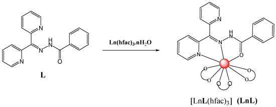
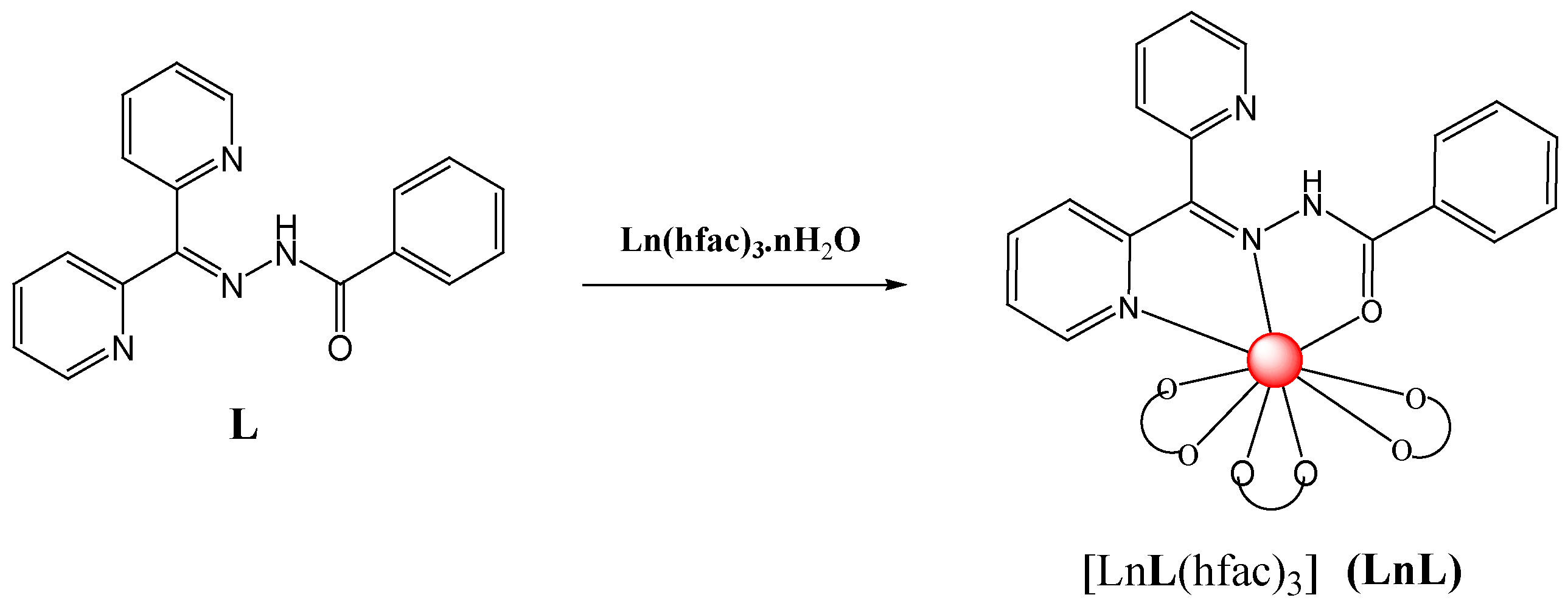
The present work reports the preparation of new lanthanide(III) complexes from the SB ligand L, benzoic acid (di-pyridin-2-yl-methylene)-hydrazide, and the ancillary ligand hexafluoroacetyl acetonato, hfac, as shown in Scheme 1. The formula of the Sm(III) complex was determined by X-ray crystallography to be [LnL(hfac)3], where Ln = Sm, Eu, Gd and Tb. With the objective of investigating the effect of polymerization on the photo-luminescence properties of the metals, the Ln-SB complexes were further incorporated into PMMA polymeric films at 2% (w/w) to produce highly luminescent hybrid materials. Limiting the dopant to 2% (w/w) was sufficient to manipulate the optical properties of the PMMA matrix without severely disturbing the polymer’s chain packing. It is expected that the pyridine substituent on ligand L will interact with PMMA via weak H bonding interactions and hence will modify their structural rigidities in the solid state and consequently the luminescent properties of the hybrid materials. The work also focused on the thermal stability of the free complexes and the hybrid materials.
Scheme 1.
The synthetic route for the Ln(III) SB complexes LnL (Ln = Sm, Eu, Gd and Tb).
2. Results and Discussion
2.1. Synthesis and Structure Characterization
A class of Ln(III) hydrazine complexes, where Ln = Sm, Eu, Gd and Tb, was successfully synthesized, and their formulae were confirmed by X-ray crystallography (Figure 1) and mass spectroscopy (Figure S1). An illustration of the synthetic routes for the complexes is shown in Scheme 1. Complexation of ligand L with Ln(hfac)3·nH2O, where Ln = Sm, Eu, Gd and Tb, yielded mononuclear compounds. Pale yellowish blocks of [SmL(hfac)3] (SmL) were obtained by slow air diffusion of the reaction mixture within a week.
Figure 1.
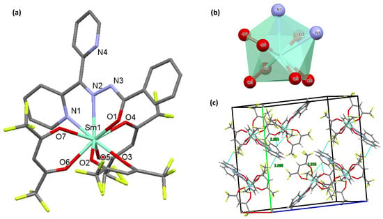
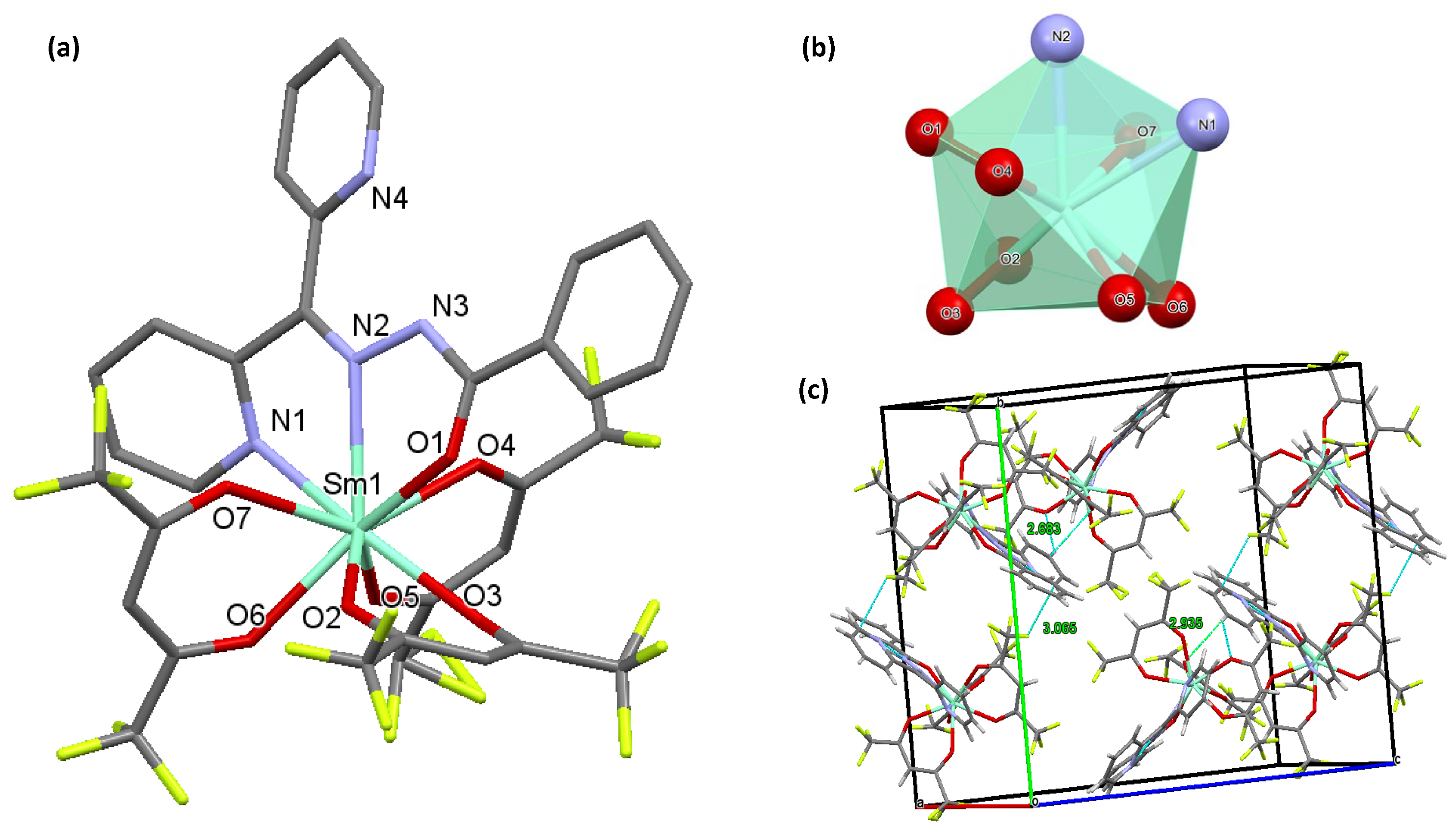
(a) Presentation of the molecular structure of [SmL(hfac)3] (SmL). (b) View of the coordination polyhedral around Sm(III). (c) Packing diagram of the complex exhibiting weak H bonding interactions to form an infinite network along the bc-plane.
Infrared results: The infrared (IR) spectra of the SB ligand L and its complexes in the solid state are presented in Figures S2–S6. The imine (C=N) absorption was found at 1526 cm−1, while the C=O group appeared at 1682 cm−1. The N–H bond appeared broad at 3539 cm−1, indicating hydrogen bonding interactions with the attached pyridyl unit of L. However, the LnL complexes show similar stretching bands and intensities to the free ligand. The stretching frequency of the coordinating carbonyl group (C=O) in LnL appeared at 1650 cm−1. In contrast, the COO− groups from the coordinating b-diketone ligands exhibit an asymmetric stretching vibration at ~1468 cm−1. Nevertheless, the IR spectra for the LnL-PMMA polymers were also investigated, as presented in Figure S3. Essentially, the spectra have similar peak positions to the LnL complexes, and the only observed differences appear mainly in the peaks’ intensities, combined with a new band around 1719–1724 cm−1 that contributes to the C=O vibrating band of PMMA. C=O stretching is observed at 1695 cm−1 in pure PMMA, which shifts to 1719–1724 cm−1 in the polymeric hybrid materials. This means that LnL is successfully embedded into the PMMA backbone through molecular interactions with the oxygen atoms of the carbonyl group in PMMA [14]. Interestingly, a sharp band appeared at 2937–2948 cm−1 from the N–H bond in LnL. This behavior indicates hydrogen bonding interactions between PMMA and the attached pyridyl unit in L, leaving the N–H bonds alone [18].
Crystal structure of SmL: The complex crystallizes in the monoclinic space group P21/n. The crystallographic refinement data for the reported structure, the Sm(III) complex, are shown in Table 1. The asymmetric unit comprises the complex molecule of [SmL(hfac)3]. The Sm(III) ion is in a nine-coordinate environment of SmN2O7, bound to ligand L, NNO and three chelating hfac units, as shown in Figure 1a. The coordination geometry surrounding Sm(III) is defined as a distorted capped square antiprism with C2 symmetry around the metal core. The ligand and three hfac units are twisted against each other in a facial fashion. Interestingly, the complex exhibits internal H bonding interactions between the amine proton (N–H) and the pyridyl substituent, where NH⋯Npy equals 1.957 Å. However, variable bond distances around Sm(III) are observed within the complex, as shown in Table 2. The Sm–Ohfac bond distances are in the range of 2.387(6)–2.461(5) Å and are shorter than those of the Sm–N bonds, 2.623(6) and 2.656(6) Å. The Sm–Oac bond is 2.419(5) Å. Interestingly, the packing diagram of SmL in Figure 1b exhibits variable weak interactions between the molecules. There are detectable H bonding interactions of pyH⋯F (3.065 Å) and pyH⋯O (2.683 and 2.935 Å) between the aromatic rings of the ligand and the hfac units. The repeated molecules create an infinite network along the bc-plane, with no clear evidence of voids in the crystal lattice.
Table 1.
Crystal data and structure refinement for SmL.
Table 2.
Bond lengths [Å] and angles [°] for SmL.
Thermogravimetry Analysis
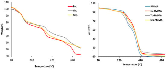
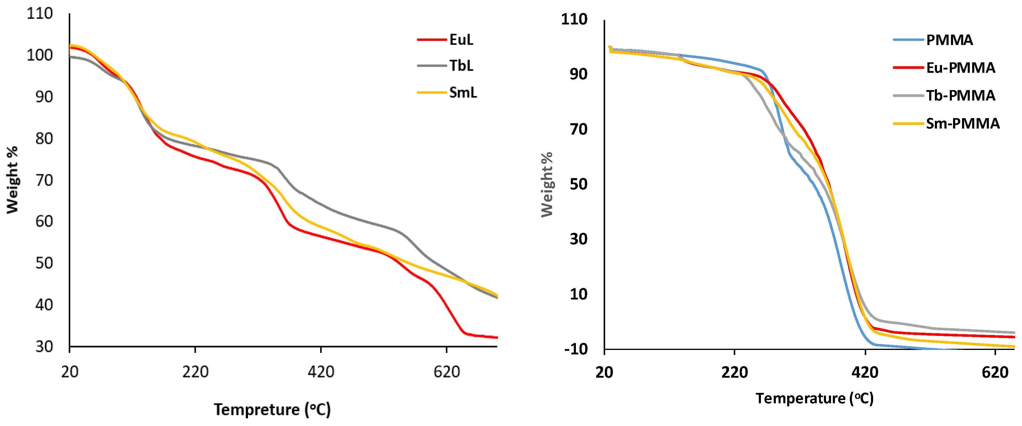
The thermal stabilities of the Ln(III)-SB complexes and their PMMA polymeric films were investigated. The thermogravimetric analysis (TGA) of TbL, EuL and SmL was performed under an inert N2 atmosphere, and the results are shown in Figure 2 (left). The TG curves show three main mass loss steps. The first major weight loss stage begins at 140 °C. A gradual mass loss of ~20% likely corresponds to initial decomposition of hfac units. The second major weight loss step takes place between 340 and 550 °C. A sharper drop in mass to ~50% represents the main ligand decomposition step. Finally, all the complexes undergo continuous weight loss down to 30%, leaving behind lanthanide oxide residues. However, the LnL-PMMA polymeric films show promising thermal properties. The TGA curves for the polymers exhibit one abrupt mass loss at 250 °C, which is extensively beyond the decomposition temperature of LnL complexes. This fact reveals that the LnL complexes are embedded in the PMMA polymer matrix by the H bonding interactions between LnL and the oxygen atoms of C=O in the PMMA polymer, as seen previously [18]. In conclusion, LnL-PMMA polymeric films have higher thermal stability compared to undoped LnL complexes, leading to interesting applications in opto-electronic devices.
Figure 2.
(Left): The TGA curves of LnL complexes. (Right): The TGA curves of LnL-PMMA polymers, where Ln = Eu, Tb and Sm. All these data were recorded under an inert N2 atmosphere.
2.2. Photophysical Study
The photophysical features, such as absorption and emission spectra, lifetime measurements (τLn) and quantum yields (QLn), of the Ln(III) complexes and their PMMA polymeric films are presented in Table 3.
Table 3.
Summary of the photophysical data for the Ln(III) SB complexes and their PMMA polymeric films at RT.
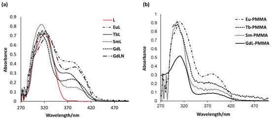
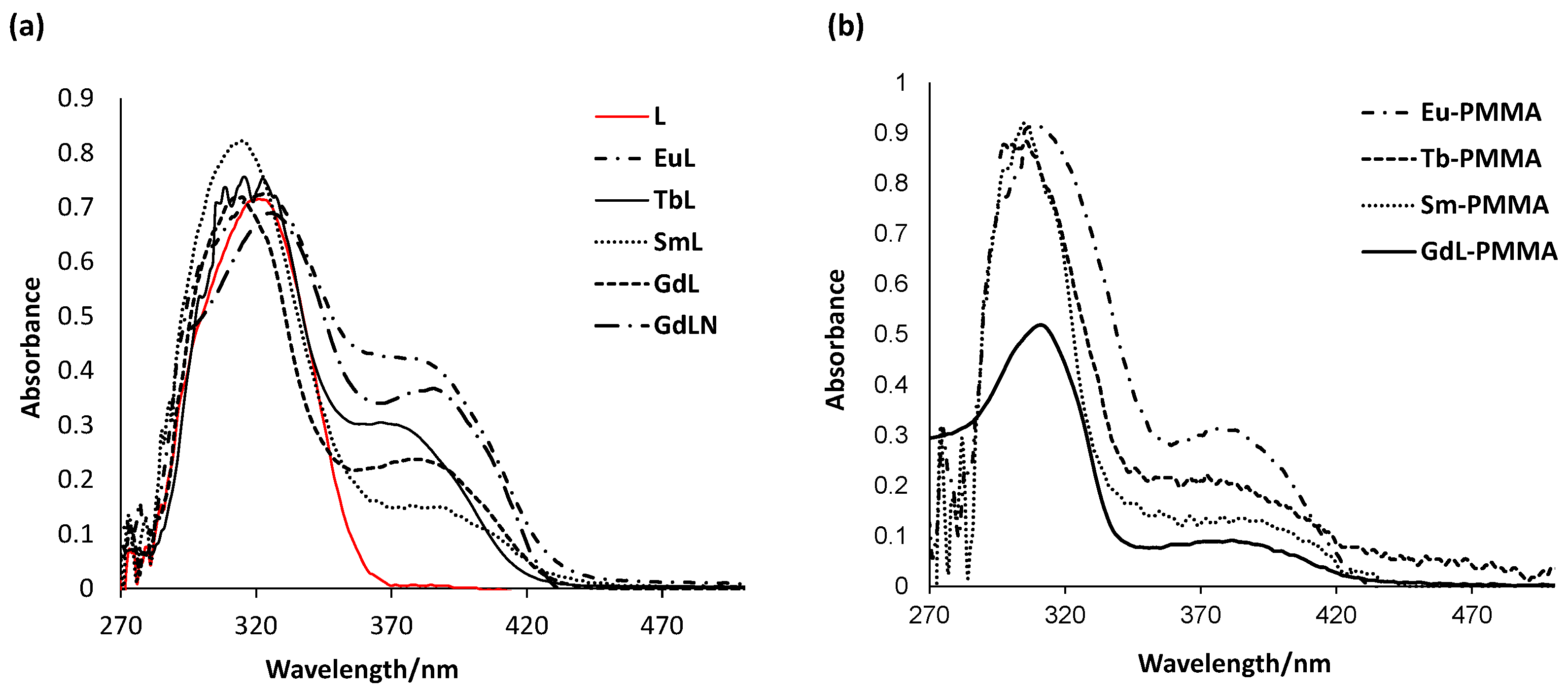
Absorption spectroscopy: The absorption spectra of ligand L and its Ln(III) complexes in methanol (10−5 M) are shown in Figure 3a. The ligand shows a broad intra-ligand charge transfer band (ILCT) centered at 318 nm. Upon complexation with the Ln(III) metals, the spectra for the LnL complexes are alike and exhibit an intense ILCT absorbance band around 320 nm. In addition, a new lower-energy band centered at 385 nm appeared that is attributed to the ligand-to-metal charge transfer (LMCT) state [19]. Interestingly, upon doping the LnL complexes into PMMA, the polymers exhibit a blue shift of the ILCT to 310 nm, in combination with a slightly blue-shifted LMCT band to 380 nm, as shown in Figure 3b.
Figure 3.
The UV/Vis absorption spectra of (a) ligand L and its Ln(III) SB complexes and (b) the LnL-PMMA polymers in MeOH at 298 K.
Photophysical properties of ligand L: In order to determine the main sensitizer of the Ln(III) metal, viz the [hfac]– unit or ligand L, we recorded the phosphorescence spectra of three Gd(III) complexes: mainly [GdL(hfac)3] (GdL), [GdL(NO3)3] (GdLN) and [Gd(hfac)3(H2O)2]. The emission spectra of the three Gd(III) complexes were recorded experimentally in ethanol at 77 K and are shown in Figure S7. We decided to excite the three Gd(III) complexes at the low-energy band at 385 nm. This band does not appear in the absorption spectrum of the [hfac]– unit [Figure S8a]; consequently, it reflects the electronic structure of the coordinating SB ligand, L.
Interestingly, the excitation of [GdL(NO3)3] reveals an intense band that is centered at 460 nm (21,700 cm−1), as shown in Figure S8b. In contrast, [Gd(hfac)3(H2O)2] shows a very weak band centered at 510 nm (19,600 cm−1). Nevertheless, the mixed-ligand complex [GdL(hfac)3] (GdL) exhibits a broad band centered at 456 nm (21,900 cm−1). Based on these findings, we deduce that the triplet state energy (T1) of the Schiff base ligand L is the main source of the luminescence band in the mixed-ligand complex [GdL(hfac)3] (GdL) [20,21]. The T1 emission band is blue-shifted to 456 nm as a result of the coordinating [hfac]– and L in GdL, leading to a delocalized p-aromatic excited state [22].
Furthermore, the singlet state energy (S1) of ligand L in GdL is found at 25,900 cm−1 (385 nm). The energy difference between the S1 and T1 states is about 4000 cm−1, signifying that an effective intersystem crossing (ISC) takes place during the sensitization process [12]. However, doping LnL into the PMMA matrix has subsequent effects on the triplet excited state energy (T1) of ligand L. The investigated phosphorescence spectrum of the GdL-PMMA polymer recorded in ethanol at 77K is shown in Figure S8c. The T1 state energy is calculated to be 21,900 cm−1 (455 nm), exhibiting no changes from the original GdL complex.
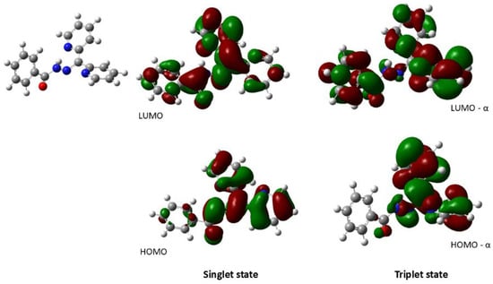
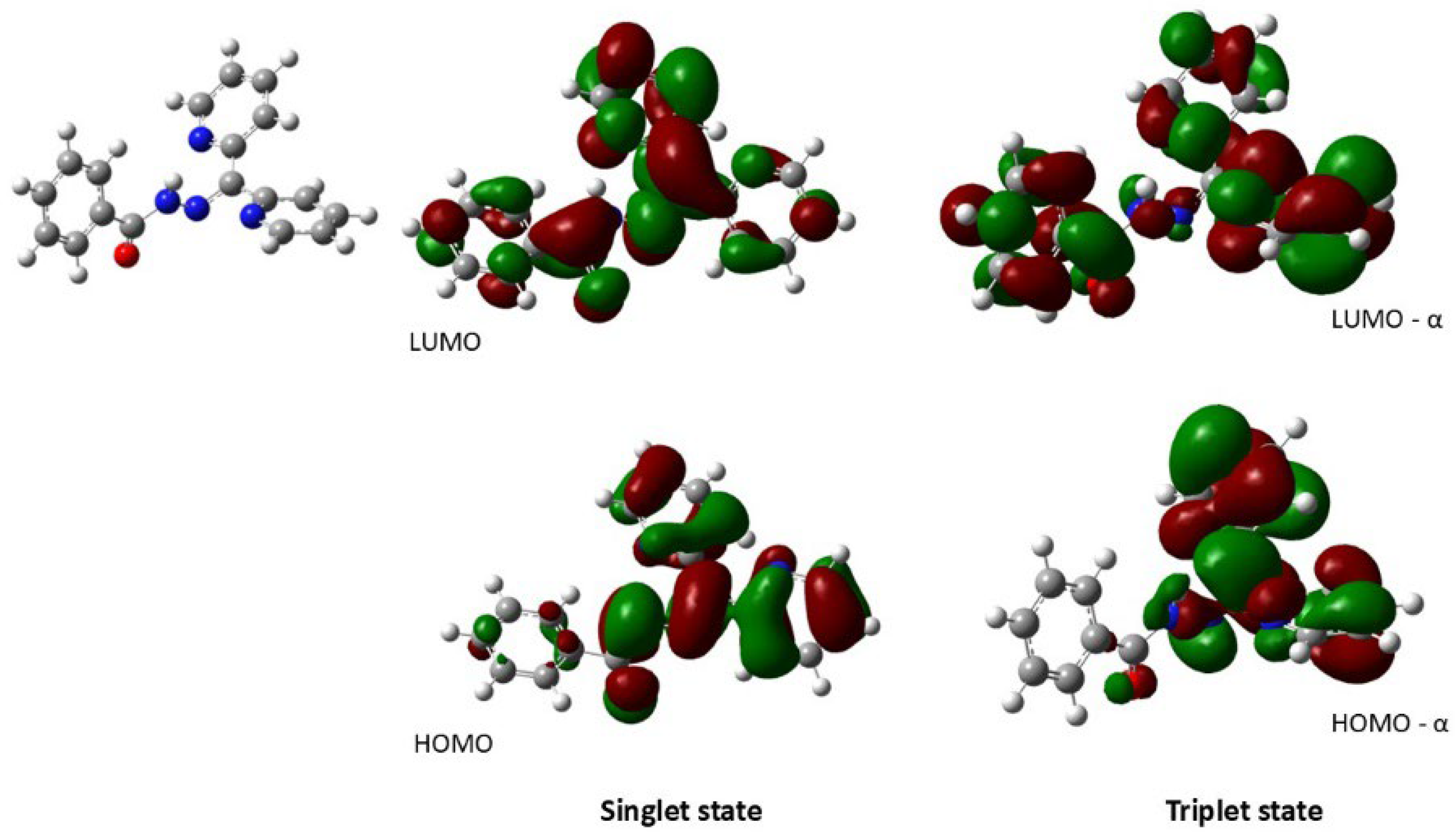
DFT calculations: The time-dependent DFT calculations for ligand L were performed at the UAPFD level with a 6-311 + g(2d,p) basis in the gas phase. The frontier orbitals (LUMO and HOMO) of the ligand are shown in Figure 4. The electron density of the HOMO orbitals of the ligand is mainly located on the pyridyl rings, and the LUMO orbitals are located on the whole ligand’s backbone. The lower-energy S0 ⟶ S1 transition (HOMO to LUMO) equals 338.40 nm (3.66 eV), as shown in Table 4. These transitions are due to intra-ligand charge transfer transitions, ILCTs. However, the calculated T1 state for L is 567.81 nm (2.18 eV). The LUMO orbitals of the excited T1 transitions are mainly found along the ligand’s backbone, primarily on the donor atoms. Coordination to the Ln(III) metals will certainly proceed to an efficient energy transfer process through the T1 ⟶ *f pathway.
Figure 4.
Molecular orbital plots of the HOMO and LUMO orbitals of the singlet and triplet state levels of ligand L in the gas phase.
Table 4.
Summary of the calculated electronic transitions for ligand L at the optimized ground state geometry of UAPFD with a 6-311 + g(2d,p) basis in the gas phase.
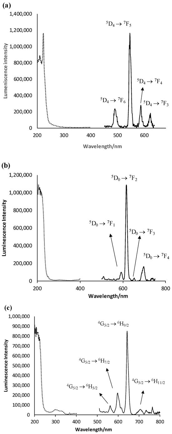
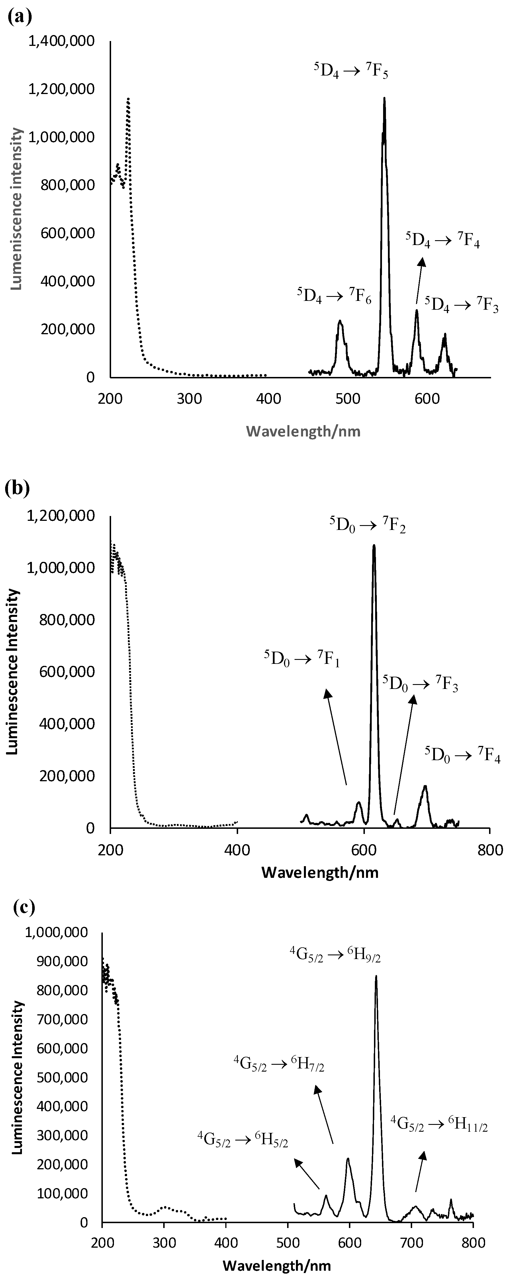
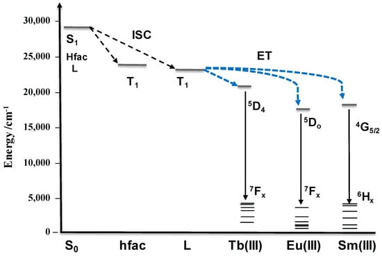
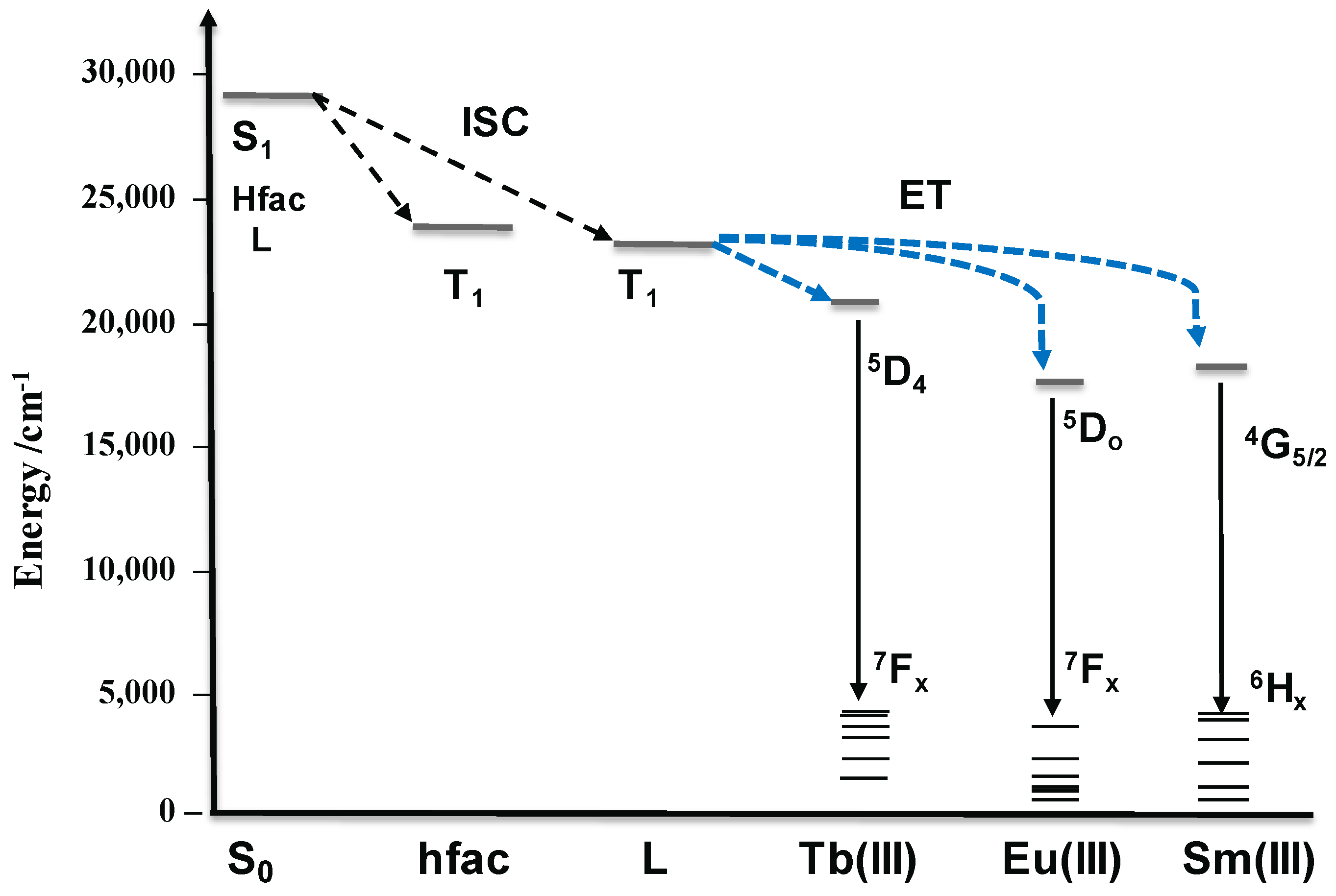
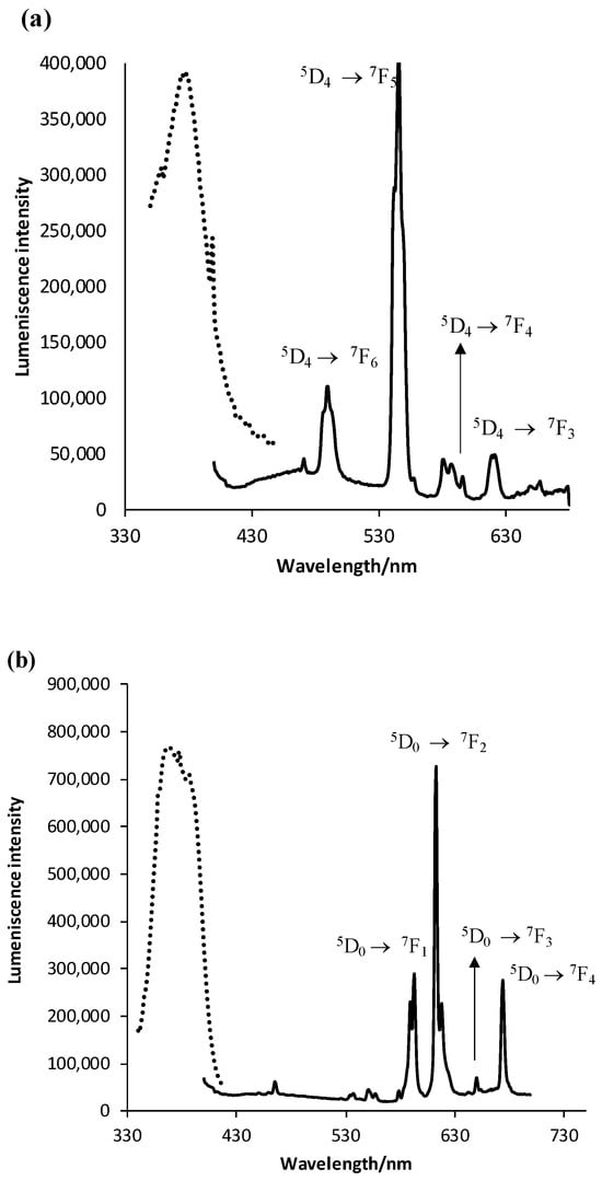
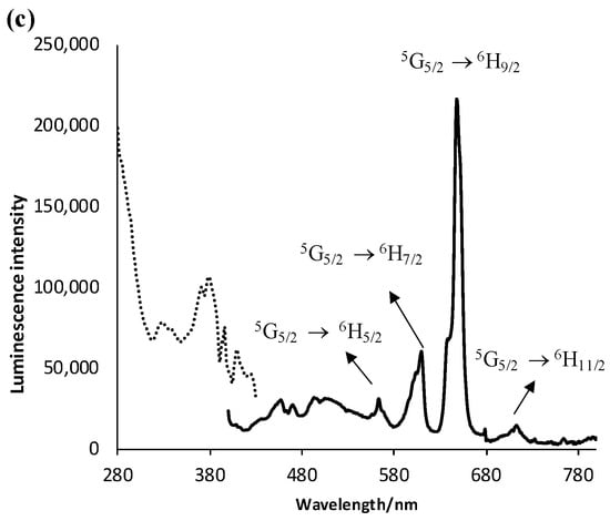
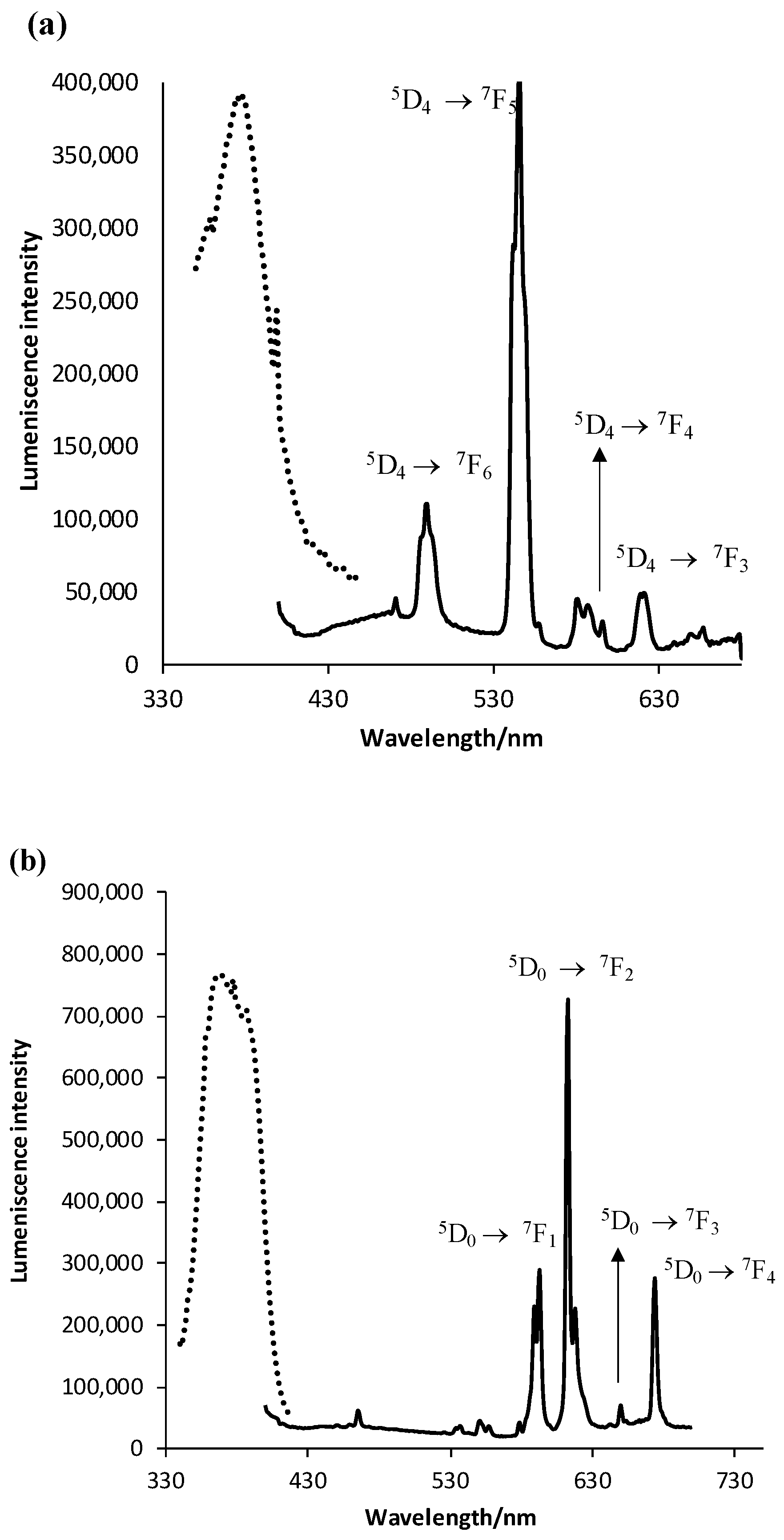
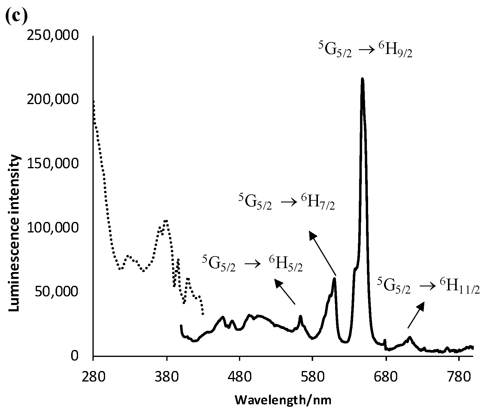
Metal-centered luminescence: The excitation and emission spectra of the Ln(III) complexes SmL, EuL and TbL in the solid state at RT are shown in Figure 5. Interestingly, under a UV lamp, all the complexes exhibit an intense colored light emission from the lanthanide ion. The excitation of TbL at 382 nm shows green emission from the characteristic luminescent 5D4 ⟶ 7F6-2 transitions. The luminescent lines are made from the combination of magnetic and electric dipole transitions within Tb(III); therefore, no possible indication of the coordination environment of Tb(III) could be seen. However, the energy gap between the T1 state and the lowest excited state of Tb(III), ΔE(T1– 5D4), equals 1500 cm−1. This amount of energy perfectly initiates the sensitization process of the metal and eliminates non-radiative processes; hence, it explains the intensive emission from Tb(III) [23]. The energy transfer pathways are shown in the Jablonski diagram in Figure 6.
Figure 5.
The excitation and luminescence spectra of the Ln(III) complexes (a) TbL, (b) EuL and (c) SmL in the solid state at room temperature.
Figure 6.
Schematic energy level diagram of the sensitization pathway for Tb(III), Eu(III) and Sm(III) centers via T1 of ligand L.
The excitation of EuL at 385 nm exhibits intense red luminescence from the 5D0 ⟶ 7F0-4 transitions. To comprehend the sensitization mechanism, the energy gap between the lowest excited state of Eu(III) and the T1 state, viz ΔE(T1– 5D0), is calculated. The ΔE(T1– 5D0) gap is 4400 cm−1. Again, this amount of energy for T1 (21,900 cm−1) is too high to effectively deactivate any non-radiative processes from the 5D0 level of Eu(III). Therefore, the energy transfer to Eu(III) is not effective.
In contrast, the excitation of SmL at 387 nm exhibits weak luminescence from the 4G5/2 ⟶ 6Hj (j = 5/2, 7/2, 9/2 and 11/2) transitions. Interestingly, the electric dipole transition of 4D5/2 ⟶ 6H9/2 appears split at 643 nm. In order to comprehend the sensitization mechanism, the energy gap between the lowest excited state of Sm(III) and the T1 state, viz ΔE(T1– 5G5/2) is calculated. The ΔE(T1– 5G5/2) gap is 4200 cm−1. This means that the T1 value (21,900 cm−1) is too high to effectively induce the radiative processes from the 5G5/2 level of Sm(III).
The excitation spectra of the LnL SB complexes were also investigated by exciting the metal center at the intense f-f* transitions. The spectra are essentially identical to their respective absorption spectra, showing a weak broad band centered at 385 nm. These results confirm the electron population in the excited Ln(III) centers by the excited T1 state of L [24].
The luminescence decay curves of the radiative lifetime (τobs) for each Ln(III) center at the intense f-f* transitions were also recorded in the solid state. The observed lifetime of TbL from the 5D4 ⟶ 7F5 transition is the longest, equaling 0.505 ms. The Tb(III) emission is very intense due to the perfect energy match between the T1 and 5D4 states, which eliminates any non-radiative deactivation pathways during the excitation process. On the other hand, EuL exhibits a shorter lifetime value of 0.349 ms at 5D0 ⟶ 7F2. Lastly, the luminescence lifetime from Sm(III) is very poor, and the QSm is almost quenched. As discussed earlier, the energy gap between the excited T1 and 5G5/2 states for Sm(III) is comparable to the energy gap between the excited T1 and 5D0 states for Eu(III), viz 4400 and 4600 cm−1, respectively. But unfortunately, the sensitization of Sm(III) is not efficient. In fact, Sm(III) complexes are known to have very weak luminescence and lifetime values because Sm(III) is more sensitive to the multi-phonon relaxation process in its complexes [25].
Ln(III)-PMMA polymeric films
The Ln(III)-SB complexes (2 wt.%) were blended in a PMMA solution to produce transparent polymeric films. The excitation and emission spectra of the Ln(III)-PMMA films in the solid state at RT are shown in Figure 7. In general, the luminescence intensity and lifetime measurements for the PMMA polymers are enhanced. The excitation of TbL-PMMA at 381 nm shows that the intense luminescence lines from the 5D4 ⟶ 7F6-2 transitions are considerably broadened. A weak broad band centered at 480 nm is observed due to emission from the organic PMMA polymer. However, the excitation of EuL-PMMA at 381 nm exhibits sharp lines from the 5D0 ⟶ 7F1-4 transitions, in addition to a relatively weak broad band from the PMMA matrix. On the other hand, the excitation of SmL-PMMA at 382 nm exhibits luminescence lines from the 4G5/2 ⟶ 6Hj (j = 5/2, 7/2, 9/2 and 11/2) transitions combined with a broad weak band from PMMA centered at 480 nm. Nevertheless, the excitation spectra of the LnL-PMMA polymers were obtained by exciting the metal center at the intense f-f* transitions. Generally, the spectra are also similar to their respective absorption spectra, which confirm the energy transfer from the excited T1 state of L to the lowest excited state of each Ln(III) center.
Figure 7.
The excitation and luminescence spectra of the doped LnL-PMMA polymers (a) TbL-PMMA, (b) EuL-PMMA and (c) SmL-PMMA in the solid state at room temperature.
In order to monitor the local symmetry of the Ln(III) centers and the molecular interactions of the LnL complexes within the PMMA matrix, the luminescence properties of the EuL-PMMA polymer were investigated. In fact, the electric dipole transition 5D0 ⟶ 7F2 is very sensitive to changes in the coordination symmetry of Eu(III). Meanwhile, the magnetic dipole transition 5D0 ⟶ 7F1 is not affected by such changes and could be used as a reference to monitor the photophysical changes in the 5D0 ⟶ 7F2 transition. The more intense the electric dipole transition, the more nonsymmetric the environment around the metal center [23]. The intensity ratios (ηEu) between the intensities of (5D0 ⟶ 7F2)/(5D0 ⟶ 7F1) for EuL and EuL-PMMA were calculated, which are equal to 12.9 and 6.8, respectively. These values are significantly high, pointing to a non-centromeric coordination environment around the Eu(III) center. The new changes in the site symmetry of Eu(III) in EuL-PMMA point to new changes in the coordination polyhedron of the Eu(III) center. Intensive intermolecular interactions occur within the polymer, leading to distortions in the coordination sphere around Eu(III) [26].
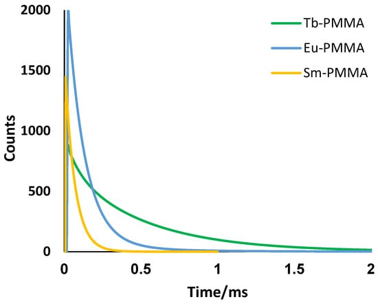
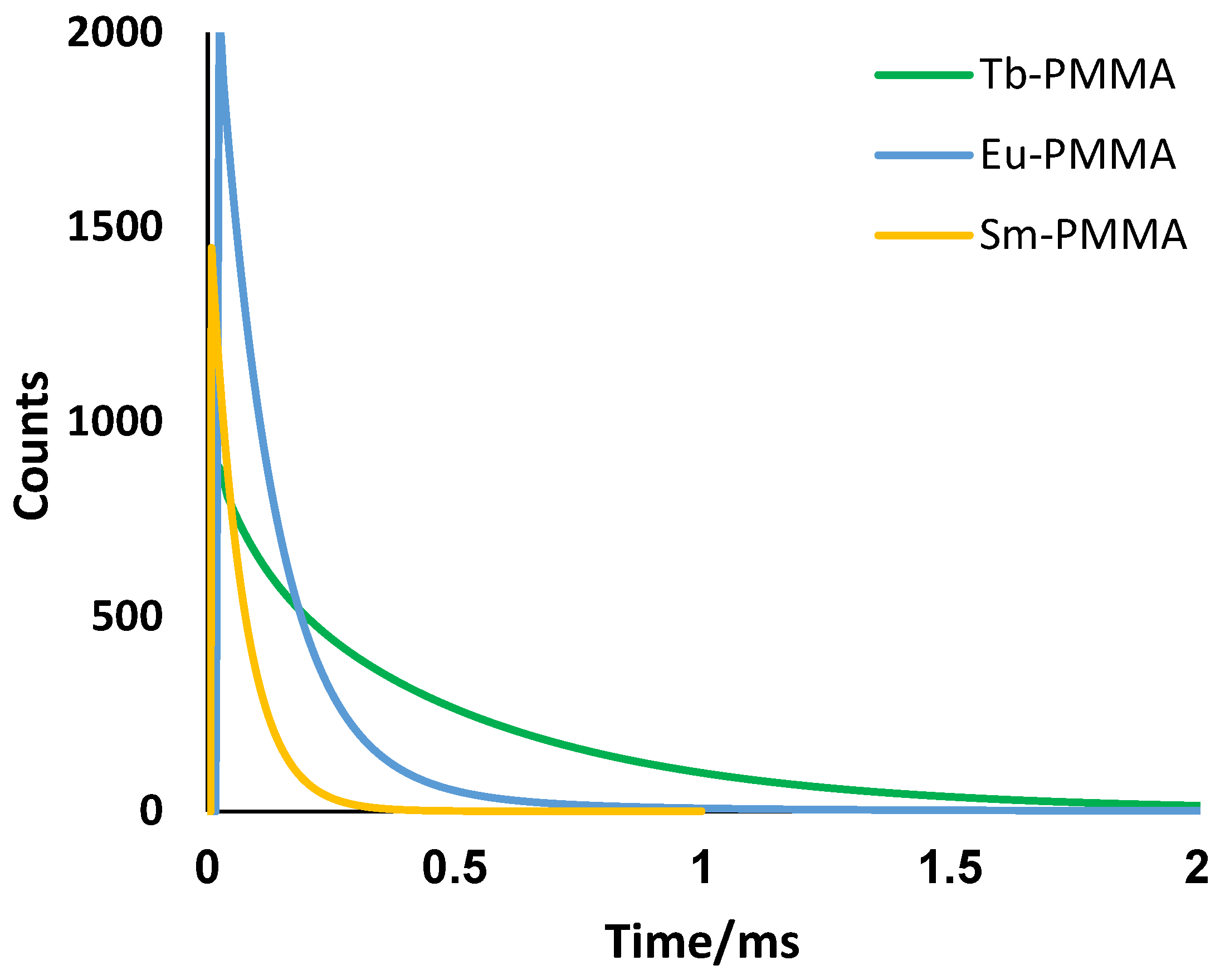
Furthermore, the luminescence decay curves for the TbL-PMMA, EuL-PMMA and SmL-PMMA polymers at the intense f-f* transitions were recorded in the solid state, as shown in Figure 8. It is interesting that the lifetime increased from each Ln(III) metal center. To clearly understand the extent of the PMMA matrix on the energy transfer process, we utilized the steady-state emission spectra and τobs for EuL and the EuL-PMMA polymer to calculate important photophysical parameters, such as the Judd–Ofelt (J-O) parameters (Ωx), radiative decay rate (kR), non-radiative decay rate (kNR) and intrinsic emission quantum yield (QEu) for the Eu(III) center, as shown in Table 5. The experimental Judd–Ofelt (J-O) parameter values of Ω2 and Ω4, were calculated from the luminescence intensity ratios of the electric–dipole transitions 5D0 → 7FJ (J= 2 and 4) and the magnetic–dipole transition 5D0 → 7F1 according to the equation [27]:
where χ is the Lorentz local field correction factor, χ = n(n2 + 2)2/9, n is the refractive index, e is the elementary charge and ℏ is Planck’s constant. The quantity |⟨5D0||U(λ)||7Fλ⟩|2 is the squared reduced matrix element, whose values are equal to 0.0032 and 0.0023 for λ = 2 and 4, respectively [27]. The χ values were calculated using the index of refraction, n = 1.50 and 1.49, for EuL and EuL-PMMA, respectively. Usually, large values for the Ω2 parameter are associated with (i) an enhanced ligand covalency, (ii) a lower local asymmetric coordination environment for the Eu(III) center [15] and (iii) the hypersensitive nature of the 5D0 → 7F2 transition [28]. The values of Ω2 for Eu(III) in EuL and EuL-PMMA equal 3.65 × 10⁻²⁰ cm2 and 15.89 × 10⁻²⁰ cm2, respectively. The value of Ω2 for Eu(III) in the doped PMMA polymer is extremely large, indicating strong hydrogen bonding interactions between L and the oxygen atoms in PMMA. Consequently, the polarizability of the coordinating ligands increases, causing stronger Eu(III)–L covalent interactions [29]. Furthermore, the high value of Ω2 for Eu(III) points to an asymmetric coordination environment around the Eu(III) center. These results agree with the obtained intensity ratio (ηEu) values.
Figure 8.
Luminescence decay curves of TbL-PMMA at 545 nm (Green), EuL-PMMA at 613 nm (blue) and SmL-PMMA at 610 nm (orange) in the solid state at room temperature.
Table 5.
Summary of the experimental photophysical parameters of the Eu(III) CPs at RT.
The Judd–Ofelt parameter, Ω4, for the Eu(III) center was also used to investigate the (i) structural changes and (ii) the local electron density of the coordinating ligands [30]. The calculated Ω4 parameter for EuL-PMMA similarly increased and accordingly confirmed the molecular packing around the complex, as well as the rigidity of the polymer. Nevertheless, the new environment and surrounding media around Eu(III) in the polymer extremely affected the radiative (AR) and non-radiative (ANR) decay rates from the metal. These values were determined from the emission spectra of the Eu(III) centers by applying the following equations:
The AR and ANR values grant a deep insight to the energy transfer process from L to the excited 5D0 level of Eu(III). For instance, the EuL complex exhibited a large non-radiative rate of ANR = 2380.43 s−1 compared to the EuL-PMMA polymer’s rate of ANR = 2204.10 s−1. Furthermore, the radiative rate from the metal in EuL-PMMA, ANR = 661.22 s−1, is increasing. Obviously, the PMMA matrix is involved in the energy transfer process to Eu(III) [31]. The PMMA matrix would induce the overall structural rigidity of the polymers, resulting in a rapid and intense photon population from T1 to the excited 5D0 level of Eu(III). Consequently, the non-radiative processes are suppressed, and more photons are relaxed to the ground state [32,33]. This behavior is further reflected in the intrinsic emission quantum yield of (QEu). The term is defined as the ratio between the radiative (AR) and the sum of the radiative (AR) and non-radiative (ANR) decay rates as follows:
Interestingly, the intrinsic emission quantum yield of EuL-PMMA (QEu = 23) is 3 times more than the value of EuL (QEu = 8). Usually, the QEu value refers to the extent of non-radiative transitions in the excited states during the sensitization process. The higher QEu value of the EuL-PMMA polymer reflects the vital role that the structure’s rigidity plays in the energy transfer process [34]. Once the 5D0 excited state of Eu(III) is populated, the induced structure’s rigidity minimizes the non-radiative deactivation pathways for Eu(III) (lower ANR rate); consequently, the energy transfer from L to the 5D0 level of Eu(III) is induced. Subsequently, the EuL-PMMA polymer undergoes an efficient sensitization process.
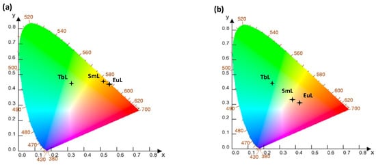
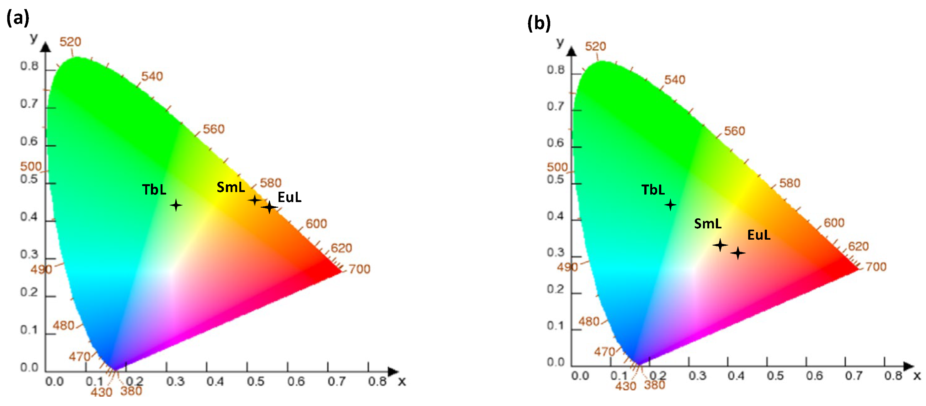
Emission colors: To estimate the capability of the LnL-PMMA polymers as phosphors in opto-electronic devices, their color coordinates were calculated using a CIE 1931 colorimetric diagram, as shown in Figure 9. The colorimetric coordinates are also presented in Table S3. TbL-PMMA emits green light at CIE coordinates (0.2655, 0.4212), while EuL-PMMA exhibits significant red emission at CIE coordinates (0.4331, 0.3091). These results suggest that these polymers are desirable for opto-electronic applications such as LED devices.
Figure 9.
CIE 1931 chromaticity diagrams of the (a) Ln(III) complexes and (b) LnL-PMMA polymers, all in the solid state at RT.
3. Conclusions
In summary, four Ln-SB complexes, namely, TbL, EuL, SmL and GdL, have been successfully synthesized and thoroughly characterized. TbL is extensively luminescent due to an efficient overlap between its excited 5D4 state and the T1 (21,900 cm−1) excited states of L. The doping of LnL complexes into the PMMA polymeric films was confirmed. Hydrogen bonding interactions take place between the Ln-SB complexes and the PMMA matrix, as confirmed by their IR spectroscopy. However, the LnL-PMMA polymers exhibit induced emission and lifetimes from the metals. For instance, the intrinsic emission quantum yield of Eu(III) in EuL-PMMA (23%) is three times higher than in EuL (8%). This behavior is ascribed to the induced rigidity of the polymers that consequently inhibits any internal non-radiative processes during the excited states. Nevertheless, the enhancement in the optical properties and thermal stabilities of TbL-PMMA and EuL-PMMA indicate their potential applications as phosphors in the production of LED devices.
4. Experimental Methods
General details: All organic reagents, metal salts and solvents were purchased from commercial suppliers and used as received without further purification. Infrared spectra for the complexes were recorded on a Bruker FT-IR spectrometer in the range 4000–400 cm−1 (Bruker, Billerica, MA, USA). The mass spectra of the complexes were obtained using Liquid Chromatography–Mass Spectrometry (LC-MS-MS) 6460 Triple Quad LC/MS and 1290 Infinity DAD (Agilent Technologies, Santa Clara, CA, USA). Elemental analysis of carbon, hydrogen and nitrogen was performed using the 2400 CHNS/O Series II Elemental Analyzer (Perkin Elmer, Springfield, IL, USA). TG-DTA analysis was performed on a TA SDT 2960 Analyzer (Artisan Technology, Campaign, IL, USA) in the range 25–1000 °C, with a heating rate of 10 °C/min under N2. Proton Magnetic Resonance (1H NMR) spectra were recorded using an AVANCE-III HD 700 MHz FT-NMR spectrometer (Bruker, Billerica, MA, USA).
5. Synthesis of SB Ligand (L)
A solution of benzoic acid hydrazide (0.073 g; 0.534 mmol) in EtOH (5 mL) was added dropwise to a solution of di(2-pyridyl ketone) (0.10 g; 0.534 mmol) in EtOH (15 mL). Two drops of acetic acid were added to speed up the reaction. After 4 h of refluxing, the reaction mixture was cooled to RT. The resultant bright yellow powder precipitate was filtered off and dried under suction filtration to give a yellow powder. Yield: 0.173 g (90%). C18H14ON4 (302.33 g/mol) Exp (Calc): %C 71.55 (71.34), %H 4.63 (4.48), %N 18.52 (18.00). ESI-MS: m/z 302.900 {L}–. FT–IR cm−1 (solid): 3063 (w), 1682 (s), 1577(w), 1530(w), 1462 (s), 1423(s), 1249(s), 1140 (s), 1074(w), 997(m), 890(m), 808(s), 703(s), 650(s), 578(w) and 512(w). 1H NMR (DMSO4-d6): 9.7 (1H, s, NH), 7.53–8.89 (8H, m, 2py), and 7.41–7.63 (5H, m, ph).
Synthesis of LnL complexes: The general procedure for preparing the Ln(III) complexes involved first dissolving ligand L in MeOH at 80 °C. After that, an Ln(hfac)3·xH2O salt was added to the solution at 50 °C, followed by continued stirring for 24 h. For example, L (0.10 g, 0.33 mmol) was dissolved in MeOH (10 mL), and then Tb(hfac)3·2H2O (0.27, 0.33 mmol) was added. The reaction mixture was stirred for 24 h at 50 °C and left under air diffusion to produce colorless blocks within a week. The complexes are air- and moisture-stable solids, and their characterization data are as follows:
[TbL(hfac)3] (TbL): Colorless blocks. Yield: 0.35 g (90%). TbC33H17N4O7F18 (1112.93 g/mol) exp (calc): %C 36.60 (36.84), %H 1.57 (1.45), and %N 5.18 (4.98). ES-MS m/z 1093.600 {[TbL-H2O]}–. FT-IR cm−1 (solid): 3362(b), 1651(m), 1518(w), 1468(m), 1251(m), 1251(m), 1138(m), 1003(w), 769(m), 656(w), 580(w) and 500 (w).
[EuL(hfac)3] (EuL): Pale yellowish blocks. Yield: 0.30 g (88%). EuC33H17N4O7F18 (1074.90 g/mol) exp (calc): %C 36.84 (36.24), %H 1.58 (1.98), and %N 5.21 (4.99). ES-MS m/z 1103.300 {[EuL- MeOH}+. FT-IR cm−1 (solid): 3241(b), 1653(m), 1536 (w), 1468(m), 1251(m), 1136(s), 1002(w), 771(m), 655(w), 574(w) and 514(w).
[SmL(hfac)3] (SmL): Pale yellowish blocks. Yield: 0.30 g (88%). SmC33H17N4O7F18 (1073.36 g/mol) exp (calc): %C 36.91 (36.92), %H 1.58 (1.55), and %N 5.22 (5.01). ES-MS m/z 1072.900 {[SmL]}–. FT-IR cm−1 (solid): 1661(m), 1501(m), 1489 (m) 1263(m), 1150(m), 1002(w), 771(m), 711(m) and 671(w).
[GdL(hfac)3] (GdL): Pale yellow powder. Yield: 0.55 g (96%). GdC33H17N4O7F18 (1080.31 g/mol) exp (calc): %C 36.96 (36.32), %H 2.38 (2.31), and %N 5.19 (5.07). ES-MS m/z 710.500 {[GdL(hfac)MeOH]}–. FT-IR cm−1 (solid): 3557(br), 1653(m), 1557(w), 1460(s), 1251(m), 1138(m), 1025(m), 796(m), 565(s), 582 (5) and 500(w).
[GdL(NO3)3] (GdL): White powder. Yield: 0.40 g (90%). GdC18H16N7O11·H2O (506.308 g/mol) exp (calc): %C 42.76 (42.69), %H 3.180 (3.185), and %N 19.31 (19.37). ES-MS m/z 667.100 {[Gd(NO3)3(H2O)L]}+. FT-IR cm−1 (solid): 2957(s), 2232(w), 1778(s), 1657(s), 1491(s), 1458(s), 1395(s), 1213(m), 1169(w), 1030(s), 1080(s), 865(s) 778(w), 750(s), and 510(w).
Synthesis of LnL-PMMA polymers: The LnL complexes were blended in a PMMA solution to prepare transparent films suitable for opto-electrical applications as follows: PMMA (0.30 g) was dissolved in acetone (100 mL), followed by the addition of the LnL complex (2% w/w). The resulting solution was heated at 80 °C for 30 min, followed by air diffusion to offer a transparent LnL-PMMA polymer. For example, 0.30 g of PMMA was dissolved in acetone (100 mL); then, TbL (6.02 mg, 0.0054 mmol) was added. The reaction mixture was stirred for 30 min at 80 °C and left under air diffusion to produce a white thin film within one week. The films are air- and moisture-stable solids, and their characterization data are as follows:
TbL-PMMA: White film. Yield: 0.300 g (90%). FT-IR cm−1 (solid): 3440(w), 2948(w), 1719(s), 1458(m), 1434(w), 1382(w), 1287(w), 1196(w), 1142(s), 887(w), 748(w) and 420(w).
EuL-PMMA: White film. Yield: 0.304 g (92%). FT-IR cm−1 (solid): 3426(w), 2937(w), 1721(s), 1452(m), 1442(w), 1430(w), 1239(w), 1155(w), 1138(s), 887(w), 746(w) and 410(w).
SmL-PMMA: White film. Yield: 0.301 g (92%). FT-IR cm−1 (solid): 3428(w), 2942(w), 1723(s), 1462(m), 1440(w), 1378(w), 1241(w), 1167(w), 1140(s), 890(w) and 748(w).
GdL-PMMA: White film. Yield: 0.302 g (93%). FT-IR cm−1 (solid): 3430(w), 2950(w), 1724(s), 1460(m), 1434(w), 1386(w), 1251(w), 1161(w), 1141(s), 886(w), 751(w) and 419(w).
Single-crystal X-ray diffraction analyses: Data for single crystals of TbC were collected at RT using a IPDS II diffractometer (STOE, Darmstadt, Germany) equipped with Mo-Kα radiation. The data were integrated using the Stoe X-AREA 1.30 [35] software package. The numerical absorption correction was applied using the X-RED 1.28 software package [36]. The data were corrected for Lorentz and polarization effects. The structures were solved by direct methods and subsequent difference Fourier maps and then refined on F2 by a full-matrix least-squares procedure using anisotropic displacement parameters with SHELXS-97 [37]. All hydrogen atoms were positioned geometrically in idealized positions and refined using the riding model approximation, with Uiso(H) = 1.2 Ueq(C). All refinements were performed using the WinGX 2018.3 [38] suite of programs. For the molecular graphics, the program’s Mercury package 2026 4.0 was used [39]. The non-hydrogen atoms were refined as anisotropic.
Photophysical measurements: UV–visible absorption spectra were recorded on a Varian Cary 5000 UV–visible spectrophotometer (Mettler Toledo, Columbus, OH, US) in the range of 250–800 nm. Quartz cuvettes of 1 cm path length were used, and solvent background corrections were applied. Steady-state emission spectra and phosphorescence lifetimes (τobs) for Ln(III) f–f transitions in solid states were recorded on an FS5 fluorimeter (Edinburgh Instruments, Livingston, UK) at RT. Lifetimes were measured by recording the decay at the maximum of the emission spectra. The goodness of fit was judged by the value of the reduced chi-squared (χ2). The triplet state energies of the SB ligands were recorded from their Gd(III) complexes in ethanol (1 × 10⁻⁵ M) at 77 K using an NMR tube.
Supplementary Materials
The following supporting information can be downloaded at https://www.mdpi.com/article/10.3390/cryst16080548/s1: Figure S1: Mass spectrometry spectra of ligand L and the LnL complexes. Figure S2: 1H NMR (500 MHz) spectrum of ligand L. Figures S3–S7: IR spectra of the ligand and complexes in the solid state. Figure S8: Absorption spectra of different Gd(III) complexes in ethanol. Summary of TD DFT results and comments on Checkcif alerts. Table S1: Mass-to-charge (m/z) of the fragments of L and the Ln(III) complexes. Table S2: Summary of the important IR stretching frequencies. Table S3: Summary of the CIE1931 chromaticity coordinates for the LnL complexes and the LnL-PMMA polymers in the solid.
Author Contributions
N.A.A.R., data curation and writing—original draft; J.H., data curation and visualization; N.K.A.-R., project administration, funding acquisition, conceptualization, and writing—review and editing. All authors have read and agreed to the published version of the manuscript.
Funding
This work was supported by Sultan Qaboos University (Grant No. IG/SCI/CHEM/23/01).
Data Availability Statement
Data will be made available on request from the corresponding authors.
Acknowledgments
The corresponding author greatly appreciates the financial support from Sultan Qaboos University, Oman, through the internal grant and the PhD scholarship for Najat A. Al Riyami.
Conflicts of Interest
The authors declare no conflicts of interest.
References
- Zhao, W.; Shao, H.; Yu, G.; Hou, Y.; Wang, S. The coordination and luminescence of the Eu (III) complexes with the polymers (PMMA, PVP). Polymers 2018, 10, 508. [Google Scholar] [CrossRef] [Scilit] [PubMed]
- Li, L.; Feng, S.; Liu, H. Hybrid lanthanide complexes based on a novel β-diketone functionalized polyhedral oligomeric silsesquioxane (POSS) and their nanocomposites with PMMA via in situ polymerization. RSC Adv. 2014, 4, 39132–39139. [Google Scholar] [CrossRef] [Scilit]
- Al-Rasbi, N.K.; Adams, H.; Suliman, F.O. Synthesis, structure and tunable white-light emission of dinuclear Eu (III) Schiff base complex. Dye. Pigment. 2014, 104, 83–88. [Google Scholar] [CrossRef] [Scilit]
- Incel, A.; Emirdag-Eanes, M.; McMillen, C.D.; Demir, M.M. Integration of triboluminescent EuD4TEA crystals to transparent polymers: Impact sensor application. ACS Appl. Mater. Interfaces 2017, 9, 6488–6496. [Google Scholar] [CrossRef] [Scilit] [PubMed]
- Cardoso Dos Santos, M.; Runser, A.; Bartenlian, H.; Nonat, A.M.; Charbonnière, L.J.; Klymchenko, A.S.; Reisch, A. Lanthanide-complex-loaded polymer nanoparticles for background-free single-particle and live-cell imaging. Chem. Mater. 2019, 31, 4034–4041. [Google Scholar] [CrossRef] [Scilit]
- Marinotto, D.; Marin, C.A.; Rau, I.; Colombo, A.; Fagnani, F.; Roberto, D.; Dragonetti, C. Large enhancement of the luminescence properties of an Eu (III) dye upon association with the DNA-CTMA matrix. Molecules 2025, 30, 1395. [Google Scholar] [CrossRef] [Scilit] [PubMed]
- Yang, D.; Li, H.; Li, H. Recent advances in the luminescent polymers containing lanthanide complexes. Coord. Chem. Rev. 2024, 514, 215875. [Google Scholar] [CrossRef] [Scilit]
- Shiraishi, N.; Iikura, D.; Karashimada, R.; Iki, N. Doping a highly charged, water-soluble lanthanide complex into a hydrophobic polymer for the preparation of luminescent films. J. Lumin. 2024, 269, 120521. [Google Scholar] [CrossRef] [Scilit]
- Fu, G.; Su, P.; Lü, X.; Wong, W.-K. NIR luminescence comparison of Zn-Nd Schiff-base complex doped or single-nodal grafted into PMMA. Inorg. Chem. Comm. 2017, 76, 12–14. [Google Scholar] [CrossRef] [Scilit]
- Gil-Kowalczyk, M.; Łyszczek, R.; Jusza, A.; Piramidowicz, R. Thermal, spectroscopy and luminescent characterization of hybrid PMMA/lanthanide complex materials. Materials 2021, 14, 3156. [Google Scholar] [CrossRef] [Scilit] [PubMed]
- Tilioua, A. Investigation of the thermo-physical properties of poly (methyl methacrylate)-based Plexiglass to improve the performance of solar cells. Mater. Sci. Energy Technol. 2021, 4, 349–356. [Google Scholar] [CrossRef] [Scilit]
- Al Riyami, N.A.; Al-Rasbi, N.K. Factors that influence the energy transfer pathways in luminescent lanthanide Schiff base complexes. Inorg. Chim. Acta 2024, 572, 122324. [Google Scholar] [CrossRef] [Scilit]
- Hegarty, I.N.; Bradberry, S.J.; Lovitt, J.I.; Delente, J.M.; Willis-Fox, N.; Daly, R.; Gunnlaugsson, T. Luminescent (metallo-supramolecular) cross-linked lanthanide hydrogels from a btp (2, 3-bis (1, 2, 3-triazol-4-yl) picolinamide) monomer give rise to strong Tb (iii) and Eu (iii) centred emissions. Mater. Chem. Front. 2023, 7, 906–916. [Google Scholar]
- Zhang, H.J.; Fan, R.Q.; Wang, X.M.; Wang, P.; Wang, Y.L.; Yang, Y.L. Preparation, characterization, and properties of PMMA-doped polymer film materials: A study on the effect of terbium ions on luminescence and lifetime enhancement. Dalton Trans. 2015, 44, 2871–2879. [Google Scholar] [CrossRef] [Scilit] [PubMed]
- Gibelli, E.B.; Kai, J.; Teotonio, E.E.; Malta, O.L.; Felinto, M.C.; Brito, H.F. Photoluminescent PMMA polymer films doped with Eu3+-β-diketonate crown ether complex. J. Photochem. Photobiol. A Chem. 2013, 251, 154–159. [Google Scholar] [CrossRef] [Scilit]
- Garcia-Torres, J.; Bosch-Jimenez, P.; Torralba-Calleja, E.; Kennedy, M.; Ahmed, H.; Doran, J.; Della Pirriera, M. Modulating the photoluminescence of europium-based emitting polymers: Influence of the matrix on the photophysical properties. J. Photochem. Photobiol. A Chem. 2014, 275, 103–113. [Google Scholar] [CrossRef] [Scilit]
- Sarkar, U.; Bhattacharya, S. White light emission from luminescent lanthanide molecular complex/terbium doped graphene quantum dots/PMMA composites. Small 2025, 21, 2503419. [Google Scholar] [CrossRef] [Scilit] [PubMed]
- Chen, W.; Fan, R.; Zhang, H.; Dong, Y.; Wang, P.; Yang, Y. Tunable white-light emission PMMA-supported film materials containing lanthanide coordination polymers: Preparation, characterization, and properties. Dalton Trans. 2017, 46, 4265–4277. [Google Scholar] [CrossRef] [Scilit] [PubMed]
- Liu, J.-X.; Mei, S.-L.; Chen, X.-H.; Yao, C.-J. Recent advances of near-infrared (NIR) emissive metal complexes bridged by ligands with N-and/or O-donor sites. Crystals 2021, 11, 155. [Google Scholar] [CrossRef] [Scilit]
- Congiu, M.; Alamiry, M.; Moudam, O.; Ciorba, S.; Richardson, P.R.; Maron, L.; Jones, A.C.; Richards, B.S.; Robertson, N. Preparation and photophysical studies of [Ln (hfac) 3DPEPO], Ln= Eu, Tb, Yb, Nd, Gd; interpretation of total photoluminescence quantum yields. Dalton Trans. 2013, 42, 13537–13545. [Google Scholar] [CrossRef] [Scilit] [PubMed]
- Li, Y.; Liu, Y.-F.; Yu, C.-L.; Zhang, S.M.; Wang, K. Syntheses, crystal structures, and luminescence of Hexafluoroacetylacetone Dinuclear lanthanide complexes bridged by “Back-to-Back” Bisterpyridine. Cryst. Growth Des. 2024, 24, 6164–6178. [Google Scholar] [CrossRef] [Scilit]
- Zaim, A.; Nozary, H.; Juenee, L.; Besnard, C.; Lemonnier, J.-F.; Petoud, S.; Piguet, C. N-heterocyclic Tridentate Aromatic Ligands Bound to [Ln (hfac) 3] Units: Thermodynamic, Structural and Luminescent Properties. Eur. J. Inorg. Chem. 2012, 18, 7155–7168. [Google Scholar] [CrossRef] [Scilit] [PubMed]
- Abusail, N.; Al Riyami, N.A.; Khan, M.; Al-Rasbi, N.K. An efficient pink luminescent Eu (iii) coordination polymer excited on a blue LED chip. J. Mater. Chem. C 2023, 11, 4094–4103. [Google Scholar] [CrossRef] [Scilit]
- Ahmed, Z.; Carvalho, R.D.S.; Dos Santos, A.M.; Gambassi, F.; Bandini, E.; Marvelli, L.; Cremona, M. Highly luminescent europium (III) complexes in solution and PMMA-doped films for bright red electroluminescent devices. Molecules 2023, 28, 4371. [Google Scholar] [CrossRef] [Scilit] [PubMed]
- Arrico, L.; De Rosa, C.; Di Bari, L.; Melchior, A.; Piccinelli, F. Effect of the counterion on circularly polarized luminescence of europium (III) and samarium (III) complexes. Inorg. Chem. 2020, 59, 5050–5062. [Google Scholar] [CrossRef] [Scilit] [PubMed]
- Saraiva, L.F.; Beltrame, A.C.F.; Bispo-Jr, A.G.; Canisares, F.S.M.; Carneiro Neto, A.N.; Moura, R.T., Jr.; Kraka, E.; Lima, S.A.M.; Pires, A.M. Unfolding the Photophysical Behavior of Luminescent Polymeric Films Containing β-Diketonate Tetrakis EuIII Complexes via Multilayer Quantum Mechanics. ACS Omega 2025, 10, 30563–30575. [Google Scholar] [CrossRef] [Scilit] [PubMed]
- Blois, L.; Carneiro Neto, A.N.; Longo, R.L.; Costa, I.F.; Paolini, T.B.; Brito, H.F.; Malta, O.L. On the Experimental Determination of 4f–4f Intensity Parameters from the Emission Spectra of Europium (III) Compounds. Opt. Spectrosc. 2022, 130, 10–17. [Google Scholar] [CrossRef] [Scilit]
- Kiran, R.; Shenoy, V.; Sayyed, M.I.; Almuqrin, A.H.; Kamath, S.D. Judd–Ofelt analysis and the spectroscopic elucidation of β-BaB2O4:Eu3+ phosphors for optoelectronic applications. RSC Adv. 2025, 15, 43377–43389. [Google Scholar] [CrossRef] [Scilit] [PubMed]
- Assunção, I.P.; Costa, I.F.; Blois, L.; Felinto, M.C.F.; Deflon, V.M.; Ando, R.A.; Brito, H.F. High red luminescence intensity under sunlight exposure of a PMMA polymer doped with a tetrakis Eu3+ β-diketonate complex containing a benzimidazolium counterion. RSC Adv. 2025, 15, 435–445. [Google Scholar] [CrossRef] [Scilit] [PubMed]
- Langyan, R. Synthesis, characterization, luminescence study and Judd-Ofelt analysis of highly luminescent Eu(III) complexes. Next Mater. 2025, 8, 100749. [Google Scholar] [CrossRef] [Scilit]
- Trejo-García, P.M.; Palomino-Merino, R.; De la Cruz, J.; Espinosa, J.E.; Aceves, R.; Moreno-Barbosa, E.; Moreno, O.P. Luminescent Properties of Eu3+-Doped Hybrid SiO2-PMMA Material for Photonic Applications. Micromachines 2018, 9, 441. [Google Scholar] [CrossRef] [Scilit] [PubMed]
- Assunção, I.P.; Costa, I.F.; Santos, P.R.; Teotonio, E.E.; Felinto, M.C.F.; Kynast, U.; Brito, H.F. Luminescent Analysis of Eu3+ and Tb3+ Flufenamate Complexes Doped in PMMA Polymer: Unexpected Terbium Green Emission under Sunlight Exposure. ACS Appl. Opt. Mater. 2022, 1, 354–366. [Google Scholar] [CrossRef] [Scilit]
- Sarkar, U.; Singh, M.; Adak, M.K.; Bhattacharya, S. Lanthanide-Tetradentate Schiff Base-PMMA Composite Films for Tunable White Light Emission. Adv. Opt. Mater. 2025, 13, 2402592. [Google Scholar] [CrossRef] [Scilit]
- Zhang, J.L.; Chen, B.W.; Luo, X.; Du, K. Eu(III) complex-doped PMMA having fast radiation rate and high emission quantum efficiency. Chin. Chem. Lett. 2012, 23, 945–948. [Google Scholar] [CrossRef] [Scilit]
- Stoe Cie GmbH. X–AREA: Program for the Acquisition and Analysis of Data, Version 1.30; Stoe Cie GmbH: Darmstadt, Germany, 2005.
- Stoe Cie GmbH. & X-RED. 1.28 B: Program for Data Reduction and Absorption Correction; Stoe & Cie GmbH: Darmstadt, Germany, 2005. [Google Scholar]
- Sheldrick, G.M. A short history of SHELX. Acta Cryst. 2008, A64, 112–122. [Google Scholar] [CrossRef] [Scilit] [PubMed]
- Farrugia, L.J. WinGX suite for small-molecule single-crystal crystallography. J. Appl. Crystallogr. 2012, 45, 849–854. [Google Scholar]
- Macrae, C.F.; Sovago, I.; Cottrell, S.J.; Galek, P.T.A.; McCabe, P.; Pidcock, E.; Platings, M.; Shields, G.P.; Stevens, J.S.; Towler, M.; et al. Mercury 4.0: From visualization to analysis, design and prediction. Appl. Cryst. 2020, 53, 226–235. [Google Scholar] [CrossRef] [Scilit] [PubMed]

Disclaimer/Publisher’s Note: The statements, opinions and data contained in all publications are solely those of the individual author(s) and contributor(s) and not of MDPI and/or the editor(s). MDPI and/or the editor(s) disclaim responsibility for any injury to people or property resulting from any ideas, methods, instructions or products referred to in the content. |
© 2026 by the authors. Licensee MDPI, Basel, Switzerland. This article is an open access article distributed under the terms and conditions of the Creative Commons Attribution (CC BY) license.