


Abstract
The study aimed to evaluate the effect of chlorhexidine (CHX), chlorin p6-mediated photodynamic therapy (PDT), magnesium oxide nanoparticles (MgONPs), and Clp6-functionalized MgONPs on smear layer removal and shear bond strength of a two-step etch-and-rinse adhesive to caries-affected dentin. Seventy-five human permanent molars with occlusal carious lesions and ICDAS scores of four and five were included. Twenty-five samples were used to prepare dentin discs 2 mm in thickness. The remaining samples, along with 25 discs, were arbitrarily allocated into five disinfectant groups, with n = 15 per group (10 teeth and 5 discs). Group I: Control, Group II: 2% CHX, Group III: Clp6-mediated PDT, Group IV: MgONPs, and Group V: Clp6-functionalized MgONPs. SL removal assessment, nanoparticle characterization, and EDX were performed using SEM. Fifty CAD were etched, followed by fifth-generation adhesive application and composite build-up. SBS and failure modes were evaluated with a universal testing machine and stereomicroscope, respectively. Group 4 (MgONPs) specimens displayed the maximum cleaning of SL (1.11 ± 0.13) and the highest SBS (10.32 ± 0.18 MPa). However, minimum SL removal (2.87 ± 0.94) and bond strength (7.42 ± 0.25 MPa) were exhibited by Group 1 (No disinfectant) samples. MgONPs possess the potential to be used as a cavity disinfectant, as they efficiently remove SL from CAD and augment the bond integrity outcomes.
1. Introduction
The partial caries removal (PCR) technique is grounded in the principles of minimally invasive dentistry and is designed to prevent unnecessary pulp exposure, thereby preserving pulp vitality [1]. In this approach, only the outermost layer of heavily infected and irreversibly demineralized dentin is excavated, while the underlying caries-affected dentin (CAD), which retains the potential for remineralization, is deliberately preserved [2]. Nevertheless, residual bacteria within deep carious lesions may continue to promote disease progression [3]. Furthermore, the altered structural composition of CAD—characterized by reduced mineral content, disrupted collagen architecture, and morphological changes in dentinal tubules—can impede resin infiltration and consequently reduce the resin–dentin shear bond strength (SBS), thereby compromising the long-term durability of restorations [4,5]. The smear layer (SL) generated during cavity preparation acts as a physical barrier to adhesive penetration [6]. Effective pretreatment of CAD is essential to achieve adequate disinfection through SL removal and optimized bond strength outcomes.
Chlorhexidine (CHX) is regarded as the gold standard cavity disinfectant and is widely employed owing to its potent antimicrobial activity against Streptococcus mutans, as well as its ability to inhibit matrix metalloproteinases (MMPs) and cysteine cathepsins [7]. However, existing evidence remains inconclusive regarding its influence on hybrid layer stability and resin–dentin bond strength [8,9]. Moreover, limited and conflicting data are available concerning its efficacy in removing the SL from cavity dentin surfaces [6,10]. These limitations justify the exploration of alternative and complementary disinfection strategies. Photodynamic therapy (PDT) represents one such alternative, utilizing a non-toxic photosensitizer (PS) activated by a low-level laser in the presence of oxygen to generate reactive oxygen species that induce bacterial cell death [11]. PDT has demonstrated broad efficacy against oral microorganisms and enhances root canal disinfection without inducing bacterial resistance [12,13]. Among available photosensitizers, chlorin p6 (Clp6) is widely utilized owing to its anionic charge and broad-spectrum antibacterial activity [14]. However, its potential as a cavity disinfectant—specifically its effects on SL removal and resin–dentin bond strength—has not yet been investigated.
In recent years, nanotechnology has emerged as a prominent area of investigation in dental research and clinical practice. Nanoparticles (NPs) offer superior dentin disinfection capabilities attributable to their high surface area-to-volume ratio, which enhances bactericidal activity and enables deeper penetration into dentinal tubules [15,16]. Among these, metal oxide nanoparticles have demonstrated considerable promise as irrigant adjuncts, owing to their potent antimicrobial properties and favourable biocompatibility [17]. A laboratory investigation by Alhamdan et al. reported that magnesium oxide nanoparticles (MgONPs), used in conjunction with EDTA as a root canal disinfectant, achieved significantly superior smear layer removal than controls [18]. Despite these encouraging findings, research on the use of MgONPs as a cavity disinfectant—and their effects on SL removal and SBS to CAD—remains limited, underscoring the need for further investigation.
Advances in bio-nanotechnology have created new opportunities for enhancing the efficacy of antimicrobial photodynamic therapy. The limited penetration of photosensitizer molecules and insufficient light propagation within dentinal tubules continue to restrict the effectiveness of conventional phototherapy in root canal treatment [19,20]. The incorporation of photosensitizers into nanoparticle carriers (1–100 nm) has been shown to improve delivery efficiency compared to free PS molecules, with polymeric nano capsules, nanospheres, and metallic or metal oxide nanoparticles representing the most commonly employed nanocarrier systems [21,22]. This nanoparticle-mediated approach has been reported to produce synergistically enhanced antibacterial effects relative to the free photosensitizer alone [23,24]. However, the utility of such nanoparticle–photosensitizer combinations as cavity disinfectants—and their specific effects on SL removal and resin–dentin bond strength—has not yet been established.
This study aimed to investigate the effects of different cavity disinfectants (CHX, Clp6, MgONPs, and Clp6-functionalized MgONPs) on SL removal and the SBS of an etch-and-rinse adhesive system to CAD. The present study was designed under the null hypothesis that SL removal and the SBS of a fifth-generation etch-and-rinse adhesive to CAD would not differ significantly among the tested disinfectants—namely CHX, Clp6, MgONPs, and Clp6-functionalized MgONPs—compared to a non-disinfected control.
2. Materials and Methods
Study Design and Reporting Standards—This research was designed as an in vitro experimental study conducted in accordance with the Checklist for Reporting In Vitro Studies (CRIS) guidelines. Ethical approval was obtained from the Institutional Review Board of King Saud University. All procedures were performed under standardized laboratory conditions.
Sample Size Determination—The required sample size was determined using G*Power software (version 3.1; Heinrich Heine University, Düsseldorf, Germany) based on a one-way ANOVA model with an effect size of f = 0.40, a type I error rate (α) of 0.05, and a statistical power of 80%. Based on data from previous SBS studies conducted on CAD, a minimum of 10 teeth per group was indicated for SBS, and 5 sample discs for SL removal efficacy via SEM [6,25].
Tooth Selection and Storage—Seventy-five human permanent mandibular molars were extracted and included in this study. Inclusion criteria required the presence of occlusal carious lesions classified as International Caries Detection and Assessment System (ICDAS) scores 4 or 5, as determined by two calibrated examiners under standardized lighting conditions with a dental explorer and mirror following air-drying. Teeth were excluded if they exhibited cracks (detected clinically or under ×10 magnification stereomicroscopy), previous restorations, structural defects, hypocalcification, or evidence of pulpal pathology [26]. Following extraction, teeth were cleaned of soft tissue remnants and calculus using an ultrasonic scaler (Superior Instruments Co., New York, NY, USA) and stored in 0.1% thymol solution (Merck KGaA, Darmstadt, Germany) at 4 °C. All specimens were used within three months of extraction to prevent tissue degradation [27].
Caries Excavation and Preparation of Dentin Substrates—Caries excavation was performed by a single trained operator to minimize inter-operator variability. The outer, heavily infected dentin layer—identified by its characteristic dark brown to black discoloration and soft tactile consistency upon probing—was removed using a sharp spoon excavator. Excavation continued until a firmer, slightly moist, and lighter-coloured dentin surface was reached, indicating transition to CAD. Clinical identification of the CAD boundary was confirmed using a commercially available caries-disclosing dye (Caries Detector, Kuraray Noritake Dental Inc., Tokyo, Japan), applied with a microbrush for 10–15 s, as per the manufacturer’s instructions. The dye selectively stains irreversibly denatured collagen in infected dentin; excavation was continued until the complete absence of dye uptake was confirmed in all areas designated for CAD preservation, in accordance with the principles of selective caries removal [28,29].
Following excavation, occlusal enamel was removed using a slow-speed precision diamond saw (IsoMet 1000; Buehler, Lake Bluff, IL, USA) under continuous water irrigation to expose a flat CAD surface. To standardize the surface topography and create a clinically representative SL, all CAD surfaces were abraded with 600-grit silicon carbide abrasive paper (Struers, Ballerup, Denmark) under continuous water cooling for 60 s using a polishing machine at a standardized pressure [6].
Dentin Disc Preparation for SL Assessment—From twenty-five of the included teeth, dentin discs of standardized thickness 2 mm were sectioned perpendicular to the long axis of the tooth using the slow-speed diamond saw under water irrigation. Disc thickness was verified using a digital calliper (±0.01 mm). Five discs per experimental group (n = 5) were allocated for scanning electron microscopy (SEM) evaluation of SL removal [30].
Group Allocation and Randomization—The remaining fifty teeth designated for SBS testing, together with the twenty-five dentin discs, were randomly allocated into five experimental groups (n = 15 per group: 10 teeth + 5 discs) using computer-generated randomization. Group allocation was as follows:
Group I (Control): No disinfectant applied.
Group II: 2% Chlorhexidine gluconate (CHX).
Group III: Chlorin p6 (Clp6)-mediated photodynamic therapy (PDT).
Group IV: Magnesium oxide nanoparticles (MgONPs).
Group V: Clp6-functionalized MgONPs (Clp6-MgONPs).
2.1. Disinfectant Application Protocols
Group I—Control: No disinfectant was applied. The cavity surface was left untreated following SL standardization.
Group II—2% Chlorhexidine Gluconate (CHX): A 2% CHX solution (Consepsis; Ultradent Products Inc., South Jordan, UT, USA) was applied to the CAD surface using a disposable micro brush with a gentle scrubbing motion for 60 s. Excess solution was removed by light air-drying for 5 s, taking care not to desiccate the dentin surface [31].
Group III—Clp6-Mediated Photodynamic Therapy (PDT): Following the protocol described by Qamar et al. [14]. Clp6 photosensitizer solution (concentration: 0.1 mg/mL in distilled water) was applied to the CAD surface using a calibrated micropipette to ensure consistent volume delivery. The photosensitizer was allowed to pre-irradiate on the surface for 60 s to permit adequate tissue penetration and binding. Photoactivation was subsequently performed using a 670 nm continuous-wave diode laser (Denfotex Light System Ltd., Livingston, UK) delivered through a 400 µm fibre optic tip. The laser was maintained at an output power of 65 mW, and the irradiation protocol comprised: 60 s of exposure, a 30 s pause to minimize thermal accumulation, followed by a further 60 s exposure (total energy dose: 7.8 J). The fibre tip was held at a standardized distance of 1 mm from the dentin surface and rotated in a circumferential motion to ensure homogeneous irradiance distribution. Treated surfaces were subsequently rinsed with distilled water for 10 s and gently blot-dried [14].
Group IV—Magnesium Oxide Nanoparticles (MgONPs): MgONPs were procured from Sigma-Aldrich (St. Louis, MO, USA; CAS No. 1309-48-4). A working suspension was prepared by dispersing 1.25 mg of nano powder in 1 mL of ultrapure water (resistivity ≥ 18.2 MΩ·cm), yielding a concentration of 1.25 mg/mL. The suspension was probe-sonicated (Sonics Vibra-Cell; Sonics & Materials Inc., Newtown, CT, USA) at 40% amplitude for 10 min immediately before use to ensure uniform nanoparticle dispersion and prevent agglomeration. The suspension was applied to the CAD surface using a micro brush and allowed to interact for 60 s. Specimens were then rinsed with distilled water for 10–15 s and gently blot-dried [18,32].
Group V—Clp6-Functionalized MgONPs (Clp6-MgONPs): The Clp6-MgONP conjugate was synthesized as follows. MgONPs (1% w/v) were dispersed in ultrapure water and probe-ultrasonicated for 20–30 min to achieve colloidal stability. In a separate dark-condition preparation, Clp6 photosensitizer (0.1–0.5 mg/mL; Tokyo Chemical Industry Co., Ltd., Tokyo, Japan) was dissolved in dimethyl sulfoxide (DMSO; Sigma-Aldrich) and protected from ambient light throughout synthesis. The Clp6 solution was added dropwise to the MgONP suspension under continuous magnetic stirring at 400 rpm, and the mixture was maintained under dark conditions at room temperature for 4–6 h to facilitate photosensitizer adsorption onto nanoparticle surfaces. Following incubation, the suspension was probe-sonicated for an additional 10 min, then centrifuged at 10,000 rpm for 15 min at 4 °C (Eppendorf Centrifuge 5430 R; Eppendorf AG, Hamburg, Germany). The resulting pellet was washed three times with ultrapure water to remove unbound Clp6, re-dispersed in ultrapure water, and stored at 4 °C in amber vials protected from light until use. Application and photoactivation protocols were identical to those described for Group III [33].
Hydrodynamic particle size distribution and polydispersity index (PDI) of bare MgONPs and Clp6-MgONPs suspensions were determined by dynamic light scattering (DLS), while surface zeta potential was measured by electrophoretic light scattering (ELS) using a Zetasizer Nano ZS (Malvern Panalytical, Malvern, UK). All measurements were performed at 25 °C in ultrapure water at a nanoparticle concentration of 0.1 mg/mL. Each sample was measured in triplicate, with a minimum of three runs per measurement, and results are expressed as mean ± standard deviation. The shift in hydrodynamic diameter, PDI, and zeta potential between bare MgONPs and Clp6-MgONPs was used as confirmatory evidence of successful photosensitizer conjugation onto the nanoparticle surface [34].
2.2. Nanoparticle Characterization
Morphological Characterization by SEM—The surface morphology of MgONPs and Clp6-Functionalized MgONPs was examined using SEM (Quanta 200/Apreo; Thermo Fisher Scientific, Waltham, MA, USA). Nanoparticles were dispersed in ethanol and ultrasonicated for 10–15 min to minimize agglomeration. A 10 µL aliquot was deposited onto a clean aluminum stub and air-dried at room temperature. Samples were sputter-coated with a 10 nm gold–palladium layer (Quorum Q150T ES; Quorum Technologies Ltd., Lewes, UK) before imaging at an accelerating voltage of 10–15 kV at different magnifications [35].
Elemental Analysis by Energy-Dispersive X-Ray Spectroscopy (EDX)—The elemental composition of MgONPs was confirmed by EDX integrated with the SEM system. Elemental mapping was performed to detect characteristic peaks for magnesium (Mg), oxygen (O), and additional elements.
Smear Layer Evaluation by SEM—Following disinfectant application, dentin disc specimens designated for SEM evaluation (n = 5 per group) were immersed in 2.5% glutaraldehyde solution in 0.1 M phosphate-buffered saline (PBS, pH 7.4) at 4 °C for 12–24 h for chemical fixation. Fixed specimens were rinsed three times with PBS, 10 min per rinse. Sequential dehydration was performed through an ascending ethanol series (50%, 70%, 90%, and 100% v/v), with each step lasting 15 min, followed by two changes in hexamethyldisilane (HMDS; Sigma-Aldrich) as a chemical drying agent and air-drying overnight. Dried discs were mounted on aluminum stubs and sputter-coated with gold–palladium (10 nm) [36]. SEM imaging was performed at ×1000 and ×5000 magnification under high vacuum mode at an accelerating voltage of 10–15 kV. Smear layer coverage was evaluated using the Hülsmann five-point scoring system by two calibrated, blinded examiners who were independent of the study protocol [37]:
Score 1: No smear layer; all dentinal tubules patent. Score 2: Minimal smear layer; majority of tubules open. Score 3: Uniform smear layer; few tubules visible. Score 4: Complete smear layer coverage; no open tubules. Score 5: Heavy, non-homogeneous smear layer. Inter-examiner reliability was assessed using Cohen’s weighted kappa statistic (κ). A κ value ≥ 0.86 was considered indicative of excellent agreement; in cases of discrepancy, a consensus score was reached through discussion with a third independent examiner.
Adhesive Procedure and Composite Resin Build-Up—SBS specimens were prepared from 50 teeth (n = 10 per group). All bonding procedures were performed under standardized conditions by a single trained operator. Cavity surfaces were etched with 37% phosphoric acid gel (Scotchbond Universal Etchant; 3M ESPE, St. Paul, MN, USA) for 15 s, thoroughly rinsed with water spray for 10 s, and blot-dried with absorbent paper points to maintain surface moisture without desiccation. A fifth-generation, two-step etch-and-rinse adhesive (Adper Single Bond 2; 3M ESPE) was applied in two consecutive coats using a disposable microbrush, air-thinned for 5 s to evaporate solvent, and light-polymerized for 20 s using a calibrated LED curing unit (3M ESPE Elipar DeepCure-S; output irradiance ≥ 1200 mW/cm2, verified before use with a radiometer). A Tygon tube mould (internal diameter: 5 mm; height: 2 mm) was positioned centrally over the adhesive-coated surface, and micro hybrid composite resin (Filtek Z350 XT; 3M ESPE) was placed in two incremental horizontal layers (each ≤1 mm) and light-cured for 20 s per increment. All specimens were subsequently stored in distilled water in a thermostatically controlled incubator at 37 °C for 24 h to ensure complete resin polymerization before mechanical testing [38,39]
Thermocycling—Following 24 h storage, all SBS specimens underwent thermocycling to simulate clinically relevant oral ageing. Specimens were subjected to 10,000 thermal cycles between 5 °C and 60 °C in a programmable thermocycler (SD Mechatronik GmbH, Westerham, Germany), with a dwell time of 30 s at each temperature extreme and a transfer interval of 5 s between baths, in accordance with ISO/TR 11405 recommendations [40].
Shear Bond Strength Testing—Each specimen was sectioned to expose the resin–dentin interface and secured in a custom-designed Ciucchi’s jig using cyanoacrylate adhesive to ensure precise perpendicular alignment of the bonded interface relative to the applied shear force. Shear load was applied using a universal testing machine (Tinius Olsen H5KS; Tinius Olsen Ltd., Redhill, UK) at a constant crosshead speed of 0.5 mm/min until bond failure. The maximum load at failure was recorded in Newtons (N) and converted to SBS values expressed in megapascals (MPa) by dividing the failure load by the bonded cross-sectional area, measured using digital image analysis software [41,42].
Failure Mode Analysis—Following SBS testing, all fractured specimens were examined under a stereomicroscope (SMZ745; Nikon Corporation, Tokyo, Japan) at ×40 magnification by a blinded examiner. Failure modes were classified as (1) adhesive failure—fracture at the resin–dentin interface with no dentin substrate remaining on the composite; (2) cohesive failure in dentin and resin—fracture entirely within the dentin substrate or within the composite resin; and (3) mixed failure—combined adhesive and cohesive components. The proportion of each failure mode per group was recorded and reported as percentages [43]. Modes of failure were assessed by independent evaluators blinded to the study protocol. Inter-examiner reliability was assessed using Cohen’s weighted kappa statistic (κ). A κ value ≥ 0.91 was considered indicative of excellent agreement; in cases of discrepancy, a consensus score was reached through discussion with a third independent examiner.
3. Results
3.1. Surface Characterization NPs
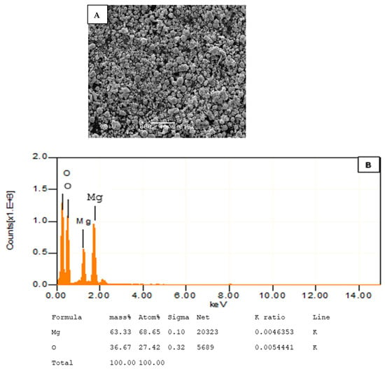
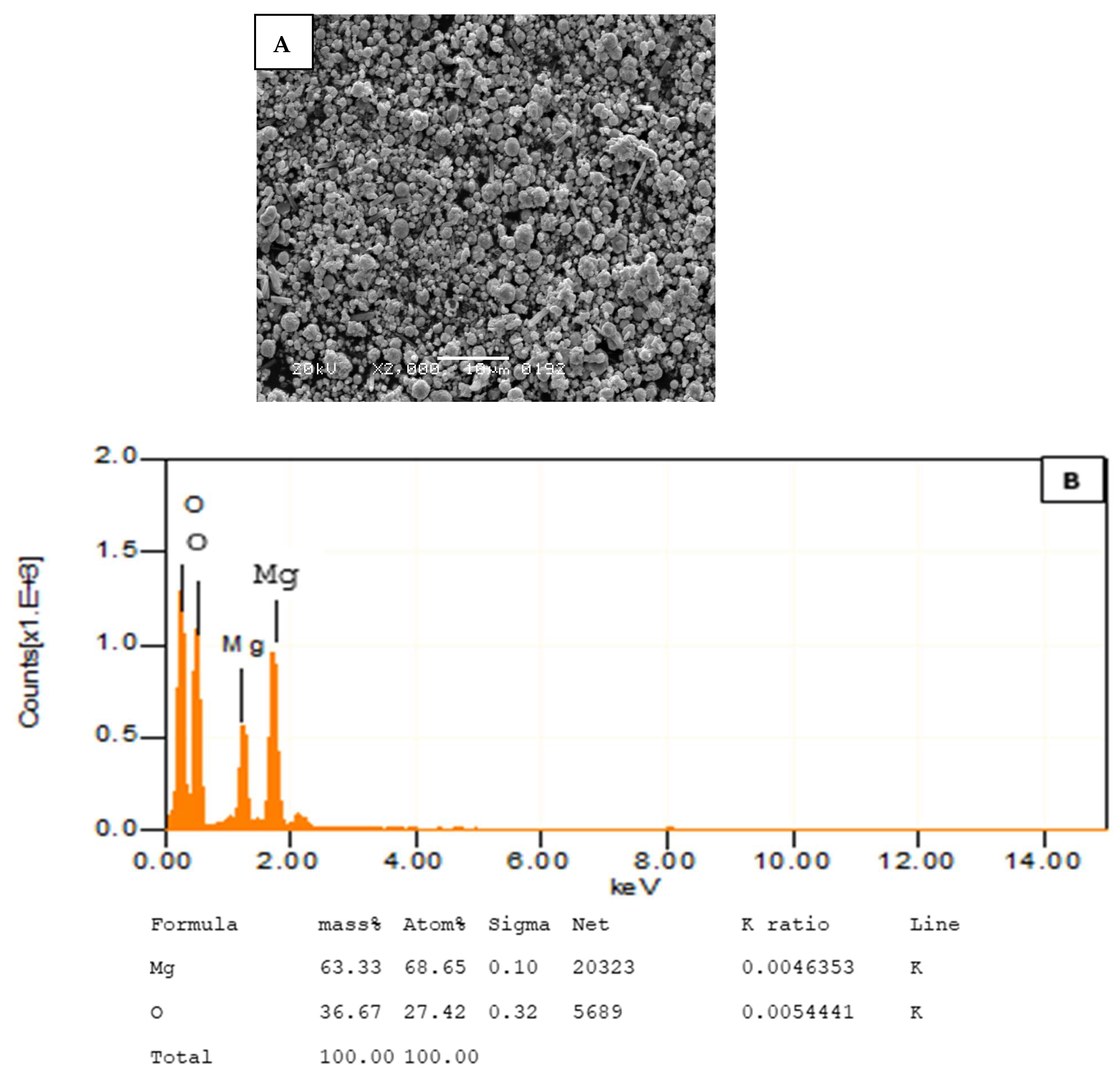
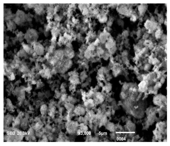
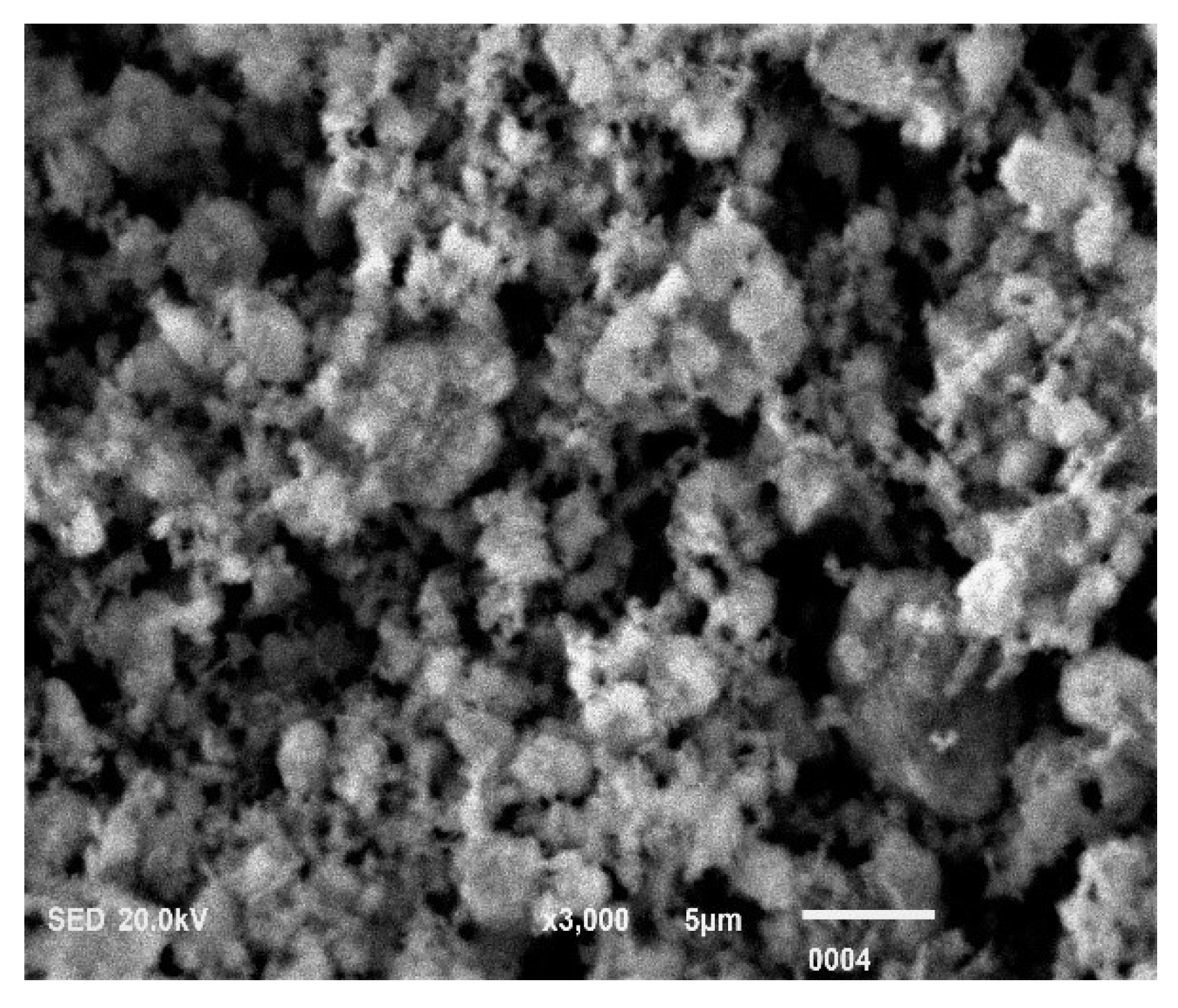
SEM micrograph of MgONPs shows predominantly spherical to pseudo-spherical morphology. Some particles exhibit irregular/agglomerated clusters resembling cauliflower-like aggregates (Figure 1A). EDX shows elemental distribution in MgONPs. Mg is showing a major component in MgONPs, followed by Oxygen (Figure 1B). The SEM image of CLP6-MgONPs shows notably rougher and more irregular surface texture compared to bare MgO, which indicates coating of the nanoparticle surface by CLP6 (Collagen-Like Peptide 6). The cauliflower/sponge-like aggregate morphology is characteristic of peptide-coated nanoparticles (Figure 2).
Figure 1.
(A): The SEM micrograph (×2000; 20 kV; scale bar 10 µm) reveals MgONPs exhibiting a predominantly spherical to sub-spherical morphology with a broad size distribution ranging from approximately 100 nm to 800 nm. The particles display a characteristic tendency toward agglomeration, forming loosely packed clusters interconnected by interparticle contact points, with visible open voids between clusters. (B): EDX analysis of MgONPs shows two major components in MgO. Mg 63.3% and O2 36.67%.
Figure 2.
SEM (×3000) of CLP6-functionalized magnesium oxide nanoparticles (CLP6-MgONPs). The micrograph reveals an irregular, cauliflower-like granular morphology with interparticulate agglomeration. A visible surface coating layer conformally distributed over the nanoparticle clusters confirms successful CLP6 peptide functionalization of the MgONP surface. The retained porous interparticulate architecture underlying the coating suggests preservation of the nanoparticulate framework.
3.2. SL Removal Assessment
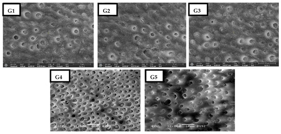
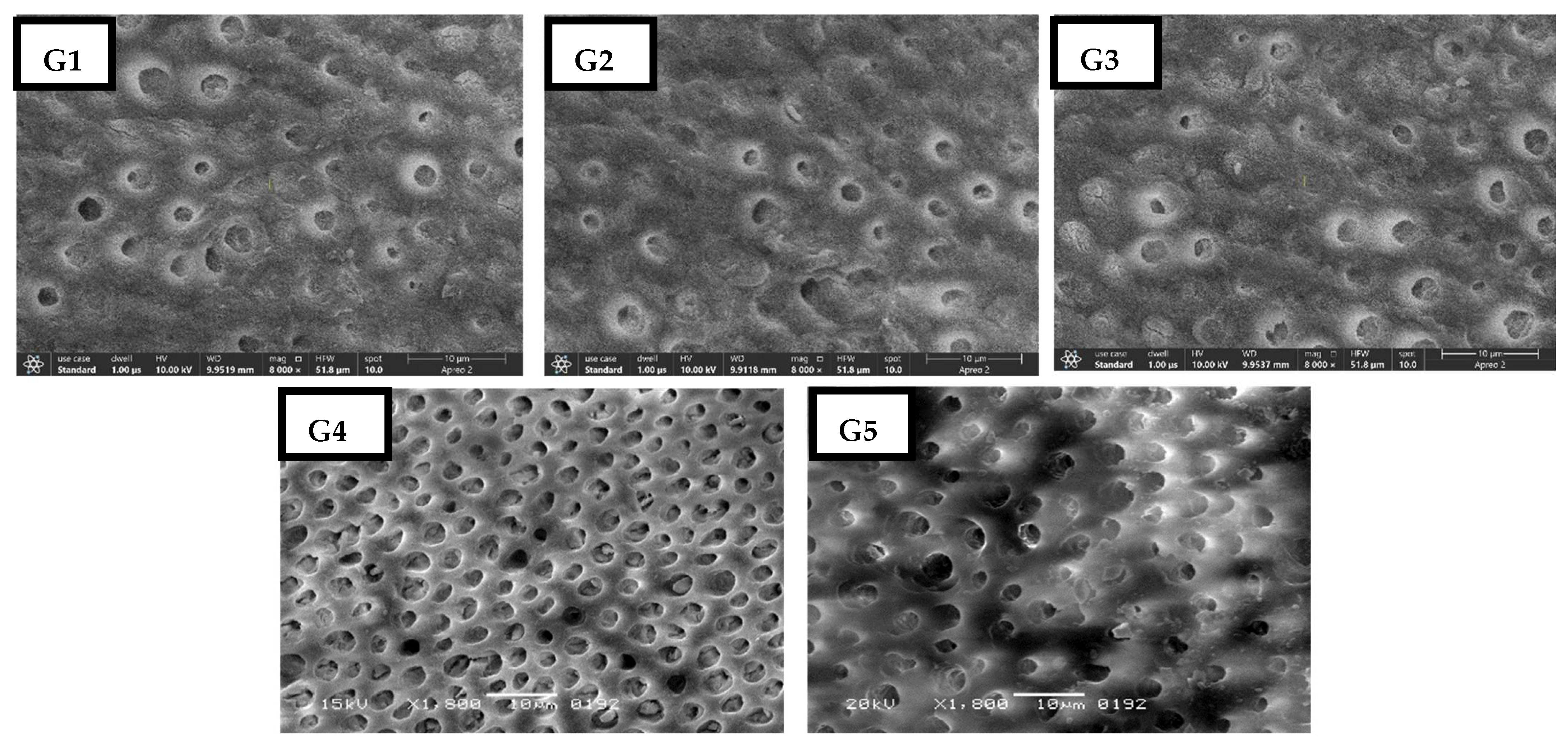
Removal of SL from the CAD discs after disinfection using different regimes is exhibited in Table 1. Group 4 (MgO-NPs) test specimens exhibited the highest level of SL cleaning (1.11 ± 0.13). The SEM image reveals dentinal tubules that are well-opened with distinct orifices, minimal amorphous debris, and a visible intertubular dentin collagen structure (Figure 3(G1–G5)). However, minimum SL removal was exhibited by Group 1 (No disinfectant) (2.87 ± 0.94) samples. In comparing the groups, it was found that Group 1, Group 2 (CHX) (2.73 ± 0.99), and Group 3 (Clp6-mediated PDT) (2.84 ± 0.86) exhibited similar levels of SL removal from cavity dentin, with no statistically significant differences observed among them (p > 0.05). The SEM micrograph reveals that the tubule orifices are either partially or completely covered by a thick, amorphous SL. The surface texture is uniform and muddy, with unclear boundaries between peritubular and intertubular dentin, which aligns with high SL scores (Figure 3(G1–G5)). Conversely, Group 5, which consists of Clp6-functionalized MgONPs, demonstrated a statistically significant improvement in SL elimination, with a value of 1.56 ± 0.17, when compared to groups 1, 2, and 3. Nonetheless, its effectiveness was statistically lower than that of samples treated solely with MgO-NPs (p < 0.05). The SEM micrograph for Group 5, featuring CLP6-functionalized MgONPs, reveals tubules that are partially opened, positioned between the MgONP-only group and the less effective groups (Control/CHX/CLP6-PDT) (Figure 3(G1–G5)).
Table 1.
Removal of SL from CAD using different cavity sterilants.
Figure 3.
SEM image of (G1–G3)—Control/CHX/CLP6-PDT): These images likely show dentin tubule orifices that are partially or fully obliterated by a thick, amorphous smear layer. The surface texture appears homogeneous and muddy, with indistinct peritubular/intertubular dentin boundaries consistent with high smear layer scores. (G4)—MgONPs: Well-opened dentinal tubules with clear orifices, minimal amorphous debris, and visible intertubular dentin collagen architecture. (G5)—CLP6-functionalized MgONPs: SEM micrograph shows partially opened tubules intermediate between the MgONP-only group and the poor performers (Control/CHX/CLP6-PDT). Interestingly, CLP6 functionalization slightly improved the SL removal efficacy compared to MgONPs alone.
3.3. SBS Testing
SBS of the fifth-generation adhesive to CAD after using different disinfectants is exhibited in Table 2. Group 4 (MgONPs) test specimens displayed the maximum bond strength (10.32 ± 0.18 MPa). However, minimum bond integrity was exhibited by Group 1 (No disinfectant) (7.42 ± 0.25 MPa) samples. Intergroup comparison analysis revealed that Group 1 (7.42 ± 0.25 MPa), Group 2 (CHX) (7.26 ± 0.27 MPa), and Group 3 (Clp6-mediated PDT) (7.23 ± 0.32 MPa) showed statistically comparable bond strength results. (p > 0.05) However, Group 5 (Clp6-functionalized MgONPs) (8.95 ± 0.23 MPa) and Group 4 showed statistically significant differences in SBS both between them and compared with the other groups (p < 0.05).
Table 2.
SBS of fifth-generation adhesive to cariously affected dentin after using different disinfectants.
3.4. Failure Mode Assessment
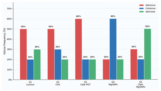
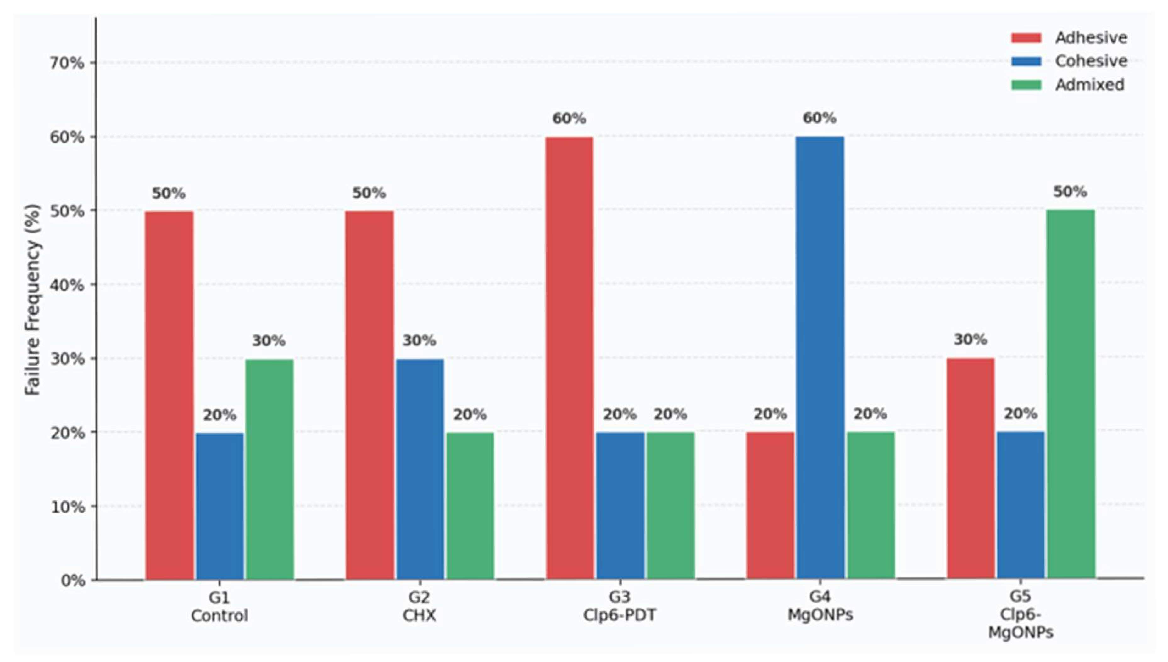
Groups 1, 2, and 3 (Control, CHX, Clp6-PDT) all share adhesive failure as their dominant mode (50%, 50%, 60%, respectively). This means the resin–dentin interface remained the weakest point despite disinfection, suggesting these treatments did not meaningfully strengthen the bond or adequately remove the smear layer. Group 4 (MgONPs) is the clear outlier. With 60% cohesive failure and only 20% adhesive failure, it is the only group where bond failure shifted away from the interface into the dentin itself—the gold standard outcome in bond strength research. This directly supports the SBS data showing Group 4 had the highest bond strength values. Group 5 (Clp6-MgONPs) shows predominantly admixed failure (50%), placing it between the weaker groups and Group 4. The photosensitizer functionalization appears to partially modify—but not optimize—the beneficial effect of MgONPs alone (Figure 4).
Figure 4.
Grouped bar chart—failure mode frequency by experimental group. Each cluster represents one group, with three bars per cluster corresponding to adhesive (red), cohesive (blue), and admixed (green) failure modes. Values above bars indicate exact percentages.
4. Discussion
The present study was designed under the null hypotheses that SL removal efficiency and SBS of a fifth-generation etch-and-rinse adhesive to CAD would not differ significantly among the tested cavity disinfectants—CHX, chlorin p6-mediated PDT, MgONPs, and Clp6-functionalized MgONPs—compared to an untreated control. Based on the outcomes of the present investigation, both null hypotheses were partially rejected. CHX- and Clp6-PDT-treated specimens demonstrated SL removal and bond strength values comparable to the control group, while MgONPs and Clp6-functionalized MgONPs produced significantly superior SL removal efficiency and SBS outcomes relative to the control, indicating a meaningful biological and mechanical effect attributable to nanoparticle-based disinfection strategies.
SBS testing is a well-established and clinically relevant methodology in dental materials research, as it quantifies the capacity of an adhesive system to resist lateral displacement forces at the bonded interface [44]. Such forces closely simulate the complex biomechanical loading experienced by resin composite restorations during mastication, occlusal contact, and parafunctional activity [45]. Bond integrity at the resin–CAD interface is fundamentally governed by three interdependent structural parameters: the completeness of SL removal, the quality and depth of hybrid layer formation, and the extent of resin tag penetration into patent dentinal tubules [46,47]. Deficiency in any of these parameters compromises the durability of the adhesive restoration.
Among all tested agents, MgONPs demonstrated the most favourable outcomes, yielding significantly superior SL removal and the highest SBS values to CAD. These findings are consistent with the laboratory-based observations of Alhamdan et al., who reported that MgONPs employed as a root canal irrigation adjunct effectively eliminated the SL and enhanced push-out bond strength of root canal sealers [48]. A comparable conclusion was drawn by Sorour et al., who documented satisfactory SL elimination following MgONP application in endodontic contexts [49]. The superior performance of MgONPs in the present study can be attributed to several converging physicochemical mechanisms. MgONPs exhibit a strongly alkaline surface chemistry (pH 10.5–12.5), and upon hydration in aqueous or biological environments, they undergo partial dissolution to form magnesium hydroxide, which facilitates chemical dissolution of the inorganic component of the SL [50]. This alkaline dissolution mechanism has been proposed to disrupt the calcium-phosphate matrix of the SL, thereby exposing the underlying dentinal tubule orifices and improving adhesive penetration [51]. Furthermore, the characteristically high surface area-to-volume ratio of nanoscale particles enhances intimate contact with the irregular dentinal surface topography, augmenting cleansing efficiency beyond what is achievable with conventional ionic disinfectants [52]. The resulting improvement in tubule patency promotes deeper resin tag formation and a more continuous hybrid layer, directly translating into the superior SBS values observed in Group 4 [48].
The CHX-treated group demonstrated SL removal and bond strength outcomes that were not significantly different from the untreated control, consistent with the growing body of evidence questioning CHX’s efficacy as a SL removal agent. Multiple investigations have reported an inability of CHX to dissolve or displace the SL from dentin surfaces, attributable to its mechanism of action being primarily bacteriostatic and bactericidal through membrane disruption rather than through mineral dissolution [6,53]. The diminished bond integrity observed in the CHX group is further supported by the findings of Salas et al., who reported that final irrigation with 2% CHX significantly reduced dentinal tubule penetration of endodontic sealer in the coronal and middle root thirds [54]. This reduced penetration was attributed to the affinity of CHX molecules for dentinal tubule walls, where their adsorption may sterically hinder subsequent adhesive monomer infiltration and resin tag development, ultimately attenuating bond strength [54,55]. However, it is acknowledged that conflicting evidence exists in the literature; certain investigations have reported a positive influence of CHX on adhesive bond integrity, potentially attributable to its MMP-inhibiting activity, preserving collagen scaffold integrity within the hybrid layer. The discrepancy across studies may reflect differences in CHX concentration, application protocol, dentin substrate condition, and adhesive system composition [56,57].
Clp6-mediated PDT produced SL removal and SBS outcomes comparable to CHX and the untreated control, suggesting that, at the concentration and irradiation parameters employed in this study, Clp6-PDT did not confer a meaningful advantage in terms of adhesive bonding to CAD. A plausible explanation for this finding relates to the known affinity of chlorin-based photosensitizers for divalent calcium ions present in dentin mineral. The formation of Clp6–calcium complexes or precipitates at the dentin surface may create a physical barrier impeding adhesive monomer diffusion into the demineralized collagen network, thereby restricting hybrid layer development and resin tag formation [58]. This interpretation is consistent with published evidence demonstrating that certain chlorin e6 derivatives—including Zn(II) chlorin e6 methyl ester—produced significantly lower push-out bond strength values compared to sodium hypochlorite-treated controls, supporting the notion that residual photosensitizer at the dentin interface may actively compromise bonding performance through physical occlusion rather than chemical interference [14,58]. The inherent limitation of light propagation in dentinal tubules and the restricted penetration depth of the photosensitizer at clinically applicable concentrations may additionally limit the practical utility of Clp6-PDT as a standalone cavity disinfectant in deep carious lesions [59].
The Clp6-functionalized MgONP formulation demonstrated SL removal and bond strength values that were intermediate between the MgONPs-alone group and the CHX/Clp6 groups, with statistically significant differences from MgONPs alone but superior performance relative to the conventional disinfectant groups. This intermediate performance profile can be interpreted through the competing and partially antagonistic effects of the two constituents within the combined formulation. The MgONP component retains its inherent alkaline dissolution mechanism and high surface reactivity, contributing to SL disruption and tubule exposure [52]. However, the Clp6 photosensitizer conjugated onto the nanoparticle surface, upon photoactivation, may induce partial occlusion of the exposed dentinal tubule orifices—either through photosensitizer precipitation or through the deposition of oxidative reaction byproducts—thereby attenuating the resin infiltration capacity otherwise achieved by MgONPs alone [14,58]. The net result is a moderate degree of tubule patency, hybrid layer formation, and SBS, explaining why the combined formulation occupies a statistically and clinically intermediate position between the nanoparticle-only and conventional disinfectant groups. Future optimization of the Clp6 loading concentration and irradiation parameters may help decouple the photodynamic antibacterial benefit from the undesirable adhesive interference, potentially yielding a formulation that combines superior disinfection with preserved bonding capacity.
The failure mode distribution across experimental groups was coherent with and corroborated the SBS findings. MgONP-treated specimens exhibited predominantly cohesive failure within the dentin substrate (60%), indicating that the adhesive interface had attained a strength exceeding that of the surrounding dental tissue—the most favourable failure pattern in bond strength research. Clp6-functionalized MgONP specimens demonstrated predominantly admixed failure (50%), consistent with their intermediate bond quality. In contrast, CHX- and Clp6-PDT-treated specimens, along with the untreated control, exhibited predominantly adhesive failure (50%, 60%, and 50%, respectively), reflecting an adhesive interface that remained the weakest structural element in the bonded assembly. These failure mode patterns are in agreement with the laboratory-based findings reported by Meerbeek et al. [60] and Carrilho et al. [61], who similarly observed a direct correspondence between elevated bond strength and a shift toward cohesive failure modes in resin–dentin bonding studies.
Several inherent limitations of the present study warrant acknowledgment. As an in vitro investigation, the experimental conditions cannot fully reproduce the complexity of the oral environment, which includes continuous salivary enzyme activity, fluctuating pH, thermal cycling, sustained masticatory loading, and microbial colonization—all of which influence the long-term clinical performance of adhesive restorations. Biological variability in CAD composition, even within the standardized ICDAS 4–5 inclusion criteria, may introduce heterogeneity in dentinal tubule density, residual mineral content, and collagen integrity, potentially contributing to variance in bond strength measurements. The use of geometrically standardized flat dentin substrates and a single fifth-generation etch-and-rinse adhesive system limits the direct clinical extrapolation of these findings to the diverse cavity morphologies and adhesive systems encountered in practice. Furthermore, only single concentrations of CHX, MgONPs, and Clp6 were evaluated; concentration–response relationships and the optimal therapeutic window for each agent remain to be established. Accordingly, while the present findings offer clinically relevant insights into the differential effects of these disinfection strategies on smear layer removal and CAD bond integrity, prospective in vivo and long-term clinical investigations are necessary to validate their translational applicability. The findings of this study suggest several directions for future research. The incorporation of MgONPs into Bis-GMA/TEGDMA-based resin composite matrices represents a promising avenue, as evidenced by Saini et al., who demonstrated that nano tricalcium phosphate and silver nanoparticle-reinforced composites exhibited enhanced tribological, mechanical, and thermal properties alongside inherent antimicrobial activity—a material science framework directly applicable to MgONP-modified restorative systems [62]. Concentration-response optimization of Clp6-functionalized MgONPs within adhesive systems, long-term ageing studies under simulated oral conditions, and randomized clinical trials with extended follow-up are necessary to confirm the translational applicability of these laboratory findings.
5. Conclusions
MgONPs demonstrated superior smear layer removal and shear bond strength to caries-affected dentin compared to all tested agents, followed by Clp6-functionalized MgONPs, while CHX and Clp6-mediated PDT performed comparably to the untreated control. Failure mode analysis corroborated these findings, with MgONPs producing predominantly cohesive failure—indicative of optimal bond integrity. These results highlight the potential of MgONPs as a promising cavity disinfectant; however, in vivo validation and long-term clinical studies are necessary before definitive clinical recommendations can be made.
Author Contributions
Conceived and designed the study: F.A.; Conducted the experiments: F.A. and M.H.A.; Analyzed the data: M.H.A. and F.A.; Contributed to the writing of the manuscript: F.A. and M.H.A.; Read and approved the final version of the manuscript: M.H.A. and F.A. All authors have read and agreed to the published version of the manuscript.
Funding
The research was funded by the Ongoing Research Funding Programme (ORF-2026-815) at King Saud University, Riyadh, Saudi Arabia, for their financial support.
Data Availability Statement
Data can be made available upon request to the corresponding author.
Acknowledgments
The authors gratefully acknowledge the Ongoing Research Funding Programme (ORF-2026-815) at King Saud University, Riyadh, Saudi Arabia, for their financial support.
Conflicts of Interest
The authors declare no conflicts of interest.
References
- Frencken, J.E.; Peters, M.C.; Manton, D.J.; Leal, S.C.; Gordan, V.V.; Eden, E. Minimal Intervention Dentistry for Managing Dental Caries—A Review: Report of a FDI Task Group. Int. Dent. J. 2012, 62, 223–243. [Google Scholar] [CrossRef] [Scilit]
- Maashi, M.S.; Elkhodary, H.M.; Alamoudi, N.M.; Bamashmous, N.O. Chemomechanical Caries Removal Methods: A Literature Review. Saudi Dent. J. 2023, 35, 233–243. [Google Scholar] [CrossRef] [Scilit]
- Turkistani, A.; Sonbul, H.M.; Almarzouki, M. Influence of Chlorhexidine Dentin Disinfection on Universal Adhesive Performance: Interfacial Adaptation and Bond Strength Assessments. PLoS ONE 2024, 19, e0315036. [Google Scholar] [CrossRef] [Scilit]
- Alshahrani, A.; Abrar, E.; Maawadh, A.M.; Al-Hamdan, R.S.; Almohareb, T.; AlFawaz, Y.; Naseem, M.; Vohra, F.; Abduljabbar, T. Management of Caries Affected Dentin (CAD) with Resin Modified Glass Ionomer Cement (RMGIC) in the Presence of Different Caries Disinfectants and Photosensitizers. Photodiagn. Photodyn. Ther. 2020, 32, 101978. [Google Scholar] [CrossRef] [Scilit]
- Al Ahdal, K.; Maawadh, A.M.; Al Deeb, L.; Alshamrani, A.S.; Almohareb, T.; Alrahlah, A. Effect of Malachite Green, Ocimum Sanctum, and Er, Cr: YSGG Laser on Antimicrobial Activity against S. mutans and CAD Disinfection Bonded to Resin Restoration. Photodiagn. Photodyn. Ther. 2023, 42, 103571. [Google Scholar] [CrossRef] [Scilit] [PubMed]
- Dafer Al Wadei, M.H.; Qasim, M.; Siddiqui, K.; Sainudeen, S.; Qureshi, A.; Naseem, M.; Panhwar, M. Photodynamic Therapy, Ozonated Water, and Diode Laser Efficacy in Removing Smear Layer in Carious Dentin and Their Effect on Bond Integrity to Composite Resin. Photobiomodul. Photomed. Laser Surg. 2025, 43, 425–433. [Google Scholar] [CrossRef] [Scilit]
- Ghorbanzadeh, A.; Fekrazad, R.; Bahador, A.; Ayar, R.; Tabatabai, S.; Asefi, S. Evaluation of the Antibacterial Efficacy of Various Root Canal Disinfection Methods Against Enterococcus faecalis Biofilm. An Ex-Vivo Study. Photodiagn. Photodyn. Ther. 2018, 24, 44–51. [Google Scholar] [CrossRef] [Scilit] [PubMed]
- Boutsiouki, C.; Frankenberger, R.; Lücker, S.; Krämer, N. Effect of Chlorhexidine-Containing Etch-and-Rinse Adhesives on Dentin Microtensile Bond Strength After Biological Loading. J. Adhes. Dent. 2023, 25, b3801065. [Google Scholar] [CrossRef] [Scilit] [PubMed]
- Ebrahimi, M.; Majidinia, S.; Sarraf, A. Effect of Chlorhexidine on Immediate and Delayed Bond Strength Between Resin and Dentin of Primary Teeth: A Systematic Review and Meta-Analysis. Front. Dent. 2022, 19, 39. [Google Scholar] [CrossRef] [Scilit]
- Dursun, E.; Le Goff, S.; Ruse, D.N.; Attal, J.P. Effect of Chlorhexidine Application on the Long-Term Shear Bond Strength to Dentin of a Resin-Modified Glass Lonomer. Oper. Dent. 2013, 38, 275–281. [Google Scholar] [CrossRef] [Scilit]
- Memon, J.; Shabbir, T.; Ishrat, M.; Aslam, H.; Khowaja, A.A.; Leemani, M.J. Caries Affected Disinfection Using Phycocyanin Activated by PDT, Holy Basil, and Ti-Sapphire Laser on Adhesive Bond Strength, Microleakage, and Bond Failure. Photodiagn. Photodyn. Ther. 2023, 43, 103691. [Google Scholar] [CrossRef] [Scilit]
- Alrahlah, A.; Naseem, M.; Tanveer, S.A.; Abrar, E.; Charania, A.; AlRifaiy, M.Q.; Vohra, F. Influence of Disinfection of Caries Effected Dentin with Different Concentration of Silver Diamine Fluoride, Curcumin and Er, Cr:YSGG on Adhesive Bond Strength to Resin Composite. Photodiagn. Photodyn. Ther. 2020, 32, 102065. [Google Scholar] [CrossRef] [Scilit]
- Alanazi, A.M.; Khan, A.A.; Mahmood, A.; Kamal, M.A.; Baig, E.A. The Antibacterial Efficiency of Chitosan Photodynamically Activated Phycocyanin, and Morinda Oleifera Against S. mutans and the Bonding Strength Between Composite Resin and Caries-Affected Dentin. Pak. J. Med. Sci. 2024, 40, 962–966. [Google Scholar] [CrossRef] [Scilit] [PubMed]
- Qamar, Z.; Abdul, N.S.; Kakti, A.; Reddy, R.N.; Shenoy, M.; Zeeshan, T.; Ramadan, A.H. Effect of Photosensitizers on Modulating Bond Strength of Different Luting Cements Used for Cementation of Resin Modified Composite Fiber Posts. Eur. Rev. Med. Pharmacol. Sci. 2022, 26, 4597–4605. [Google Scholar] [CrossRef] [Scilit] [PubMed]
- Besinis, A.; De Peralta, T.; Handy, R.D. The Antibacterial Effects of Silver, Titanium Dioxide and Silica Dioxide Nanoparticles Compared to the Dental Disinfectant Chlorhexidine on Streptococcus mutans Using a Suite of Bioassays. Nanotoxicology 2014, 8, 1–16. [Google Scholar] [CrossRef] [Scilit] [PubMed]
- Pourhajibagher, M.; Salehi Vaziri, A.; Takzaree, N.; Ghorbanzadeh, R. Physico-Mechanical and Antimicrobial Properties of an Orthodontic Adhesive Containing Cationic Curcumin Doped Zinc Oxide Nanoparticles Subjected to Photodynamic Therapy. Photodiagn. Photodyn. Ther. 2019, 25, 239–246. [Google Scholar] [CrossRef] [Scilit]
- Danhier, F.; Ansorena, E.; Silva, J.M.; Coco, R.; Le Breton, A.; Préat, V. PLGA-Based Nanoparticles: An Overview of Biomedical Applications. J. Control. Release 2012, 161, 505–522. [Google Scholar] [CrossRef] [Scilit]
- Alhamdan, M.M. Canal Disinfectants: Potassium Titanyl Phosphate Laser, Magnesium Oxide Nanoparticles, and Aloe-Emodin PDT on Smear Layer Removal and Bond Strength of Glass Fiber Post to Root Dentin. Photodiagn. Photodyn. Ther. 2025, 53, 104615. [Google Scholar] [CrossRef] [Scilit]
- Beach, M.A.; Nayanathara, U.; Gao, Y.; Zhang, C.; Xiong, Y.; Wang, Y.; Such, G.K. Polymeric Nanoparticles for Drug Delivery. Chem. Rev. 2024, 124, 5505–5616. [Google Scholar] [CrossRef] [Scilit]
- Sen, D.; Patil, V.; Smriti, K.; Varchas, P.; Ratnakar, R.; Naik, N.; Kumar, S.; Saxena, J.; Kapoor, S. Nanotechnology and Nanomaterials in Dentistry: Present and Future Perspectives in Clinical Applications. Eng. Sci. 2022, 20, 14–24. [Google Scholar] [CrossRef] [Scilit]
- Tjäderhane, L.; Nascimento, F.D.; Breschi, L.; Mazzoni, A.; Tersariol, I.L.S.; Geraldeli, S.; Tezvergil-Mutluay, A.; Carrilho, M.; Carvalho, R.M.; Tay, F.R.; et al. Strategies to Prevent Hydrolytic Degradation of the Hybrid Layer—A Review. Dent. Mater. 2013, 29, 999–1011. [Google Scholar] [CrossRef] [Scilit]
- Akram, Z.; Aati, S.; Ngo, H.; Fawzy, A. PH-Dependent Delivery of Chlorhexidine from PGA Grafted Mesoporous Silica Nanoparticles at Resin-Dentin Interface. J. Nanobiotechnol. 2021, 19, 43. [Google Scholar] [CrossRef] [Scilit]
- Kirchherr, A.K.; Briel, A.; Mäder, K. Stabilization of Indocyanine Green by Encapsulation Within Micellar Systems. Mol. Pharm. 2009, 6, 480–491. [Google Scholar] [CrossRef] [Scilit] [PubMed]
- Gholami, A.; Ghezelbash, K.; Asheghi, B.; Abbaszadegan, A.; Amini, A. An In Vitro Study on the Antibacterial Effects of Chlorhexidine-Loaded Positively Charged Silver Nanoparticles on Enterococcus faecalis. J. Nanomater. 2022, 2022, 6405772. [Google Scholar] [CrossRef] [Scilit]
- Al Deeb, L.; Bin-Shuwaish, M.S.; Abrar, E.; Naseem, M.; Al-Hamdan, R.S.; Maawadh, A.M.; Al Deeb, M.; Almohareb, T.; Al Ahdal, K.; Vohra, F.; et al. Efficacy of Chlorhexidine, Er Cr YSGG Laser and Photodynamic Therapy on the Adhesive Bond Integrity of Caries Affected Dentin. An In-Vitro Study. Photodiagn. Photodyn. Ther. 2020, 31, 101875. [Google Scholar] [CrossRef] [Scilit]
- AlQhtani, F.A.B.A.; Alkahtani, Z.M.; Sainudeen, S.; Abdulla, A.M.; Kamran, M.A.; Naseem, M. To Evaluate ΜTBS, Microleakage, and Resin Tag Length via Scanning Electron Microscopy of Different Resin Sealants Clinpro and Embrace Bonded to the Intact Enamel in Primary Molars. Microsc. Res. Tech. 2025, 88, 2504–2511. [Google Scholar] [CrossRef] [Scilit] [PubMed]
- Alkhudhairy, F.; Naseem, M.; Bin-Shuwaish, M.; Vohra, F. Efficacy of Er Cr: YSGG Laser Therapy at Different Frequency and Power Levels on Bond Integrity of Composite to Bleached Enamel. Photodiagn. Photodyn. Ther. 2018, 22, 34–38. [Google Scholar] [CrossRef] [Scilit]
- Alrefeai, M.H.; Aljamhan, A.S.; Alhabdan, A.; Alzehiri, M.H.; Naseem, M.; Alkhudhairy, F. Influence of Methylene Blue, Riboflavin, and Indocyanine Green on the Bond Strength of Caries Affected Dentin When Bonded to Resin-Modified Glass Ionomer Cement. Photodiagn. Photodyn. Ther. 2022, 38, 102792. [Google Scholar] [CrossRef] [Scilit]
- Vohra, F.; Alghamdi, A.; Aldakkan, M.; Alharthi, S.; Alturaigi, O.; Alrabiah, M.; Al-Aali, K.A.; Alrahlah, A.; Naseem, M.; Ab-duljabbar, T. Influence of Er: Cr: YSGG laser on adhesive strength and microleakage of dentin bonded to resin com-posite. Vitr. Study Photodiagn. Photodyn. Ther. 2018, 23, 342–346. [Google Scholar] [CrossRef] [Scilit]
- Alanazi, A.M.; Khan, N.A.; Khan, A.A.; Bhutto, K.; Askary, S.H.; Askary, G.; Abrar, E.; Mahmood, S.J.; Qureshi, A. Titanium Oxide and Chitosan Nanoparticles Loaded in Methylene Blue Activated by Photodynamic Therapy on Caries Affected Dentin Disinfection, Bond Strength, and Smear Layer Removal Efficacy. Photodiagn. Photodyn. Ther. 2024, 50, 104343. [Google Scholar] [CrossRef] [Scilit]
- Al-Kheraif, A.A.; Alsunbul, H.; Alhaidry, H.; Khan, A.A. Comparative Evaluation of Modern Disinfection Methods on Smear Layer Removal and Bond Strength to Caries-Affected Dentin: A KTP Laser, Nanocare Gold, Photodynamic Therapy Study. Pak. J. Med. Sci. 2026, 42, 365–370. [Google Scholar] [CrossRef] [Scilit]
- Navayath, M.; Ramesh, S.R.; Kalaiselvam, R.; Ashok, R.; Rajendran, M.R.; Balaji, L. Evaluation of Nanomagnesium Oxide in Combination with Garlic Extract as an Endodontic Irrigant: An In Vitro Study. J. Contemp. Dent. Pract. 2023, 24, 459–466. [Google Scholar] [CrossRef] [Scilit]
- Wang, Y.; Li, P.; Kong, L. Chitosan-Modified PLGA Nanoparticles with Versatile Surface for Improved Drug Delivery. AAPS PharmSciTech 2013, 14, 585–592. [Google Scholar] [CrossRef] [Scilit]
- Wang, M.; Shen, J.; Thomas, J.C.; Mu, T.; Liu, W.; Wang, Y.; Pan, J.; Wang, Q.; Liu, K. Particle Size Measurement Using Dynamic Light Scattering at Ultra-Low Concentration Accounting for Particle Number Fluctuations. Materials 2021, 14, 5683. [Google Scholar] [CrossRef] [Scilit]
- Alkhudhairy, F. Experimental and Chitosan-Infused Adhesive with Dentin Pretreated with Femtosecond Laser, Methylene Blue-Activated Low-Level Laser, and Phosphoric Acid. Photobiomodul. Photomed. Laser Surg. 2024, 42, 634–642. [Google Scholar] [CrossRef] [Scilit]
- Alfawaz, Y.F.; Almutairi, B.; Kattan, H.F.; Zafar, M.S.; Farooq, I.; Naseem, M.; Vohra, F.; Abduljabbar, T. Dentin Bond Integrity of Hydroxyapatite Containing Resin Adhesive Enhanced with Graphene Oxide Nano-Particles—An SEM, EDX, Micro-Raman, and Microtensile Bond Strength Study. Polymers 2020, 12, 2978. [Google Scholar] [CrossRef] [Scilit] [PubMed]
- Hülsmann, M.; Rümmelin, C.; Schäfers, F. Root Canal Cleanliness after Preparation with Different Endodontic Handpieces and Hand Instruments: A Comparative SEM Investigation. J. Endod. 1997, 23, 301–306. [Google Scholar] [CrossRef] [Scilit] [PubMed]
- Aljamhan, A.S.; Alrefeai, M.H.; Alhabdan, A.; Alzehiri, M.H.; Naseem, M.; Vohra, F.; Alkhudhairy, F. Interaction of Zirconium Oxide Nanoparticle Infiltrated Resin Adhesive with Dentin Conditioned by Phosphoric Acid and Er, Cr: YSGG Laser. J. Appl. Biomater. Funct. Mater. 2022, 20, 1–8. [Google Scholar] [CrossRef] [Scilit] [PubMed]
- Abrar, E.; Naseem, M.; Baig, Q.A.; Vohra, F.; Maawadh, A.M.; Almohareb, T.; AlRifaiy, M.Q.; Abduljabbar, T. Antimicrobial Efficacy of Silver Diamine Fluoride in Comparison to Photodynamic Therapy and Chlorhexidine on Canal Disinfection and Bond Strength to Radicular Dentin. Photodiagn. Photodyn. Ther. 2020, 32, 102066. [Google Scholar] [CrossRef] [Scilit]
- Attia, A. Bond Strength of Three Luting Agents to Zirconia Ceramic—Influence of Surface Treatment and Thermocycling. J. Appl. Oral Sci. 2011, 19, 388–395. [Google Scholar] [CrossRef] [Scilit]
- Alrahlah, A.; Awad, M.M.; Vohra, F.; Al-Mudahi, A.; Al Jeaidi, Z.A.; Elsharawy, M. Effect of Self Etching Ceramic Primer and Universal Adhesive on Bond Strength of Lithium Disilicate Ceramic. J. Adhes. Sci. Technol. 2017, 31, 2611–2619. [Google Scholar] [CrossRef] [Scilit]
- Naiboğlu, P.; Koşar, T.; Yücel, A. Shear Bond Strength of Calcium Silicate-Based Cements to Composite Resin Using a Universal Adhesive in Different Application Modes: An in Vitro Study. Aust. Dent. J. 2023, 69, 102–111. [Google Scholar] [CrossRef] [Scilit]
- Mokeem, L.S.; Garcia, I.M.; Melo, M.A. Degradation and Failure Phenomena at the Dentin Bonding Interface. Biomedicines 2023, 11, 1256. [Google Scholar] [CrossRef] [Scilit]
- Jain, M.; Patel, D.; Sharma, T. The Call for Standardization of Shear Bond Strength Testing Protocols in Orthodontics. J. Indian Orthod. Soc. 2023, 57, 39–42. [Google Scholar] [CrossRef] [Scilit]
- Ismail, A.M.; Bourauel, C.; Elbanna, A.; Eldin, T.S. Micro versus Macro Shear Bond Strength Testing of Dentin-Composite Interface Using Chisel and Wireloop Loading Techniques. Dent. J. 2021, 9, 140. [Google Scholar] [CrossRef] [Scilit] [PubMed]
- Alkhudhairy, F.; Neiva, G.F. Effect of Er, Cr: YSGG, Nd: YAG, and Diode Laser Against Different Photosensitizers on Tensile and Shear Bond Strength of Bonded Composite to Caries Affected Dentin. Eur. Rev. Med. Pharmacol. Sci. 2023, 27, 8350–8359. [Google Scholar] [CrossRef] [Scilit]
- Keskin, G.; Uçar Gündoğar, Z.; Yaman, M. Bonding of an Ion-Releasing Restorative Material to Caries-Affected Dentin Disinfected with Photodynamic Therapy, Er,Cr:YSGG Laser, and Chemical Disinfectants. Photodiagn. Photodyn. Ther. 2021, 34, 102261. [Google Scholar] [CrossRef] [Scilit]
- Al-Kheraif, A.A.; Mohamed, B.A.; Khan, A.A.; Al-Shehri, A.M. Role of Riboflavin; Curcumin photosensitizers and Ozone when used as canal disinfectant on push-out bond strength of glass fiber post to radicular dentin. Photodiagn. Photodyn. Ther. 2022, 37, 102592. [Google Scholar] [CrossRef] [Scilit] [PubMed]
- Souror, K.K.; El Gendy, A.; Nabil, M. The Efficacy of Nano Magnesium Oxide and Nano Silver Fluoride on Canal Cleanliness and Smear Layer Removal (An In Vitro Study). Egypt. Dent. J. 2024, 70, 2853–2866. [Google Scholar] [CrossRef] [Scilit]
- Monzavi, A.; Eshraghi, S.; Hashemian, R.; Momen-Heravi, F. In Vitro and Ex Vivo Antimicrobial Efficacy of Nano-MgO in the Elimination of Endodontic Pathogens. Clin. Oral Investig. 2015, 19, 349–356. [Google Scholar] [CrossRef] [Scilit]
- Noori, A.J.; Kareem, F.A. The Effect of Magnesium Oxide Nanoparticles on the Antibacterial and Antibiofilm Properties of Glass-Ionomer Cement. Heliyon 2019, 5, e02568. [Google Scholar] [CrossRef] [Scilit]
- Sharifian, S.; Loghmani, A.; Nayyerain, S.; Javanbakht, S.; Daneii, P. Application of Magnesium Oxide Nanoparticles in Dentistry: A Literature Review. Eur. J. Gen. Dent. 2023, 12, 1–6. [Google Scholar] [CrossRef] [Scilit]
- Poppolo Deus, F.; Ouanounou, A. Chlorhexidine in Dentistry: Pharmacology, Uses, and Adverse Effects. Int. Dent. J. 2022, 72, 269–277. [Google Scholar] [CrossRef] [Scilit]
- Salas, H.; Castrejon, A.; Fuentes, D.; Luque, A.; Luque, E. Evaluation of the Penetration of CHX 2% on Dentinal Tubules Using Conventional Irrigation, Sonic Irrigation (EDDY) and Passive Ultrasonic Irrigation (PUI) Techniques: An in Vitro Study. J. Clin. Exp. Dent. 2021, 13, e37–e42. [Google Scholar] [CrossRef] [Scilit] [PubMed]
- Örs, S.A.; Yılmaz, G.A.; Şengül, N.; Keleş, A.; Eren, S.K. Effect of Final Irrigation Protocols on the Bond Strength and Dentinal Tubule Penetration of Root Canal Sealers: An Ex Vivo Laboratory Study. BMC Oral Health 2026, 26, 216. [Google Scholar] [CrossRef] [Scilit]
- Khaddam, M.; Salmon, B.; Le Denmat, D.; Tjaderhane, L.; Menashi, S.; Chaussain, C.; Rochefort, G.Y.; Boukpessi, T. Grape Seed Extracts Inhibit Dentin Matrix Degradation by MMP-3. Front. Physiol. 2014, 5, 425. [Google Scholar] [CrossRef] [Scilit] [PubMed]
- Moazami, F.; Sahebi, S.; Ahzan, S. Tooth Discoloration Induced by Imidazolium Based Silver Nanoparticles as an Intracanal Irrigant. J. Dent. 2018, 19, 280–286. [Google Scholar]
- Abdulwahed, A. Canal Pretreatment Using Cold Atmospheric Argon Plasma, Chitosan and Chlorine P6 Activated by Photodynamic Therapy with EDTA as a Final Irrigant on the Pushout Bond Strength of Fiber Post. Photodiagn. Photodyn. Ther. 2023, 42, 103517. [Google Scholar] [CrossRef] [Scilit] [PubMed]
- Krishnan, U.; Saji, S.; Clarkson, R.; Lalloo, R.; Moule, A.J. Free Active Chlorine in Sodium Hypochlorite Solutions Admixed with Octenidine, SmearOFF, Chlorhexidine, and EDTA. J. Endod. 2017, 43, 1354–1359. [Google Scholar] [CrossRef] [Scilit]
- Van Meerbeek, B.; Peumans, M.; Poitevin, A.; Mine, A.; Van Ende, A.; Neves, A.; De Munck, J. Relationship Between Bond-Strength Tests and Clinical Outcomes. Dent. Mater. 2010, 26, 664–678. [Google Scholar] [CrossRef] [Scilit]
- Carvalho, R.M.; Manso, A.P.; Geraldeli, S.; Tay, F.R.; Pashley, D.H. Durability of Bonds and Clinical Success of Adhesive Restorations. Dent. Mater. 2012, 28, 72–86. [Google Scholar] [CrossRef] [Scilit] [PubMed]
- Saini, S.; Yadav, R.; Sonwal, S.; Meena, A.; Huh, Y.S.; Brambilla, E.; Ionescu, A.C. Tribological, Mechanical, and Thermal Properties of Nano Tricalcium Phosphate and Silver Particulates Reinforced Bis-GMA/TEGDMA Dental Resin Composites. Tribol. Int. 2024, 199, 110010. [Google Scholar] [CrossRef] [Scilit]

Disclaimer/Publisher’s Note: The statements, opinions and data contained in all publications are solely those of the individual author(s) and contributor(s) and not of MDPI and/or the editor(s). MDPI and/or the editor(s) disclaim responsibility for any injury to people or property resulting from any ideas, methods, instructions or products referred to in the content. |
© 2026 by the authors. Licensee MDPI, Basel, Switzerland. This article is an open access article distributed under the terms and conditions of the Creative Commons Attribution (CC BY) license.