


Abstract
Three complexes of Zn(II) with the general formulas {[Zn(H2O)4(4,4′-BiPy)](NO3)2·2NA·H2O}n (1), [Zn(NA)2Br2]·2NA (2), and [Zn2(CH3COO)4(NA)2]·2H2O (3), where 4,4′-BiPy = 4,4′-bipyridine and NA = nicotinamide, have been synthesized and characterized by means of single-crystal X-ray diffraction (complexes 1 and 2), elemental analysis, FT-IR, fluorescence, 1H and 13C NMR spectroscopy (complexes (2) and (3)), and thermogravimetric analysis. A previously reported complex, [Zn(NA)2(H2O)4](NO3)2·2H2O, was used as a precursor for the synthesis of coordination compounds (1) and (2). X-ray data show that (1) is a 1D polynuclear compound, whereas complex (2) is mononuclear. The Zn(II) ions adopt a slightly distorted octahedral geometry in the polymer and a slightly distorted tetrahedral geometry in the monomer. The antimicrobial activity of complexes (2) and (3) was also evaluated, and complex (3) exhibited superior antimicrobial properties, particularly against the C. albicans strain.
1. Introduction
Zn(II) complexes have been intensively studied in recent years because of their wide range of applications: as catalysts [1], as storage material for industrial [2] or therapeutic applications [3], as sensors for water pollutants [4], and so on.
As a d10 metal, Zn(II) does not exhibit a preference for a particular coordination number since its crystal field energy stabilization (CFSE) is zero. However, a survey of the Cambridge Structural Database (CSD) including 28,706 metal complexes—of which 1425 are Zn(II) coordination compounds—revealed that Zn(II) most commonly adopts a tetrahedral geometry. More precisely, in 59% of the complexes, the metal ion adopts tetrahedral coordination, while 23% of them have a coordination number of six and exhibit octahedral geometry [5].
The design and synthesis of coordination polymers have greatly increased in recent years due to their wide range of applications [6]. The 1D, 2D, and 3D frameworks display interesting structures, influenced by ligand morphology (topology of the donor atoms), flexibility, and bonding ability. The molecular and supramolecular architectures of these compounds are imposed by stereochemical preferences of the metal ions, morphology of the bridging ligands, the presence of supramolecular synthons, and hydrogen and/or π-π stacking interactions. One method of obtaining such complexes with extended structures is to link metal ions with various coordination numbers and stereochemical preferences by using spacers [7]. A well-known class of spacers is that of exo-bidentate bipyridyl ligands, including 1,2-bis(4-pyridyl)ethane [8], trans-1,2-bis(4-pyridyl)ethene [9], and 4,4′-bipyridine [10,11]. 4,4′-Bipyridine can generate networks ranging from 1D to 3D and exhibits a variety of architectures: linear chain (the most common), zig-zag chain, square grid, molecular antenna, ladder, and others. The anions of transition metal ions also play an important role in the formation of this type of coordination polymer. Nitrate and perchlorate are considered silent spectators as they do not influence network development, while carboxylate and halide ions often interfere with this process [12]. Zn(II) coordination polymers with 4,4′-bipyridine as a connector have been reported recently. Two 2D Zn(II) polynuclear compounds with sql and bey topologies, where malonate and 4,4′-bipyridine act together as building blocks, have been synthesized and characterized [13]. Two additional zinc polymers, one 2D and one 1D, containing chloride and 4,4′-bipyridine, and 1,10-phenantroline and 4,4′-bipyridine as mixed ligands, respectively, were tested as catalysts for the thermal decomposition of ammonium perchlorate [14].
Zn(II) complexes display notable antimicrobial activity. For instance, five complexes of Zn(II) with ibuprofen and 2-aminopyridine, 2-aminomethylpyridine, and 2,2′-bipyridine were tested for antibacterial activity against three Gram-positive and three Gram-negative strains. The most effective among them was the complex with ibuprofen and 2,2′-bipyridine [15]. Similarly, certain iminopyridine Zn(II) complexes exhibit moderate biocidal activity against planktonic cells of S. aureus and E. coli [16], whereas a zinc–glucose–citrat complex showed better antibacterial activity than ZnCl2 against the same bacterial strains [17].
Zn(II) complexes also exhibit antiviral activity. Complexes of Zn(II) with EPDTC or TDT, where EPDTC = N-ethyl-N-phenyl dithiocarbamic acid and TDT = toluene-3,4-dithiolate, have been shown to limit the replication of coronaviruses SARS-CoV and MERS-CoV [18,19]. A hexyl-dimethylcyclam complex of Zn(II) displays good activity against HIV-1 virus [20], while a mononuclear Zn(II) compound with dihydroquercetin shows in vivo activity against the H3N2 strain of the influenza virus [21].
Over time, many coordination compounds of Zn(II) with nicotinamide as a co-ligand have been synthesized and characterized due to their multiple applications and intriguing structures. Recently, a mononuclear Zn(II) complex with nicotinamide and furan-2-carboxylate as mixed ligands was synthesized and successfully tested for its inhibitory activity against some enzymes, like carbonic anhydrase, cholinesterase, and glutathione reductase [22]. Several mononuclear Zn(II) complexes containing nicotinamide and halide ligands have also been reported, with the general formula [Zn(NA)2×2], where X = Cl, I and NA = nicotinamide [23,24], as well as a related compound, [Zn(NA)2Cl2]·2NA [25]. The [Zn(NA)2Cl2] complex was tested for antibacterial activity, and its clinically relevant action against the cutaneous Gram-positive bacterium C. acnes was demonstrated [26]. The synthesis of a mixed nicotinamide–bromide complex, [Zn(NA)2Br2], has also been reported [27], along with an ionic compound obtained using 1-ethyl-3-methylimidazolium bromide ([EMMI]Br) as a solvent, with the formula (EMMI)[ZnBr3(NA)2] [28].
Two mononuclear zinc acetate complexes were also synthesized and characterized. The compound [Zn(ac)2(H2O)2(NA)]·2H2O [29] was synthesized using a 1:2 zinc acetate/nicotinamide molar ratio in a water/ethanol (1:1) solvent mixture, while the [Zn(NA)2(ac)2] complex [30] was obtained by mixing ethanolic solutions of zinc acetate and nicotinamide in a 1:2 molar ratio, where ac denotes acetate. The Zn(II) adopts a distorted bipyramidal pentagonal geometry and a distorted tetrahedral geometry, respectively.
As a part of our research on nicotinamide complexes, we have synthesized and characterized three coordination compounds. Starting from a mononuclear complex with nicotinamide and nitrate, [Zn(NA)2(H2O)4](NO3)2·2H2O, where NA = nicotinamide [31], we obtained a 1D polynuclear complex, {[Zn(H2O)4(4,4′-BiPy)](NO3)2·2NA·H2O}n (1), where 4,4′-BiPy = 4,4′-bipyridine, and a mononuclear complex, [Zn(NA)2Br2]·2NA (2). Additionally, using zinc(II) acetate, we synthesized a binuclear complex, [Zn2(NA)2(ac)4]·2H2O (3), where ac = acetate. The structure of this complex is known, although it has not been previously characterized [32].
1.1. Synthesis and Characterization of Complexes
The chemicals for the synthesis of the complexes were purchased from Sigma-Aldrich (Darmstadt, Germany) as reagent-grade and were used as received, without further purification.
[Zn(NA)2(H2O)4](NO3)2·2H2O: 1 mmol (0.2975 g) of Zn(NO3)2·6H2O and 2 mmol (0.244 g) of nicotinamide were dissolved in 10 mL of an ethanol/water solution (1:1, v/v). The resulting solution was refluxed for 1 h then cooled to room temperature, filtered off, and left to evaporate slowly. After three weeks, colorless crystals suitable for X-ray diffraction were obtained. Single-crystal X-ray diffraction confirmed the composition and structure of the compound [31].
{[Zn(H2O)4(4,4′-BiPy)](NO3)2·2NA·H2O}n (1): A 10 mL suspension of 4,4′-bipyridine (0.078 g, 0.5 mmol) was added to a 10 mL solution of [Zn(NA)2(H2O)4](NO3)2·2H2O (0.27 g, 0.5 mmol). The pH of the solution, determined with pH paper, was around 6. The reaction mixture was stirred at room temperature for 10 min, then refluxed for 2 h. After cooling to room temperature, the solution was filtered off and left to evaporate slowly. After about one month, colorless crystals suitable for single-crystal X-ray diffraction were obtained. Yield: 18.00% (62 mg).
C22H30N8O13Zn. Found (%): C, 39.2; H, 4.5; N, 16.4. Calculated (%): C, 38.82; H, 4.41; N, 16.47. IR(cm−1): ν (O-H), 3400–3100, br; υas (N-H), 3361 m; υs (N-H), 3170 m; υ (C=O), 1702 m; υ (C=O), 1681 m; υ (C=N) + δ (O-H), 1612 m; ν (C=C), 1535 w; υ (C=C), 1489 w; υas(NO2) + ν (C-C), 1384 vs; ν (C-NH2), 1214 m; υ (C-NH2), 1201 w; δ (C-H), 1137 w; υs(NO2), 1071 m; γ (C-H), 1028 w; γ (C-H), 825 m; υ, ρ (ONO) 807 m; γ (N-H), 701 w; δ (CCC), 642 m; δ (CCC), 625 w.
[Zn(NA)2Br2]·2NA (2): A mixture of [Zn(NA)2(H2O)4](NO3)2·2H2O (0.27 g, 0.5 mmol) and KBr (0.119 g, 1 mmol) was ground together in a mortar. To this mixture, 10 mL of dry ethanol was added, and the resulting suspension was stirred at room temperature for 15 min. The pH of the solution, determined with pH paper, was approximately 5. The suspension was then transferred to a flask and refluxed for 1 h. After cooling, the solution was filtered off and allowed to evaporate slowly at low temperature (4–6 °C). After one week, colorless crystals suitable for X-ray diffraction appeared. Yield: 24% (42 mg).
C24H24N8O4Br2Zn. Found (%): C, 39.5; H, 3.2; N, 15.5. Calculated (%): C, 40.35; H, 3.36; N, 15.69. IR(cm−1): υas (N-H), 3357 m; υs (N-H), 3157 s; υ (C=O), 1704 vs; υ (C=N), 1622 m; υ (C=N), 1602 m; δ (N-H), 1593 m; ν (C=C), 1566 w; υ (C=C), 1474 w; ν (C-C), 1437 m; ν (C-C), 1384 vs; υ (C-NH2), 1214 m; δ (C-H), 1136 m; δ (C-H), 1103 w; δ (C-H) + υ (C-C), 1053 m; γ (C-H), 1025 w; γ (C-H), 828 w; γ (C-H), 796 w; ρw(NH2), 717 w; β (C=O) + δ (N-H), 694 m; δ (CCC), 656 m; δ (CCC), 623 w; δ (C-NH2) + γ (C=O), 530 w; ν (Zn-N), 429 w.
[Zn2(CH3COO)4(NA)2]·2H2O (3): A 10 mL methanolic solution of Zn(CH3COO)2·2H2O (0.329 g, 1.5 mmol) was mixed with a 10 mL water solution of nicotinamide (0.122 g, 1 mmol). The pH of the solution, determined with pH paper, was approximately 5. The resulting mixture was magnetically stirred at room temperature for 10 min and then refluxed for 1 h. After cooling to room temperature, the solution was filtered off and left to evaporate slowly. After about one month, colorless crystals suitable for X-ray diffraction were obtained. Yield (based on nicotinamide): 15% (49 mg). Single-crystal X-ray diffraction confirmed the composition of the compound [32].
C20H28N4O12Zn. Found (%): C, 35.8; H, 4.5; N, 7.7. Calculated (%): C, 35.59; H, 4.15; N, 8.30. IR(cm−1): ν (O-H), 3600–3100, br; ν (O-H), 3501, m; νas(N-H), 3429 m; υas (N-H), 3341 m; υs (N-H), 3190 m; ν(C-H), 3011 w; υ (C=O), 1698 s; υ (C=N) + δ (O-H), 1626 vs; υas (COO), 1605 vs; ν (C=C), 1450 vs; νs(COO), 1427 vs; ν (C-C), 1386 s; ν (C-N), 1348 s; υ (C-NH2), 1204 m; δ (C-H), 1141 m; δ (C-H), 1095 w; δ (C-H) + υ (C-C), 1057 m; ρr(CH3), 1032 w; γ (C-H), 970 m; ν (C-C), 955 m; γ (C-H), 836 m; β (C=O) + δ (N-H), 695 s; δ (OCO), 672 s; δ (CCC), 636 w; δ (CCC), 620 m; δ (C-NH2) + γ (C=O), 524 w; δ (C-N-C), 501 w; ν (Zn-N), 426 w.
1.2. Single-Crystal X-Ray Diffraction and Refinement
X-ray diffraction measurements were performed on a Rigaku XtaLAB Synergy-S diffractometer (Rigaku Polska, Wroclaw, Poland) operating with a Mo-Kα (λ = 0.71073 Å) micro-focus sealed X-ray tube. The structure was solved by direct methods and refined by full-matrix least squares techniques based on F2. The non-H atoms were refined with anisotropic displacement parameters. Calculations were performed using the SHELX-2018 and SHELX-2019 crystallographic software package. A summary of the crystallographic data and the structure refinement is given in Table 1. CCDC reference number: 2503120-2503121.
Table 1.
Crystallographic data for complexes (1) and (2).
1.3. Fourier-Transform Infrared Spectroscopy (FT-IR)
FT-IR spectra for the complexes were recorded on a Bruker Tensor 37 spectrophotometer in the 4000–400 cm−1 region. The FT-IR spectrum of the residue of complex (3) was recorded with a GX Spectrum (Perkin Elmer, Waltham, MA, USA) spectrometer, in the 4000–400 cm−1 region. The zinc oxide spectrum was taken from its database.
1.4. NMR Spectroscopy
1H NMR spectra were measured in DMSO-d6 using a Bruker Advance UltraShield Puls III spectrometer (Karlsruhe, Germany), operating at 500 MHz. The chemical shifts (δ) are reported as ppm values, and the residual solvent peaks were used as an internal reference. For complex 2, the NMR spectrum was also recorded in a mixture of DMSO-d6 and D2O. The ratio between deuterated solvents was DMSO-d6 − D2O = 6:1.
1.5. Photoluminescence
The fluorescence spectra were collected on powder using a JASCO FP-6500 spectrofluorometer.
1.6. Thermogravimetric Analysis (TG)
The heating curves (TG) were recorded using a Metler Toledo TGA/SDATA 851 instrument, Columbus, OH, USA. The measurements were carried out in a nitrogen atmosphere (60 mL/min) at a heating rate of 10 K/min. The amounts used were 9.9899 mg (complex 1), 2.6216 mg (complex 2), and 8.7211 mg (complex 3).
1.7. Elemental Analysis
A FlashSmart Thermo Fisher Scientific elemental analyzer, Bremen, Germany, was used for chemical analyses (C, H, and N). Elemental analysis was performed in duplicate for each complex, and the reported values represent averages.
1.8. Antimicrobial Activity
1.8.1. Qualitative Assessment of Antimicrobial Activity
Microbial suspensions were prepared and standardized to 1.5 × 10⁸ CFU/mL for qualitative evaluation of antibacterial activity, according to the 0.5 McFarland nephelometric standard. Cultures cultivated on a solid medium (Mueller–Hinton) for 18–24 h were used to prepare microbial suspensions. CLSI (Clinical and Laboratory Standards Institute, 2025) criteria [33] for antibiotic activity control were used to evaluate antibacterial activity using the diffusion method adapted for 10 mg/mL stock solutions in DMSO for each sample. A solvent control was used for each variant. Spots of each stock solution (5 μL) were applied to the test strain-inoculated medium. A favorable outcome was noted when an inhibition zone formed surrounding the compound spot on the culture medium, and the diameter zones (DIZ, mm) were assessed.
1.8.2. Quantitative Assessment of Antimicrobial Activity
The serial two-fold microdilution approach was used in a liquid medium (tryptone soy broth for bacterial strains and Sabouraud broth for yeast strains) on a 96-well plate for quantitative analysis. With a working concentration range of 5–0.08 mg/mL, stock solutions in DMSO were employed at a concentration of 10 mg/mL. Serial dilutions with solvent used were prepared simultaneously under the same conditions (solvent control) for each strain. Each well received ten microliters of a microbial suspension standardized to 1.5 × 10⁸ CFU/mL. The negative and positive controls were culture media (10 μL of NaCl 0.9%) and untreated microbial growth (10 μL of microbial suspension). MIC values were determined both macroscopically, as the lowest concentration showed no appreciable microbial growth, and spectrophotometrically, by measuring absorbance at 620 nm using the FlexStation 3 UV-Vis spectrophotometer (Molecular Devices, San Jose, CA, USA). The data was analyzed using Prism GraphPad 10.0 software’s Inhibitor vs. Response-Variable Slope (four-parameter) analysis function to determine the IC50, or the concentration required to stop 50% of the untreated microbial inoculum’s growth.
1.8.3. Evaluation of Microbicidal Activity
To measure the minimum microbicidal concentration (MMC), 5 µL of culture from each well was plated onto solid medium. The plates were incubated at 37 °C for 22 ± 2 h. MMC is the lowest concentration at which no microbial colonies could be detected.
1.8.4. Microbial Adherence to Inert Surface
After MIC evaluation, microbial adherence was evaluated using the slime technique. The samples were stained with 0.1% crystal violet after being fixed with CH3OH. After staining the resuspended biomass in acetic acid was measured at 490 nm.
1.8.5. Statistical Analysis
The data was displayed as mean ± standard deviation (SD) based on duplicate analyses. The statistical assessment was conducted with GraphPad Prism 10.0. Tukey’s multiple comparisons analysis and a two-way ANOVA were applied to compare the samples’ IC50 values to the solvent control. Statistical significance was selected for a p-value less than 0.05.
2. Results
2.1. Structural Characterization
A summary of the crystallographic data and the structure refinement is given in Table 1.
2.2. FT-IR Spectra
The FT-IR spectra of complexes (1) and (3) exhibit broad bands in the 3600–3100 cm−1 region, attributable to O–H valence vibrations of lattice water molecules. In both spectra, bands appearing at 1612 cm−1 and 1626 cm−1 correspond to the overlapping stretching vibrations of the C=N group of the nicotinamide pyridine ring and to the bending vibrations of the O–H bonds of water molecules. In addition, the IR spectrum of complex 3 displays a sharp band at 3501 cm−1, indicating the presence of uncoordinated water molecules. The presence of amide groups in the nicotinamide ligands is confirmed by the strong bands at 1702 and 1681 cm−1 for complex (1), 1704 cm−1 for complex (2), and 1698 cm−1 for complex (3), which are assigned to the C=O stretching mode. The FT-IR spectra of all complexes display strong and medium intensity bands in the 3429–3341 cm−1 range, corresponding to the asymmetric N-H stretching vibration (νas(N-H)), and in the 3157–3190 cm−1 range, attributed to the symmetric stretching vibration (νs(N-H)) of the nicotinamide molecules [34]. In addition, all complexes show maxima in the 1200–1214 cm−1 region, assigned to the C–NH2 valence vibration, together with other bands corresponding to deformation vibrations characteristic of the amide group in the nicotinamide ligand [35]. The spectrum of complex (1) also exhibits distinct bands which indicate the presence of the nitrate anion. The peaks at 1384, 1071, and 807 cm−1 are assigned to the asymmetric, symmetric, and rocking vibrations, respectively, of the ionic nitrate group [36]. In the case of complex (3), two strong bands at 1605 cm−1 and 1427 cm−1 indicate the presence of acetate groups. These are assigned to the asymmetric and symmetric stretching vibrations, νas(COO) and νs(COO), respectively. The difference between the wave numbers (Δν = 178 cm−1) suggests a bridging coordination mode for the acetate ligands [34].
The IR spectra of complexes (1)–(3) are presented in the Supplementary Information (Figures S1–S3). The main bands of the complexes, compared with those of nicotinamide, are listed in Table S1.
2.3. Crystal Structure Descriptions
2.3.1. Complex (1)
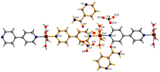
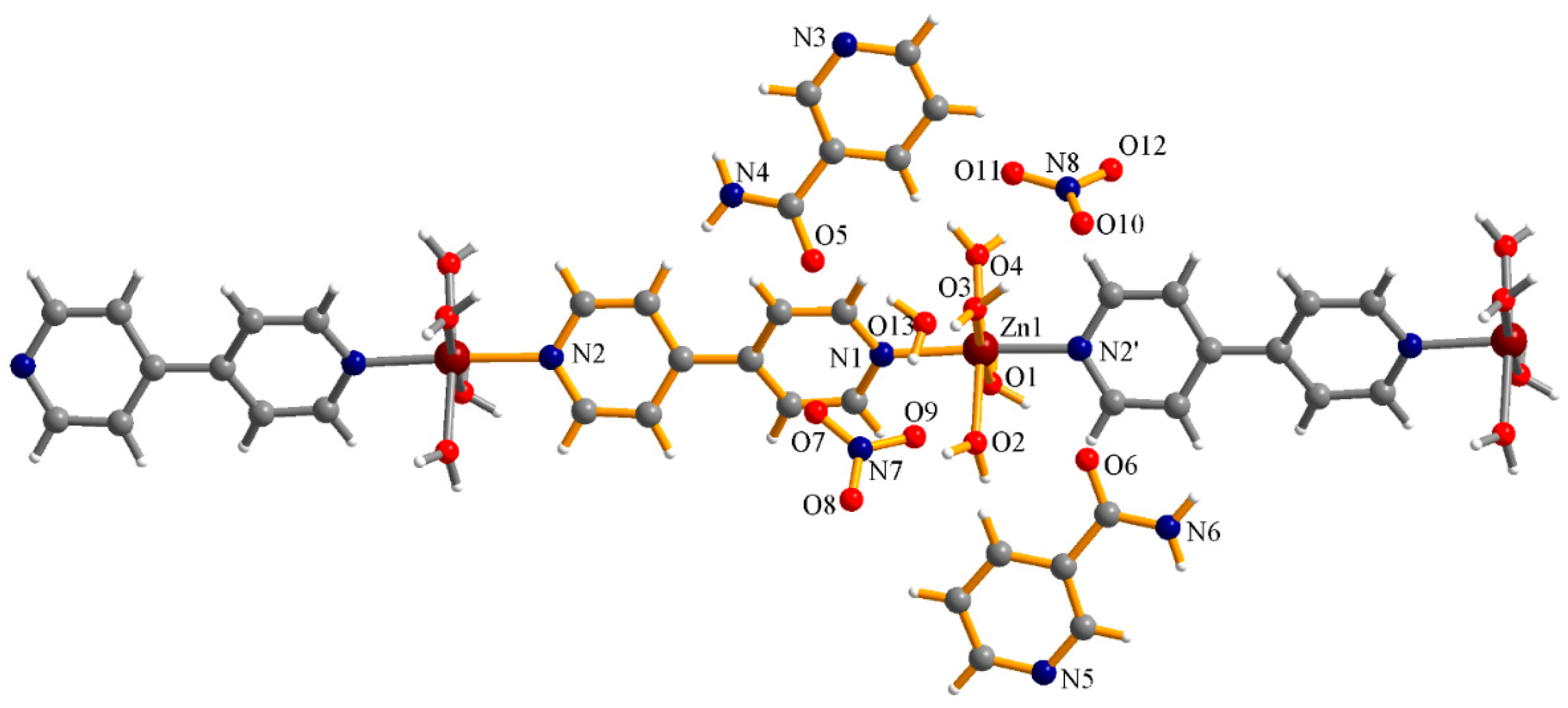
The polynuclear coordination compound {[Zn(H2O)4(4,4′-BiPy)](NO3)2·2NA·H2O}n crystallizes in the triclinic system, space group P-1. The asymmetric unit consists of one Zn(II), four coordinated water molecules, and one 4,4′-bipyridine molecule, which is bonded to Zn(II) through one of its heterocyclic nitrogen atoms. The other nitrogen atom of the 4,4′-bipyridine ligand coordinates with a neighboring metal center, ensuring the development of the polynuclear chain and confirming the role of this ligand as a spacer. The asymmetric unit contains also two uncoordinated nicotinamide molecules, one lattice water molecule, and two nitrate anions (Figure 1). This structural arrangement indicates that the 4,4′-bipyridine spacer replaces the nicotinamide molecules from the coordination sphere of the precursor complex, [Zn(NA)2(H2O)4](NO3)2·2H2O. An attempt to retain nicotinamide coordinated to Zn(II) by performing the synthesis in dry ethanol instead of water was unsuccessful as single-crystal X-ray diffraction again revealed the formation of complex (1).
Figure 1.
The structure of the 1D polynuclear compound {[Zn(H2O)4(4,4′-BiPy)](NO3)2·2NA·H2O}n (1) (the asymmetric unit is labeled in orange). Atoms colors: O, red; C, grey; N, blue; Zn, dark red.
In this compound, the Zn(II) center adopts a slightly compressed octahedral geometry. The equatorial plane is made up of the four oxygen atoms of the coordinated water molecules, while in the axial positions the nitrogen atoms from the two 4,4′-bipyridine ligands are located. The Zn-N bond lengths are shorter than the Zn-O bond lengths (Table 2).
Table 2.
Selected bond distances (Å) and angles (°) for the compound {[Zn(H2O)4(4,4′-BiPy)](NO3)2·2NA·H2O}n.
The Zn-O and Zn-N bond lengths are comparable to those reported for other one-dimensional Zn(II) coordination polymers in which 4,4′-bipyridine acts as a spacer. For example, in the compound with the formula catena-Poly[[tetraaqua(µ-4,4′-bipyridine-k2 N:N’)zinc(II)] fumarate tetrahydrate] [37], the two Zn-N distances are 2.133(3) Å and 2.146(3) Å, respectively, slightly longer than those observed for complex (1). The Zn-O bonds in this compound are of two types, Zn-O1 = 2.186(2) Å and Zn-O2 = 2.0697(18) Å, whereas in the {[Zn(H2O)4(4,4′-BiPy)](NO3)2·2NA·H2O}n compound all Zn-O bond lengths are slightly different, ranging from 2.1186(12) to 2.1781(18) Å. The angles between oxygen atoms located along an axis are almost linear, with values of 179.01(9)° and 178.43(8)° (174.76(7)° and 175.83(7)° in complex (1)), while the N-Zn-O angles, as in complex (1), are close to 90°. The coordination environment of the Zn(II) ion consists of four coordinated water molecules and the 4,4′-bipyridine spacer, similarly to complex (1), resulting in a distorted octahedral geometry. The counterion in this structure is fumarate, in contrast to complex (1), where nitrate plays this role. The asymmetric unit also contains four lattice water molecules. As in complex (1), the water molecules are involved in numerous hydrogen bonds, which contributes to the stability of the crystal lattice [37].
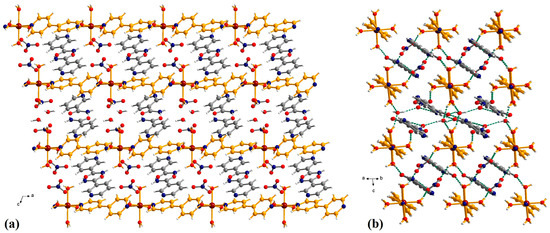
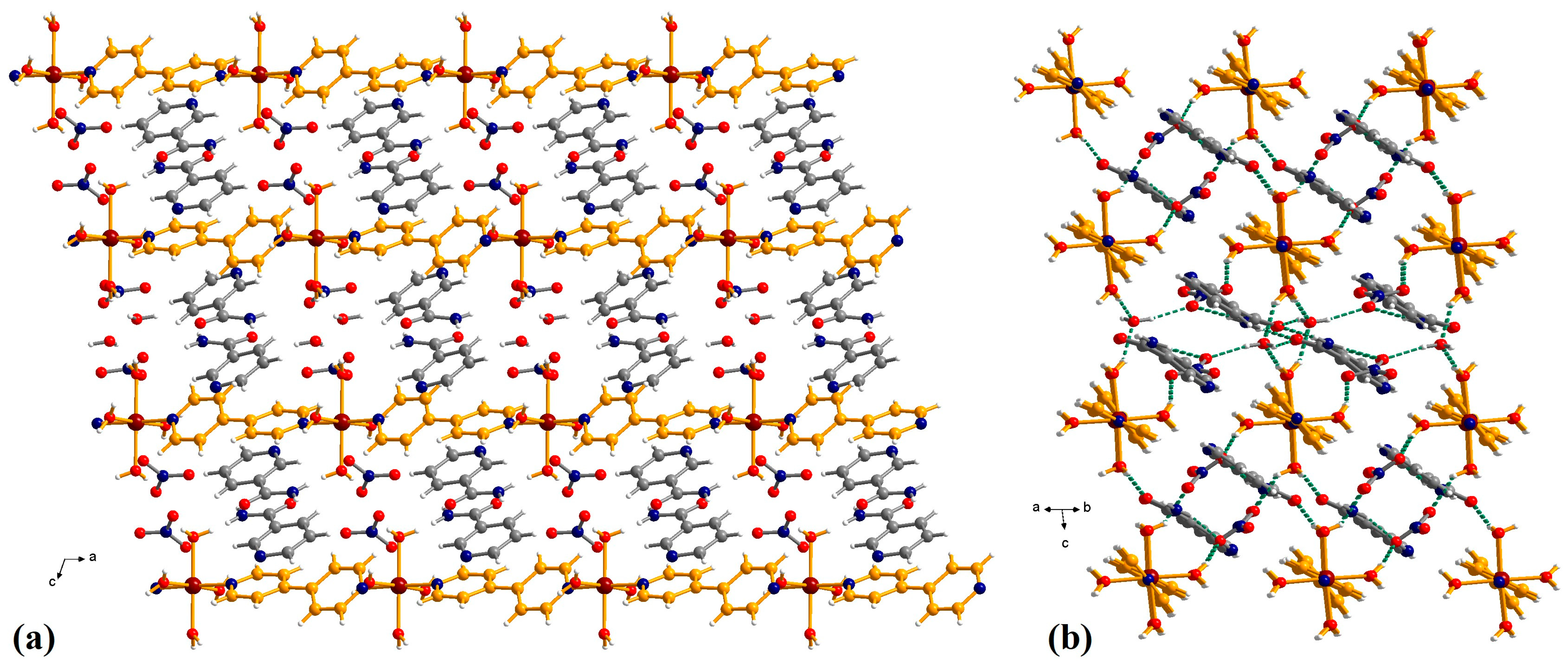
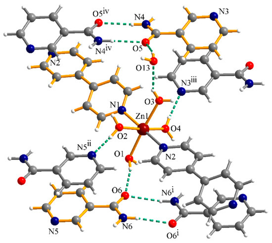
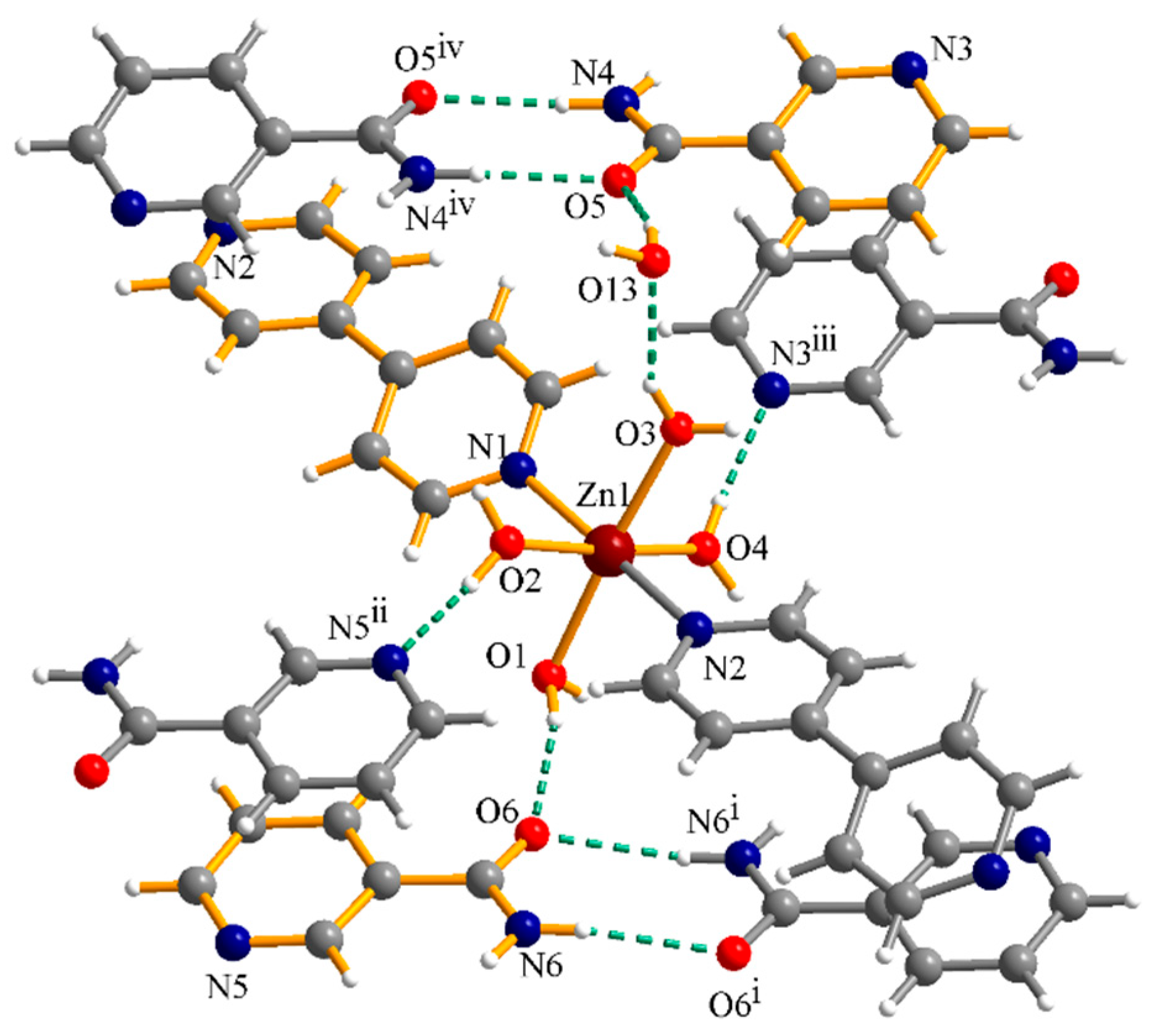
The linear {Zn(H2O)4(4,4′-BiPy)}n chains are oriented in the same direction within the crystal (Figure 2). The stability of the crystal lattice is ensured by numerous hydrogen bonds and π-π interactions. The hydrogen interactions are established between coordinated water molecules, lattice water molecules, nitrate anions, amide groups, and pyridine nitrogen atoms of the nicotinamide (Figure 2b). The nicotinamide molecules are organized in supramolecular dimers through π-π interactions. The separations within the supramolecular dimers are 3.31–3.43 Å and 3.52–3.63 Å. The nicotinamide supramolecular dimers are also in involved in π-π interactions with 4,4′-bipyridine ligands (3.12–3.62 Å and 3.25–3.65 Å). The NA molecules also generate supramolecular dimers by hydrogen interactions (Figure 3). Selected data for hydrogen interactions are listed in Table 3.
Figure 2.
Views of the packing diagrams in crystal (1): along the crystallographic b axis (a) and along the polynuclear chains showing the hydrogen interactions and π-π stacking (b). Atoms colors: O, red; C, grey; N, blue; Zn, dark red.
Figure 3.
Details of hydrogen interactions involving the nicotinamide in crystal (1).
Table 3.
Selected geometrical data of the hydrogen interactions for compound (1).
2.3.2. Complex (2)
For this complex, we aimed to test the feasibility of synthesizing the compound by mortaring the precursor complex q, [Zn(NA)2(H2O)4](NO3)2·2H2O, with KBr. We used a 1:2 molar ratio as we initially expected to obtain the previously reported complex, [Zn(NA)2Br2]. Unexpectedly, this approach led to the formation of the complex described in the present manuscript, [Zn(NA)2Br2]·2NA. The complex [Zn(NA)2Br2] was synthesized in hot ethanol, by mixing nicotinamide with a saturated solution of ZnBr2 in the appropriate molar ratio [27]. The chloride complex, [Zn(NA)2Cl2]·2NA, was prepared by reacting ZnSO4·7H2O with L-lysine monohydrochloride and nicotinamide in a water/methanol mixture (1:4) The molar ratio of the reagents was zinc sulfate: L-lysine.HCl: nicotinamide = 1:1:2 [25].
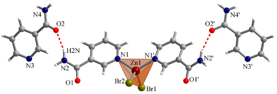
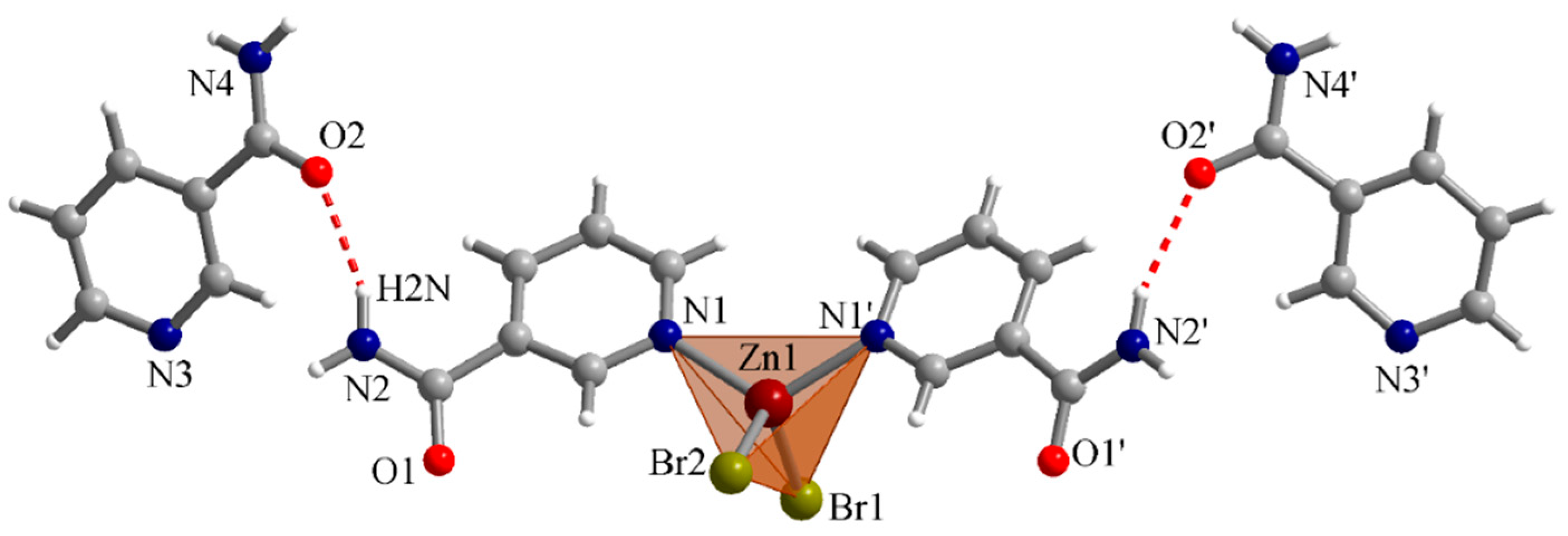
Compound (2) crystallizes in the orthorhombic Pnma space group, with four mononuclear complexes per unit cell. The Zn(II) ion is tetracoordinated by two nicotinamide ligands and two bromide ions, the nicotinamide molecules being coordinated through their pyridine nitrogen atoms. In the lattice there are two other uncoordinated nicotinamide molecules for each mononuclear complex. The metal center adopts a slightly distorted tetrahedral geometry, as illustrated in Figure 4. Selected bond lengths and angle values for complex (2) are summarized in Table 4.
Figure 4.
The structure of the complex [Zn(NA)2Br2]·2NA (2) (symmetry code: ‘ = x, 1.5 − y, z).
Table 4.
Some relevant bond lengths and angle values in the complex [Zn(NA)2Br2]·2NA.
As shown by the data in Table 4, the Zn-N and Zn-Br bond lengths range from 2.059(2) to 2.3598(6) Å, with the Zn-N distances being equal and the Zn-Br bonds showing minimal variation. The bond angles deviate slightly from the ideal tetrahedral value of 109°28′, falling in the range 106.26°–117.01°. The Zn-Br distance is in very good agreement with that reported for a similar complex, [Zn(NA)2Br2] [27]: 2.3598(6) Å, compared with 2.3592(5) Å. Similarly, the Zn-N bond lengths are very close to each other: 2.059(2) Å in our complex and 2.065(3) Å in the previously reported compound. In contrast, some differences are observed in the bond angles around the metal ion within the tetrahedral environment. Although the Br-Zn-Br angle values are comparable, 117.01° in complex 2 versus 118.9° in the reference compound, the N-Zn-N values differ significantly. In complex 2 this angle is 106.26°, whereas in the reference complex it is smaller, 96.1°. This difference results in a higher degree of distortion of the Zn(II) tetrahedral geometry in [Zn(NA)2Br2} compound compared to that observed for the complex described in this work. In the Supporting Information comparative data with other reported [Zn(NA)2(halide)2] complexes are presented.
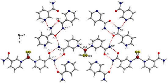
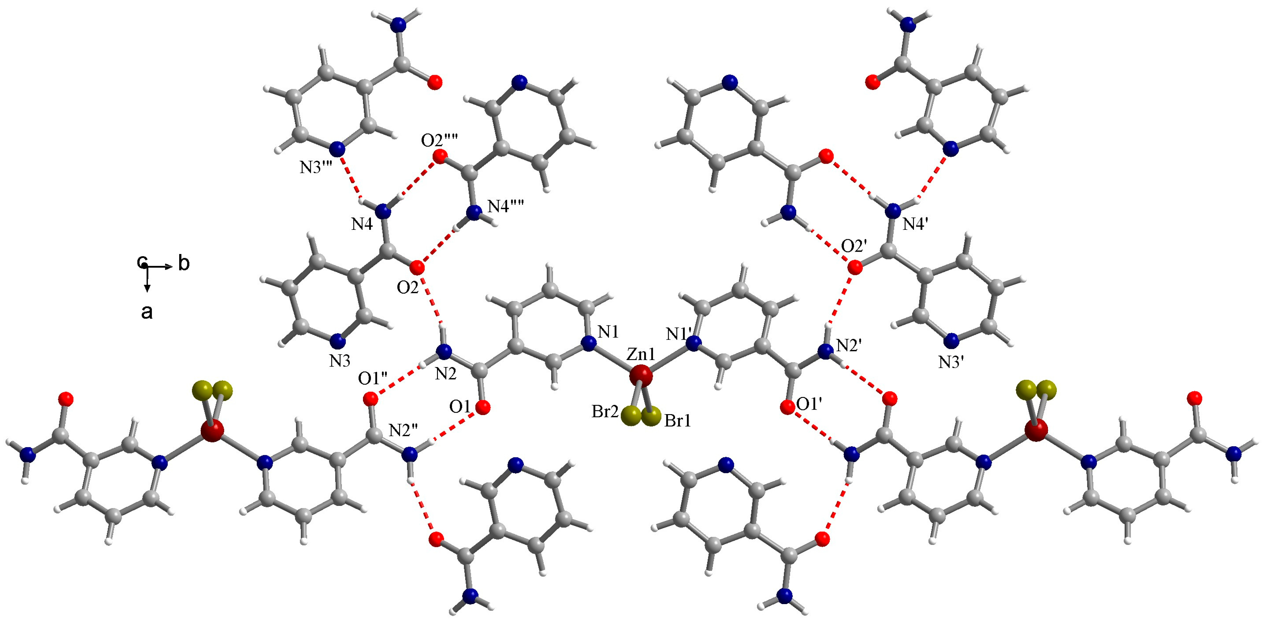
In complex (2), both the coordinated and uncoordinated nicotinamide molecules are involved in hydrogen interactions (Figure 5). The coordinated NA molecules participate in hydrogen interactions through the amide groups, while the uncoordinated NA molecules have also pyridine nitrogen atoms involved in hydrogen bonding. Details of these interactions are listed in the following table (Table 5).
Figure 5.
Details of hydrogen interactions involving the nicotinamide in crystal (2).
Table 5.
Selected geometrical data of the hydrogen interactions for compound (2).
As shown by these data, both amide hydrogen atoms (H1N and H2N) of one coordinated nicotinamide ligand form hydrogen bonds with oxygen atoms belonging to two other nicotinamide molecules, coordinated and uncoordinated. The amide hydrogen atoms (H3N and H4N) of uncoordinated NA are involved in hydrogen interactions with one pyridine nitrogen atom and one oxygen atom from the other two uncoordinated NA molecules.
2.3.3. Complex (3)
For complex 3, a zinc acetate/nicotinamide molar ratio of 3:2 was selected in order to obtain a trinuclear complex with acetate bridges. In contrast, when a 1:2 molar ratio was used, a mononuclear compound was obtained, [Zn(NA)2(CH3COO)2].
The structure of complex (3) is shown in the Supplementary Information (Figure S4).
2.4. Solution Phase Magnetic Resonance Spectroscopy (NMR)
(1):1H-NMR (500 MHz, DMSO-d6, δ ppm, J Hz): 9.02 (s, 2H, H-1), 8.72–8.68 (m, 6H, H-5, and HPy), 8.23 (dt, 2H, H-3, 2.0, 7.8 Hz), 8.20 (s, 2H, NH2), 7.86 (d, 4H, HPy, 6.2 Hz), 7.62 (s, 2H, NH2), 7.51 (dd, 2H, H-4, 4.9 Hz, 7.7 Hz) ppm.
(1):13C-NMR (125 MHz, DMSO-d6, δ ppm): 166.39, 151.76, 150.34, 148.62, 144.59, 135.35, 129.73, 123.49, 121.48. ppm.
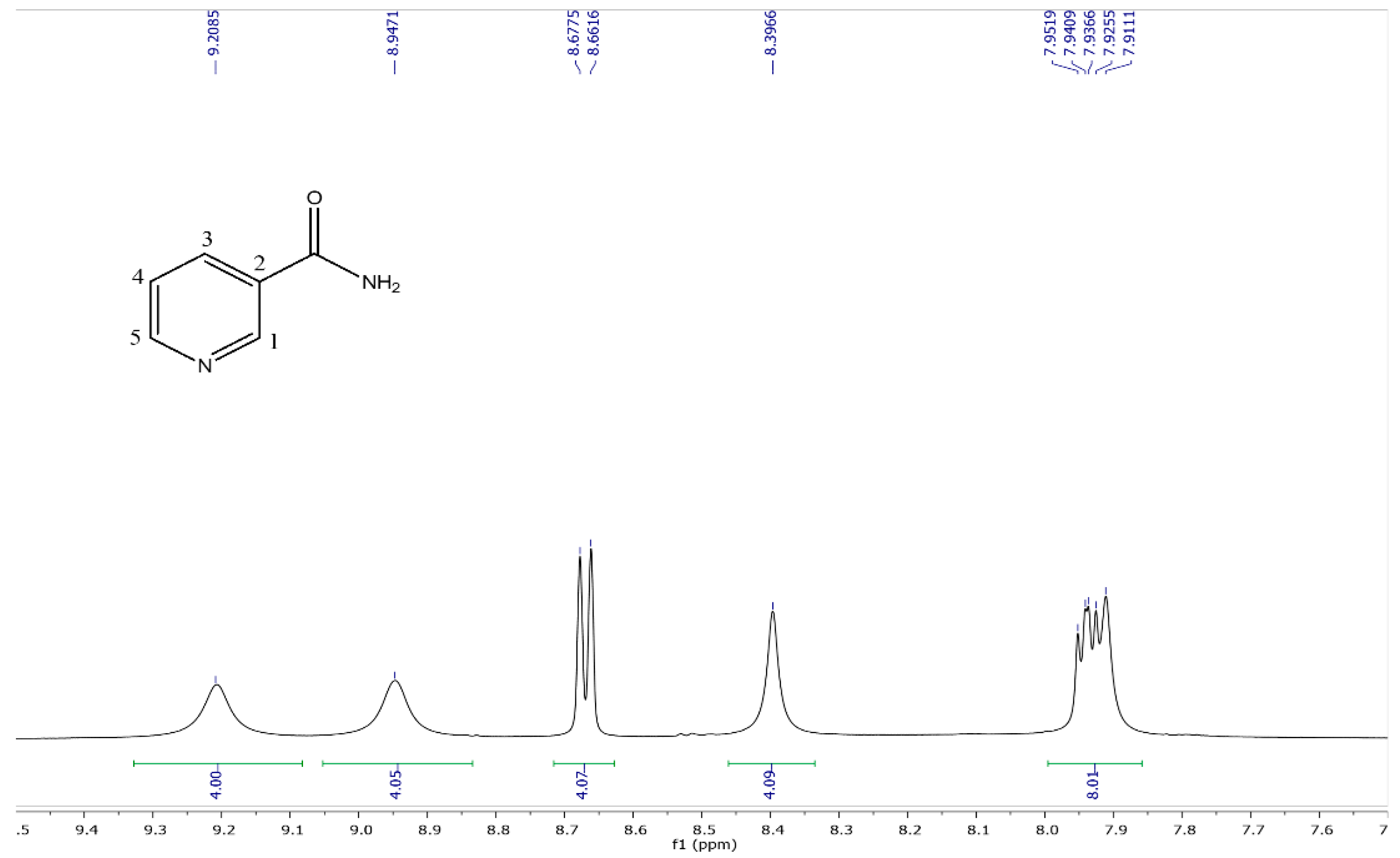
(2): 1H-NMR (500 MHz, DMSO-d6, δ ppm, J Hz): 9.21 (bs, 4H, H-1, 1.7 Hz), 8.95 (bs, 4H, H-5, 1.5 Hz, 4.8 Hz), 8.67 (d, 4H, H-3, 8.0 Hz), 8.40 (s, 4H, NH2), 7.94 (dd, 4H, H-4, 5.5 Hz, 7.7 Hz), 7.91 (s, 4H, NH2) ppm.
(2): 13C-NMR (125 MHz, DMSO-d6, δ ppm): 164.45, 147.15, 144.47, 140.70, 131.70, 125.70. ppm.
(3): 1H-NMR (500 MHz, DMSO-d6, δ ppm, J Hz): 9.02 (d, 2H, H-1, 1.7 Hz), 8.70 (dd, 2H, H-5, 1.5 Hz, 4.8 Hz), 8.22 (dt, 2H, H-3, 2.0, 7.9 Hz), 8.17 (s, 2H, NH2), 7.62 (s, 2H, NH2), 7.52 (ddd, 2H, H-4, 0.7 Hz, 4.8 Hz, 7.9 Hz), 1.81 (s, 12H, CH3) ppm.
(3): 13C-NMR (125 MHz, DMSO-d6, δ ppm): 166.34, 151.79, 148.61, 135.36, 129.77, 123.50, 22.43 ppm.
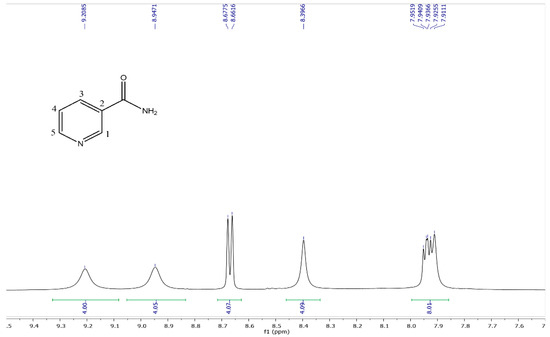
For the dissolution products of the polynuclear complex (1), HPy denotes the hydrogen atoms of 4,4′-bipyridine. The chemical structure of nicotinamide and the numbering of its atoms are shown below in Figure 6, together with the 1H-NMR spectrum of complex (2). The other NMR spectra are provided in the Supplementary Information (Figure S5–S9).
Figure 6.
1H-NMR spectrum of compound [Zn(NA)2Br2·2NA (2).
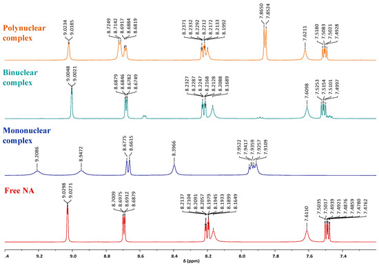
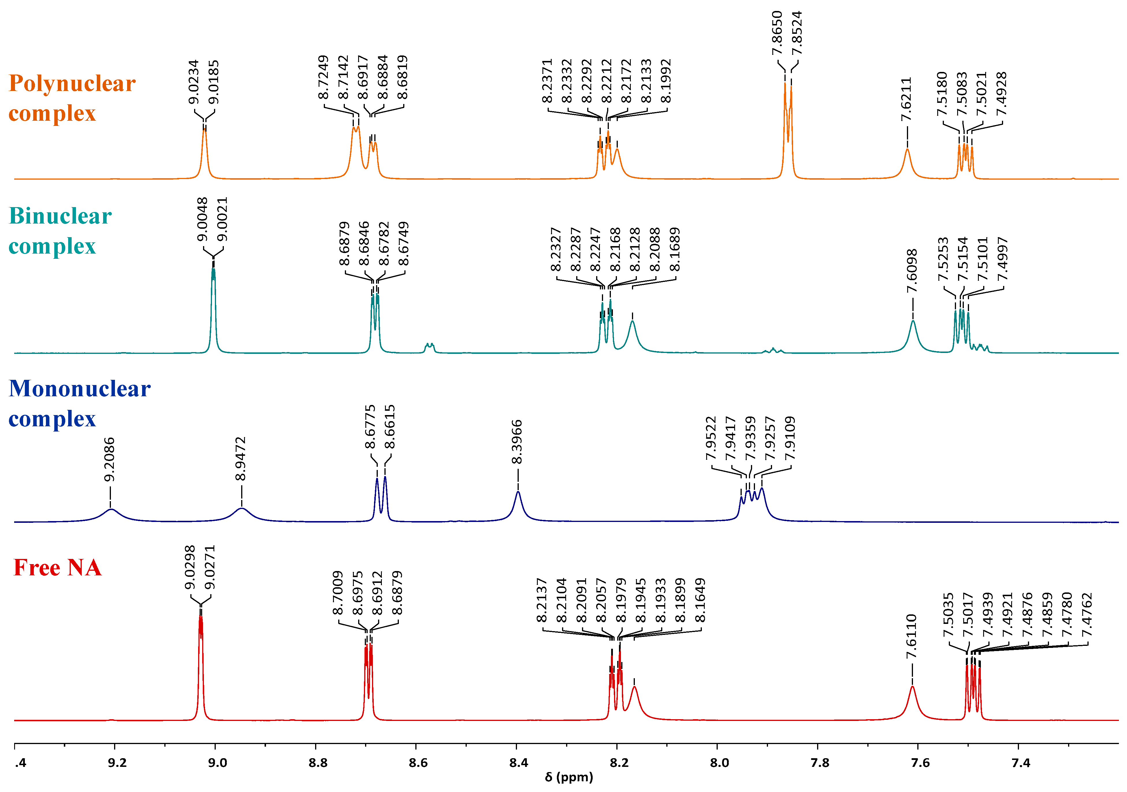
In the 1H NMR spectrum of complex (2), six distinct signal positions can be identified, corresponding to the six types of hydrogen atoms. In contrast, the spectrum of complex (3) displays an additional strong signal at 1.81 ppm, attributed to the hydrogen atoms of the acetate groups. All maxima are shifted relative to those of free nicotinamide, indicating its presence in the structure of complexes. Due to the electron-withdrawing inductive effect of the bromine atoms, the protons of nicotinamide in the mononuclear complex are more deshielded compared to those in the acetate and polynuclear complexes. A comparison of the 1H NMR spectrum of nicotinamide with those of complexes (1)–(3) is presented in Figure 7.
Figure 7.
1H-NMR spectra of free NA (red), mononuclear (blue), acetate (green), and polynuclear (orange) complexes.
By recording 1H-NMR spectrum of complex (3) for five consecutive days, we demonstrated its structural stability in DMSO (Figure S10). We also recorded the 1H-NMR spectrum for the acetate complex in DMSO-d6 and D2O addition (Figure S11). The disappearance of the amino protons from nicotinamide moieties due to the exchange proton deuterium was noticed. Due to this exchange, the chemical shifts were also affected, with deuterium causing upfield shifts (lower ppm values). Another 1H NMR spectrum was recorded after 29 days, and it can be noticed that the complex is stable in solution (Figure S12). Initially complex 3 is dimeric, but the NMR data suggests that in solution it may transfer to a monomeric species, [Zn(CH3COO)2(H2O)2(NA)] [29].
2.5. Thermogravimetric Analysis
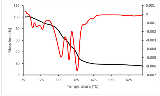
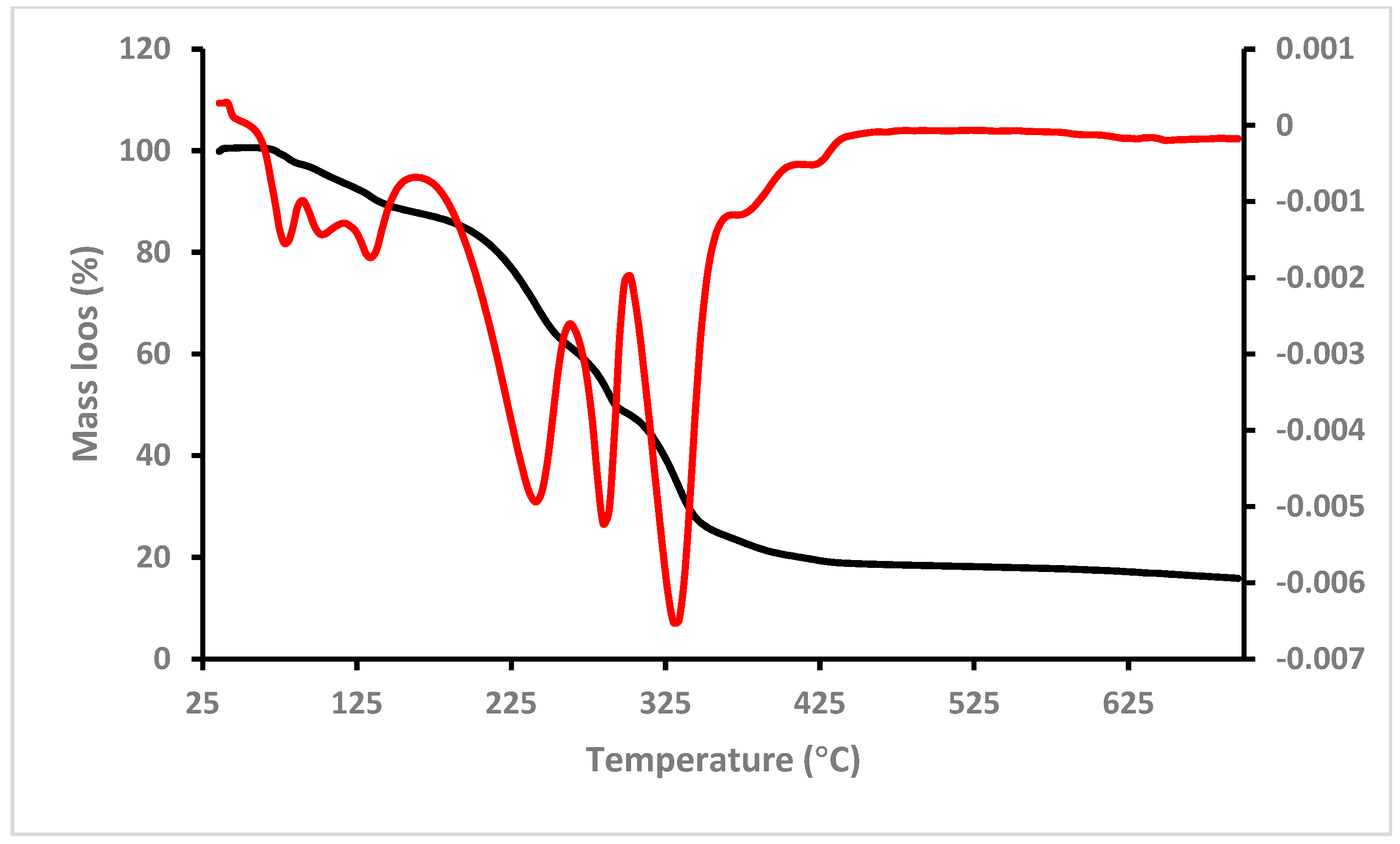
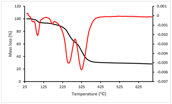
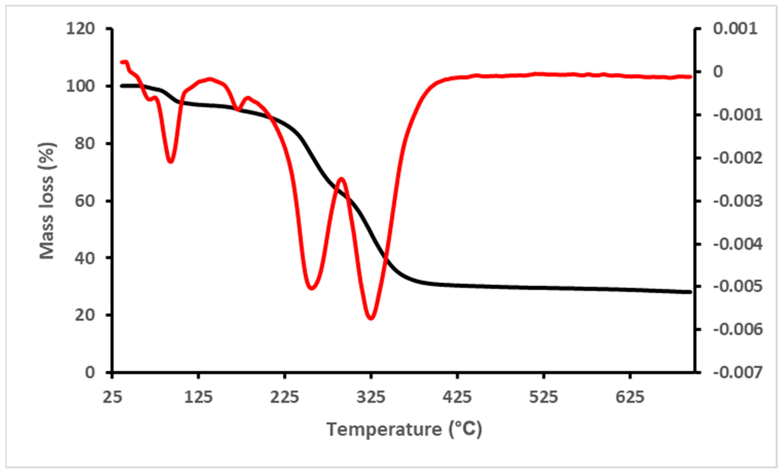
The TG curves of complexes (1) and (2) confirm the presence of the lattice and coordinated water molecules in their structure. The TG curve of complex (1) is shown in Figure 8.
Figure 8.
TG (black) and DTG (red) curves of complex {[Zn(H2O)4(4,4′-BiPy)](NO3)2·2NA·H2O}n (1).
As can be seen in the figure above, the polynuclear complex exhibits three decomposition steps in the range 65–161 °C. The first step occurs between 65 and 86 °C and is due to the loss of the lattice water molecule (found = 2.96%; calculated = 2.64%). The next two steps develop in the ranges 86–124 °C and 124–161 °C, and are attributed to the removal of the coordinated water molecules. The experimental mass losses are 4.87% and 5.16%, totaling 10.03%, while the calculated value is 10.56%. Beyond this, the TG curve shows three more decomposition steps, occurring between 161 and 266 °C, 266 and 301 °C, and 301 and 450 °C. The observed mass loss for the first two stages is 39.94%, which is probably due to the elimination of 4,4′-bipyridine along with one nicotinamide molecule (calculated = 40.88%). In the final decomposition stage, which begins at 301 °C and ends around 450 °C, the mass loss (29.24%) is due to the removal of the second nicotinamide molecule and the decomposition of zinc nitrate.
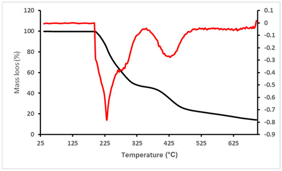
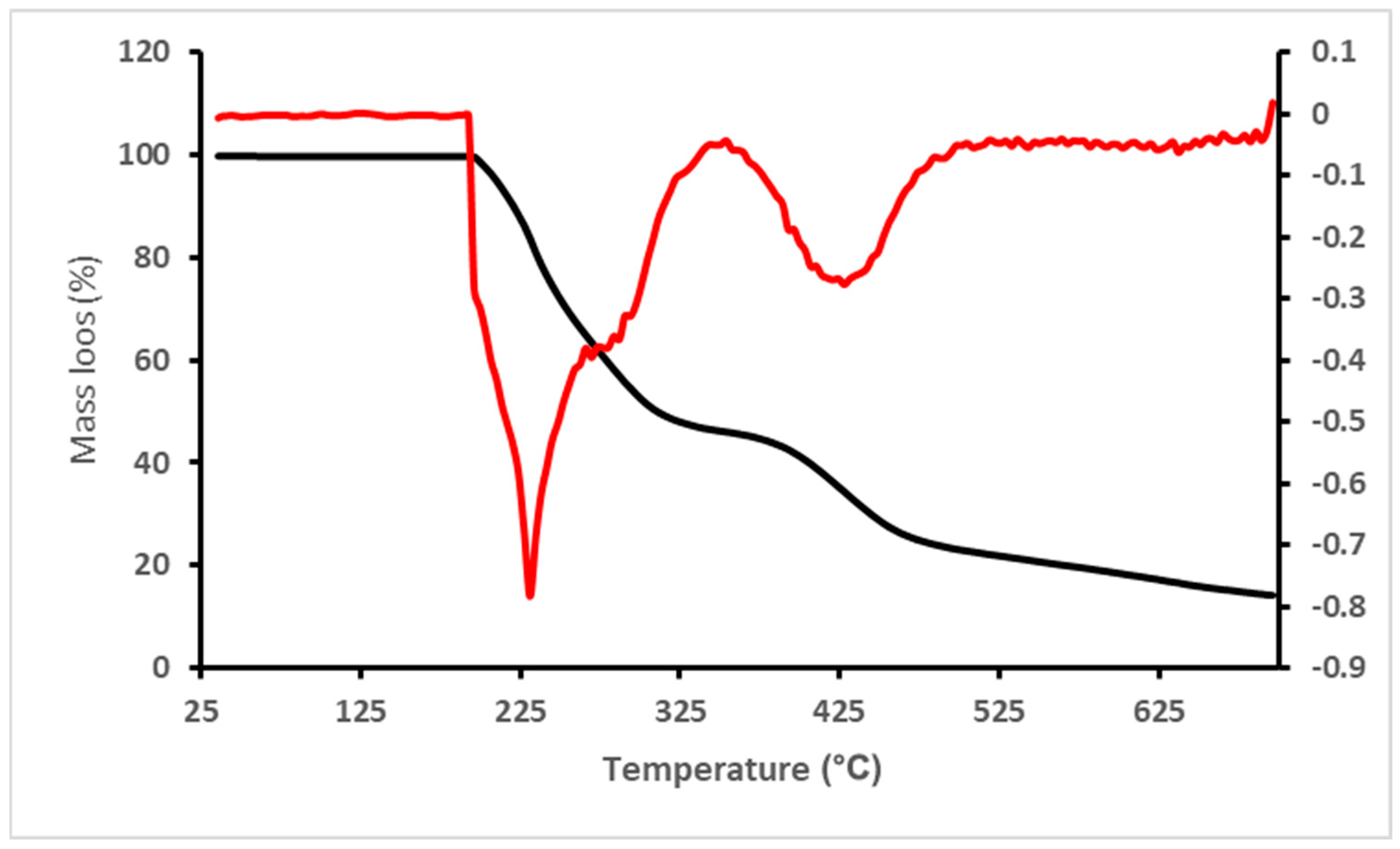
The thermogram of complex (2), [Zn(NA)2Br2]·2NA (Figure 9), shows that it is stable up to 199 °C. Above this temperature, two decomposition stages can be observed. The first occurs between 199 and 360 °C and shows a mass loss of 54.2%, which roughly corresponds to the elimination of three of the four nicotinamide molecules (calculated = 51.28%). The second stage develops between 360 and 500 °C, with a mass loss of 22.76%, attributed to the removal of the remaining nicotinamide ligand, overlapping with the onset of zinc bromide decomposition. Above 500 °C, the decomposition continues slowly until the end of the measurement (700 °C). The TG and DTG curves of complex (2) are shown in the figure below.
Figure 9.
TG (black) and DTG (red) curves of complex [Zn(NA)2Br2]·2NA (2).
The decomposition of complex (3), [Zn2(ac)4(NA)2]·2H2O, takes place in three steps. The first step occurs between 72 and 114 °C and corresponds to the elimination of the two lattice water molecules (found = 5.24%; calculated = 5.55%). Between 114 and 160 °C, the complex remains stable, after which two major decomposition steps are observed. The first occurs between 160 and 290 °C, and the second between 290 and 400 °C. The mass loss in the first step is 30.14%, while in the second is 31.82%. The elimination of nicotinamide takes place mainly during the first step (calculated = 37.73%), while the second step overlaps with the decomposition of zinc acetate. By recording the IR spectrum of the residue of complex (3) and comparing it with database spectra, we concluded that it consists of ZnO (Figure S13). Also, due to its black color, it probably contains residual carbon as well.
We also recorded the TG curve of [Zn(CH3COO)2]·2H2O in a nitrogen atmosphere and compared it with that of complex (3). As shown in Figure S14, the second mass-loss step of zinc acetate dihydrate occurs between 215 °C and 320 °C, overlapping with the decomposition of nicotinamide. Moreover, the TG curve indicates a sublimation process preceding the decomposition stage of zinc acetate [38].
The thermogram of complex (3) is shown in Figure 10.
Figure 10.
TG (black) and DTG (red) curves of complex [Zn2(ac)4(NA)2]·2H2O (3).
2.6. Photoluminescence
The room temperature photoluminescence of compounds was investigated in the solid state using different excitation wavelengths in the 270–350 nm range, but only compound (2) presents weak luminescent properties. The emission spectrum resulting from excitation at 300 nm shows an asymmetric band with maxima at 475 nm. The band is attributed to intra-ligand transitions. Aromatic compounds containing a heterocyclic nitrogen atom with a non-bonding electron pair typically exhibit two emission maxima, corresponding to π*-π and π*-n transitions. The broad and asymmetric shape of the band suggests an overlap of these two emission peaks [39]. The spectrum is presented in the Supplementary Information (Figure S15).
2.7. Antimicrobial Investigation
In biological tests, the names of the complexes refer exclusively to the solid samples, without implying that the same structure was maintained in solution, where speciation processes can occur. The antimicrobial activity of complex 1 (based on 4,4′-bipyridine) was not studied in this work since it was included purely for structural and crystallo-chemical purposes, and its stability in aqueous solutions does not make it suitable for biological assessments (high cytotoxicity). According to the qualitative assessment of antibacterial activity, the metal precursors showed larger inhibition zones than the final complexes due to the decrease in the availability of the Zn2+ ion after complexation (Zn(CH3COO)2 vs. [Zn2(CH3COO)4(NA)2]·2H2O). By stabilizing the metal ion and limiting its release into the biological environment, nicotinamide and anions (acetate and bromide) reduce direct interaction with bacterial cellular targets. Additionally, the synthesized complexes have larger molecular sizes and lower solubility, which limits their diffusion through the medium and contributes to smaller inhibition zones (Table 6). Similar results have been highlighted in the specialized literature for stable Zn(II) complexes, where biological activity is inversely proportional to the thermodynamic stability of the complex [40].
Table 6.
The outcomes of qualitative antimicrobial activity against tested microbial strains.
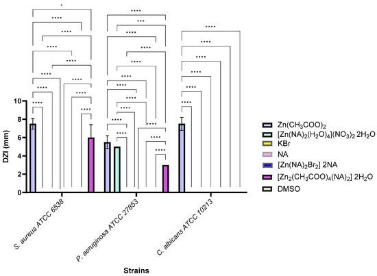
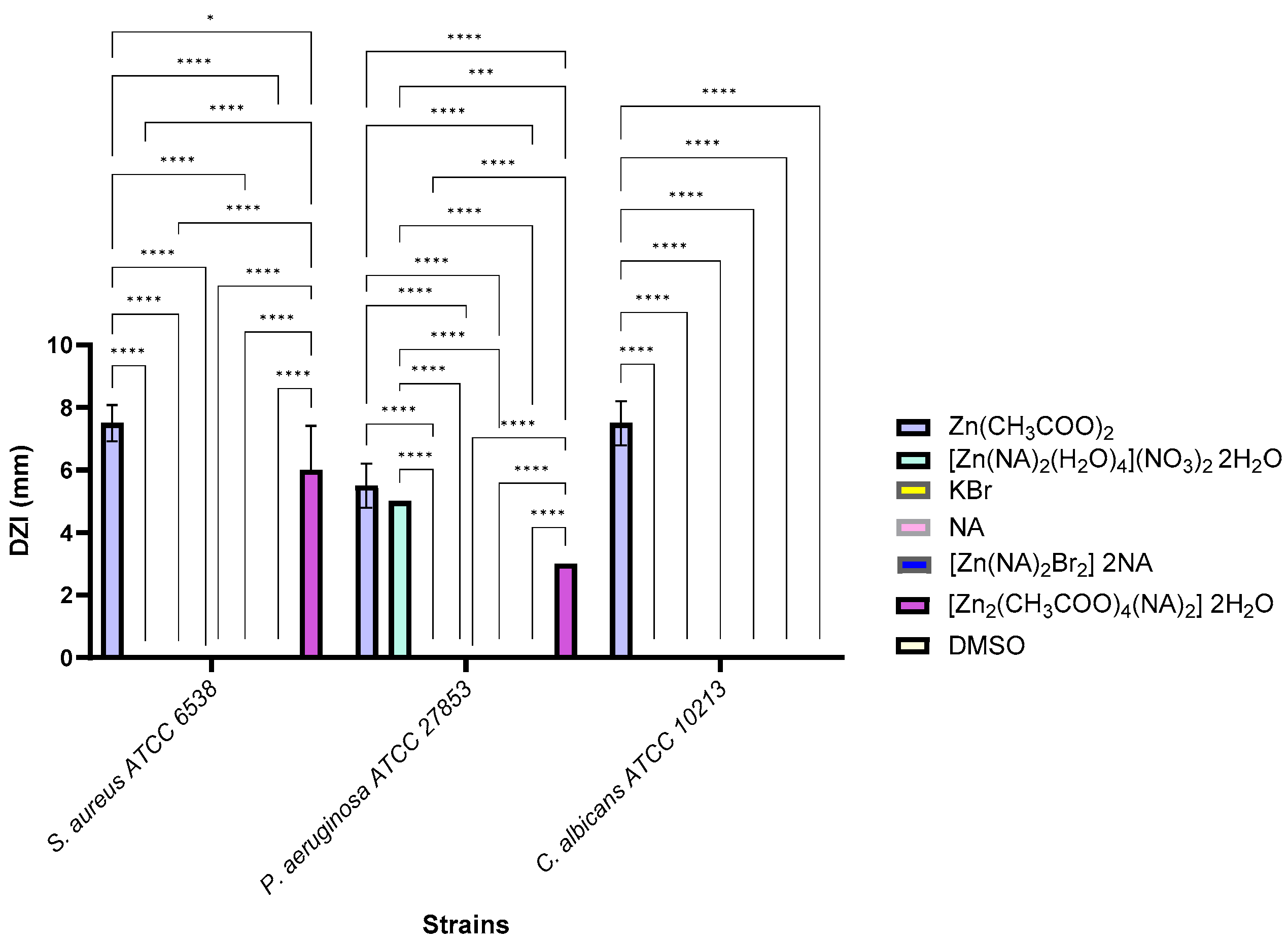
In contrast to the brominated complex [Zn(NA)2Br2]·2NA, zinc acetate complex showed better antibacterial activity. While [Zn(NA)2(H2O)4]2(NO3)2·2H2O appeared to show activity against the Gram-negative strain in the direct-contact spot assay, this effect was restricted to the contact area and did not represent a true inhibition zone, whereas Figure 11 showed that Zn(CH3COO)2 exhibits antimicrobial activity against all three tested strains. The selective antibacterial activity of the complex [Zn(NA)2(H2O)4](NO3)2·2H2O against P. aeruginosa can be associated with the particularities of the Gram-negative bacteria’s outer membrane and the fact that the sample was administered in DMSO, which can facilitate membrane permeabilization and complex transport, an effect observed in a recent study for ZnSO4 and ZnCl2 against E. coli strain [41]. The lack of activity for free nicotinic acid, DMSO, and the reduced activity of zinc salts used as controls indicate that the observed effect was dependent on the complex structure.
Figure 11.
Comparative qualitative assessment of the solvent’s (DMSO) and samples’ antimicrobial efficiency against various microbial strains. Statistical significance was defined as * p < 0.05, *** p < 0.001, and **** p < 0.0001.
For S. aureus, the Zn(NO3)2 salt, corresponding to the dissociation of the complex [Zn(NA)2(H2O)4](NO3)2·2H2O, showed no detectable antibacterial activity (DIZ = 0.00 mm), while Zn(NO3)2, tested at the same mass concentration (10 mg/mL), induced a small inhibition zone (5.00 ± 1.41 mm). However, this difference cannot be interpreted as a direct comparison of biological potency as the effective concentration of Zn(NO3)2 or free Zn2+ present in the complex medium is lower and cannot be quantified under the experimental conditions used. The results suggest that Zn2+ species derived from Zn(NO3)2 can contribute to the inhibition of S. aureus, while complexation with the NA ligand can reduce the availability of the Zn2+ ion. In the case of P. aeruginosa, both ZnBr2 and Zn(NO3)2 showed lower activity compared to [Zn(NA)2(H2O)4](NO3)2·2H2O, suggesting that the labile Zn2+ species resulting from dissociation were not solely influenced by the free Zn2+ ion, but the effect may be due to the nature of the ligand and the complex architecture (Table 6).
Complexation resulted in a significant decrease in the compound’s antibacterial activity against the S. aureus strain (p < 0.05) as well as the P. aeruginosa and C. albicans strains (p < 0.0001). In the case of the complex [Zn(NA)2Br2]·2NA, activity was significantly reduced against P. aeruginosa (p < 0.0001). Qualitatively, the compound [Zn(NA)2Br2]·2NA did not exhibit antimicrobial activity. This behavior can be attributed to the more labile nature of Zn–O (acetate) bonds compared to Zn–Br, which allows for easier release of biologically active Zn2+ ions [42]. Furthermore, the acetate complex has increased solubility and more effective diffusion into the agar medium, resulting in greater DIZ [40]. The dimeric structure enables for a larger concentration of Zn2+ to interact with the bacterial cell wall. Similar findings have been reported for Zn(II) complexes containing carboxylate ligands, where biological activity was associated with metal ion mobility and moderate complex stability [16,40]. Generally, studies on Zn(II) complexes/polymers report that coordination complexes are often more antibacterial than the free ligand or simple Zn salts, partly due to the controlled release of Zn2+ and the generation of reactive oxygen species (ROS) that affect microbial cells [43].
Analysis of the minimum inhibitory concentration (MIC), minimum microbicidal concentration (MMC), and minimal biofilm eradication concentration (MBEC) values revealed that the metal precursors have antibacterial activity similar to the final complexes. In Table 7, MIC values that were lower than the solvent’s are highlighted in blue, revealing that none of the compounds showed activity against the Gram-negative bacterium P. aeruginosa. Gram-negative bacteria typically exhibit increased resistance to antibiotics and synthetic compounds due to the presence of a lipopolysaccharide outer membrane that limits permeability and facilitates the active expulsion of antimicrobial agents [44]. Although the MIC values obtained in broth were similar between the metal precursors and the final complexes, agar tests indicated smaller inhibition zones for the complexes against the S. aureus and C. albicans strains. The discrepancy was attributed to transport and speciation factors: the complexes diffuse more slowly and can interact with the agar matrix, reducing the active fraction. In a liquid medium, the speciation equilibrium favors the partial release of Zn2+, which equalizes the activity and explains the similar MICs to the precursors. These findings are in line with the literature, which demonstrates that the lability and availability of the metal ion determine the biological activity of Zn(II) complexes and that agar techniques might understate the activity of compounds with low diffusion [45]. A similar trend was also described for other Zn(II) coordination polymers; for example, SLUG-39 ([Zn(bipy)(OH2)42+]1.5[ClO4−]3·(bipy)3(H2O)) and SLUG-40 ([Zn1.5(C2H3O2)2(bipy)2+][ClO4−]·H2O) complexes showed lower MIC values against S. epidermidis and E. coli than zinc acetate, demonstrating that modifications to the coordination medium can significantly improve antibacterial performance [46]. Similarly, Zn(II) complexes based on chitosan were reported to exhibit enhanced antibacterial activity compared to the ligand alone, particularly against S. aureus, where Zn-containing films (12ChiZn) almost completely suppressed colony formation after 24 h, while the neat polymer showed minimal inhibition [47]. Although all stock solutions were prepared at 10 mg/mL, the corresponding molarities differ (0.0456 M for Zn(CH3COO)2·2H2O vs. 0.0155 M for [Zn2(CH3COO)4(NA)2]·2H2O), which may partially reduce potency but does not account for the lack of inhibition on agar. MIC/MBEC values confirm antifungal activity, while the absence of a detectable zone results from the poor diffusion of the high-molecular-weight dimer and the intrinsic resistance of C. albicans, whose chitin/β-glucan-rich cell wall requires substantially higher local concentrations for inhibition.
Table 7.
Minimum inhibitory concentration (MIC), minimum microbicidal concentration (MMC), and minimum biofilm eradication concentration (MBEC) as indicators of quantitative antimicrobial activity. Color-coded presentation of antimicrobial activity, where blue indicates MIC values and yellow indicates MBEC values of the samples that are lower than those of the solvent control. The green highlights MBEC values that are lower than both the solvent control and the corresponding MIC of the same sample.
The zinc acetate complex has higher activity than the brominated complex because of the acetate ligand’s greater solubility in an aqueous environment and its more labile character. Conversely, the more stable and hydrophobic brominated molecule [Zn(NA)2Br2]·2NA exhibited less activity, particularly against yeast strains. Carboxylate ligand complexes generally exhibit more pronounced activity than those with more stable ligands, such as halides, these results being correlated with previous studies [40,42].
From Table 7 it can be observed that none of the compounds exhibit antimicrobial activity, and the microbial adherence of the C. albicans cells was inhibited at values lower than those generated by the solvent (DMSO); these values are highlighted in yellow. In the case of the precursor [Zn(NA)2(H2O)4](NO3)2·2H2O, it was observed that the MBEC value is lower than the MIC, which suggests pronounced activity against adherent forms compared to planktonic ones. Among the tested complexes, [Zn2(CH3COO)4(NA)2]·2NA exhibits the best antibiofilm activity, with an MBEC value of 0.3125 mg/mL, similar to the MIC value. Similar MIC values for Zn(NO3)2 and ZnBr2 indicate an antimicrobial effect dominated by the Zn2+ against planktonic cells, while higher MBEC values reflect reduced efficacy in inhibiting microbial adherence. For S. aureus and C. albicans, the MICs were comparable to those of the complex ([Zn2(CH3COO)4(NA)2]·2H2O), without being able to attribute superior activity to the Zn salts, considering their lower molecular weight and effectively higher concentration of Zn2+. In Table 7, blue highlights the MIC values, and yellow highlights the MBEC values of the samples, which were lower than those for the solvent (DMSO). Green highlights the MBEC values of the samples which were lower than MIC values. The antimicrobial activity observed in this study for the bromine-containing Zn(II)-nicotinamide complexes had MIC values of approximately 1.25 mg/mL and measurable MBEC values for most of the strains tested, but this differs from the data reported in the scientific literature for the chloride analogs [Zn(NA)2Cl2], for which MIC values could not be determined (>1 mg/mL) against the S. aureus, E. faecalis, E. coli, and P. aeruginosa strains [26] or evaluations based solely on inhibition zones [48] for chloride analogues, where a similar trend to our study was observed, with NA showing weaker activity compared to the complex combinations studied. These differences can be attributed to variations in the nature of the halogen, the coordination environment, and the experimental methodology used, without implying a direct structure–activity correlation.
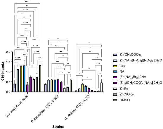
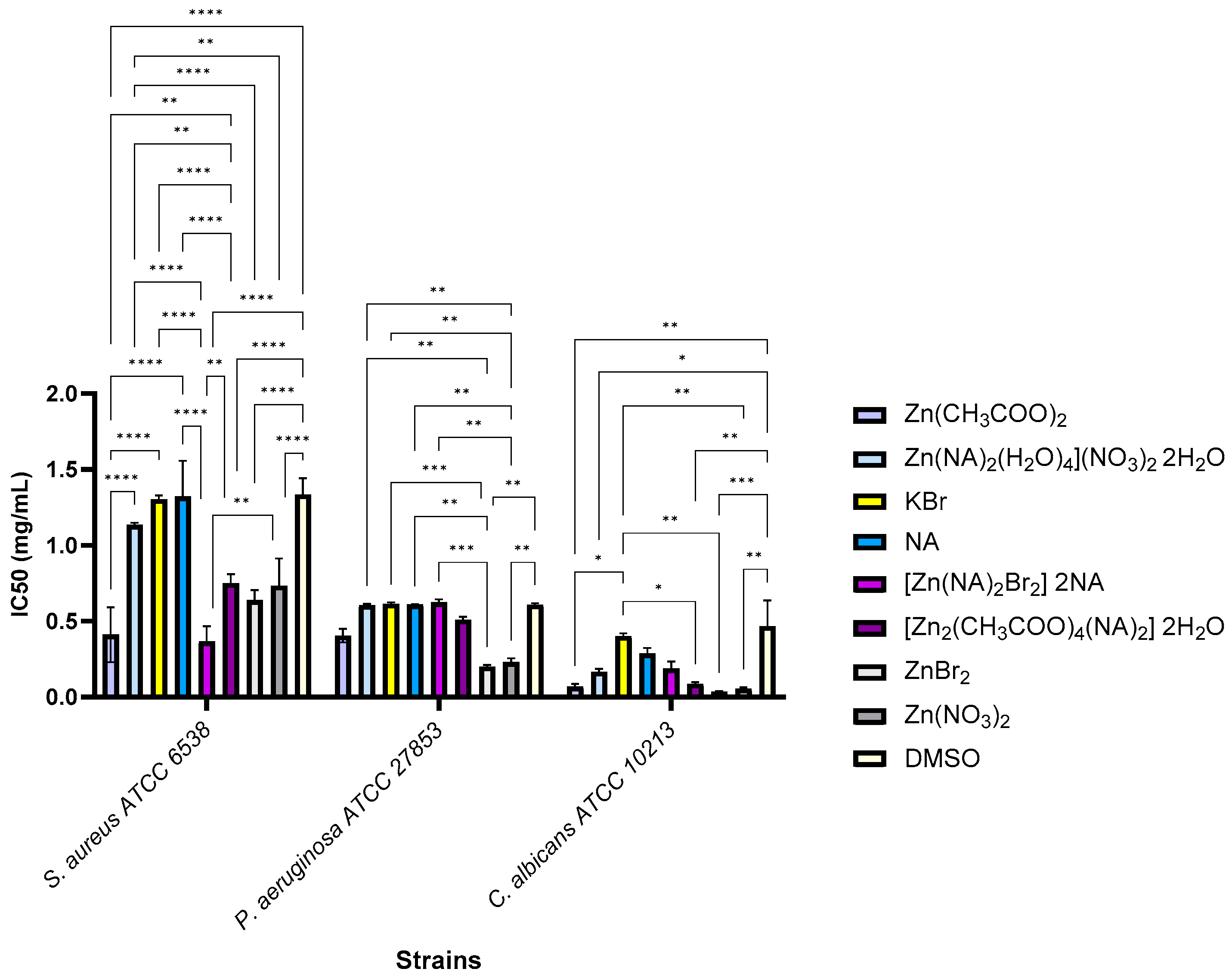
The IC50 data showed that metal precursors (Zn(CH3COO)2 and [Zn(NA)2(H2O)4](NO3)2·2H2O) were more effective than final complexes against Gram-positive bacteria (S. aureus) and fungal strains (C. albicans). Figure 12 showed that the complex [Zn2(CH3COO)4(NA)2]·2H2O and its precursor exhibited the highest activity, with IC50 values much lower than the solvent used for the yeast strain (p < 0.05). For the S. aureus strain, a considerable increase in the inhibitory concentration (IC50) of the precursor relative to the final complex [Zn(CH3COO)4(NA)2]·2H2O was observed (p < 0.01), while for the complex [Zn(NA)2Br2]·2NA, the IC50 value was considerably lower than that of the precursor (p < 0.01). This behavior correlates with the increased precursor lability and the greater availability of Zn2+ ions in the tested environment. In the case of Gram-negative bacteria, the IC50 values remained similar for all samples, confirming the intrinsic resistance conferred by the lipopolysaccharide outer membrane (p > 0.05). The acetate complex [Zn2(CH3COO)4(NA)2]·2H2O exhibits better activity than the brominated complex [Zn(NA)2Br2]·2NA, which was explained by its higher solubility and the more labile nature of the acetate ligand, which favors the release of Zn2+. The results support the idea that the biological activity of Zn(II) complexes is inversely proportional to their thermodynamic stability and directly reliant on the metal ion’s availability. The investigated Zn salts showed significantly lower IC50 values compared to the corresponding complexes against S. aureus and P. aeruginosa strains (p < 0.01); however, this difference does not directly reflect superior biological activity as the comparison is influenced by molecular weight differences, which lead to a higher effective concentration of Zn2+ at the same applied dose. In the case of C. albicans, the differences are insignificant (p > 0.05).
Figure 12.
Antimicrobial activity against tested microbial strains, as measured by IC50, and its relationship to solvent control (* p < 0.05, ** p < 0.01, *** p < 0.001, **** p < 0.0001).
Possible mechanisms according to the specialized literature for Zn(II) systems are controlled by complex lability and the availability of the Zn2+ ion [16,49,50]. More labile complexes, such as those with carboxylate ligands, release Zn2+ more efficiently and exhibit superior activity compared to those with more stable ligands (e.g., bromides), a phenomenon also reported for the SLUG-39/40 polymers, where modifying the coordination environment significantly increased the antimicrobial potency [46]. Additionally, Zn(II) complexes can exhibit greater activity than their precursors through controlled release of Zn2+ and generation of ROS, a mechanism highlighted in studies on Zn(II) complexes [50]. The limitations observed in agar tests are explainable by the low diffusion of bulky structures and possible interactions with the agar matrix, a factor known to lead to underestimation of the actual activity of compounds with reduced mobility [51]. An equilibrium between complex stability, ligand type, and Zn2+ ion availability controlled the antimicrobial activity of NA-based Zn(II) complexes. Compared to metal precursors, complexation with stable ligands reduces activity. Carboxylate ligands and dimeric structures may promote the release of Zn2+, resulting in stronger antibacterial action. This study’s contribution was to experimentally investigate the direct relationship between the biological response, ligand type, and complex structure. These findings serve as a foundation for future designs of Zn(II) complexes with regulated antibacterial activity.
3. Conclusions
Three Zn(II) complexes containing nicotinamide, 4,4′-bipyridine, bromide, and acetate as mixed ligands were synthesized and characterized by single-crystal X-ray diffraction, elemental analysis, FT-IR, fluorescence, 1H and 13C NMR spectroscopy, and thermogravimetric analysis. Using the spacer 4,4′-bipyridine and starting from the mononuclear complex [Zn(NA)2(H2O)4](NO3)2·2H2O, we successfully synthesized a 1D polynuclear compound in which adjacent metal centers are linked by the spacer. The same precursor, [Zn(NA)2(H2O)4](NO3)2·2H2O, was employed to obtain a mononuclear complex, where nitrate ions were substituted by bromide and entered the coordination sphere. In the polymeric compound, the Zn(II) center adopts a slightly distorted octahedral geometry, whereas in the monomeric complex it exhibits a slightly distorted tetrahedral environment. Both the bromide complex and the binuclear acetate-nicotinamide complex, [Zn(CH3COO)4(NA)2]·2H2O, were evaluated for their antimicrobial activity. The results indicate a direct correlation between biological efficacy and the availability and lability of the Zn2+ ion, suggesting that zinc precursors exhibit superior antibacterial activity compared to the final complexes. The zinc acetate complex showed the best activity among the compounds synthesized, especially against the yeast strain C. albicans. This behavior was linked to the more labile and more soluble nature of the acetate ligand. Additionally, the structural characteristics of their cell walls impact the reaction of yeast, Gram-positive, and Gram-negative strains.
Supplementary Materials
The following supporting information can be downloaded at https://www.mdpi.com/article/10.3390/cryst16020110/s1; Figure S1: IR spectrum of complex {[Zn(H2O)4(4,4′-BiPy)](NO3)2·2NA·H2O}n; Figure S2: IR spectrum of complex [Zn(NA)2Br2]·2NA; Figure S3: IR spectrum of complex [Zn2(CH3COO)4(NA)2]·2H2O]; Figure S4: The structure of complex [Zn2(CH3COO)4(NA)2]·2H2O]; Figure S5: 13C-NMR spectrum of compound [Zn2(CH3COO)4(NA)2]·2H2O]; Figure S6: 1H-NMR spectrum of compound [Zn2(CH3COO)4(NA)2]·2H2O]; Figure S7: 13C-NMR spectrum of complex [Zn(NA)2Br2]·2NA; Figure S8: 1H-NMR spectrum of compound {[Zn(H2O)4(4,4′-BiPy)](NO3)2·2NA·H2O}n; Figure S9: 13C-NMR spectrum of compound {[Zn(H2O)4(4,4′-BiPy)](NO3)2·2NA·H2O}n; Figure S10: 1H-NMR spectra of complex (3) recorded in time; Figure S11: 1H-NMR spectra of binuclear complex in DMSO-d6 (red) and binuclear complex in DMSO-d6 + D2O (blue); Figure S12: 1H-NMR spectra of the binuclear complex in DMSO-d6 + D2O recorded initially (dark blue) and after 29 days (light blue); Figure S13: IR spectra of the residue of complex (3) compared with that of ZnO; Figure S14: TG decomposition curves of complex 3 (red line) and zinc acetate dihydrate (black line); Figure S15: Fluorescence spectrum of complex [Zn(NA)2Br2]·2NA; Figure S16: Details of hydrogen interactions involving the nicotinamide in [Zn(NA)2Cl2]·2NA; Figure S17: The supramolecular zig-zag chains formed by hydrogen interaction in crystal [Zn(NA)2Br2]; Table S1: Some IR maxima of nicotinamide and complexes (1)–(3). Table S2: Some relevant bond lengths (Å) in the complexes [Zn(NA)2(halide)2].
Author Contributions
Conceptualization: L.P. and A.M.M.; methodology: L.P.; software: L.P., A.M.M., I.C.M., A.H., and M.G.; validation: L.P., A.M.M., I.C.M., A.H., and M.G.; investigation: L.P., A.M.M., I.C.M., A.H., and M.G.; writing—original draft preparation: L.P., A.M.M., and I.C.M.; writing—review and editing: L.P., A.M.M., and I.C.M. All authors have read and agreed to the published version of the manuscript.
Funding
This work was funded by the PN 23.06 Core Program—ChemNewDeal and PN 23.04 within the National Plan for Research, Development, and Innovation 2022–2027, developed with the support of Ministry of Research, Innovation, and Digitization.
Data Availability Statement
The original contributions presented in this study are included in the article/Supplementary Materials. Further inquiries can be directed to the corresponding author.
Conflicts of Interest
The authors declare no conflicts of interest.
References
- Wang, J.; Lu, L.; He, J.R.; Wu, W.P.; Gong, C.; Fang, L.; Pan, Y.; Kumar Singh, A.; Kumar, A. Photocatalytic performances of two new Cd (II) and Zn (II)-based coordination polymers. J. Mol. Struct. 2019, 1182, 79–86. [Google Scholar]
- Zhou, P.; Li, X.; Dong, Y. Photoluminescence and Gas Sorption Properties of a New Zinc (II) Coordination Polymer Constructed from Biphenyl-3, 3′, 5, 5′-tetracarboxylate Ligand. Z. Anorg. Allg. Chem. 2015, 641, 596–600. [Google Scholar]
- Lawson, S.; Newport, K.; Schueddig, K.; Rownaghi, A.A.; Rezaei, F. Optimizing ibuprofen concentration for rapid pharmacokinetics on biocompatible zinc-based MOF-74 and UTSA-74. Mater. Sci. Eng. 2020, 117, 111336. [Google Scholar] [CrossRef] [Scilit] [PubMed]
- Parmar, B.; Bisht, K.K.; Rachuri, Y.; Suresh, E. Zn (II)/Cd (II) based mixed ligand coordination polymers as fluorosensors for aqueous phase detection of hazardous pollutants. Inorg. Chem. Front. 2020, 7, 1082–1107. [Google Scholar]
- Dudev, M.; Wang, J.; Dudev, T.; Lim, C. Factors governing the metal coordination number in metal complexes from Cambridge Structural Database analyses. J. Phys. Chem. B 2006, 110, 1889–1895. [Google Scholar] [CrossRef] [Scilit]
- Vasile Scaeteanu, G.; Maxim, C.; Badea, M.; Olar, R. Zinc(II) carboxylate coordination polymers with versatile applications. Molecules 2023, 28, 1132. [Google Scholar] [CrossRef] [Scilit]
- Ye, B.-H.; Tong, M.-L.; Chen, X.-M. Metal-organic molecular architectures with 2,2′-bipyridil-like and carboxylate ligands. Coord. Chem. Rev. 2005, 249, 545–565. [Google Scholar]
- García-Hernán, A.; Brito-Santos, G.; de la Rubia, E.; Aguilar-Galindo, F.; Castillo, O.; Lifante-Pedrola, G.; Sanchiz, J.; Guerrero-Lemus, R.; Amo-Ochoa, P. Determining Factors to Understand the External Quantum Efficiency Values: Study Carried Out with Copper(I)-I and 1,2-Bis(4-pyridyl)ethane Coordination Polymers as Downshifters in Photovoltaic Modules. Inorg. Chem. 2024, 63, 4646–4656. [Google Scholar]
- Peedikakkal, A.M.P.; Song, Y.M.; Xiong, R.G.; Gao, S.; Vittal, J.J. Cobalt (II) Coordination Polymers Containing trans-1,2-bis(4-pyridyl) ethene and Their Magnetic Properties. Eur. J. Inorg. Chem. 2010, 2010, 3856–3865. [Google Scholar]
- Demakov, P.A.; Ryadun, A.A.; Fedin, V.P. Zn(II) coordination polymer with π-stacked 4,4′-bipyridine dimers: Synthesis, structure and luminescent properties. Polyhedron 2022, 219, 115793. [Google Scholar]
- Adarsh, N.N.; Dastidar, P. Coordination polymers: What has been achieved in going from innocent 4,4′-bipyridine to bis-pyridyl ligands having a non-innocent backbone? Chem. Soc. Rev. 2012, 41, 3039–3060. [Google Scholar]
- Biradha, K.; Sarkar, M.; Rajput, L. Crystal engineering of coordination polymers using 4,4′-bipyridine as a bond between transition metals. Chem. Commun. 2006, 40, 4169–4179. [Google Scholar] [CrossRef] [Scilit] [PubMed]
- Chistyakov, A.S.; Zorina-Tikhonova, E.N.; Vologzhanina, A.V.; Kiskin, M.A.; Eremenko, I.L. 2D Coordination Polymers of Zn(II) with Diethylmalonic Acid Dianions and 4,4′-bipyridine: Synthesis and Structure. Russ. J. Coord. Chem. 2024, 50, 469–475. [Google Scholar] [CrossRef] [Scilit]
- Farhadi Abkanar, F.; Eslami, A.; Kubicki, M. Direct synthesis of Zn(II) and Cu(II) coordination polymers based on 4,4′-bipyridine and 1,10-phenanthroline and evaluating their effects as catalyst on ammonium perchlorate thermal decomposition. J. Therm. Anal. Calorim. 2020, 140, 1779–1789. [Google Scholar]
- Abu Ali, H.; Omar, S.N.; Darawsheh, M.D.; Fares, H. Synthesis, characterization and antimicrobial activity of zinc (II) ibuprofen complexes with nitrogen-based ligands. J. Coord. Chem. 2016, 69, 1110–1122. [Google Scholar] [CrossRef] [Scilit]
- de la Mata Moratilla, S.; Casado Angulo, S.; Gómez-Casanova, N.; Copa-Patiño, J.L.; Heredero-Bermejo, I.; de la Mata, F.J.; García-Gallego, S. Zinc(II) Iminopyridine Complexes as Antibacterial Agents: A Structure-to-Activity Study. Int. J. Mol. Sci. 2024, 25, 4011. [Google Scholar]
- Zhang, Y.; Li, X.; Li, J.; Khan, H.Z.; Ma, F.; Liu, X. A novel zinc complex with antibacterial and antioxidant activity. BMC Chem. 2021, 15, 17. [Google Scholar] [CrossRef] [Scilit]
- Lee, C.C.; Kuo, C.J.; Hsu, M.F.; Liang, P.H.; Fang, J.M.; Shie, J.J.; Wang, A.H. Structural basis of mercury- and zinc-conjugated complexes as SARS-CoV 3C-like protease inhibitors. FEBS Lett. 2007, 581, 5454–5458. [Google Scholar] [CrossRef] [Scilit]
- Pormohammad, A.; Monych, N.K.; Turner, R.J. Zinc and SARS-CoV-2: A molecular modeling study of Zn interactions with RNA-dependent RNA-polymerase and 3C-like proteinase enzymes. Int. J. Mol. Med. 2021, 47, 326–334. [Google Scholar]
- Ross, A.; Choi, J.H.; Hunter, T.M.; Pannecouque, C.; Moggach, S.A.; Parsons, S.; DeClercq, E.; Sadler, P.J. Zinc (II) complexes of constrained antiviral macrocycles. Dalton Trans. 2012, 41, 6408–6418. [Google Scholar] [CrossRef] [Scilit]
- Trofimova, N.N.; Babkin, V.A.; Kiselev, O.I. Complex compounds of zinc and copper (II) ions with dihydroquercetin and their antiviral activity. Russ. Chem. Bull. 2015, 64, 1430–1436. [Google Scholar] [CrossRef] [Scilit]
- Nar, M.; Senturk, M.; Kaya, A.A.; Öztürkkan, F.E.; Kaya, E.Ç.; Şahin, E. Nicotinamide complexes of metal (II) furan-2-carboxylates: Synthesis, determination of crystal structures, Hirshfeld surface analysis, and in vitro and in silico evaluation of inhibitory properties on CA I, CA II, AChE, BChE, and GR enzymes. Struct. Chem. 2025, 36, 1–20. [Google Scholar] [CrossRef] [Scilit]
- İde, S.; Atac, A.; Yurdakul, Ş. Spectroscopic and structural studies on dichlorobis (nicotinamide) zinc (II). J. Mol. Struct. 2002, 605, 103–107. [Google Scholar] [CrossRef] [Scilit]
- Paşaoğlu, H.; Güven, S.; Heren, Z.; Büyükgüngör, O. Synthesis, spectroscopic and structural investigation of ZnI2(nicotinamide)2, ZnI2(isonicotinamide)2 and [Zn(H2O)2(picolinamide)2]I2. J. Mol. Struct. 2006, 794, 270–276. [Google Scholar] [CrossRef] [Scilit]
- Saini, A.; Pal Sharma, R.; Kaur, P.; Bansal, P.; Krishan, B.; Kaur, P.; Ferretti, V.; Kandwal, P. Synthesis, characterization, X-ray structural analysis, DFT and BSA binding study of a Zn(II) complex, [Zn(II)Cl2(nia)2].2nia. J. Coord. Chem. 2021, 74, 2741–2763. [Google Scholar] [CrossRef] [Scilit]
- de Oliveira Neto, J.G.; Rodrigues, J.A.; Viana, J.R.; Barros, J.D.; Lage, M.R.; de Sousa, F.F.; Dutra, R.P.; Souto, E.B.; dos Santos, A.O. Antibacterial [Zn(nicotinamide)2Cl2] complex for the treatment of skin conditions: An experimental-theoretical study of physicochemical, microbiological and in silico pharmacokinetic properties. J. Mol. Liq. 2024, 403, 124846. [Google Scholar] [CrossRef] [Scilit]
- Şahin, E.; İde, S.; Ataç, A.; Yurdakul, Ş. Structural features of dibromobis (nicotinamide) zinc (II) complex. J. Mol. Struct. 2002, 616, 253–258. [Google Scholar] [CrossRef] [Scilit]
- Li, C.; Cui, F.; Zhang, H.; Xuan, X. Ionothermal synthesis, properties and vibrational spectra of zinc (II) complex with nicotinamide. Spectrochim. Acta A Mol. Biomol. Spectrosc. 2015, 134, 367–371. [Google Scholar] [CrossRef] [Scilit]
- Pricop, L.; Mădălan, A.M.; Hanganu, A. A new seven-coordinated complex of Zn(II) with nicotinamido and acetato ligands: Syhthesis and characterization. Rev. Roum. Chim. 2022, 67, 365. [Google Scholar]
- Pricop, L.; Miclăuș, M.O.; Badea, M.E.; Hanganu, A.; Ganciarov, M. Nicotinamide, acetate and their two new complexes with Zn(II) and Cd(II). Rev. Roum. Chim. 2024, 69, 97. [Google Scholar] [CrossRef] [Scilit]
- Dziewulska-Kulaczkowska, A.; Mazur, L.; Ferenc, W. Thermal, spectroscopic and structural studies of zinc(II) complex with nicotinamide. J. Therm. Anal. Calor. 2009, 96, 255–260. [Google Scholar] [CrossRef] [Scilit]
- Chakrabarty, D.; Nagase, H.; Kamijo, M.; Endo, T.; Ueda, H. Crystal Structure of Tetrakis [μ-(acetato-O: O′)] bisnicotinamide-N1-zinc (II)] dihydrate. Anal. Sci. X-Ray Struct. Anal. Online 2005, 21, x167–x168. [Google Scholar] [CrossRef] [Scilit]
- CLSI Supplement M100; Performance Standards for Antimicrobial Susceptibility Testing, 35th ed. Clinical and Laboratory Standards Institute: Wayne, PA, USA, 2025.
- Nakamoto, K. Infrared and Raman Spectra of Inorganic and Coordination Compounds, Part B–Applications in Coordination, Organometallic, and Bioinorganic Chemistry, 6th ed.; John Wiley & Sons, Inc.: Hoboken, NJ, USA, 2009; pp. 79–82. [Google Scholar]
- Ramalingam, S.; Periandy, S.; Govindarajan, M.; Mohan, S. FT-IR and FT-Raman vibrational spectra and molecular structure investigation of nicotinamide: A combined experimental and theoretical study. Spectrochim. Acta A Mol. Biomol. Spectrosc. 2010, 75, 1552–1558. [Google Scholar] [CrossRef] [Scilit] [PubMed]
- Mihaylov, M.Y.; Zdravkova, V.R.; Ivanova, E.Z.; Aleksandrov, H.A.; Petkov, P.S.; Vayssilov, G.N.; Hadjiivanov, K.I. Infrared spectra of surface nitrates: Revision of the current opinions based on the case study of ceria. J. Catal. 2021, 394, 245–258. [Google Scholar] [CrossRef] [Scilit]
- Tian, Y.; Wu, Y.P.; Li, D.S.; Wang, H.; Wang, J.W. catena-Poly[[tetra-aqua-(μ-4,4′-bipyridine-κN:N’)zinc(II)] fumarate tetra-hydrate]. Acta Crystallogr. Sect. E 2008, 64, m662-3. [Google Scholar] [CrossRef] [Scilit] [PubMed]
- Arii, T.; Kishi, A. The effect of humidity on thermal process of zinc acetate. Thermochim. Acta 2003, 400, 175–185. [Google Scholar] [CrossRef] [Scilit]
- Marcenko, R.; Lysova, A.A.; Samsonenko, D.G.; Dybtsev, D.N. Synthesis, structural diversity, luminiscent properties and antibacterial effects of cadmium(II) and silver(I) coordination compounds with bis(1,2,3-benzotriazol-1-yl)alkanes. Polyhedron 2019, 177, 114330. [Google Scholar] [CrossRef] [Scilit]
- Claudel, M.; Schwarte, J.V.; Fromm, K.M. New Antimicrobial Strategies Based on Metal Complexes. Chemistry 2020, 2, 849–899. [Google Scholar] [CrossRef] [Scilit]
- Deumić, S.; El Sayed, A.; Hsino, M.; Kulesa, A.; Crnčević, N.; Vladavić, N.; Borić, A.; Avdić, M. Investigating the Effect of Zinc Salts on Escherichia coli and Enterococcus faecalis Biofilm Formation. Appl. Sci. 2025, 15, 8383. [Google Scholar] [CrossRef] [Scilit]
- Turner, R.J. The good, the bad, and the ugly of metals as antimicrobials. Biometals 2024, 37, 545–559. [Google Scholar] [CrossRef] [Scilit]
- Wang, D.; Xing, X.; Ye, X.; Chen, Z.; Gou, Z.; Wu, D. Synthesis, characterization and antibacterial activity of Zn (II) coordination polymer. J. Inorg. Biochem. 2019, 194, 153–159. [Google Scholar] [CrossRef] [Scilit]
- Maher, C.; Hassan, K.A. The Gram-negative permeability barrier: Tipping the balance of the in and the out. mBio 2023, 14, e0120523. [Google Scholar] [CrossRef] [Scilit] [PubMed]
- Hossain, T.J. Methods for screening and evaluation of antimicrobial activity: A review of protocols, advantages, and limitations. Eur. J. Microbiol. Immunol. 2024, 14, 97–115. [Google Scholar] [CrossRef] [Scilit] [PubMed]
- Colinas, I.R.; Rojas-Andrade, M.D.; Chakraborty, I.; Oliver, S.R.J. Two structurally diverse Zn-based coordination polymers with excellent antibacterial activity. Cryst. Eng. Comm. 2018, 20, 3353–3362. [Google Scholar]
- Mutlu, N.; Liverani, L.; Kurtuldu, F.; Galusek, D.; Boccaccini, A.R. Zinc improves antibacterial, anti-inflammatory and cell motility activity of chitosan for wound healing applications. Int. J. Biol. Macromol. 2022, 213, 845–857. [Google Scholar] [CrossRef] [Scilit]
- Lawal, A.; Obaleye, J.A.; Adediji, J.F.; Amolegbe, S.A.; Bamigboye, M.O.; Yunus-Issa, M.T. Synthesis, Characterization and Antimicrobial Activities of Some Nicotinamide–metal Complexes. J. Appl. Sci. Environ. Manag. 2014, 18, 205–208. [Google Scholar]
- Shehu, A.; Joshua, O.O.; Uduma, U.A. Unveiling the potential of a tri-seed synthesis approach: Synthesis of zinc nanoparticles with enhanced antimicrobial activity. Discov. Chem. 2025, 2, 162. [Google Scholar]
- Ancona, J.R.; Alvarez, P. Synthesis and antibacterial activity of metal complexes of cefazolin. Trans. Met. Chem. 2002, 27, 856–860. [Google Scholar]
- Bonev, B.; Hooper, J.; Parisot, J. Principles of assessing bacterial susceptibility to antibiotics using the agar diffusion method. J. Antimicrob. Chemother. 2008, 61, 1295–1301. [Google Scholar] [CrossRef] [Scilit]

Disclaimer/Publisher’s Note: The statements, opinions and data contained in all publications are solely those of the individual author(s) and contributor(s) and not of MDPI and/or the editor(s). MDPI and/or the editor(s) disclaim responsibility for any injury to people or property resulting from any ideas, methods, instructions or products referred to in the content. |
© 2026 by the authors. Licensee MDPI, Basel, Switzerland. This article is an open access article distributed under the terms and conditions of the Creative Commons Attribution (CC BY) license.