


Abstract
In this work, TiO2 powders modified with different acids (hydrochloric, nitric and sulfuric) were prepared using the sol–gel method. The XRD spectra demonstrated that the proportion of TiO2 anatase phase was significantly increased after modification with different acids. SEM confirmed that the grain size was significantly refined, a porous structure was formed, and the agglomeration of the powders was improved. XPS confirmed that the different acids broke the Ti-O bond of the original TiO2, and the new bond formation inhibited the transformation of the rutile phase during the annealing process. The results of photocatalytic activity tests confirmed that the degradation efficiencies were significantly improved for the all samples after modified by the acids, where the 5% H2SO4-TiO2 shows the best degradation efficiency of 79.5% for tetracycline (TC) antibiotic under 500 W mercury lamp irradiation within 28 min, compare to the hydrochloric, nitric acids.
1. Introduction
In recent years, photocatalytic technology has received widespread attention as a green and efficient method for environmental purification and energy conversion. Among many materials, titanium dioxide (TiO2) has been widely studied and applied in the field of photocatalysis due to its excellent photocatalytic performance, chemical stability, nontoxicity, and low cost [1,2,3]. However, the high photogenerated carrier recombination rate of TiO2 limits its further photocatalytic application [4]. Consequently, enhancing the photocatalytic efficiency of TiO2 remains a key focus in materials and environmental research. Acid modification is an effective method to improve the photocatalytic performance of TiO2 [5,6,7], by modulating its crystal structure, surface properties, and defect states [3,5,8]. The modification effect of different acids on TiO2 varies [3]; for example, hydrochloric acid (HCl) can change the surface charge distribution of TiO2 by providing chloride ions, which affects the separation and migration of its photogenerated carriers [5], while nitric acid (HNO3) can modulate the energy band structure of TiO2 by introducing nitrogen to enhance its photocatalytic activity [3,8]. In addition, sulfuric acid (H2SO4) can form sulfate ions to enhance the surface active sites of TiO2 and promote a photocatalytic reaction [5,9].
The anatase and rutile phases are the two main crystalline forms of TiO2, of which the anatase phase has a high photocatalytic activity, while the rutile phase is more stable [5,10]. The introduction of acid can modulate the crystallization process of TiO2, thus affecting its phase composition and grain size and, consequently, its photocatalytic properties [3,6,8,11]. For example, lower pH values favor the formation of the anatase phase, while higher pH values may promote the generation of the rutile phase [3,8].
Sol–gel processing is currently the most widely employed soft-chemical route to synthesize nanocrystalline TiO2 powders, thin films and aerogels [1,6,12]. By hydrolysing titanium alkoxides in alcoholic media, the method affords accurate control over stoichiometry, homogeneity and phase composition at temperatures below 700 °C [12,13].
Post-synthesis acid treatment (HCl, HNO3, H2SO4, H3PO4, etc.) has emerged as a simple but powerful tool to tailor the surface chemistry and crystal phase of sol–gel-derived TiO2 [5,13,14,15,16]. Chloride ions slow down condensation and stabilize the anatase nuclei [5], nitrate ions favor oxygen-rich surfaces that delay rutile growth [3,8], while sulfate ions chemisorb as Ti–O–SO3 bridges that introduce lattice strain and additional surface acid sites [16,17,18]. Consequently, acid-modified sol–gel TiO2 usually exhibits smaller crystallite size, higher anatase fraction, stronger UV–vis absorption and improved photocatalytic activity than its untreated counterpart [12,13,15,17].
Although several studies have investigated the effects of acids on the photocatalytic properties of TiO2, most have focused on a single acid, and comparative investigations on the individual roles of different acids under identical synthesis conditions remain scarce. Therefore, the present work aims to comparatively evaluate the effects of three commonly used acids (hydrochloric, nitric, and sulfuric) on the physical and photocatalytic properties of TiO2 powders prepared using the sol–gel method; no mixed-acid treatments were involved [6,11]. We prepared a series of TiO2 powders modified with different acids and systematically characterized their crystal structure, surface chemistry, light absorption, and photocatalytic activity. The results clarify how each acid influences the photocatalytic performance of TiO2, offering both theoretical insights and experimental guidance for designing high-activity TiO2 photocatalysts.
In this work, acid (hydrochloric acid, nitric acid, and sulfuric acid)-modified TiO2 powders were fabricated using the sol–gel method and selected for annealing treatment at 650 °C. Subsequently, the structure, morphology, and photocatalytic properties of the TiO2 powders were characterized via XRD, SEM, EDS, XPS, and UV. Finally, tetracycline (TC) was chosen as the degradation target to investigate the effect of different acids on the degradation ability of TiO2 powder [18].
2. Experimental Details
2.1. Powder’s Preparation
The reagents used for to prepare the acid-modified TiO2 powder were butyl titanate (99.0%, Tianjin Zhiyuan, Tianjin, China), anhydrous ethanol (99.8%, Tianjin Zhiyuan), acetyl acetone (99%, Merck, Darmstadt, Germany), hydrochloric acid (37%, Merck), nitric acid (65% Merck), and sulfuric acid (40% Merck). First, 0.02 mol of butyl titanate was stirred with 20 mL of anhydrous ethanol (99.8%, Tianjin Zhiyuan); then, 1.5 mL of acetylacetone (0.015 mol) was added and stirred for 20 min to prepare solution A. Then, 10 mL of anhydrous ethanol and 2 mL of deionized water (0.11 mol) were stirred for 20 min to prepare solution B. Next, solutions B and A were mixed for 20 min. Then, the acid was added to adjust the pH value to 2~3. The amounts of acid added were 0, 10% HCl, 10% HNO3, 5% H2SO4, and 10% H2SO4, respectively. Finally, the mixture was stirred for 30 min to prepare the TiO2 precursor solution. The reaction process is roughly shown in Equations (1) and (2):
Ti(OC4H9)4 + 4H2O → Ti(OH)4 + 4C4H9OH,
Ti(OH)4→TiO2 + 2H2O.
The prepared solution was aged for 72 h at room temperature, dried in a drying oven (Zhejiang Sangke Experimental Instrument, Hangzhou, China) at 80 °C for 24 h, and then ground into fine powder. Finally, the ground powder was placed in a muffle furnace (Hefei Kejing KSL-1000 X, Hefei, China) and heated to 650 °C at a rate of 5 °C per minute, held for 2 h, and then naturally cooled to room temperature.
2.2. Powders Characterizations
The crystal structure, as well as the crystallinity and crystalline phase, of the TiO2 powders were analyzed using X-ray diffraction spectroscopy (XRD, Bruker D8 Advance, Bruker, Billerica, MA, USA), and the structural elements, as well as the valence states, were characterized via photoelectron spectroscopy (XPS, Thermo ESCALAB 250Xi, Thermo Electron, Waltham, MA, USA). All spectra were acquired using a flood-gun charge neutralizer (1.2 eV, 50 µA) under 2 × 10⁻⁸ mbar analysis pressure. The binding energies were calibrated to the adventitious C 1s line at 284.8 eV; the calibration accuracy was ±0.05 eV. The topography of the sample was observed via scanning electron microscope (SEM, Thermal Field Emission Zeiss Gemini 300, Carl Zeiss, Oberkochen, Germany), and the photocatalytic activity was tested using UV–Vis absorption spectroscopy (UV-visible Absorption Spectrometer, Lambda 650, PerkinElmer, PE, Waltham, MA, USA).
Photocatalytic tests were carried out at 25 ± 1 °C in a self-contained XPA-7(G7) photo-reactor (Nanjing Xujiang, Nanjing, China) equipped with a 500 W high-pressure Hg lamp. A quartz-cooling jacket was used to remove IR irradiation and maintain the reaction temperature. The emission spectrum of the lamp (Recorded using the “Maya 2000 Pro” type fiber optic spectrometer produced by Oxson Optics Company) is dominated by a narrow band at 365 nm (FWHM ≈ 30 nm) with minor peaks at 313, 405 and 436 nm; 92 ± 2% of the radiant energy lies in the UV-A region (320–400 nm). The incident light intensity at the surface of the suspension was 35 ± 1 mW cm−2 (320–400 nm) as measured with a PL-MW2000 radiometer (Beijing Perfect-light, Beijing, China).
In a typical run, 40 mL of tetracycline solution (50 mg L−1) and 10 mg of catalyst (loading 0.25 g L−1) were placed in a 50 mL Pyrex tube. The suspension was magnetically stirred in the dark for 30 min to establish adsorption/desorption equilibrium, after which the lamp was turned on. At 7 min intervals (7, 14, 21, 28 min) 4 mL aliquots were withdrawn, filtered through 0.22 µm PTFE syringe filters, and analyzed by UV–vis spectroscopy (Lambda 650, Perkin-Elmer, Waltham, MA, USA).
The grain size was calculated using Scherrer’s formula (Equation (3)):
where D is the microcrystal size, λ is the wavelength of the X-rays, θ is the Bragg angle, and β is the half-peak full width of the peak [18,19,20]. The lattice constants (ɑ, c) and cell volume (V) were calculated according to Equations (4)–(6):
3. Results and Discussion
3.1. X-Ray Powder Diffraction Analysis
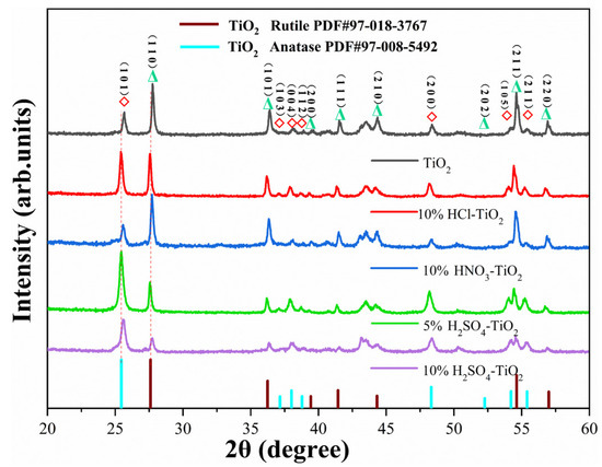
The XRD spectra demonstrated that the proportion of the TiO2 anatase phase was significantly increased after modification with the different acids, which indicates that the phase composition strongly depends on the acid type and concentration (Figure 1 and Table 1) [12,13,14,15,17,21]. The XRD crystallographic peaks at 2θ = 27.606°, 36.242°, 39.438°, 41.443°, 44.324°, 54.631°, and 57.000° for the pure and acid-modified powders correspond to the (110), (101), (200), (111), (210), (211), and (220) crystalline facets of rutile PDF#97-008-5492. The diffraction peaks at 2θ = correspond to the (101), (103), (004), (112), (200), (202), (105), and (211) crystallographic surfaces of anatase PDF#97-018-3767. Here, 5% H2SO4-TiO2 contained the highest anatase fraction (~90 wt%) and the smallest anatase crystallite size (27 nm) [1,12]. In contrast, 10% HNO3-TiO2 and 10% HCl-TiO2 showed a remarkable increase in rutile content (≈35 wt% and ≈30 wt%, respectively) with the concomitant growth of rutile crystallites (>50 nm) [13,21]. The 10% H2SO4 sample occupied an intermediate position (≈20 wt% rutile) [22]. Compared with the rest of the powders, the anatase diffraction peaks were most intense for 5% H2SO4-TiO2, whereas those for the HCl- and HNO3-modified samples exhibited a slight intensity decrease [12]. Moreover, Figure 1 shows that sulfuric-acid modification increases the overall diffraction-peak intensity, indicating improved crystallinity and, consequently, better photocatalytic degradation performance of the synthesized powder.
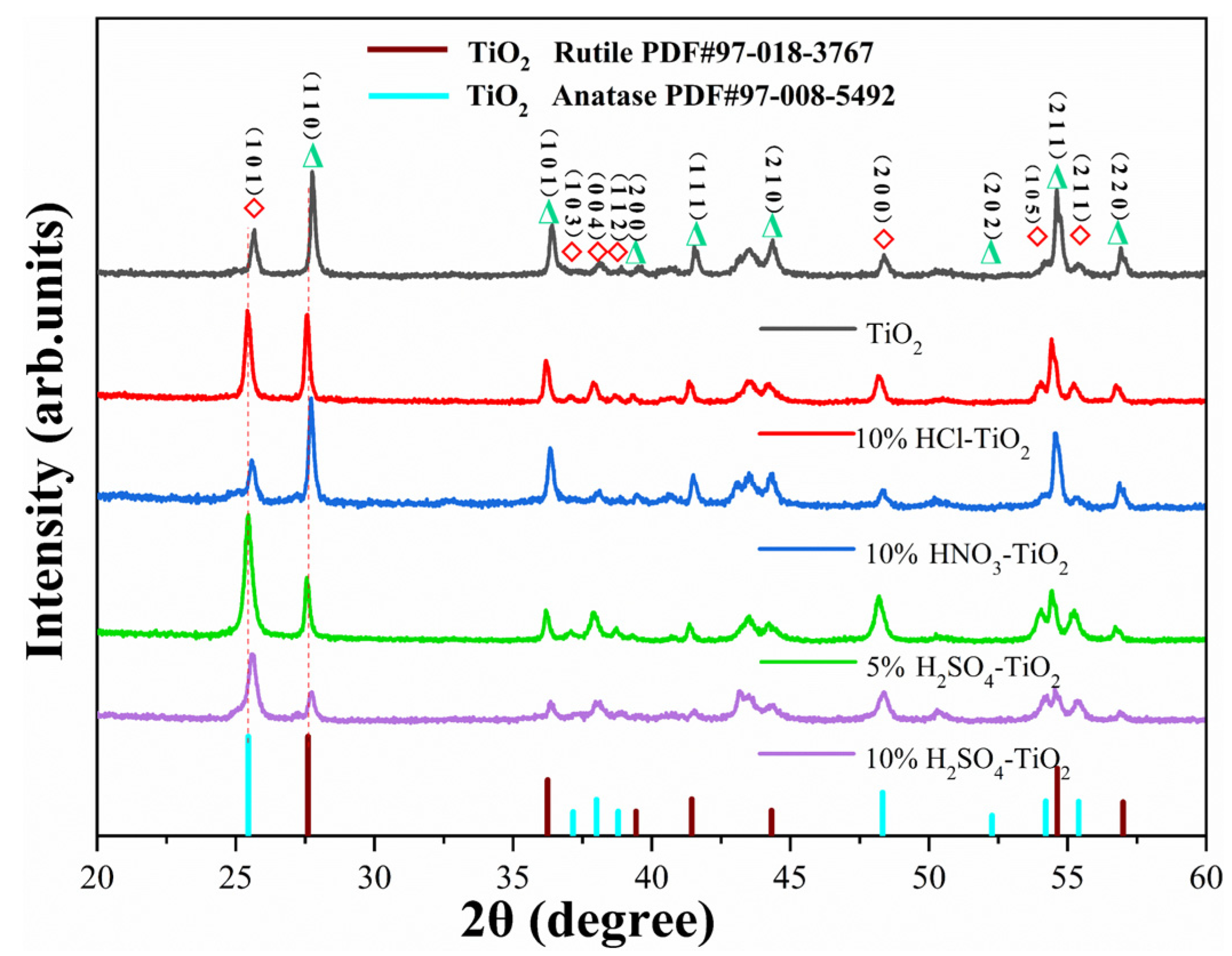
Figure 1.
XRD spectra of the pure and acid-modified TiO2 powders.
Table 1.
Unit cell parameters, micro-crystallite sizes, and band gaps of the rutile and anatase phases.
The lattice parameters extracted from XRD (Table 1) reveal that, when the H2SO4 concentration is raised from 5% to 10%, the anatase unit-cell contracts along both the crystallographic a-axis (in-plane) and c-axis (out-of-plane); this shrinkage matches the high-angle shift in the (1 0 1) and (0 0 4) anatase reflections [23]. For the sample of 5% H2SO4-TiO2, the XRD result shows measurable elongation along the c-axis direction (9.491 ± 0.03 Å), which is comparable with the standard anatase value (9.461 Å, PDF#97-008-5492), while the a- and b-axes remain unchanged within the error range [23]. This anisotropic strain is attributed to the formation of a surface-adsorbed sulfate species (Ti-O-SO3 bridging configuration, as confirmed by S 2p XPS at 168.19 eV, Figure 2((b)-5)), which induces lattice distortion through surface stress [23]. The elongation along the c-axis suggests that the sulfate groups create an extended region that propagates into the subsurface region, consistent with the literature reports of surface-adsorbed species inducing measurable lattice strain in TiO2 [23,24,25,26]. In contrast, the XRD for the HCl- and HNO3-modified samples have smaller deviations from the reference lattice constants, indicating that Cl− and NO3− may mainly be in surface adsorption form [23]. Table 1 shows that acid modification leads to an uniform reduction in the grain size: TiO2 produces a larger grain (48.4 nm), while 5% H2SO4-TiO2 produces the smallest grains (24.9 nm), and the 10% H2SO4-TiO2, 10% HNO3-TiO2, and 10% HCl-TiO2, respectively, produce moderate grains (26.7 nm, 41.6 nm, and 33.7 nm) [1,12]. The results reflect the differences in the nucleation/growth kinetics caused by each acid, rather than the uncertainty of the analysis; the estimated standard error of the Scherch value is less than 2 nm, which confirms the reliability of the observed trend [20]. The modification of different acids affects the bonding tightness, thus weakening the interaction forces and generating micro-strains that float within the lattice parameters [27].
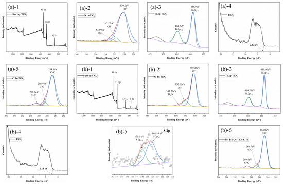
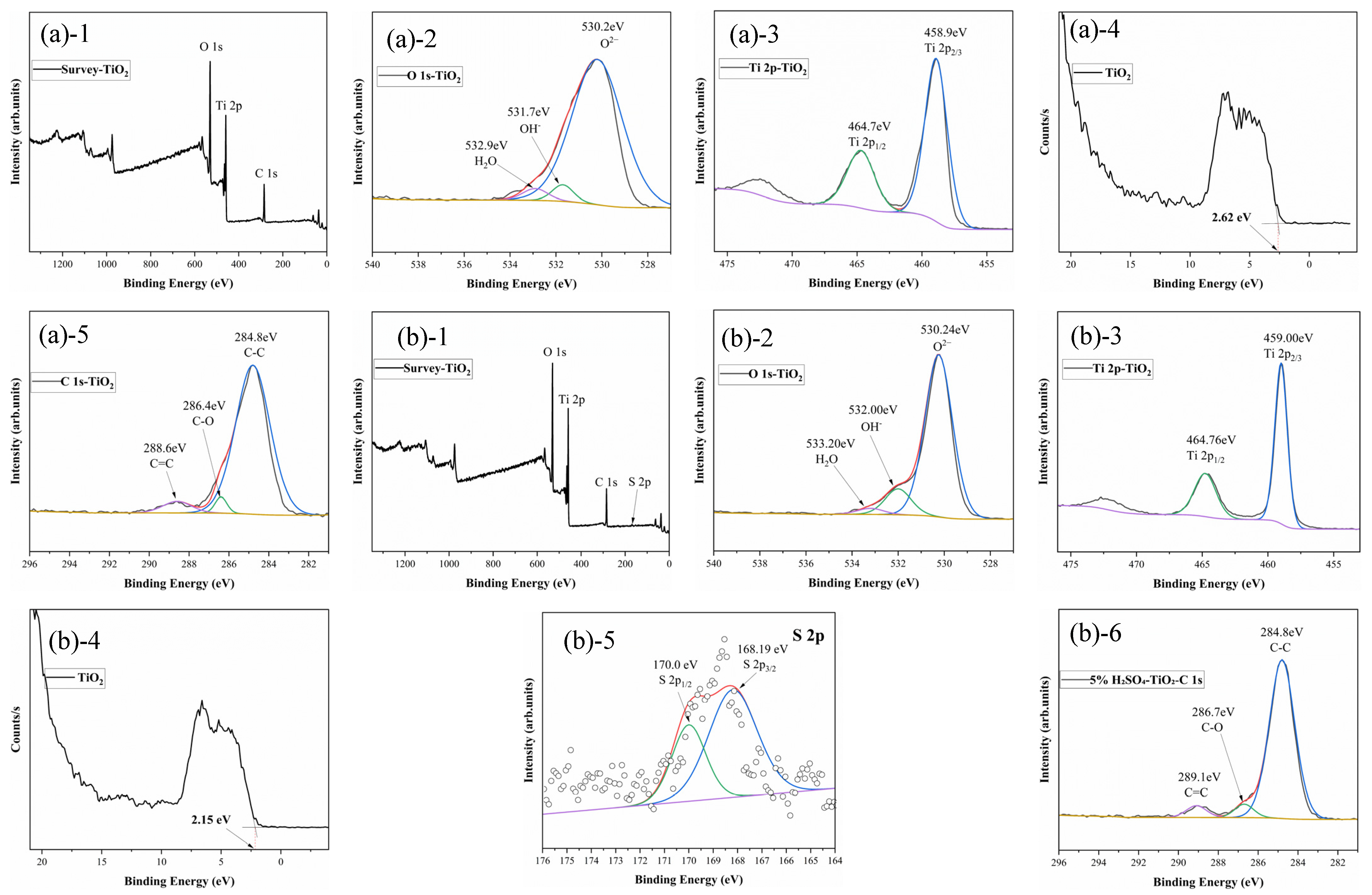
Figure 2.
X-ray photoelectron spectroscopy of the TiO2 powders: a (1–5) pure, b (1–6) 5 mol% H2SO4-modified TiO2. Charge correction: C 1s = 284.8 eV; flood-gun neutralizer employed.
3.2. X-Ray Photoelectron Spectroscopy Analysis
Figure 2a(1–5) illustrates the XPS spectrum of the pure TiO2 powder. The figure shows that the pure TiO2 powder mainly consists of two elements, Ti and O. After charge correction to adventitious C 1s = 284.8 eV, the Ti 2p3/2 and Ti 2p1/2 peaks of pristine TiO2 are located at 458.9 ± 0.05 eV and 464.7 ± 0.05 eV, respectively, yielding a spin–orbit splitting of 5.7 eV; these values are consistent with the standard Ti4+ spectrum reported by Biesinger et al [26]. The characteristic peaks of O 1s are located at 530.2 eV O2−, 531.7 eV OH-, and 532.9 eV H2O, which is consistent with the characteristic binding energy of O2− [26].
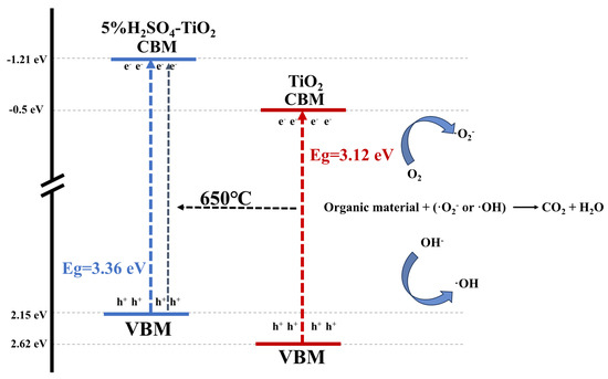
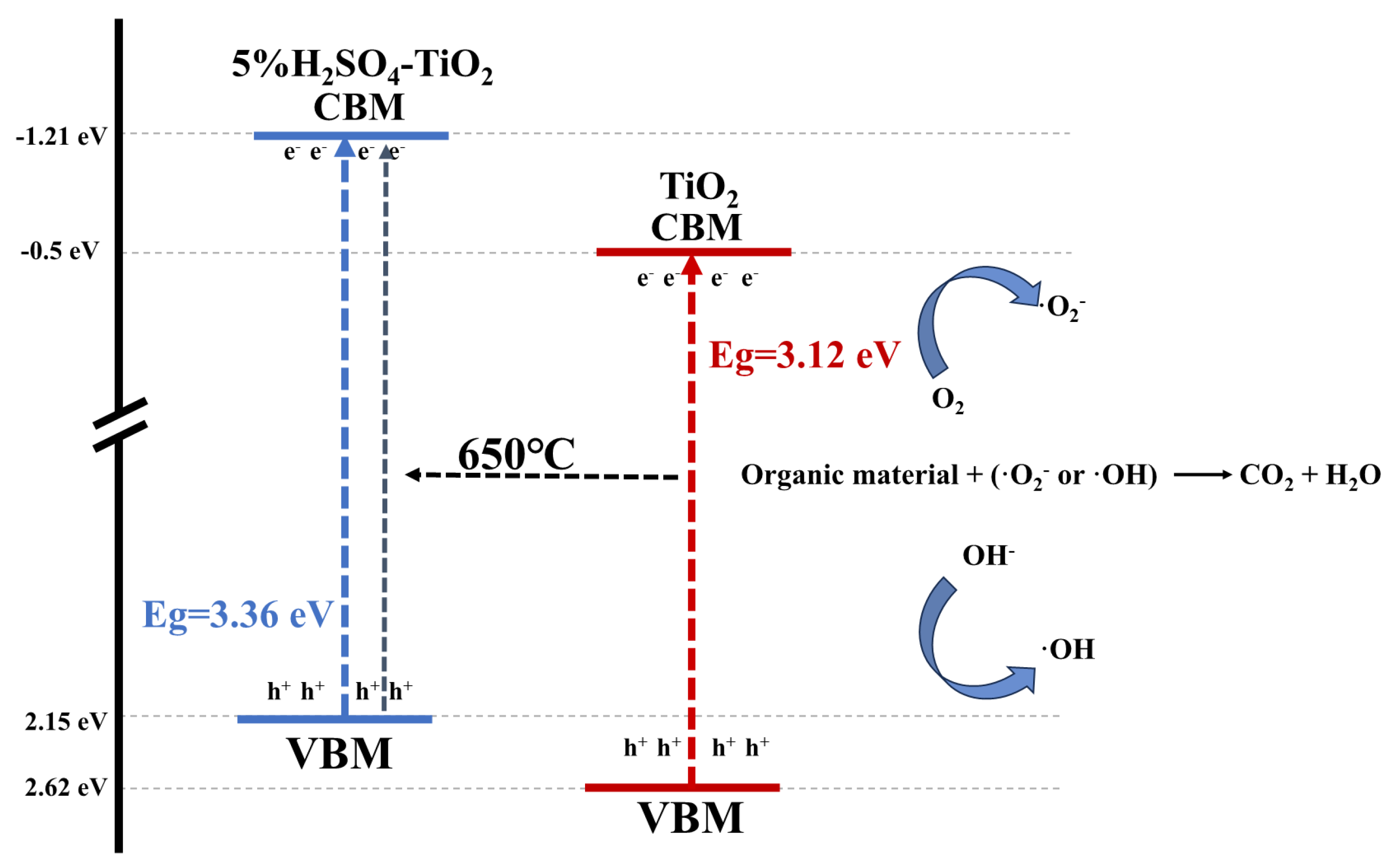
Figure 2b(1–6) demonstrates the XPS spectrum of the 5% H2SO4-TiO2 powder. The positions of the Ti 2p characteristic peaks of the 5% H2SO4-TiO2 powder were slightly right-shifted at 459 eV (Ti 2p3/2) and 464.7 eV (Ti 2p1/2), respectively, as compared with that of the pure TiO2 powder [28]. The right shift indicates that the introduction of H2SO4 resulted in a change in the chemical environment of Ti atoms on the TiO2 surface; in addition, the introduction of SO42− ions altered the charge distribution on the TiO2 surface [29]. The O 1s region was refitted with the three components after Shirley background subtraction (Figure 2((a)-2)): lattice O2− at 529.9 ± 0.1 eV, surface hydroxyls/adsorbed water at 531.7 ± 0.1 eV (≈4.37% area), and molecular H2O at 532.9 ± 0.1 eV (≈3.71% area) [28]. The peak area content of the surface–OH in the original TiO2 is approximately 4.37%, which is lower than the 12.71% in the 5% H2SO4-TiO2 [30]. The 531.7 eV component is ascribed to dissociative adsorbed H2O on defect sites, consistent with the recent surface-science assignments [25] and not to oxygen vacancies, which cannot be detected in ex situ XPS, owing to instantaneous healing by ambient H2O [30]. The S 2p region in Figure 2((b)-5) was re-fit under the physical constraint of 2p3/2:2p1/2 = 2:1 (area). A single doublet at 168.19 eV (2p3/2) and 170.00 eV (2p1/2) with an area ratio of 66.88%: 33.12% perfectly fit the experimental data, confirming that the sulfur exists exclusively as the surface-adsorbed SO42− in a Ti–O–SO3 bridging configuration. No additional intensity was detected in the 160–165 eV window, ruling out the formation of Ti–S bonds [29]. This assignment is supported by the synchrotron-XPS results on sulfate-functionalized TiO2 [31,32]. Thus, sulfate withdraws the electron density from neighboring Ti4+ centers without implying lattice sulfur substitution [29]. The introduction of S elements can increase the surface-active sites of TiO2 and thus improve its photocatalytic activity [31,32]. It has been shown that the introduction of S can significantly improve the photocatalytic performance of TiO2, especially in the UV region [1]. In addition, the introduction of H2SO4 led to the doping of SO42− ions, which combined with Ti atoms on the surface of TiO2 to form Ti-O-SO3 bonds. The formation of Ti-O-SO3 bonds (confirmed by S 2p XPS at 168.19 eV, Figure 2((b)-5)) not only changes the surface charge distribution of TiO2 through electron withdrawal from neighboring Ti4+ centers but may also introduce oxygen vacancies or defect states through the associated charge redistribution [31,33]. Oxygen vacancies can act as trap centers for photogenerated carriers, thus affecting the photocatalytic performance [33]. Therefore, optimizing the concentration of H2SO4 is the key to achieving high-performance TiO2 photocatalysts. However, a too high S content may lead to over-passivation of the TiO2 surface, thus reducing the photocatalytic activity [28]. The subplots (a)-4 and (b)-4 in Figure 2, respectively, present the XPS valence band spectra (VB-XPS) for the pure TiO2 and 5% H2SO4-modified TiO2. The linear extrapolation method (Figure 2((a)-4,(b)-4)) shows that the valence band top (VBM) for the pure TiO2 is located at 2.62 eV, while for the 5% H2SO4-modified sample, the VBM value shifts to 2.15 eV, with a decrease of 0.47 eV [28]. The reason for this phenomenon may that the successful introduction of sulfate groups forms Ti–O–SO3 bonds, which leads the 5% H2SO4-TiO2 to have the highest photocatalytic activity [29]. Note that high-resolution XPS measurements were conducted on the 10% HCl-TiO2 and 10% HNO3-TiO2 powders. Above the detection limit, no obvious Cl 2p signals were detected due to the low concentration, and only the noise-level intensity was shown in the N 1s region, which made curve fitting impossible [28]. These results indicate that the surface Cl and N concentrations are lower than approximately 0.5% of the atomic percentage, consistent with the trace adsorption of NO3−/Cl− rather than lattice incorporation. To quantify the residual anion content, SEM-EDS analysis was performed on randomly selected 5 μm × 5 μm regions of each sample; the results of the SEM-EDS elemental analysis are summarized in Table S1. As a contrast, for the 10% hydrochloric acid-treated TiO2 and 10% nitric acid-treated TiO2, these extremely small surface concentrations are unlikely to cause measurable band gap narrowing or to become the dominant carrier separation center [28]; instead, the observation of a moderate enhancement in the degradation efficiency (Table 2) is attributed to the increased surface hydroxyl groups and the slightly stable rutile phase (Figure 1 and Table 1), which may be promoted by the low concentration of acid adsorption [28].
Table 2.
Degradation data of C/C0.
3.3. Scanning Electron Microscope Analysis
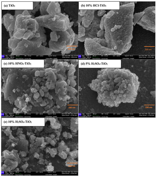
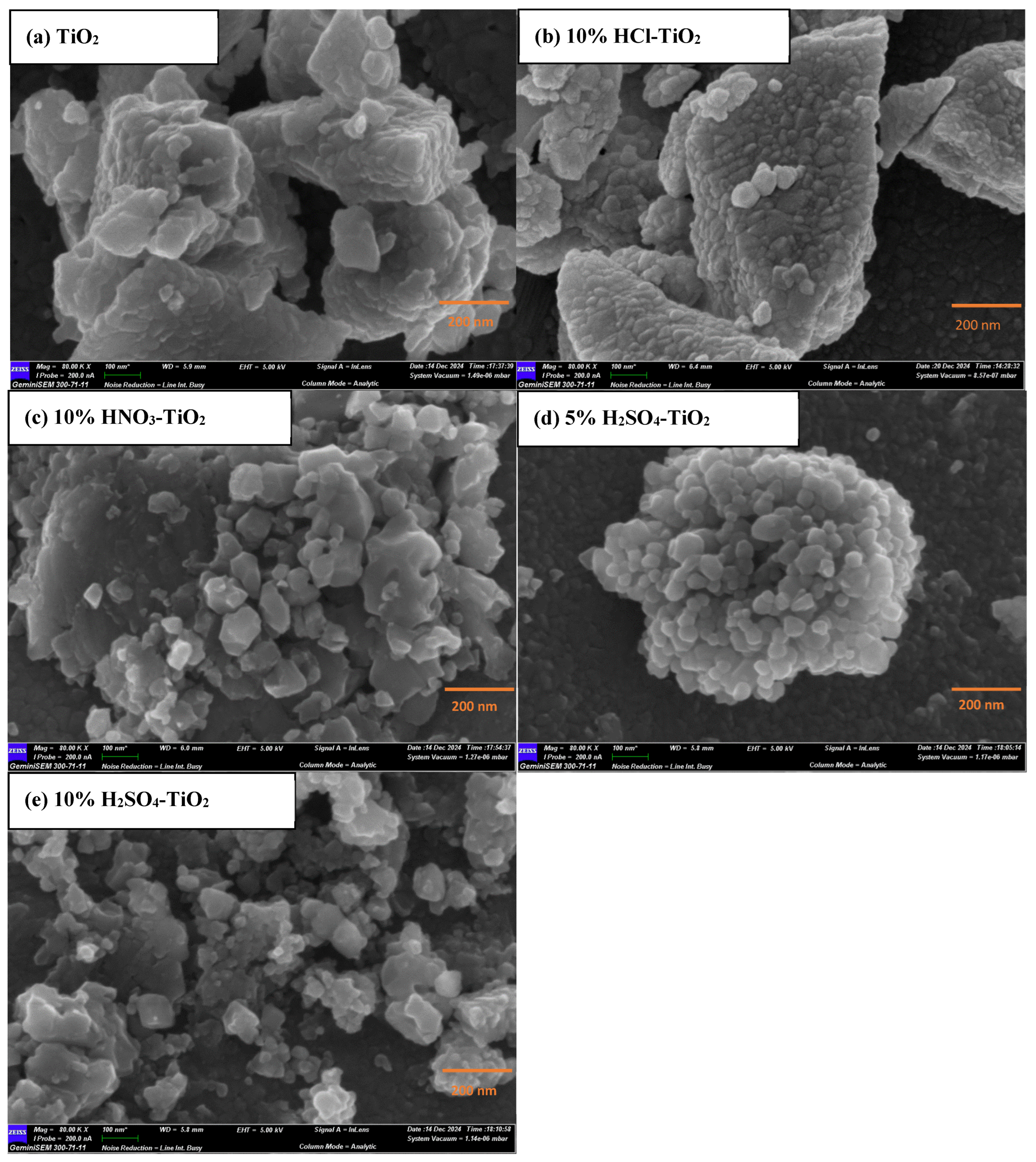
Figure 3 presents SEM images of the pure and acid-modified TiO2 powders. The pure TiO2 powders exhibit severe agglomeration with an average grain size of 54 ± 8 nm, whereas the acid-modified samples show markedly refined particles (27 ± 5 nm for 5%H2SO4-TiO2) [19,34]. Different acids significantly affected the microstructure and morphology of TiO2. HCl promoted the aggregation of TiO2 particles during hydrolysis, leading to an increase in the particle size, but this irregular structure might increase the light scattering path and thus improve the light absorption efficiency [1]. HNO3 induced a change in the direction of the TiO2 crystal growth during hydrolysis, which increased the specific surface area and the formation of more active sites to facilitate the segregation and migration of photogenerated carriers, which might improve the photocatalytic performance [15]. In contrast, for the H2SO4, during hydrolysis, sulfuric acid supplies additional sulfate ions that serve as templating agents, directing the crystal growth of TiO2 to produce 20–30 nm particles, thereby increasing the light-scattering path length and specific surface area, thus improving the light absorption and photocatalytic performance [14]. These results agree with the XRD results [16,19,34].
Figure 3.
SEM images of the TiO2 powders: (a) pure, (b) 10 mol% HCl-TiO2, (c) 10 mol% HNO3-TiO2, (d) 5 mol% H2SO4-TiO2, and (e) 10 mol% H2SO4-TiO2. Scale bars: 200 nm.
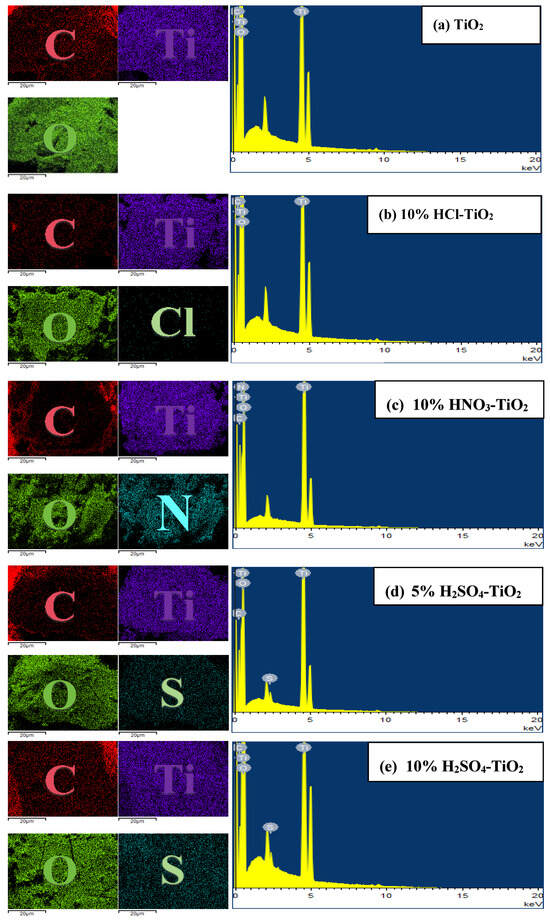
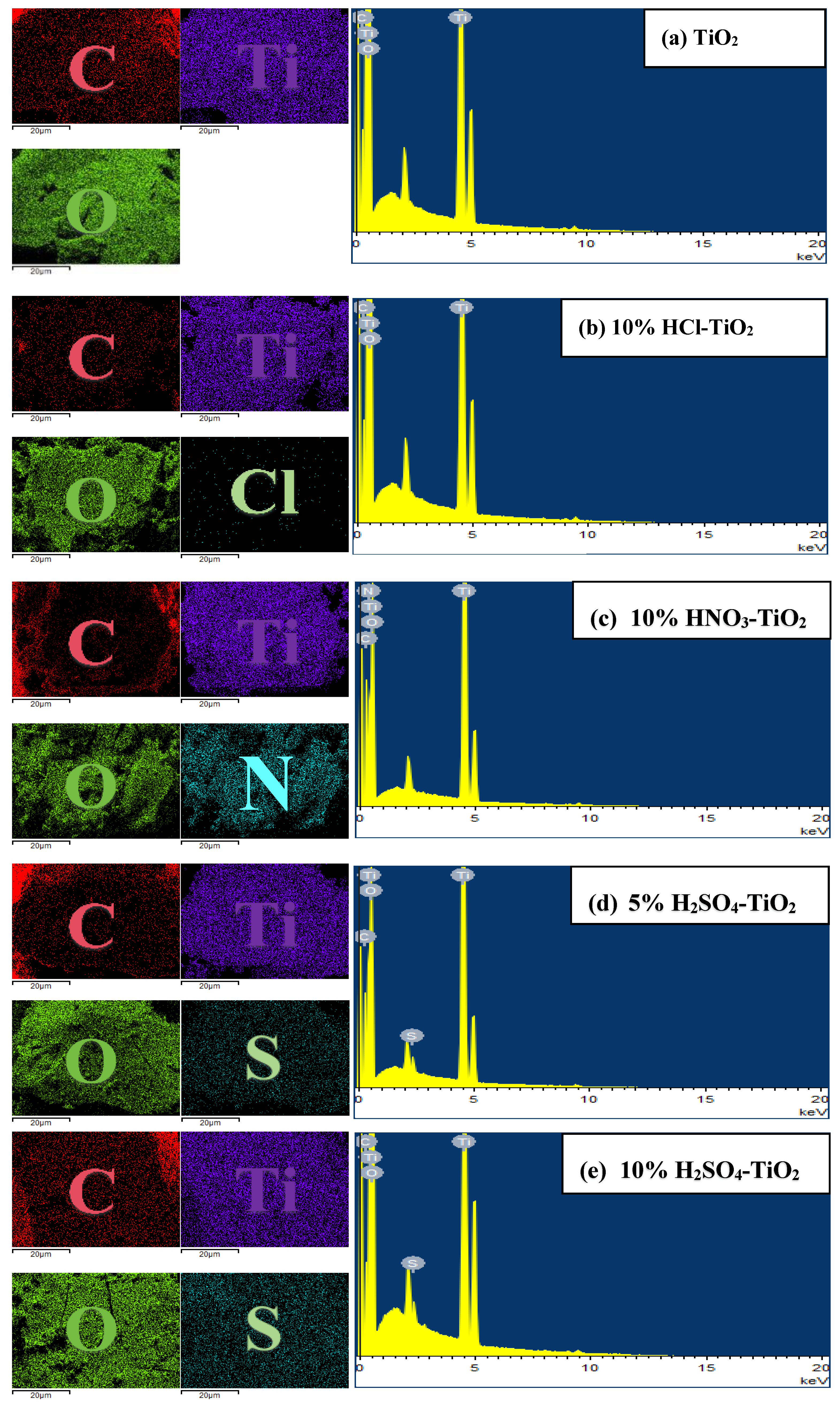
Figure 4 illustrates the EDS spectra of the pure and acid-modified TiO2 powders. The pure TiO2 samples mainly comprise two elements: Ti and O. The result is consistent with the chemical formula of TiO2 and indicates that there are no significant impurity elements present in the pure TiO2 sample [35]. Figure 4b–e demonstrates the EDS spectra of the TiO2 powders modified with different acids. In addition to the Ti and O elements, the presence of Cl, N, and S elements is observed. These elements not only affect the surface charge distribution of TiO2 but also modulate its energy band structure, thus affecting the separation and migration efficiency of the photogenerated carriers [35,36]. In addition, different acid modifications have significant effects on the surface-active sites of TiO2, thus improving its photocatalytic activity [37]. However, too high an acid concentration may lead to the over-passivation of the TiO2 surface, thus reducing the photocatalytic activity [37]. Therefore, optimizing the concentration of acid is the key to achieving high-performance TiO2 photocatalysts [38]. The mechanism of the effect of different acid modifications on the elemental composition and surface properties of TiO2 was confirmed via EDS analysis.
Figure 4.
SEM-EDS images of the pure and acid-modified TiO2 powders (a–e): C Kα1, O Kα1, Ti Kα1, and acid Lα.
3.4. Optical Properties Analysis
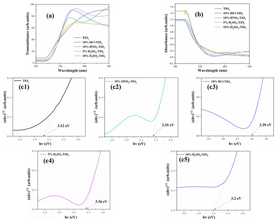
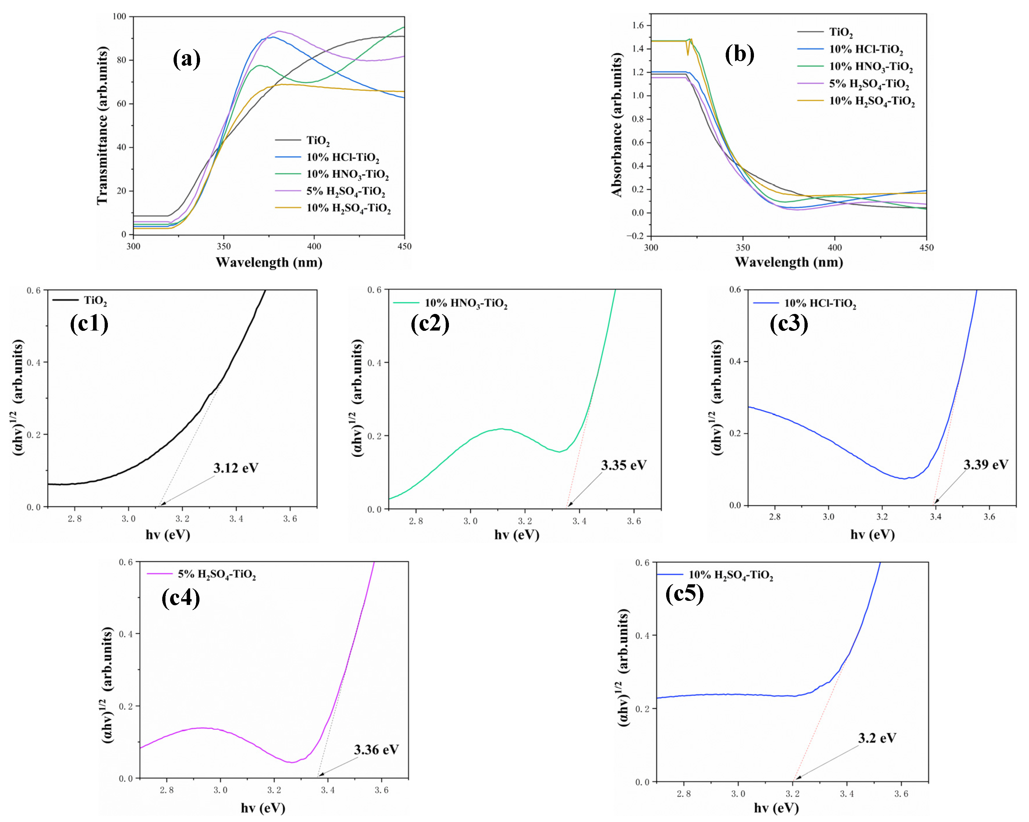
Figure 5 shows the ultraviolet-visible absorption spectra for pure TiO2 and the TiO2 powders modified with different acids. Figure 5b shows that the pure TiO2 powder has a distinct absorption peak in the ultraviolet region (about 387 nm), which is caused by the bandgap transition of TiO2. With different acid modifications, the absorption edges of the TiO2 powders underwent different degrees of redshift, and the absorption intensity was enhanced. In particular, 5% H2SO4-TiO2 shows the most significant redshift and the strongest absorption intensity, indicating that its absorption ability for visible light was significantly enhanced [19]. The absorption edges of 10% HCl-TiO2, 10% HNO3-TiO2, 5% H2SO4-TiO2, and 10% H2SO4-TiO2 all underwent redshift. During hydrolysis, Cl−, NO3−, and SO42− ions are adsorbed on the TiO2 surface [35]. For the sulfate sample, the XPS shows only surface-adsorbed SO42− (168.2/169.3 eV, S 2p, Figure 2((b)-5)) with no Ti-S (<165 eV) signal, indicating Ti-O-SO3 bridging rather than sulfur substitution. The Cl 2p and N 1s spectra of the HCl- and HNO3-modified samples were not recorded [28,35,37]; thus, no statements about the Ti-Cl or Ti-N bonds are made. The improved activity is attributed to (i) increased surface hydroxyls (531.7 eV O 1s) and (ii) enhanced band bending caused by adsorbed anions, both of which facilitate carrier separation [28]. The formation of the new chemical bonds not only changes the surface charge distribution of TiO2 but also may introduce surface defect states, which can act as capture centers for photogenerated carriers and prolong their lifetime. Meanwhile, the introduction of acid has an effect on the forbidden band width, which makes the carriers more likely to jump at lower excitation energies and enhances the photocatalytic degradation of TiO2 powder [39]. The introduction of SO42− ions with H2SO4 modification significantly increases the active sites on the surface of TiO2, which not only increases the separation efficiency of photogenerated carriers but also improves the light absorption ability [35].
Figure 5.
(a) Transmission spectra, (b) absorption spectra, and (c1–c5) optical band gap of the pure and acid-modified TiO2 powder.
The systematic widening of the band gap after acid modification (3.12 eV → 3.36–3.39 eV) is attributed to a pronounced Burstein–Meyer (BM) shift rather than to a change, in intrinsic TiO2 electronic structure. Sulfate, chloride and nitrate species chemisorbed on the particle surface withdraw electron density from the neighboring Ti4+ centers (XPS Ti 2p shift in Figure 2b), creating an internal electric field that bends the bands upward [40,41,42,43]. Consequently, the filling of the conduction band is reduced and higher photon energy is required to excite electrons across the gap, i.e., an apparent gap widening [16,17,18]. A similar BM-type blue-shift has repeatedly been reported for non-metal-adsorbed TiO2 obtained by the sol–gel route [17,23].
Analyzing the ultraviolet–visible absorption spectra, different acid modifications have different degrees of influence on the optical properties of TiO2 [28], mainly reflected in the redshift of the absorption edge and the enhancement in the absorption intensity [37]. The absorption edge of 5% H2SO4-TiO2 redshifted to about 430 nm, and the absorption intensity was significantly enhanced, indicating that its absorption ability for visible light was the strongest [44,45]. These changes are closely related to the regulation of the TiO2 surface charge distribution, band structure, and surface-active sites via acid modification, providing an important optical basis for improving the photocatalytic performance of TiO2 [28].
3.5. Photocatalytic Performance Analysis
For each photocatalytic run, 10 mg of the TiO2 powder was dispersed in 40 mL of tetracycline solution (50 mg L−1), corresponding to a catalyst loading of 0.25 g L−1. Tetracycline (TC, with the chemical formula C22H24N2O8, pKa1 = 3.3, pKa2 = 7.7, pKa3 = 9.7) was selected as the model pollutant because its conjugated π system exhibits a strong absorption band at 357 nanometers, and its photochemical behavior under titanium dioxide irradiation has been well documented in the literature [46,47]. During the adsorption process, the carboxyl and phenolic hydroxyl groups of tetracycline will form bidentate surface complexes (≡Ti–O–C=O) with the ≡Ti–OH site [48]. Under UV illumination the degradation proceeds via two synergistic routes [49]: direct electron injection from excited TC into the TiO2 conduction band (TC → TC+• + e−) followed by reduction of O2 to •O2− [49]; Oxidation by valence-band holes or surface-bound •OH radicals generated from H2O/OH− [49].
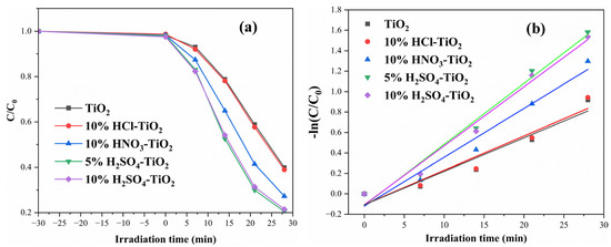
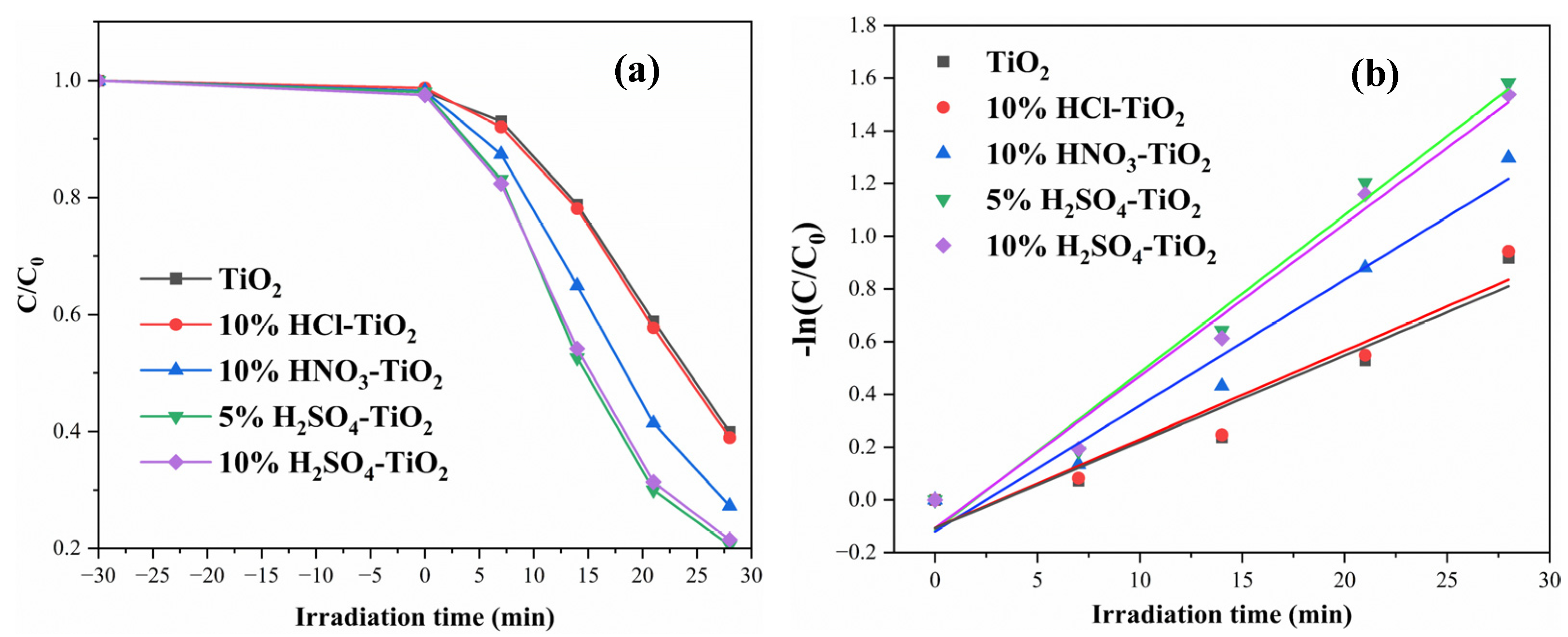
Successive attacks on the N-dimethyl amino and C-4 amide moieties lead to demethylation, hydroxylation and ring-opening intermediates ultimately yielding CO2 and H2O [49]. The observed rate of TC disappearance therefore integrates the yields of •OH/•O2− and the efficiency of interfacial charge separation, making it a reliable probe for comparing photocatalytic activities [49]. Figure 6 shows the degradation performance of (TC) by pure TiO2 and different acid-modified TiO2 powders under visible light irradiation. The degradation data in Figure 6a are shown in Table 2, and the degradation data in Figure 6b are presented in Table 3. The Figure shows that 5% H2SO4-TiO2 exhibits the best photocatalytic performance, with a degradation efficiency of 79.5% within 28 min. The result is significantly higher than that of the other powders (Table 4), namely, pure TiO2 (60.1%), 10% HCl-TiO2 (61.1%), 10% HNO3-TiO2 (72.7%), and 10% H2SO4-TiO2 (78.5%). The linear relationship between −ln(C/C0) and irradiation time (R2 ≥ 0.927, Figure 6b) confirms that the photodegradation of tetracycline under the current experimental conditions follows pseudo-first-order kinetics. The corresponding apparent rate constant (k) and correlation coefficient are summarized in Table 5. From 0.0328 min−1 (bare TiO2) to 0.0598 min−1, the rate constant almost doubled. (5% sulfuric acid-titanium dioxide), which is consistent with the excellent photocatalytic activity observed in Figure 6a [17,20,27].
Figure 6.
UV–Vis absorption spectra residual percentage: (a) Concentration ratio C/C0 of TC versus irradiation time; (b) corresponding −ln(C/C0) kinetic plots.
Table 3.
Degradation rate data of −ln(C/C0).
Table 4.
Maximum degradation efficiency of pure TiO2 and acid-modified TiO2 powders.
Table 5.
Pseudo-first-order rate constants and regression coefficients for tetracycline photodegradation.
The XRD analysis indicated that 5% H2SO4-TiO2 mainly consists of the anatase phase, and the crystalline peak intensity of the anatase phase of 5% H2SO4-TiO2 is significantly higher than that of other powders (see Figure 1). This indicates that the introduction of 5% H2SO4 significantly improves the crystallinity of TiO2, especially the crystallinity of the anatase phase. Since the anatase phase has higher photocatalytic activity, the increase in crystallinity enhances the photocatalytic performance [34,45]. In addition, the grain size calculated using the Scherrer formula indicates that the grain size of the anatase phase of 5% H2SO4-TiO2 is approximately 24.9 nm (see Table 1), which is consistent with the results of the SEM analysis (see Figure 3d). The size is smaller than the pure TiO2 and other acid-modified TiO2 samples. A smaller grain size can increase the specific surface area, thereby providing more active sites, facilitating the separation and migration of photogenerated carriers, and further enhancing the photocatalytic performance [44,50]. The XPS analysis showed that the Ti 2p characteristic peaks on the surface of 5% H2SO4-TiO2 have significantly right-shifted (Figure 2), indicating that SO42− ions combine with Ti atoms on the TiO2 surface to form Ti-O-S bonds (specifically, Ti-O-SO3 bridging configurations, as confirmed by the S 2p binding energy at 168.2 eV, Figure 2((b)-5)) [45]. The Ti–O–SO3 chemical bond formation not only changes the charge distribution on the TiO2 surface but may also introduce oxygen vacancies or defect states [28]. The oxygen vacancies can act as capture centers for photogenerated carriers, prolonging their lifetime, thereby enhancing the photocatalytic performance [28]. However, due to the lack of photoluminescence, electrochemical impedance spectroscopy, and transient photocurrent facilities, direct quantitative evidence for carrier-separation enhancement was not provided in this work [23]. Nevertheless, the right shift in Ti 2p and the appearance of oxygen-vacancy-related signals in XPS (Figure 2b(1–6)) strongly imply that 5% H2SO4-TiO2 possesses more surface defects and shorter bulk-diffusion lengths, which are known to facilitate electron–hole separation. These indirect signatures are consistent with the highest photocatalytic rate observed, and dedicated charge-transfer measurements will be performed in follow-up studies to quantify the separation efficiency [45]. Meanwhile, the EDS analysis also confirmed the successful introduction of S elements, further supporting the XPS results (Figure 2((b)-5)). In conclusion, the reasons for the best photocatalytic performance of 5% H2SO4-TiO2 can be attributed to the increase in crystallinity, the formation of surface chemical bonds, the reduction in particle size, and the enhancement in the light absorption ability [34]. These factors enable 5% H2SO4-TiO2 to exhibit excellent performance in the photocatalytic degradation of (TC) [44,45]. Although both sulfuric-acid samples exhibited higher anatase content than the others, the photocatalytic efficiency dropped from 79.5% (5%) to 78.5% (10%). The XRD showed that 10% H2SO4-TiO2 possessed a larger anatase crystallite size (29.3 nm) than the 5% sample (24.9 nm) and a measurable c-axis expansion (+0.03%, Table 1), implying increased lattice strain [51,52]. Concurrently, the S 2p XPS (Figure 2b) revealed that the additional sulfur remained as surface-adsorbed SO42− (168–169 eV), rather than forming new Ti–S bonds [45]. These extra sulfate species can act as recombination centers for photogenerated carriers and reduce the number of surface-active sites, offsetting the benefit of the slightly higher anatase fraction [50]. Consequently, 5% H2SO4 represents the optimum concentration that maximizes the anatase content while minimizing the carrier-recombination losses [53].
To clarify the degradation pathway of tetracycline (TC) over the optimum 5% H2SO4–TiO2 catalyst, radical-scavenging experiments were carried out under the same irradiation conditions described in Section 2.2. tert-Butanol (TBA), p-benzoquinone (BQ) and EDTA were employed as quenchers for •OH, •O2− and h+, respectively (see Supplementary Information, Figure S1).
Photocatalytic mechanism with acid-treated TiO2 and TC: under 500 W mercury-lamp irradiation (λ ≤ 400 nm) TiO2 generates electrons (e−) in the conduction band and holes (h+) in the valence band (hν → e− + h+) [54]. Chemisorbed sulfate, chloride or nitrate groups withdraw electron density from neighboring Ti4+ centers, creating an upward band-bending that suppresses e−/h+ recombination and lengthens carrier lifetime [54,55,56]. The resulting surface-trapped holes (Ti–O•–SO3−, ≡Ti–O•–Cl− or ≡Ti–O•–NO3−) directly oxidize adsorbed tetracycline (TC) to TC•+ intermediates while simultaneously converting surface H2O into •OH radicals; conduction-band electrons reduce O2 to •O2− [55,57]. These •OH and •O2− species attack the TC ring system, leading ultimately to CO2, H2O and NH4+ [58]. The highest rate constant observed for 5% H2SO4-TiO2 (0.0598 min−1) correlates with the greatest surface coverage of Ti–O–SO3 bridges that provide the strongest internal field and the longest-lived charge carriers, thereby accelerating TC degradation [54,56,59].
3.6. Discussion of the Mechanism
During the photocatalytic process, TiO2 photogenerates electron and hole pairs under the irradiation of ultraviolet or visible light [60,61,62]. These photogenerated charge carriers can participate in redox reactions, thereby degrading organic pollutants [47]. As shown in Figure 7, the specific reaction mechanism is as follows:
TiO2 + hν → TiO2(e− + h+)
h+ + OH− → ⋅OH
h+ + H2O → ⋅OH + H+
e− + O2 → ⋅O2−
⋅O2− + 2H+ → H2O2
Figure 7.
The photocatalytic mechanism of acid-modified TiO2 powder under simulated ultraviolet light irradiation.
Pollutant degradation mechanism:
⋅O2− + pollutant → degradation products
⋅OH + pollutant → degradation products
In recent years, many studies have also reported methods to improve the photocatalytic performance of TiO2 through acid modification. For example, Sherly et al. [60] studied the effect of Cl− ion doping on the photocatalytic performance of TiO2 and determined that the introduction of Cl− ions significantly enhanced the photocatalytic activity of TiO2 under visible light. Yang et al. [61]. successfully regulated the band structure of TiO2 through N element doping, significantly enhancing its absorption ability in the visible light region. Our results of HCl and HNO3 modification are consistent with the above research results, further verifying the positive impact of acid modification on the photocatalytic performance of TiO2. Jothivel et al. [45] studied the effect of SO42− ion-doping on the photocatalytic performance of TiO2 and determined that the introduction of SO42− ions not only increased the active sites on the surface of TiO2 but also significantly enhanced its absorption ability for visible light [50]. Our result of H2SO4 modification is highly consistent with the results of Jothivel et al., indicating that SO42− ions have significant advantages in improving the photocatalytic performance of TiO2.
4. Conclusions
In this work, the physical and photocatalytic properties of TiO2 powders modified with different acids (hydrochloric, nitric, and sulfuric) were systematically investigated. The results showed that acid modification significantly improved the crystallinity of the anatase phase of TiO2, especially 5% H2SO4-TiO2, which had the smallest grain size and the highest crystallinity. XPS shows that sulfate is present as surface-adsorbed SO42− (Ti-O-SO3), while chloride and nitrate species are likewise surface-bound; no evidence for lattice Cl-, N-, or S-substitution was obtained. These adsorbed anions, together with increased surface hydroxyls, promote band bending and carrier separation, leading to higher photocatalytic efficiency. However, the results of the UV–Vis absorption spectroscopy and photocatalytic degradation test of TC analysis showed that the acid modification enhanced the absorption of visible light by TiO2, in which the 5% H2SO4-TiO2 sample had the highest absorption of visible light and best photocatalytic performance. The slightly lower activity observed at 10% H2SO4 is attributed to the increased lattice strain and surface-adsorbed sulfate that favors carrier recombination. The distinctive advance of the present work is the systematic, side-by-side comparison of three single-acid modifications (HCl, HNO3, H2SO4) applied at identical sol–gel synthesis conditions, coupled with quantitative kinetic evidence that sulfate surface groups generate the strongest internal electric field (XPS shift +0.47 eV) and the highest pseudo-first-order rate constant (0.0598 min−1, ≈1.8× that of bare TiO2). For the first time we correlate the sulfate-induced lattice strain (c-axis expansion, Table 1) with prolonged charge-carrier lifetime and faster TC mineralisation, providing a clear structure–activity blueprint for designing high-performance TiO2 photocatalysts without resorting to mixed-acid or noble-metal co-catalysts. The results indicate that acid modification significantly enhances the photocatalytic efficiency of TiO2 by optimizing the crystal structure, surface chemistry, and light-absorbing properties, providing an important theoretical and experimental basis for the development of high-performance TiO2 photocatalysts.
Supplementary Materials
The following supporting information can be downloaded at: https://www.mdpi.com/article/10.3390/cryst16020109/s1, Figure S1: Free radical scavenging experiment results for TC degradation under ultraviolet irradiation in 5% H2SO4-TiO2 conditions; Table S1: SEM-EDS elemental composition (at.%) of acid-modified TiO2 powders.
Author Contributions
This research was jointly completed by all authors, with specific contributions as follows: B.J. proposed the research concept and design, was responsible for data collection and experiment implementation, and completed the initial draft writing and revision. M.M. supervised the research design, guided the data analysis methods, and critically reviewed and revised the content of the paper. Y.B. provided experimental equipment and technical support and assisted in data organization and result verification. Y.J. provided professional advice on the research conclusions and was responsible for proofreading the paper format, reviewing the citation norms of references, and coordinating the submission process. All authors are responsible for the authenticity of the research data and the completeness of the paper content. Each author meets the academic journal’s requirements for authorship, and there are no other contributors not listed as authors. All authors have read and agreed to the published version of the manuscript.
Funding
This work was supported by the Natural Science Foundations (Grant No. 2025D01C45) of Science and Technology Department of Xinjiang, China.
Data Availability Statement
The data of this article are available from the corresponding author upon reasonable request.
Conflicts of Interest
The authors declare that they have not accepted any financial support, material gifts, or other forms of funding from any external organizations or individuals in the course of this research and the writing of the paper. The authors have no interest in any commercial institutions, academic organizations, or individuals that may have an influence on the results of the study, and there are no conflicts of interest that may affect the objectivity and impartiality of the study.
References
- Szołdra, P.; Frąc, M.; Pichór, W. Effect of sol composition on the properties of TiO2 powders obtained by the sol-gel method. Powder Technol. 2021, 387, 261–269. [Google Scholar] [CrossRef] [Scilit]
- Mahmoud, H.A.; Narasimharao, K.; Ali, T.T.; Khalil, K.M.S. Acidic peptizing agent effect on anatase-rutile ratio and photocatalytic performance of TiO2 nanoparticles. Nanoscale Res. Lett. 2018, 13, 48. [Google Scholar] [CrossRef] [Scilit]
- Huang, B.-S.; Su, E.-C.; Huang, Y.-Y.; Tseng, H.-H. Tailored Pt/TiO2 photocatalyst with controllable phase prepared via a modified sol–gel process for dye degradation. J. Nanosci. Nanotechnol. 2018, 18, 2235–2240. [Google Scholar] [CrossRef] [Scilit]
- Zhang, J.; Yan, S.; Fu, L.; Wang, F.; Yuan, M.; Luo, G.; Xu, Q.; Wang, X.; Li, C. Photocatalytic degradation of Rhodamine B on anatase, rutile, and brookite TiO2. Chin. J. Catal. 2011, 32, 983–991. [Google Scholar] [CrossRef] [Scilit]
- Noor, N.H.M.; Saputra, J.; Kamil, S.A.; Mahmood, M.R.; Fadzil, A.F.M.; Supardan, S.N. The morphological and structural properties of chromium (Cr) doped titanium dioxide (TiO2) nanoparticles prepared via sol–gel method at various concentrations. Malays. J. Chem. 2024, 26, 185–193. [Google Scholar]
- Mutuma, B.K.; Shao, G.N.; Kim, W.D.; Kim, H.T. Sol–gel synthesis of mesoporous anatase–brookite and anatase–brookite–rutile TiO2 nanoparticles and their photocatalytic properties. J. Colloid Interface Sci. 2015, 442, 1–7. [Google Scholar] [CrossRef] [Scilit] [PubMed]
- Shahi, S.K.; Kaur, N.; Singh, V. Fabrication of phase and morphology controlled pure rutile and rutile/anatase TiO2 nanostructures in functional ionic liquid/water. Appl. Surf. Sci. 2016, 360, 953–960. [Google Scholar] [CrossRef] [Scilit]
- Jing, L.; Li, S.; Song, S.; Xue, L.; Fu, H. Investigation on the electron transfer between anatase and rutile in nano-sized TiO2 by means of surface photovoltage technique and its effects on the photocatalytic activity. Sol. Energy Mater. Sol. Cells 2008, 92, 1030–1036. [Google Scholar] [CrossRef] [Scilit]
- He, J.; Du, Y.; Bai, Y.; An, J.; Cai, X.; Chen, Y.; Wang, P.; Yang, X.; Feng, Q. Facile formation of anatase/rutile TiO2 nanocomposites with enhanced photocatalytic activity. Molecules 2019, 24, 2996. [Google Scholar] [CrossRef] [Scilit] [PubMed]
- Almeida, L.A.; Viol, J.; Cremona, M.; Menezes, F.A.F.; Guimarães, A.O.; Llorca, J.; Marinkovic, B.A. Enhanced photocatalytic activity of TiO2 anatase nanoparticles modified with malonic acid under reduced power visible light: Synthesis, characterization and degradation of tetracycline and chlorophenol. J. Photochem. Photobiol. A Chem. 2024, 452, 115617. [Google Scholar] [CrossRef] [Scilit]
- Wang, S.; Lian, J.S.; Zheng, W.T.; Jiang, Q. Photocatalytic property of Fe doped anatase and rutile TiO2 nanocrystal particles prepared by sol–gel technique. Appl. Surf. Sci. 2012, 263, 260–265. [Google Scholar] [CrossRef] [Scilit]
- Zhang, A.-X.; Li, L. Influence of acid catalyst on crystalline microstructure and photocatalytic property of nano-TiO2. J. Changsha Univ. Sci. Technol. 2014, 1, 88–92. [Google Scholar]
- Wang, Z.; Xia, D.; Chen, G.; Yang, T.; Chen, Y. The effects of different acids on the preparation of TiO2 nanostructure in liquid media at low temperature. Mater. Chem. Phys. 2008, 111, 313–316. [Google Scholar] [CrossRef] [Scilit]
- Tsega, M.; Dejene, F.B. Influence of acidic pH on the formulation of TiO2 nanocrystalline powders with enhanced photoluminescence property. Heliyon 2017, 3, e00246. [Google Scholar] [CrossRef] [Scilit] [PubMed]
- Yalcin, M. The effect of pH on the physical and structural properties of TiO2 nanoparticles. J. Cryst. Growth 2022, 585, 126603. [Google Scholar] [CrossRef] [Scilit]
- Dontsova, T.; Kutuzova, A.; Hosseini-Bandegharaei, A. Characterization and properties of titanium(IV) oxide, synthesized by different routes. Chem. Chem. Technol. 2021, 15, 465–474. [Google Scholar] [CrossRef] [Scilit]
- Hossain, M.S.; Ahmed, S. Easy and green synthesis of TiO2 (Anatase and Rutile): Estimation of crystallite size using Scherrer equation, Williamson–Hall plot, Monshi-Scherrer Model, size-strain plot, Halder-Wagner Model. Results Mater. 2023, 20, 100492. [Google Scholar] [CrossRef] [Scilit]
- Arellano, U.; Wang, J.A.; Asomoza, M.; Chen, L.F.; González, J.; Manzo, A.; Solís, S.; Lara, V.H. Crystalline structure, surface chemistry and catalytic properties of Fe3+ doped TiO2 sol–gel catalysts for photooxidation of 2,4–dichlorophenoxyacetic acid. Mater. Chem. Phys. 2018, 214, 247–259. [Google Scholar] [CrossRef] [Scilit]
- Samantaray, S.K.; Mohapatra, P.; Parida, K. Physico-chemical characterisation and photocatalytic activity of nanosized SO42−/TiO2 towards degradation of 4-nitrophenol. J. Mol. Catal. A Chem. 2003, 198, 277–287. [Google Scholar] [CrossRef] [Scilit]
- Inagaki, M.; Nonaka, R.; Tryba, B.; Morawski, A.W. Dependence of photocatalytic activity of anatase powders on their crystallinity. Chemosphere 2006, 64, 437–445. [Google Scholar] [CrossRef] [Scilit]
- Yanagida, S.; Sano, K.; Takei, T.; Kumada, N. Preparation and photocatalytic properties of rutile TiO2 with a unique morphology and SrTiO3–TiO2 composites obtained by acid treatment of SrTiO3. Mater. Res. Bull. 2020, 125, 110762. [Google Scholar] [CrossRef] [Scilit]
- Masahashi, N.; Mizukoshi, Y.; Semboshi, S.; Ohtsu, N. Enhanced photocatalytic activity of rutile TiO2 prepared by anodic oxidation in a high concentration sulfuric acid electrolyte. Appl. Catal. B Environ. 2009, 90, 255–261. [Google Scholar] [CrossRef] [Scilit]
- Xu, B.; Duan, X.; Zhou, T.; Hao, J.; Qin, H.; Zhao, Y.; Ye, W.; Cao, J. Optically active oxygen defects in titanium dioxide doped with inorganic acid ions. Nanomaterials 2024, 14, 1020. [Google Scholar] [CrossRef] [Scilit] [PubMed]
- Yu, F.; Wang, J.; Zhao, K.; Yin, J.; Jin, C.; Liu, X. A novel approach for the preparation of phase-tunable TiO2 nanocomposite crystals with superior visible-light-driven photocatalytic activity. Chin. J. Catal. 2013, 34, 1216–1223. [Google Scholar] [CrossRef] [Scilit]
- Rodríguez-Barajas, N.; Becerra-Solano, L.; Gutiérrez-Mercado, Y.K.; Macías-Carballo, M.; Gómez, C.M.; Pérez-Larios, A. Study of the interaction of Ti–Zn as a mixed oxide at different pH values synthesized by the sol–gel method and its antibacterial properties. Nanomaterials 2022, 12, 1948. [Google Scholar] [CrossRef] [Scilit]
- Biesinger, M.C.; Lau, L.W.M.; Gerson, A.R.; Smart, R.S.C. Resolving surface chemical states in XPS analysis of first row transition metals, oxides and hydroxides: Sc, Ti, V, Cu and Zn. Appl. Surf. Sci. 2010, 257, 887–898. [Google Scholar] [CrossRef] [Scilit]
- Chackrabarti, S.; Zargar, R.A.; Mearaj, T.; Arfat, Y.; Hafiz, A.K.; Khan, M.I. Unraveling optical and electrical dynamics in synthesized TiO2 nanopowder through sol–gel chemistry. MRS Adv. 2024, 9, 991–996. [Google Scholar] [CrossRef] [Scilit]
- Cui, H.; Cao, Y.; Jing, L.; Luan, Y.; Li, N. Effects of inorganic acid modification on photocatalytic performance of TiO2 and its activity-enhanced mechanism related to adsorbed O2. ChemPlusChem 2013, 79, 318–324. [Google Scholar] [CrossRef] [Scilit]
- Liao, H.; Xu, H.; Zhang, X.; Dai, W.; Zhang, Z. Stable bidentate coordination sulfated TiO2 for highly durable photocatalytic degradation of gaseous acetone. Appl. Catal. A Gen. 2023, 657, 119158. [Google Scholar] [CrossRef] [Scilit]
- Idriss, H. On the wrong assignment of the XPS O1s signal at 531–532 eV attributed to oxygen vacancies in photo- and electro-catalysts for water splitting and other materials applications. Surf. Sci. 2021, 712, 121894. [Google Scholar] [CrossRef] [Scilit]
- Yan, X.; Yuan, K.; Lu, N.; Xu, H.; Zhang, S.; Takeuchi, N.; Kobayashi, H.; Li, R. The interplay of sulfur doping and surface hydroxyl in band gap engineering: Mesoporous sulfur-doped TiO2 coupled with magnetite as a recyclable, efficient, visible light active photocatalyst for water purification. Appl. Catal. B Environ. 2017, 218, 20–31. [Google Scholar] [CrossRef] [Scilit]
- Sekar, P.P.; Parasuraman, V.; Aslam, M.A.; Sheraz, M.; Swamidoss, C.M.A.; Lee, W.R.; Kim, S. Porous sulfur-doped titanium dioxide for improving photocatalytic VOC removal and biological disinfection under low intensity fluorescent light. Surf. Interfaces 2022, 35, 102468. [Google Scholar] [CrossRef] [Scilit]
- Andronic, L.; Lelis, M.; Enesca, A.; Karazhanov, S. Photocatalytic activity of defective black-titanium oxide photocatalysts towards pesticide degradation under UV/VIS irradiation. Surf. Interfaces 2022, 32, 102123. [Google Scholar] [CrossRef] [Scilit]
- Žener, B.; Matoh, L.; Reli, M.; Sever Škapin, A.; Cerc Korošec, R. Metal and non-metal modified titania: The effect of phase composition and surface area on photocatalytic activity. Acta Chim. Slov. 2022, 69, 217–226. [Google Scholar] [CrossRef] [Scilit] [PubMed]
- Wang, X.; Yu, J.C.; Liu, P.; Wang, X.; Su, W.; Fu, X. Probing of photocatalytic surface sites on SO42−/TiO2 solid acids by in situ FT-IR spectroscopy and pyridine adsorption. J. Photochem. Photobiol. A Chem. 2006, 179, 339–347. [Google Scholar] [CrossRef] [Scilit]
- Zhao, D.; Chen, C.; Wang, Y.; Ji, H.; Ma, W.; Zang, L.; Zhao, J. Surface modification of TiO2 by phosphate: Effect on photocatalytic activity and mechanism implication. J. Phys. Chem. C 2008, 112, 5993–6001. [Google Scholar] [CrossRef] [Scilit]
- Kozlov, D.; Bavykin, D.; Savinov, E. Effect of the acidity of TiO2 surface on its photocatalytic activity in acetone gas-phase oxidation. Catal. Lett. 2003, 86, 169–172. [Google Scholar] [CrossRef] [Scilit]
- Romanovska, N.I.; Manoryk, P.A.; Ermokhina, N.I.; Yaremov, P.S.; Grebennikov, V.M. Effect of structural and dimensional characteristics of TiO2 and its photocatalytic activity in the oxidation of tetracycline. Theor. Exp. Chem. 2019, 55, 345–353. [Google Scholar] [CrossRef] [Scilit]
- Devi, L.G.; Kavitha, R. Enhanced photocatalytic activity of sulfur doped TiO2 for the decomposition of phenol: A new insight into the bulk and surface modification. Mater. Chem. Phys. 2014, 143, 1300–1308. [Google Scholar] [CrossRef] [Scilit]
- Jung, S.M.; Grange, P. Evidence of correlation between electronic density and surface acidity of sulfated TiO2. Catal. Lett. 2001, 76, 27–30. [Google Scholar] [CrossRef] [Scilit]
- Zhao, W.; Zhong, Q.; Pan, Y.; Zhang, R. Systematic effects of S-doping on the activity of V2O5/TiO2 catalyst for low-temperature NH3-SCR. Chem. Eng. J. 2013, 228, 815–823. [Google Scholar] [CrossRef] [Scilit]
- Wang, X.D.; He, S.Y.; Yang, D.Z.; Gu, P.F. Space radiation damage in ZnO induced by subthreshold electrons: Defect identity and optical degradation. Radiat. Res. 2011, 176, 264–268. [Google Scholar] [CrossRef] [Scilit] [PubMed]
- Yun, H.; Li, J.; Chen, H.-B.; Lin, C.-J. A study on the N-, S- and Cl-modified nano-TiO2 coatings for corrosion protection of stainless steel. Electrochim. Acta 2007, 52, 6679–6685. [Google Scholar] [CrossRef] [Scilit]
- Zouheir, M.; Assila, O.; Tanji, K.; El Gaidoumi, A.; Araña, J.; Doña Rodríguez, J.M.; Smått, J.-H.; Huynh, T.-P.; Kherbeche, A. Bandgap optimization of sol–gel-derived TiO2 and its effect on the photodegradation of formic acid. Nano Futures 2021, 5, 025004. [Google Scholar] [CrossRef] [Scilit]
- Jothivel, S.; Velmurugan, R.; Selvam, K.; Krishnakumar, B.; Swaminathan, M. Preparation, characterization and photocatalytic activity of acidic sulfated nano titania for the degradation of Reactive Orange 4 under UV light. Sep. Purif. Technol. 2011, 77, 245–250. [Google Scholar] [CrossRef] [Scilit]
- Hu, L.; Xiao, R.; Wang, X.; Wang, X.; Wang, C.; Wen, J.; Gu, W.; Zhu, C. MXene-induced electronic optimization of metal-organic framework-derived CoFe LDH nanosheet arrays for efficient oxygen evolution. Appl. Catal. B Environ. 2021, 298, 120599. [Google Scholar] [CrossRef] [Scilit]
- Hanh, N.T.; Khai, N.M.; Anh, T.N.; Vinh, L.T.; Huan, N.H.; Pham, T.-D. TiO2 deposited on activated sewage sludge for effective photocatalytic degradation of tetracycline. Chem. Eng. Technol. 2022, 45, 1969–1975. [Google Scholar] [CrossRef] [Scilit]
- Luo, Z.; Li, L.; Wei, C.; Li, H.; Chen, D. Role of active oxidative species on TiO2 photocatalysis of tetracycline and optimization of photocatalytic degradation conditions. J. Environ. Biol. 2015, 36, 1091–1098. [Google Scholar]
- Wu, S.; Hu, H.; Lin, Y.; Zhang, J.; Hu, Y.H. Visible light photocatalytic degradation of tetracycline over TiO2. Chem. Eng. J. 2020, 382, 122842. [Google Scholar] [CrossRef] [Scilit]
- Xu, Z.; Chen, Y.; Zhong, J.; Chen, J.; Li, M.; Wang, Q.; Yang, H. Plasma surface treatment facilitated visible light-driven H2 production over TiO2. Surf. Interfaces 2023, 36, 102626. [Google Scholar] [CrossRef] [Scilit]
- Wang, G.; Lv, S.; Shen, Y.; Li, W.; Lin, L.; Li, Z. Advancements in heterojunction, cocatalyst, defect and morphology engineering of semiconductor oxide photocatalysts. J. Mater. 2024, 10, 315–338. [Google Scholar] [CrossRef] [Scilit]
- Fuentes, K.M.; Venuti, D.; Betancourt, P. Black titania with increased defective sites for phenol photodegradation under visible light. React. Kinet. Mech. Catal. 2020, 131, 423–435. [Google Scholar] [CrossRef] [Scilit]
- Liu, Y.; Ren, M.; Zhang, X.; Yang, G.; Qin, L.; Meng, J.; Guo, Y. Supramolecule self-assembly approach to direct Z-scheme TiO2/g-C3N4 heterojunctions for efficient photocatalytic degradation of emerging phenolic pollutants. Appl. Surf. Sci. 2022, 593, 153401. [Google Scholar] [CrossRef] [Scilit]
- Chen, Q.; Liu, L.; Liu, L.; Zhang, Y. A novel UV-assisted PEC-MFC system with CeO2/TiO2/ACF catalytic cathode for gas phase VOCs treatment. Chemosphere 2020, 255, 126930. [Google Scholar] [CrossRef] [Scilit]
- Jiang, L.; Li, J.; Wang, K.; Zhang, G.; Li, Y.; Wu, X. Low boiling point solvent mediated strategy to synthesize functionalized monolayer carbon nitride for superior photocatalytic hydrogen evolution. Appl. Catal. B Environ. 2020, 260, 118181. [Google Scholar] [CrossRef] [Scilit]
- Günsel, A.; Atmaca, G.Y.; Taslimi, P.; Bilgiçli, A.T.; Gülçin, İ.; Erdoğmuş, A.; Yarasir, M.N. Synthesis, characterization, photo-physicochemical and biological properties of water-soluble tetra-substituted phthalocyanines: Antidiabetic, anticancer and anticholinergic potentials. J. Photochem. Photobiol. A Chem. 2020, 396, 112511. [Google Scholar] [CrossRef] [Scilit]
- Garcia-Segura, S.; Lanzarini-Lopes, M.; Hristovski, K.; Westerhoff, P. Electrocatalytic reduction of nitrate: Fundamentals to full-scale water treatment applications. Appl. Catal. B Environ. 2018, 236, 546–568. [Google Scholar] [CrossRef] [Scilit]
- Wang, D.-C.; Yang, X.; Yu, H.-Y.; Gu, J.; Qi, D.; Yao, J.; Ni, Q. Smart nonwoven fabric with reversibly dual-stimuli responsive wettability for intelligent oil-water separation and pollutants removal. J. Hazard. Mater. 2020, 383, 121123. [Google Scholar] [CrossRef] [Scilit]
- Zhu, J.; Zhu, Z.; Zhang, H.; Lu, H.; Zhang, W.; Qiu, Y.; Zhu, L.; Küppers, S. Calcined layered double hydroxides/reduced graphene oxide composites with improved photocatalytic degradation of paracetamol and efficient oxidation-adsorption of As(III). Appl. Catal. B Environ. 2018, 225, 550–562. [Google Scholar] [CrossRef] [Scilit]
- Sherly, R.A.; Padma, C.M.; Raja, D.H.; Davidson, D.J. Photodegradation of Methyl violet using Ag modified TiO2 nanotubes by UV and UV/H2O2. Chem. Phys. Impact 2023, 7, 100366. [Google Scholar] [CrossRef] [Scilit]
- Yang, Y.; Yang, X.; Jia, Q.; Zheng, S.; Lin, Z.; Qin, Z. Enhanced photocatalytic performance of (N, F) co-doped TiO2 loaded on coal-based hierarchical porous carbon foam under simulated sunlight. Vacuum 2023, 207, 111577. [Google Scholar] [CrossRef] [Scilit]
- Higashimoto, S. Titanium-dioxide-based visible-light-sensitive photocatalysis: Mechanistic insight and applications. Catalysts 2019, 9, 201. [Google Scholar] [CrossRef] [Scilit]

Disclaimer/Publisher’s Note: The statements, opinions and data contained in all publications are solely those of the individual author(s) and contributor(s) and not of MDPI and/or the editor(s). MDPI and/or the editor(s) disclaim responsibility for any injury to people or property resulting from any ideas, methods, instructions or products referred to in the content. |
© 2026 by the authors. Licensee MDPI, Basel, Switzerland. This article is an open access article distributed under the terms and conditions of the Creative Commons Attribution (CC BY) license.