


Abstract
The enzymatic production of low-molecular-weight chitosan and chitooligosaccharides (COS), with broad application potential in agriculture, food, medicine, and cosmetics, has emerged as an attractive alternative to chemical chitosan depolymerization owing to its substrate specificity and environmentally benign catalytic action. However, the functional properties of available chitosanases need to be enhanced to meet the demands of industrial COS manufacturing under high temperature and substrate concentrations. In this work, we performed directed evolution on a recombinant Bacillus subtilis chitosanase to increase chitosan hydrolysis performance and thermal resistance. Three rounds of directed evolution screening (~9000 clones) yielded variants MT1, MT2, and MT3 with higher specific activity, achieved through Vmax improvement and increased T1/2 at 60 °C. HPLC, DLS, and MALDI-TOF results indicate differences in the hydrolysis kinetics and size distribution of COS products over reaction time, suggesting a narrower distribution and a lower average molecular weight. Molecular dynamics simulations and docking studies revealed potential modulation of chitosanase activity via changes in the opening and closing dynamics of the active-site cleft. These results suggest that future efforts targeting the cleft interface could significantly advance both the catalytic performance and the mechanistic understanding of GH46 family chitosanases.
1. Introduction
Chitosan is a linear polysaccharide obtained through the partial or complete deacetylation of chitin. Chitosan has gained substantial attention due to its unique physicochemical properties, low toxicity, biodegradability, biocompatibility, high adsorption capacity, and microbial resistance [1,2,3]. The amino group (-NH2) of chitosan is available for chemical reactions with aldehydes and ketones (Schiff’s base), chelation of metals, alkylation, sulfonation, carboxymethylation, grafting, acetylation, quaternization, etc. [4,5]. Furthermore, the chemical groups along the backbone can be modified with specific agents to form functionalized or physical hydrogels [6,7]. Thus, chitosan has become a highly attractive biomaterial with applications including drug and enzyme delivery vehicles, tissue engineering scaffolds, adhesives, texturing agents, adsorbents, chelators, and supports for enzyme immobilization.
Chitosan is soluble in aqueous acid solutions such as acetic, citric, formic, and lactic acids [8]. However, chitosan presents low water solubility at neutral to alkaline conditions, limiting its potential for broad applications. This solubility is defined by the chitosan degree of deacetylation (DD), the protonation of free amino groups, and its molecular size or degree of polymerization [9]. On the other hand, chitosan derivatives, such as low-molecular-weight chitosan and chitooligosaccharides (COS), are highly soluble in water, present multiple bioactivities, and have been described as molecular recognition agents, cell signaling and morphogenesis mediators, immune response elicitors, and host–pathogen interaction modulators, as well as having other applications in agriculture, food, cosmetics, and biomedicine [10,11,12,13].
Low-molecular-weight chitosan can be obtained through acid hydrolysis of chitosan; however, achieving a homogeneous oligomer size distribution by this method remains challenging. Therefore, the development of new approaches to produce COS of specific molecular weight is urgently needed [14]. Enzymatic degradation of chitosan has been proposed as a sustainable alternative for the manufacture of COS and low-molecular-weight chitosan. Chitosanases are enzymes that cleave β-1,4-glycosidic linkages in chitosan in an endo (internal cleavage) or exo (chain-end cleavage) fashion. Excellent information on the different types of chitosanases, their structures, and catalytic mechanisms, as well as examples of their improvement, can be found in a recent review [2].
Through advances in understanding the catalytic mechanisms and structure–function relationships of chitosanases across the glycoside hydrolase (GH) families GH8, GH46, GH5, and GH75, several candidates for chitosan degradation and COS production have been identified, each with distinct substrate and product selectivity, particularly to produce functional COS [2,15,16].
GH46 family chitosanases, despite being the most extensively characterized group of chitosan-degrading enzymes [16], face several technical challenges that hinder their application to industrial-scale chitooligosaccharide (COS) production. The main bottleneck is the inherent thermostability limitation of wild-type enzymes. For the B. subtilis chitosanase BsCsn46A, wild-type (WT) chitosanase activity diminishes by 50% after only 30 min of incubation at 50 °C, and only the reported engineered A129L mutant retained stability after prolonged exposure to 40 °C for up to 3 h [17]. For B. subtilis CsnMY002 (referred to here as BSchito), the reported half-life at 55 °C is approximately 116 min, indicating more suitable thermal resistance; however, significant activity losses occur above 55 °C [16].
Furthermore, BSchito was reported to reach 60.5% substrate conversion at 200 min under high chitosan concentration (25 mg/mL, 2.5%), indicating potential for improvement [16].
The catalytic and structural properties of enzymes can be improved through protein engineering, which aims to generate variants with superior performance relative to the starting enzyme [18]. This can be achieved through iterative screening of randomly mutated enzyme variants or through rational modifications, such as by designing or modifying the target enzyme’s catalytic site [19,20].
Directed evolution is the most commonly employed strategy in enzyme engineering [21,22]. Through iterative cycles of diversification and high-throughput screening, variants with improved properties relative to the parent enzyme can be identified. Decades of research have shown that directed evolution is a robust enzyme engineering strategy and, in addition, can provide valuable insights into structure–function relationships in recently isolated and less studied enzymes [23,24,25]. Furthermore, researchers can target specific enzyme residues or residues identified by in silico, structural, or computational chemistry, using rational and semi-rational protein engineering [26]. These strategies are particularly effective at improving catalytic properties of enzymes, such as substrate specificity and stereo- and regioselectivity, and can even generate entirely new catalytic activities [27]. The knowledge and experience generated by protein engineering and the advancement of computational biochemistry, artificial intelligence, and machine learning have recently enabled a new wave of protein design, which promises a qualitative leap forward in protein technology and biocatalysis, fueled by the data generated in previous research and new analysis and structure–function prediction strategies [28,29,30].
Rational engineering of GH46 chitosanases has improved both thermostability and catalytic efficiency. For BsCsn46A, saturation mutagenesis at the C-terminal residue Lys242 produced six beneficial mutants (Lys242Ala, Lys242Asp, Lys242Glu, Lys242Phe, Lys242Pro, and Lys242Thr), where Lys242Pro achieved the highest specific activity reported to date at 17,820 U/mg, alongside a 2.27% improvement in thermostability [31]; prior work at Pro121 had similarly enhanced catalytic activity through increased flexibility at the catalytic junction [32]. Computational design using FireProt and PROSS further identified the A129L mutant, which conferred a 20% increase in activity in the 55–80 °C range and was the only variant to retain stability after 3 h at 40 °C, whereas the wild type lost 50% activity after only 30 min at 50 °C [17]. Modulation of the extra loop region (194 N–200 T) in BsCsn46A through deletion and insertion mutations shifted the chitobiose-to-chitotriose ratio from the wild type’s 1:1 to 2:3 and 15:7, respectively, demonstrating that loop architecture governs product distribution, albeit at a severe cost to specific activity [33]. For BSchito (CsnMY002), structure-based rational design at the +3 subsite via G21K substitution redirected cleavage from a “3 + 3” to a “2 + 4” mode, increasing chitobiose yield with purity exceeding 98% [34], while MD simulation-guided combinatorial mutagenesis generated Mut6 (A49G/K70A/S84A/N89G/D199R/N221G) and Mut2 (A196H/N221G), increasing both catalytic efficiency and thermal resistance, as well as substrate conversion at high chitosan concentrations [16]. Nevertheless, several shortcomings persist. Thermostability improvements remain insufficient for sustained operation above 60 °C, and gains in catalytic activity and thermal robustness often come at the expense of each other. Additionally, product specificity engineering tends to drastically reduce overall activity, as observed for BsCsn46A loop mutants [33]. To date, no single mutant has simultaneously resolved all these industrial bottlenecks [16].
In this work, we conducted a three-round directed evolution campaign to improve the specific activity and thermal resistance of a recombinant BSchito, generating variants with improved performance. These variants combine increased specific activity at a high chitosan concentration (1.5%, 15 mg/mL) and thermal resistance, as indicated by residual activity after incubation at 60 °C. We identified novel amino acid substitutions, characterized their effects on hydrolysis products, and proposed strategies to further improve this enzyme for chitosan degradation, with a focus on industrial process performance.
2. Results and Discussion
This section presents results in four parts: construction, screening, and selection of the BSchito mutant library; characterization and comparison of enzymatic activity and thermal resistance profiles of the selected variants relative to WT BSchito; characterization of the resulting chitosan hydrolysates; and a structure–function analysis proposing explanations for the observed improvements and strategies for further optimization of enzymatic chitosan hydrolysis.
2.1. Library Screening and Results
Random mutagenesis libraries were constructed as described in the Materials and Methods Section using an MnCl2 concentration of 0.1 mM, since this concentration yielded approximately 40.5% active variants in a test population, close to the target of 50% [35,36]. The obtained library showed a mutation rate of 1.62 mutations per kilobase. The screening strategy is summarized in Figure 1. The enzyme-containing supernatant was mixed with 1.5% chitosan in acetic acid, previously adjusted to pH 6.0, for 120 min at 30 °C. This approach enabled the identification of variants with a greater cumulative release of reducing ends, which was quantified using the 3,5-dinitrosalicylic acid (DNS) assay. Microtiter plate wells containing active variants appeared dark orange and showed no visible precipitate after the DNS reaction was developed; inactive variants were readily identified because chitosan precipitated dramatically during the final step of the DNS assay, and the solution remained light yellow. A stronger DNS signal may indicate either a faster chitosanase variant or one with extended processivity beyond that of WT BSchito, potentially producing lower-molecular-weight products.
Figure 1.
Scheme of the directed evolution strategy for improving BSchito. The WT BSchito gene (pelB-bschito) was used as a starting point for error-prone polymerase chain reaction (epPCR). The resulting PCR products were cloned into pET28a using megaprimer PCR of the whole plasmid (MEGAWHOP) and transformed into E. coli C41. Resulting single colonies (3240, 2700, and 2880 clones for rounds 1, 2, and 3, respectively) were picked, transferred to microtiter plates, and stored at −80 °C. The libraries were screened for chitosanase activity and for residual activity after incubation at 50 °C (round 1), 52 °C (round 2), and 54 °C (round 3). The activity was quantified using a microtiter plate adaptation of the DNS assay and analyzed. DNA from variants with confirmed improved performance was sequenced, and amino acid substitutions were identified and used as input for the next directed evolution round.
The selection of 30 °C as the assay temperature was primarily driven by the need to minimize thermal stress during enzyme activity measurements, particularly for residual activity screening. Since each microtiter plate underwent two screening rounds—one with and one without thermal challenge—30 °C served as the non-challenged reference condition (100% residual activity) and was subsequently adopted for all further characterization experiments.
For substrate concentration, 1.5% chitosan (15 mg/mL) was chosen to reflect industrially relevant conditions, as commercial chitosan products typically range from 1 to 3% (10–30 mg/mL). Screening under near-application conditions reduces the likelihood of encountering artifacts, such as substrate inhibition or diffusion limitations, that can arise when evolving enzymes at lower, non-representative concentrations, in line with the “you get what you screen for” principle in directed evolution.
Viscosity limitations imposed by the selected medium-molecular-weight chitosan (190,000–375,000 Da, ~200 mPa·s) further justified this concentration, as exceeding 1.5% chitosan introduced liquid-handling inconsistencies with a coefficient of variation exceeding 10%. Consequently, substrate saturation experiments were capped at 2.0% chitosan. Given these concentration boundaries and literature-reported ranges for chitosanase characterization, improvements in rather than were anticipated from the screening campaign [37]. A second key parameter is the stability of the generated BSchito variants. Enzyme stability is an important property, not only for the hydrolysis process itself but also for stabilizing the enzyme through physical methods such as spray-drying or pelletization. Thus, a second parallel screening was added to assess the resistance of the variants to a 30 min, 50 °C incubation by measuring the residual activity of each variant at 30 °C after cool-down. The screening temperature was chosen because, under these conditions, WT BSchito shows 20% residual activity, high enough to measure consistently and low enough to detect a potential increase in residual activity. Using this strategy, 3240 clones were screened in the first round, 2700 in the second, and 2880 in the third. Each round presented progressively stronger challenges, with the temperature of the thermal challenge increasing to 52 °C in the second round and 54 °C in the third screening round to select chitosanase variants with increased thermal resistance and to reduce the probability of reselecting input (parent) variants. Selected variants from each round served as the starting point for the next. From these screenings, 15, 22, and 16 clones were selected in rounds 1, 2, and 3, respectively, fulfilling both activity and residual activity criteria. After sequencing, 3, 4, and 5 unique variants (i.e., distinct amino acid substitution combinations) were identified. Notably, the identification of identical amino acid substitutions across independent microtiter plates within the same library suggests reproducibility and consistency in both the screening strategy and its execution.
2.2. Selected Variants
Table 1 summarizes the amino acid substitutions found in selected variants for each evolution round. Selected variants from round 1 carry an average of two amino acid substitutions per variant, consistent with the mutation frequency targeted in the design of the epPCR library. The first screening round identified 22 distinct positions with amino acid substitutions among the selected variants, the second round identified 14, and the third round identified 9 (Figure 2). This decrease in diversity of amino acid substitutions during the screening round may result from using selected variants as starting points, thereby limiting the inclusion of new substitutions, except for those that stabilize [38]. Although this result is from a single directed evolution campaign and not all substitutions are beneficial, several sequence hotspots are evident (Figure 2): positions 4–7, 66–71, 83–88, 103–107, 175–178, 203, and 220–221.
Table 1.
BSchito variants identified for each directed evolution round with detailed amino acid substitutions. Variants used as input for the following round are marked with an asterisk (*).
Figure 2.
Location of the identified amino acid substitutions found in selected BSchito variants in this study, in relation to the amino acid sequence in BSchito (242 amino acids). Positions marked in green were found in the first screening round, yellow in the second, and orange in the third. The positions of the catalytic residues Glu19 and Asp35 are highlighted in blue.
Interestingly, identified amino acid substitutions disproportionately targeted charged amino acids (Lys, Arg, Glu, and Asp; 14 amino acid substitutions out of 29 in the first screening round). Lysine (seven unique instances in the first round) was a common target in this campaign. Polar amino acids were the second most frequent targets, with 11 instances (Gln, Asn, Ser, and His), whereas only 4 hydrophobic substitution targets (Phe, Ala, Leu, and Ile) were identified. This target distribution has been described for both random and rational approaches to improving enzyme performance, especially when thermal challenges are involved, with a focus on surface charge optimization and reduced repulsion interactions among charged amino acids [39,40,41].
Very recently, a computational design study of BSchito (CsnMY002) was conducted, proposing positions to improve enzyme performance based on consensus sequences and flexibility analyses of amino acid residues [16]. Coincidentally, some of these positions correspond to, or are very close to, those identified as relevant in this study, such as positions 70, 83, 84, 88 (89), 175, 196, and 221, indicating an interesting convergence between rational and random mutagenesis strategies for target residue identification. Although two improved variants are described in the cited study, we cannot directly compare them with our results because of differences in the enzyme construct (His-tag presence [41]) and in assay and thermal-challenge conditions (pH and challenge time). Nevertheless, our results support their amino acid residue selection strategy and the flexibility of residues near the substrate-binding cleft in both catalysis and thermal resistance. In our screening, variants MT1, MT2, and MT3 (Table 1) were selected based on overall performance.
2.3. Characterization of Selected Variants
The performance of variants MT1, MT2, and MT3 was compared to that of WT BSchito. For this, supernatants from each variant were concentrated and semi-purified using a size-exclusion column (Sephacryl S200). The obtained enzyme preparations were analyzed by Sodium Dodecyl Sulfate–Polyacrylamide Gel Electrophoresis (SDS-PAGE), and the chitosanase concentration for each preparation was estimated using ImageJ v1.54k (Figure S1). The enzyme preparations were diluted to 0.14 mg/mL for each experiment.
First, a kinetic study of chitosan hydrolysis, yielding glucosamine equivalents, was performed using 1.5% chitosan as the substrate (Figure 3A). The results showed that all three selected variants exhibited higher initial rates of chitosan degradation and that these initial rates are linear for the first 60 min; by contrast, WT BSchito shows a linear initial rate up to 180 min. Considering the first 60 min of reaction, the apparent specific activities for WT BSchito, MT1, MT2, and MT3 are 32.42 ± 1.63, 79.77 ± 3.51, 91.03 ± 3.90, and 95.65 ± 5.87 U/mg, respectively.
Figure 3.
The chitosan hydrolysis performance of MT1 (red), MT2 (blue), and MT3 (green) was compared to WT BSchito (black). (A) A kinetic study of 1.5% chitosan hydrolysis over time was performed. (B) A substrate saturation curve for WT BSchito and the variants was also assessed at chitosan concentrations of 0.5–2.0% chitosan. (C) The residual activity of the chitosanase variants was compared with that of WT BSchito by incubating the enzyme preparations for 30 min at a fixed temperature, cooling, and then measuring residual activity as the release of glucosamine equivalents after 60 min of incubation at 30 °C with 1.5% chitosan. Enzyme preparations of WT BSchito, MT1, MT2, and MT3 were incubated at 60 °C for different times, then cooled, and residual activity was measured at 30 °C in the presence of 1.5% chitosan (D). Data are presented as mean ± standard deviation (SD) with n = 3. The statistical significance of the differences between WTBSchito and its variants is shown in Figure S2.
The performance of the variants was also assessed at substrate concentrations of 0.5–2.0% chitosan (Figure 3B). For a 60 min reaction, the amount of released glucosamine equivalents increases with substrate concentration to 1.3% for WT BSchito. In contrast, saturation occurs at 1.5% chitosan for MT1, MT2, and MT3, suggesting that the variants achieved a Vmax improvement rather than a Km improvement, consistent with their similar behavior to WT BSchito at lower chitosan concentrations.
2.4. Thermal Resistance of BSchito Variants
Thermal resistance was assessed by incubating the BSchito variants for 30 min at a fixed temperature, cooling, and then measuring residual activity as the release of glucosamine equivalents after 60 min of incubation at 30 °C with 1.5% chitosan (Figure 3C). For the enzyme preparations, WT BSchito maintained almost 100% residual activity after 30 min incubation at 50 °C, as previously reported [42], showing a decrease in residual activity at 55 °C (~65%), whereas the selected variants maintained >95% residual activity. After incubation at 60 °C, WT BSchito showed about 23% residual activity, whereas the variants MT1, MT2, and MT3 showed 70, 67, and 84% residual activity, respectively. This trend is consistent with the results observed in the screening and rescreening in round 3 of evolution; however, the enzyme preparations exhibited a higher overall residual activity compared to the cell culture supernatant, for example, after 30 min of incubation at 52 °C. This discrepancy may be attributable to the difference in pH between the cell culture supernatant (pH 7–8) and the enzyme preparation (pH 5.5), as well as to differences in total protein content and composition between the two preparations, both of which could influence residual activity measurements.
The greatest difference in residual activity between WT BSchito and its variants was observed when the enzyme preparations were incubated at 60 °C (Figure 3D). WT BSchito lost more than 50% of its residual activity after 5 min of incubation at 60 °C, dropping to less than 20% after 20 min and continuing to decline thereafter. MT1 and MT2 exhibited similar inactivation curves at 60 °C, with residual activity below 50% only after 120 min of incubation. For MT3, no decrease in residual activity was observed below 90% up to 120 min of incubation at 60 °C. This experiment highlights differences in process stability between WT BSchito and variants generated by directed evolution at high temperatures, with an observed T1/2 at 60 °C of approximately 5 min for WT BSchito, which increased to ~68 min for MT1, ~100 min for MT2, and >120 min for MT3, confirming both the increased heat resistance of the selected variants and the effectiveness of the screening strategy.
Observed kinetic parameters were calculated for WT BSchito, MT1, MT2, and MT3 from the substrate saturation data. The predominant improvement for the selected variants was in Vmax: MT3 showed a 322.4% increase, MT2 a 137.9% increase, and MT1 a 72.7% increase, all relative to WT BSchito. On the other hand, observed Km values increased proportionally for MT3, MT2, and MT1, probably due to the high chitosan concentration used during screening and selection of improved variants. This is supported by the similar Vmax/Km values obtained for all variants, which did not differ significantly from WT BSchito (Table 2).
Table 2.
Observed kinetic parameters for WT BSchito and the identified variants. Assay conditions were 0.15 M acetate buffer, pH 6.0, at 30 °C; chitosan concentration: 0.5 to 2.0% (5 to 20 mg/mL). The enzyme concentration was 0.046 µM.
With respect to thermal resistance, as measured by residual activity after incubation at 60 °C, MT3 shows at least a 24-fold improvement in T1/2, suggesting that the screening strategy successfully identified variants with simultaneous increases in activity and thermal resistance, thereby making them suitable for chitosan hydrolysis at these reaction temperatures.
Engineering of GH46 chitosanases through amino acid substitutions has frequently improved catalytic activity and thermal resistance, yet these gains often come at the expense of one another, as a recurring activity-stability trade-off. Regarding activity improvements, saturation mutagenesis at Ser196 of B. amyloliquefaciens BaCsn46A produced the Ser196Ala mutant with a 1.89-fold increase in Vmax and a 1.29-fold increase in kcat/Km, attributed to enhanced hydrogen bonding and reduced unfavorable substrate interactions, while Ser196Phe and Ser196Tyr increased specific activity by 105.63% and 144.76%, respectively, without thermostability gains [34]. Similarly, saturation mutagenesis at Lys242 of B. subtilis BsCsn46A yielded the Lys242Pro mutant with a specific activity of 17,820 U/mg (~137% of wild type), driven by significant increases in Vmax without appreciable changes in Km [31]. Computational MD simulation-guided design of CsnMY002 produced the double mutant Ala196His/Asn221Gly (Mut2), achieving a kcat/Km of 708.67 mL·mg−1·s−1, a 1.52-fold increase over the wild type [16]. In the same work, the sextuple mutant Ala49Gly/Lys70Ala/Ser84Ala/Asn89Gly/Asp199Arg/Asn221Gly (Mut6) of CsnMY002 achieved half-lives at 55 °C and 75 °C of 209 min and 86 min (1.80- and 1.62-fold over wild type) and a T50 increase of 10 °C, but at the cost of a reduced kcat/Km of 328.97 mL·mg−1·s−1 (~71% of wild type) [16]. Computational design of BsCsn46A via FireProt and PROSS identified Ala129Leu as retaining ~50% activity after 2 h at 50 °C versus only 40 min for the wild type, while multiple mutants Lys70Ala/Ser126Ala and Lys70Ala/Ser126Ala/Lys104Pro similarly extended thermal half-life to ~2 h at 50 °C [17]. Crucially, the Lys242Pro and Ser196Ala substitutions stand out as rare exceptions to the trade-off, simultaneously improving both catalytic efficiency and thermostability, whereas most other substitutions either enhance activity at the expense of stability or vice versa [31,34].
2.5. Characterization of the Resulting Chitosan Hydrolysates
In addition to characterizing specific activity and thermal resistance, we analyzed the resulting chitosan hydrolysates to compare the hydrolysis products generated by WT BSchito and its variants. Chitosan (1.5%, w/v) was hydrolyzed by WT BSchito or its variants, and samples were collected at different reaction times and then stopped by high-temperature incubation (95 °C). High-performance liquid chromatography (HPLC) analysis confirmed the progressive depolymerization of chitosan with increasing reaction time (Figure S3). However, in our setup, no clear differences in hydrolysis kinetics between BSchito and its variants were detected by HPLC under these conditions. Under the conditions tested, the hydrolysates obtained after 24 h showed an average molecular weight of approximately 3.2 kDa. This method did not resolve significant differences in the average molecular weight of the hydrolysates produced by WT BSchito and its variants, suggesting that the mutations primarily affect enzyme stability and processivity rather than the extent of chitosan depolymerization.
2.5.1. Dynamic Light Scattering Analysis of Chitosan Hydrolysates Obtained Using BSchito and Its Variants
Particle size distribution of the hydrolysates as a function of hydrolysis time was analyzed by dynamic light scattering (DLS) using a Zetasizer Pro (Figure 4). The obtained particle size distributions showed polydispersity indices (PDIs) below 0.5 for all samples, indicating relatively narrow particle populations under the analyzed conditions. For WT BSchito, particle size decreased progressively with reaction time, reaching a stable distribution by 4 h; at 6 h, a secondary population of very small particles appeared, suggesting further hydrolysis, likely of the already hydrolyzed low-molecular-weight chitosan. For the hydrolysate obtained using MT1, a decrease in size was observed at 30 min (Figure 4A), with a further decrease at 2 h (Figure 4B), yielding a distribution similar to that of WT BSchito at 4 h (Figure 4C). Notably, at a 6 h reaction time, a complete shift toward smaller average molecular sizes was observed (Figure 4D), which was not achieved by hydrolysis with WT BSchito. A similar narrow distribution with a smaller particle size was observed for the hydrolysate obtained with MT3, with no secondary populations. These results suggest that the improved variants promote a more homogeneous fragmentation of polymer chains, yielding smaller particle populations than WT BSchito.
Figure 4.
Particle size distribution of 1.5% chitosan hydrolysates at different reaction times: 30 min (A), 2 h (B), 4 h (C), and 6 h (D), analyzed by dynamic light scattering (DLS) using a Zetasizer Pro. For each panel, the yellow line represents the initial chitosan particle distribution, whereas hydrolysates obtained using WT BSchito, MT1, MT2, and MT3 are represented by black, red, blue, and green lines, respectively. A decrease in particle size is observed as hydrolysis time progresses, with hydrolysates obtained using MT1 and MT3 exhibiting a distinct, narrow size distribution after 6 h of hydrolysis (D).
For hydrolysates obtained with MT2, a rapid initial decrease in particle size was observed at 30 min, followed by no further change at 2 h, and stabilization to a narrower size distribution than that of WT BSchito by 4 h. However, in contrast to WT BSchito, no secondary population was observed at 6 h.
2.5.2. Matrix-Assisted Laser Desorption/Ionization Time-of-Flight (MALDI-TOF) Mass Spectrometry Analysis of Chitosan Hydrolysates Produced by WT BSchito and Its Variants
MALDI-TOF spectra for the 6 h hydrolysates showed a series of characteristic ion peaks of multimers with different degrees of polymerization (DP) formed by the combination of GlcN (D-glucosamine) and GlcNAc (N-acetyl-D-glucosamine) (Figure 5). After hydrolysis, the observed molecular weight of all analyzed samples was <1 kDa, confirming chitosan degradation. We observed a mixture of heterochitooligomers with DPs of 3–5. It should be noted that MALDI-TOF spectra were interpreted primarily to identify the oligomer species formed and to compare the general product patterns among the different enzyme variants. Differences in ionization efficiency among chitooligosaccharides and their sodium adducts may influence signal intensities; therefore, peak intensities were not used for strict quantitative comparisons of the relative abundance of individual DP species. Based on the clustering of ion peaks in the 500–800 m/z region of the spectra and the relative ion intensities, we observed differences in oligomer distribution among the hydrolysates [43]. The more intense signals corresponded to DP3 isomers, especially the fully deacetylated trimer (also sodiated), followed by the sodiated diacetylated trimer. Interestingly, the hydrolysate obtained with MT2 (Figure 5C) was the only one with a higher intensity for the di-acetylated trimer at 6 h. In contrast, it also dominated the MT1 hydrolysates at shorter reaction times (Figure S4). In general, the spectra showed DP3 and DP4 as the dominant species; however, DP5 isomers were also observed for hydrolysates obtained with WT BSchito and MT2 (m/z = 957), which is consistent with the larger particle sizes observed for these enzyme variants at 6 h, suggesting that the hydrolysis products of MT1 and MT3 are shifted, on average, toward a smaller molecular size. The presence of DP5 species in WT BSchito and MT2 hydrolysates suggests a slightly lower extent of fragmentation under these conditions, which is consistent with the larger particle populations observed by DLS. Chitooligomers with DP < 2 could not be determined by this method due to potential matrix interference [43]. Differences in product distribution among the chitosanase variants could be due to the introduced amino acid substitutions, Glu83Gly and Glu206Gly, which are located near the substrate-binding cleft and may affect subsites and the enzyme’s cleavage pattern [44]. Alternative MALDI-TOF configurations have enabled the analysis of broader DP populations, as reported in studies that analyzed the distribution of N-acetyl-D-glucosamine and deacetylated glucosamine units in COS with DP values of 5–12 [45].
Figure 5.
MALDI-TOF spectra of a 6 h enzymatic chitosan hydrolysis obtained using (A) WT BSchito, (B) MT1, (C) MT2, and (D) MT3. The identified chitooligosaccharide isomers are shown alongside their corresponding m/z values.
2.5.3. Antioxidant Activity of the Resulting Chitosan Hydrolysates
Chitosan hydrolysates produced by WT BSchito and its variants (MT1, MT2, and MT3) showed antioxidant activity as evaluated by the oxygen radical absorbance capacity (ORAC) method (Figure 6). The values obtained ranged from 12 to 40 μmol Trolox equivalents (TE)/g, while unhydrolyzed chitosan (control) had a value of 0.38 μmol TE/g. These variations can be attributed to changes in enzymatic hydrolysis, which increased antioxidant activity by generating lower-molecular-weight chito-oligosaccharides (COS) and increasing the availability of reactive functional groups [46]. Consistent with this interpretation, MALDI-TOF analysis showed that the hydrolysates were mainly composed of oligomers in the DP3–DP5 range, which have been reported to exhibit enhanced antioxidant properties compared with higher-molecular-weight chitosan [47]. The hydrolysates obtained with the WT enzyme showed ORAC values between 18 and 37 μmol TE/g, with the highest values observed around 4–6 h of hydrolysis, suggesting that antioxidant activity stabilizes once a certain degree of depolymerization of the released COS is reached. The mutant enzymes derived from WT exhibited different ORAC values: MT1 produced hydrolysates with slightly higher activity than the parental enzyme (22–38 μmol TE/g), MT2 showed the lowest values (12–33 μmol TE/g), whereas MT3 showed an intermediate range (18–37 μmol TE/g). These results are consistent with the variability reported in other studies on enzymatically produced chitosan and COS, where similar ranges of antioxidant activity were observed depending on the production method and the structural characteristics of the resulting oligomers [48,49]. The differences observed among the mutants are attributed to the introduced amino acid substitutions, which could alter their cleavage specificity, generating COS with different degrees of polymerization and deacetylation patterns, thereby modifying their antioxidant capacity through mechanisms such as hydrogen donation, metal chelation, and free radical neutralization [50]. Because the substitutions Glu83Gly and Glu206Gly are located near the substrate-binding cleft, they may subtly modify substrate positioning and cleavage pattern, leading to variations in the DP distribution and acetylation pattern of the resulting COS, which in turn influence their antioxidant activity. COS with a higher content of free amino groups (-NH2) exhibit superior antioxidant activity due to their greater ability to donate electrons and form stable complexes with reactive species [51].
Figure 6.
Antioxidant activity of hydrolysates produced using WT BSchito (black) and its variants MT1 (red), MT2 (blue), and MT3 (green) was evaluated by the ORAC method at different reaction times. Antioxidant activities were expressed as μmol Trolox equivalents per gram of dried hydrolysate.
2.6. Structure–Function Analysis and Computational Characterization of Conformational Changes in the Identified BSchito Variants
The crystal structure of the WT Bacillus subtilis chitosanase (Protein Data Bank: 7C6C) [42] revealed a two-domain architecture that harbored the catalytic residues Glu19 and Asp35 in the active-site cleft (Figure 7A) [16]. The enzyme’s function is thought to be intrinsically linked to the dynamics of this cleft, which adopts distinct “closed” and “open” states to modulate substrate access and catalysis. Thus, achieving a catalytically relevant binding pose requires precise positioning of the substrate, minimizing the distance between Glu19 and the target glycosidic bond [52].
Figure 7.
(A) The three-dimensional ribbon structure of the wild-type chitosanase (PDB: 7C6C). The protein comprises two primary domains, colored green and tan. The active site is in the cleft between these two domains. The catalytically important residue Glu 19 and the structurally relevant Asp 35 are shown in ball-and-stick representation, with the distance between them indicated by a yellow dashed line. This distance is a key metric for defining the “open” and “closed” conformations of the active site. Red circles highlight the positions of amino acid residues that were substituted during the directed evolution experiments to generate the improved variants MT1 (Lys5Glu/Glu83Gly), MT2 (Lys5Glu/Glu83Gly/Asn221Asp), and MT3 (Glu83Gly/Glu206Gly). (B) Structural superposition of the “closed” (solid ribbon) and “open” (transparent ribbon) conformations for both the wild type (left) and the engineered MT3 variant (right). The key distance between the Cα atoms of residues Glu19 and Asp35 is used to quantify the extent of the active-site cleft’s opening and closing motion. The analysis reveals a dramatic difference in the dynamic range between the two enzymes. The active-site cleft of WT BSchito fluctuates between a closed state at 14.4 Å and an open state at 18.2 Å, corresponding to a dynamic range of 3.8 Å. Variant MT3 (right) displays a significantly amplified dynamic range. While its closed state is nearly identical to the WT BSchito, its open state extends to 21.5 Å, resulting in a total dynamic range of 6.9 Å. (C) Structural comparison of the catalytically relevant binding poses in WT BSchito and variant MT3. Superposition of the final docked poses of chitohexaose (GlcN)6 within the active sites of the wild-type (WT) BSchito and the engineered MT3 variant. The left side shows the overall binding context within the enzyme’s structure. The right side provides a detailed, magnified view of the key interactions.
To propose potential mechanisms by which variants MT1 (Lys5Glu/Glu83Gly), MT2 (Lys5Glu/Glu83Gly/Asn221Asp), and MT3 (Glu83Gly/Glu206Gly) alter conformational dynamics to enhance enzymatic activity, specifically for chitosan degradation, a comprehensive computational analysis was performed. Triplicate 200 ns all-atom molecular dynamics (MD) simulations on the wild-type (WT) enzyme (PDB: 7C6C) and three variants (MT1, MT2, and MT3) were performed. To ensure a stable starting configuration, all systems underwent a multi-step energy minimization protocol: 5000 steepest descent steps, followed by simulated annealing until convergence (energy change < 0.01 kcal/mol per iteration). No positional or harmonic restraints were applied to the catalytic residues Glu19 and Asp35 or to any other protein region during the production runs, thereby allowing an unbiased exploration of the conformational landscape.
Analysis of these simulations revealed a change in conformational plasticity in the most active variant, MT3. Unlike WT or other variants, the MD trajectory of MT3 captured a dynamic equilibrium between two distinct conformational states, which we have termed “closed” and “open” (Figure 7B). These states can be quantitatively defined by the distance between the Cα atoms of the key catalytic residues, Glu19 and Asp35, which demarcate the active-site cleft (Figure S5). The “closed” state, representing the average and most populated conformation, was characterized by a compact 14.6 Å separation between these residues. In contrast, the simulation also revealed transient excursions to a significantly wider “open” state, with the distance increasing to 21.5 Å. This dynamic “breathing” motion was not observed in the WT BSchito simulation, suggesting the mutations in MT3 could have unlocked a new degree of functional flexibility.
To understand the functional roles of these two states, we performed molecular docking of a chitohexaose substrate (GlcN)6 into representative structures. The results indicate a specialized role for each conformation in the catalytic cycle. The “open” state, with its wider active-site cleft, may provide a more accessible portal for the initial capture of the long polymeric substrate. This step has been proposed to be rate-limiting for WT BSchito and other GH46 chitosanases [2]. Conversely, docking the substrate into the “closed” state revealed the formation of a near-perfect pre-catalytic complex (Figure 7C). In this conformation, the substrate’s glycosidic bond was positioned just 3.0 Å from the catalytic carboxylate of Glu19, an ideal distance for efficient hydrolysis (Figure S6). These results suggest that the enhanced activity of MT3 arises from a sophisticated two-state dynamic mechanism. The introduced amino acid substitutions enable the enzyme to transiently adopt an “open” conformation to capture its substrate efficiently and subsequently shift to a compact “closed” conformation that precisely aligns the substrate for optimal catalysis. This ability to dynamically switch between states that (a) favor substrate binding and (b) favor catalysis may explain the observed increase in hydrolysis performance of the MT3 variant and its different hydrolysis products. To quantitatively assess the stability of the catalytically relevant enzyme-substrate complex, we calculated the binding free energy (ΔGbinding) of chitohexaose to the “closed” (State A) conformations of both the wild type and MT3 variant using the molecular mechanics/Poisson–Boltzmann surface area (MM/PBSA) method [53]. The analysis yielded a binding free energy of −418 kJ/mol for WT BSchito, compared to a significantly more favorable −480 kJ/mol for the MT3 variant. This substantial energetic difference indicates that the final pre-catalytic complex in MT3 could be more stable and favorable. This energetic advantage complements the geometric optimization observed in our docking studies (a 3.0 Å catalytic distance), providing a dual mechanism for the variant’s enhanced activity. The amino acid substitutions in MT3 have therefore engineered a “closed” state that is superior in both substrate positioning precision and final complex stability. Detailed structural analyses of all variants, including characterizations of the active-site tunnel, are provided in the Supplementary Materials (Figures S4 and S5).
A comparison of root mean square fluctuation (RMSF) in molecular dynamics (MD) simulations of WT BSchito and MT3 variants indicated improved rigidity and stability of the N-terminal region of MT3 (Figure 8). Two spikes in the RMSF profile were observed near residues 150 and 200, corresponding to loop regions that facilitate the open conformation of MT3. The increased fluctuations observed in the MT3 variant, localized around residues 150 and 200, provide the mechanical basis for its transition to an open conformation. While the rest of the protein remained more stable and compact, these specific peaks indicate regions of high flexibility that allow the protein to “breathe”. The dashed box region near Glu206 and Asn221 identifies a critical hinge or loop expansion that likely facilitates this open state.
Figure 8.
Structural stability and conformational dynamics of BSchito vs. the MT3 variant. (A) Integrated C-alpha root mean square fluctuation (RMSF) comparison across three 200 ns replicates. The MT3 variant (red) shows a global reduction in backbone fluctuations compared to the wild type (blue), indicating enhanced overall structural stability. (B) Detailed view of the stabilizing amino acid substitutions (red circles), including the hydrogen-bonding network between Glu19 and Asp35, which likely anchors the flexible loop regions. (C,D) Conformational mapping of localized flexibility. The high peaks in the RMSF plot (black and dashed boxes) correspond to specific loop regions (residues 150 and 200) that facilitate the “open” conformation of the MT3 variant, in contrast to its otherwise rigidified core. Shaded areas of the corresponding colors in (A) represent the standard deviation between triplicate MD runs.
Crucially, while most of the MT3 structure was rigidified, the introduction of glycine at these strategic positions created “functional hinges” due to the residue’s high torsional freedom. This directly correlates with the two localized high peaks observed in the RMSF plot near residues 150 and 200; these hinges act as mechanical pivot points that facilitate the high-amplitude opening of the active-site cleft. Temporal analysis of the MD trajectories suggested a conformational selection model in which the MT3 variant sampled the “open” state more frequently than WT BSchito. This increased sampling frequency enhanced the kinetic accessibility of the active-site for substrate binding. Once the substrate was positioned within the cleft, the globally stabilized and rigidified MT3 scaffold, particularly the anchored catalytic loop residues Glu19 and Asp35, maintained a pre-organized geometry that reduced the entropic penalty of the catalytic reaction. Thus, the mutations in MT3 achieved a synergistic effect: increasing overall stability while simultaneously optimizing cleft dynamics to enable more efficient substrate entry and catalysis. Consequently, the MT3 variant exhibited a distinct structural profile: it maintained a more stable, rigid core than the wild type and employed localized flexibility near the catalytic cleft to facilitate the conformational change required for its “open” state. This selective stabilization suggests that MT3 is not just more stable but structurally optimized to support a more accessible and functional architecture.
Overall, the computational results suggest that the Glu83Gly and Glu206Gly substitutions in MT3 modulate the enzyme’s conformational dynamics, likely resulting in a wider cleft in the “open” state and a more catalytically favorable substrate orientation in the closed state. Additionally, RMSF analysis revealed a distribution of peak flexibility across the enzyme, particularly around positions 83 and 206, which may contribute to the observed increase in thermal resistance. The latter observation is consistent with a recent study on the same enzyme, which proposed a similar hypothesis to identify candidate positions for mutagenesis to increase enzyme stability [16].
3. Materials and Methods
3.1. Chemicals
Acetonitrile, 2,5-dihydroxybenzoic acid, trifluoroacetic acid (TFA), isopropyl-β-D-thiogalactoside (IPTG), kanamycin sulfate, tryptone, and yeast extract were purchased from Merck (Darmstadt, Germany). Molecular-mass-marker proteins were purchased from New England Biolabs (Ipswich, MA, USA) (190–11 kDa), and DNA Marker (10–0.5 kbp), Q5 High-Fidelity DNA Polymerase (2000 U mL−1). Taq DNA Polymerase, ThermoPol Buffer (5000 U mL−1), DpnI (20,000 U mL−1), and the host strain E. coli Neb5α were purchased from New England Biolabs (Ipswich, MA, USA). Chitosan from shrimp shell (Sigma 417963) was purchased from Sigma-Aldrich (Darmstadt, Germany). Sodium hydroxide pellets were purchased from Loba Chemie (Mumbai, India). Sodium chloride and potassium sodium (+) tartrate were purchased from CDH (New Delhi, India), and 3,5-dinitrosalicylic acid 98% was purchased from Sigma-Aldrich (Steinheim, Germany). The expression vector pET-28a and the host strain E. coli C41 are stored in our laboratory.
3.2. Chitosanase Gene and Plasmid Construction
The CsnMY002 Bacillus subtilis chitosanase (Uniprot O07921) amino acid sequence was used as a template for the recombinant construct. The signal peptide sequence MKISMQKADFWKKAAISLLVFTMFFTLMMSETVFA was removed and replaced by a pelB sequence for periplasmatic production in E. coli MGKYLLPTAAAGLLLLAAQPAH. The resulting protein was reverse-translated into an open reading frame (ORF) optimized for E. coli expression, and the gene (Figure S7A) was synthesized and cloned into pET28a (+) using NdeI and EcoRI, yielding pET28a-BSchito for recombinant production of chitosanase. The resulting amino acid sequence (BSchito) is shown in Figure S7B. To maintain consistency with the numbering scheme used in the crystal structure (PDB: 7C6C), residue Ala24 of the expressed construct was renumbered as Ala1; all subsequent residue numbers in this manuscript follow this convention.
3.3. Cultivation and Recombinant Protein Production in 96-Well Microtiter Plates
C41 E. coli colonies transformed with the target plasmid pET28α-BSchito or the corresponding random mutagenesis libraries were manually picked using sterile toothpicks and resuspended in 96-well flat-bottom microtiter master plates containing 150 μL of LB medium (10 g L−1 tryptone, 5 g L−1 yeast extract, 10 g L−1 NaCl) supplemented with 50 μg mL−1 kanamycin. Plates were incubated at 37 °C for 16 h with shaking at 800 rpm. Following cultivation, 100 μL of 50% (v/v) glycerol was added to each well, and master plates were stored at −80 °C until further use. Heterologous expression of BSchito was performed by replicating the master plates into 200 μL V-bottom expression plates using a 96-well pin replicator. Each well contained 180 μL of LB medium supplemented with 50 μg mL−1 kanamycin. Plates were incubated at 37 °C for 5 h with shaking at 800 rpm. Protein expression was induced by the addition of isopropyl β-D-1-thiogalactopyranoside (IPTG) to a final concentration of 50 μM. Sixteen hours post-induction, plates were centrifuged at 3220× g and 15 °C for 20 min (Eppendorf 5810R, Hamburg, Germany). The cell pellets were discarded, and the supernatants were transferred to new microtiter plates for enzymatic activity determination.
3.4. Enzymatic Assay of Chitosanase
Enzymatic chitosan degradation was performed in microplates containing 50 μL of 1.5% (w/v) chitosan (Sigma 417963) prepared in 0.15 M acetic acid/acetate adjusted to pH 6.0. The reaction was initiated by adding either chitosanase-containing culture supernatant (10 µL) or semi-purified chitosanase (5 µL); the final reaction volume was brought to 60 µL with H2O when necessary. The reaction mixtures were incubated for 60 min at 30 °C with orbital shaking at 800 rpm in a microtiter plate shaker (MS160HS, RADOBIO SCIENTIFIC, Shanghai, China). Reactions were terminated by heating the plates at 95 °C for 10 min in a thermocycler (Aeris-BG096, Esco Micro, Singapore). Samples were stored at −20 °C until DNS analysis.
For both BSchito screening and characterization, the enzymatic reaction was performed in 0.15 M acetic/acetate buffer, pH 6.0, at 30 °C. For substrate saturation experiments, chitosan at concentrations of 0.5–2.0% (5–20 mg/mL) was used.
The observed kinetic parameters for BSchito were calculated by fitting the substrate saturation curve to the Michaelis-Menten equation in GraphPad Prism (version 9.5.1).
3.5. DNS Analysis
Chitosanase activity was quantified using the dinitrosalicylic acid (DNS) assay to determine the increase in reducing sugars released during enzymatic degradation of chitosan over time. Briefly, 55 μL of DNS reagent (aqueous solution of 1.6% sodium hydroxide, 1% 3,5-dinitrosalicylic acid, and 30% potassium sodium tartrate) [42] was added to each reaction well, followed by a brief spin down. Plates were incubated at 85 °C for 5 min, then cooled to room temperature. Samples were diluted by adding 90 μL of distilled water and centrifuged at 3220× g for 5 min at room temperature. Subsequently, 150 μL of the resulting supernatant was transferred to a new plate, and absorbance at 540 nm was measured using a microplate reader (Infinite 200 Pro, Tecan Life Sciences, Männedorf, Switzerland). Reducing sugar concentrations, expressed as D-glucosamine equivalents, were calculated using a D-glucosamine calibration curve. The enzyme activity of one unit of chitosanase was defined as the amount of enzyme required to produce 1 µmol D(+)-glucosamine equivalent per min.
3.6. Random Mutagenesis Library Generation
The plasmid pET28α-chitosanase, carrying the Bacillus subtilis chitosanase gene construct, was used as the template for random mutagenesis. Chitosanase mutant libraries were generated by error-prone PCR (epPCR) using the primers T7Prom_pET_EPPCR_FW (5′-GAAATTAATACGACTCAC-3′) and T7Term_pET_EPPCR_RV (5′-CAAAAAACCCCTCAAGACCCG-3′). PCR reactions were performed in ThermoPol buffer containing 0.2 mM dNTPs, 1 μM of each primer, 0.5 ng plasmid template, and 0.01 U of Taq polymerase in a total volume of 25 μL. MnCl2 was added to a final concentration of 0.01, 0.05, 0.1, 0.15, and 0.2 mM, as well as a control reaction without MnCl2. Amplification was carried out using the following thermal cycling conditions: initial denaturation at 95 °C for 30 s; 25 cycles of 95 °C for 10 s, 57 °C for 30 s, and 68 °C for 50 s; followed by a final extension at 68 °C for 5 min. The resulting PCR products were digested with DpnI (20 U, 15 h at 37 °C) to remove template DNA, and the enzyme was inactivated at 80 °C for 20 min. Samples were purified using a DNA Clean and Concentrator kit (Zymo Research, Irvine, CA, USA). The cleaned libraries were cloned into pET28α-chitosanase using the MEGAWHOP (megaprimer PCR of whole plasmid) method [54] as described previously [55]. MEGAWHOP reactions contained PCR buffer, 25 ng plasmid template, 10 ng epPCR product, 0.5 mM dNTPs, and 0.75 U Q5 polymerase in a total volume of 25 μL. The cycling conditions were: 98 °C for 30 s; 25 cycles of 98 °C for 15 s, 63 °C for 30 s, and 72 °C for 4 min, followed by a final extension at 72 °C for 5 min. The resulting products were digested with DpnI and purified for transformation.
3.7. Recombinant Production of Chitosanase Variants
The cloned libraries were transformed into chemically competent E. coli NEB5α cells and plated on LB agar containing 50 μg mL−1 kanamycin. Plasmids extracted from the pooled combination of the resulting colonies were subsequently transformed into E. coli C41 cells. Individual colonies were picked and resuspended in 96-well flat-bottom microtiter plates containing 150 μL of LB medium supplemented with 50 μg mL−1 kanamycin and incubated for 16 h at 37 °C with shaking at 800 rpm.
The MnCl2 concentration used for epPCR library generation was selected based on the value at which approximately 50% of the resulting colonies were active [36]. Each plate included six controls: three wells producing WT BSchito chitosanase as a positive control and three wells containing only culture medium supernatant as negative controls. Before storage, 100 μL of sterile 50% (v/v) glycerol was added to each well and mixed, and the plates were stored at −80 °C. Library generation was repeated for three successive rounds, using selected mutants from each round as templates for subsequent epPCR cycles.
3.8. Screening and Selection of Variants
Microtiter plates containing E. coli C41 clones harboring BSchito variants were replicated for recombinant enzyme production. V-bottom microtiter plates were used, each well containing 180 μL of LB medium supplemented with 50 μg mL−1 kanamycin. The plates were incubated at 37 °C for 5 h and shaken at 800 rpm. Enzyme production was induced by the addition of IPTG to a final concentration of 50 μM. Sixteen hours after induction, the plates were centrifuged at 3220× g and 15 °C for 20 min (Eppendorf 5810R, Hamburg, Germany). The cell pellets were discarded, and the supernatants were transferred to fresh microtiter plates for determination of enzyme activity, which was quantified using the DNS assay described above. The coefficient of variation (CV) for the activity of a microtiter plate containing BSchito WT in each well was calculated as the standard deviation divided by the mean and expressed as a percentage. This CV (4.14%) was used as a benchmark to screen mutant BSchito libraries, selecting variants whose activities exceeded that of WT BSchito by more than four times the CV (i.e., >4 × 4.14% above the mean WT activity).
3.9. Thermal Resistance Screening for BSchito Mutant Libraries
Additionally, BSchito variants were analyzed to assess their residual enzymatic activity following thermal stress. Residual activity was evaluated by incubating cell culture-containing supernatant or BSchito enzyme preparations at 52–60 °C for varying periods (0–6 h), followed by a quick cool down to 8 °C. The samples were then incubated with 50 μL of chitosan substrate (1.5%, w/v) prepared in 0.15 M acetic acid buffer (pH 6.0) in 96-well flat-bottom microtiter plates. Reactions were carried out for 60 min at 30 °C under orbital shaking at 800 rpm. The reactions were terminated by heating the samples to 95 °C, and enzyme activity was quantified using the DNS assay described above. Mutant clones exhibiting residual activity higher than that of the WT chitosanase were selected for further analysis. This selection criterion was applied throughout all rounds of mutagenesis.
3.10. Sequencing and Data Analysis
Plasmid DNA isolated from selected variants was sequenced and analyzed using CloneManager 9 Professional Edition (Scientific & Educational Software, Cary, NC, USA).
3.11. High-Performance Liquid Chromatography (HPLC)
Chitosan hydrolysates produced by BSchito or its variants were diluted to 0.1% (w/v) and injected into an HPLC system (JASCO LC-4000 Series) equipped with a quaternary pump (PU-4180), an autosampler (AS-4050 with loop 100 µL), a refractive index detector (RI-4030), an oven column (CO-4061), an interface LC-Net II/ADC, and a Shodex Asahipak GF-7M-HQ column (particle size, 9 µm; 300 mm × 7.5 mm). The mobile phase was 0.5 M NaNO3 in isocratic mode (injection volume, 15 µL; flow rate, 0.2 mL/min).
3.12. Dynamic Light Scattering (DLS)
The particle size distribution of chitosan hydrolysates was determined by dynamic light scattering (DLS) using a Zetasizer Pro (ZSU3305, Malvern Panalytical, Malvern, UK). Hydrolysates were diluted to 0.1% (w/v) at different reaction times. Measurements were performed at 25 °C in aqueous dispersions.
3.13. Matrix-Assisted Laser Desorption/Ionization Time-of-Flight Mass Spectrometry (MALDI-TOF MS)
MALDI-TOF mass spectra of chitosan hydrolysates produced by BSchito or its variants were acquired using a Bruker Microflex LRF instrument (Bruker Daltonics, Bremen, Germany) operating in positive ion mode. Sample aliquots were mixed 1:1 with a 2,5-dihydroxybenzoic acid (2,5-DHB) matrix solution. The matrix was prepared by dissolving 4 mg of DHB in 200 µL of TA30 (30:70, v/v acetonitrile:water containing 0.1% TFA). One microliter of matrix solution was applied to the target plate, dried, and then overlaid with 1 μL of sample. All spectra were recorded in reflector mode with a laser intensity of 60% and a mass detection range of 450–5000 Da. Samples were not desalted before analysis. Therefore, during spectral interpretation, the possible formation of sodium adducts ([M + Na]+) was considered when assigning oligosaccharide signals. Analysis was performed using flexAnalysis (Bruker).
3.14. Antioxidant Properties of the Chitosan Hydrolysates
The ORAC (oxygen radical absorbance capacity) assay was performed for chitosan hydrolysates produced by BSchito or its variants, according to the method described by [56] in a multi-plate reader (PerkinElmer, Victor TM χ3, Turku, Finland). To each well of a 96-well black polystyrene microplate (OptiPlate™ 96-F HB, PerkinElmer, Turku, Finland), 40 µL of sample and 200 µL of fluorescein (100 μmol L−1) prepared in phosphate buffer (75 mmol L−1, pH 7.4) were added and left to incubate for 20 min at 37 °C. Then, 35 µL of [2,2′-Azobis (dihydrochloride-2-methylpropionamidine)] (AAPH) with a concentration of 0.36 mol L−1 was added to initiate the reaction. Fluorescein was read every 60 s with an excitation wavelength of 485 nm and an emission filter of 535 nm until the fluorescein reading decreased to less than 5% of the initial value. A calibration curve was performed with Trolox at concentrations ranging from 10 to 250 μM. Antioxidant activity was expressed as μmol of Trolox equivalents per g of dry sample.
3.15. Computational Analysis of BS Chito and Its Variants
3.15.1. Preparation of Wild-Type and Mutant Structures
The initial atomic coordinates for the wild-type (WT) B. subtilis chitosanase were obtained from the Protein Data Bank (PDB ID: 7C6C) [42]. The structure was prepared by removing crystallographic water molecules and other non-protein atoms.
In silico models for the three mutant variants (MT1, MT2, and MT3) were generated from the prepared WT structure using the “swapaa” command in UCSF Chimera [57]. For each amino acid substitution, the resulting side-chain conformation was selected as the highest-probability rotamer from a backbone-dependent library, thereby minimizing steric clashes with the surrounding environment.
3.15.2. Molecular Dynamics (MD) Simulations
To obtain equilibrated and stable models and to explore the conformational landscape of each variant, all-atom MD simulations were performed using the YASARA Structure package [58]. Each system (WT, MT1, MT2, and MT3) was prepared as follows: (i) centering the protein in a cubic simulation cell with periodic boundary conditions, extending 5 Å beyond the protein surface in all directions; (ii) solvating the cell with the explicit TIP3P water model [59]. (iii) neutralizing the system by adding counterions and setting the physiological NaCl concentration to 0.9%; and (iv) applying the AMBER14 force field for the protein at a constant temperature of 298 K and pressure of 1 bar [60]. Each of the four systems underwent a full 200 ns MD simulation. The resulting trajectories were used to generate both an average conformation and a final energy-minimized structure for subsequent structural and functional analyses.
3.15.3. Molecular Docking Studies
To investigate the substrate binding mode, molecular docking was performed using AutoDock 4, as implemented within the YASARA package, as follows: (i) Substrate Preparation: A 3D model of the chitohexaose substrate, (GlcN)6, was built and energy minimized. (ii) Docking Protocol: The average, equilibrated structures of WT BSchito and the enzyme variants from the MD simulations were used as receptors. The docking grid box was centered on the active site and encompassed the key catalytic residues. For each receptor, 200 independent docking runs were performed using the Lamarckian genetic algorithm. (iii) Pose Analysis: The resulting docked poses were clustered and ranked based on the AutoDock scoring function. The top-ranked poses were visually inspected using UCSF Chimera to analyze key interactions, binding orientation, and proximity to catalytic residues, particularly Glu19.
3.15.4. Binding Free Energy Calculations Using Molecular Mechanics/Poisson–Boltzmann Surface Area (MM/PBSA)
To estimate the binding free energy (ΔGbinding) of the (GlcN)6 substrate to the “closed” state (State A) of WT BSchito and MT3 enzymes, the MM/PBSA method was employed [53]. The input coordinates for the enzyme-substrate complexes were generated by docking (GlcN)6 into the representative “closed” state structures of WT and MT3, which were derived from clustering the MD trajectories. The calculations were performed using the MM/PBSA implementation within the YASARA Structure package. This method calculates the binding free energy as the difference between the free energy of the complex and the sum of the free energy of the individual receptor and ligand in solution. The energy terms include gas-phase molecular mechanics energy, polar solvation energy calculated using the Poisson–Boltzmann model, and non-polar solvation energy estimated from the solvent-accessible surface area (SASA). The final reported values represent the effective binding energy and do not include an explicit calculation of the conformational entropy term, as the analysis was performed on a single docked pose.
3.16. Semi-Purification of WT BSchito and Its Variants
Cell culture supernatants (50 mL) containing WT BSchito, MT1, MT2, or MT3 were concentrated to 2.2 mL using Vivaspin 15 with a molecular weight cutoff of 3.000 Dalton (Sartorius, Göttingen, Germany). A 10 mL Sephacryl column was equilibrated for 5 column volumes with 50 mM acetate buffer, pH 5.5, at a flow rate of 2 mL/min using an AKTA Prime system (GE Healthcare, Chicago, IL, USA) with a collection sampler. A 2 mL concentrated supernatant sample was injected into the column in 50 mM acetate buffer, pH 5.5, at a flow rate of 2 mL/min. Flow-through samples with UV absorbance at 280 nm greater than 30 mAU were collected into 2 mL fractions, and the column was washed with 50 mM acetate buffer, pH 5.5, until the absorbance was less than 5 mAU. The collected fractions were assessed for chitosanase activity using the DNS assay, and the fraction with the highest activity (fraction 3 for all samples) was used for further analysis.
4. Conclusions
In this work, we performed directed evolution of a recombinant B. subtilis chitosanase (BSchito) to improve chitosan hydrolysis performance and thermal resistance. After three rounds of screening, random mutagenesis libraries containing approximately 9000 clones yielded variants MT1, MT2, and MT3, which were subsequently selected. These variants exhibit higher specific activity than WT BSchito, and substrate saturation experiments indicate increased Vmax, consistent with the high substrate concentration used in screening. Additionally, MT3 did not show a significant decrease in activity after 120 min of incubation at 60 °C, suggesting a stabilizing effect of the Glu83Gly and Glu206Gly substitutions, whereas MT1 and MT2 showed intermediate improvement (T1/2 at 60 °C close to 100 min), suggesting that the Lys5Glu and Asn221Asp substitutions are not stabilizing.
Regarding the resulting hydrolysates obtained with the different enzymes, HPLC, DLS, and MALDI-TOF analyses revealed differences in hydrolysis kinetics and product size distributions. After extended reaction times, MT1 and MT3 produced hydrolysates with a narrower size distribution and lower average molecular weight than those obtained with WT BSchito or MT2, which yielded products of higher average particle size and molecular weight. Antioxidant potential analysis of the hydrolysates showed no pronounced differences in maximum values. Nevertheless, the data suggest that excessive hydrolysis—degrading the COS to fragments smaller than DP3—could decrease overall antioxidant potential at longer reaction times, particularly for MT3.
Molecular dynamics simulations and docking studies were conducted to evaluate structure–function relationships and the effects of the amino acid substitutions, particularly in MT3, and revealed a potential modulation of chitosanase activity through changes in the opening and closing of the active-site cleft. Specifically, the substitutions at positions 83 and 206, located at this interface, appear to alleviate local steric clashes and electrostatic repulsions in WT BSchito arising from the bulky, negatively charged side chains of glutamic acid. By replacing these residues with glycine, the amino acid with the smallest side chain, the MT3 variant allows the structural core to adopt a more energetically favorable, compact state. This is evidenced by the global reduction in mean RMSF values, indicating a rearrangement in the enzyme’s flexibility profile that likely results in the observed increase in thermal resistance.
The findings of this study, particularly the identification of amino acid positions near the cleft interface as functionally relevant, could inform future rational or AI-guided protein engineering efforts targeting this interface to further improve both catalytic performance and the mechanistic understanding of GH46 chitosanases. Such improvements would support the development of greener, more efficient enzymatic processes for chitosan depolymerization and expand the added-value applications of chitosan derivatives across food, agricultural, and biomedical industries.
Supplementary Materials
The following supporting information can be downloaded at: https://www.mdpi.com/article/10.3390/catal16030280/s1, Figure S1. SDS-PAGE analysis of semi-purified BSchito WT, MT1, MT2, and MT3, and Bovine Serum Albumin (BSA) of known concentrations; Figure S2. Statistical analyses were performed using GraphPad Prism (version 9.5.1) for the experiments reported in Figure 3 using one-way analysis of variance (ANOVA) to compare differences among experimental groups (variants vs. WT BSchito). Figure S3. Chromatographic profiles of chitooligosaccharides obtained through enzymatic hydrolysis of chitosan using BSchito; Figure S4. MALDI-TOF spectra of 30 min, 2 h, 4 h, and 6 h enzymatic chitosan hydrolysis obtained using WT BSchito, MT1, MT2, and MT3; Figure S5. Two conformations of the MT3 variant observed in MD simulations: the “open” state and the “closed” state; Figure S6. Substrate docking and catalytic pose in the “closed” conformation of the MT3 variant; Figure S7. The modified CsnMY002 Bacillus subtilis chitosanase (Uniprot O07921) amino acid sequence was used as a template for the recombinant DNA construct.
Author Contributions
Conceptualization, R.M., A.O. and C.B.; Formal analysis, A.S.S., M.D.D. and J.P.; Investigation, C.V., V.V., J.N.-G., J.P. and A.S.S.; Data curation, V.V. and C.V.; Writing—original draft preparation, R.M.; Writing—review and editing, A.S.S., C.V., C.B., M.D.D. and A.O.; Visualization, R.M. and A.S.S.; Supervision, R.M., C.B. and V.V.; Project administration, R.M.; Funding acquisition, R.M. and C.B. All authors have read and agreed to the published version of the manuscript.
Funding
This research was funded by ANID Chile, Regular FONDECYT 1230483. This study was supported by the SATREPS Project “Recovering High-Value Bioproducts for Sustainable Fisheries in Chile (ReBiS)” funded by JST/JICA (Grant Number JPMJSA2206).
Data Availability Statement
Data are available from the corresponding authors upon request.
Acknowledgments
During the preparation of this manuscript, the authors used Grammarly (https://app.grammarly.com/) to proofread the English language. The authors have reviewed and edited the output and take full responsibility for the content of this publication.
Conflicts of Interest
The authors declare no conflicts of interest.
References
- Brasselet, C.; Pierre, G.; Dubessay, P.; Dols-Lafargue, M.; Coulon, J.; Maupeu, J.; Vallet-Courbin, A.; de Baynast, H.; Doco, T.; Michaud, P.; et al. Modification of Chitosan for the Generation of Functional Derivatives. Appl. Sci. 2019, 9, 1321. [Google Scholar] [CrossRef] [Scilit]
- Nguyen, O.T.; Nargotra, P.; Chen, P.-T.; Shieh, C.-J.; Liu, Y.-C.; Kuo, C.-H. Advances in Chitosanase Research: From Structure and Function to Green Biocatalytic Production of Chitooligosaccharides. Catalysts 2025, 15, 863. [Google Scholar] [CrossRef] [Scilit]
- de Maria Aguiar Carvalho, C.; da Silva, B.B.; Brianezi, S.F.S.; Sanfelice, R.C.; Balogh, D.T.; Assis, L.; Tim, C.R.; Pavinatto, A. Chitosan-based structures for skin repair: A literature review. Int. J. Biol. Macromol. 2025, 306, 141426. [Google Scholar] [CrossRef] [Scilit]
- Shariatinia, Z. Carboxymethyl chitosan: Properties and biomedical applications. Int. J. Biol. Macromol. 2018, 120, 1406–1419. [Google Scholar] [CrossRef] [Scilit]
- Pandey, R.; Mathur, G. Current Trends in Chitosan Functionalization Methods and Their Applications. Starch-Stärke 2025, 77, 2300248. [Google Scholar] [CrossRef] [Scilit]
- Cesquim, M.G.; Lopes, L.R.; da Silva, L.D.; da Silva, M.B.; Donato, G.D.; Machado, W.F.; Noronha, P.B.; de Carvalho, L.T.; Medeiros, S.d.F. Advances in chitosan/gelatin scaffold design: Bridging manufacturing, crosslinking, and structure—Function dynamics. Carbohydr. Polym. 2026, 373, 124658. [Google Scholar] [CrossRef] [Scilit]
- Woźniak, A.; Biernat, M. Methods for crosslinking and stabilization of chitosan structures for potential medical applications. J. Bioact. Compat. Polym. 2022, 37, 151–167. [Google Scholar] [CrossRef] [Scilit]
- Pakizeh, M.; Moradi, A.; Ghassemi, T. Chemical extraction and modification of chitin and chitosan from shrimp shells. Eur. Polym. J. 2021, 159, 110709. [Google Scholar] [CrossRef] [Scilit]
- Kaczmarek, M.B.; Struszczyk-Swita, K.; Li, X.; Szczęsna-Antczak, M.; Daroch, M. Enzymatic Modifications of Chitin, Chitosan, and Chitooligosaccharides. Front. Bioeng. Biotechnol. 2019, 7, 243. [Google Scholar] [CrossRef] [Scilit] [PubMed]
- Li, X.; Min, M.; Du, N.; Gu, Y.; Hode, T.; Naylor, M.; Chen, D.; Nordquist, R.E.; Chen, W.R. Chitin, Chitosan, and Glycated Chitosan Regulate Immune Responses: The Novel Adjuvants for Cancer Vaccine. Clin. Dev. Immunol. 2013, 2013, 387023. [Google Scholar] [CrossRef] [Scilit]
- Winkler, A.J.; Dominguez-Nuñez, J.A.; Aranaz, I.; Poza-Carrión, C.; Ramonell, K.; Somerville, S.; Berrocal-Lobo, M. Short-Chain Chitin Oligomers: Promoters of Plant Growth. Mar. Drugs 2017, 15, 40. [Google Scholar] [CrossRef] [Scilit]
- Doan, C.T.; Tran, T.N.; Nguyen, A.D.; Wang, S.-L. Enzymatic Production of Chitooligosaccharide Using a GH Family 46 Chitosanase from Paenibacillus elgii and Its Antioxidant Activity. Catalysts 2024, 14, 761. [Google Scholar] [CrossRef] [Scilit]
- Li, B.; Cui, J.; Xu, T.; Xu, Y.; Long, M.; Li, J.; Liu, M.; Yang, T.; Du, Y.; Xu, Q. Advances in the preparation, characterization, and biological functions of chitosan oligosaccharide derivatives: A review. Carbohydr. Polym. 2024, 332, 121914. [Google Scholar] [CrossRef] [Scilit] [PubMed]
- Wang, J.; Chen, H.; Wang, N.; Pan, X.; Xia, B.; Xu, K.; Zhong, B. Chitosan Oligosaccharides: A Natural Rich, High-Efficiency, and Safe Frontrunner for the Futural Fruits/Vegetables Preservation. Food Bioprocess Technol. 2025, 18, 1104–1124. [Google Scholar] [CrossRef] [Scilit]
- Chen, Y.; Zi, X.; Chen, M.; Ming, D.; Sun, X.; Jiang, L.; Fu, Y. Enhancing Chitosanase Activity via Directed Evolution and Its Molecular Mechanisms. J. Agric. Food Chem. 2025, 73, 25530–25538. [Google Scholar] [CrossRef] [Scilit]
- Xie, J.; Liu, J.; Wang, S.; Wang, G. Improved Enzymatic Properties of Chitosanase CsnMY002 from Bacillus subtilis via Computational Design. Int. J. Mol. Sci. 2025, 26, 1588. [Google Scholar] [CrossRef] [Scilit]
- Duan, S.Y.; Zhang, X.S.; Yuan, Y.Q.; Jing, S.Y.; Qiao, M.H.; Ji, R. Improving the Thermostability and Catalytic Performance of the Bacillus subtilis Chitosanase BsCsn46A via Computational Design. Appl. Biochem. Microbiol. 2024, 60, 889–896. [Google Scholar] [CrossRef] [Scilit]
- Martínez, R.; Schwaneberg, U. A roadmap to directed enzyme evolution and screening systems for biotechnological applications. Biol. Res. 2013, 46, 395–405. [Google Scholar] [CrossRef] [Scilit] [PubMed]
- Bernal, C.; Rodríguez, K.; Martínez, R. Integrating enzyme immobilization and protein engineering: An alternative path for the development of novel and improved industrial biocatalysts. Biotechnol. Adv. 2018, 36, 1470–1480. [Google Scholar] [CrossRef] [Scilit]
- Miller, D.C.; Athavale, S.V.; Arnold, F.H. Combining chemistry and protein engineering for new-to-nature biocatalysis. Nat. Synth. 2022, 1, 18–23. [Google Scholar] [CrossRef] [Scilit]
- Farinas, E.T.; Bulter, T.; Arnold, F.H. Directed enzyme evolution. Curr. Opin. Biotechnol. 2001, 12, 545–551. [Google Scholar] [CrossRef] [Scilit] [PubMed]
- Bornscheuer, U.T.; Hauer, B.; Jaeger, K.E.; Schwaneberg, U. Directed Evolution Empowered Redesign of Natural Proteins for the Sustainable Production of Chemicals and Pharmaceuticals. Angew. Chem. Int. Ed. 2019, 58, 36–40. [Google Scholar] [CrossRef] [Scilit]
- Tee, K.L.; Schwaneberg, U. Directed Evolution of Oxygenases: Screening Systems, Success Stories and Challenges. Comb. Chem. High Throughput Screen. 2007, 10, 197–217. [Google Scholar] [CrossRef] [Scilit]
- Wong, T.S.; Zhurina, D.; Schwaneberg, U. The Diversity Challenge in Directed Protein Evolution. Comb. Chem. High Throughput Screen. 2006, 9, 271–288. [Google Scholar] [CrossRef] [Scilit]
- Zhang, P.; Ji, Y.; Meng, S.; Li, Z.; Hirtz, D.; Elling, L.; Schwaneberg, U. A phylogeny-based directed evolution approach to boost the synthetic applications of glycosyltransferases. Green Chem. 2023, 25, 8108–8116. [Google Scholar] [CrossRef] [Scilit]
- Rajakumara, E.; Abhishek, S.; Nitin, K.; Saniya, D.; Bajaj, P.; Schwaneberg, U.; Davari, M.D. Structure and Cooperativity in Substrate–Enzyme Interactions: Perspectives on Enzyme Engineering and Inhibitor Design. ACS Chem. Biol. 2022, 17, 266–280. [Google Scholar] [CrossRef] [Scilit] [PubMed]
- Buller, R.; Lutz, S.; Kazlauskas, R.J.; Snajdrova, R.; Moore, J.C.; Bornscheuer, U.T. From nature to industry: Harnessing enzymes for biocatalysis. Science 2023, 382, eadh8615. [Google Scholar] [CrossRef] [Scilit]
- Yang, J.; Li, F.-Z.; Long, Y.; Arnold, F.H. Illuminating the universe of enzyme catalysis in the era of artificial intelligence. Cell Syst. 2025, 17, 101372. [Google Scholar] [CrossRef] [Scilit]
- Baek, M.; DiMaio, F.; Anishchenko, I.; Dauparas, J.; Ovchinnikov, S.; Lee, G.R.; Wang, J.; Cong, Q.; Kinch, L.N.; Schaeffer, R.D.; et al. Accurate prediction of protein structures and interactions using a three-track neural network. Science 2021, 373, 871–876. [Google Scholar] [CrossRef] [Scilit]
- Kouba, P.; Kohout, P.; Haddadi, F.; Bushuiev, A.; Samusevich, R.; Sedlar, J.; Damborsky, J.; Pluskal, T.; Sivic, J.; Mazurenko, S. Machine Learning-Guided Protein Engineering. ACS Catal. 2023, 13, 13863–13895. [Google Scholar] [CrossRef] [Scilit]
- Guo, J.; Gao, W.; Zhang, X.; Pan, W.; Zhang, X.; Man, Z.; Cai, Z. Enhancing the thermostability and catalytic activity of Bacillus subtilis chitosanase by saturation mutagenesis of Lys242. Biotechnol. J. 2024, 19, 2300010. [Google Scholar] [CrossRef] [Scilit] [PubMed]
- Guo, J.; Wang, Y.; Zhang, X.; Gao, W.; Cai, Z.; Hong, T.; Man, Z.; Qing, Q. Improvement of the Catalytic Activity of Chitosanase BsCsn46A from Bacillus subtilis by Site-Saturation Mutagenesis of Proline121. J. Agric. Food Chem. 2021, 69, 11835–11846. [Google Scholar] [CrossRef] [Scilit]
- Gao, W.; Ding, F.; Wu, J.; Ma, W.; Wang, C.; Man, Z.; Cai, Z.; Guo, J. Modulation of a Loop Region in the Substrate Binding Pocket Affects the Degree of Polymerization of Bacillus subtilis Chitosanase Products. J. Agric. Food Chem. 2024, 72, 4358–4366. [Google Scholar] [CrossRef] [Scilit]
- Jing, G.; Wenjun, G.; Yi, W.; Kepan, X.; Wen, L.; Tingting, H.; Zhiqiang, C. Enhancing Enzyme Activity and Thermostability of Bacillus amyloliquefaciens Chitosanase BaCsn46A Through Saturation Mutagenesis at Ser196. Curr. Microbiol. 2023, 80, 180. [Google Scholar] [CrossRef] [Scilit]
- Sellés Vidal, L.; Isalan, M.; Heap, J.T.; Ledesma-Amaro, R. A primer to directed evolution: Current methodologies and future directions. RSC Chem. Biol. 2023, 4, 271–291. [Google Scholar] [CrossRef] [Scilit]
- Rodríguez-Núñez, K.; Bernal, C.; Martínez, R. Immobilized Biocatalyst Engineering: High throughput enzyme immobilization for the integration of biocatalyst improvement strategies. Int. J. Biol. Macromol. 2021, 170, 61–70. [Google Scholar] [CrossRef] [Scilit]
- Goddard, J.-P.; Reymond, J.-L. Enzyme assays for high-throughput screening. Curr. Opin. Biotechnol. 2004, 15, 314–322. [Google Scholar] [CrossRef] [Scilit] [PubMed]
- Cui, H.; Cao, H.; Cai, H.; Jaeger, K.-E.; Davari, M.D.; Schwaneberg, U. Computer-Assisted Recombination (CompassR) Teaches us How to Recombine Beneficial Substitutions from Directed Evolution Campaigns. Chem. A Eur. J. 2020, 26, 643–649. [Google Scholar] [CrossRef] [Scilit] [PubMed]
- Liu, Z.; Fu, X.; Yuan, M.; Liang, Q.; Zhu, C.; Mou, H. Surface charged amino acid-based strategy for rational engineering of kinetic stability and specific activity of enzymes: Linking experiments with computational modeling. Int. J. Biol. Macromol. 2021, 182, 228–236. [Google Scholar] [CrossRef] [Scilit]
- Jakob, F.; Martinez, R.; Mandawe, J.; Hellmuth, H.; Siegert, P.; Maurer, K.-H.; Schwaneberg, U. Surface charge engineering of a Bacillus gibsonii subtilisin protease. Appl. Microbiol. Biotechnol. 2013, 97, 6793–6802. [Google Scholar] [CrossRef] [Scilit]
- Cui, H.; Jaeger, K.-E.; Davari, M.D.; Schwaneberg, U. CompassR Yields Highly Organic-Solvent-Tolerant Enzymes through Recombination of Compatible Substitutions. Chem. A Eur. J. 2021, 27, 2789–2797. [Google Scholar] [CrossRef] [Scilit] [PubMed]
- Li, Y.; Gou, Y.; Liu, Z.; Xie, T.; Wang, G. Structure-based rational design of chitosanase CsnMY002 for high yields of chitobiose. Colloids Surf. B Biointerfaces 2021, 202, 111692. [Google Scholar] [CrossRef] [Scilit]
- Liang, T.-W.; Lo, B.-C.; Wang, S.-L. Chitinolytic Bacteria-Assisted Conversion of Squid Pen and Its Effect on Dyes and Pigments Adsorption. Mar. Drugs 2015, 13, 4576–4593. [Google Scholar] [CrossRef] [Scilit] [PubMed]
- Su, H.; Sun, J.; Guo, C.; Wang, Y.; Secundo, F.; Dong, H.; Mao, X. Structure-based mining of a chitosanase with distinctive degradation mode and product specificity. Appl. Microbiol. Biotechnol. 2023, 107, 6859–6871. [Google Scholar] [CrossRef] [Scilit]
- Chen, M.; Zhu, X.; Li, Z.; Guo, X.; Ling, P. Application of matrix-assisted laser desorption/ionization time-of-flight mass spectrometry (MALDI-TOF-MS) in preparation of chitosan oligosaccharides (COS) with degree of polymerization (DP) 5–12 containing well-distributed acetyl groups. Int. J. Mass Spectrom. 2010, 290, 94–99. [Google Scholar] [CrossRef] [Scilit]
- Naveed, M.; Phil, L.; Sohail, M.; Hasnat, M.; Baig, M.M.F.A.; Ihsan, A.U.; Shumzaid, M.; Kakar, M.U.; Mehmood Khan, T.; Akabar, M.D.; et al. Chitosan oligosaccharide (COS): An overview. Int. J. Biol. Macromol. 2019, 129, 827–843. [Google Scholar] [CrossRef] [Scilit]
- Park, P.J.; Je, J.Y.; Kim, S.K. Free radical scavenging activity of chitooligosaccharides by electron spin resonance spectrometry. J. Agric. Food Chem. 2003, 51, 4624–4627. [Google Scholar] [CrossRef] [Scilit]
- Singh, A.; Benjakul, S.; Prodpran, T. Chitooligosaccharides from squid pen prepared using different enzymes: Characteristics and the effect on quality of surimi gel during refrigerated storage. Food Prod. Process. Nutr. 2019, 1, 5. [Google Scholar] [CrossRef] [Scilit]
- Casettari, L.; Gennari, L.; Angelino, D.; Ninfali, P.; Castagnino, E. ORAC of chitosan and its derivatives. Food Hydrocoll. 2012, 28, 243–247. [Google Scholar] [CrossRef] [Scilit]
- Fang, Z.; Cong, W.; Zhou, H.; Zhang, J.; Wang, M. Biological activities, mechanisms and applications of chitooligosaccharides in the food industry. J. Funct. Foods 2024, 116, 106219. [Google Scholar] [CrossRef] [Scilit]
- Muanprasat, C.; Chatsudthipong, V. Chitosan oligosaccharide: Biological activities and potential therapeutic applications. Pharmacol. Ther. 2017, 170, 80–97. [Google Scholar] [CrossRef] [Scilit]
- Singh, R.; Weikert, T.; Basa, S.; Moerschbacher, B.M. Structural and biochemical insight into mode of action and subsite specificity of a chitosan degrading enzyme from Bacillus spec. MN. Sci. Rep. 2019, 9, 1132. [Google Scholar] [CrossRef] [Scilit]
- Genheden, S.; Ryde, U. The MM/PBSA and MM/GBSA methods to estimate ligand-binding affinities. Expert Opin. Drug Discov. 2015, 10, 449–461. [Google Scholar] [CrossRef] [Scilit] [PubMed]
- Miyazaki, K. Chapter Seventeen—MEGAWHOP Cloning: A Method of Creating Random Mutagenesis Libraries via Megaprimer PCR of Whole Plasmids. In Methods in Enzymology; Voigt, C., Ed.; Academic Press: Cambridge, MA, USA, 2011; Volume 498, pp. 399–406. [Google Scholar]
- Rodríguez-Núñez, K.; Martínez, R.; Bernal, C. Unlocking the application potential of transaminases: Improving thermal and pH stability of transaminase from Vibrio fluvialis through random mutagenesis. Biochem. Eng. J. 2025, 222, 109839. [Google Scholar] [CrossRef] [Scilit]
- Zhang, M.W.; Zhang, R.F.; Zhang, F.X.; Liu, R.H. Phenolic Profiles and Antioxidant Activity of Black Rice Bran of Different Commercially Available Varieties. J. Agric. Food Chem. 2010, 58, 7580–7587. [Google Scholar] [CrossRef] [Scilit]
- Sadr, A.S.; Eslahchi, C.; Ghassempour, A.; Kiaei, M. In silico studies reveal structural deviations of mutant profilin-1 and interaction with riluzole and edaravone in amyotrophic lateral sclerosis. Sci. Rep. 2021, 11, 6849. [Google Scholar] [CrossRef] [Scilit]
- Land, H.; Humble, M.S. YASARA: A Tool to Obtain Structural Guidance in Biocatalytic Investigations. Methods Mol. Biol. 2018, 1685, 43–67. [Google Scholar] [CrossRef] [Scilit] [PubMed]
- Mark, P.; Nilsson, L. Structure and Dynamics of the TIP3P, SPC, and SPC/E Water Models at 298 K. J. Phys. Chem. A 2001, 105, 9954–9960. [Google Scholar] [CrossRef] [Scilit]
- Wang, X.; Xiong, D.; Zhang, Y.; Zhai, J.; Gu, Y.-C.; He, X. The evolution of the Amber additive protein force field: History, current status, and future. J. Chem. Phys. 2025, 162, 030901. [Google Scholar] [CrossRef] [Scilit]
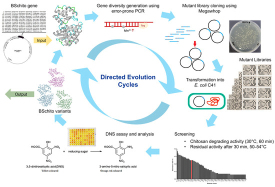
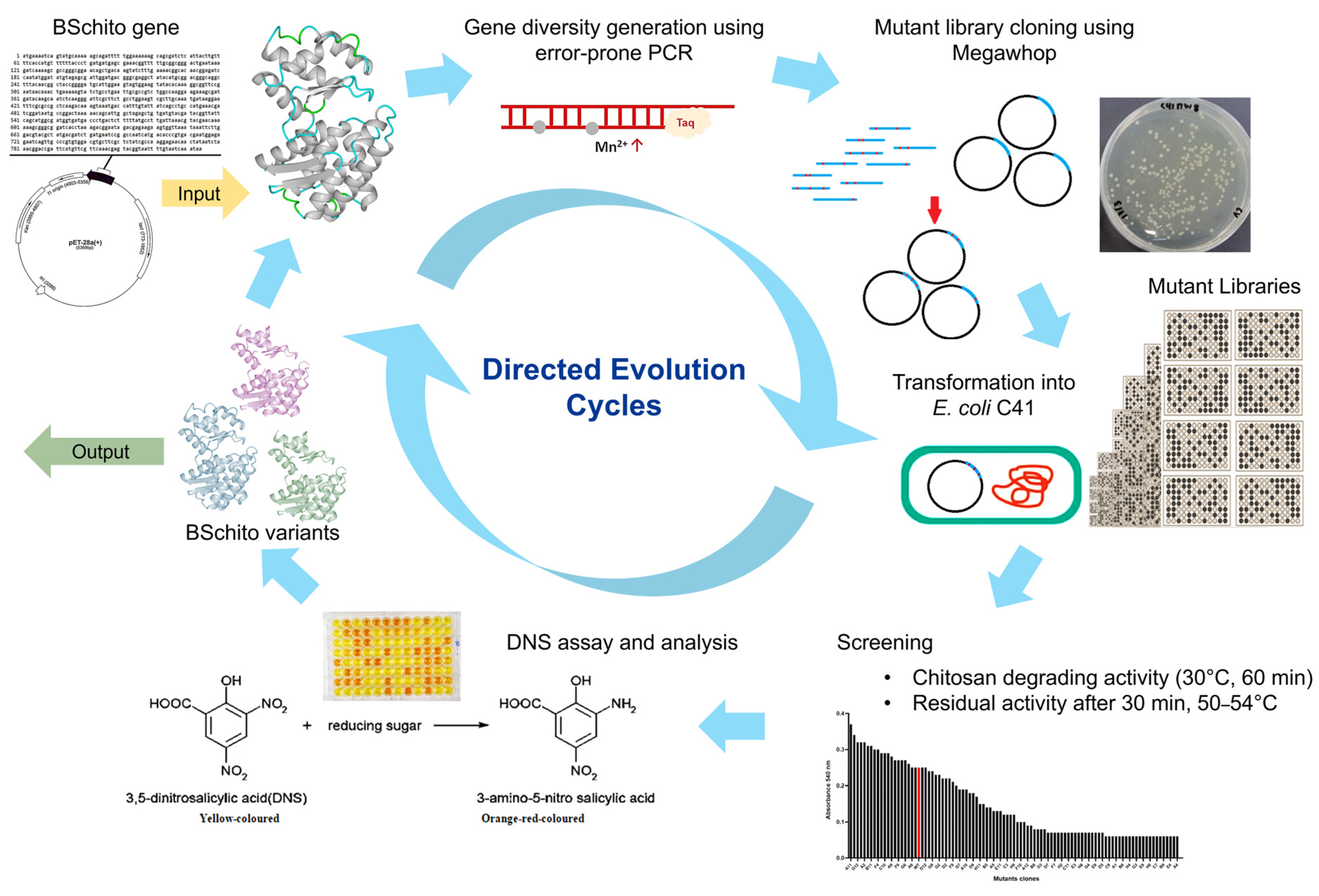
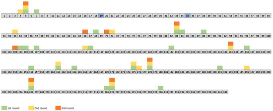
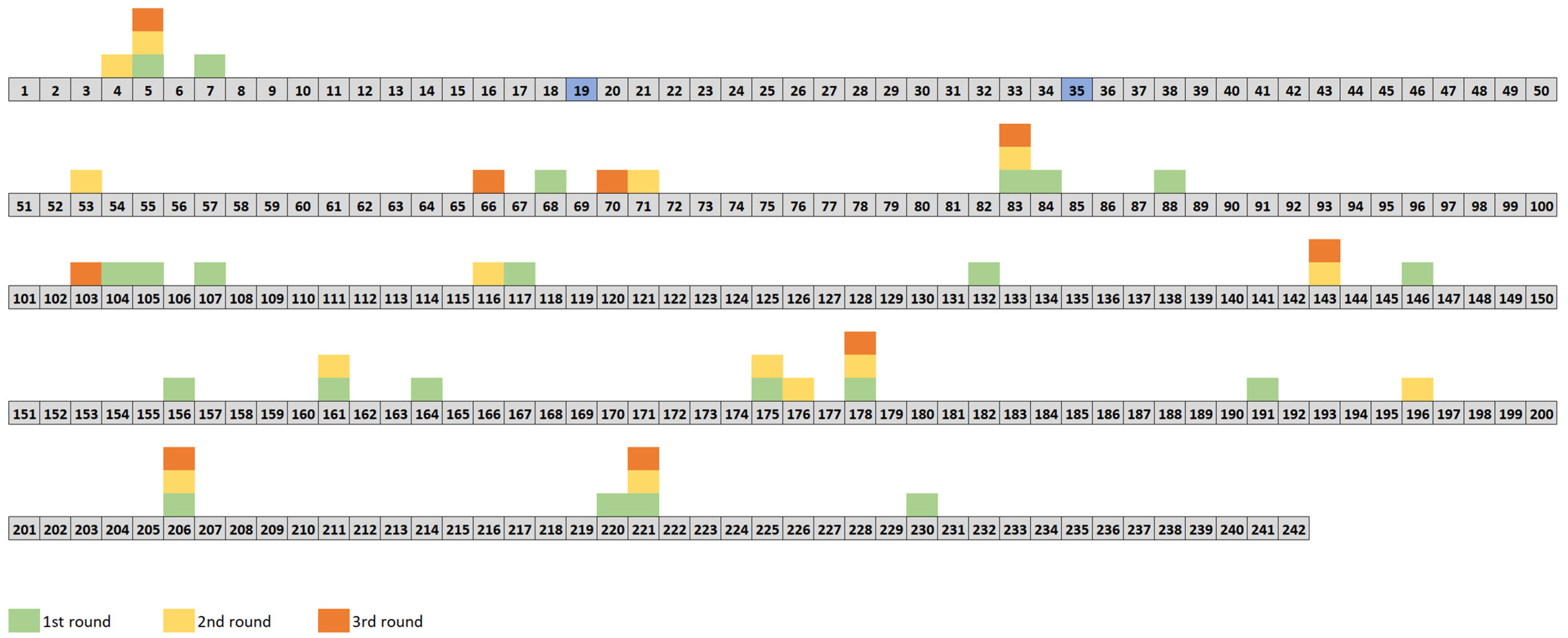
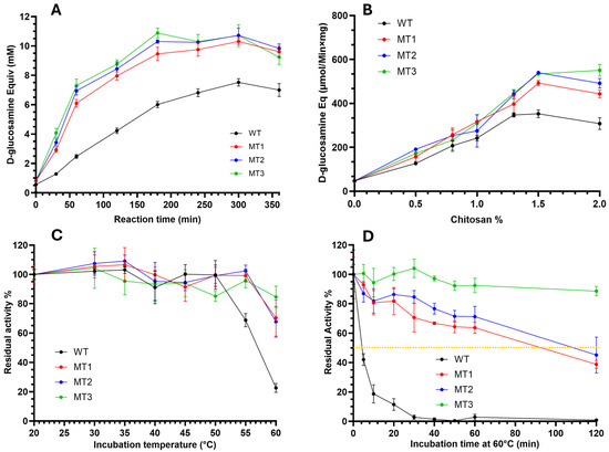
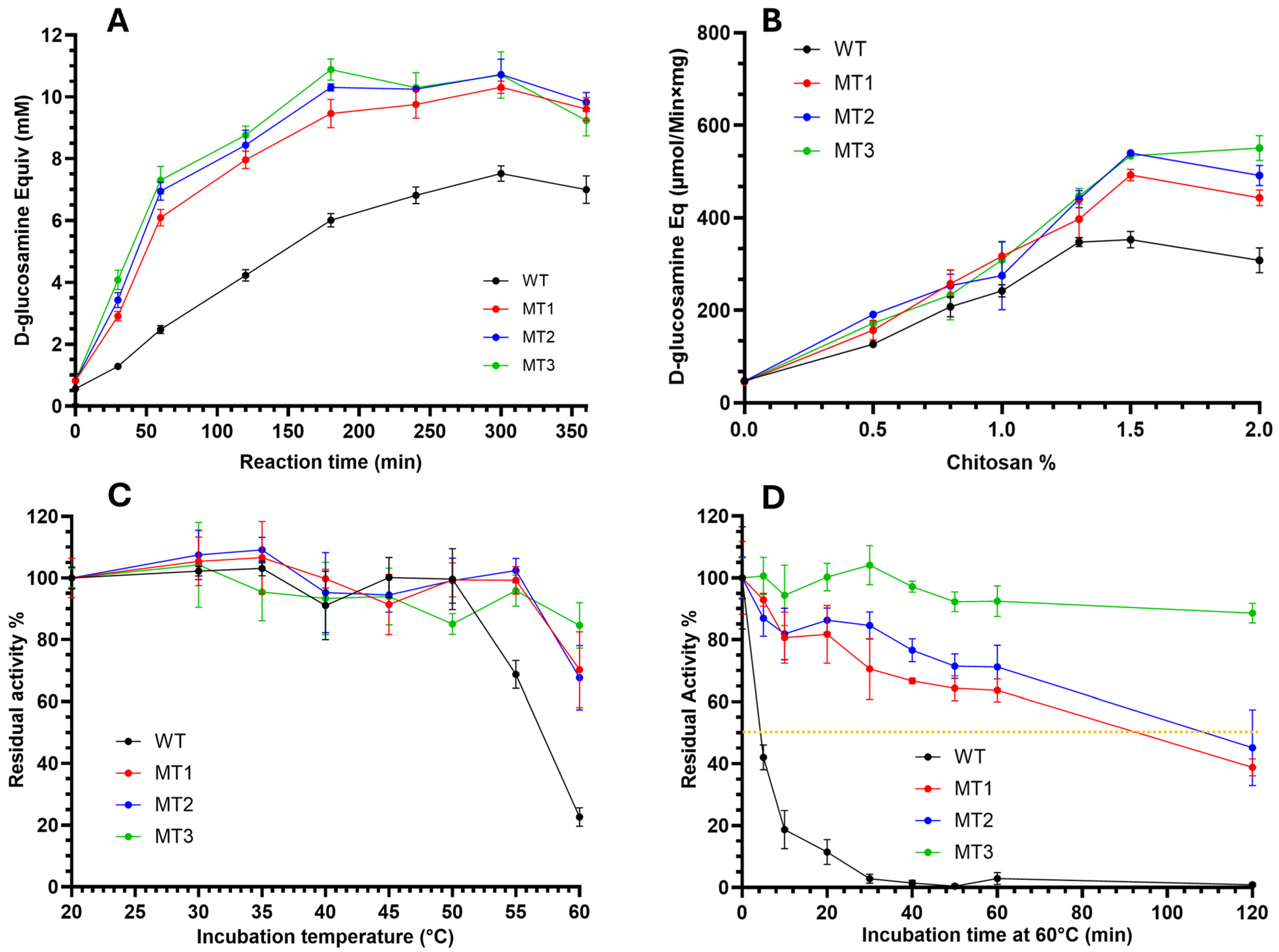
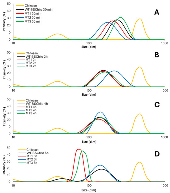
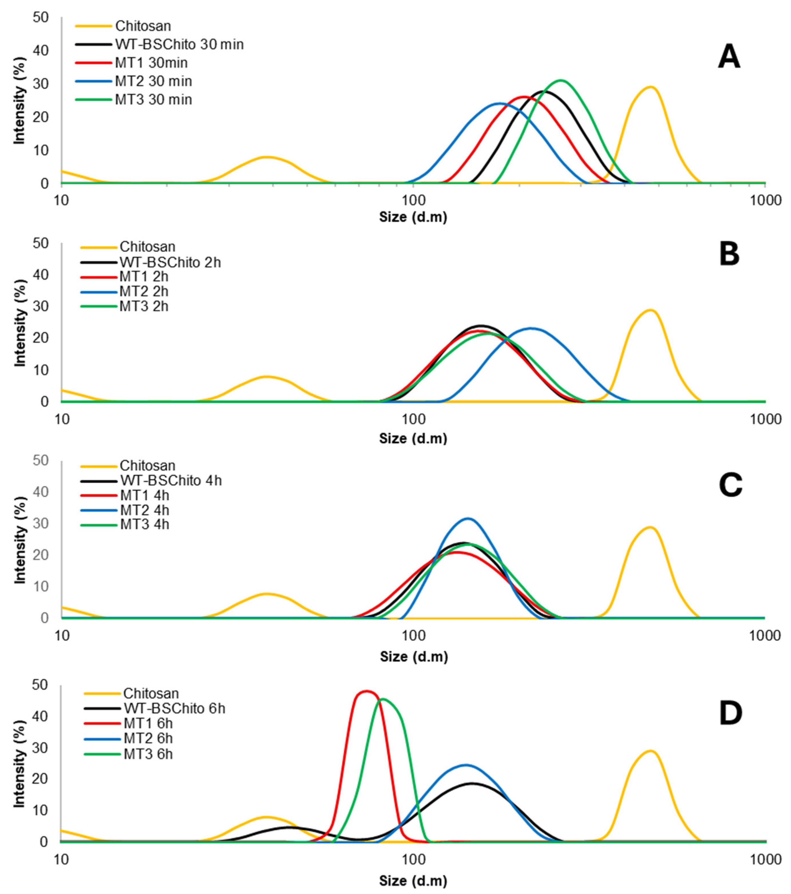
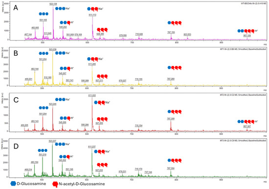
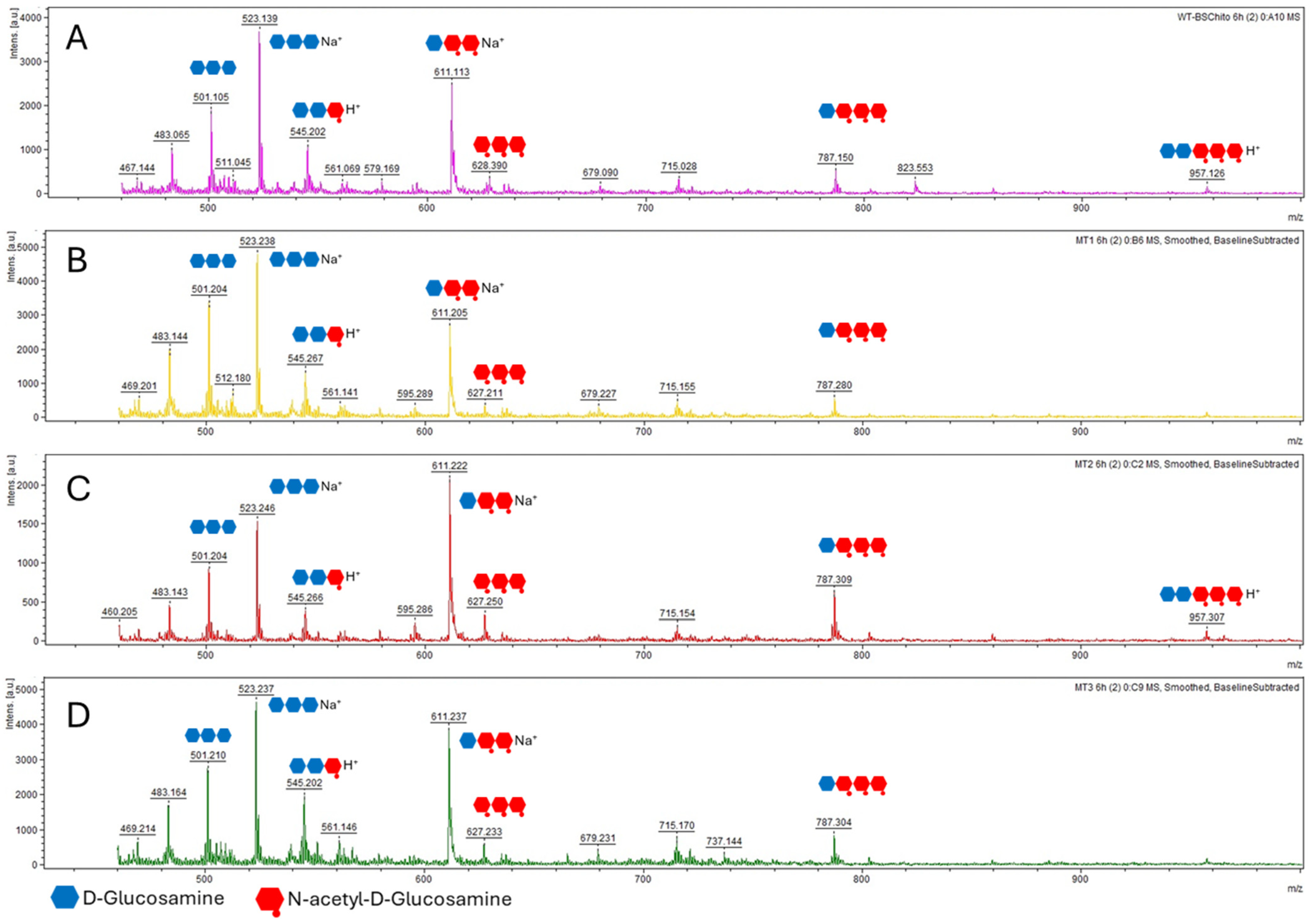
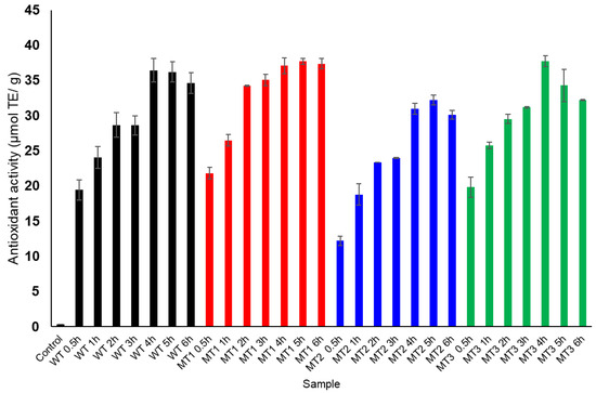
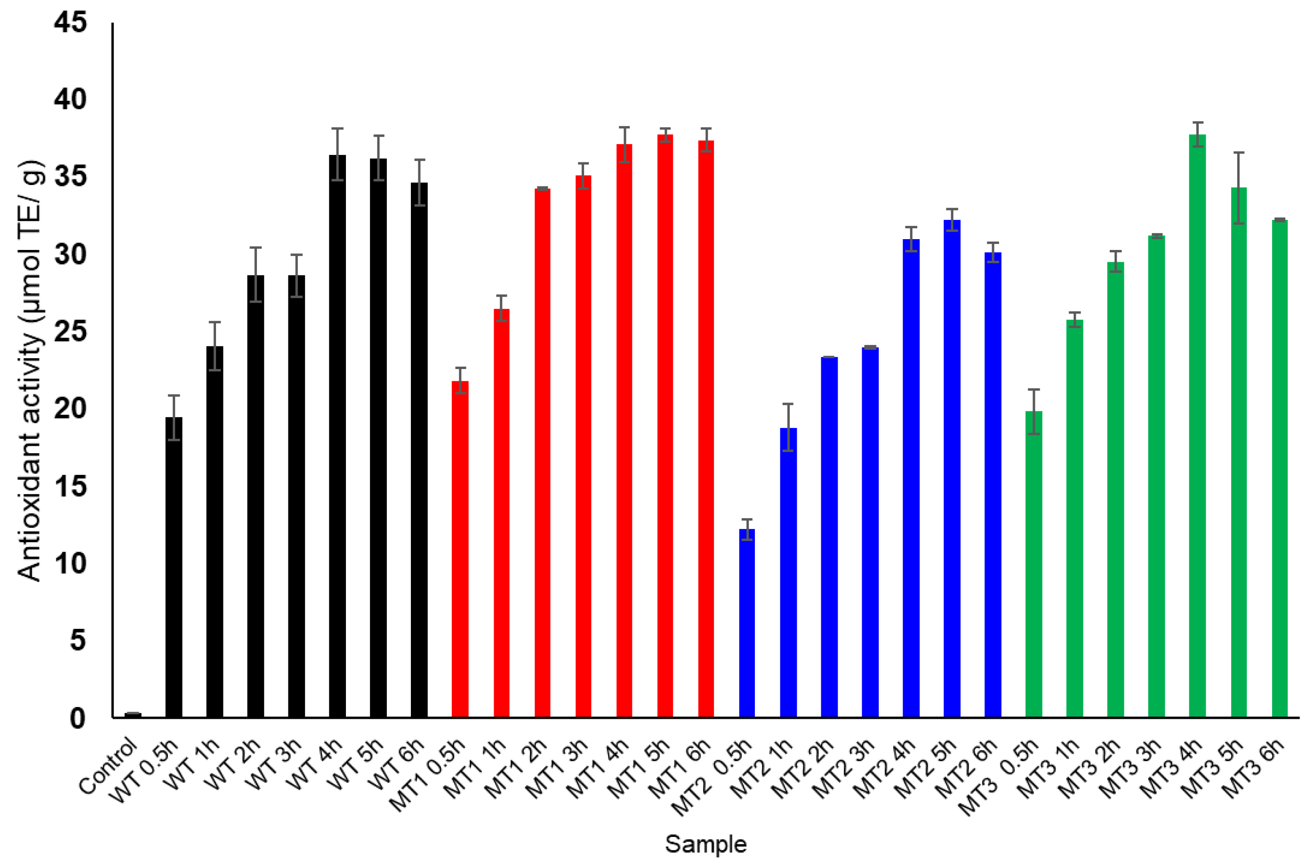
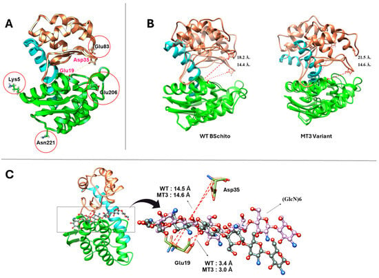
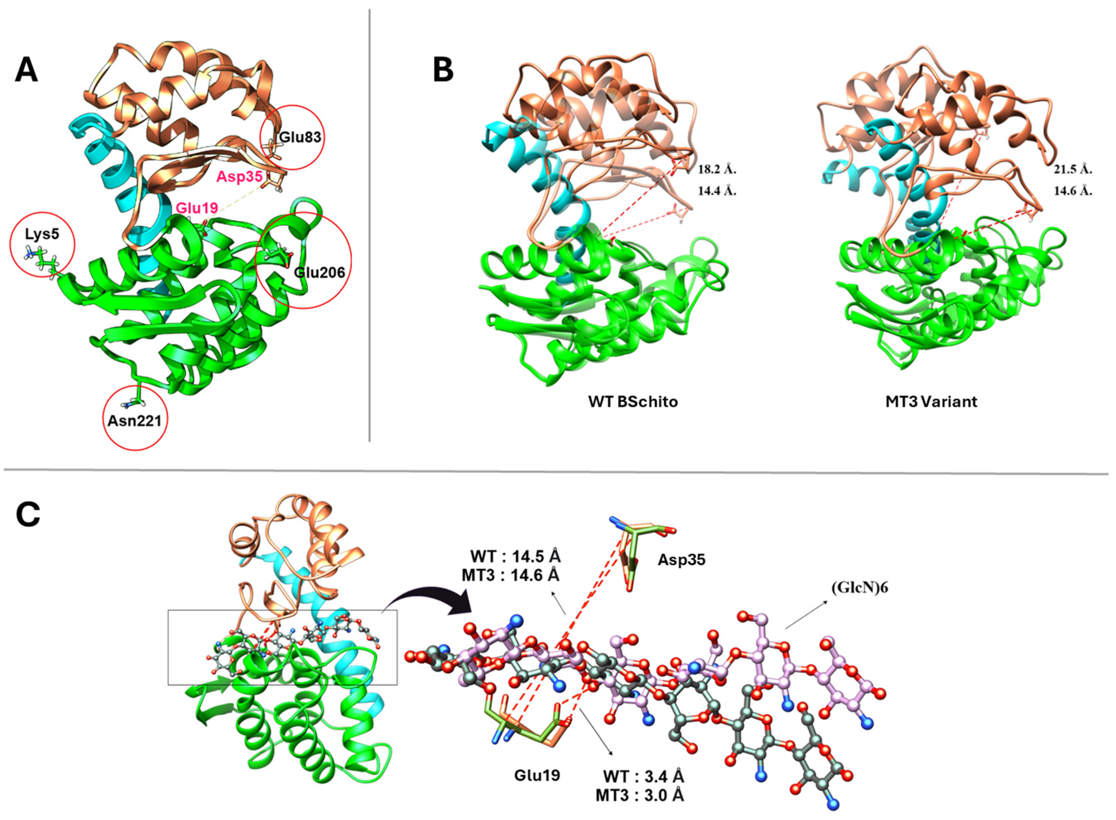
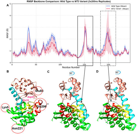
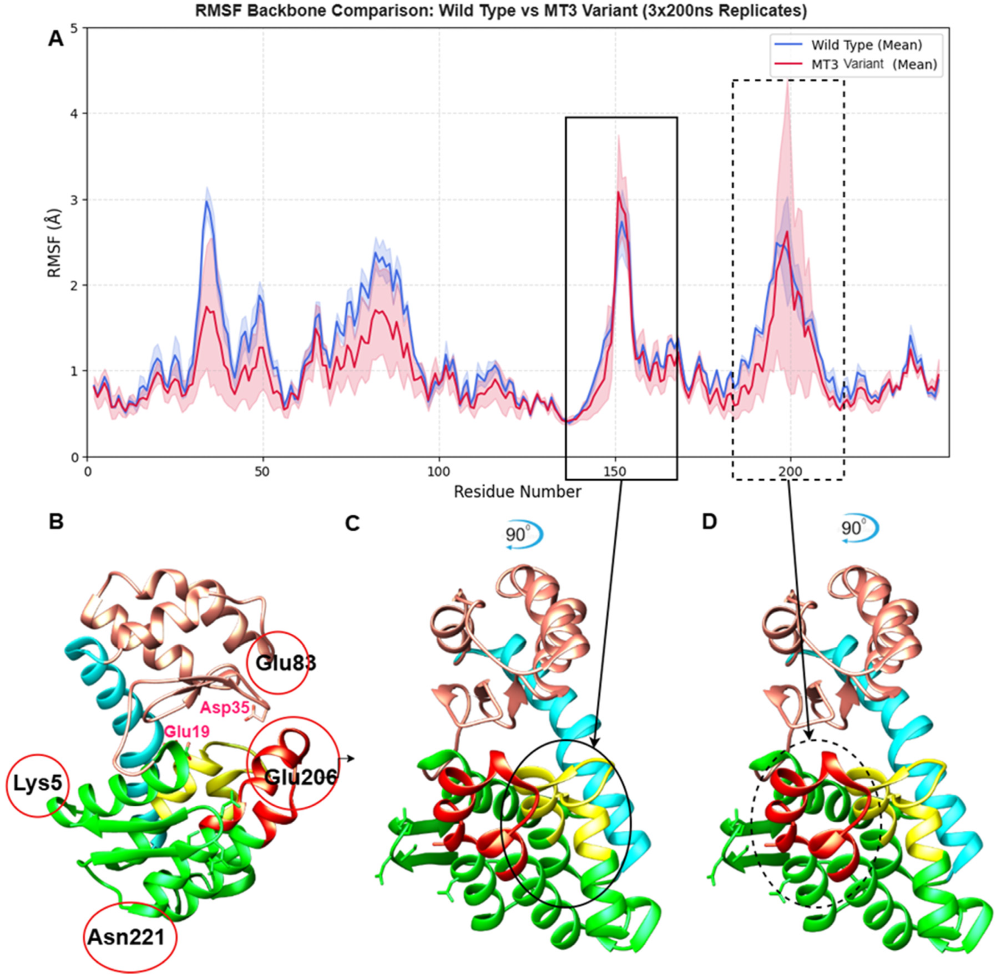

Disclaimer/Publisher’s Note: The statements, opinions and data contained in all publications are solely those of the individual author(s) and contributor(s) and not of MDPI and/or the editor(s). MDPI and/or the editor(s) disclaim responsibility for any injury to people or property resulting from any ideas, methods, instructions or products referred to in the content. |
© 2026 by the authors. Licensee MDPI, Basel, Switzerland. This article is an open access article distributed under the terms and conditions of the Creative Commons Attribution (CC BY) license.