


Simple Summary
This retrospective study evaluated the safety and efficacy of the FOLFIRI-3 regimen (split-dose irinotecan) in 147 patients with unresectable metastatic colorectal cancer, stratified by UGT1A1 polymorphisms. Patients were classified into Wild-type, Single-heterozygous (SH), and Homo/Compound heterozygous (HCH) groups. The HCH group maintained a relative dose intensity of 57.7% with manageable toxicity, supporting the regimen’s safety for high-risk patients. Notably, the SH group demonstrated significantly longer overall survival compared to the Wild-type group (median 30.4 vs. 21.7 months, p = 0.0058), likely due to prolonged exposure within the therapeutic window. FOLFIRI-3 serves as a valuable alternative for toxicity management in high-risk patients and offers superior survival benefits for single heterozygotes, highlighting its potential as a tailored strategy in personalized medicine.
Abstract
Purpose: UGT1A1 gene polymorphisms are established risk factors for irinotecan (CPT-11)-induced toxicity. The FOLFIRI-3 regimen, incorporating split-dose CPT-11 administration on Days 1 and 3, was developed to improve safety and efficacy. This study evaluated the clinical outcomes of FOLFIRI-3 stratified by UGT1A1 genotype. Methods: We retrospectively analyzed 147 patients with unresectable metastatic colorectal cancer (mCRC) treated with first-line FOLFIRI-3 between 2005 and 2020 who underwent UGT1A1 genotyping for *6 and *28 variants. During this period, FOLFIRI-3 was adopted as the standard first-line regimen for all eligible patients at our institution to standardize toxicity management. Patients were classified into three groups: Wild-type (negative for both variants; n = 82), Single-heterozygous (SH; n = 56), and Homo/Compound heterozygous (HCH; n = 8) group. Results: The HCH group maintained a relative dose intensity (RDI) of 57.7% with manageable toxicity. While time to treatment discontinuation (TTD) did not differ significantly among groups, overall survival (OS) was significantly longer in the SH group compared to the Wild-type group (Median Survival Time: 30.4 vs. 21.7 months, p = 0.0058). Conclusions: For high-risk HCH patients, our findings regarding the tolerability of FOLFIRI-3 are strictly preliminary due to the small sample size and require further validation in larger, multi-institutional cohorts. Despite this limitation, this regimen serves as a valuable alternative for high-risk patients and provides superior survival benefits for single heterozygotes, likely due to optimized pharmacokinetics.
1. Introduction
Colorectal cancer remains a leading cause of cancer-related mortality worldwide. While the therapeutic landscape is rapidly evolving with the introduction of immunotherapies and targeted agents [1], the combination of fluorouracil (5-FU), leucovorin (LV), and irinotecan (CPT-11)—known as the FOLFIRI regimen—remains a fundamental backbone of first-line chemotherapy for metastatic colorectal cancer (mCRC). Clinical trials, such as those by Douillard et al. [2], the GERCOR study [3], and others [4,5,6,7,8], have demonstrated its survival benefit. However, chemotherapy-induced toxicity poses a persistent challenge in clinical practice [9]. Irinotecan is notably associated with severe, dose-limiting toxicities, particularly neutropenia and delayed diarrhea. The metabolism of irinotecan is heavily dependent on uridine diphosphate glucuronosyltransferase 1A1 (UGT1A1). This enzyme glucuronidates SN-38, the active metabolite, into the inactive SN-38 glucuronide (SN-38G). Polymorphisms in the UGT1A1 gene, specifically UGT1A1 *28 and UGT1A1 *6, significantly reduce glucuronidation activity, leading to the accumulation of SN-38 [10,11]. There are substantial ethnic differences in the distribution of these variant alleles [12]. The *28 variant is common in Caucasian populations (30–35%), whereas its frequency is lower in Asians [12]. Conversely, the *6 variant is specific to East Asian populations (15–20%) and is virtually absent in Caucasians [13,14]. In Japanese cancer patients, both *6 and *28 are critical predictors of severe neutropenia [13,15]. Based on these findings and the report by Akiyama et al. [16], it has become common practice in Japan to perform UGT1A1 polymorphism testing and to consider dose reduction for homozygous or compound heterozygous (HCH) patients, in accordance with the manufacturer’s labeling recommendations [17].
However, dose reduction may compromise antitumor efficacy. To optimize the therapeutic index, researchers have focused on the sequence-dependent synergy between irinotecan and 5-FU. In vitro studies suggested that cytotoxicity is enhanced when irinotecan is administered before 5-FU [18,19,20]. Yet, clinical evaluation in a randomized phase II study revealed that toxicities were exacerbated when 5-FU was followed by irinotecan [21]. These results suggested that while cytotoxicity might be higher, the safety profile is worse with that sequence. Consequently, Mabro et al. initially designed the FOLFIRI-2 regimen, consisting of irinotecan (180 mg/m2) administered on Day 3 at the end of the 46-h 5-FU infusion [22]. Although FOLFIRI-2 showed encouraging efficacy in pretreated mCRC, the toxicity profile was unacceptably high, with Grade 3–4 diarrhea in 31%, Grade 3–4 neutropenia in 52%, and febrile neutropenia in 14% of patients. To maintain the therapeutic advantage of administering irinotecan after 5-FU while improving tolerability, the FOLFIRI-3 regimen was developed. FOLFIRI-3 consists of irinotecan (100 mg/m2) as a 60-min infusion on Day 1 administered concurrently with leucovorin, followed by a 46-h continuous infusion of 5-FU, and a second infusion of irinotecan (100 mg/m2) on Day 3 at the end of the 5-FU infusion [23,24]. This split-dose strategy was adopted to mitigate the severe toxicity observed with the single high-dose administration on Day 3 in the FOLFIRI-2 regimen. We hypothesized that this fractionation strategy, by avoiding a single high-dose administration, may allow for safer administration in high-risk patients with UGT1A1 polymorphisms and potentially enhance efficacy in intermediate-risk patients. This study analyzes the clinical outcomes of FOLFIRI-3 stratified by UGT1A1 status.
2. Materials and Methods
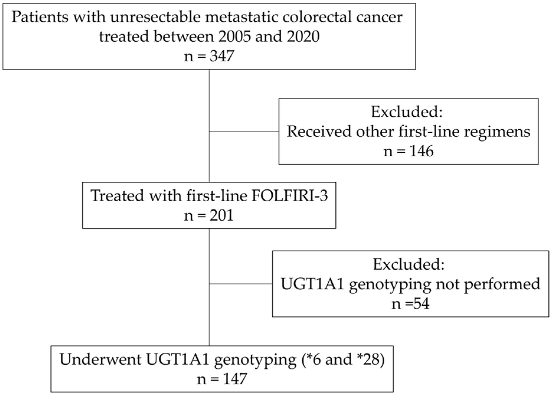
2.1. Study Population (Figure 1)
We retrospectively reviewed patients with unresectable mCRC treated at our institutions between 2005 and 2020. Among 347 patients who underwent systemic chemotherapy during this period, 201 were treated with first-line FOLFIRI-3. During this period, FOLFIRI-3 was our institutional standard to standardize toxicity management. However, alternative regimens (e.g., standard FOLFIRI or FOLFOX) were utilized for patients who declined the split-dose schedule or the logistical requirement of a Day 3 hospital visit, as well as those who found irinotecan-induced alopecia unacceptable or wished to avoid potent cytotoxic agents such as irinotecan or oxaliplatin. Of the 201 patients, 147 who underwent UGT1A1 genotyping for *6 and *28 were included in this analysis. Since 2010, following the general recommendation for dose reduction in the Japanese drug package insert [17], our institutional policy has been to perform preemptive dose reduction for confirmed HCH patients. In the absence of a standardized national guideline for dose-reduction ratios, our institutional consensus was to initiate irinotecan at a two-level dose reduction (approximately 64% of the standard dose) for HCH patients. The final determination of the regimen and minor adjustments remained at the discretion of the treating physician, who fine-tuned this baseline dose based on the patient’s clinical status (e.g., age, performance status, and prior treatment history). Furthermore, active chemotherapy was generally not recommended for fragile patients with an ECOG Performance Status (PS) of 3. However, for the 7 patients with PS 3 included in this cohort, treatment was initiated only after providing a thorough explanation of the high risk of severe toxicities and obtaining strict informed consent. This decision was ethically justified by the need to respect the strong and explicit requests from the patients and their families to pursue treatment. In all such cases, treatment was carefully initiated at reduced doses with rigorous clinical monitoring to prioritize patient safety.
Figure 1.
Flow diagram of patient selection.
2.2. Treatment Regimen (FOLFIRI-3)
The FOLFIRI-3 regimen followed the protocol described by Mabro et al. [23]:
- Day 1, irinotecan 100 mg/m2 as a 1-h infusion, administered concurrently with LV 200 mg/m2 as a 2-h infusion, followed by 5FU 2000 mg/m2 as a 46-h continuous infusion.
- Day 3, irinotecan 100 mg/m2 as a 1-h infusion was repeated, at the end of the 5FU infusion. Cycles were repeated every two weeks, combined with molecular targeted drugs according to standard guidelines [25].
2.3. Genotyping and Stratification
The UGT1A1 genotypes (*6 and *28) were determined by PCR-based melting curve analysis using a previously described method [26]. Stratification was defined as follows: Wild-type (negative for both variant alleles), Single-heterozygous (SH; *1/*6 or *1/*28), and Homo/Compound heterozygous (HCH; *6/*6, *28/*28, or *6/*28). The detailed classification of these genotypes, their zygosity, and assigned metabolic phenotypes are summarized in Table 1.
Table 1.
UGT1A1 genotypes.
2.4. Statistical Analysis
Overall survival (OS) and time to treatment discontinuation (TTD) were estimated using the Kaplan–Meier method and compared among groups using the log-rank test. To identify independent prognostic factors for OS, variables with a p-value < 0.05 in the univariate log-rank test were subsequently included in a multivariate analysis using the Cox proportional hazards model. Hazard ratios (HRs) and 95% confidence intervals (CIs) were calculated. Comparison of the relative dose intensity (RDI) between groups was performed using the Mann–Whitney U test. Adverse events (AEs) were graded according to Common Terminology Criteria for Adverse Events (CTCAE) version 5 [27]. All statistical analyses were performed using JMP Student Edition version 19 (SAS Institute Inc., Cary, NC, USA).
3. Results
3.1. Patient Characteristics
The genotype distribution was as follows: Wild-type 82 patients (55.8%), SH 56 patients (38.1%), and HCH 8 patients (5.4%). The HCH group included 7 compound heterozygotes (*6/*28) and 1 homozygote (*6/*6). Baseline characteristics showed 15.6% had a Performance Status (PS) ≥ 2 and 23.1% with prior adjuvant chemotherapy. Detailed characteristics are shown in Table 2.
Table 2.
Patient characteristics.
3.2. Treatment Efficacy
Regarding the best overall response by Response Evaluation Criteria in Solid Tumor (RECIST) version 1.1 [28], Progressive Disease (PD) was observed in 10 patients (6.8%), Stable Disease (SD) in 45 (30.6%), Partial Response (PR) in 68 (46.3%), and Complete Response (CR) in 7 (4.8%). The objective response rate (ORR) was 51.0%, and the disease control rate (DCR) was 81.6%.
3.3. Survival Outcomes
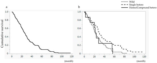
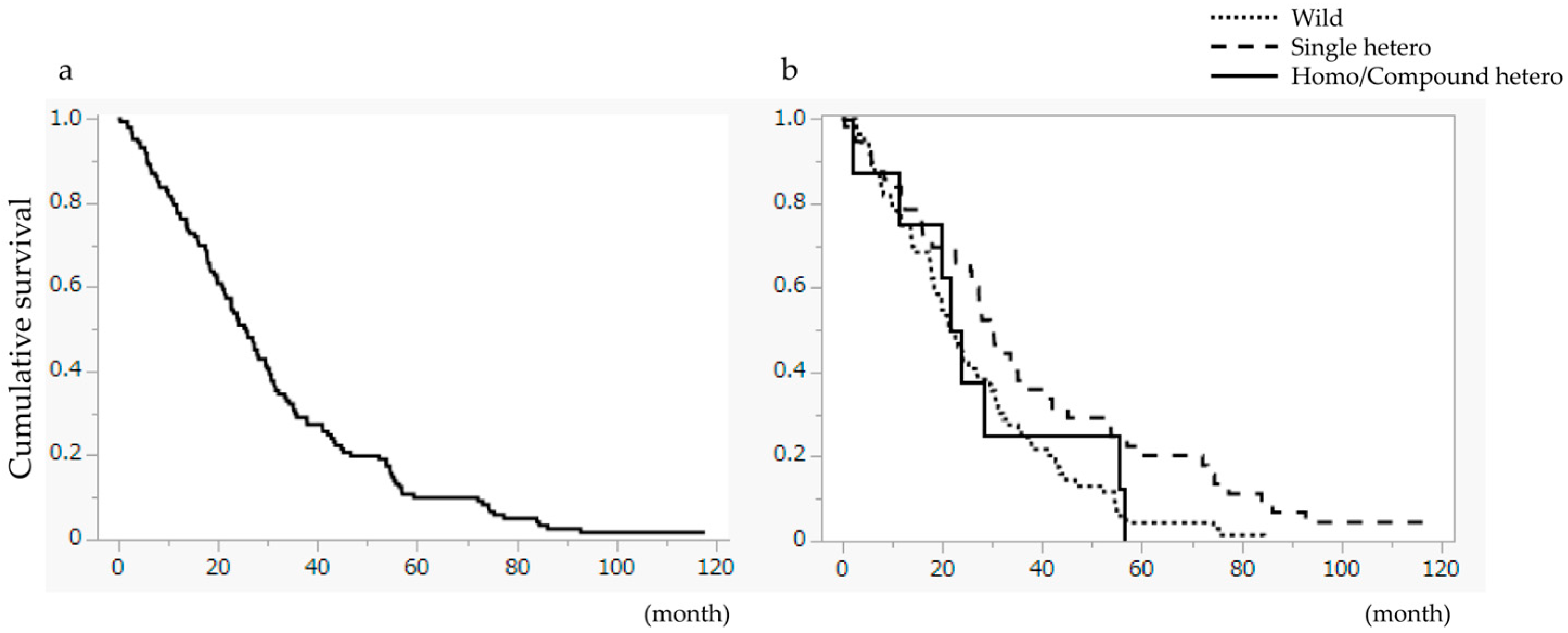
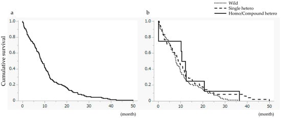
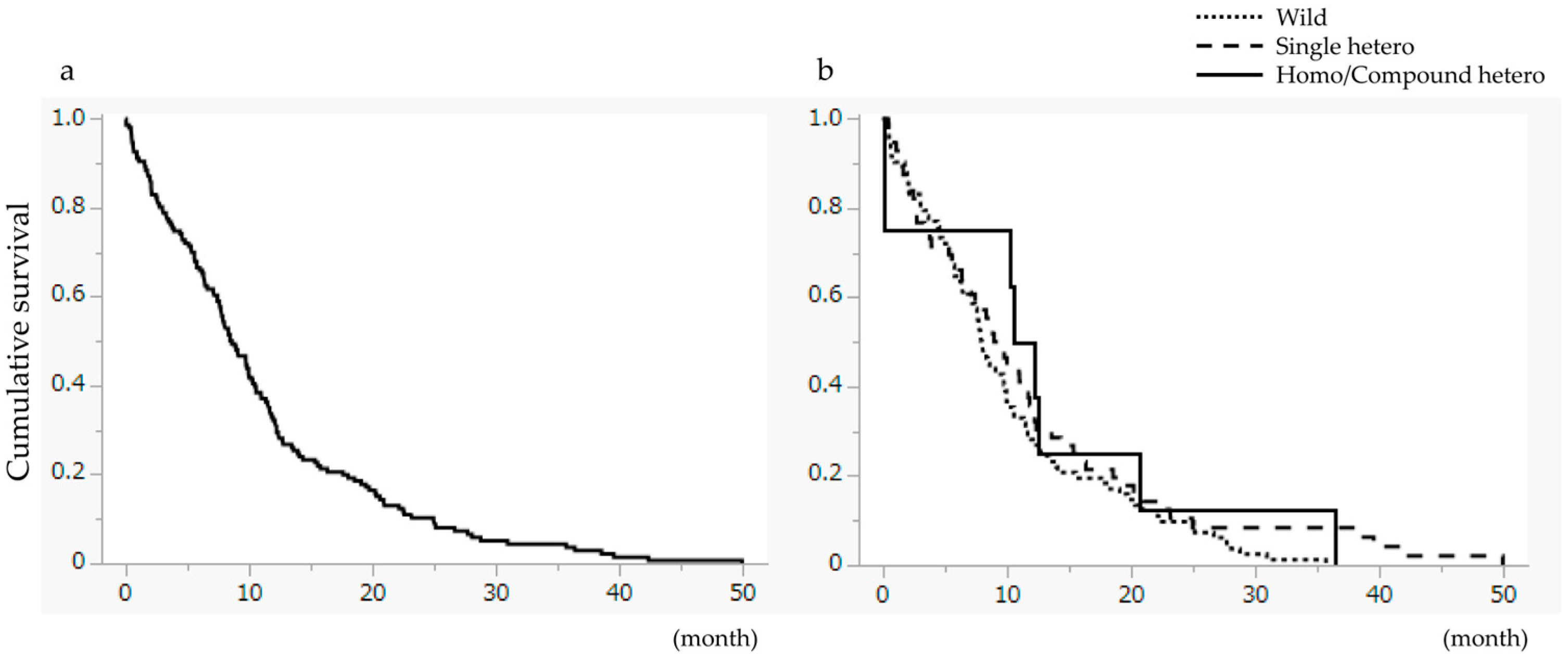
For the entire cohort, the Median Survival Time (MST) was 25.7 months (Figure 2a). Stratified analysis revealed a significant difference in OS (p = 0.0058), with the SH group (MST 30.4 months) performing significantly better than the Wild-type group (MST 21.7 months). The HCH group achieved an MST of 25.7 months (Figure 2b). TTD did not differ significantly among the three groups (Figure 3).
Figure 2.
Overall survival (OS). (a) Overall survival for all patients. The median survival time (MST) was 25.7 months. (b) Comparison of OS based on UGT1A1 genotype.
Figure 3.
Time to treatment discontinuation (TTD). (a) TTD for all patients. The median TTD was 8.6 months. (b) Comparison of TTD based on UGT1A1 genotype. No significant difference was observed among the groups.
A significant difference was observed among groups (p = 0.0171, log-rank test). The MST was 21.7 months in the Wild-type group, 30.4 months in the single heterozygote (SH) group, and 22.8 months in the homo/compound heterozygote (HCH) group. OS was significantly longer in the SH group compared to the Wild-type group (p = 0.0058).
Furthermore, univariate and multivariate analyses were performed to identify prognostic factors for OS (Table 3). In the univariate analysis, ECOG PS (0, 1 vs. 2, 3), side of primary tumor (right vs. left), presence of liver metastasis, presence of peritoneal metastasis, BRAF status (wild vs. mutant), microsatellite instability (MSS vs. MSI), UGT1A1 genotype, and main pathology were significantly associated with OS (p < 0.05). These significant variables were incorporated into the multivariate Cox proportional hazards model. The multivariate analysis revealed that ECOG PS ≥ 2 (HR 3.66, p = 0.0008), presence of liver metastasis (HR 3.16, p = 0.0009), mucinous/signet-ring/poorly differentiated histology (HR 2.64, p = 0.0264), and UGT1A1 genotype remained as independent prognostic factors for OS (p = 0.0068), confirming the robust survival advantage in the SH group.
Table 3.
Univariate and multivariate analyses of prognostic factors for overall survival.
3.4. Safety and Dose Intensity
Grade 3 or higher AEs occurred in 64 patients (78.1%) in the Wild-type group, 43 patients (78.2%) in the SH group, and 8 patients (100%) in the HCH group. Despite the high incidence in the HCH group, all events were manageable (primarily neutropenia), and no treatment-related deaths occurred (Table 4). The median RDI for irinotecan in the entire cohort was 66.7% (range, 16.6–100%), and an RDI of ≥85% was achieved in 22 cases (15.5%). By group, the median RDI in the Wild-type group was 66.7% (range 16.6–100), in the SH group was 67.3% (range 20.6–95.6), and in the HCH group was 57.7% (range 31.5–94.2). There were no significant differences in RDI among the three groups.
Table 4.
Adverse Events (CTCAE Grade ≥ 3).
4. Discussion
This study re-evaluates the clinical utility of the FOLFIRI-3 regimen in the era of UGT1A1-guided personalized medicine. Originally reported by Mabro et al. [23], FOLFIRI-3 enhances safety by splitting the irinotecan dose. While standard FOLFIRI-1 remains the standard practice in Japan due to the logistical burden of Day 3 visits [25], our institutional use of FOLFIRI-3 provided a unique dataset across UGT1A1 genotypes without selection bias.
Patients with HCH genotypes are at high risk for life-threatening toxicity [13,29,30]. All HCH patients in our study experienced a 100% rate of Grade 3+ AEs, and their median RDI was numerically lower than that of the other groups (57.7%), reflecting the frequent need for dose delays or reductions. However, the absence of treatment-related deaths suggests that the split-dose approach may help avoid fatal outcomes, although severe toxicities remain highly prevalent. This favorable safety profile stands in contrast to the high toxicity previously observed with the single high-dose strategy (FOLFIRI-2) [22], suggesting that fractionating the dose helps mitigate the severity of adverse events. As a real-world retrospective cohort, our analysis included three HCH patients who initially received full doses of irinotecan prior to the availability of genotyping results or established guidelines. While the inclusion of these cases artificially inflated the median RDI of the HCH group (57.7%), the absence of fatal toxicities even in these “worst-case scenarios” underscores a potential safety margin of the split-dose strategy. However, given the 100% incidence of Grade 3 or higher adverse events, FOLFIRI-3 does not eliminate the need for extreme caution, strict monitoring, and appropriate dose reductions in this high-risk population.
While direct comparisons with historical cohorts are limited by differences in patient populations and supportive care over time, the incidences of severe neutropenia (36.1%) and diarrhea (17.0%) in our FOLFIRI-3 cohort did not exceed the rates generally reported in landmark trials of standard FOLFIRI-1 [2,3,4]. However, given the added logistical burden of a Day 3 irinotecan administration, this baseline safety profile alone does not justify the routine adoption of FOLFIRI-3 for all unselected mCRC patients. Instead, we believe the true clinical rationale for the split-dose strategy emerges in the SH subgroup. A novel finding is the superior survival in the SH group (MST 30.4 months) compared to the Wild-type group. Crucially, our multivariate analysis demonstrated that the UGT1A1 genotype is an independent prognostic factor for OS, even after adjusting for strong clinical variables such as ECOG PS, liver metastasis, and tumor histology. This aligns with clinical reports such as those by Cecchin et al. [31], who observed that UGT1A1 *28 variant carriers achieved significantly better clinical benefit, response rate, and time to progression (TTP) compared to wild-type patients when treated with irinotecan-based therapy. Cecchin et al. proposed that the mechanism explaining the better outcome of variant carriers could be reduced intratumoral inactivation of SN-38 in variant carriers, as UGT1A1 is expressed in CRC cells and glucuronidation has been regarded as a mechanism of resistance. Their study ruled out a systemic antitumor effect since UGT1A1 *28 did not show a strong association with systemic SN-38 AUC. Furthermore, Toffoli et al. [32] and Paoluzzi et al. [33] have shown that *UGT1A1* variants are associated with a higher biliary index (BI) and increased systemic exposure to SN-38. We hypothesize that SH patients might benefit from a “moderate metabolic delay”. In Wild-type patients, rapid glucuronidation may limit tumor exposure to SN-38. In contrast, SH patients likely maintain effective SN-38 levels for a longer duration (increased AUC). This allows them to remain within a “therapeutic window”—maximizing efficacy without exceeding the toxic threshold seen in HCH patients. However, it is important to emphasize that this pharmacokinetic mechanism is highly speculative. Despite this limitation, our clinical survival data suggest that FOLFIRI-3 may be the optimal regimen for SH patients without the need for dose attenuation. Considering the increasing use of intensive triplet regimens (e.g., FOLFOXIRI) and the inherent logistical burden of a Day 3 visit, FOLFIRI-3 is not intended to replace standard FOLFIRI for all unselected patients. Consequently, rather than a universal standard, we propose that FOLFIRI-3 should be positioned as a tailored, highly targeted therapeutic option specifically for SH patients to maximize their survival benefits.
This study has several limitations. First, it is a retrospective analysis from a single center. Second, as a real-world observational study, the cohort inherently includes clinical heterogeneity, such as elderly patients and a small subset with a Performance Status of 3. While this limits the homogeneity of the cohort, our multivariate analysis ensured that these variables were statistically adjusted for when evaluating the primary impact of the UGT1A1 genotype. Third, the HCH sample size was small (n = 8), which severely limits statistical power and the generalizability of conclusions for this high-risk population. Therefore, our findings regarding the safety of FOLFIRI-3 in HCH patients must be considered preliminary and require rigorous validation in larger, multi-institutional cohorts. Fourth, we did not perform a direct internal comparison between FOLFIRI-3 and FOLFIRI-1. Because FOLFIRI-3 was strictly adopted as our institutional standard regimen during the study period, only one patient in our database received standard FOLFIRI-1, precluding any meaningful comparative analysis. Consequently, the true relative advantages of FOLFIRI-3 over FOLFIRI-1 must be evaluated in future prospective randomized studies. Fifth, plasma SN-38 concentrations were not measured directly; our mechanistic explanations are based on the established literature. Therefore, our hypothesis that SH patients benefit from optimal SN-38 exposure remains strictly hypothetical and cannot be definitively confirmed without direct PK measurements. Finally, due to the retrospective design, we could not objectively assess patients’ quality of life (QOL) to fully evaluate the impact of the additional logistical burden imposed by the Day 3 irinotecan administration. However, the acceptable and comparable time to treatment discontinuation (TTD) observed across all genotypes suggests that treatment adherence was reasonably well-maintained in real-world practice.
5. Conclusions
Despite the logistical burden, FOLFIRI-3 is a valuable strategy. For HCH patients, while it may help prevent fatal events, the high incidence of severe toxicities and the necessity for dose reductions (as reflected by the lower RDI) indicate that extreme caution and strict monitoring remain essential. Importantly, these findings are preliminary, and the true safety profile and clinical utility for HCH patients must be validated in larger, multi-institutional cohorts. For SH patients, it offers a survival advantage likely due to optimized exposure. We propose FOLFIRI-3 as a tailored option for UGT1A1 single-heterozygous patients.
Author Contributions
Conceptualization, G.O. and H.M.; methodology, G.O.; software, G.O.; validation, G.O., H.M. and K.S.; formal analysis, G.O.; investigation, T.I. and K.M.; resources, G.O., T.T., T.M., K.O., Y.I., A.H., T.I. and K.M.; data curation, G.O.; writing—original draft preparation, G.O.; writing—review and editing, G.O.; visualization, G.O.; supervision, H.M., M.M., K.H. and K.S.; project administration, G.O. All authors have read and agreed to the published version of the manuscript.
Funding
This research received no external funding.
Institutional Review Board Statement
The study was conducted in accordance with the Declaration of Helsinki, and approved by the Institutional Review Board of Chiba University Hospital (No. 5-47, 27 January 2026).
Informed Consent Statement
The requirement for written informed consent was waived by the IRB due to the retrospective nature of the study, and an opt-out method was used for patient recruitment.
Data Availability Statement
The data are not publicly available due to ethical restrictions.
Acknowledgments
We would like to acknowledge the use of Google’s Gemini language model for assistance with refining the clarity of the English language, improving grammatical structure, and optimizing the expression of the text within this manuscript. The authors confirm that the core research design, data collection, analysis, interpretation of the results, and all conclusions presented herein were conducted solely by the human authors. All authors take full responsibility for the content and accuracy of this publication.
Conflicts of Interest
The authors declare no conflicts of interest.
Abbreviations
The following abbreviations are used in this manuscript:
| 5-FU | Fluorouracil |
| AE | Adverse event |
| BI | Biliary index |
| CPT-11 | Irinotecan |
| CR | Complete response |
| CTCAE | Common Terminology Criteria for Adverse Events |
| DCR | Disease control rate |
| ECOG | Eastern Cooperative Oncology Group |
| HCH | Homo/compound heterozygote |
| LV | Leucovorin |
| mCRC | Metastatic colorectal cancer |
| MSI | Microsatellite instability |
| MSS | Microsatellite stable |
| MST | Median survival time |
| ORR | Objective response rate |
| OS | Overall survival |
| PD | Progressive disease |
| PR | Partial response |
| PS | Performance status |
| RDI | Relative dose intensity |
| RECIST | Response Evaluation Criteria in Solid Tumors |
| SD | Stable disease |
| SH | Single heterozygote |
| SN-38G | SN-38 glucuronide |
| TTD | Time to treatment discontinuation |
| TTP | Time to progression |
| UGT1A1 | Uridine diphosphate glucuronosyltransferase 1A1 |
References
- Nikolouzakis, T.K.; Chrysos, E.; Docea, A.O.; Fragkiadaki, P.; Souglakos, J.; Tsiaoussis, J.; Tsatsakis, A. Current and Future Trends of Colorectal Cancer Treatment: Exploring Advances in Immunotherapy. Cancers 2024, 16, 1995. [Google Scholar] [CrossRef] [Scilit]
- Douillard, J.; Cunningham, D.; Roth, A.; Navarro, M.; James, R.; Karasek, P.; Jandik, P.; Iveson, T.; Carmichael, J.; Alakl, M.; et al. Irinotecan combined with fluorouracil compared with fluorouracil alone as first-line treatment for metastatic colorectal cancer: A multicentre randomised trial. Lancet 2000, 355, 1041–1047. [Google Scholar] [CrossRef] [Scilit]
- Tournigand, C.; André, T.; Achille, E.; Lledo, G.; Flesh, M.; Mery-Mignard, D.; Quinaux, E.; Couteau, C.; Buyse, M.; Ganem, G.; et al. FOLFIRI Followed by FOLFOX6 or the Reverse Sequence in Advanced Colorectal Cancer: A Randomized GERCOR Study. J. Clin. Oncol. 2004, 22, 229–237. [Google Scholar] [CrossRef] [Scilit]
- Yamazaki, K.; Nagase, M.; Tamagawa, H.; Ueda, S.; Tamura, T.; Murata, K.; Nakajima, T.E.; Baba, E.; Tsuda, M.; Moriwaki, T.; et al. Randomized phase III study of bevacizumab plus FOLFIRI and bevacizumab plus mFOLFOX6 as first-line treatment for patients with metastatic colorectal cancer (WJOG4407G). Ann. Oncol. 2016, 27, 1539–1546. [Google Scholar] [CrossRef] [Scilit]
- Heinemann, V.; von Weikersthal, L.F.; Decker, T.; Kiani, A.; Vehling-Kaiser, U.; Al-Batran, S.-E.; Heintges, T.; Lerchenmüller, C.; Kahl, C.; Seipelt, G.; et al. FOLFIRI plus cetuximab versus FOLFIRI plus bevacizumab as first-line treatment for patients with metastatic colorectal cancer (FIRE-3): A randomised, open-label, phase 3 trial. Lancet Oncol. 2014, 15, 1065–1075. [Google Scholar] [CrossRef] [Scilit]
- Muro, K.; Salinardi, T.; Singh, A.R.; Macarulla, T. Safety of Aflibercept in Metastatic Colorectal Cancer: A Literature Review and Expert Perspective on Clinical and Real-World Data. Cancers 2020, 12, 844. [Google Scholar] [CrossRef] [Scilit] [PubMed]
- Van Cutsem, E.; Köhne, C.-H.; Hitre, E.; Zaluski, J.; Chien, C.-R.C.; Makhson, A.; D’Haens, G.; Pintér, T.; Lim, R.; Bodoky, G.; et al. Cetuximab and chemotherapy as initial treatment for metastatic colorectal cancer. N. Engl. J. Med. 2009, 360, 1408–1417. [Google Scholar] [CrossRef] [Scilit] [PubMed]
- Loupakis, F.; Cremolini, C.; Masi, G.; Lonardi, S.; Zagonel, V.; Salvatore, L.; Cortesi, E.; Tomasello, G.; Ronzoni, M.; Spadi, R.; et al. Initial therapy with FOLFOXIRI and bevacizumab for metastatic colorectal cancer. N. Engl. J. Med. 2014, 371, 1609–1618. [Google Scholar] [CrossRef] [Scilit]
- Han, C.J.; Ning, X.; Burd, C.E.; Spakowicz, D.J.; Tounkara, F.; Kalady, M.F.; Noonan, A.M.; McCabe, S.; Von Ah, D. Chemotoxicity and Associated Risk Factors in Colorectal Cancer: A Systematic Review and Meta-Analysis. Cancers 2024, 16, 2597. [Google Scholar] [CrossRef] [Scilit] [PubMed]
- Nelson, R.S.; Seligson, N.D.; Bottiglieri, S.; Carballido, E.; Cueto, A.D.; Imanirad, I.; Levine, R.; Parker, A.S.; Swain, S.M.; Tillman, E.M.; et al. UGT1A1 Guided Cancer Therapy: Review of the Evidence and Considerations for Clinical Implementation. Cancers 2021, 13, 1566. [Google Scholar] [CrossRef] [Scilit]
- Iyer, L.; King, C.D.; Whitington, P.F.; Green, M.D.; Roy, S.K.; Tephly, T.R.; Coffman, B.L.; Ratain, M.J. Genetic predisposition to the metabolism of irinotecan (CPT-11). Role of uridine diphosphate glucuronosyltransferase isoform 1A1 in the glucuronidation of its active metabolite (SN-38) in human liver microsomes. J. Clin. Investig. 1998, 101, 847–854. [Google Scholar] [CrossRef] [Scilit] [PubMed]
- Beutler, E.; Gelbart, T.; Demina, A. Racial variability in the UDP-glucuronosyltransferase 1 (UGT1A1) promoter: A balanced polymorphism for regulation of bilirubin metabolism? Proc. Natl. Acad. Sci. USA 1998, 95, 8170–8174. [Google Scholar] [CrossRef] [Scilit] [PubMed]
- Minami, H.; Sai, K.; Saeki, M.; Saito, Y.; Ozawa, S.; Suzuki, K.; Kaniwa, N.; Sawada, J.-I.; Hamaguchi, T.; Yamamoto, N.; et al. Irinotecan pharmacokinetics/pharmacodynamics and UGT1A genetic polymorphisms in Japanese: Roles of UGT1A1*6 and *28. Pharmacogenet. Genom. 2007, 17, 497. [Google Scholar] [CrossRef] [Scilit]
- Iyer, L.; Hall, D.; Das, S.; Mortell, M.A.; Ramírez, J.; Kim, S.; Dirienzo, A.; Ratain, M. Phenotype-genotype correlation of in vitro SN-38 (active metabolite of irinotecan) and bilirubin glucuronidation in human liver tissue with UGT1A1 promoter polymorphism. Clin. Pharmacol. Ther. 1999, 65, 576–582. [Google Scholar] [CrossRef] [Scilit] [PubMed]
- Ando, Y.; Saka, H.; Ando, M.; Sawa, T.; Muro, K.; Ueoka, H.; Yokoyama, A.; Saitoh, S.; Shimokata, K.; Hasegawa, Y. Polymorphisms of UDP-glucuronosyltransferase gene and irinotecan toxicity: A pharmacogenetic analysis. Cancer Res. 2000, 60, 6921–6926. [Google Scholar] [PubMed]
- Akiyama, Y.; Fujita, K.; Nagashima, F.; Yamamoto, W.; Endo, H.; Sunakawa, Y.; Yamashita, K.; Ishida, H.; Mizuno, K.; Araki, K.; et al. Genetic testing for UGT1A1*28 and *6 in Japanese patients who receive irinotecan chemotherapy. Ann. Oncol. 2008, 19, 2089–2090. [Google Scholar] [CrossRef] [Scilit]
- NIPRO Co., Ltd. Irinotecan Hydochloride Package Insert. Available online: https://www.pmda.go.jp/PmdaSearch/iyakuDetail/ResultDataSetPDF/530100_4240404A1130_3_10 (accessed on 13 February 2026). (In Japanese)
- Guichard, S.; Cussac, D.; Hennebelle, I.; Bugat, R.; Canal, P. Sequence-dependent activity of the irinotecan-5FU combination in human colon-cancer model HT-29 in vitro and in vivo. Int. J. Cancer 1997, 73, 729–734. [Google Scholar] [CrossRef] [Scilit]
- Mullany, S.; Svingen, P.A.; Kaufmann, S.H.; Erlichman, C. Effect of adding the topoisomerase I poison 7-ethyl-10-hydroxycamptothecin (SN-38) to 5-fluorouracil and folinic acid in HCT-8 cells: Elevated dTTP pools and enhanced cytotoxicity. Cancer Chemother. Pharmacol. 1998, 42, 391–399. [Google Scholar] [CrossRef] [Scilit]
- Mans, D.R.A.; Grivicich, I.; Peters, G.J.; Schwartsmann, G. Sequence-dependent growth inhibition and DNA damage formation by the irinotecan–5-fluorouracil combination in human colon carcinoma cell lines. Eur. J. Cancer 1999, 35, 1851–1861. [Google Scholar] [CrossRef] [Scilit]
- Falcone, A.; Di Paolo, A.; Masi, G.; Allegrini, G.; Danesi, R.; Lencioni, M.; Pfanner, E.; Comis, S.; Del Tacca, M.; Conte, P. Sequence Effect of Irinotecan and Fluorouracil Treatment on Pharmacokinetics and Toxicity in Chemotherapy-Naive Metastatic Colorectal Cancer Patients. J. Clin. Oncol. 2001, 19, 3456–3462. [Google Scholar] [CrossRef] [Scilit]
- Mabro, M.; Louvet, C.; André, T.; Carola, E.; Gilles-Amar, V.; Artru, P.; Krulik, M.; de Gramont, A. Bimonthly Leucovorin, Infusion 5-Fluorouracil, Hydroxyurea, and Irinotecan (FOLFIRI-2) for Pretreated Metastatic Colorectal Cancer. Am. J. Clin. Oncol. 2003, 26, 254. [Google Scholar] [CrossRef] [Scilit]
- Mabro, M.; Artru, P.; André, T.; Flesch, M.; Maindrault-Goebel, F.; Landi, B.; Lledo, G.; Plantade, A.; Louvet, C.; De Gramont, A. A phase II study of FOLFIRI-3 (double infusion of irinotecan combined with LV5FU) after FOLFOX in advanced colorectal cancer patients. Br. J. Cancer 2006, 94, 1287–1292. [Google Scholar] [CrossRef] [Scilit]
- Bidard, F.-C.; Tournigand, C.; André, T.; Mabro, M.; Figer, A.; Cervantes, A.; Lledo, G.; Bengrine-Lefevre, L.; Maindrault-Goebel, F.; Louvet, C.; et al. Efficacy of FOLFIRI-3 (irinotecan D1,D3 combined with LV5-FU) or other irinotecan-based regimens in oxaliplatin-pretreated metastatic colorectal cancer in the GERCOR OPTIMOX1 study. Ann. Oncol. 2009, 20, 1042–1047. [Google Scholar] [CrossRef] [Scilit] [PubMed]
- Hashiguchi, Y.; Muro, K.; Saito, Y.; Ito, Y.; Ajioka, Y.; Hamaguchi, T.; Hasegawa, K.; Hotta, K.; Ishida, H.; Ishiguro, M.; et al. Japanese Society for Cancer of the Colon and Rectum (JSCCR) guidelines 2019 for the treatment of colorectal cancer. Int. J. Clin. Oncol. 2020, 25, 1–42. [Google Scholar] [CrossRef] [Scilit]
- Ishige, T.; Itoga, S.; Kawasaki, K.; Sawai, S.; Nishimura, M.; Nomura, F.; Matsushita, K. Multiplex PCR and multicolor probes melting for the simultaneous detection of five UGT1A1 variants. Anal. Biochem. 2019, 587, 113448. [Google Scholar] [CrossRef] [Scilit]
- National Cancer Institute. Common Terminology Criteria for Adverse Events (CTCAE), Version 5.0; National Cancer Institute: Bethesda, MD, USA, 2017.
- Eisenhauer, E.A.; Therasse, P.; Bogaerts, J.; Schwartz, L.H.; Sargent, D.; Ford, R.; Dancey, J.; Arbuck, S.; Gwyther, S.; Mooney, M.; et al. New response evaluation criteria in solid tumours: Revised RECIST guideline (version 1.1). Eur. J. Cancer 2009, 45, 228–247. [Google Scholar] [CrossRef] [Scilit]
- Marcuello, E.; Altés, A.; Menoyo, A.; Del Rio, E.; Gómez-Pardo, M.; Baiget, M. UGT1A1 gene variations and irinotecan treatment in patients with metastatic colorectal cancer. Br. J. Cancer 2004, 91, 678–682. [Google Scholar] [CrossRef] [Scilit]
- Innocenti, F.; Undevia, S.D.; Iyer, L.; Xian Chen, P.; Das, S.; Kocherginsky, M.; Karrison, T.; Janisch, L.; Ramírez, J.; Rudin, C.M.; et al. Genetic Variants in the UDP-glucuronosyltransferase 1A1 Gene Predict the Risk of Severe Neutropenia of Irinotecan. J. Clin. Oncol. 2004, 22, 1382–1388. [Google Scholar] [CrossRef] [Scilit]
- Cecchin, E.; Innocenti, F.; D’Andrea, M.; Corona, G.; De Mattia, E.; Biason, P.; Buonadonna, A.; Toffoli, G. Predictive Role of the UGT1A1, UGT1A7, and UGT1A9 Genetic Variants and Their Haplotypes on the Outcome of Metastatic Colorectal Cancer Patients Treated With Fluorouracil, Leucovorin, and Irinotecan. J. Clin. Oncol. 2009, 27, 2457–2465. [Google Scholar] [CrossRef] [Scilit] [PubMed]
- Toffoli, G.; Cecchin, E.; Corona, G.; Russo, A.; Buonadonna, A.; D’Andrea, M.; Pasetto, L.M.; Pessa, S.; Errante, D.; De Pangher, V.; et al. The Role of UGT1A1*28 Polymorphism in the Pharmacodynamics and Pharmacokinetics of Irinotecan in Patients With Metastatic Colorectal Cancer. J. Clin. Oncol. 2006, 24, 3061–3068. [Google Scholar] [CrossRef] [Scilit] [PubMed]
- Paoluzzi, L.; Singh, A.S.; Price, D.K.; Danesi, R.; Mathijssen, R.H.J.; Verweij, J.; Figg, W.D.; Sparreboom, A. Influence of Genetic Variants in UGT1A1 and UGT1A9 on the In Vivo Glucuronidation of SN-38. J. Clin. Pharmacol. 2004, 44, 854–860. [Google Scholar] [CrossRef] [Scilit] [PubMed]

Disclaimer/Publisher’s Note: The statements, opinions and data contained in all publications are solely those of the individual author(s) and contributor(s) and not of MDPI and/or the editor(s). MDPI and/or the editor(s) disclaim responsibility for any injury to people or property resulting from any ideas, methods, instructions or products referred to in the content. |
© 2026 by the authors. Licensee MDPI, Basel, Switzerland. This article is an open access article distributed under the terms and conditions of the Creative Commons Attribution (CC BY) license.