


Simple Summary
Bone metastasis is a fatal complication of cancer that causes fractures and severe pain. Historically, it was thought that cancer cells reached bones solely through the bloodstream. However, patients with lymph node metastases are at high risk for bone lesions, yet surgical removal of these nodes often fails to prevent further spread. This suggests that lymph nodes function as more than passive filters; they may actively modify cancer cells to survive in the bone environment. This review introduces the Lymphatic–Bone Axis, describing how the lymph node environment alters cancer cells to express specific bone-homing proteins and acquire bone-like traits. We also discuss evidence that cancer cells can enter the bloodstream directly from lymph nodes through specialized blood vessels, bypassing standard routes. These insights suggest that new treatments should target these adaptive processes within the lymphatic system before cancer cells colonize bone.
Abstract
Bone metastasis is a devastating complication of advanced osteotropic malignancies, notably breast, prostate, lung carcinomas, and malignant melanoma, and remains a primary driver of mortality. Historical paradigms have conceptualized skeletal dissemination almost exclusively as a hematogenous process wherein circulating tumor cells colonize receptive bone marrow niches. However, this model fails to reconcile why lymph node metastasis consistently serves as a potent predictor of bone involvement even though therapeutic lymphadenectomy rarely prevents distant spread. This discordance suggests that lymph nodes function not merely as passive reservoirs but as active ‘evolutionary gateways’ that sculpt bone-tropic metastatic clones. In this review, we introduce the Lymphatic–Bone Axis, a framework integrating lymphatic biology into models of bone metastasis. We synthesize emerging evidence elucidating how the lymph node microenvironment primes tumor cells through CCR7-CXCR4 switching, induction of osteomimicry programs, and metabolic reprogramming that favors survival within the bone marrow. We also discuss preclinical data demonstrating direct intranodal intravasation via high endothelial venules (HEVs), providing a rapid route into the systemic circulation that bypasses the thoracic duct. Beyond consolidating current knowledge, we outline a research agenda for dissecting this axis, including longitudinal single-cell transcriptomic mapping and functional assessments of lymph node-derived tumor cells. Finally, we consider translational implications, highlighting why bone-targeted agents alone may prove insufficient once cells are conditioned within lymphatic niches. By mechanistically linking lymphatic priming to skeletal colonization, this review informs the rational design of multimodal therapeutic approaches that jointly target lymphatic transit and the bone microenvironment.
1. Introduction
Bone metastasis is a major clinical challenge in advanced cancers [1]. For patients with advanced prostate or breast cancer, this complication is particularly frequent, affecting over 70% of individuals [2,3,4]. The development of skeletal lesions often leads to severe morbidity, including pathological fractures, spinal cord compression, and debilitating pain [5,6]. Current treatments, such as bone-resorbing agents, primarily manage established lesions rather than prevent their initial formation [7,8]. While less prevalent than in breast or prostate carcinomas, bone involvement in advanced melanoma is also a significant clinical entity associated with poor prognosis. This limitation underscores the need for a more complete understanding of the metastatic process [1,7].
Historically, cancer dissemination has been explained by the hematogenous model, which posits that cancer cells metastasize by entering the bloodstream to colonize distant organs [9,10]. Within this framework, the bone’s highly vascularized marrow is viewed as a receptive site for these circulating tumor cells [11].
However, a singular focus on this blood-borne route fails to adequately explain robust clinical data suggesting that lymph node metastasis (LNM) is a powerful predictor for subsequent bone metastasis [12]. This link is evident across several key cancer types. In prostate cancer, for which bone is the most common site of distant spread, lymph node status has been identified as a significant independent predictor of bone metastasis [13,14]. This clinical correlation is also exceptionally strong in breast cancer and melanoma [15]. For these patients, a negative sentinel lymph node (SLN) biopsy correlates with a significantly lower incidence of distant metastasis, with recurrence rates reported as low as 5–10% [16]. This strongly suggests that for these cancers, the SLN may serve as the major gateway for systemic dissemination [17]. This consistent clinical link where the status of the regional lymph node strongly predicts the development of distant skeletal disease motivates a deeper investigation into the lymphatic system’s functional role in disseminating cells to bone, particularly as a direct anatomical pathway is not obvious [18,19,20].
This review will overview current models of bone metastasis and interactions within the bone microenvironment, before integrating the underappreciated role of the lymphatic system to provide a holistic perspective on this lethal stage of cancer.
2. Current Perspectives on Bone Metastasis
2.1. The Hematogenous Metastatic Cascade
The conventional model of bone metastasis describes a multi-step sequence primarily driven by hematogenous dissemination (Figure 1A) [21]. The process is thought to begin with cancer cells detaching from the primary tumor and penetrating the walls of intratumoral or nearby blood vessels, a step known as intravasation [22]. The survival and destination of these circulating tumor cells (CTCs) are not random [23,24]. Evidence suggests that bone-tropic metastatic seeds may be pre-selected within the primary tumor [25]. For instance, cancer cells with high Src kinase activity [26] or expression of specific chemokine receptors like CXCR4 [27] may be inherently primed to survive and home to the bone microenvironment, which is rich in their corresponding ligands, such as CXCL12 [28].
Figure 1.
Contrasting Models of Metastatic Dissemination to the Bone Marrow. (A) The classical hematogenous metastasis model. Cancer cells from the primary tumor microenvironment intravasate directly into the bloodstream. These circulating tumor cells (CTCs) disseminate systemically, adhere to the bone marrow vasculature, and extravasate to colonize the bone, a process often characterized by a period of inefficiency or dormancy. (B) The proposed lymphatic–hematogenous hybrid model. Cancer cells from the primary tumor exploit chemokine gradients such as CCL21–CCR7 and CCL1–CCR8 to invade afferent lymphatic vessels and migrate to the draining lymph node. Within the lymph node microenvironment, tumor cells interact with stromal and immune components such as T cells and dendritic cells, undergoing a ‘priming’ process. This selection and adaptation phase is hypothesized to enrich for cells with specific acquired traits essential for distant colonization, including resistance to anoikis and ferroptosis, upregulation of the osteomimetic transcription factor RUNX2, and activation of the CXCL12–CXCR4 axis, a shared homing pathway for both lymph nodes and bone marrow. These ‘primed’ lymph node-metastatic cells subsequently bypass the thoracic duct by intravasating directly into the intranodal blood vasculature via high endothelial venules (HEVs). Upon entering the systemic circulation, they follow conventional hematogenous routes to efficiently colonize the bone marrow microenvironment, potentially bypassing the dormancy phase observed in the classical model. Arrows indicate the direction of cancer cell dissemination. Color coding highlights distinct dissemination routes, including hematogenous circulation (pink) and lymphatic pathways (green).
Upon reaching the skeleton, these CTCs adhere to the endothelium of bone marrow vessels [29]. This is followed by extravasation, a process of migration from the vessel into the marrow stroma [30]. The bone marrow vasculature is heterogeneous [31], containing distinct capillary types such as type H and type L vessels [32]. The subsequent tumor cell adhesion and extravasation are not random events but are thought to be mediated by highly specific molecular interactions [33,34]. Key among these are the binding of endothelial E-selectin [35] and the interaction of integrins, such as VLA-4(α4β1) [36], on tumor cells with their ligands, like VCAM-1 [37], on marrow stromal and endothelial cells [38]. This homing process is further directed by potent chemokine gradients, most notably the previously mentioned CXCL12-CXCR4 axis [39].
Once inside the marrow, cells may colonize distinct niches that determine their fate [40]. The perivascular niche, for example, has been associated with a state of cellular dormancy [41], where cells can remain quiescent for extended periods that can last from years to even decades [42]. This quiescent state is thought to be actively induced by signals from the niche [43], such as thrombospondin 1 (TSP1) secreted by endothelial cells or TGFβ2 from perivascular cells [44]. In contrast, the osteogenic niche, located near osteoblasts on the endosteal surface, is linked to reactivation and proliferation [40]. This outgrowth is promoted by direct cell-to-cell contact, including the formation of heterotypic adherens and gap junctions, as well as signaling through pathways like Notch [45]. These interactions are dynamic and can vary significantly between different metastatic lesions [33]. These biological features collectively align with observed clinical patterns of dissemination. Clinically, metastases show a strong preference for the axial skeleton such as the spine, pelvis, and ribs, which is highly vascularized and rich in hematopoietic marrow [4,33]. This distribution is often attributed to the vertebral-venous (Batson’s) plexus, a low-pressure valveless system that allows venous blood from the breast and pelvis to bypass the lungs and gain access to the spine [46]. However, this model alone cannot explain why lymph node status is such a powerful predictor of bone metastasis (Table 1), suggesting that additional, lymphatic-dependent mechanisms may be operative.
Table 1.
Incidence of bone metastasis and quantitative association of lymph node metastasis (LNM) and lymphovascular invasion (LVI) with skeletal risk.
2.2. The Bone Microenvironment and Pre-Metastatic Niche
The bone is considered a uniquely receptive site for metastasis due to its complex microenvironment [61,62]. The intrinsic properties of the BME include active hematopoiesis and continuous remodeling a balanced process of bone resorption by osteoclasts and formation by osteoblasts [63,64]. The bone matrix also contains a rich depot of growth factors [65]. During remodeling, matrix-embedded factors such as TGF-β, IGFs, Bone Morphogenetic Proteins (BMPs), Fibroblast Growth Factors (FGFs), and Platelet-Derived Growth Factors (PDGFs) can be liberated. While in vitro and animal models robustly suggest these factors encourage the growth of colonized tumor cells [62,66,67], whether the local physiological concentrations of these factors within the in vivo human bone microenvironment are strictly sufficient to independently drive metastasis remains a challenging question and a subject of ongoing investigation.
Beyond these inherent features, the BME is not a passive recipient [61]. Evidence suggests it can be actively conditioned by the primary tumor before metastatic cells arrive [68]. This concept is known as pre-metastatic niche (PMN) formation [68,69].
Bone-specific conditioning mechanisms have been most extensively studied in osteolytic metastasis models, where tumor-derived factors such as RSPO2 promote osteoclast-mediated pre-metastatic niche formation through LGR4–RANKL signaling [70]. This process is initiated by tumor-secreted factors and represents a bone-specialized mechanism, distinct from soft-tissue pre-metastatic niche formation, which typically relies on the recruitment of bone marrow–derived cells (BMDCs) [39]. For instance, in breast cancer models, hypoxic primary tumors secrete the enzyme lysyl oxidase (LOX) [71], which is reported as essential for the formation of osteolytic PMN lesions [72]. Other systemically secreted factors like PTHrP may also “prime” the bone by activating osteoblasts to release RANKL [73], thereby inducing osteoclast differentiation even before cancer cells are present [74]. For osteoblastic metastases, commonly seen in prostate cancer [75], less is known about PMN formation, but candidate factors secreted by primary tumors include WNT proteins [76], BMPs [77], and endothelin-1 [78], which may promote an osteoblast-receptive environment.
This organ-specific preparation is a key feature of the PMN, distinguishing it from other sites. Studies show that tumor-derived exosomes [79], for example, can be directed to specific organs based on the integrins they express on their surface. Exosomal integrins like α6β4 promote homing to the lung PMN [80], whereas ανβ5 targets exosomes to the liver PMN [81]. This highlights that the pre-metastatic conditioning is a highly targeted process. The successful establishment of metastasis may therefore depend on both the properties of the cancer cell and this acquired organ-specific receptivity of the BME.
2.3. Lesion Heterogeneity and Cancer-Specific Patterns
The interaction between colonized cancer cells and the BME results in distinct lesion types [62,82]. Osteolytic lesions, common in breast cancer, are characterized by bone destruction [83]. This process appears to be driven by tumor-secreted factors that enhance osteoclast activity, creating a self-reinforcing vicious cycle [84]. Conversely, osteoblastic (or sclerotic) lesions, predominant in prostate cancer, involve excessive and disorganized bone formation stimulated by tumor-derived factors [84,85]. Notably, even in cancers that preferentially cause osteolytic lesions (like breast cancer), osteoblastic lesions (like prostate cancer), mixed lesions with both components are also generally present [68,86].
Bone metastasis is a frequent event in breast, prostate, lung, kidney, and thyroid cancers, among others. Clinically, metastases show a strong preference for the axial skeleton (such as the spine, pelvis, and ribs), which is highly vascularized and rich in hematopoietic marrow. This distribution is often attributed to the vertebral-venous (Batson’s) plexus, a low-pressure valveless system that allows venous blood from the breast and pelvis to bypass the lungs and gain access to the spine.
While the axial skeleton is the common target, specific distribution patterns vary by primary tumor type. For breast cancer, common sites include the spine, hips, and skull, with one large study identifying the most frequent sites as the ribs (13.4%), thoracic spine (12.4%), pelvis (12.2%), and lumbar spine (12.1%) [87]. Prostate cancer shows a similar axial pattern, with the most common sites being the spine (32.6%; specifically lumbar 18.3% and thoracic 12.2%), ribs (25.7%), and pelvis (16.1%) [88]. Lung cancer also frequently metastasizes to the ribs (62.3%), thoracic spine (53.8%), and lumbar spine (40.4%) [89]. Renal cell carcinoma commonly metastasizes to the spine (thoracic 39.5%, lumbar 36.8%), followed by the pelvis (26.3%) and ribs (23.7%) [90]. Finally, differentiated thyroid cancer primarily targets the spine (34.6%; specifically thoracic 10.4% and lumbar 8.6%), pelvis (25.5%), and thorax (sternum and ribs, 18.3%) [91].
2.4. Experimental Models and Their Limitations
To investigate these complex processes, researchers employ a range of experimental models [92,93,94]. In vitro models allow for controlled study of specific cellular interactions [95]. Recent advances in this area include sophisticated organ-on-a-chip platforms and 3D co-culture systems that can better mimic aspects of the BME [95,96]. Ex vivo models, using cultured bone fragments, provide a native matrix context but lack systemic circulation [61].
In vivo models in animals are essential for studying the complete process [61,97,98,99,100]. The choice of model often depends on the research question, and their success rates can be highly variable [63,100]. Left ventricle injection introduces cells into the arterial circulation, modeling the later stages of dissemination [92]. Intratibial injection places cells directly into the bone marrow to focus on tumor-bone interactions, offering high reproducibility but introducing confounding tissue injuries. The most comprehensive approach is orthotopic injection, where cells are implanted in the primary organ site, allowing for recapitulation of the entire metastatic cascade. However, this model often suffers from high experimental variability and nonsynchronous metastasis, making specific stages difficult to study quantitatively.
A critical limitation across many of these models is that their outcomes do not always mirror clinical patterns [100,101]. While human metastases favor the axial skeleton, many experimental models report a high incidence of lesions in the long bones of the limbs [101]. This discrepancy complicates the preclinical testing of therapies, as drugs may show efficacy in a model that does not accurately reflect the BME of the most clinically relevant metastatic sites [61]. Emerging techniques like spatial transcriptomics are beginning to map these microenvironments at a single-cell resolution, which may help bridge this translational gap [61].
While these models have provided valuable insights into the hematogenous route of metastasis, they may not fully capture alternative dissemination pathways [64,102]. The next section will therefore examine emerging evidence for the lymphatic system’s role in bone metastasis.
3. Lymphatic Biology in the Skeletal System
3.1. Anatomical Breakthrough: The Discovery of Lymphatic Vessels in Bone
For over a century, the skeletal system was considered an organ devoid of lymphatic vasculature, a view that stemmed from the technical challenges of visualizing these delicate structures within mineralized tissue [103]. This long-held paradigm was fundamentally overturned just recently in a pivotal 2023 study from Biswas et al., which used high-resolution imaging to demonstrate the existence of functional lymphatic vessels in both mouse and human bone [104]. This study revealed that bone lymphatic vessels expand dramatically in response to stress, such as radiation or chemotherapy, a process driven by signaling molecules like IL-6 [104]. Furthermore, these vessels actively support regeneration by secreting lymphangiocrine factors specialized signaling molecules released by lymphatic cells to influence the behavior of other nearby cells like CXCL12, which are critical for the recovery of both bone and hematopoietic stem cells [104]. This anatomical breakthrough provides a credible basis for exploring the direct involvement of the lymphatic system in bone pathology, including metastasis [20,104,105]. This discovery raises the possibility that cancer cells may exploit these intraosseous lymphatic networks not only for drainage but as signaling hubs that influence tumor cell behavior within the bone marrow niche itself.
3.2. Immune Trafficking, Homeostasis, and Biomarker Transport
The discovery of these vessels suggests they may function as critical regulators of skeletal immune surveillance and fluid homeostasis [106], a role consistent with lymphatic function in other tissue [107]. While the Biswas et al. study focused primarily on the role of these vessels in regeneration, their discovery is significant as it provides a previously missing anatomical basis for understanding how cellular and molecular traffic, including potential tumor cell dissemination, is regulated within the bone microenvironment [34]. For instance, the presence of this network suggests a potential route for the migration of dendritic cells specialized immune cells that present antigens and for the resolution of local inflammation, which are known functions of lymphatic vessels in other tissues [108,109]. This provides a plausible framework for how the skeletal system, a primary immune organ, manages the egress and trafficking of immune cells [20,110,111,112]. Furthermore, this lymphatic network may serve as a key conduit for various molecular and cellular components from the BME, which could reflect the state of skeletal health or disease [113]. This has significant clinical implications; for instance, the transport of specific disease-associated signatures via intraosseous lymphatics could potentially be developed into novel liquid biopsy approaches for the early diagnosis of skeletal diseases [113,114]. However, direct evidence for this transport route is currently lacking, and it remains a speculative but highly important area for future investigation [113,114,115].
4. The Lymphatic System’s Involvement in Skeletal Metastasis
4.1. Lymph Node and Lymphatic Invasion and Their Possible Link to Bone Metastasis
Lymphangiogenesis, the formation of new lymphatic vessels, is a hallmark of tumor progression [116,117]. In many cancers, this is not a passive byproduct of inflammation but a process actively promoted by cancer cells. The vascular endothelial growth factor (VEGF)-C and VEGF-D ligands and their receptor VEGFR-3 are the canonical regulators of this process [118]. Notably, in osteotropic malignancies, high VEGF-C expression has been consistently correlated with lymph node metastasis (LNM), particularly in breast [119,120] and prostate cancer [121,122]. Similarly, in melanoma, VEGF-C overexpression is a well-established driver of tumor lymphangiogenesis [123,124] and sentinel lymph node metastasis [125], further supporting the lymphatic route of dissemination [125,126]. Given the robust association between LNM and subsequent bone metastasis (Table 1), this raises the possibility of an indirect mechanistic link. However, direct clinical evidence specifically linking VEGF-C levels to skeletal recurrence as opposed to general distant spread remains limited and requires further investigation.
This correlation is mechanistically plausible given that interfering with this pathway impacts distant metastasis in preclinical models. In mouse models of breast cancer and melanoma, blocking the VEGF-C/VEGFR-3 signaling pathway was effective in inhibiting lymphangiogenesis and reducing regional lymph node metastasis [127,128]. However, the impact on distant organ colonization was variable. While some studies demonstrated that inhibiting this axis significantly reduced metastasis to distant organs, predominantly to the lung [129,130], others reported that the effect was largely confined to regional control without significantly altering the systemic metastatic burden. These findings provide mechanistic evidence in visceral metastasis models that the tumor-induced lymphatic pathway can facilitate systemic spread; however, whether these pathways specifically facilitate osteotropism remains undefined and requires validation using bone-tropic cancer models such as MDA-MB-231-BO breast cancer or PC3-ML prostate cancer cells.
Bone metastasis is a frequent event in breast, prostate, lung, kidney, thyroid cancers and melanoma. The incidence rates and risk associations for these malignancies are synthesized in Table 1. It is important to note that these figures primarily reflect the cumulative incidence in patients with advanced or metastatic disease. The specific anatomical definitions of the primary drainage lymph nodes for these malignancies are summarized in Table 2. As detailed in this table, these basins are not arbitrary surgical landmarks but are validated by anatomical mapping studies such as lymphoscintigraphy and SPECT/CT as the initial biological gateways for dissemination [131,132,133]. Their clinical importance is further underscored by the quantitative data presented in Table 1, where tumor involvement within these specific regions is consistently associated with a significantly increased risk of subsequent bone metastasis. For instance, in breast cancer, involvement of the axillary basin is a major risk factor (OR 2.60) [134], and in prostate cancer, metastasis to the true pelvic nodes serves as the strongest independent predictor of progression to distant bone lesions [50]. Notably, in melanoma, the high hazard ratios (HR) reported in Table 1 underscore that regional lymph node involvement is the strongest independent predictor for subsequent distant dissemination, outpacing other prognostic factors.
Table 2.
Anatomical definitions of primary lymphatic drainage basins in major osteotropic cancers.
4.2. The Role of Lymph Node Dissection and Its Impact on Cancer Metastasis
Lymph node dissection (LND) has historically been performed for staging, debulking, and with curative intent. However, the therapeutic benefit of LND in preventing distant metastasis remains a subject of intense debate, as summarized in Table 3. High-level evidence from randomized trials indicates that extensive surgical clearance often fails to improve overall survival in unselected populations. The recently reported SOUND trial in early breast cancer found no significant difference in 5-year distant disease-free survival between sentinel lymph node biopsy (SLNB) and observation (97.7% vs. 98.0%) [149]. Notably, approximately 98% of patients in this trial received breast radiotherapy, and the majority of hormone receptor–positive patients received endocrine therapy, suggesting that modern adjuvant treatments effectively eradicated microscopic nodal disease [149]. Parallel findings were observed in melanoma; the MSLT-II trial demonstrated that completion lymph node dissection (CLND) in sentinel-node-positive patients did not result in a significant survival benefit compared to observation (HR 1.08) [150]. These findings imply that in the era of effective systemic therapy, physical surgical removal may become redundant for survival extension.
Table 3.
Therapeutic impact of lymph node dissection on distant metastasis and survival outcomes.
However, the therapeutic benefit of LND appears to be context-dependent. While prophylactic removal may fail if systemic dissemination has already occurred, removing established nodal disease can eliminate an active reservoir of disseminating cells [159]. This is best exemplified in prostate cancer. Although a randomized phase 3 trial by Touijer et al. found no significant benefit of extended dissection regarding biochemical recurrence [160], an updated analysis revealed that extended dissection significantly reduced the risk of distant metastasis (HR 0.75, 95% CI 0.64–0.88) (Table 3) [153].This dichotomy reveals a critical insight: removing a metastatic reservoir provides a tangible benefit in blocking the secondary wave of spread. Similarly, in thyroid cancer, LND confers meaningful benefits in locoregional disease control [144,158], establishing distinct clinical roles beyond survival extension. Thus, the value of LND depends on eliminating the metastatic reservoir rather than mere prophylactic staging.
4.3. Molecular Drivers of Tumor Lymphangiogenesis and Their Link to Bone Tropism
The transition from lymphatic dissemination to bone metastasis is hypothesized to be orchestrated by specific molecular axes that govern both directionality and survival. Chief among these is the proposed ‘Chemokine Receptor Switch,’ wherein tumor cells sequentially exploit distinct chemokine gradients to navigate from primary site to lymph node and ultimately to bone [161,162]. Mechanistically, VEGF-C/VEGFR-3 signaling induces lymphangiogenesis [163], physically expanding exit routes for cancer cells. However, structural expansion alone is insufficient; the directionality of metastasis is proposed to be governed by sequential chemokine receptor expression, termed the Chemokine Receptor Switch. Initially, tumor cells express CCR7 to migrate toward CCL21-rich lymph nodes [164]. Within the nodal microenvironment, it is hypothesized that cancer cells undergo phenotypic reprogramming, downregulating CCR7 and upregulating CXCR4 [165].
Supporting evidence exists at the endpoints of this proposed trajectory: bulk tissue analyses document CXCR4 upregulation in established bone metastases compared to primary tumors [166], while CCR7 expression has been confirmed in nodal metastases [167]. However, these cross-sectional observations cannot establish causality or temporal sequence. The precise dynamics of the proposed CCR7→CXCR4 transition specifically, whether it occurs during lymph node residence remain undefined. To definitively validate this trajectory, future studies utilizing single-cell RNA sequencing (scRNA-seq) are required to demonstrate the transition from a primary CCR7high/CXCR4low phenotype to a nodal CCR7low/CXCR4high state [164,168].
To further distinguish between these hypotheses, beyond the phenotypic changes observable by scRNA-seq, clonal tracking studies become essential. This switch is critical because CXCL12, the ligand for CXCR4, is constitutively expressed at high levels in the bone marrow niche [169]. However, alternative models must be considered. Cancer cells may acquire CXCR4 expression stochastically in the primary tumor, with CXCL12 gradients in both lymph nodes and bone marrow independently selecting for pre-existing CXCR4+ clones [170]. Distinguishing between active “education” in the lymph node versus passive selection requires clonal tracking studies.
4.4. Remodeling of the Lymphovascular Niche as a Determinant of Bone Metastasis
While the chemokine switch directs cancer cells toward the bone [171], their capacity to survive and colonize the bone marrow is likely acquired through extensive remodeling of the lymphovascular niche [172]. In response to tumor-secreted cytokines and growth factors, the lymph node is actively reshaped into an immunosuppressive state [173,174], characterized by the upregulation of immunosuppressive mediators such as indoleamine 2,3-dioxygenase (IDO) [175], which depletes tryptophan and inhibits T-cell proliferation, and transforming growth factor-β (TGF-β) [176], which promotes regulatory T-cell differentiation and directly induces EMT in tumor cells. This environment is hypothesized to drive metabolic reprogramming toward fatty acid oxidation (FAO) [177]. While FAO dependence has been documented in hematopoietic stem cells within the bone marrow niche [178], whether lymph node-resident tumor cells undergo similar metabolic adaptation remains speculative and requires metabolomic profiling of matched samples to confirm.
A key hypothesis proposes that the lymph node microenvironment initiates a phenotypic shift toward osteomimicry. Current evidence for osteomimicry comes primarily from analysis of established bone metastases, where Runx2 expression is well-documented [179,180]. However, the critical question whether this program is induced within the lymph node or only after arrival in bone remains unanswered. The lymph node priming hypothesis posits that environmental stressors such as relatively low oxygen tension in specific lymph node niches and fluid shear stress within lymphatic sinuses could activate transcription factors upstream of Runx2, such as HIF-1α [181] and mechanosensitive YAP/TAZ [182,183]. Supportive evidence for this mechanism includes observations that hypoxia can modulate Runx2 expression in mesenchymal stem cells, potentially upregulating it under specific conditions in vitro and that lymph nodes and bone marrow share CXCL12-rich niches [168].
Furthermore, phylogenetic studies in a subset of patients have shown that nodal and distant metastases, including bone lesions, can be more closely related to each other than to the primary tumor, although in most cases distant metastases appear to arise directly from the primary tumor [184]. This suggests that while not the exclusive route, the remodeled lymph node may serve as a pivotal intermediate stage where cancer cells acquire osteotropic traits before systemic dissemination (Figure 1B). However, direct demonstration of Runx2 induction within lymph nodes is lacking. To bridge this gap, critical experiments are required, including immunohistochemistry comparing Runx2 expression across matched primary, nodal, and bone samples, in vivo lineage tracing using Runx2-reporter mice, and scRNA-seq trajectory analysis to identify the precise anatomical site of the osteomimetic transition. Consequently, determining the magnitude of this lymphatic contribution relative to direct hematogenous spread remains a key priority for future research.
5. The Lymphatic–Bone Axis: Hypothesized Mechanisms of the Lymphatic–Bone Axis and Future Perspectives
5.1. Experimental Evidence for the Lymph Node as a Gateway for Systemic Dissemination
Recent studies utilizing distinct methodological approaches have provided experimental evidence in mouse models suggesting that the lymph node can serve as a physical gateway for systemic dissemination [185,186]. Pereira et al. employed a photoconvertible lineage-tracing strategy using Dendra2 in melanoma and breast cancer xenografts [186]. This non-invasive approach allowed for the longitudinal tracking of naturally emigrating cancer cells, confirming that specific clones exited the node to appear as circulating tumor cells (CTCs) and subsequently seeded distant metastases. Complementing this, Brown et al. used a microinfusion model to introduce tumor cells directly into afferent lymphatics. While more artificial, this method captured the early kinetics of dissemination, revealing that tumor cells could invade high endothelial venules (HEVs) and enter the venous circulation as early as three days post-infusion. This distinction is pivotal because Pereira’s work confirms the natural occurrence of this trajectory over time, whereas Brown’s work highlights the rapidity with which nodal tumor cells can gain access to the bloodstream, providing a more direct route to the systemic circulation compared to passive drainage through the thoracic duct.
These functional assays in animal models are further supported by retrospective phylogenetic analysis in humans. Naxerova et al. reconstructed the evolutionary lineage of metastatic lesions in colorectal cancer patients, demonstrating that in approximately 35% of cases, distant metastases shared a more recent common ancestor with lymph node metastases than with the primary tumor [184]. Although this study focused on colorectal cancer, the fundamental principle of the lymph node as a reseeding hub may apply to osteotropic malignancies like breast and prostate cancer, though differences in metastatic biology warrant similar phylogenetic reconstruction in these specific contexts to confirm generalizability.
However, extrapolating these findings to the lymphatic–bone axis requires careful scrutiny. First, the mouse models used, such as 4T1 [187,188] and B16F10 [189,190], exhibit an intrinsic organotropism toward the lung, leaving the step from systemic circulation to bone colonization unverified in these systems. Second, fundamental questions remain regarding the human relevance of this pathway, specifically what proportion of bone metastases actually utilize this route versus direct hematogenous spread in patients. Furthermore, it remains to be determined whether the lymph node microenvironment is truly instructive, conferring osteotropic traits, or merely permissive, allowing pre-existing aggressive clones to pass through. Importantly, direct in vivo evidence demonstrating that circulating tumor cells derived specifically from lymph nodes possess a higher bone colonization efficiency compared to those from the primary tumor is currently lacking. Addressing these uncertainties requires the comprehensive experimental approaches outlined in the following section.
5.2. Therapeutic Approaches for Targeting the LNM-BM Axis
While these mechanistic insights remain incomplete, the clinical urgency of bone metastasis prevention necessitates the parallel development of therapeutic strategies, even as our understanding of the LNM-BM axis continues to evolve. The translation of biological concepts into clinical benefit has proven challenging, as evidenced by major adjuvant trials such as AZURE (zoledronic acid) [191,192] and D-CARE (denosumab) [193], which failed to improve bone metastasis-free survival in the overall population. This consistent failure likely reflects multiple converging factors. First, from a timing perspective, interventions may have been administered after systemic dissemination had already occurred [194]. Second, from a mechanistic standpoint, targeting the bone microenvironment alone is likely insufficient if the seed has already acquired osteotropic traits in the lymph node [193]. Third, from a trial design perspective, the lack of biomarker-driven enrichment diluted potential benefits in unselected populations [191].
Consequently, should the lymphatic priming hypothesis be definitively validated, a paradigm shift toward dual-targeting strategies would be warranted. Conceptually attractive approaches involve combining agents that block lymphatic transit, such as VEGFR-3 inhibitors, with those that modify the bone niche [195,196]. However, a major hurdle is that DTCs in the bone marrow often enter a prolonged quiescent state [197], which may persist for years or decades. Current bone-modifying agents primarily target active osteolysis and may have limited effect on dormant DTCs [198]. Understanding whether lymph node priming influences the molecular determinants of dormancy entry including p27 [199], NR2F1 expression [200] or the susceptibility to reactivation cues, notably inflammatory cytokines, osteoclastic activity [201] is therefore a priority that could inform the timing and selection of adjuvant interventions.
Finally, future trials must move beyond unselected cohorts by employing biomarker-driven strategies. Potential enrichment criteria include high VEGF-C expression in the primary tumor or radiographic evidence of lymphangiogenesis detectable by contrast-enhanced MRI. Genomic signatures associated with both lymphotropism and osteotropism, such as the co-expression of specific adhesion molecules and stemness-associated markers (e.g., the broadly expressed hyaluronic acid receptor CD44 [202] and ALDH1 [203]) and bone-homing receptors (CXCR4 [165,168], integrin αvβ3 [105]), could provide additional stratification. While differentiating lymph node-derived CTCs from primary-derived CTCs remains a technological hurdle, emerging approaches such as single-cell DNA barcoding [204], multi-region methylation profiling [205], or mitochondrial DNA heterogeneity analysis [206] may enable the retrospective tracing of CTC origin, potentially addressing this gap within the next 3–5 years, though clinical validation will require additional time.
6. Conclusions
Bone metastasis remains a major determinant of morbidity and mortality in osteo-tropic cancers; however, a strictly hematogenous model does not adequately explain the strong and reproducible clinical association between lymph node involvement and subsequent skeletal dissemination. The Lymphatic–Bone Axis proposed in this review re-frames lymph nodes as biologically active intermediates rather than passive conduits, emphasizing their capacity to shape metastatic competence. Available evidence suggests that the lymph node microenvironment can promote phenotypic adaptations in tumor cells, including remodeling of chemokine receptor programs, acquisition of bone-mimetic and stress-adaptive traits, and metabolic changes that may enhance survival within the bone marrow niche. In parallel, experimental observations of direct tumor cell entry into the bloodstream via intranodal high endothelial venules provide an anatomical explanation for why surgical nodal clearance alone often fails to prevent distant relapse.
Important questions nevertheless remain unresolved. It is still unclear whether lymph nodes actively instruct osteotropism or preferentially select for pre-existing bone-competent subclones, and the quantitative contribution of this pathway to human bone metastasis relative to direct hematogenous spread is unknown. Resolving these questions will require longitudinal analyses of matched primary tumors, lymph nodes, and bone lesions, integrated with single-cell and spatial approaches and functional comparisons of lymph node-derived tumor cells. Such mechanistic resolution has direct translational implications, suggesting that interventions focused solely on the bone microenvironment may be insufficient once tumor cells have undergone lymphatic conditioning. Therefore, integrating strategies that disrupt lymphatic transit or nodal priming with bone-targeted therapies may represent a more effective framework for preventing, rather than simply managing, skeletal metastasis.
Author Contributions
Conceptualization, J.H.H. and K.R.; methodology, A.L., J.R. and R.M.; investigation, A.L.; data curation, A.L., J.R. and R.M.; formal analysis, A.L.; writing original draft preparation, A.L.; writing review and editing, J.R., R.M., J.H.H. and K.R.; supervision, J.H.H. and K.R. All authors have read and agreed to the published version of the manuscript.
Funding
This work was supported by a grant from the Ministry of Science and ICT and the National Research Foundation of Korea (RS-2024-00359396 to K.R.); by the Korea Health Technology R&D Project through the Korea Health Industry Development Institute (KHIDI), funded by the Ministry of Health and Welfare, Republic of Korea (RS-2024-00407869 to J.H.H.); by the National Research Foundation of Korea (NRF) grant funded by the Korea government (MSIT) (RS-2024-00411137 to J.H.H.); by the Phase III Postdoctoral Fellowship grant of the SNU–SNUH Physician Scientist Training Program (SPST Program to J.H.H.); by the National Institutes of Health (R03HL177119 to J.R.); and by the National Heart, Lung, and Blood Institute (R01HL159514 and R01HL162928 to R.M.), the Leducq Foundation (to R.M.), the American Heart Association (25TPA1474605 to R.M.), and the Wild Family Foundation (to R.M.).
Data Availability Statement
No new data were created or analyzed in this study. Data sharing is not applicable to this article.
Conflicts of Interest
The authors declare no conflicts of interest.
References
- Clezardin, P.; Coleman, R.; Puppo, M.; Ottewell, P.; Bonnelye, E.; Paycha, F.; Confavreux, C.B.; Holen, I. Bone metastasis: Mechanisms, therapies, and biomarkers. Physiol. Rev. 2021, 101, 797–855. [Google Scholar] [CrossRef] [Scilit] [PubMed]
- Padhani, A.R.; Gogbashian, A. Bony metastases: Assessing response to therapy with whole-body diffusion MRI. Cancer Imaging 2011, 11, S129–S145. [Google Scholar] [CrossRef] [Scilit]
- Arakil, N.; Akhund, S.A.; Elaasser, B.; Mohammad, K.S. Intersecting Paths: Unraveling the Complex Journey of Cancer to Bone Metastasis. Biomedicines 2024, 12, 1075. [Google Scholar] [CrossRef] [Scilit] [PubMed]
- Coleman, R.E. Clinical features of metastatic bone disease and risk of skeletal morbidity. Clin. Cancer Res. 2006, 12, 6243s–6249s. [Google Scholar] [CrossRef] [Scilit]
- Fitch, M.; Maxwell, C.; Ryan, C.; Lothman, H.; Drudge-Coates, L.; Costa, L. Bone metastases from advanced cancers: Clinical implications and treatment options. Clin. J. Oncol. Nurs. 2009, 13, 701–710. [Google Scholar] [CrossRef] [Scilit] [PubMed]
- Jehn, C.F.; Diel, I.J.; Overkamp, F.; Kurth, A.; Schaefer, R.; Miller, K.; Lüftner, D. Management of Metastatic Bone Disease Algorithms for Diagnostics and Treatment. Anticancer. Res. 2016, 36, 2631–2637. [Google Scholar]
- von Moos, R.; Costa, L.; Gonzalez-Suarez, E.; Terpos, E.; Niepel, D.; Body, J.J. Management of bone health in solid tumours: From bisphosphonates to a monoclonal antibody. Cancer Treat. Rev. 2019, 76, 57–67. [Google Scholar] [CrossRef] [Scilit]
- De Mattia, G.; Maffi, M.; Mazzantini, M. Anabolic treatment for osteoporosis and fragility fracture risk: One year in review 2024. Clin. Exp. Rheumatol. 2024, 42, 1311–1316. [Google Scholar] [CrossRef] [Scilit]
- Castaneda, M.; den Hollander, P.; Kuburich, N.A.; Rosen, J.M.; Mani, S.A. Mechanisms of cancer metastasis. Semin. Cancer Biol. 2022, 87, 17–31. [Google Scholar] [CrossRef] [Scilit]
- Murata, D.; Yamaguchi, A.; Tamaru, Y.; Kusunoki, R.; Kuwai, T.; Kouno, H.; Ishikawa, A.; Kuraoka, K.; Kohno, H. Hematogenous metastasis to colon from gallbladder cancer. JGH Open 2021, 5, 626–628. [Google Scholar] [CrossRef] [Scilit]
- Bhatia, R.; Chang, J.; Munoz, J.L.; Walker, N.D. Forging New Therapeutic Targets: Efforts of Tumor Derived Exosomes to Prepare the Pre-Metastatic Niche for Cancer Cell Dissemination and Dormancy. Biomedicines 2023, 11, 1614. [Google Scholar] [CrossRef] [Scilit]
- Datta, K.; Muders, M.; Zhang, H.; Tindall, D.J. Mechanism of lymph node metastasis in prostate cancer. Future Oncol. 2010, 6, 823–836. [Google Scholar] [CrossRef] [Scilit] [PubMed]
- Ye, L.; Kynaston, H.G.; Jiang, W.G. Bone metastasis in prostate cancer: Molecular and cellular mechanisms (Review). Int. J. Mol. Med. 2007, 20, 103–111. [Google Scholar] [CrossRef] [Scilit]
- Cheng, L.; Bergstralh, E.J.; Cheville, J.C.; Slezak, J.; Corica, F.A.; Zincke, H.; Blute, M.L.; Bostwick, D.G. Cancer Volume of Lymph Node Metastasis Predicts Progression in Prostate Cancer. Am. J. Surg. Pathol. 1998, 22, 1491–1500. [Google Scholar] [CrossRef] [Scilit]
- Tahara, R.K.; Brewer, T.M.; Theriault, R.L.; Ueno, N.T. Bone Metastasis of Breast Cancer. In Breast Cancer Metastasis and Drug Resistance: Challenges and Progres; Ahmad, A., Ed.; Springer International Publishing: Cham, Switzerland, 2019; pp. 105–129. [Google Scholar]
- Davis-Malesevich, M.; Goepfert, R.; Myers, J.N. Melanoma Recurrence after Negative Sentinel Lymph Node Biopsy. Otolaryngol.–Head Neck Surg. 2011, 145, P70. [Google Scholar] [CrossRef] [Scilit]
- Leong, S.P.; Naxerova, K.; Keller, L.; Pantel, K.; Witte, M. Molecular mechanisms of cancer metastasis via the lymphatic versus the blood vessels. Clin. Exp. Metastasis 2022, 39, 159–179. [Google Scholar] [CrossRef] [Scilit]
- Suresh, R.; Ziemys, A.; Holder, A.M. Dissecting the Lymphatic System to Predict Melanoma Metastasis. Front. Oncol. 2020, 10, 576190. [Google Scholar] [CrossRef] [Scilit] [PubMed]
- Zarrer, J.; Haider, M.-T.; Smit, D.J.; Taipaleenmäki, H. Pathological Crosstalk between Metastatic Breast Cancer Cells and the Bone Microenvironment. Biomolecules 2020, 10, 337. [Google Scholar] [CrossRef] [Scilit]
- Liu, H.; Liu, L.; Zhang, Y.; Tian, Q.; Ding, Z.; Chen, J.; Kusumbe, A.P. Lymphatic-immune interactions in the musculoskeletal system. Front. Immunol. 2025, 16, 1578847. [Google Scholar] [CrossRef] [Scilit]
- van der Horst, G.; van der Pluijm, G. Preclinical Imaging of the Cellular and Molecular Events in the Multistep Process of Bone Metastasis. Future Oncol. 2012, 8, 415–430. [Google Scholar] [CrossRef] [Scilit]
- Wyckoff, J.B.; Jones, J.G.; Condeelis, J.S.; Segall, J.E. A critical step in metastasis: In vivo analysis of intravasation at the primary tumor. Cancer Res. 2000, 60, 2504–2511. [Google Scholar]
- Follain, G.; Herrmann, D.; Harlepp, S.; Hyenne, V.; Osmani, N.; Warren, S.C.; Timpson, P.; Goetz, J.G. Fluids and their mechanics in tumour transit: Shaping metastasis. Nat. Rev. Cancer 2020, 20, 107–124. [Google Scholar] [CrossRef] [Scilit] [PubMed]
- Gao, Y.; Bado, I.; Wang, H.; Zhang, W.; Rosen, J.M.; Zhang, X.H.F. Metastasis Organotropism: Redefining the Congenial Soil. Dev. Cell 2019, 49, 375–391. [Google Scholar] [CrossRef] [Scilit]
- Li, H.; Lin, X.; Yang, D.; Chen, Z.; Wang, X.; Re, F.; Wei, J.; Chen, J. Cancer-associated fibroblasts support bone tropic metastasis by acting as coordinators between the tumor microenvironment and bone matrix in breast cancer. Neoplasma 2021, 68, 10–22. [Google Scholar] [CrossRef] [Scilit]
- Zhang, X.H.F.; Wang, Q.; Gerald, W.; Hudis, C.A.; Norton, L.; Smid, M.; Foekens, J.A.; Massagué, J. Latent Bone Metastasis in Breast Cancer Tied to Src-Dependent Survival Signals. Cancer Cell 2009, 16, 67–78. [Google Scholar] [CrossRef] [Scilit] [PubMed]
- Kaifi, J.T.; Yekebas, E.F.; Schurr, P.; Obonyo, D.; Wachowiak, R.; Busch, P.; Heinecke, A.; Pantel, K.; Izbicki, J.R. Tumor-Cell Homing to Lymph Nodes and Bone Marrow and CXCR4 Expression in Esophageal Cancer. JNCI J. Natl. Cancer Inst. 2005, 97, 1840–1847. [Google Scholar] [CrossRef] [Scilit] [PubMed]
- Shi, J.; Wei, Y.; Xia, J.; Wang, S.; Wu, J.; Chen, F.; Huang, G.; Chen, J. CXCL12–CXCR4 Contributes to the Implication of Bone Marrow in Cancer Metastasis. Future Oncol. 2014, 10, 749–759. [Google Scholar] [CrossRef] [Scilit]
- Dimitroff, C.J.; Lechpammer, M.; Long-Woodward, D.; Kutok, J.L. Rolling of Human Bone-Metastatic Prostate Tumor Cells on Human Bone Marrow Endothelium under Shear Flow Is Mediated by E-Selectin. Cancer Res. 2004, 64, 5261–5269. [Google Scholar] [CrossRef] [Scilit]
- Stoletov, K.; Lewis, J.D. Invadopodia: A new therapeutic target to block cancer metastasis. Expert. Rev. Anticancer. Ther. 2015, 15, 733–735. [Google Scholar] [CrossRef] [Scilit]
- Itkin, T.; Gur-Cohen, S.; Spencer, J.A.; Schajnovitz, A.; Ramasamy, S.K.; Kusumbe, A.P.; Ledergor, G.; Jung, Y.; Milo, I.; Poulos, M.G.; et al. Distinct bone marrow blood vessels differentially regulate haematopoiesis. Nature 2016, 532, 323–328. [Google Scholar] [CrossRef] [Scilit]
- Liu, X.; Zhang, P.; Gu, Y.; Guo, Q.; Liu, Y. Type H vessels: Functions in bone development and diseases. Front. Cell Dev. Biol. 2023, 11, 1236545. [Google Scholar] [CrossRef] [Scilit]
- Mundy, G.R. Metastasis to bone: Causes, consequences and therapeutic opportunities. Nat. Rev. Cancer 2002, 2, 584–593. [Google Scholar] [CrossRef] [Scilit]
- Biswas, L.; Chen, J.; De Angelis, J.; Singh, A.; Owen-Woods, C.; Ding, Z.; Pujol, J.M.; Kumar, N.; Zeng, F.; Ramasamy, S.K.; et al. Lymphatic vessels in bone support regeneration after injury. Cell 2023, 186, 382–397.e24. [Google Scholar] [CrossRef] [Scilit] [PubMed]
- Kobayashi, H.; Boelte, K.C.; Lin, P.C. Endothelial Cell Adhesion Molecules and Cancer Progression. Curr. Med. Chem. 2007, 14, 377–386. [Google Scholar] [CrossRef] [Scilit] [PubMed]
- Arroyo, A.G.; Sánchez-Mateos, P.; Campanero, M.R.; Martín-Padura, I.; Dejana, E.; Sánchez-Madrid, F. Regulation of the VLA integrin-ligand interactions through the beta 1 subunit. J. Cell Biol. 1992, 117, 659–670. [Google Scholar] [CrossRef] [Scilit]
- Simmons, P.; Masinovsky, B.; Longenecker, B.; Berenson, R.; Torok-Storb, B.; Gallatin, W. Vascular cell adhesion molecule-1 expressed by bone marrow stromal cells mediates the binding of hematopoietic progenitor cells. Blood 1992, 80, 388–395. [Google Scholar] [CrossRef] [Scilit] [PubMed]
- Jacobsen, K.; Kravitz, J.; Kincade, P.W.; Osmond, D.G. Adhesion receptors on bone marrow stromal cells: In vivo expression of vascular cell adhesion molecule-1 by reticular cells and sinusoidal endothelium in normal and gamma-irradiated mice. Blood 1996, 87, 73–82. [Google Scholar] [CrossRef] [Scilit]
- Dong, Q.; Liu, X.; Cheng, K.; Sheng, J.; Kong, J.; Liu, T. Pre-metastatic niche formation in different organs induced by tumor extracellular vesicles. Front. Cell Dev. Biol. 2021, 9, 733627. [Google Scholar] [CrossRef] [Scilit]
- Satcher, R.L.; Zhang, X.H. Evolving cancer-niche interactions and therapeutic targets during bone metastasis. Nat. Rev. Cancer 2022, 22, 85–101. [Google Scholar] [CrossRef] [Scilit]
- Ghajar, C.M.; Peinado, H.; Mori, H.; Matei, I.R.; Evason, K.J.; Brazier, H.; Almeida, D.; Koller, A.; Hajjar, K.A.; Stainier, D.Y.R.; et al. The perivascular niche regulates breast tumour dormancy. Nat. Cell Biol. 2013, 15, 807–817. [Google Scholar] [CrossRef] [Scilit]
- Wang, H.; Zhang, W.; Bado, I.; Zhang, X.H. Bone Tropism in Cancer Metastases. Cold Spring Harb. Perspect. Med. 2020, 10, a036848. [Google Scholar] [CrossRef] [Scilit]
- Cho, I.J.; Lui, P.P.; Obajdin, J.; Riccio, F.; Stroukov, W.; Willis, T.L.; Spagnoli, F.; Watt, F.M. Mechanisms, Hallmarks, and Implications of Stem Cell Quiescence. Stem Cell Rep. 2019, 12, 1190–1200. [Google Scholar] [CrossRef] [Scilit]
- Nobre, A.R.; Risson, E.; Singh, D.K.; Di Martino, J.S.; Cheung, J.F.; Wang, J.; Johnson, J.; Russnes, H.G.; Bravo-Cordero, J.J.; Birbrair, A.; et al. Bone marrow NG2+/Nestin+ mesenchymal stem cells drive DTC dormancy via TGFbeta2. Nat. Cancer 2021, 2, 327–339. [Google Scholar] [CrossRef] [Scilit]
- Abravanel, D.L.; Belka, G.K.; Pan, T.-c.; Pant, D.K.; Collins, M.A.; Sterner, C.J.; Chodosh, L.A. Notch promotes recurrence of dormant tumor cells following HER2/neu-targeted therapy. J. Clin. Investig. 2015, 125, 2484–2496. [Google Scholar] [CrossRef] [Scilit]
- Batson, O.V. The function of the vertebral veins and their role in the spread of metastases. Ann. Surg. 1940, 112, 138–149. [Google Scholar] [CrossRef] [Scilit] [PubMed]
- Saeki, T.; Ookubo, K.; Takeuchi, H.; Fujiuchi, N. The incidence and management of bone metastasis from breast cancer. Gan Kagaku Ryoho Cancer Chemother. 2006, 33, 1054–1057. [Google Scholar]
- Liu, H.; Zhang, X.; Zhang, S.; Wang, X.; Yu, S. Factors associated with bone metastasis in breast cancer: A systematic review and meta-analysis. Ann. Palliat. Med. 2021, 10, 4435–4452. [Google Scholar] [CrossRef] [Scilit]
- da Luz, F.A.C.; da Costa Marinho, E.; Nascimento, C.P.; de Andrade Marques, L.; Delfino, P.F.R.; Antonioli, R.M.; de Araujo, R.A.; Silva, M.J.B. Prognosis value of lymphovascular invasion in patients with invasive ductal breast carcinoma according to lymph node metastasis status. Ecancermedicalscience 2022, 16, 1364. [Google Scholar] [CrossRef] [Scilit]
- Peng, C.; Juan, C.; Mao, W.; Jinghe, Y.; Renli, T. Retrospective analysis of risk factors for bone metastasis in newly diagnosed prostate cancer patients. Eur. Rev. Med. Pharmacol. Sci. 2022, 26, 3832–3839. [Google Scholar] [PubMed]
- Sathianathen, N.J.; Furrer, M.A.; Mulholland, C.J.; Katsios, A.; Soliman, C.; Lawrentschuk, N.; Peters, J.S.; Zargar, H.; Costello, A.J.; Hovens, C.M.; et al. Lymphovascular Invasion at the Time of Radical Prostatectomy Adversely Impacts Oncological Outcomes. Cancers 2023, 16, 123. [Google Scholar] [CrossRef] [Scilit]
- Tsuya, A.; Kurata, T.; Tamura, K.; Fukuoka, M. Skeletal metastases in non-small cell lung cancer: A retrospective study. Lung Cancer 2007, 57, 229–232. [Google Scholar] [CrossRef] [Scilit]
- Asamura, H.; Chansky, K.; Crowley, J.; Goldstraw, P.; Rusch, V.W.; Vansteenkiste, J.F.; Watanabe, H.; Wu, Y.-L.; Zielinski, M.; Ball, D.; et al. The International Association for the Study of Lung Cancer Lung Cancer Staging Project: Proposals for the Revision of the N Descriptors in the Forthcoming 8th Edition of the TNM Classification for Lung Cancer. J. Thorac. Oncol. 2015, 10, 1675–1684. [Google Scholar] [CrossRef] [Scilit] [PubMed]
- Wang, J.; Wang, B.; Zhao, W.; Guo, Y.; Chen, H.; Chu, H.; Liang, X.; Bi, J. Clinical significance and role of lymphatic vessel invasion as a major prognostic implication in non-small cell lung cancer: A meta-analysis. PLoS ONE 2012, 7, e52704. [Google Scholar] [CrossRef] [Scilit]
- Zhou, H.; Yang, S.; Xie, T.; Wang, L.; Zhong, S.; Sheng, T.; Fan, G.; Liao, X.; Xu, Y. Risk Factors, Prognostic Factors, and Nomograms for Bone Metastasis in Patients with Newly Diagnosed Clear Cell Renal Cell Carcinoma: A Large Population-Based Study. Front. Surg. 2022, 9, 877653. [Google Scholar] [CrossRef] [Scilit] [PubMed]
- Belsante, M.; Darwish, O.; Youssef, R.; Bagrodia, A.; Kapur, P.; Sagalowsky, A.I.; Lotan, Y.; Margulis, V. Lymphovascular invasion in clear cell renal cell carcinoma--association with disease-free and cancer-specific survival. Urol. Oncol. 2014, 32, 30.e23–30.e38. [Google Scholar] [CrossRef] [Scilit] [PubMed]
- Tong, Y.; Hu, C.; Huang, Z.; Fan, Z.; Zhu, L.; Song, Y. Novel nomogram to predict risk of bone metastasis in newly diagnosed thyroid carcinoma: A population-based study. BMC Cancer 2020, 20, 1055. [Google Scholar] [CrossRef] [Scilit]
- Yasar, M.; Aydemir, E.; Ates, C.; Mercan Saridas, F.; Hocaoglu, E.; Gulle, B.T.; Oz Gul, O.; Cander, S.; Erturk, E.; Ersoy, C. Retrospective Evaluation of Bone Metastases in Patients With Thyroid Malignancy: A Single-Center Experience. Cureus 2024, 16, e52079. [Google Scholar] [CrossRef] [Scilit]
- Gershenwald, J.E.; Thompson, W.; Mansfield, P.F.; Lee, J.E.; Colome, M.I.; Tseng, C.H.; Lee, J.J.; Balch, C.M.; Reintgen, D.S.; Ross, M.I. Multi-Institutional Melanoma Lymphatic Mapping Experience: The Prognostic Value of Sentinel Lymph Node Status in 612 Stage I or II Melanoma Patients. J. Clin. Oncol. 1999, 17, 976–983. [Google Scholar] [CrossRef] [Scilit]
- Petersson, F.; Diwan, A.H.; Ivan, D.; Gershenwald, J.E.; Johnson, M.M.; Harrell, R.; Prieto, V.G. Immunohistochemical detection of lymphovascular invasion with D2-40 in melanoma correlates with sentinel lymph node status, metastasis and survival. J. Cutan. Pathol. 2009, 36, 1157–1163. [Google Scholar] [CrossRef] [Scilit]
- Coughlin, T.R.; Romero-Moreno, R.; Mason, D.E.; Nystrom, L.; Boerckel, J.D.; Niebur, G.; Littlepage, L.E. Bone: A Fertile Soil for Cancer Metastasis. Curr. Drug Targets 2017, 18, 1281–1295. [Google Scholar] [CrossRef] [Scilit]
- Bussard, K.M.; Gay, C.V.; Mastro, A.M. The bone microenvironment in metastasis; what is special about bone? Cancer Metastasis Rev. 2008, 27, 41–55. [Google Scholar] [CrossRef] [Scilit]
- Deng, M.; Ding, H.; Zhou, Y.; Qi, G.; Gan, J. Cancer metastasis to the bone: Mechanisms and animal models (Review). Oncol. Lett. 2025, 29, 221. [Google Scholar] [CrossRef] [Scilit]
- Suva, L.J.; Washam, C.; Nicholas, R.W.; Griffin, R.J. Bone metastasis: Mechanisms and therapeutic opportunities. Nat. Rev. Endocrinol. 2011, 7, 208–218. [Google Scholar] [CrossRef] [Scilit]
- Nistala, H.; Lee-Arteaga, S.; Smaldone, S.; Siciliano, G.; Carta, L.; Ono, R.N.; Sengle, G.; Arteaga-Solis, E.; Levasseur, R.; Ducy, P.; et al. Fibrillin-1 and -2 differentially modulate endogenous TGF-beta and BMP bioavailability during bone formation. J. Cell Biol. 2010, 190, 1107–1121. [Google Scholar] [CrossRef] [Scilit] [PubMed]
- Kingsley, L.A.; Fournier, P.G.; Chirgwin, J.M.; Guise, T.A. Molecular biology of bone metastasis. Mol. Cancer Ther. 2007, 6, 2609–2617. [Google Scholar] [CrossRef] [Scilit]
- Wicha, M.S. Cancer stem cells and metastasis: Lethal seeds. Am. Assoc. Cancer Res. 2006, 12, 5606–5607. [Google Scholar] [CrossRef] [Scilit]
- Vičić, I.; Belev, B. The pathogenesis of bone metastasis in solid tumors: A review. Croat. Med. J. 2021, 62, 270–282. [Google Scholar] [CrossRef] [Scilit]
- Zhang, X. Interactions between cancer cells and bone microenvironment promote bone metastasis in prostate cancer. Cancer Commun. 2019, 39, 76. [Google Scholar] [CrossRef] [Scilit] [PubMed]
- Yue, Z.; Niu, X.; Yuan, Z.; Qin, Q.; Jiang, W.; He, L.; Gao, J.; Ding, Y.; Liu, Y.; Xu, Z. RSPO2 and RANKL signal through LGR4 to regulate osteoclastic premetastatic niche formation and bone metastasis. J. Clin. Investig. 2022, 132, e144579. [Google Scholar] [CrossRef] [Scilit] [PubMed]
- Wong, C.C.-L.; Gilkes, D.M.; Zhang, H.; Chen, J.; Wei, H.; Chaturvedi, P.; Fraley, S.I.; Wong, C.-M.; Khoo, U.-S.; Ng, I.O.-L.; et al. Hypoxia-inducible factor 1 is a master regulator of breast cancer metastatic niche formation. Proc. Natl. Acad. Sci. USA 2011, 108, 16369–16374. [Google Scholar] [CrossRef] [Scilit]
- Cox, T.R.; Gartland, A.; Erler, J.T. Lysyl Oxidase, a Targetable Secreted Molecule Involved in Cancer Metastasis. Cancer Res. 2016, 76, 188–192. [Google Scholar] [CrossRef] [Scilit] [PubMed]
- Soki, F.N.; Park, S.I.; McCauley, L.K. The multifaceted actions of PTHrP in skeletal metastasis. Future Oncol. 2012, 8, 803–817. [Google Scholar] [CrossRef] [Scilit]
- Suda, T.; Kobayashi, K.; Jimi, E.; Udagawa, N.; Takahashi, N. The Molecular Basis of Osteoclast Differentiation and Activation. In The Molecular Basis of Skeletogenesis; John Wiley & Sons, Inc.: Hoboken, NJ, USA, 2001; pp. 235–250. [Google Scholar]
- Thudi, N.K.; Martin, C.K.; Murahari, S.; Shu, S.T.; Lanigan, L.G.; Werbeck, J.L.; Keller, E.T.; McCauley, L.K.; Pinzone, J.J.; Rosol, T.J. Dickkopf-1 (DKK-1) stimulated prostate cancer growth and metastasis and inhibited bone formation in osteoblastic bone metastases. Prostate 2011, 71, 615–625. [Google Scholar] [CrossRef] [Scilit]
- Hall, C.L.; Bafico, A.; Dai, J.; Aaronson, S.A.; Keller, E.T. Prostate Cancer Cells Promote Osteoblastic Bone Metastases through Wnts. Cancer Res. 2005, 65, 7554–7560. [Google Scholar] [CrossRef] [Scilit]
- Chen, D.; Ji, X.; Harris, M.A.; Feng, J.Q.; Karsenty, G.; Celeste, A.J.; Rosen, V.; Mundy, G.R.; Harris, S.E. Differential Roles for Bone Morphogenetic Protein (BMP) Receptor Type IB and IA in Differentiation and Specification of Mesenchymal Precursor Cells to Osteoblast and Adipocyte Lineages. J. Cell Biol. 1998, 142, 295–305. [Google Scholar] [CrossRef] [Scilit] [PubMed]
- Yin, J.J.; Mohammad, K.S.; Käkönen, S.M.; Harris, S.; Wu-Wong, J.R.; Wessale, J.L.; Padley, R.J.; Garrett, I.R.; Chirgwin, J.M.; Guise, T.A. A causal role for endothelin-1 in the pathogenesis of osteoblastic bone metastases. Proc. Natl. Acad. Sci. USA 2003, 100, 10954–10959. [Google Scholar] [CrossRef] [Scilit]
- Foley, J.F. Preparing for metastasis. Sci. Signal. 2015, 8, ec347. [Google Scholar] [CrossRef] [Scilit]
- Zhao, L.; Ma, X.; Yu, J. Exosomes and organ-specific metastasis. Mol. Ther. Methods Clin. Dev. 2021, 22, 133–147. [Google Scholar] [CrossRef] [Scilit]
- Li, C.; Hou, X.; Zhang, P.; Li, J.; Liu, X.; Wang, Y.; Guan, Q.; Zhou, Y. Exosome-based Tumor Therapy: Opportunities and Challenges. Curr. Drug Metab. 2020, 21, 339–351. [Google Scholar] [CrossRef] [Scilit]
- Patel, L.R.; Camacho, D.F.; Shiozawa, Y.; Pienta, K.J.; Taichman, R.S. Mechanisms of cancer cell metastasis to the bone: A multistep process. Future Oncol. 2011, 7, 1285–1297. [Google Scholar] [CrossRef] [Scilit] [PubMed]
- Waning, D.L.; Guise, T.A. Molecular mechanisms of bone metastasis and associated muscle weakness. Clin. Cancer Res. 2014, 20, 3071–3077. [Google Scholar] [CrossRef] [Scilit] [PubMed]
- Mastro, A.M. Trafficking of Metastatic Breast Cancer Cells in Bone; Defense Technical Information Center: Fort Belvoir, VA, USA, 2006.
- Ibrahim, T.; Flamini, E.; Mercatali, L.; Sacanna, E.; Serra, P.; Amadori, D. Pathogenesis of osteoblastic bone metastases from prostate cancer. Cancer 2010, 116, 1406–1418. [Google Scholar] [CrossRef] [Scilit]
- Choong, P.F. The molecular basis of skeletal metastases. Clin. Orthop. Relat. Res. 2003, 415, S19–S31. [Google Scholar] [CrossRef] [Scilit] [PubMed]
- Nie, H.; Yuan, Y.; Li, J.; Xu, F.; Wang, T.; Bian, L.; Jiang, Z.; Zhang, S. Occurrence and distribution of bone metastases in 984 metastatic breast cancer patients. Transl. Breast Cancer Res. 2021, 2, 4. [Google Scholar] [CrossRef] [Scilit]
- Obeng, F.; Korsah, C.; Fadil, M.; Agbeteti, G.; Amenyo, O.K.; Okai, E.N.; Seshie, D.S.; Owusu Boateng, J.; Dzomeku, J.; Hammond, N.B. Pattern and Distribution of Skeletal Metastases in Patients With Prostate Cancer in Ghana: A Descriptive Analysis and a Model-Based Digital Nomogram for Oligo-Ostotic Versus Polyostotic Metastases. Cureus 2025, 17, e91287. [Google Scholar] [CrossRef] [Scilit]
- Zhou, Y.; Yu, Q.F.; Peng, A.F.; Tong, W.L.; Liu, J.M.; Liu, Z.L. The risk factors of bone metastases in patients with lung cancer. Sci. Rep. 2017, 7, 8970. [Google Scholar] [CrossRef] [Scilit]
- Chen, X.Y.; Lan, M.; Zhou, Y.; Chen, W.Z.; Hu, D.; Liu, J.M.; Huang, S.H.; Liu, Z.L.; Zhang, Z.H. Risk factors for bone metastasis from renal cell cancer. J. Bone Oncol. 2017, 9, 29–33. [Google Scholar] [CrossRef] [Scilit]
- Osorio, M.; Moubayed, S.P.; Su, H.; Urken, M.L. Systematic review of site distribution of bone metastases in differentiated thyroid cancer. Head Neck 2017, 39, 812–818. [Google Scholar] [CrossRef] [Scilit]
- Campbell, J.P.; Merkel, A.R.; Masood-Campbell, S.K.; Elefteriou, F.; Sterling, J.A. Models of bone metastasis. J. Vis. Exp. JoVE 2012, e4260. [Google Scholar] [CrossRef] [Scilit]
- Yu, Y.; Li, K.; Peng, Y.; Wu, W.; Chen, F.; Shao, Z.; Zhang, Z. Animal models of cancer metastasis to the bone. Front. Oncol. 2023, 13, 1165380. [Google Scholar] [CrossRef] [Scilit] [PubMed]
- Liu, F. The Research Progress for Animal Models of Bone Metastasis. Chin. J. Comp. Med. 2010, 20, 1–4. [Google Scholar]
- Qiao, H.; Tang, T. Engineering 3D approaches to model the dynamic microenvironments of cancer bone metastasis. Bone Res. 2018, 6, 3. [Google Scholar] [CrossRef] [Scilit]
- Yang, X.; Tang, Q.; Lai, C.; Wu, K.; Shi, X. Applications and prospects of microfluidic chips in orthopaedic diseases. Front. Mater. 2021, 7, 610558. [Google Scholar] [CrossRef] [Scilit]
- Khanna, C.; Hunter, K. Modeling metastasis in vivo. Carcinogenesis 2005, 26, 513–523. [Google Scholar] [CrossRef] [Scilit] [PubMed]
- Singh, A.S.; Figg, W.D. In vivo models of prostate cancer metastasis to bone. J. Urol. 2005, 174, 820–826. [Google Scholar] [CrossRef] [Scilit]
- Khoon, M.C.S. Experimental models of bone metastasis: Opportunities for the study of cancer dormancy. Adv. Drug Deliv. Rev. 2015, 94, 141–150. [Google Scholar] [CrossRef] [Scilit]
- Simmons, J.K.; Hildreth, B.E., 3rd; Supsavhad, W.; Elshafae, S.M.; Hassan, B.B.; Dirksen, W.P.; Toribio, R.E.; Rosol, T.J. Animal Models of Bone Metastasis. Vet. Pathol. 2015, 52, 827–841. [Google Scholar] [CrossRef] [Scilit]
- Olden, K. Human tumor bone metastasis model in athymic nude rats. JNCI J. Natl. Cancer Inst. 1990, 82, 340–341. [Google Scholar] [CrossRef] [Scilit]
- Daphu, I.; Sundstrøm, T.; Horn, S.; Huszthy, P.C.; Niclou, S.P.; Sakariassen, P.Ø.; Immervoll, H.; Miletic, H.; Bjerkvig, R.; Thorsen, F. In vivo animal models for studying brain metastasis: Value and limitations. Clin. Exp. Metastasis 2013, 30, 695–710. [Google Scholar] [CrossRef] [Scilit] [PubMed]
- Blei, F. Update April 2023. Lymphat. Res. Biol. 2023, 21, 194–226. [Google Scholar] [CrossRef] [Scilit]
- Schwaerzer, G. Lymphatic vessels identified in bones. Nat. Cardiovasc. Res. 2023, 2, 223. [Google Scholar] [CrossRef] [Scilit]
- Goldstein, R.H.; Weinberg, R.A.; Rosenblatt, M. Of mice and (wo)men: Mouse models of breast cancer metastasis to bone. J. Bone Miner. Res. 2010, 25, 431–436. [Google Scholar] [CrossRef] [Scilit]
- Huang, J.; Liao, C.; Yang, J.; Zhang, L. The role of vascular and lymphatic networks in bone and joint homeostasis and pathology. Front. Endocrinol. 2024, 15, 1465816. [Google Scholar] [CrossRef] [Scilit] [PubMed]
- Liao, S.; Padera, T.P. Lymphatic Function and Immune Regulation in Health and Disease. Lymphat. Res. Biol. 2013, 11, 136–143. [Google Scholar] [CrossRef] [Scilit] [PubMed]
- Makala, L.H.C.; Nagasawa, H. Dendritic Cells: A Specialized Complex System of Antigen Presenting Cells. J. Vet. Med. Sci. 2002, 64, 181–193. [Google Scholar] [CrossRef] [Scilit] [PubMed]
- Hey, Y.Y.; O’Neill, H.C. Murine spleen contains a diversity of myeloid and dendritic cells distinct in antigen presenting function. J. Cell. Mol. Med. 2012, 16, 2611–2619. [Google Scholar] [CrossRef] [Scilit]
- Takayanagi, H. Osteoimmunology and the effects of the immune system on bone. Nat. Rev. Rheumatol. 2009, 5, 667–676. [Google Scholar] [CrossRef] [Scilit]
- Weitzmann, M.N. Bone and the Immune System. In Bone Toxicology; Smith, S.Y., Varela, A., Samadfam, R., Eds.; Springer International Publishing: Cham, Switzerland, 2017; pp. 363–398. [Google Scholar]
- Lorenzo, J.; Horowitz, M.; Choi, Y. Osteoimmunology: Interactions of the Bone and Immune System. Endocr. Rev. 2008, 29, 403–440. [Google Scholar] [CrossRef] [Scilit]
- Mehrara, B.J.; Radtke, A.J.; Randolph, G.J.; Wachter, B.T.; Greenwel, P.; Rovira, I.I.; Galis, Z.S.; Muratoglu, S.C. The emerging importance of lymphatics in health and disease: An NIH workshop report. J. Clin. Investig. 2023, 133, e171582. [Google Scholar] [CrossRef] [Scilit]
- Angeli, V.; Lim, H.Y. Biomechanical control of lymphatic vessel physiology and functions. Cell Mol. Immunol. 2023, 20, 1051–1062. [Google Scholar] [CrossRef] [Scilit]
- Zhao, H.; Sun, M.; Zhang, Y.; Kong, W.; Fan, L.; Wang, K.; Xu, Q.; Chen, B.; Dong, J.; Shi, Y.; et al. Connecting the Dots: The Cerebral Lymphatic System as a Bridge Between the Central Nervous System and Peripheral System in Health and Disease. Aging Dis. 2024, 15, 115–152. [Google Scholar] [CrossRef] [Scilit]
- Wang, C.; Chu, M. Advances in Drugs Targeting Lymphangiogenesis for Preventing Tumor Progression and Metastasis. Front. Oncol. 2021, 11, 783309. [Google Scholar] [CrossRef] [Scilit]
- Chen, J.-M.; Luo, B.; Ma, R.; Luo, X.-X.; Chen, Y.-S.; Li, Y. Lymphatic Endothelial Markers and Tumor Lymphangiogenesis Assessment in Human Breast Cancer. Diagnostics 2022, 12, 4. [Google Scholar] [CrossRef] [Scilit]
- Conceicao, F.; Sousa, D.M.; Paredes, J.; Lamghari, M. Sympathetic activity in breast cancer and metastasis: Partners in crime. Bone Res. 2021, 9, 9. [Google Scholar] [CrossRef] [Scilit]
- Mohammed, R.A.; Green, A.; El-Shikh, S.; Paish, E.C.; Ellis, I.O.; Martin, S.G. Prognostic significance of vascular endothelial cell growth factors -A, -C and -D in breast cancer and their relationship with angio- and lymphangiogenesis. Br. J. Cancer 2007, 96, 1092–1100. [Google Scholar] [CrossRef] [Scilit]
- Nakamura, Y.; Yasuoka, H.; Tsujimoto, M.; Yang, Q.; Tsukiyama, A.; Imabun, S.; Nakahara, M.; Nakao, K.; Nakamura, M.; Mori, I.; et al. Clinicopathological significance of vascular endothelial growth factor-C in breast carcinoma with long-term follow-up. Mod. Pathol. 2003, 16, 309–314. [Google Scholar] [CrossRef] [Scilit]
- Jennbacken, K.; Vallbo, C.; Wang, W.; Damber, J.E. Expression of vascular endothelial growth factor C (VEGF-C) and VEGF receptor-3 in human prostate cancer is associated with regional lymph node metastasis. Prostate 2005, 65, 110–116. [Google Scholar] [CrossRef] [Scilit] [PubMed]
- Kostis, G.; Ioannis, L.; Helen, K.; Helen, P. The expression of vascular endothelial growth factor-C correlates with lymphatic microvessel density and lymph node metastasis in prostate carcinoma: An immunohistochemical study. Urol. Ann. 2014, 6, 224–230. [Google Scholar] [CrossRef] [Scilit]
- Skobe, M.; Hamberg, L.M.; Hawighorst, T.; Schirner, M.; Wolf, G.L.; Alitalo, K.; Detmar, M. Concurrent Induction of Lymphangiogenesis, Angiogenesis, and Macrophage Recruitment by Vascular Endothelial Growth Factor-C in Melanoma. Am. J. Pathol. 2001, 159, 893–903. [Google Scholar] [CrossRef] [Scilit] [PubMed]
- Špirić, Z.; Eri, Ž.; Erić, M. Significance of Vascular Endothelial Growth Factor (VEGF)-C and VEGF-D in the Progression of Cutaneous Melanoma. Int. J. Surg. Pathol. 2015, 23, 629–637. [Google Scholar] [CrossRef] [Scilit] [PubMed]
- Hirakawa, S.; Brown, L.F.; Kodama, S.; Paavonen, K.; Alitalo, K.; Detmar, M. VEGF-C–induced lymphangiogenesis in sentinel lymph nodes promotes tumor metastasis to distant sites. Blood 2006, 109, 1010–1017. [Google Scholar] [CrossRef] [Scilit]
- Jeltsch, M.; Kaipainen, A.; Joukov, V.; Meng, X.; Lakso, M.; Rauvala, H.; Swartz, M.; Fukumura, D.; Jain, R.K.; Alitalo, K. Hyperplasia of Lymphatic Vessels in VEGF-C Transgenic Mice. Science 1997, 276, 1423–1425. [Google Scholar] [CrossRef] [Scilit] [PubMed]
- Liu, Z.-Y.; Qiu, H.-O.; Yuan, X.-J.; Ni, Y.-Y.; Sun, J.-J.; Jing, W.; Fan, Y.-Z. Suppression of lymphangiogenesis in human lymphatic endothelial cells by simultaneously blocking VEGF-C and VEGF-D/VEGFR-3 with norcantharidin. Int. J. Oncol. 2012, 41, 1762–1772. [Google Scholar] [CrossRef] [Scilit] [PubMed]
- García-Caballero, M.; Paupert, J.; Blacher, S.; Van de Velde, M.; Quesada, A.R.; Medina, M.A.; Noël, A. Targeting VEGFR-3/-2 signaling pathways with AD0157: A potential strategy against tumor-associated lymphangiogenesis and lymphatic metastases. J. Hematol. Oncol. 2017, 10, 122. [Google Scholar] [CrossRef] [Scilit]
- Lund, A.W.; Duraes, F.V.; Hirosue, S.; Raghavan, V.R.; Nembrini, C.; Thomas, S.N.; Issa, A.; Hugues, S.; Swartz, M.A. VEGF-C Promotes Immune Tolerance in B16 Melanomas and Cross-Presentation of Tumor Antigen by Lymph Node Lymphatics. Cell Rep. 2012, 1, 191–199. [Google Scholar] [CrossRef] [Scilit]
- Stacker, S.A.; Williams, S.P.; Karnezis, T.; Shayan, R.; Fox, S.B.; Achen, M.G. Lymphangiogenesis and lymphatic vessel remodelling in cancer. Nat. Rev. Cancer 2014, 14, 159–172. [Google Scholar] [CrossRef] [Scilit] [PubMed]
- Mattei, A.; Fuechsel, F.G.; Bhatta Dhar, N.; Warncke, S.H.; Thalmann, G.N.; Krause, T.; Studer, U.E. The template of the primary lymphatic landing sites of the prostate should be revisited: Results of a multimodality mapping study. Eur. Urol. 2008, 53, 118–125. [Google Scholar] [CrossRef] [Scilit]
- Wit, E.M.K.; Acar, C.; Grivas, N.; Yuan, C.; Horenblas, S.; Liedberg, F.; Valdes Olmos, R.A.; van Leeuwen, F.W.B.; van den Berg, N.S.; Winter, A.; et al. Sentinel Node Procedure in Prostate Cancer: A Systematic Review to Assess Diagnostic Accuracy. Eur. Urol. 2017, 71, 596–605. [Google Scholar] [CrossRef] [Scilit]
- Fumado, L.; Abascal, J.M.; Mestre-Fusco, A.; Vidal-Sicart, S.; Aguilar, G.; Juanpere, N.; Cecchini, L. Sentinel Lymph Node Biopsy in Prostate Cancer Patients: Results From an Injection Technique Targeting the Index Lesion in the Prostate Gland. Front. Med. 2022, 9, 931867. [Google Scholar] [CrossRef] [Scilit]
- Chen, W.-Z.; Shen, J.-F.; Zhou, Y.; Chen, X.-Y.; Liu, J.-M.; Liu, Z.-L. Clinical characteristics and risk factors for developing bone metastases in patients with breast cancer. Sci. Rep. 2017, 7, 11325. [Google Scholar] [CrossRef] [Scilit]
- Estourgie, S.H.; Nieweg, O.E.; Olmos, R.A.; Rutgers, E.J.; Kroon, B.B. Lymphatic drainage patterns from the breast. Ann. Surg. 2004, 239, 232–237. [Google Scholar] [CrossRef] [Scilit]
- Zahoor, S.; Haji, A.; Battoo, A.; Qurieshi, M.; Mir, W.; Shah, M. Sentinel Lymph Node Biopsy in Breast Cancer: A Clinical Review and Update. J. Breast Cancer 2017, 20, 217–227. [Google Scholar] [CrossRef] [Scilit]
- Hindie, E.; Groheux, D.; Brenot-Rossi, I.; Rubello, D.; Moretti, J.L.; Espie, M. The sentinel node procedure in breast cancer: Nuclear medicine as the starting point. J. Nucl. Med. 2011, 52, 405–414. [Google Scholar] [CrossRef] [Scilit]
- Lin, B. Narrative review of patterns of lymphatic drainage in early-stage non-small cell lung cancer. AME Med. J. 2021, 6, 41. [Google Scholar] [CrossRef] [Scilit]
- Riquet, M.; Rivera, C.; Pricopi, C.; Arame, A.; Mordant, P.; Foucault, C.; Dujon, A.; Le Pimpec-Barthes, F. Is the lymphatic drainage of lung cancer lobe-specific? A surgical appraisal. Eur. J. Cardiothorac. Surg. 2015, 47, 543–549. [Google Scholar] [CrossRef] [Scilit] [PubMed]
- Rusch, V.W.; Asamura, H.; Watanabe, H.; Giroux, D.J.; Rami-Porta, R.; Goldstraw, P.; Members of IASLC Staging Committee. A Proposal for a New International Lymph Node Map in the Forthcoming Seventh Edition ofthe TNM Classification for Lung Cancer. J. Thorac Oncol. 2009, 4, 568–577. [Google Scholar] [CrossRef] [Scilit] [PubMed]
- Riquet, M.; Pricopi, C.; Arame, A.; Le Pimpec Barthes, F. From anatomy to lung cancer: Questioning lobe-specific mediastinal lymphadenectomy reliability. J. Thorac. Dis. 2016, 8, 2387–2390. [Google Scholar] [CrossRef] [Scilit]
- Karmali, R.J.; Suami, H.; Wood, C.G.; Karam, J.A. Lymphatic drainage in renal cell carcinoma: Back to the basics. BJU Int. 2014, 114, 806–817. [Google Scholar] [CrossRef] [Scilit]
- Campi, R.; Sessa, F.; Di Maida, F.; Greco, I.; Mari, A.; Takacova, T.; Cocci, A.; Fantechi, R.; Lapini, A.; Serni, S.; et al. Templates of Lymph Node Dissection for Renal Cell Carcinoma: A Systematic Review of the Literature. Front. Surg. 2018, 5, 76. [Google Scholar] [CrossRef] [Scilit]
- Fama, F.; Cicciù, M.; Giudice, G.L.; Sindoni, A.; Palella, J.; Piquard, A.; Saint-Marc, O.; Benvenga, S.; Bramanti, E.; Cervino, G.; et al. Pattern of nodal involvement in papillary thyroid cancer: A challenge of quantitative analysis. Int. J. Clin. Exp. Pathol. 2015, 8, 11629–11634. [Google Scholar] [PubMed]
- Lang, B.H.-H.; Ng, S.-H.; Lau, L.L.H.; Cowling, B.J.; Wong, K.P.; Wan, K.Y. A Systematic Review and Meta-Analysis of Prophylactic Central Neck Dissection on Short-Term Locoregional Recurrence in Papillary Thyroid Carcinoma After Total Thyroidectomy. Thyroid 2013, 23, 1087–1098. [Google Scholar] [CrossRef] [Scilit]
- Statius Muller, M.G.; Hennipman, F.A.; van Leeuwen, P.A.; Pijpers, R.; Vuylsteke, R.J.; Meijer, S. Unpredictability of lymphatic drainage patterns in melanoma patients. Eur. J. Nucl. Med. Mol. Imaging 2002, 29, 255–261. [Google Scholar] [CrossRef] [Scilit]
- Norman, J.; Cruse, C.W.; Espinosa, C.; Cox, C.; Berman, C.; Clark, R.; Saba, H.; Wells, K.; Reintgen, D. Redefinition of cutaneous lymphatic drainage with the use of lymphoscintigraphy for malignant melanoma. Am. J. Surg. 1991, 162, 432–437. [Google Scholar] [CrossRef] [Scilit] [PubMed]
- Leong, S.P.; Achtem, T.A.; Habib, F.A.; Steinmetz, I.; Morita, E.; Allen, R.E.; Kashani-Sabet, M.; Sagebiel, R. Discordancy between clinical predictions vs lymphoscintigraphic and intraoperative mapping of sentinel lymph node drainage of primary melanoma. Arch. Dermatol. 1999, 135, 1472–1476. [Google Scholar] [CrossRef] [Scilit] [PubMed]
- Gentilini, O.D.; Botteri, E.; Sangalli, C.; Galimberti, V.; Porpiglia, M.; Agresti, R.; Luini, A.; Viale, G.; Cassano, E.; Peradze, N.; et al. Sentinel Lymph Node Biopsy vs No Axillary Surgery in Patients With Small Breast Cancer and Negative Results on Ultrasonography of Axillary Lymph Nodes: The SOUND Randomized Clinical Trial. JAMA Oncol. 2023, 9, 1557–1564. [Google Scholar] [CrossRef] [Scilit]
- Faries, M.B.; Thompson, J.F.; Cochran, A.J.; Andtbacka, R.H.; Mozzillo, N.; Zager, J.S.; Jahkola, T.; Bowles, T.L.; Testori, A.; Beitsch, P.D.; et al. Completion Dissection or Observation for Sentinel-Node Metastasis in Melanoma. N. Engl. J. Med. 2017, 376, 2211–2222. [Google Scholar] [CrossRef] [Scilit]
- Herida, A.R.; Dewi, S.P.; Herida, A.F. Mortality comparison between axillary lymph node dissection (ALND) and sentinel lymph node biopsy (SND) among breast cancer pantient: A meta-analysis and systematic review study. Bali Med. J. 2024, 13, 895–899. [Google Scholar] [CrossRef] [Scilit]
- Kim, I.E., Jr.; Wang, A.H.; Corpuz, G.S.; Sprenkle, P.C.; Leapman, M.S.; Brito, J.M.; Renzulli, J.; Kim, I.Y. Association between pelvic lymph node dissection and survival among patients with prostate cancer treated with radical prostatectomy. Prostate Int. 2024, 12, 70–78. [Google Scholar] [CrossRef] [Scilit] [PubMed]
- Touijer, K.A.; Vertosick, E.A.; Sjoberg, D.D.; Liso, N.; Nalavenkata, S.; Melao, B.; Laudone, V.P.; Ehdaie, B.; Carver, B.; Eastham, J.A.; et al. Pelvic Lymph Node Dissection in Prostate Cancer: Update from a Randomized Clinical Trial of Limited Versus Extended Dissection. Eur. Urol. 2025, 87, 253–260. [Google Scholar] [CrossRef] [Scilit]
- Huang, X.; Wang, J.; Chen, Q.; Jiang, J. Mediastinal Lymph Node Dissection versus Mediastinal Lymph Node Sampling for Early Stage Non-Small Cell Lung Cancer: A Systematic Review and Meta-Analysis. PLoS ONE 2014, 9, e109979. [Google Scholar] [CrossRef] [Scilit]
- Han, H.; Zhao, Y.; Chen, H. Selective versus systematic lymph node dissection (other than sampling) for clinical N2-negative non-small cell lung cancer: A meta-analysis of observational studies. J. Thorac. Dis. 2018, 10, 3428–3435. [Google Scholar] [CrossRef] [Scilit] [PubMed]
- Shi, X.; Feng, D.; Li, D.; Zhang, F.; Wei, W. The Role of Lymph Node Dissection for Non-Metastatic Renal Cell Carcinoma: An Updated Systematic Review and Meta-Analysis. Front. Oncol. 2021, 11, 790381. [Google Scholar] [CrossRef] [Scilit] [PubMed]
- Cao, D.; Huang, Y.; Zhang, C.; Wu, J.; Yu, L.; Zhang, H.; Shi, G.; Ye, D. Adverse Effect of Lymph Node Dissection in Metastatic Renal Cell Cancer Patients Treated with Cytoreductive Nephrectomy: A Contemporary Analysis of Survival. J. Cancer 2019, 10, 4639–4646. [Google Scholar] [CrossRef] [Scilit]
- Kwon, O.; Lee, S.; Bae, J.S. The role of prophylactic central compartment neck dissection in patients with T1-T2 cN0 papillary thyroid carcinoma. Gland. Surg. 2023, 12, 344–353. [Google Scholar] [CrossRef] [Scilit]
- Klein, C.A.; Blankenstein, T.J.F.; Schmidt-Kittler, O.; Petronio, M.; Polzer, B.; Stoecklein, N.H.; Riethmüller, G. Genetic heterogeneity of single disseminated tumour cells in minimal residual cancer. Lancet 2002, 360, 683–689. [Google Scholar] [CrossRef] [Scilit]
- Touijer, K.A.; Sjoberg, D.D.; Benfante, N.; Laudone, V.P.; Ehdaie, B.; Eastham, J.A.; Scardino, P.T.; Vickers, A. Limited versus Extended Pelvic Lymph Node Dissection for Prostate Cancer: A Randomized Clinical Trial. Eur. Urol. Oncol. 2021, 4, 532–539. [Google Scholar] [CrossRef] [Scilit]
- Chen, W.; Hoffmann, A.D.; Liu, H.; Liu, X. Organotropism: New insights into molecular mechanisms of breast cancer metastasis. NPJ Precis. Oncol. 2018, 2, 4. [Google Scholar] [CrossRef] [Scilit]
- Weidle, U.H.; Birzele, F.; Kollmorgen, G.; Rüger, R. Molecular Mechanisms of Bone Metastasis. Cancer Genom. Proteom. 2016, 13, 1–12. [Google Scholar]
- Veikkola, T.; Jussila, L.; Makinen, T.; Karpanen, T.; Jeltsch, M.; Petrova, T.V.; Kubo, H.; Thurston, G.; McDonald, D.M.; Achen, M.G.; et al. Signalling via vascular endothelial growth factor receptor-3 is sufficient for lymphangiogenesis in transgenic mice. EMBO J. 2001, 20, 1223–1231. [Google Scholar] [CrossRef] [Scilit] [PubMed]
- Müller, A.; Homey, B.; Soto, H.; Ge, N.; Catron, D.; Buchanan, M.E.; McClanahan, T.; Murphy, E.; Yuan, W.; Wagner, S.N.; et al. Involvement of chemokine receptors in breast cancer metastasis. Nature 2001, 410, 50–56. [Google Scholar] [CrossRef] [Scilit]
- Taichman, R.S.; Cooper, C.; Keller, E.T.; Pienta, K.J.; Taichman, N.S.; McCauley, L.K. Use of the stromal cell-derived factor-1/CXCR4 pathway in prostate cancer metastasis to bone. Cancer Res. 2002, 62, 1832–1837. [Google Scholar] [PubMed]
- Darash-Yahana, M.; Pikarsky, E.; Abramovitch, R.; Zeira, E.; Pal, B.; Karplus, R.; Beider, K.; Avniel, S.; Kasem, S.; Galun, E.; et al. Role of high expression levels of CXCR4 in tumor growth, vascularization, and metastasis. FASEB J. 2004, 18, 1240–1242. [Google Scholar] [CrossRef] [Scilit] [PubMed]
- Gunther, K.; Leier, J.; Henning, G.; Dimmler, A.; Weissbach, R.; Hohenberger, W.; Forster, R. Prediction of lymph node metastasis in colorectal carcinoma by expressionof chemokine receptor CCR7. Int. J. Cancer 2005, 116, 726–733. [Google Scholar] [CrossRef] [Scilit]
- Sun, Y.X.; Wang, J.; Shelburne, C.E.; Lopatin, D.E.; Chinnaiyan, A.M.; Rubin, M.A.; Pienta, K.J.; Taichman, R.S. Expression of CXCR4 and CXCL12 (SDF-1) in human prostate cancers (PCa) in vivo. J. Cell. Biochem. 2003, 89, 462–473. [Google Scholar] [CrossRef] [Scilit]
- Sugiyama, T.; Kohara, H.; Noda, M.; Nagasawa, T. Maintenance of the hematopoietic stem cell pool by CXCL12-CXCR4 chemokine signaling in bone marrow stromal cell niches. Immunity 2006, 25, 977–988. [Google Scholar] [CrossRef] [Scilit]
- Klusa, D.; Lohaus, F.; Franken, A.; Baumbach, M.; Cojoc, M.; Dowling, P.; Linge, A.; Offermann, A.; Lock, S.; Husman, D.; et al. Dynamics of CXCR4 positive circulating tumor cells in prostate cancer patients during radiotherapy. Int. J. Cancer 2023, 152, 2639–2654. [Google Scholar] [CrossRef] [Scilit] [PubMed]
- Langley, R.R.; Fidler, I.J. The seed and soil hypothesis revisited--the role of tumor-stroma interactions in metastasis to different organs. Int. J. Cancer 2011, 128, 2527–2535. [Google Scholar] [CrossRef] [Scilit]
- Lee, C.-k.; Jeong, S.-h.; Jang, C.; Bae, H.; Kim, Y.H.; Park, I.; Kim, S.K.; Koh, G.Y. Tumor metastasis to lymph nodes requires YAP-dependent metabolic adaptation. Science 2019, 363, 644–649. [Google Scholar] [CrossRef] [Scilit]
- Kim, R.; Emi, M.; Tanabe, K.; Arihiro, K. Tumor-driven evolution of immunosuppressive networks during malignant progression. Cancer Res. 2006, 66, 5527–5536. [Google Scholar] [CrossRef] [Scilit]
- Wang, Y.; Jia, J.; Wang, F.; Fang, Y.; Yang, Y.; Zhou, Q.; Yuan, W.; Gu, X.; Hu, J.; Yang, S. Pre-metastatic niche: Formation, characteristics and therapeutic implication. Signal Transduct. Target. Ther. 2024, 9, 236. [Google Scholar] [CrossRef] [Scilit] [PubMed]
- Munn, D.H.; Shafizadeh, E.; Attwood, J.T.; Bondarev, I.; Pashine, A.; Mellor, A.L. Inhibition of T cell proliferation by macrophage tryptophan catabolism. J. Exp. Med. 1999, 189, 1363–1372. [Google Scholar] [CrossRef] [Scilit]
- Peinado, H.; Quintanilla, M.; Cano, A. Transforming growth factor beta-1 induces snail transcription factor in epithelial cell lines: Mechanisms for epithelial mesenchymal transitions. J. Biol. Chem. 2003, 278, 21113–21123. [Google Scholar] [CrossRef] [Scilit]
- Li, S.S.; Zhang, B.; Huang, C.; Fu, Y.; Zhao, Y.; Gong, L.; Tan, Y.; Wang, H.; Chen, W.; Luo, J.; et al. FAO-fueled OXPHOS and NRF2-mediated stress resilience in MICs drive lymph node metastasis. Proc. Natl. Acad. Sci. USA 2025, 122, e2411241122. [Google Scholar] [CrossRef] [Scilit]
- Ito, K.; Carracedo, A.; Weiss, D.; Arai, F.; Ala, U.; Avigan, D.E.; Schafer, Z.T.; Evans, R.M.; Suda, T.; Lee, C.H.; et al. A PML-PPAR-delta pathway for fatty acid oxidation regulates hematopoietic stem cell maintenance. Nat. Med. 2012, 18, 1350–1358. [Google Scholar] [CrossRef] [Scilit]
- Barnes, G.L.; Hebert, K.E.; Kamal, M.; Javed, A.; Einhorn, T.A.; Lian, J.B.; Stein, G.S.; Gerstenfeld, L.C. Fidelity of Runx2 Activity in Breast Cancer Cells Is Required for the Generation of Metastases-Associated Osteolytic Disease. Cancer Res. 2004, 64, 4506–4513. [Google Scholar] [CrossRef] [Scilit]
- Pratap, J.; Javed, A.; Languino, L.R.; van Wijnen, A.J.; Stein, J.L.; Stein, G.S.; Lian, J.B. The Runx2 osteogenic transcription factor regulates matrix metalloproteinase 9 in bone metastatic cancer cells and controls cell invasion. Mol. Cell. Biol. 2005, 25, 8581–8591. [Google Scholar] [CrossRef] [Scilit]
- Wagegg, M.; Gaber, T.; Lohanatha, F.L.; Hahne, M.; Strehl, C.; Fangradt, M.; Tran, C.L.; Schonbeck, K.; Hoff, P.; Ode, A.; et al. Hypoxia promotes osteogenesis but suppresses adipogenesis of human mesenchymal stromal cells in a hypoxia-inducible factor-1 dependent manner. PLoS ONE 2012, 7, e46483. [Google Scholar] [CrossRef] [Scilit] [PubMed]
- Yu, H.; He, J.; Su, G.; Wang, Y.; Fang, F.; Yang, W.; Gu, K.; Fu, N.; Wang, Y.; Shen, Y.; et al. Fluid shear stress activates YAP to promote epithelial-mesenchymal transition in hepatocellular carcinoma. Mol. Oncol. 2021, 15, 3164–3183. [Google Scholar] [CrossRef] [Scilit] [PubMed]
- Oliver-De La Cruz, J.; Nardone, G.; Vrbsky, J.; Pompeiano, A.; Perestrelo, A.R.; Capradossi, F.; Melajova, K.; Filipensky, P.; Forte, G. Substrate mechanics controls adipogenesis through YAP phosphorylation by dictating cell spreading. Biomaterials 2019, 205, 64–80. [Google Scholar] [CrossRef] [Scilit] [PubMed]
- Naxerova, K.; Reiter, J.G.; Brachtel, E.; Lennerz, J.K.; van de Wetering, M.; Rowan, A.; Cai, T.; Clevers, H.; Swanton, C.; Nowak, M.A.; et al. Origins of lymphatic and distant metastases in human colorectal cancer. Science 2017, 357, 55–60. [Google Scholar] [CrossRef] [Scilit]
- Brown, M.; Assen, F.P.; Leithner, A.; Abe, J.; Schachner, H.; Asfour, G.; Bago-Horvath, Z.; Stein, J.V.; Uhrin, P.; Sixt, M.; et al. Lymph node blood vessels provide exit routes for metastatic tumor cell dissemination in mice. Science 2018, 359, 1408–1411. [Google Scholar] [CrossRef] [Scilit]
- Pereira, E.R.; Kedrin, D.; Seano, G.; Gautier, O.; Meijer, E.F.J.; Jones, D.; Chin, S.-M.; Kitahara, S.; Bouta, E.M.; Chang, J.; et al. Lymph node metastases can invade local blood vessels, exit the node, and colonize distant organs in mice. Science 2018, 359, 1403–1407. [Google Scholar] [CrossRef] [Scilit]
- Pulaski, B.A.; Ostrand-Rosenberg, S. Mouse 4T1 Breast Tumor Model. Curr. Protoc. Immunol. 2000, 39, 20.2.1–20.2.16. [Google Scholar] [CrossRef] [Scilit]
- Zhang, Y.; Zhang, N.; Hoffman, R.M.; Zhao, M. Surgically-Induced Multi-organ Metastasis in an Orthotopic Syngeneic Imageable Model of 4T1 Murine Breast Cancer. Anticancer Res. 2015, 35, 4641–4646. [Google Scholar] [PubMed]
- Hart, I.R.; Fidler, I.J. Role of Organ Selectivity in the Determination of Metastatic Patterns of B16 Melanoma. Cancer Res. 1980, 40, 2281–2287. [Google Scholar]
- Fidler, I.J.; Nicolson, G.L. Brief Communication: Organ Selectivity for Implantation Survival and Growth of 816 Melanoma Variant Tumor Lines. J. Natl. Cancer Inst. 1976, 57, 1199–1202. [Google Scholar] [PubMed]
- Coleman, R.E.; Marshall, H.; Cameron, D.; Dodwell, D.; Burkinshaw, R.; Keane, M.; Gil, M.; Houston, S.J.; Grieve, R.J.; Barrett-Lee, P.J.; et al. Breast-Cancer Adjuvant Therapy with Zoledronic Acid. N. Engl. J. Med. 2011, 365, 1396–1405. [Google Scholar] [CrossRef] [Scilit] [PubMed]
- Yan, T.; Yin, W.; Zhou, Q.; Zhou, L.; Jiang, Y.; Du, Y.; Shao, Z.; Lu, J. The efficacy of zoledronic acid in breast cancer adjuvant therapy: A meta-analysis of randomised controlled trials. Eur. J. Cancer 2012, 48, 187–195. [Google Scholar] [CrossRef] [Scilit]
- Coleman, R.; Finkelstein, D.M.; Barrios, C.; Martin, M.; Iwata, H.; Hegg, R.; Glaspy, J.; Perianez, A.M.; Tonkin, K.; Deleu, I.; et al. Adjuvant denosumab in early breast cancer (D-CARE): An international, multicentre, randomised, controlled, phase 3 trial. Lancet Oncol. 2020, 21, 60–72. [Google Scholar] [CrossRef] [Scilit]
- Pantel, K.; Alix-Panabières, C. Liquid biopsy and minimal residual disease—Latest advances and implications for cure. Nat. Rev. Clin. Oncol. 2019, 16, 409–424. [Google Scholar] [CrossRef] [Scilit]
- Pytowski, B.; Goldman, J.; Persaud, K.; Wu, Y.; Witte, L.; Hicklin, D.J.; Skobe, M.; Boardman, K.C.; Swartz, M.A. Complete and Specific Inhibition of Adult Lymphatic Regeneration by a Novel VEGFR-3 Neutralizing Antibody. JNCI J. Natl. Cancer Inst. 2005, 97, 14–21. [Google Scholar] [CrossRef] [Scilit]
- Alitalo, K.K. Lymphangiogenesis and cancer metastasis. Cancer Res. 2006, 66, 1360–1361. [Google Scholar]
- Korah, R.; Boots, M.; Wieder, R. Integrin α5β1 Promotes Survival of Growth-Arrested Breast Cancer Cells: An in Vitro Paradigm for Breast Cancer Dormancy in Bone Marrow. Cancer Res. 2004, 64, 4514–4522. [Google Scholar] [CrossRef] [Scilit] [PubMed]
- Katsumi, H.; Yamashita, S.; Morishita, M.; Yamamoto, A. Bone-Targeted Drug Delivery Systems and Strategies for Treatment of Bone Metastasis. Chem. Pharm. Bull. 2020, 68, 560–566. [Google Scholar] [CrossRef] [Scilit]
- Tamamouna, V.; Pavlou, E.; Neophytou, C.M.; Papageorgis, P.; Costeas, P. Regulation of Metastatic Tumor Dormancy and Emerging Opportunities for Therapeutic Intervention. Int. J. Mol. Sci. 2022, 23, 13931. [Google Scholar] [CrossRef] [Scilit]
- Sosa, M.S.; Estrada, Y.; Parikh, F.; Bosch-Gutierrez, A.; Ekpin, E.; George, A.; Farias, E.F.; Aguirre-Ghiso, J.A. Abstract 4262: NR2F1 and SOX9 mediate reprogramming of tumor cells into dormancy: Potential role in dormant bone marrow DTCs. Cancer Res. 2012, 72, 4262. [Google Scholar] [CrossRef] [Scilit]
- Byrne, N.M.; Summers, M.A.; McDonald, M.M. Tumor Cell Dormancy and Reactivation in Bone: Skeletal Biology and Therapeutic Opportunities. JBMR Plus 2019, 3, e10125. [Google Scholar] [CrossRef] [Scilit] [PubMed]
- Chanmee, T.; Ontong, P.; Kimata, K.; Itano, N. Key Roles of Hyaluronan and Its CD44 Receptor in the Stemness and Survival of Cancer Stem Cells. Front. Oncol. 2015, 5, 180. [Google Scholar] [CrossRef] [Scilit]
- Ginestier, C.; Hur, M.H.; Charafe-Jauffret, E.; Monville, F.; Dutcher, J.; Brown, M.; Jacquemier, J.; Viens, P.; Kleer, C.G.; Liu, S.; et al. ALDH1 is a marker of normal and malignant human mammary stem cells and a predictor of poor clinical outcome. Cell Stem Cell 2007, 1, 555–567. [Google Scholar] [CrossRef] [Scilit]
- Gremmelspacher, D.; Gawron, J.; Szczerba, B.M.; Jahn, K.; Castro-Giner, F.; Kuipers, J.; Singer, J.; Marass, F.; Gvozdenovic, A.; Budinjas, S.; et al. Phylogenetic inference reveals clonal heterogeneity in circulating tumor cell clusters. Nat. Genet. 2025, 57, 1357–1361. [Google Scholar] [CrossRef] [Scilit]
- Chen, H.; Su, Z.; Li, R.; Zhang, N.; Guo, H.; Bai, F. Single-cell DNA methylome analysis of circulating tumor cells. Chin. J. Cancer Res. 2021, 33, 391–404. [Google Scholar] [CrossRef] [Scilit]
- Xu, J.; Nuno, K.; Litzenburger, U.M.; Qi, Y.; Corces, M.R.; Majeti, R.; Chang, H.Y. Single-cell lineage tracing by endogenous mutations enriched in transposase accessible mitochondrial DNA. eLife 2019, 8, e45105. [Google Scholar] [CrossRef] [Scilit] [PubMed]
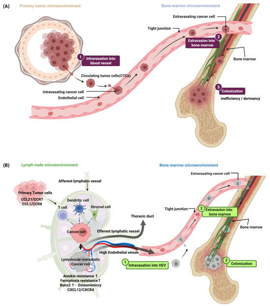
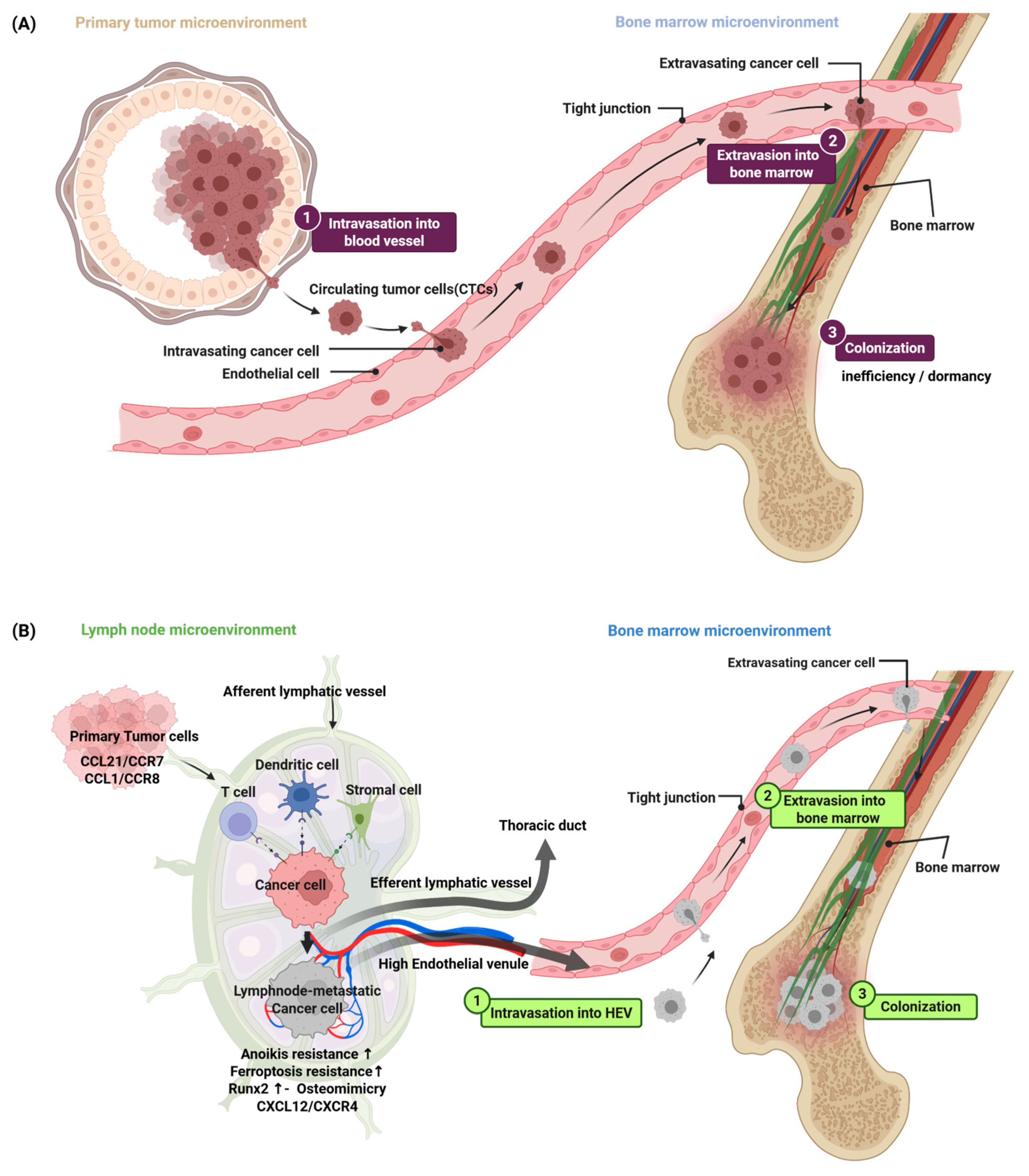

Disclaimer/Publisher’s Note: The statements, opinions and data contained in all publications are solely those of the individual author(s) and contributor(s) and not of MDPI and/or the editor(s). MDPI and/or the editor(s) disclaim responsibility for any injury to people or property resulting from any ideas, methods, instructions or products referred to in the content. |
© 2026 by the authors. Licensee MDPI, Basel, Switzerland. This article is an open access article distributed under the terms and conditions of the Creative Commons Attribution (CC BY) license.